Non-Invasive Study of the Pigments of a Painting on Copper with the Inscription “Boceto di Pablo Veronese” on the Back

 , ,
, ,
Abstract
:1. Introduction
Painting on Copper Support
2. Materials and Methods
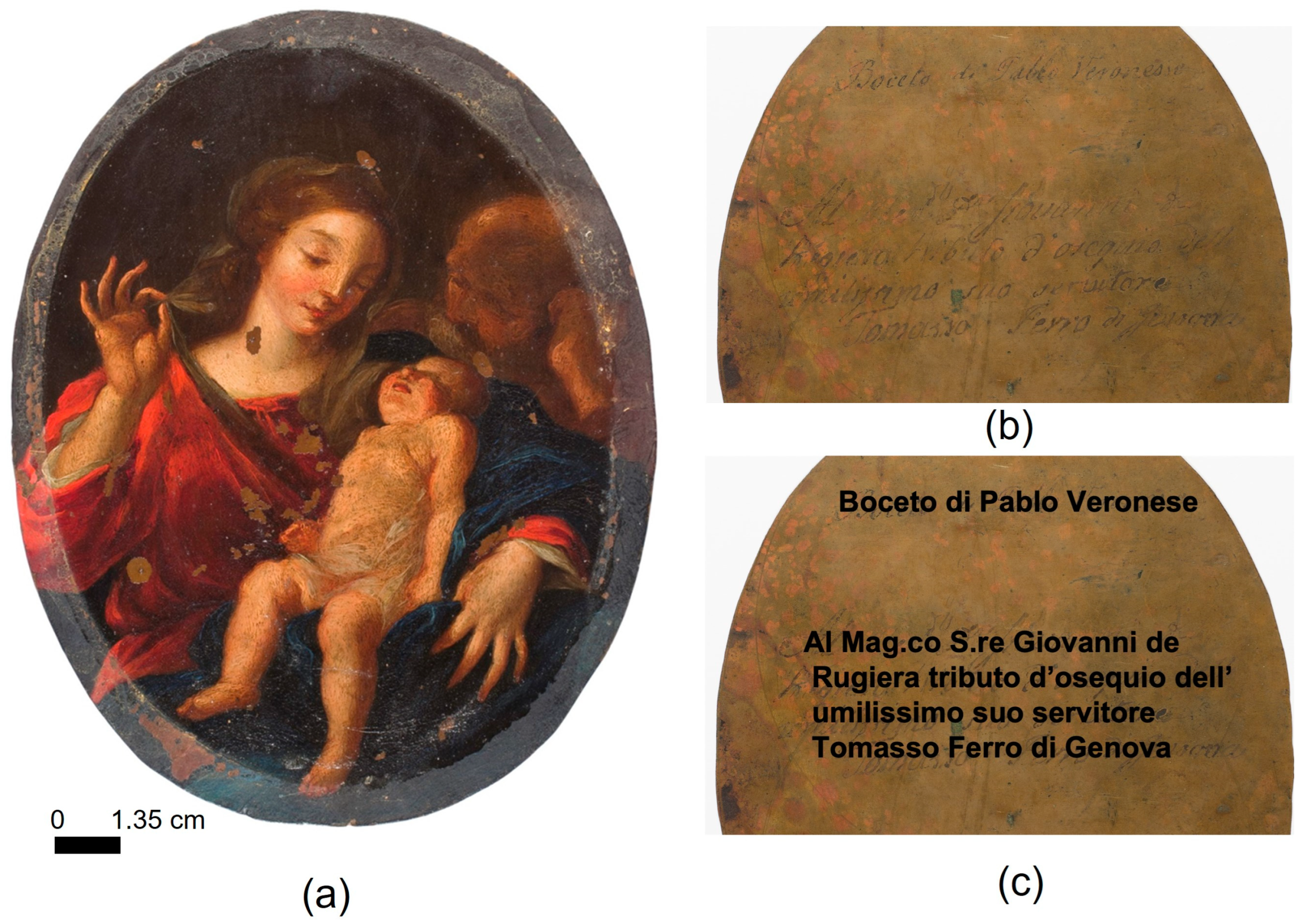
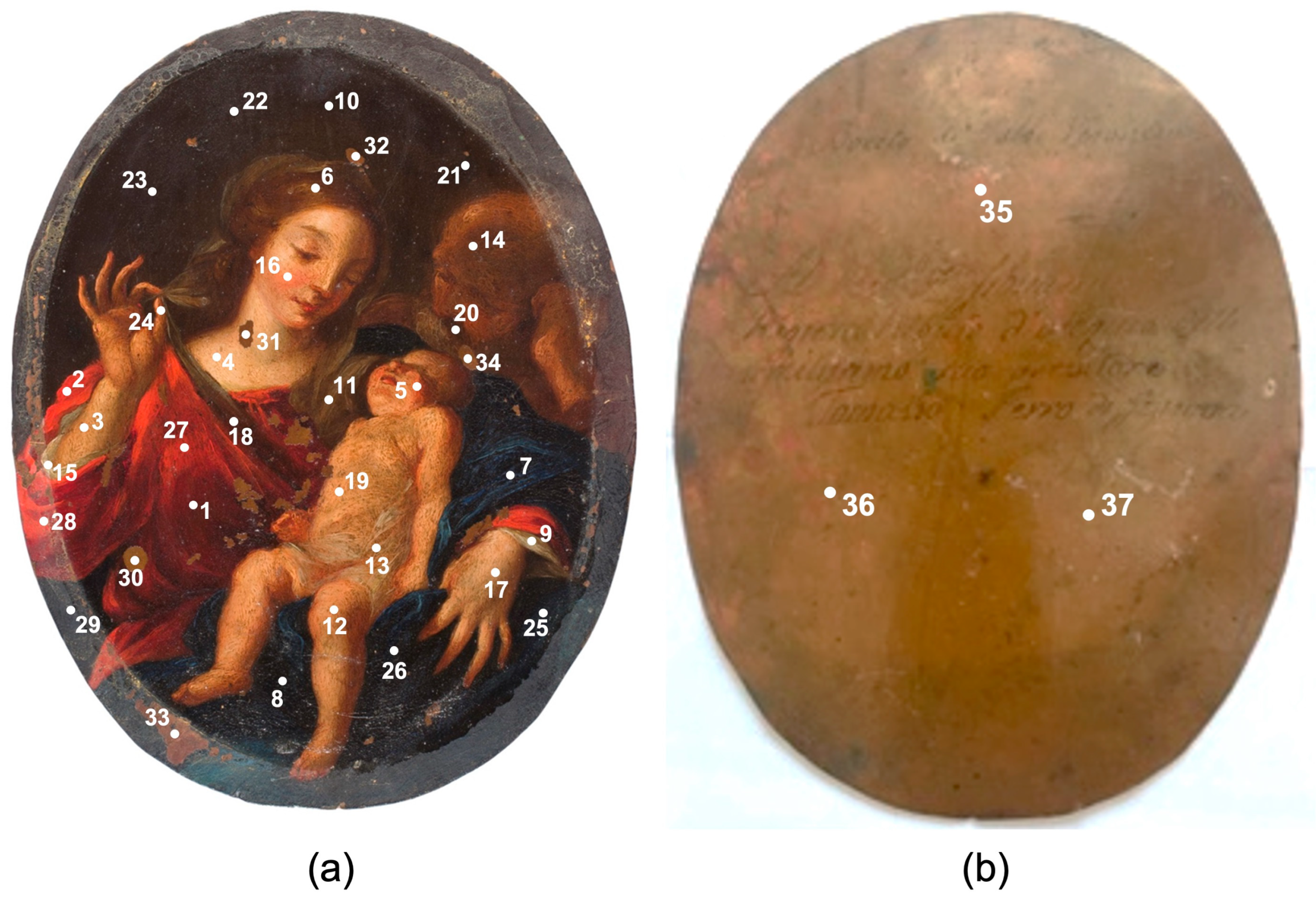
2.1. The Painting on Copper Support Investigated
2.2. Analytical Techniques
2.2.1. Hand-Held X-ray Fluorescence (XRF)
2.2.2. X-ray Diffraction (XRD)
3. Results
3.1. Support
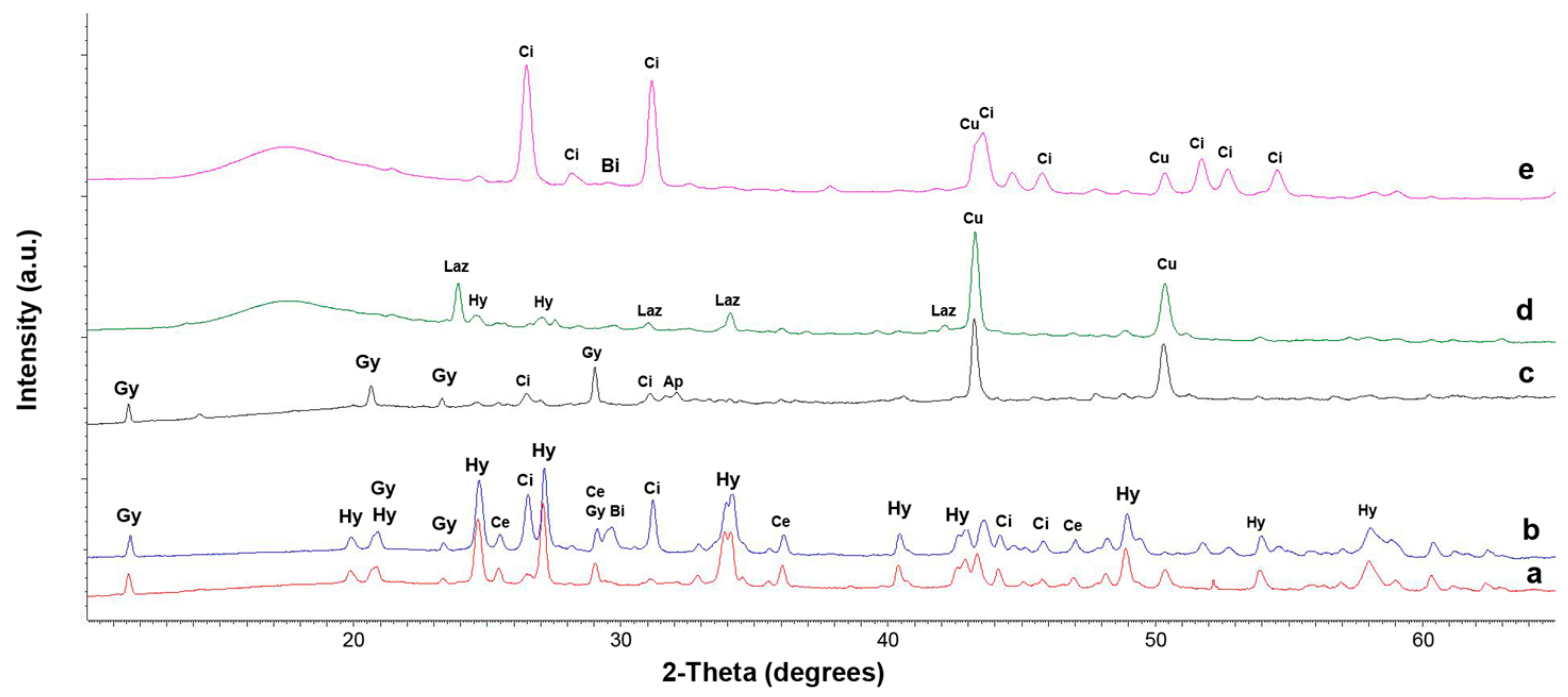
3.2. Ground Layer
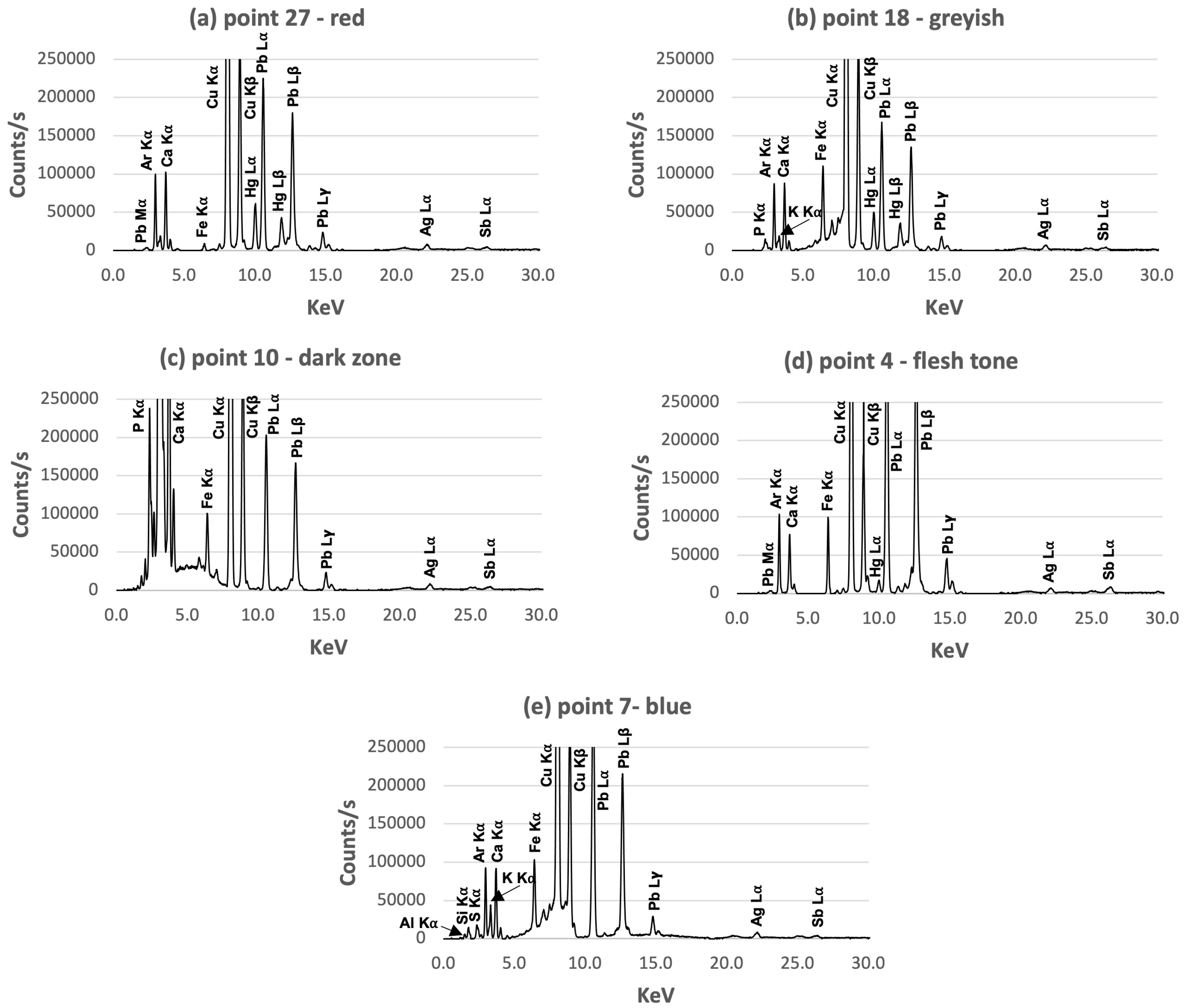
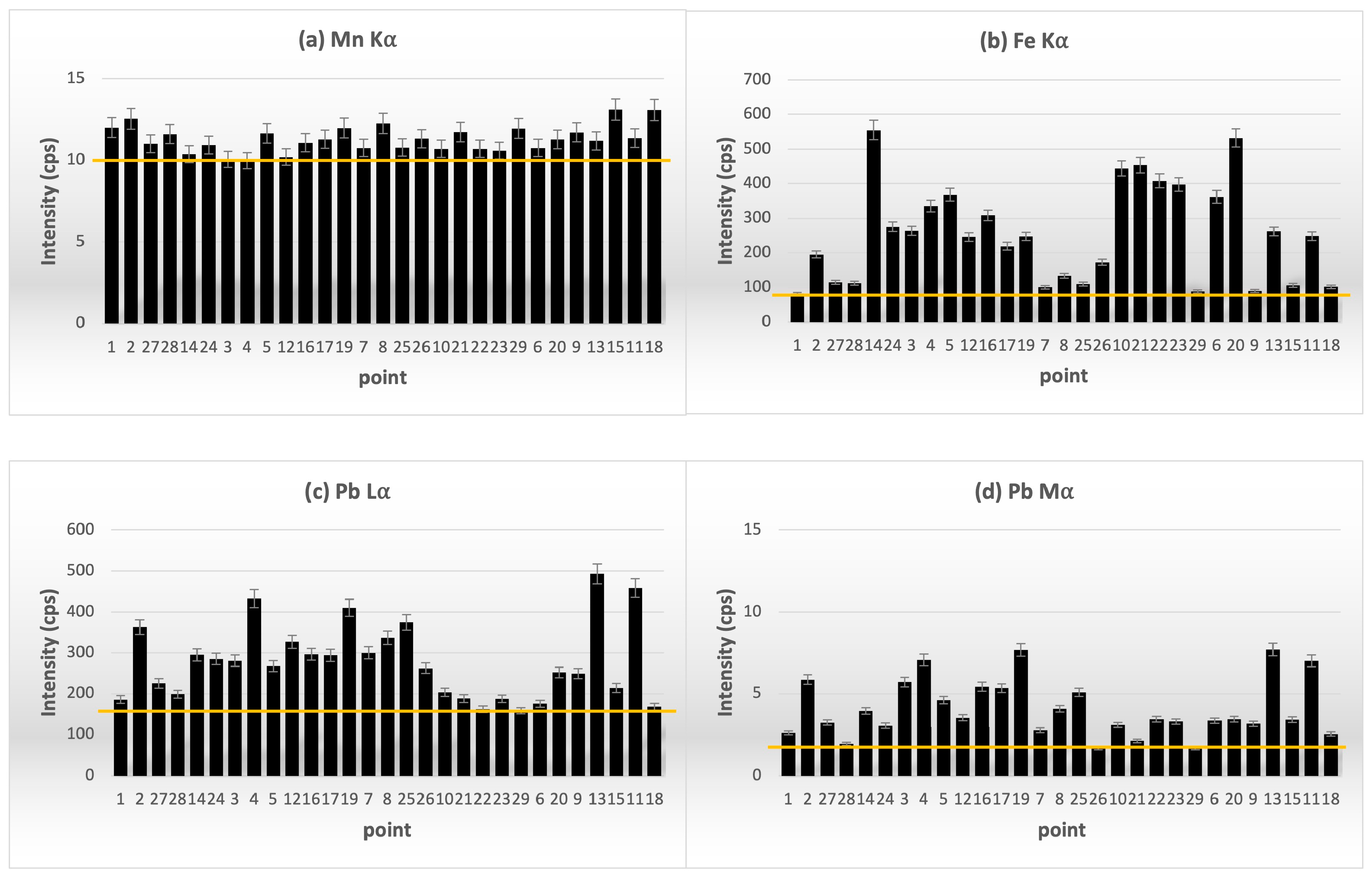
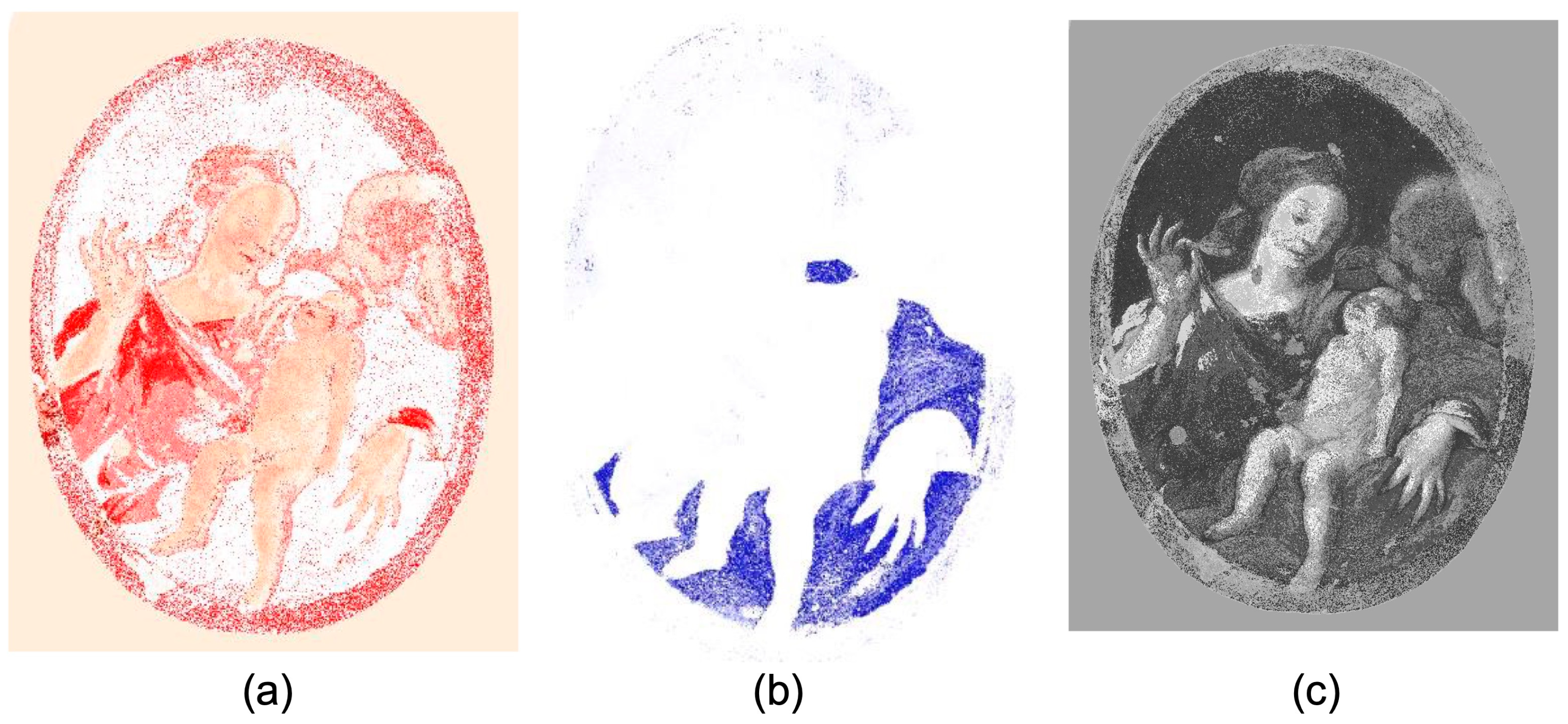
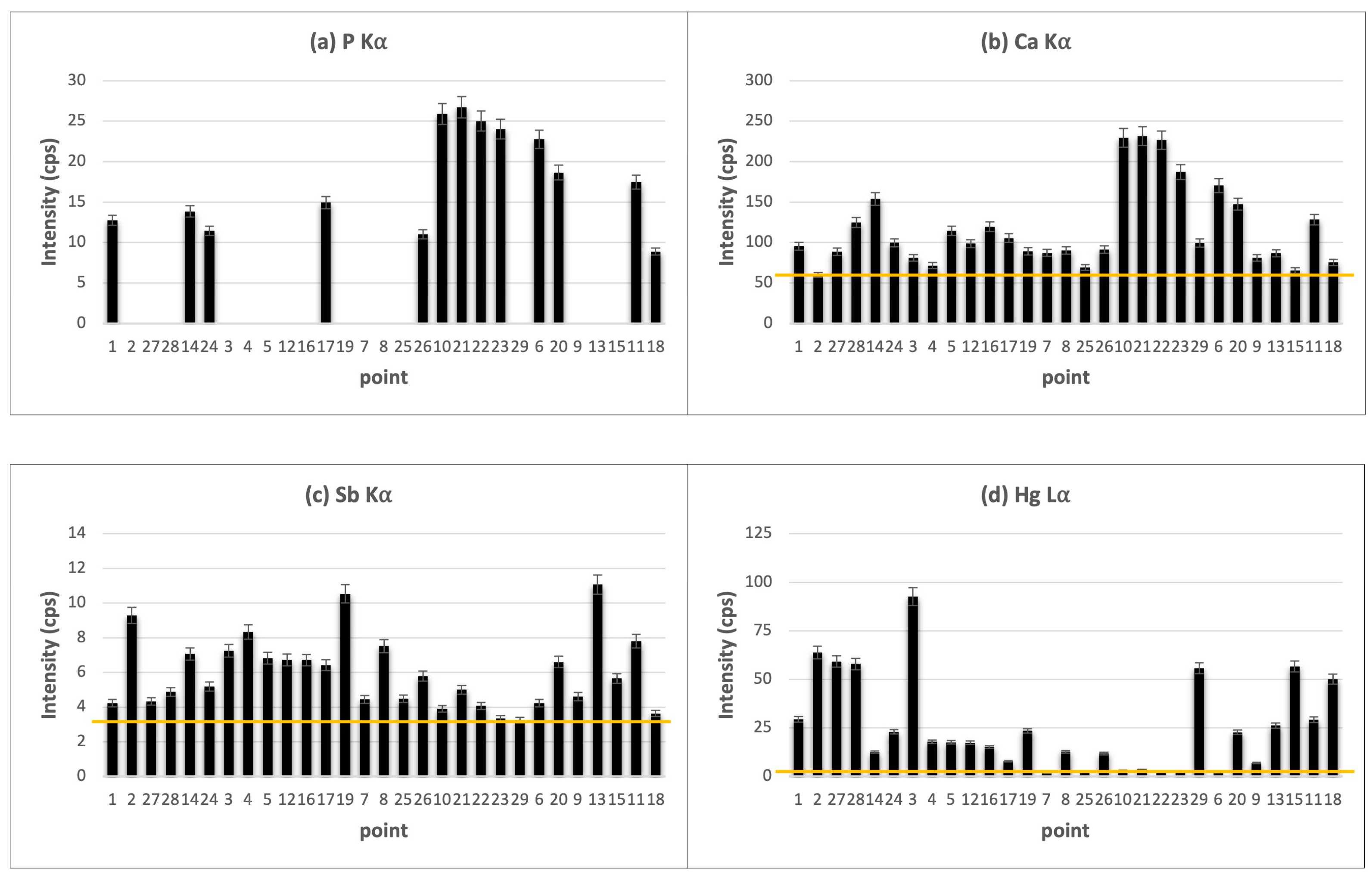
3.3. Pictorial Layer
- Flesh tones
- Red color
- Blue color
- Grayish color
- Whitish color
- Brown and dark color
4. Discussion
5. Conclusions
Author Contributions
Funding
Data Availability Statement
Acknowledgments
Conflicts of Interest
References
- López-Ramírez, M.R.; Navas, N.; Rodríguez-Simón, L.R.; Otero, J.C.; Manzano, E. Study of modern artistic materials using combined spectroscopic and chromatographic techniques. Case study: Painting with the signature “Picasso”. Anal. Method. 2015, 7, 1499–1508. [Google Scholar] [CrossRef]
- Simon, S.; Röhrs, S. Between fakes, forgeries, and illicit artifacts—Authenticity studies in a heritage science laboratory. Arts 2018, 7, 20. [Google Scholar] [CrossRef] [Green Version]
- Manzano, E.; Blanc, R.; Martin-Ramos, J.D.; Chiari, G.; Sarrazin, P.; Vilchez, J.L. A combination of invasive and non-invasive techniques for the study of the palette and painting structure of a copy of Raphael’s Transfiguration of Christ. Herit. Sci. 2021, 9, 150. [Google Scholar] [CrossRef]
- Manzano, E.; Rodríguez-Simón, L.R.; Navas, N.; Capitán-Vallvey, L.F. Non-Invasive and Spectroscopic Techniques for the Study of Alonso Cano’s Visitation from the Golden Age of Spain. Stud. Conserv. 2021, 66, 298–312. [Google Scholar] [CrossRef]
- Robledo, J.I.; Leani, J.J.; Sánchez, H.J. Possibilities of spatially-resolved energy dispersive inelastic X-ray scattering (EDIXS) spectroscopy for painting characterization. Spectrochim. Acta Part B At. Spectrosc. 2022, 198, 106552. [Google Scholar] [CrossRef]
- Van de Voorde, L.; Vekemans, B.; Verhaeven, E.; Tack, P.; De Wolf, R.; Garrevoet, J.; Vincze, L. Analytical characterization of a new mobile X-ray fluorescence and X-ray diffraction instrument combined with a pigment identification case study. Spectrochim. Acta Part B At. Spectrosc. 2015, 110, 14–19. [Google Scholar] [CrossRef]
- Bonizzoni, L.; Caglio, S.; Galli, A.; Poldi, G. A non-invasive method to detect stratigraphy, thicknesses and pigment concentration of pictorial multilayers based on EDXRF and vis-RS: In situ applications. Appl. Phys. A 2008, 92, 203–210. [Google Scholar] [CrossRef]
- Martin-Ramos, J.D.; Zafra-Gómez, A.; Vílchez, J.L. Non-destructive pigment characterization in the painting Little Madonna of Foligno by X-ray Powder Diffraction. Microchem. J. 2017, 134, 343–353. [Google Scholar] [CrossRef]
- Artioli, G. Science for the cultural heritage: The contribution of X-ray diffraction. Rend. Lincei 2013, 24, 55–62. [Google Scholar] [CrossRef]
- Andrić, V.; Gajić-Kvaščev, M.; Crkvenjakov, D.K.; Marić-Stojanović, M.; Gadžurić, S. Evaluation of pattern recognition techniques for the attribution of cultural heritage objects based on the qualitative XRF data. Microchem. J. 2021, 167, 106267. [Google Scholar] [CrossRef]
- Pitarch, A.; Ramón, A.; Álvarez-Pérez, A.; Queralt, I. Characterization of “oil on copper” paintings by energy dispersive X-ray fluorescence spectrometry. Anal. Bioanal. Chem. 2012, 402, 1481–1492. [Google Scholar] [CrossRef] [PubMed] [Green Version]
- Nakai, I.; Abe, Y. Portable X-ray powder diffractometer for the analysis of art and archaeological materials. Appl. Phys. A 2012, 106, 279–293. [Google Scholar] [CrossRef]
- Hirayama, A.; Abe, Y.; van Loon, A.; De Keyser, N.; Noble, P.; Vanmeert, F.; Nakai, I. Development of a new portable X-ray powder diffractometer and its demonstration to on-site analysis of two selected old master paintings from the Rijksmuseum. Microchem. J. 2018, 138, 266–272. [Google Scholar] [CrossRef]
- Hocquet, F.P.; Calvo del Castillo, H.; Cervera Xicotencatl, A.; Bourgeois, C.; Oger, C.; Marchal, A.; Strivay, D. Elemental 2D imaging of paintings with a mobile EDXRF system. Anal. Bioanal. Chem. 2011, 399, 3109–3116. [Google Scholar] [CrossRef]
- Gonzalez, V.; Cotte, M.; Vanmeert, F.; de Nolf, W.; Janssens, K. X-ray Diffraction Mapping for Cultural Heritage Science: A Review of Experimental Configurations and Applications. Chem.–A Eur. J. 2020, 26, 1703–1719. [Google Scholar] [CrossRef]
- Martin-Ramos, J.D.; Chiari, G. SmART_scan: A method to produce composition maps using any elemental, molecular and image data. J. Cult. Herit. 2019, 39, 260–269. [Google Scholar] [CrossRef]
- Komanecky, M.K.; Bowron, E.P.; Bargellini, C. Copper as Canvas: Two Centuries of Masterpiece Paintings on Copper, 1575–1775; The Phoenix Art Museum, Oxford University Press: Oxford, UK, 1999; ISBN 0-19-512397-2. Available online: https://www.gettextbooks.com/isbn/9780195123975/ (accessed on 20 March 2023).
- Pitarch, A.; Ramón, A.; Álvarez-Pérez, A.; Castro, K.; Madariaga, J.M.; Queralt, I. Multispectroscopic characterization of oil on copper painting. Spectrosc. Lett. 2014, 47, 38–51. [Google Scholar] [CrossRef] [Green Version]
- Bowron, E.P. A brief history of European oil paintings on copper, 1560–1775. In Copper as Canvas: Two Centuries of Masterpiece Paintings on Copper, 1575–1775; The Phoenix Art Museum; Oxford University Press: Oxford, UK, 1999; pp. 9–30. ISBN 0-19-512397-2. [Google Scholar]
- Zaccaron, S.; Grespan, C.; Ganzerla, R. Characterization of pigments on paintings on copper plate. A case study: The copy of La sepoltura di Cristo by Federico Barocci. Sci. Ca’Foscari 2013, 1, 48–56. [Google Scholar]
- Corregidor, V.; Oliveira, A.R.; Rodrigues, P.A.; Alves, L.C. Paintings on copper by the Flemish artist Frans Francken II: PIXE characterization by external mi-crobeam. Nucl. Instrum. Methods B 2015, 348, 291–295. [Google Scholar] [CrossRef]
- Donahue-Wallace, K. Copper as Canvas: Two Centuries of Masterpiece Paintings on Copper, 1575–1775; Phoenix Art Museum, Nelson-Atkins Museum of Art, The Sixteenth Century Journal; 2000; Volume 31, pp. 874–876. Available online: https://www.jstor.org/stable/2671133?origin=crossref (accessed on 20 March 2023).
- Alvarez, M.C. La restauración: Recuperación de una memoria pictórica en una nueva instancia. In En Lecciones barrocas: Pinturas sobre la vida de la Virgen de la Ermita de Egipto; Departamento editorial del Banco de la República: Bogotá, Colombia, 1990; pp. 20–39. [Google Scholar]
- Pavlopoulou, L.C.; Watkinson, D. The degradation of oil painted copper surfaces. Stud. Conserv. 2006, 51 (Suppl. S1), 55–65. [Google Scholar] [CrossRef]
- Rodriguez, S.H.; Appoloni, C.R.; Campos, P.H.O.V.; Gonçalves, B.; Kajiya, E.A.M.; Molari, R.; Winter, C. Non-Destructive and portable analyses helping the study and conservation of a Saraceni copper plate painting in the São Paulo museum of art. Microchem. J. 2020, 155, 104787–104799. [Google Scholar] [CrossRef]
- Vega, D.; Pombo Cardoso, I.; Carlyle, L. Pintura sobre cobre: Investigación sobre materiales y técnicas de aplicación de la capa de preparación a través de los tratados tradicionales y estudio analítico de dos obras atribuidas a las escuelas portuguesa y flamenca. Conserv. Patrim. 2018, 27, 23–35. [Google Scholar] [CrossRef] [Green Version]
- Spring, M.; Grout, R. The blackening of vermilion: An analytical study of the process in paintings. Natl. Gallery Tech. Bull. 2002, 23, 50–61. Available online: https://www.jstor.org/stable/42616159 (accessed on 20 March 2023).
- Ruberto, C.; Mazzinghi, A.; Massi, M.; Castelli, L.; Czelusniak, C.; Palla, L.; Raffaelli, M. Imaging study of Raffaello’s “La Muta” by a portable XRF spectrometer. Microchem. J. 2016, 126, 63–69. [Google Scholar] [CrossRef]
- Penny, N.; Roy, A.; Spring, M. Veronese’s Paintings in the National Gallery Technique and Materials: Part II. Natl. Gallery Tech. Bull. 1996, 17, 32–55. Available online: http://www.nationalgallery.org.uk/technical-bulletin/penny_roy_spring1996 (accessed on 20 March 2023).
- Armstrong, L. Passion in Venice: Crivelli to Tintoretto and Veronese (New York, Museum of Biblical Art [MOBIA], 11 February–12 June 2011). Passion in Venice: Crivelli to Tintoretto and Veronese, The Man of Sorrows in Venetian Art. Renaiss. Stud. 2012, 26, 441–449. Available online: https://www.jstor.org/stable/24420075 (accessed on 20 March 2023). [CrossRef]
- Penny, N.; Spring, M. Veronese’s Paintings in the National Gallery. Technique and Materials: Part I. Natl. Gallery Tech. Bull. 1995, 16, 4–29. Available online: http://www.nationalgallery.org.uk/technical-bulletin/penny_spring1995 (accessed on 20 March 2023).
- Mahon, D.; Centeno, S.A.; Wypyski, M.T.; Salomon, X.F.; Bayer, A. Technical study of three allegorical paintings by Paolo Veronese: The choice between virtue and vice, wisdom and strength, and mars and venus united by love. In Metropolitan Museum Studies in Art, Science, and Technology; The Metropolitan Museum of Art: New York, NY, USA, 2010; Volume 1, ISBN 978-1-58839-365-4. Available online: https://www.researchgate.net/publication/260418022_Technical_Study_of_Three_Allegorical_Paintings_by_Paolo_Veronese_The_Choice_Between_Virtue_and_Vice_Wisdom_and_Strength_and_Venus_and_Mars_United_by_Love (accessed on 20 March 2023).
- Zagora, J. Historical Development of Coloured Grounds in Italian Painting from the 15th to the mid-18th Century–Present Insights and Open Questions. Portal Godišnjak Hrvat. Restaur. Zavoda 2017, 73–94. Available online: http://www.hrz.hr/images/portal/portal_08_zagora1.pdf (accessed on 20 March 2023). [CrossRef]
- Komanecky, M.K.; Horovitz, I.; Eastaugh, N. Catalogue “Painting Techniques History, Materials and Studio Practice: Contributions to the IIC Dublin Congress, 7–11 September 1998”; International Institute for Conservation of Historic and Artistic Works: London, UK, 1998; pp. 136–139. ISBN 0950052582. [Google Scholar]
- Uffelman, E.S.; Court, E.; Marciari, J.; Miller, A.; Cox, L. Handheld XRF Analyses of Two Veronese Paintings. In Collaborative Endeavors in the Chemical Analysis of Art and Cultural Heritage Materials; American Chemical Society: Washington, DC, USA, 2012; pp. 51–73. [Google Scholar] [CrossRef]
- Osticioli, I.; Pagliai, M.; Comelli, D.; Schettino, V.; Nevin, A. Red lakes from Leonardo’s Last Supper and other Old Master Paintings: Micro-Raman spectroscopy of anthraquinone pigments in paint cross-sections. Spectrochim. Acta Part A Mol. Biomol. Spectrosc. 2019, 222, 117273. [Google Scholar] [CrossRef]
- Sandalinas, C.; Ruiz-Moreno, S. Lead tin-antimony yellow, historical manufacture, molecular characterization and identification in seventeenth-century Italian paintings. Stud. Conserv. 2003, 49, 41–52. [Google Scholar] [CrossRef]
- Hradil, D.; Grygar, T.; Hradilová, J.; Bezdička, P.; Grűnwaldová, V.; Fogaš, I.; Miliani, C. Microanalytical identification of Pb-Sb-Sn yellow pigment in historical European paintings and its differentiation from lead tin and Naples yellows. J. Cult. Herit. 2007, 8, 377–386. [Google Scholar] [CrossRef]
- Ruiz-Moreno, S.; Pérez-Pueyo, R.; Gabaldón, A.; Soneira, M.J.; Sandalinas, C. Raman laser fibre optic strategy for non-destructive pigment analysis. Identification of a new yellow pigment (Pb, Sn, Sb) from the Italian XVII century painting. J. Cult. Herit. 2003, 4, 309–313. [Google Scholar] [CrossRef]


| Color | No. | Elements Detected | ||||||||||
|---|---|---|---|---|---|---|---|---|---|---|---|---|
| Ca | S | Fe | Mn | Pb | P | Hg | Sb | Si | Al | K | ||
| Red | 1 | m | M | m | tr | m | M | M | tr | tr | tr | tr |
| 2 | m | M | M | tr | M | - | M | tr | - | - | - | |
| 27 | m | M | m | tr | m | - | M | tr | tr | - | tr | |
| 28 | m | M | m | tr | m | - | M | tr | tr | - | tr | |
| Flesh tone | 3 | m | M | M | tr | M | - | M | tr | - | - | - |
| 4 | m | M | M | tr | M | - | m | tr | - | - | - | |
| 5 | m | M | M | tr | M | - | m | tr | tr | - | tr | |
| 12 | m | M | M | tr | M | - | m | tr | tr | tr | tr | |
| 14 | M | M | M | tr | M | M | m | tr | tr | - | tr | |
| 16 | m | M | M | tr | M | - | m | tr | tr | - | - | |
| 17 | m | M | M | tr | M | M | m | tr | tr | tr | tr | |
| 19 | m | M | M | tr | M | - | m | tr | - | - | - | |
| 24 | m | M | M | tr | M | M | m | tr | - | - | - | |
| Blue | 7 | m | M | m | tr | M | - | tr | tr | tr | tr | tr |
| 8 | m | M | m | tr | M | - | m | tr | tr | tr | ||
| 25 | m | M | m | tr | M | - | tr | tr | - | tr | tr | |
| 26 | m | M | m | tr | M | M | m | tr | tr | tr | ||
| Grayish | 11 | M | M | M | tr | M | M | m | tr | tr | tr | tr |
| 18 | m | M | m | tr | m | M | M | tr | tr | tr | tr | |
| Whitish | 9 | m | M | m | tr | M | - | m | tr | tr | tr | tr |
| 13 | m | M | M | tr | M | - | m | tr | - | - | - | |
| 15 | m | M | m | tr | M | - | M | tr | tr | tr | tr | |
| Dark zone | 10 | M | M | M | tr | m | M | tr | tr | - | - | - |
| 21 | M | M | M | tr | m | M | tr | tr | - | - | - | |
| 22 | M | M | M | tr | m | M | tr | tr | - | - | - | |
| 23 | M | M | M | tr | m | M | tr | tr | - | - | - | |
| 29 | m | M | m | tr | m | - | M | tr | tr | tr | ||
| Brown | 6 | M | M | M | tr | m | M | tr | tr | - | - | - |
| 20 | M | M | M | tr | M | M | m | tr | tr | - | tr | |
| Colour | No. | Minerals Detected * | ||||||||||
|---|---|---|---|---|---|---|---|---|---|---|---|---|
| Gy | Hy | Ce | Ci | Laz | A | Ap | Bi | Si | H | Ca | ||
| Red | 1 | 32.0 | 21.0 | 4.6 | 12.8 | - | 13.6 | - | 1.6 | 14.4 | - | - |
| 2 | 19.5 | 9.5 | 4.9 | 65.2 | - | - | - | 1.0 | - | - | - | |
| 27 | 25.7 | - | 7.2 | 90.4 | - | - | - | 2.4 | - | - | - | |
| 28 | 31.4 | - | 4.9 | 63.7 | - | - | - | - | - | - | - | |
| Flesh tone | 3 | 41.0 | 44.0 | 7.0 | 2.0 | - | - | - | 6.0 | - | - | - |
| 4 | 36.8 | 38.4 | 7.7 | 2.2 | - | - | - | 1.9 | 5.7 | 7.4 | - | |
| 5 | 44.0 | 34.0 | 11.0 | 6.0 | - | - | - | 5.0 | - | - | - | |
| 12 | 29.6 | 49.3 | 10.4 | 4.5 | - | - | - | 6.0 | - | - | - | |
| 14 | 86.1 | 3.2 | 2.2 | 6.5 | - | - | - | 2.1 | - | - | - | |
| 16 | 41.2 | 33.2 | 10.1 | 11.0 | - | - | - | 4.4 | - | - | - | |
| 17 | 47.9 | 69.8 | 22.8 | 4.4 | - | - | - | 3.0 | - | - | - | |
| 19 | 51.3 | 26.9 | 12.8 | 3.9 | - | - | - | 5.0 | - | - | - | |
| 24 | 33.3 | 35.9 | 17.1 | 9.0 | - | - | - | 4.7 | - | - | - | |
| Blue | 7 | 23.0 | - | - | - | 61.6 | - | - | - | 15.4 | - | - |
| 8 | 15.6 | - | - | - | 62.7 | - | - | - | 37.3 | - | - | |
| 25 | 1.6 | - | 2.9 | - | 82.8 | - | - | - | 12.8 | - | - | |
| 26 | 18.3 | 9.0 | 4.0 | - | 67.0 | - | - | - | 19.9 | - | - | |
| Grayish | 11 | 29.8 | 22.2 | 10.7 | 1.7 | 15.3 | - | 6.2 | - | 14.0 | - | |
| 18 | 29.6 | 13.5 | 5.5 | 2.9 | 15.2 | - | 13.5 | - | 19.8 | - | - | |
| Whitish | 9 | 23.5 | 62.3 | 8.3 | - | - | - | - | - | - | - | 5.9 |
| 13 | 39.0 | 41.0 | 12.5 | 3.0 | - | - | - | 4.5 | - | - | - | |
| 15 | 26.0 | 38.1 | 12.2 | - | - | 16.0 | - | - | - | - | 7.7 | |
| Dark zone | 10 | 54.0 | - | 3.0 | 2.5 | - | - | 25.5 | - | 15.0 | - | - |
| 21 | 48.6 | 23.1 | 7.6 | - | 51.2 | - | - | - | 18.0 | - | - | |
| 22 | 50.0 | 13.0 | 5.0 | 2.0 | - | - | 30.0 | - | - | - | - | |
| 23 | 53.0 | 9.9 | 6.4 | 2.1 | - | - | 28.6 | - | - | - | - | |
| 29 | 10.6 | 5.0 | 2.0 | 1.1 | 60.0 | - | - | - | 21.3 | - | - | |
| Brown. | 6 | 68.0 | 10.0 | 5.0 | 3.0 | - | 12.6 | - | 1.4 | - | - | - |
| 20 | 59.1 | 7.4 | - | 3.6 | - | - | 27.7 | 2.0 | - | - | - | |
Disclaimer/Publisher’s Note: The statements, opinions and data contained in all publications are solely those of the individual author(s) and contributor(s) and not of MDPI and/or the editor(s). MDPI and/or the editor(s) disclaim responsibility for any injury to people or property resulting from any ideas, methods, instructions or products referred to in the content. |
© 2023 by the authors. Licensee MDPI, Basel, Switzerland. This article is an open access article distributed under the terms and conditions of the Creative Commons Attribution (CC BY) license (https://creativecommons.org/licenses/by/4.0/).
Share and Cite
Blanc, R.; Manzano, E.; López-Montes, A.; Domínguez-Gasca, N.; Vílchez, J.L. Non-Invasive Study of the Pigments of a Painting on Copper with the Inscription “Boceto di Pablo Veronese” on the Back. Heritage 2023, 6, 4787-4801. https://doi.org/10.3390/heritage6060254
Blanc R, Manzano E, López-Montes A, Domínguez-Gasca N, Vílchez JL. Non-Invasive Study of the Pigments of a Painting on Copper with the Inscription “Boceto di Pablo Veronese” on the Back. Heritage. 2023; 6(6):4787-4801. https://doi.org/10.3390/heritage6060254
Chicago/Turabian StyleBlanc, Rosario, Eloisa Manzano, Ana López-Montes, Nazaret Domínguez-Gasca, and José Luis Vílchez. 2023. "Non-Invasive Study of the Pigments of a Painting on Copper with the Inscription “Boceto di Pablo Veronese” on the Back" Heritage 6, no. 6: 4787-4801. https://doi.org/10.3390/heritage6060254