1. Introduction
Beans play a major role as staple foods throughout the world. They are generally acknowledged as good sources of protein, while their traditional aqueous extracts (soups and broths) are rich in polysaccharides, including starch. Proteins extracted from faba beans have good emulsification capacity at neutral pH, with a capacity to produce emulsion gels in the presence of λ-carrageenan [
1]. Mung bean protein isolates have been shown to have emulsification and foaming potential, which conditionally with their hydrolysis [
2]. Lupin extracts show promise as emulsifiers [
3], while saponins are an emerging field of naturally-derived surfactants [
4]. Overall, the field of biosurfactants appears to attract attention [
5]. There has been recent interest in the potential of larger particles from beans to stabilize emulsions via a Pickering mechanism, e.g., adzuki beans coupled with zein [
6]. Pigeon pea protein (PiPI) and soybean protein (SPI) have also been reported to be good emulsifiers [
7] with SPI being one of the most studied plant-based food proteins.
White beans are a major component of the culinary tradition of South Europe, North Africa and the Middle East. Their aqueous extracts have found their way into the gastronomic traditions of many civilizations of this area, and they have been used so as thickeners and (largely unknowingly) as emulsifiers. This emulsification capacity has been highlighted in recent studies [
8,
9]; however, to the best understanding of the present authors, no systematic study exist on the basis of the emulsifying action of the emulsifiers of that major crops, that is the correlation of the macromolecular compositional attributes of the extracts to their interfacial properties, and thereon to the emulsification capacity of specific extract populations and their emulsion stability.
Knowledge of the latter can add to the fundamental understanding of that particular niche of the chemistry of gastronomy, and potentially allow for the development of new, green emulsifiers. Considering the above, the aim of this work is to (i) to obtain extracts with emulsification potential from while beans; (ii) to identify the principal component groups in the extracts; (iii) to assess the emulsification capacity of the extracts in relation to pH, simulating their application in neutral and in acidified products; and (iv) to attribute any emulsification and emulsion stabilizing properties to specific component groups of the extracts.
2. Materials and Methods
2.1. Materials and Extraction
White beans originating from Kastoria, Greece, were obtained from the local market. All the reagents used in this work were purchased from Sigma Aldrich (St Louis, MO, USA). The beans were processed with a home blender and were then pulverized with a Pulverisette ball mill (Fritsch GmbH, Idar-Oberstein, Germany). Then, 100 g of this solid was mixed with 500 mL deionized water, and stirred for 30 min at 60 °C. The extract-rich supernatant was separated, and the remaining residue was extracted again (30 min at 60 °C) with 500 mL water each time. The three supernatants were then combined, frozen, and freeze-dried. The obtained bean extract (BE) was then stored in airtight containers.
2.2. Emulsion Preparation
Appropriate amounts of BE extract were dissolved into buffers set at pH 3 and 7 (10 m citrate and tris respectively). Miglyol, acting as oil phase, was added into the solutions, in order to reach an oil volume fraction of φ = 0.2. These were agitated in a magnetic stirrer as to prepare an emulsion pre-mix. Then, this was sonicated using a Hielscher UP-100H ultrasonic homogenizer (Hielscher Ultrasonics GmbH, Teltow, Germany) for 60 s.
2.3. Zeta Potential Measurements
A Brookhaven ZetaPALS device (Brookhaven Instruments Corporation, Brookhaven, Holtsville, NY, USA) was used for the measurement of zeta potential. The measurements were held at a 25 °C, assuming a bulk refractive index of 1.33 and a medium viscosity of 0.89 mPa s. The samples were diluted using an appropriate buffer prior to measurement, in order to minimize the effects of multiple scattering.
2.4. Particle Sizing
The particle size distribution of the oil droplets/droplet flocs was measured using a Malvern Mastersizer 2000 (Malvern Instrument, Malvern, Worcestershire, UK) apparatus, with the sample fed into the measuring device by a Hydro MU liquid sampler (Malvern Instrument, Malvern, Worcestershire, UK). The continuous phase refractive index was set at 1.33, while the dispersed phase refractive index was set at 1.42. An absorbance value of 0.1 was used for the particles. The calculation of particle size distribution from the angular scattering data was made on the basis of the Mie equations. The samples were diluted as to achieve an obscuration between 7% and 10%, using an appropriate buffer prior to measurement, in order to minimize the effects of multiple scattering.
2.5. Fourier Transform Infra Red Spectroscopy (FT-IR)
The bean extracts were analyzed with a FT-IR 6700 spectrometer (JASCO, Essex, UK). A small amount of the sample was placed on the surface of an Attenuated Total Reflectance (ATR) accessory MIRacle ™-Universal ATR (Pike Technologies, Madison, WI, USA) equipped with a 3-Reflection Diamond/ZnSe Performance Crystal Plate. The Spectra were obtained in triplicate over a range from 4000 to 400 cm−1 in transmittance mode. A total of 64 scans with 4 cm−1 resolution were acquired for each spectrum against a background of a dry and clean cell. The spectra were corrected for CO2, H2O, and ATR effects (in this order) using the Spectra Manager software (V.2.15.01, JASCO, Essex, UK). These were then subjected to Savitsky–Golay smoothing and baseline corrections with the same software.
2.6. Confocal Laser Scanning Microscopy (CLSM)
The micrographs were obtained by an inverted Zeiss LSM 700 confocal microscope (Carl Zeiss, CZ Microscopy GmbH, Jena, Germany). A minute amount (0.01% w/v) of Nile Red was added in the emulsions under study by gentle stirring. The emulsions were placed on a glass slide which was then covered with a coverslip prior to examination.
2.7. Interfacial Measurements
The oil–water dynamic interfacial tension measurements were performed by a pendant-drop tensiometer (CAM 200, KSV, Biolin Scientific, Stockholm, Sweden). Then, 1 g L−1 of the extracts were dissolved in 5 mL ultrapure water, as obtained from an Ultraclear Ro DI 30 device (Evoqua Lab, Pittsburgh, PA, USA). These were then filtered with either a 1 μm or a 0.2 μm syringe filter. The experiments were performed at 20 ± 1 °C and were performed at least twice. The data were analyzed with a drop shape analysis software (Attension Theta Software, V. 4.1.9.8, Biolin Scientific, Stockholm, Sweden). The curves were fitted to the Young–Laplace equation.
2.8. Size Exclusion Chromatography
The size exclusion chromatograph (SEC) was composed of the following components, set in tandem: a SpectraSystem SCM 1000 degasser (Thermo Separation Products, San Jose, CA, USA); pump: Shimadzu LC-20AR (Shimadzu Corporation, Tokyo, Japan); frit: 2 μm (Idex, Oak Harbor, WA, USA); a GPC/SEC PL-Aquagel-OH 50 × 7.5 (8 μm) guard column (Varian Inc, Palo Alto, CA, USA); two tandem GPC/SEC PL-Aquagel-OH 300 × 7.5 mm analytical columns (Varian Inc, Palo Alto, CA, USA); a Model 605 column oven (Scientific Systems Incorporated, State College, PA, USA) operating at a temperature of 30 °C; a UV detector (Rigas Labs, Thessaloniki, Greece); and a BI-MwA multiangle laser light scattering (MALLS) detector (Brookhaven Instruments Corporation, Brookhaven, Holtsville, NY, USA). The data were analyzed with an appropriate chromatographic/scattering analysis software (Brookhaven Instruments Corporation, Brookhaven, Holtsville, NY, USA). Each run involved the injection of sample using a 200 μL loop at an eluent (ultrapure water) flow rate of 0.8 mL min−1.
3. Results and Discussion
The obtained bean extracts (BE) were used as emulsifiers in oil-in-water emulsions, set at pH 3 (simulating acidified foods) and pH 7 (simulating neutral foods).
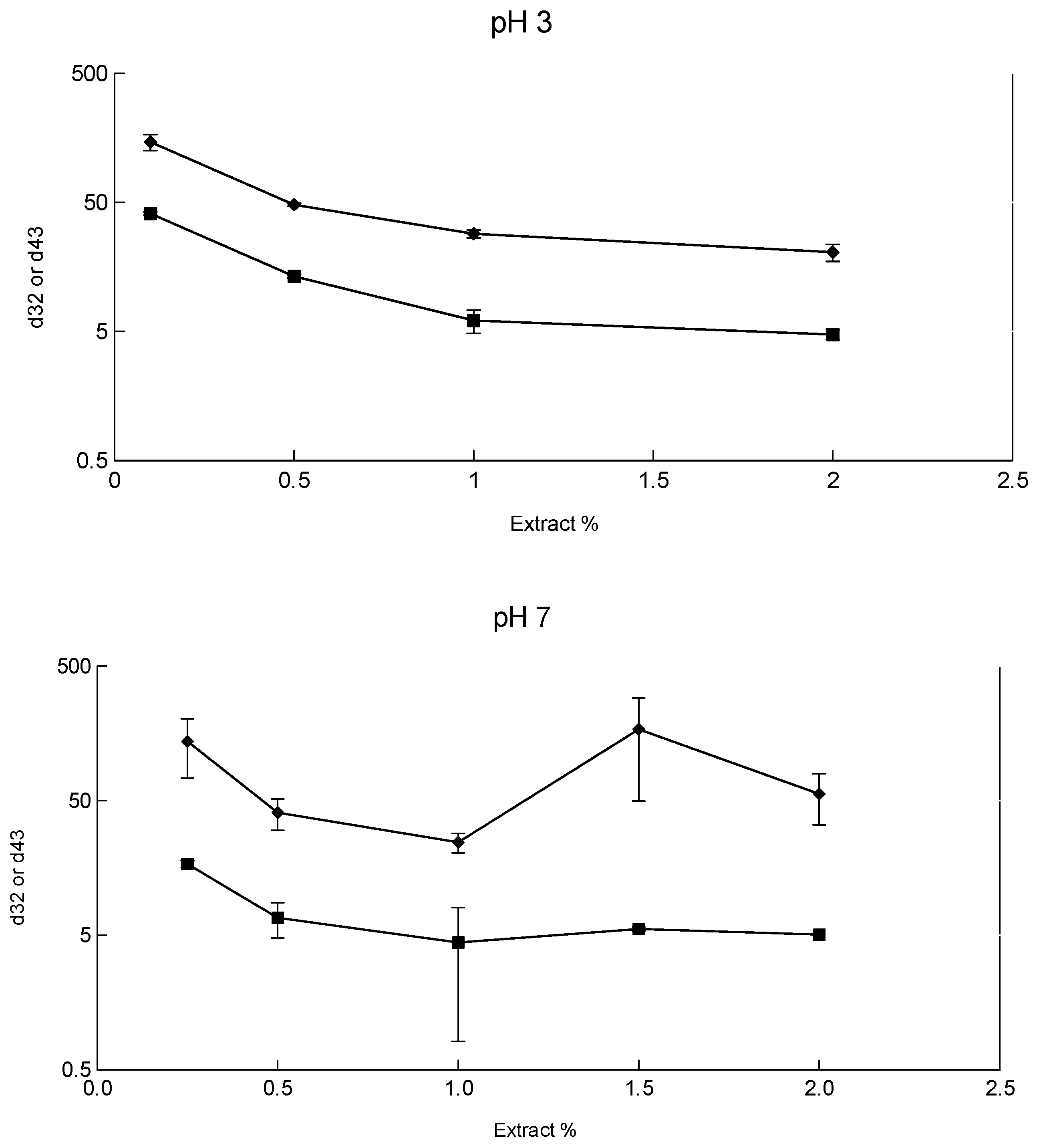
Figure 1a shows the dependence of the measured
d32 and
d43 with increasing BE concentration. Concentrations of BE of 1%
w/v and above allow the lowering of the measured particle sizes below 10 μm, with 2% leading to particles of
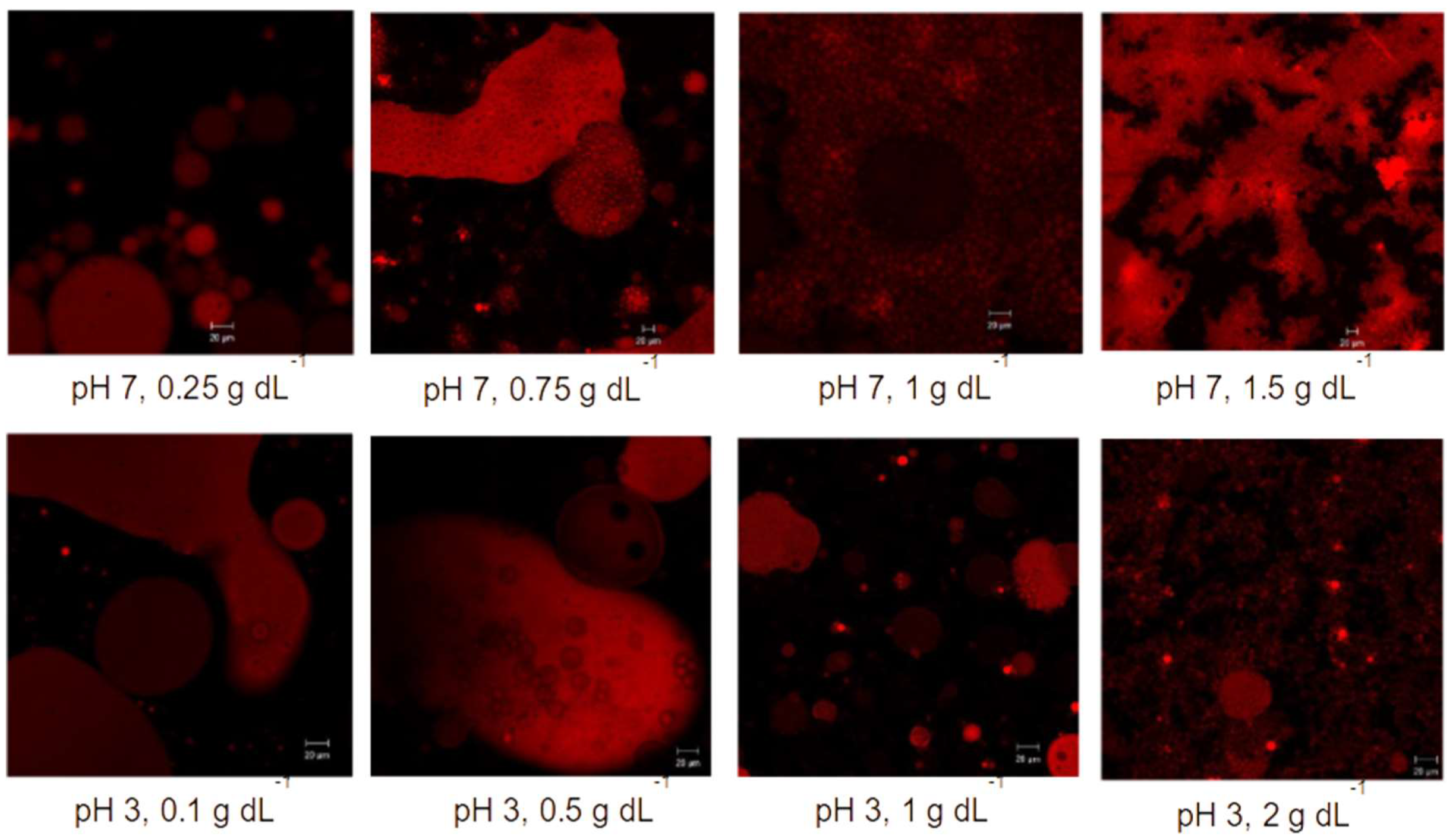
d32 = 5 μm. An examination of confocal micrographs obtained for these emulsions (
Figure 2) shows that, up to BE concentrations of 0.5–0.75%, extract content is not sufficient as to allow for the formation of very small droplets, an observation in line with the macroscopic image of the emulsion and the light scattering data. Of interest is the existence of water-in-oil clusters (black/aqueous spheres in red phase-separated large red/oil droplets), suggesting the existence of low-HLB components in BE. The authors of the present work are not aware of another natural extract which has the potential to effectively stabilize water-in-oil emulsions. This attribute of BE should be further explored in the near future, as it shows promise for the development of green low-HLB emulsifiers, a constant need of the chocolate and condiments industry, among other stakeholders. It will not be further discussed here, though, as it will draw the focus away from the set aims of this paper. A further increase of BE results in smaller droplets, while an interesting trend is the emulsifier-mediated adhesion of smaller droplets around the larger ones. This is in effect flocculation between droplets of different sizes and brings in mind recent findings of Ye and co-workers on the small oil droplet Pickering stabilization of larger ones [
10,
11]. Given the potential of droplet-stabilized emulsions to delay the in vitro digestion of oil [
12], this brings forward another potential functionality of BE extracts, which appear to readily produce such emulsions.
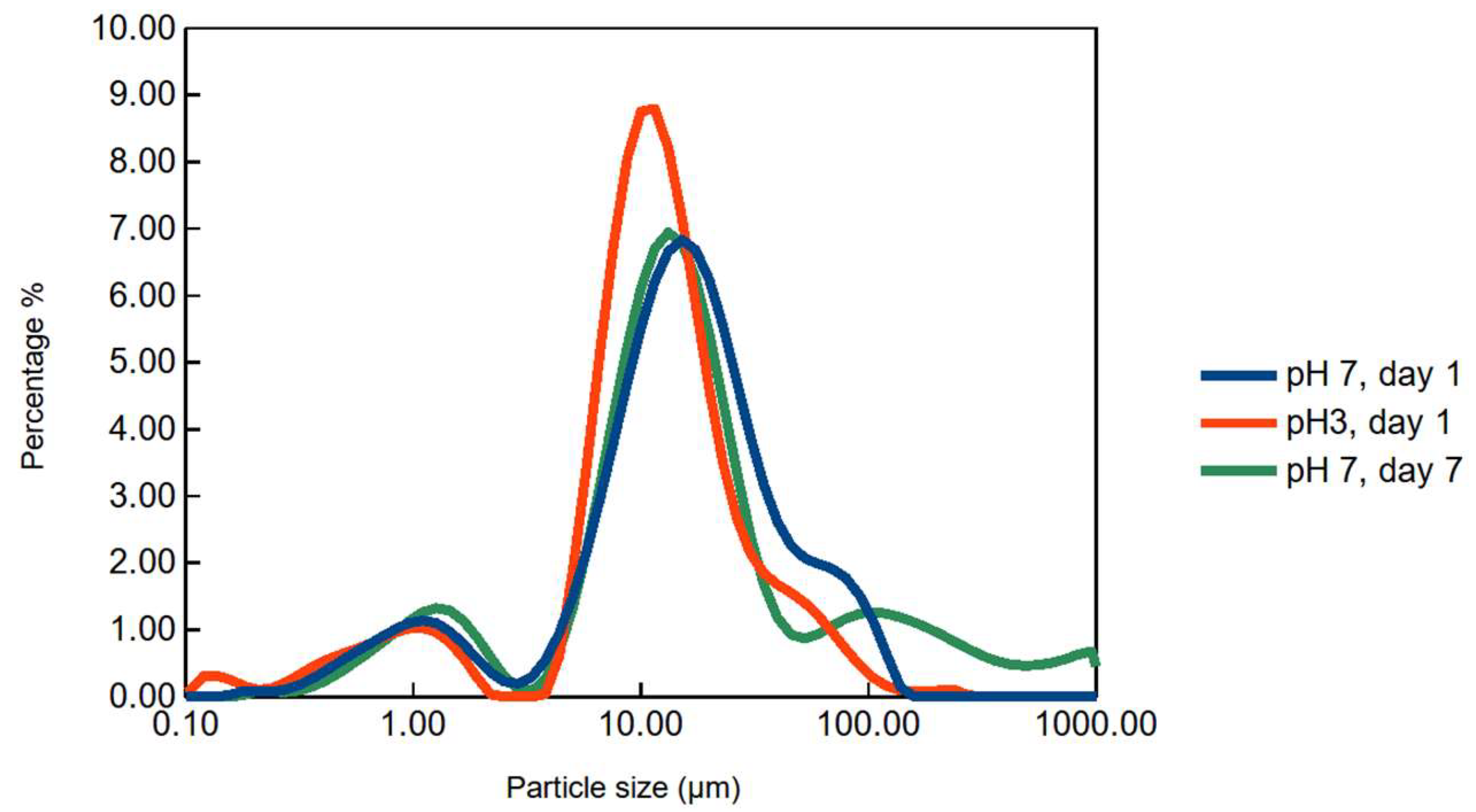
Figure 3 shows typical particle size distributions of such emulsions. A particle size distribution of a 7 days-aged emulsion (preserved by 0.1% sodium azide) is also shown. Although some aggregates form, seen as peaks above 100 μm, the main populations remain stable at about 10 μm. A second peak at around 100 microns on day 7 indicates flocculation at pH 7.
The dissolution of BE and emulsification at pH 7 produces fairly stable emulsions, especially above 1% BE. These are stable over the duration of several days, despite the relatively large particles produced. In that aspect, they are reminiscent of emulsions produced with olive processing waste [
13]. The zeta potential of such emulsions is close to zero. This should be related to the presence of non-charged macromolecules, possibly starch, at the interface, in addition to proteins or any other amphiphilic entities. The droplet-covered (potentially droplet-stabilized) emulsions observed at pH 3 are also observed at pH7, with characteristic examples shown for 0.75%
v/v BE. At high BE concentrations, the adherence between the droplets leads to the formation of flocculated emulsions. This is monitored as an increase in
d43 in the light scattering results between 1% and at 1.5% BE (
Figure 1b and
Figure 3), which should be attributed to the flocs observed in
Figure 2. The covering of larger droplets with smaller ones at 0.75%, itself a form of flocculation, should not be attributed to depletion, since there is no excess of unabsorbed material at such low BE concentrations; depletion, however, should not be ruled out at higher BE concentrations. Dilution of the 2% BE sample with buffer set at pH 7 leads to the reduction of flocculation, while the droplet-covered droplets are still present. This suggests that two modes of flocculation co-exist: a clustering of droplets due to direct interactions between the interfacially-adsorbed materials (droplet-covered droplets), and a depletion flocculation caused by excess unabsorbed components of BE at higher concentrations.
To account for depletion, one should consider that part of BE extracts is starch, although, as seen in SEC, it is not the principal component of BE. Starch shows promise to act as affective Pickering emulsifier, especially after appropriate physicochemical modifications [
14], but it is generally considered to be interfacially inert. Its presence in the bulk phase, as an unabsorbed component of BE, is reasonably expected to cause depletion flocculation as most unabsorbed (bulk phase) polysaccharides.
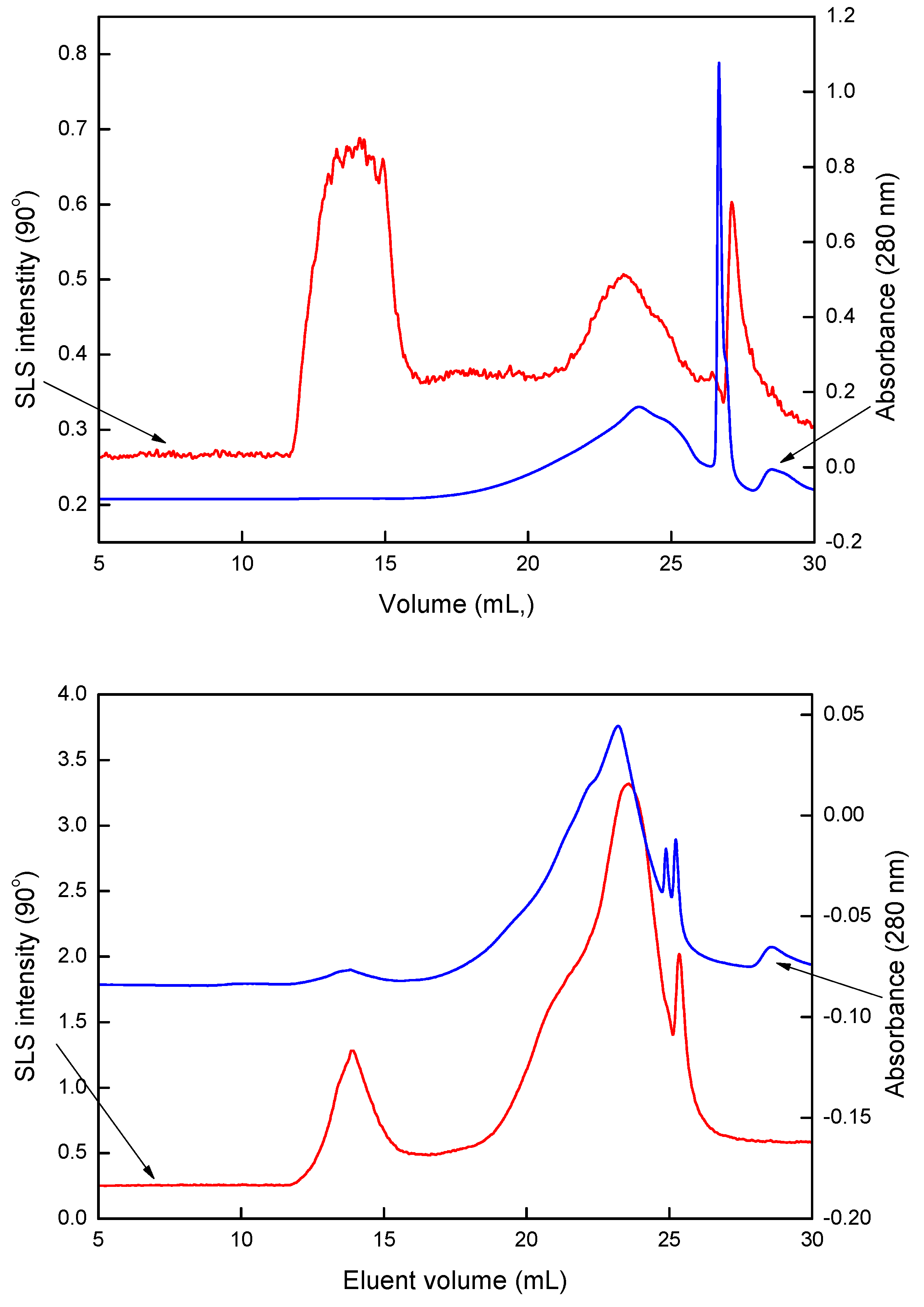
Figure 4 (top) shows size exclusion chromatography (SEC) data obtained from a bean extract (BE) at pH 7. Two chromatograms are presented, one collected at a scattering angle of 90°, using a static light scattering (SLS) detector and a second depicting the eluent increment’s absorbance at 280 nm. Three main peaks elute: The first one elutes at 15 min, a time also corresponding to the elution of 2 MDa dextrans. Although the direct comparison of elution times between different macromolecules should be avoided [
15], it is safe to attribute this peak to polysaccharidic self-assemblies (one should bear in mind that starch is a component of aqueous bean extracts). A second peak appears at elution times corresponding to dextrans of some tens of kDa (between 20 and 25 mL). This can be readily attributed to proteins, which is verified by the absorbance at 280 nm due to the aromatic amino acids of the extracted proteins. A third peak elutes between 25 and 30 mL is the system, and should be attributed to oligosaccharides and oligopeptides, along with other smaller molecules (‘system peak’ in SEC terminology). The above are a description of the macromolecular populations of BE: One population of very large polysaccharidic entities, one of much smaller proteins, and a number of smaller molecules eluting from the bean matrix.
The addition of the anionic surfactant sodium dodecyl sulphate (SDS) leads to displacement of the proteins from the oil droplet interface [
16]. To this end, a BE-stabilized emulsion at pH 7 was gently centrifuged as to separate the cream layer and dispersed in an appropriate buffer containing 1% SDS. The emulsion was then stirred and centrifuged. The serum, at that time containing the macromolecular populations which were interfacially adsorbed before SDS application, was collected and measured in SEC, giving the chromatograms shown in
Figure 4b. Although the scales between
Figure 4a and
Figure 4b are different, it is clear that the composition of the interfacial layer (shown in
Figure 4b) is entirely different from the extract overall composition (shown in
Figure 3a). More specifically, the peak corresponding to the proteins (24 mL) is dominant at the interface, while the polysaccharide peak (15 mL) is existent, but weak. That suggests that either a large part of the polysaccharide remains unabsorbed, or that the desorbed polysaccharide opened up its configuration and no longer scatters light (as do, i.e., β-glucans). In order to check the latter scenario, SEC plots were collected from the serum of a BE emulsion prior to SDS addition. The polysaccharide was indeed found in the bulk phase, so it can be concluded that a large part of it remains unabsorbed. The presence of unabsorbed polysaccharides in the bulk phase can readily account for the depletion flocculation that was suggested as the reason for the droplet aggregation at higher BE concentrations.
Polysaccharides such as starch are not amphiphilic and are generally not considered good emulsifiers; however, recent developments in understanding starch-based Pickering emulsifiers [
17,
18,
19] support the observation that a part of the polysaccharide remained adsorbed at the interface (and, possibly, did not desorb with SDS). Thus, based on the small but existing SLS peak at 15 mL (
Figure 3b), one can confidently claim that the oil–water interface of the BE-stabilized emulsions also contains starch and/or other bean polysaccharides, along with protein. The involvement of non-charged polysaccharides at the interface can explain the relative stability of the BE-stabilized emulsions at pH 3 discussed earlier. While proteins below their isoelectric points typically lead to extensive flocculation, polysaccharidic emulsifiers are efficient at low pH [
20].
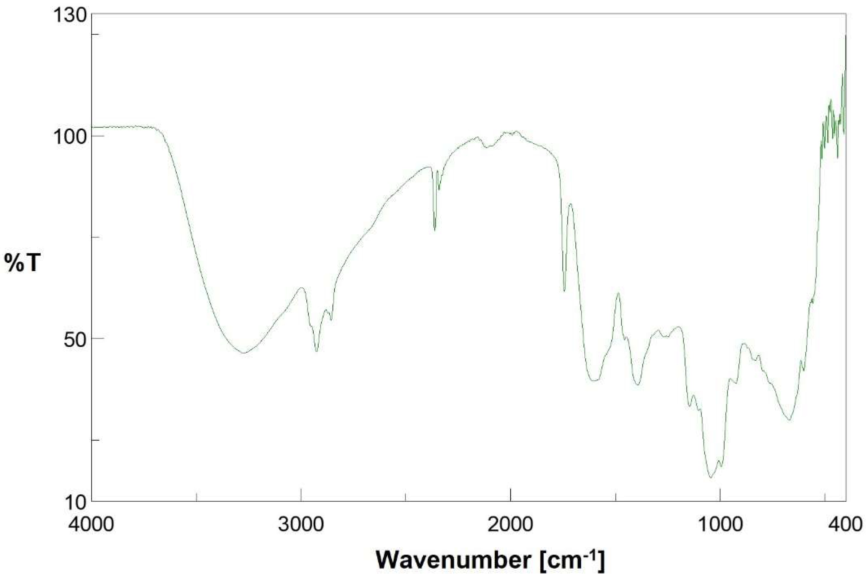
The SEC data on the extract composition can be validated by FT-IR measurements.
Figure 5 shows a typical FT-IR spectrum of a bean extract. In it, one may discern major polysaccharide peaks at 1030 cm
−1 [
21], and protein peaks corresponding to the amide I and amide II regions (1500 to 1650 cm
−1) [
22]. The peaks between 2850 and 3000 cm
−1 are due to C–H bonds, –CH
2 and –CH
3 groups, while the broad peak between 3000 and 3500 cm
−1 corresponds to O–H stretching [
23]. The above agree with the co-existence of polysaccharides, proteins and their by-products (sugars, peptides) as principal components in the extract under study.
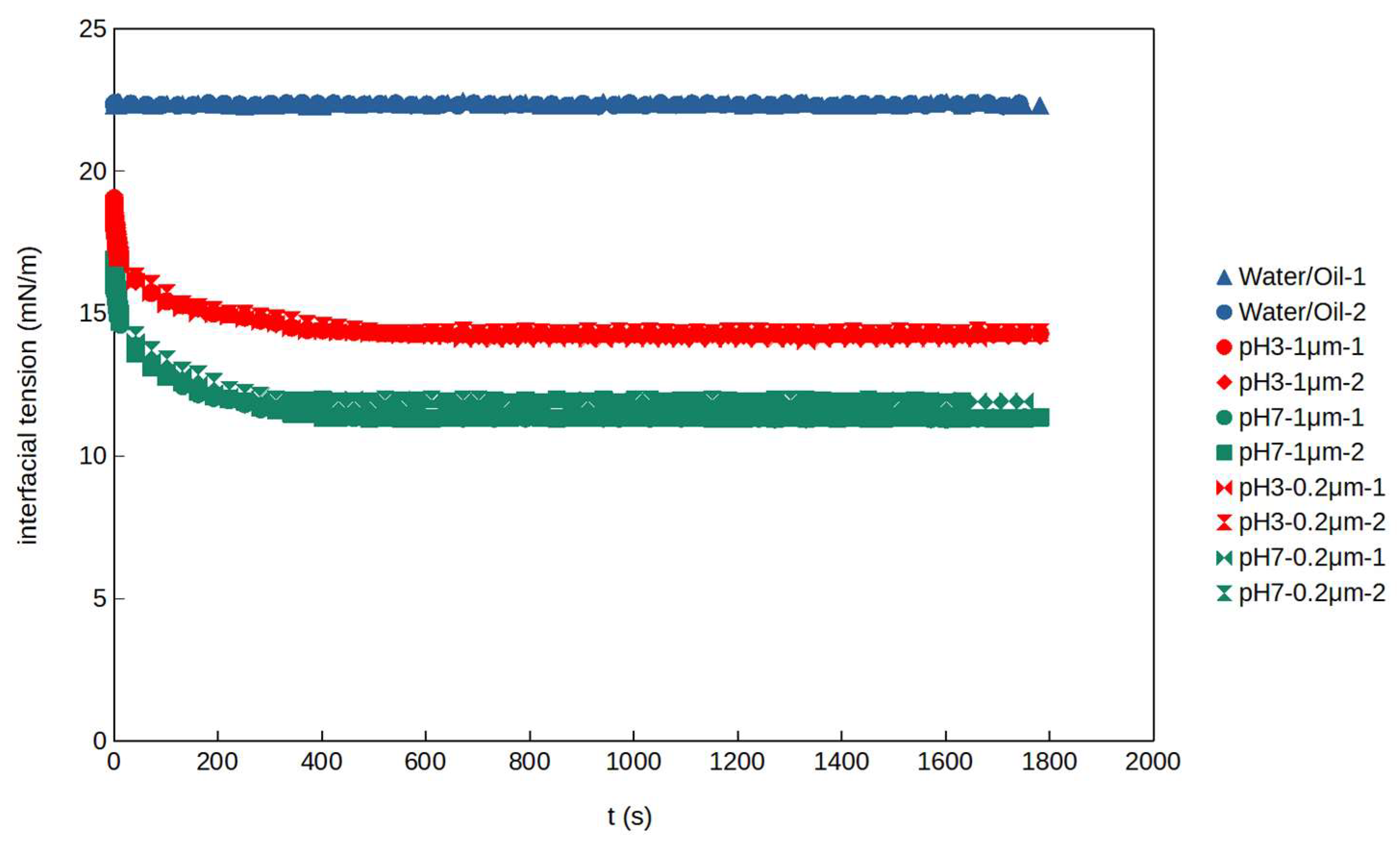
The adsorption of proteins and smaller molecules, such as system peak peptides, tends to lower the oil–water interfacial tension, while Pickering stabilization is not typically associated to significant changes in the interfacial tension, it is worth following the time-dependent oil–water interfacial tension for BE systems simulating the interfaces of the studied emulsions.
Figure 6 presents the dynamic interfacial tension vs. time for BE extracts. It is shown that the BE extracts lower the interfacial tension to intermediate levels. The decrease of interfacial tension is higher at a pH 7, and this is in line with the trend observed for the d
3.2 and d
4,3 values for emulsions prepared at the same BE extract concentrations (1%). Filtering the extract with 1μm and 0.2 μm filter did not have any impact on interfacial tension indicating the absence of significant adsorption of 0.2–1 μm particles on the interface.

 ,
, 


