Review

Surface Modification of 3D Printed PLA Objects by Fused Deposition Modeling: A Review

Department of Chemical Engineering, Gebze Technical University, Gebze 41400, Kocaeli, Turkey
*
Author to whom correspondence should be addressed.
Colloids Interfaces 2019, 3(2), 43; https://doi.org/10.3390/colloids3020043
Submission received: 16 February 2019 / Revised: 10 March 2019 / Accepted: 25 March 2019 / Published: 29 March 2019
(This article belongs to the Special Issue Colloids and Interfaces in Printing Technology)

Abstract

:
Polylactic acid (PLA) filaments are very popular as a thermoplastic source used in the 3D printing field by the “Fused Deposition Modeling” method in the last decade. The PLA market is expected to reach 5.2 billion US dollars in 2020 for all of its industrial uses. On the other hand, 3D printing is an expanding technology that has a large economic potential in many industries where PLA is one of the main choices as the source polymer due to its ease of printing, environmentally friendly nature, glossiness and multicolor appearance properties. In this review, we first reported the chemical structure, production methods, general properties, and present market of the PLA. Then, the chemical modification possibilities of PLA and its use in 3D printers, present drawbacks, and the surface modification methods of PLA polymers in many different fields were discussed. Specifically, the 3D printing method where the PLA filaments are used in the extrusion-based 3D printing technologies is reviewed in this article. Many methods have been proposed for the permanent surface modifications of the PLA where covalent attachments were formed such as alkaline surface hydrolysis, atom transfer polymerization, photografting by UV light, plasma treatment, and chemical reactions after plasma treatment. Some of these methods can be applied for surface modifications of PLA objects obtained by 3D printing for better performance in biomedical uses and other fields. Some recent publications reporting the surface modification of 3D printed PLA objects were also discussed.

1. Introduction

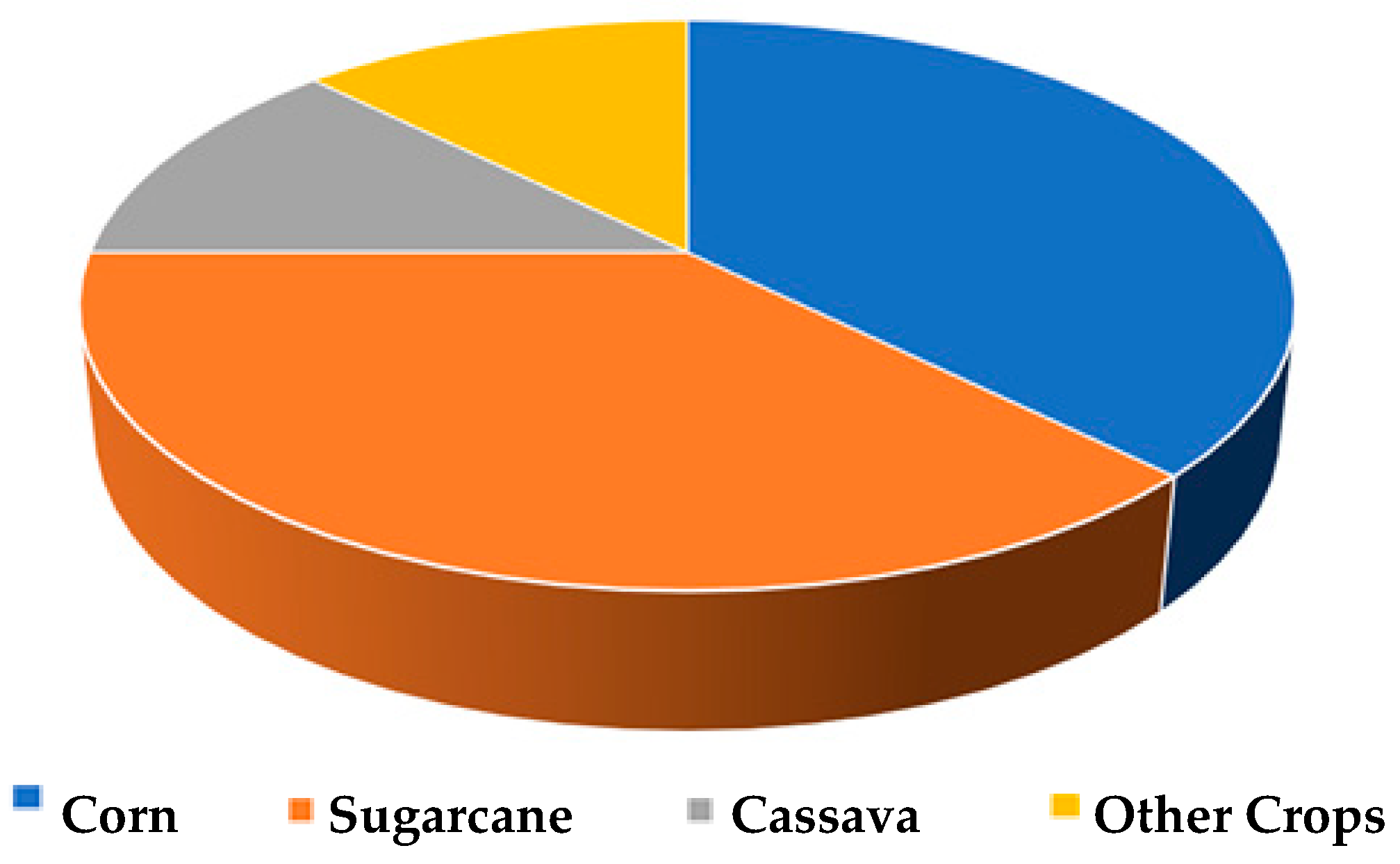
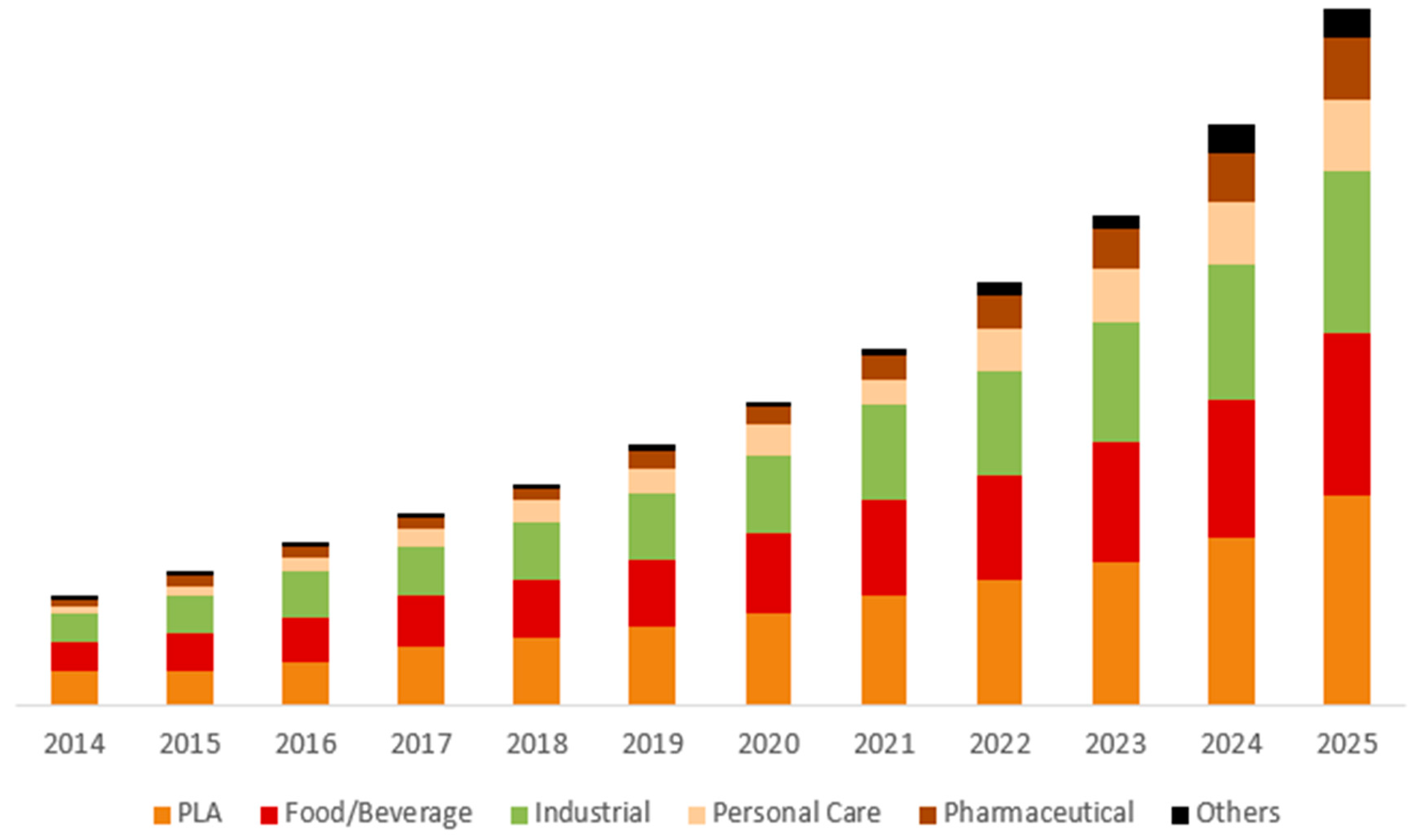
1.1. PLA Production, General Properties, and Present Market

Poly(lactic acid) is a biobased, biocompatible and biodegradable polymer which is generally produced from renewable sources (e.g., corn, sugar cane, wheat, and rice). The production of poly(lactic acid) is environmentally advantageous since it is obtained from natural sources and also due to the consumption of large amounts of carbon dioxide gas during its production [1,2,3,4,5,6,7,8,9,10,11,12,13,14,15]. Polymers based on lactic acids are called either poly(lactic acid) or polylactide and abbreviated as PLA in both cases. The main applications of PLA have been limited to medical uses such as implant devices and tissue scaffolds until the last decade, mostly due to its high production cost and insufficient mechanical resistance of the low molecular weight PLA [2,3,4,5,7,8,9,10,11,16]. However, the industrial usage of PLA was largely widened after the development of new methods which allowed the economical production of a high molecular weight PLA polymer [10,11,12,13,14,15].
At present, high molecular weight PLA is generally produced by polycondensation and/or ring-opening polymerization of lactic acid (HO–CH3–CH–COOH) which is a simple chiral molecule existing as two enantiomers, l- and d-lactic acid, differing in their effect on polarized light where the l- isomer rotates the plane of polarized light clockwise, and the d- isomer rotates it counterclockwise. There is another form of lactic acid which consists of an equimolar (racemic) mixture of d and l isomers and this form is optically inactive [3,8,9,10,11].
Lactic acid is produced commercially by a fermentation process from natural products, or via petrochemical feedstocks, however, the petrochemical route to lactic acid is not economically feasible at present. In addition, the lactic acid produced by the petrochemical route gives the racemic mixture of l- and d- enantiomers, which is not very useful in the industry. Starch (mainly corn starch) is converted into lactic acid by bacterial fermentation using a strain of Lactobacillus in the fermentation route. The lactic acid produced by this route is mostly (99.5%) the l-isomer which introduces the high mechanical strength into the PLA polymer after polymerization [17,18].
The PLA polymer can be obtained by two different polymerization routes where the first one is the direct condensation of lactic acid using solvents under high vacuum and the second is the formation of the lactide which is a cyclic dimer intermediate where no solvent is used [8,9,10,11,12,13,14,15]. The second method is advantageous because the catalytic ring-opening polymerization of the purified lactide results in high molecular weight PLA [19,20]. It is possible to control the molecular weight, the sequence and also the ratio of d- and l-lactic acid units in the final PLA polymer by selecting the catalyst type, concentration and controlling the residence time and temperature during the polymerization [3,21]. The stereochemistry of PLA is important because the stereoregularity enables the formation of a highly crystalline PLA [21]. When the d- content is high in the polymer (>20%), a fully amorphous PLA can be obtained, whereas highly crystalline PLA can only be obtained when the d- content is low (<2%) [3,8,17,18]. The commercially available PLA polymer is relatively hard, with a glass transition temperature in the range 60–70 °C and a melting temperature of 170–180 °C. PLA needs 25–55% less energy consumption than that of the petroleum-based polymers during its production and it is predicted that this energy need will fall to less than 10% in the near future [22]. It is expected that low energy requirements will also decrease the future cost of PLA [23].
PLA is an “eco-friendly” biopolymer since it is biodegradable, recyclable, and compostable. It is also biocompatible with body fluids and can be used in biomedical applications. It does not produce toxic or carcinogenic effects in local tissues and the degradation products should not interfere with tissue healing [2,4,17,23]. PLA degrades usually by hydrolysis and sometimes by microbial attack. PLA hydrolyzes to its constituent hydroxyl acid when used in the interior of a human body. The resultant metabolites are incorporated into the tricarboxylic acid cycle after hydrolysis and excreted from the body [7,24]. Correspondingly, the Food and Drug Administration (FDA) has confirmed that the use of PLA is appropriate in cases of direct contact with the biological fluids [8]. In addition, PLA has good thermal processibility when compared to other biopolymers and can be processed by widely used industrial processes like injection molding, film extrusion, blow molding, thermoforming, fiber spinning, and film forming, etc. [3,6,10,11,16,22]. High tensile strength, a high Young’s modulus and good transparency properties have made the PLA polymer a preferred choice in many applications, such as packaging and household products, braces, wound dressings and seams [25,26,27,28,29]. In addition, it was used in different sectors such as an automotive component and breakable plastic bag material [30,31,32].
PLA polymer is not only biodegradable, but also bacteriostatic, flame retardant and has weather resistance [33,34,35,36]. Composites, in which one of its raw materials is PLA, have also found application in the electronics industry. A flame retardant PLA composite was developed by NEC for electronic products [37]. The PLA matrix was reinforced with carbon fiber and when a carbon fiber reinforcement of 30% was applied, it was observed that its mechanical properties were as good as stainless steel. This has been an important step in improving the heating and cooling problem in small technological devices where the low heat transfer is a problem [10].
However, the PLA polymer has some limitations for industrial applications such as having a poor toughness, fragility, poor melt strength, low heat bending temperature, narrow processing window, and low thermal stability [10,11]. PLA is a rather brittle material with a less than 10% elongation at its break point and this low toughness property limits its use in many industrial applications that need plastic deformation at higher stress levels.
On the other hand, PLA has a slow degradation rate and this property of a polymer is mostly considered as the decisive criterion for the possibility of its use in biomedical applications. PLA degrades by the hydrolysis of the backbone ester groups and the rate of its degradation depends on factors like the crystallinity, molecular weight, molecular weight distribution, morphology, water diffusion rate and stereoisomeric content of the used PLA. Due to the slow degradation rate, it may take years for PLA to degrade completely in a living organism and there are studies to speed up this degradation rate [7,26,27]. Bergsma et al. [26] and Incardona et al. [27] noted that approximately 3 years after the placement of a PLA-based implant, a second operation was needed to remove the implant from the patient. In addition, the slow degradation rate is also a problem with the disposal of consumer commodities such as packaging films [1,2,3,4,5,6,10,11,13,16,22].
PLA is a relatively hydrophobic polymer with a static water contact angle in the range of 75–85° which results in a low cell affinity in biomedical applications and sometimes cause an inflammatory response from the surrounding tissue with direct contact [33,34]. Although it has carbonyl and methyl groups as the pendant chemical groups on the backbone chain, PLA is somewhat a chemically inert polymer that lacks enough reactive side-chain groups, which prevents its easy surface and bulk modifications.
The global lactic acid market size was US $ 2.22 billion in 2017 and the global lactic acid market is estimated to reach US $ 9.8 billion by 2025 according to the report prepared by Grand View Research, Inc. [38] as given in Figure 1 and Figure 2. Lactic acid is used in cosmetics, chemicals, food and beverages, pharmaceutical, and medical industries and the PLA production. Lactic acid is used as a pH regulator, flavoring agent and an inhibitor of residual bacteria in the food and beverages sectors (bread, butter, yogurt, canned vegetables, sweets, beer and soft drinks). Lactic acid is increasingly used in cosmetics and personal care because of its antimicrobial and moisturizing properties.
The PLA market is expected to reach 5.2 billion US dollars in 2020. The main applications are divided into sectors such as packaging, agriculture, electronics, textiles, bio-medical and others. Packaging applications have the highest share (65.2%) in the global market in 2014 due to its good mechanical, thermal and barrier properties [39]. There is a consumer demand for the use of green-packaging in the market and the PLA package performance was improved by blending with other polymers and adding other compounds to increase its mechanical resistance. However, the low barrier properties of PLA toward moisture and gases cause limitations in many food packaging applications. PLA containers were tried to be used as microwaveable containers and disposable drinking cups but there were some troubles in its use since PLA is susceptible to heat deformation [4,6,13].
PLA is used in the medical industry, such as implants, bone grafting, and fracture fixation devices for the healing of tendons and ligaments in order to replace metal-based medical devices in combination with other polymers such as polyglycolic acid, glass fiber, collagen, carbon fiber, and hydroxyapatite ceramic [2,4,7,8,9,10,11,12,13,14,25,26,29,31,36].
PLA can be processed into fibers by spinning and it is a promising textile source due to its ability to absorb organic compounds and water due to its polarity and liquid wicking properties. However, major synthetic textile polymers, such as nylon and PET are intended to be replaced by PLA with little success. Although PLA is seen as a promising alternative due to its breathability, the dyeing and finishing of PLA is difficult because of its rapid degradation in comparison with the synthetic fibers and also because its ironing temperature is limited to temperatures which are lower than those acceptable for PET and cotton fibers. At present, PLA textiles are being used by garment industries (i.e., apparel, homeware) and also for knitted and embroidered fabrics due to its good retention and crimp properties. The automotive industry tested the environmentally friendly PLA polymer for the car interior parts such as carpets, floor and trim parts, but obtained little success due to the low durability of PLA [8,10,13,14,18,21,27,35].
PLA was tested for environmental remediation applications to remove the contaminants from the environment and also to develop tow fibers for cigarette filters to replace the presently used cellulose acetate tows. PLA was also used to produce water-based paint using an isocyanate reactant [13].

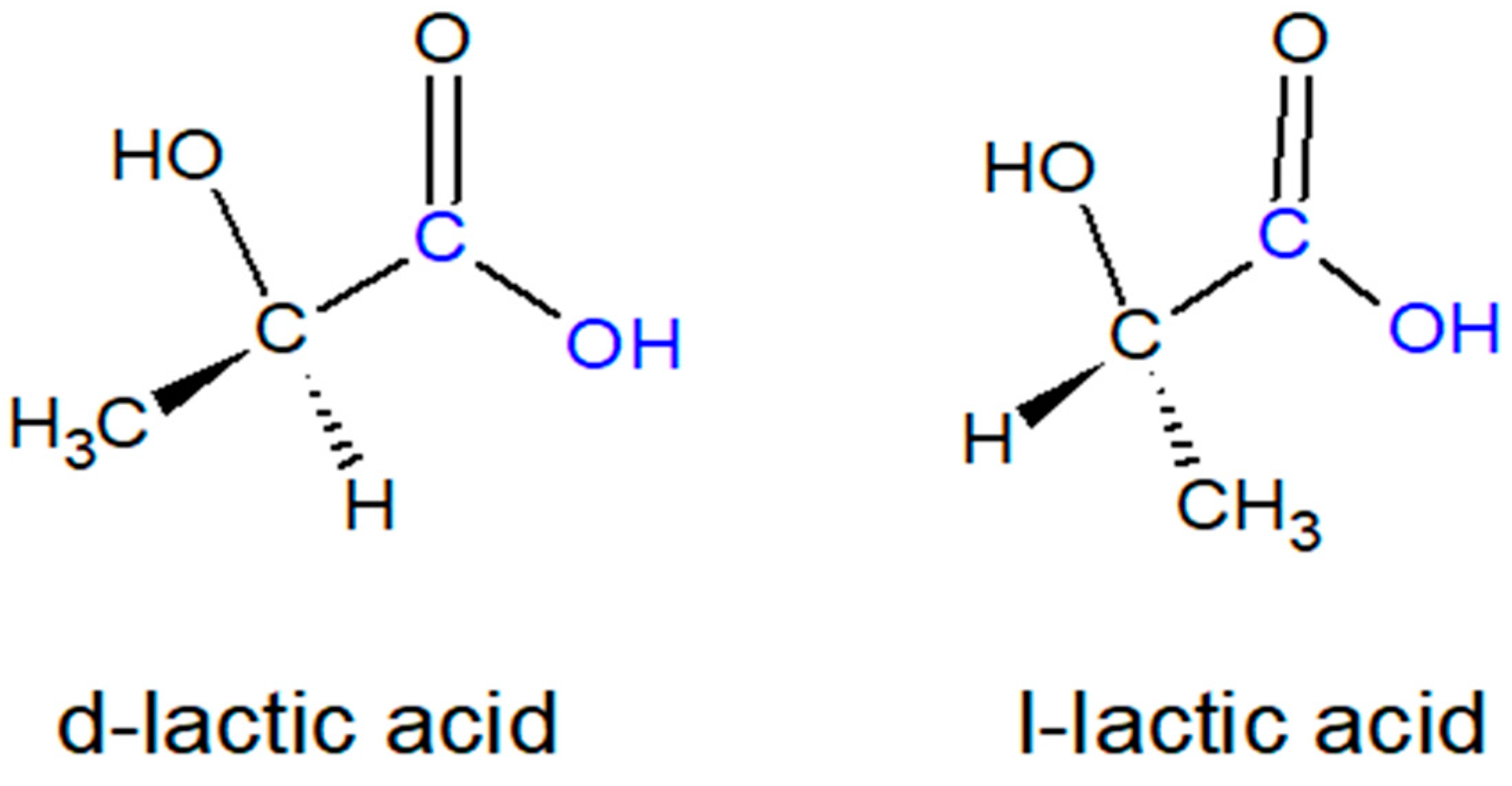
1.2. Chemical Structure of PLA and Chemical Modification Possibilities

The main reason for all the advantages and limitations of PLA is directly related to its chemical structure and correspondingly to the chemical nature of its monomer lactic acid (2-hydroxypropanoic acid, HO–CH3–CH–COOH). Lactic acid is the simplest 2-hydroxycarboxylic acid with a chiral carbon atom and exists in two enantiomeric forms, l- and d- differing in their effect on polarized light where the l- isomer rotates the plane of polarized light clockwise, and the d- isomer rotates it counterclockwise as seen in Figure 3. The lactic acid molecule has a hydroxyl and an acid functional group in it, which may result in intermolecular and intramolecular esterification reactions. Dimeric lactic acid (lactoyl lactic acid) is formed by a condensation reaction by the removal of water. The cyclic dimer (lactide) can be formed by the intramolecular esterification of lactoyl lactic acid or by the breakdown of higher oligomers. A solution of lactic acid at equilibrium consists of monomeric lactic acid, dimeric lactic acid or lactoyl lactic acid, higher oligomers of lactic acid, and lactide. The dehydrated, cyclic dimer of lactic acid is commonly called lactide (3,6-dimethyl-1,4-dioxane-2,5-dione). Due to the two asymmetric carbon atoms in the molecule, lactide exists in three different forms. In addition to the three dia-stereomeric structures mentioned above, a racemate of d-lactide and l-lactide also exists: dl-lactide.
Distillation and melt crystallization are currently used for lactide purification: the crude lactide from the synthesis is distilled in the first column to remove the acids and water, and then in the second column, under low pressures, meso-lactide is separated from lactide. On the other hand, lactide crystallizes easily in the melt in falling film crystallizers or vertical column crystallizers with a scraper. Pure lactide is miscible with benzene, toluene, xylene, methylene chloride, chloroform, tetrahydrofuran, ethyl acetate, methanol, isopropanol, acetone, and butanone. Lactide hydrolyzes to lactic acid in water at room temperature.
By applying the ring opening polymerization of lactide, PLA can be produced at a desired high molecular weight with a continuous process [40]. If PLA consists entirely of the l-lactic acid monomer then it is called PLLA, and if it is composed of the d-lactic acid monomer, it is called PDLA. The high purity of the l-isomer reduces the effort to be spent during the purification of the final product [3,8,9,10,11]. The PLA macromolecule has aliphatic ester nature. Fourier transform infrared spectroscopy (FTIR) spectrum in the 1870–1540 cm−1 C=O stretching absorption band in the region, especially in 3571 cm−1 –OH stretch, 2996.53 and 2987.75 cm−1 CH stretch band are the characteristic peaks [31].
Polymerizations of lactide can be carried out by using the melt polymerization, bulk polymerization, solution polymerization, and suspension polymerization methods however melt polymerization was found to be the best method. In general, commercial PLA is a blend of PLLA and PDLA or copolymer PDLLA, obtained by the polymerization of LLA and DLLA. PLA with a PLLA content higher than 90% tends to be crystalline and the melting temperature (Tm), glass transition temperature (Tg), and crystallinity of PLA decrease with the decreasing amounts of PLLA. Many important properties of PLA are controlled by the ratio of d- to l-enantiomers used and the sequence of arrangement of these enantiomers in the polymers. Several types of additives could be utilized to improve the characteristics of the PLA. These well-established additives are antioxidants, heat stabilizers, light stabilizers, impact modifiers and several others [10].

1.3. The Use of PLA in 3D Printers and the Present Drawbacks

In the last decade, PLA filaments have been gaining popularity as a thermoplastic source to be used in 3D printing (in fused deposition modeling, FDM approach) where its characteristics, such as ease of printing, glossiness and multicolor appearance, make PLA one of the main choices for this purpose. A 3D printing machine working with the FDM method is basically a computer controlled gantry, carrying one or two miniature extruder head nozzles, one for the modeling material and sometimes the other for the support material. Three-dimensional objects are fabricated by extruding a molten thermoplastic filament through a heated nozzle in a predetermined pattern onto a platform. The extruded filament cools, solidifies and bonds with the adjoining material as the material is deposited so that the base plate moves down (or the nozzle moves up) by an increment equal to the height of the filament when a whole layer is deposited and, later, the next layer is deposited again. The temperature history of the interfaces plays an important role in determining the bonding quality between these deposited layers and the formation of bonds among the polymer filaments in the FDM parts which are driven by the thermal energy of the extruded material. The bonding quality also depends on the molecular diffusion and randomization of the polymer chains across the interface and on the growth of the neck formed between the adjacent filaments [41,42,43]. In general, thermoplastics such as ABS, PLA [41,42,43,44,45], polyamide (PA) [46] and polycarbonate (PC) [47] are used as filaments in the 3D printers. However, at present, PLA filaments are going to be the most popular material used, with an annual growth rate of 19.5% [48]. PLA is in competition with the acrylonitrile-butadiene-styrene copolymer (ABS) filaments in the FDM technology, however, a high accuracy for dimensional parts can be achieved only with PLA because it poses less warp behavior than the ABS filaments. It is also safer than ABS [49]. It is well-known that the glass transition temperature of a polymer should be low and the viscosity when it melts should be suitable for layering for a thermoplastic material which is suitable for 3D printing technology. PLA has a 150–160 °C melting point which is low when compared with other polymers used in the thermoplastic 3D printing industry and requires less energy consumption during printing. This makes PLA polymer preferable especially in off-grid applications [50].
However, PLA filaments have some disadvantages: PLA has a poor toughness and elongation at break of less than 10% [51,52]. Although this value is actually comparable to that of poly(ethylene terephthalate) [6], the use of PLA in low toughness and high stress levels remains limited. It is expected that the range of the use of PLA filaments will expand with the development of 3D printing technology [43,53,54,55].

1.4. Reasons for the Surface Modification of PLA

In this review, we will examine the published literature on the surface modifications of the PLA polymer in various industries in detail in the sections given below. In general, surface modification methods of the PLA can be divided into two classes: covalently bonded and non-covalent bonded (physically attached). The methods used for the surface modification of the PLA polymer are examined in Section 2. These are mainly alkaline surface hydrolysis, atom transfer polymerization (ATRP), photografting by UV light, various forms of plasma treatment, and chemical reaction after plasma treatment. The use of PLA polymers after the surface modifications was also reported in this section. A few papers reporting the surface modification of 3D printed PLA objects are given in Section 4.

2. Surface Modification of PLA Solids

PLAs have widespread uses in many industrial fields. The successful application of PLA relies not only on its mechanical properties, but also on the controlled surface properties such as hydrophilicity, surface free energy, reactive functionalities and roughness. The bulk modifications of PLA are mostly dependent on the processes to improve toughness and the degradation rate. However, its surface modification was applied to control hydrophilicity, surface free energy, chemical heterogeneity, roughness, and to introduce reactive chemical groups to enable further covalent reactions. There are a variety of surface modification methods to control the surface properties of PLA such as surface coating, surface alkali hydrolysis treatment, graft polymerization, low-temperature plasma treatment and various surface chemical reactions. The improvements in hydrophilicity and the introduction of reactive groups are beneficial especially for the biomedical applications of PLA when it is used in the human body where the surface characteristics are critical for cell affinity and cell adhesion [22,56]. However, the hydrophilicity, biocompatibility, and cell affinity of PLA are still not good enough for some tissue engineering and other biotechnology uses and awaits proper surface modifications.
Since we are interested with the surface modification of 3D printed PLA objects in this review, we excluded all the PLA bulk modification methods such as copolymerization, stereochemical manipulation, blending with plasticizers, other polymers, fillers and other composite materials from the review content. On the other hand, some good reviews were published on the surface modification of PLA polymers in the past [22,57,58,59,60,61], however, these reviews are mostly on the general plasma treatments of many biopolymers or specific biomedical applications of PLA except the publication of Rasal et al. which reports on the specific PLA bulk and surface modifications published till to 2010 [22]. Thus, there is a need to review the literature regarding PLA surface modifications after 2010 and also to select some suitable methods to be applied for the PLA objects which are printed by the FDM approach. In this review, we initially discuss the most effective surface modification methods which were given in the older literature and then present more recent publications in this field.
In general, surface-modification methods can be classified as chemical methods (with covalent attachment) and physical methods (non-covalent attachment of functional groups). However, in practice, it was determined that more than half of the publications are related to the plasma treatments of PLA which are accepted to be non-permanent. The gains obtained by the plasma surface modification is partially lost due to surface rearrangements since the macromolecular motions are thermally activated to minimize the interfacial energy and makes the effect of plasma treatment non-permanent at room temperature applications [62,63,64,65].

2.1. Surface Modification with Chemical Methods Giving Covalent Attachment

2.1.1. Alkaline Surface Hydrolysis and Atom Transfer Polymerization (ATRP)

PLA itself does not have any reactive side-chain groups and alkaline surface hydrolysis is a simple way to impart two types of reactive hydrophilic functional groups, e.g., carboxylic acids (–COOH) and hydroxyls (–OH) on the PLA main chain by cleaving the ester bond of the aliphatic polyester [57]. The resulting new chemical groups can be used to conjugate the bioactive molecules, such as l-lysine, collagen and peptides, which can be recognized by the cell adhesion receptors in biomedical applications. It was found that a mild alkali treatment could not break the ester bonds effectively in a short time, however, if strong alkali is used, it caused the bulk degradation of the PLA. Thus, a mixture of aqueous 0.25 M NaOH/ethanol can be successfully used for this purpose [66]. Ethanol was applied to assist the hydroxide nucleophilic attack on the ester bond. After alkali treatment, the surface hydrophilicity and the surface energy of the PLA can be improved significantly, but its surface roughness changes. Such a treatment was effective to enhance the cell affinity of the PLA cell scaffolds [66].
The PLA polymer cannot be dissolved in water, alcohols, and unsubstituted hydrocarbons but can be dissolved in many low-cost organic solvents such as benzene, toluene, p-xylene, chloroform, dichloromethane, trichloromethane, dioxane, and ethyl acetate [10,11,22]. Racemic PLA (poly-d,l-lactic acid) can be dissolved in polar solvents such as acetone, dimethylformamide, and tetrahydrofuran [67]. Thus, these solvents may be used during covalent binding reactions onto PLA.
After the formation of the carboxylic acid groups on the PLA, these can be conjugated with species containing amine (–NH2) or hydroxyl (–OH) groups. In some cases, the acid groups are first activated with phosphorous pentachloride [68], thionyl chloride [69], or water-soluble carbodiimides [70] and then subsequently reacted with amines or hydroxyls. Chitosan can be covalently attached to PLA surfaces after alkaline surface hydrolysis followed by acid-chitosan conjugation [71]. Reactive amine groups can be introduced onto PLA surfaces by aminolysis followed by conjugation with biocompatible macromolecules such as chitosan, collagen and gelatin [72,73].
Electrospun PLA nano-fiber surfaces were also modified with amine groups and then conjugated with polyethylene glycol to convert the partially hydrophobic surface into a hydrophilic one. Prolonged cell viability was seen on such surfaces [74]. The PLA film was first treated with an alkaline solution to form the carboxyl groups on the surface and then inoculated the Xtal Fluoro-E gelatin onto it giving a hydrophilic surface [75]. A method was proposed for the immobilization of simple molecules on the PLA surface to prevent its degradation. The method is a two-step procedure where the surface is activated anionically under certain conditions in the first step and then click chemistry was applied in the second step to modify the surface of the PLA [56].
Gelatin was added to the PLA film surfaces in order to improve its cell adhesion properties after the covalent bonding of sodium methylacrylate (MAAS) to the surface by atom transfer radical polymerization (ATRP). The results show that the unreacted PLA and also the PLA-g-P(MAA) surfaces were not suitable for cell adhesion, but when the surface is coated with gelatin, the cell adhesion properties improved considerably [76]. PLA membranes were grafted by the zwitterionic polymer to improve the fouling resistance and hemocompatibility. Surface zwitterionization was carried out by dopamine inspired bromo alkyl initiator anchoring and the subsequent ATRP of zwitterionic poly(sulfobetainemethacrylate). ATR-FTIR, XPS and SEM analyses were applied onto the samples and it was confirmed that zwitterionic PSBMA was successfully grafted onto the PLA membrane. The hydrophilicity, anti-fouling and hemocompatibility of the PLA membranes were improved for their biomedical and blood-contacting applications such as hemodialysis [77].

2.1.2. Photografting by UV Light

Photografting by UV light enables the permanent alteration of surface chemistry with a low-cost operation [78]. Photoactivation occurs on the PLA surface to form reactive groups followed by the grafting of selected materials having different functionalities. There are two types of surface grafting methods: “grafting to” or “grafting from”. For the first one, polymer chains of specified molecular weights and compositions are covalently attached to the surface [79]. Although it is useful, it is difficult to obtain high grafting densities on a PLA surface with a “grafting to” process because of the presence of steric hindrance and chain diffusion limitations [80]. The second method, which is the “grafting from” approach, involves the growing of polymer chains from the surface by graft polymerization (especially by photopolymerization) of suitable monomers, and this method overcomes the limitations of the “grafting to” approach. However, in the “grafting from” approach, the used photoinitiators must be immobilized onto the PLA substrate in order to initiate the subsequent polymerization of the vinyl monomers from the surface, which is also a difficult process.
The “grafting to” approach was applied to produce superhydrophobic PLA fabrics by using hydrophobic silica particles possessing vinyl functional groups on the surfaces which were synthesized via a sol-gel process [81]. In another publication, a single step vapor phase photografting reaction was reported to graft poly(acrylamide), poly(maleic anhydride), and poly(N-vinylpyrrolidone) onto the PLA surface in the vapor phase to avoid the detrimental solvent effects [82]. Benzophenone was used as the photoinitiator under UV irradiation. Static contact angles decreased down to 25–50° from around 80° after grafting. This method was also applied to graft poly(N-vinylpyrrolidone) chains onto PLA to obtain a substrate for the attachment of keratinocytes and skin fibroblast cells [83].
For the “grafting from” approach, the grafting of PLA with an acrylic acid monomer to form hydrophilic polyacrylic acid brushes on the PLA were studied several times in the liquid phase reactions [51,84,85]. PLA films were activated by argon-plasma and then were immersed in an aqueous acrylic acid solution and exposed to UV to produce grafted chains of poly(acrylic acid) [84]. In another study, a PLA substrate was placed in a glass reactor where the acrylic acid monomer and benzophenone initiator were added and finally the reactor was irradiated with UV light for 20 min. It was determined that surface grafting with poly(acrylic acid) changed the degradation of the product patterns and accelerated the formation of water-soluble degradation products [85]. Similarly, a PLA film was formed by a solvent casting technique, and then the surfaces of these films were inoculated with a UV cured photopolymerization reaction using poly (acrylic acid) (PAA) and poly (acrylamide) (PAAm). PAA was grafted to the PLA surface in approximately 2–3 h, while PAAm was vaccinated in 3–5 h depending on the activation method. As a result, it has been observed that the hydrophilicity or wettability of the grafted surfaces has improved considerably [86]. Acrylic acid and maleic anhydride were photografted onto a PLA surface in order to increase its surface wettability by applying the “grafting from” approach where the aniline tetramer was used to make a conductive PLA surface [87]. PLA-poly[(3-hydroxybutyrate)-co-(3-hydroxyhexanoate) films were also formed by photografting in the solvent phase and the solvents affecting the film hardness were investigated [51].
A two-stage method was used for the same purpose to graft N-vinyl caprolactam onto a PLA surface. In the first step, UV rays were exposed after adding benzophenone to the sample surface and N-vinyl caprolactam monomer was grafted onto the PLA surface in the second step. As a result, the contact angle of the grafted PLA has decreased significantly and the successful grafting on the surface was confirmed according to the FTIR-ATR analysis results [88].
The photopolymerization grafting of poly (acrylic acid) (PAA) and poly (acrylamide) (PAAm) was studied in order to determine the interactions between the chains. The presence of the attractive long-range interactions was observed in the hydrophobic PLA/PLA and hydrophobic/hydrophilic PLA/PLA-g-PAA systems [89].

2.2. Surface Modification of PLA with Plasma Treatment

2.2.1. Advantages of Plasma Treatment

The surface modifications of biomaterials are of interest to the biomedical industry. Plasma treatment is one of the most used methods to impart specific chemical functionality to surfaces which are used in the biomedical industry. The formation of plasma depends on the ionization of gases. Gas molecules will become ionized (lose one or more electrons) and so carry a net positive charge if enough external energy is added. If a sufficient number of the molecules in a gas are ionized to affect the overall electrical characteristics of this gas, then the result is called “plasma” which often referred to as the fourth state of matter. In general, plasma contains positive ions, electrons, neutral gas atoms or molecules, excited gas atoms and molecules and UV light. Plasmas glow because light is emitted as these excited neutral particles relax to a lower energy state. All of these components can interact with a solid surface during plasma treatment. It is possible to tune precisely or select the effects of the plasma treatment on the surface by choosing the type of the gas mixture, power, pressure, etc.
Plasma changes only the surface of the treated substrates and does not alter their bulk properties. In general, plasma is effective at temperatures close to ambient temperature without damaging most of the heat-sensitive biomaterials. Plasma treatment and plasma polymerizable coatings offer suitable flexibility, efficiency and environmental awareness. The most important feature of plasma processing is the surface functionality that any chemical processing cannot offer. Plasmas can change almost any substrate geometry without the problem of line-of-sight processes compared to other energy sources (such as UV, e-beam and laser). However, there are some disadvantages of the plasma surface treatment method such as the need for a vacuum environment, the initial high cost of the plasma chamber, the irregularity of the modified surface and, sometimes, an unidentified chemistry [90,91,92,93].
Low-pressure (LP) plasma techniques [91,92] are non-thermodynamic equilibrium processes which are performed in weakly ionized gases to modify the surface properties of any substrate under medium-low vacuum conditions (10-1 Torr). LP plasmas generally allow for the design of a new class of materials in the form of surfaces, with unique properties and little correlation with conventional ones. It is possible to make changes in the properties of the chemical composition, such as wettability, metal adhesion, dye-ability, refractive index, hardness, chemical inertness, lubrication and “biocompatibility”. A prosthesis or a biomedical device can be tolerated by the biological area and fulfills its intended function for a reasonable time after a suitable plasma treatment [93].

2.2.2. Surface Modification of PLA with Plasma Treatment

Yang et al. studied the effect of the ammonia plasma method onto a poly(d,l-lactide) film surface to improve its affinity to cells. They measured the contact angle and calculated the surface free energy using the harmonic mean equations. The chemical composition of the sample surface was examined by X-ray photoelectron spectroscopy analysis [62]. They observed that O- and N-containing groups such as hydroxyl and amine were formed on the surface of the plasma treated films. They also examined the effect of the methods to maintain the hydrophilicity of the surface. They showed that protection is possible at low temperatures at 0–4 °C. It was observed that the ammonia plasma treatment could support the binding and growth of cells, in cell affinity tests using the mouse 3T3 fibroblast cells [62]. In another study, the same group produced poly(l-lactic acid) and poly(l-lactic-co-glycolic acid) (70/30) cell scaffolds that have different pore structures to be used in tissue engineering. They also applied ammonia plasma treatment to alter the surface properties and improve the cell affinity of the scaffolds [94]. The hydrophilicity and surface free energy of the surfaces were increased after plasma. Cell culture results showed that pores of less than 160 micrometers were suitable for the growth of the human skin fibroblast cell. Cell seeding efficiency was maintained above 99% which is a better value than the efficiency achieved by the common preheating method with ethanol. The plasma-treatment method also assisted in solving the problem of cell loss during cell seeding and avoided the negative effects of waste ethanol on the cell culture [95].
Wan et al. used a series of gases under various conditions of electrical power, pressure and time for the plasma treatment of PLLA [95]. The cells exposed to the NH3 plasma-modified PLLA and the results indicated that the cell retention on the modified PLLA was much higher than the unmodified. Increased surface hydrophilicity, high surface free energy polar component and N-containing groups were obtained and have an important role in increasing the cell resistance against shear stress [96]. Safinia et al. have developed a novel low-pressure radio frequency plasma treatment protocol to effectively modify the surface of large pore PDLLA scaffolds using air or water in ammonia plasma treatment [65]. PDLLA foams were found to have a high porosity, void ratio and coherent pores ownership criteria to be used as tissue engineering scaffolds and the surface of the main polymer was hydrophobic. After exposure to plasma, it was found that an increasing number of polar functional groups and improved wetting formed on the treated surfaces. The number of polar functional groups on the surface (and hence the drop in water contact angles) increased as the plasma exposure time increased. The authors characterized the change in the surface composition and the wettability of the polymer structures by zeta potential and contact angle measurements. However, it was determined that the storage of the polymer structures in the ambient air caused a significant hydrophobic recovery, while only a partial hydrophobic recovery occurred in water [65].
Ferreira et al. treated the poly PLLA and poly (hydroxyl butyrate-co- hydroxyl valerate) surfaces with oxygen and nitrogen plasma. Physical and chemical surface properties were evaluated by contact angle tests, scanning electron microscopy (SEM), atomic force microscopy (AFM) and X-ray photoelectron spectroscopy (XPS). Plasma treatments altered the surface morphology by changing both the angle of contact and impart roughening and affected the placement of polar groups thus increasing the wettability of both surfaces [96]. Geyter et al., have used different gas atmospheres (air, nitrogen, helium and argon) and a medium pressure dielectric barrier discharge (DBD) method to change the surface properties of PLA. They examined the plasma treated surfaces with the contact angle, XPS and AFM measurements. It was found that the discharge gas may have a significant effect on the chemical composition of the resultant PLA surfaces, while the air and argon plasmas introduced oxygen-containing groups, nitrogen discharges add only nitrogen groups to the PLA surface. Helium plasma also imparted functionalities with a small amount of nitrogen [97].
In the study of increasing the surface wettability of PDLLA films, Guowei et al. treated PLA films with nitrogen plasma (PLA-N2) and nitrogen/hydrogen mixed plasma (PLA-N2/H2). The surface properties and structure of PLA-N2 and PLA-N2/H2 were measured by static contact angle, surface free energy calculations, XPS and AFM. It was found that the surface hydrophilicity of both of the PLA-N2 and PLA-N2/H2 surfaces was higher than that of the pure PLA and the surface hydrophilicity of PLA-N2 films were better than PLA-N2/H2 [98].
Jiao et al. treated PLLA films with ammonia plasma to investigate their effects on surface properties and cytotoxicity [99]. They examined the surface morphology and surface roughness of the films with AFM. The variation of contact angles associated with the mass loss density, plasma treatment power and exposure time were also examined. Cyto-compatibility was evaluated by cell adhesion, proliferation and cell cycle. The results showed that the surface morphology and roughness of PLLA surfaces increased with the plasma processing power. The number of resulting amino groups on the surface of the treated PLLA film increased with the increase in the plasma treatment strength and duration. The contact angles showed an inverse trend and they increased with the storage duration and returned to its initial value after about 2 weeks. Cell experiments have shown that plasma-treated PLLA surfaces were more susceptible to cell adhesion and proliferation [99].
Jacobs et al. applied plasma modification using helium and argon gases to improve the inadequate surface properties of PLA for biomedical uses [100]. They have changed the surface properties of the PLA layer by the application of the dielectric barrier discharge method at medium pressures. After the plasma treatment, the water contact angle measurements showed the increased hydrophilic character of the PLA surface. XPS analysis indicated that the oxygen content increased. The cells on plasma-modified PLA exhibited a much better morphology compared to unmodified PLA samples after 1 day. However, after 7 days of culture, no significant difference was observed between the untreated and plasma-modified PLA samples, indicating that plasma treatment improved the initial cell attachment, but it did not seem to affect the cell proliferation [100].
Tenn et al. applied a hydrophobic plasma process to change the surface properties of the PLA film, by using CF4, CF4/H2, CF4/C2H2, tetramethyl silane (TMS) plasmas [101]. Plasma parameters such as power, gas flow and process time were optimized according to the water contact angle measurements. XPS measurements revealed the presence of the fluorine (–CF2–, –CF3) or silicon (SiOxCy) functional groups on the surface of the film after the fluorine or TMS plasma treatment. Thermal properties of the treated PLA films were investigated by differential scanning calorimetry (DSC) measurements and it was concluded that they were not affected by the plasma treatment. Water permeability measurements showed an improvement in the PLA barrier properties by decreasing its water solubility for all of the plasma processes used and especially after the application of CF4/C2H2 plasma. No effect of plasma treatment on the oxygen barrier properties of the PLA film was observed even at high relative humidity up to 90% [101].
Jordá-Vilaplana et al. used air atmospheric plasma technique to increase the surface free energy of PLA by optimizing process parameters in terms of nozzle-substrate distance and sample feed rate [60]. The macroscopic effects of the process parameters were determined using contact angle measurements and surface free energy calculations. In addition, chemical changes in the top layers were investigated by using XPS and FTIR. Surface topography changes were evaluated with SEM and AFM. The results show that there was a significant increase in surface free energy and this method was shown to be applied in a continuous industrial process with no waste production [60].
In a study by Bolbasov et al., the surface modifications of the biodegradable PLA and polycaprolactone (PCL) polymers were reported by the application of the radio-frequency thermal radiation discharge plasma method. This plasma reduced the substrate hydrophobicity and increase the surface free energy to increase the living cell affinity of PLA [102]. Izdebska-Podsiadly et al. investigated the effects of argon low-temperature plasma on the PLA film properties and studied the duration of storage to change the value of the surface free energy (SFE) and its effect on polar and dispersion components of SFE [103]. The contact angle measurements and SFE calculations were performed immediately after the plasma treatment and also after 1, 7, 14, 30 and 60 days. The chemical changes were investigated using XPS and surface roughness was determined by a confocal microscope. The results showed that a significant improvement occurred in PLA film wettability, accompanied by a 40% reduction in the water contact angle and an increase of 43% in SFE. In addition, the highest SFE value was obtained by the plasma treatment was after 6 min, but the results obtained with a shorter processing time such as 2–4 min were more stable over time [103].

2.2.3. Surface Modification of PLA with Plasma and Chemical Reaction

An improved surface modification method for PDLLA which combines the ammonia plasma treatment with the collagen anchorage was proposed in 2002 [104]. Ammonia plasma pretreatment imparted the chemical groups containing hydrophilic O and N atoms and increased the roughness of the PDLLA surface. Afterward, the plasma-pretreated surface can easily bind collagen. The surface free energy of the modified surface was further improved after the collagen was anchored onto the surface. Since the positively charged groups increased the interaction between the PDLLA surface and the negatively charged cells, the interactions with the cells also improved [104]. The same group also investigated the extent of the ammonia plasma treatment on three-dimensional PLGA scaffolds [105]. The results showed that the exchange depth reached about 4.0 mm under suitable plasma treatment conditions. In addition, plasma treatment and collagen anchorage modification effectively facilitated the cell transplantation and the cells could be distributed evenly along the scaffold. It was suggested that the flow chamber system could be used as a potential tool to compare various surface modification methods [105].
The reaction of chitosan plasma coupling was performed for the surface modification of PLLA by Ding et al. [106]. The structure of the modified PLLA surfaces was characterized by contact angle measurements and XPS. Two cell lines, L929 (mouse fibroblasts) and L02 (human hepatocytes), were cultured on the modified PLLA surface and the cells grown on this substrate could multiply at almost the same rate as the cells cultured on a glass surface. These results showed that the modified substrate could be used to control cell morphology and had potential applications in tissue engineering [107]. In another interesting study, Li et al. treated the PLLA film with oxygen plasma before making the surface hydrophobic by using organosilanes. They then immobilized gelatin on this surface to improve the cytofunction of the PLLA film which was characterized by AFM and water contact angle measurements. It was reported that hydrophobization of the PLLA surface by the organosilane modification improved the wettability properties after gelatin immobilization [108].

3. 3D Printed PLA Objects

PLA objects have become increasingly used in biopolymer research due to its biocompatibility and sustainability properties with the discovery of new processing paths based on the 3D printing methodology. There are three main 3D printing (additive manufacturing) routes, extrusion-based 3D printing techniques, powder-based laser sintering and stereolithography (SLA) processes [43].

3.1. PLA in Extrusion-Based Printing Technologies

In this category, PLA filament is fed to the 3D printing machine by means of a clamping cylinder and is melted at the desired temperature extrusion head and the solid portion of the filament pushes the melt into the nozzle. The nozzle can be moved horizontally along the xy plane. During this movement, the molten material is deposited on the table, which can be moved in the vertical z-direction [108]. “Fused deposition modeling” (FDM), “Fused filament fabrication” (FFF) and “Melt extrusion production” (MEM) names are widely used for this category to indicate the technologies based on the material extrusion. These processes are basically the same and only their trade names are different. All of the supplied thermoplastic material solidifies quickly after accumulation. This allows the production of complex 3D objects.
Extrusion-based printing techniques where PLA is used as raw material are widely used in teaching aids [109], construction of laboratory equipment [110], agricultural instruments [111] and surgical equipment [112]. Due to the biocompatibility of PLA, its use in tissue engineering and “patient-specific” implants is also very popular [113,114]. However, it is necessary to evaluate the strength and fracture properties of the 3D printed PLA parts with the FDM technique. The quality and strength of the thermoplastic components are affected by several machine-related factors. For example, the material extrusion temperature significantly affects the mechanical strength of the final 3D product in addition to the printing orientation and layer thickness which can be experimentally optimized [115,116,117]. Although the given operation settings represent the best total combination according to the test results, some settings still vary according to the user preferences. For instance, if aesthetic is important, a lower layer thickness and a lower printing speed provide a higher resolution with a better surface quality during 3D printing. In such cases, the layer thickness can be reduced without any loss of the mechanical strength of the final product [117,118].
Many statistical models have been used to study the effect of process parameters on the mechanical properties of the PLA parts produced by FDM [118,119] and to observe the resultant fatigue [120], distortion and degradation properties [121]. In order to increase the mechanical properties of the final products, 3D printable PLA-graphene composites were used to improve the mechanical resistance, flame retardancy and electrical conductivity of the printed objects [122]. However, there is a general acceptance that the high variability in the experimental 3D printing results is a result of the influence of other less controllable factors such as a medium moisture level [123].
3D printing techniques of PLA using polymer solutions instead of filaments were also proposed by using a suitable nozzle. This technique has the advantage that the polymer can be printed without degradation at much lower temperatures. In general, chloroform is used as the solvent for PLA. Such structures are very popular in the regenerative medicine applications of the biodegradable templates for tissue regeneration [124,125]. Perhaps the most difficult part of the solution printing process is to adjust the viscosity of the PLA printing ink accurately. The changing of the process temperature or adding a plasticizer was used to control the viscosity. For these methods, the correct plasticizer addition is important; because the plasticizer allows the polymer to be treated at low temperatures without the risk of thermal degradation [126]. Low-density polyethylene glycol (PEG) seems to be the most efficient additive for the PLA polymer in comparison with the different plasticizers tested. PEG is a biocompatible, hydrophilic polymer that is soluble in chloroform [127,128]. It is possible to obtain homogeneous PLA/PEG blends at low temperatures for the production of 3D scaffolding. The addition of PEG increased the surface roughness and wettability of the PLA surfaces, however, the newly formed structures were not homogenous enough and the mechanical properties were significantly reduced [126,127,128].

3.2. Use of Powder-Based Techniques and Photopolymerization in 3D Printing of PLA

PLA objects can be printed by processes other than the FDM method where PLA is used in the filament form. One of these processes is known as the “Powder-Based Additive Manufacturing Technique”, which depends on powder melting at a specific location by the help of laser, and the other one is “Stereolithography (SLA)”, where a liquid PLA layer is deposited instead of a solid powder layer by the laser application. We have to refer to these two methods in this review by citing some indicative examples of the published literature since we also have to consider the modification of the PLA object surfaces obtained by these methods. However, a comprehensive review of these two techniques is out of the scope of this review.
When PLA is used in powder-based techniques, a thin layer of polymeric PLA powder (typically 100 µm) is placed as a powder bed and a computer-controlled laser scans this area and heats the powder at the pre-determined locations with the help of a CAD program. When the powder particles melt at these points, then they combined with each other to form the desired object. An advantage of this technique in comparison with the extrusion-based methods is the presence of non-sintered powders in the powder bed to support the subsequent layers without the need for external support structures. Only a few publications on the laser sintering of the PLA are present in the literature and mostly related to the application of tissue engineering where a biocompatible and biodegradable porous scaffold was produced to serve as a temporary template for the binding and development of cells and subsequent production of tissue [129,130]. However, it was found that PLA is not a very suitable polymer for laser sintering because it is difficult to grind PLA into a powder form even in a cryogenic environment leading to a poor level of sphericity as a consequence of the poor flow characteristics [131]. Bai et al. investigated the effect of the addition of nanoclay to the PLA on the mechanical properties of sintered parts after 3D printing. They showed that the addition of nanoclay improved the elastic modulus of the produced PLA parts, however, the flexibility of the modules have been reported to be significantly lower (100–700 MPa) than the standard injection molded parts (about 3000 MPa) in the literature [132].
When PLA was also is used in the stereolithography 3D printing applications, laser is used similar to the laser powder sintering process, but a liquid PLA layer is deposited instead of a solid powder layer. After the desired parts were printed by the application of photopolymerization and photocrosslinking, then the unpolymerised resin is removed from the printed object and afterward, the objects are generally kept in a UV oven to cure the unreacted chemical groups and to increase the mechanical strength of the 3D printed object [133]. The materials which are used in this approach must contain photocurable parts and allows photocrosslinking. However, the types of suitable resins for this process is relatively limited and most of them are based on the low molecular weight acrylate or epoxy-based polymers which form glassy networks upon polymerization and crosslinking. On the other hand, when PLA polymer is used in the SLA technology, it has the potential for a number of interesting applications because a wide variety of 3D structures can be prepared like cell seeding or culturing [133,134,135,136,137]. In order to modify the PLA polymer which is used in the SLA approach, many chemical approaches have been tried. For example, cross-linked PLA networks were formed by photo-initiated radical polymerization of polylactide oligomers which are functionalized with unsaturated groups such as methacrylate [134], fumarate [135] or hydroxy-acrylate [136]. In another interesting report, it was proposed that ceramic 3D objects can be fabricated by SLA using a ceramic slurry containing alumina powder, UV curable monomer, diluent, photoinitiator and dispersant, after the removal of organic components and sintering. However, a diluent is required to make the resin viscosity lower than about 5 Pas in order to be used in the SLA approach [137].

4. Surface Modification of 3D Printed PLA Objects

The surface modification of the PLA objects printed by the FDM method has a large application potential. It is well-known that there is a need of biocompatible and biofunctioning 3D printed objects but the surface properties of PLA cannot meet these requirements since pure PLA is a typical hydrophobic polymer without any cell-recognition signals which limits its use in the biomaterials field. Thus, the surface modification approaches for post-3D printing are applied to functionalize 3D-printed PLA objects to render them to be used in many biomedical applications. In general, the surface modification of PLA can be carried out by depositing functional materials such as biologically active antibodies, peptides, nucleic acids or their derivatives onto the 3D-printed components. Such coatings can promote the interaction between the 3D-printed PLA components and the biological entities. The formation of polydopamine coatings on the PLA surface was the most applied method for the surface modifications of 3D printed PLA objects. Recently, some other methods were also developed for the surface modification of PLA objects both in the biomedical and various other fields as given below.

4.1. The Use of Polydopamine Coatings for the Surface Modification of 3D-Printed PLA Objects in Biomedical Applications

Polydopamine is a bio-inspired material that has a strong adhesion to virtually any type of surface. The application of polydopamine coatings was inspired by the adhesive nature of catechols and amines in the mussel adhesive proteins which enable the adhesion of the mussels to rocks in wet conditions. It was found that a polydopamine coating can be deposited onto any type and shape of a surface via the oxidative self-polymerization of dopamine at a slightly basic pH. Polydopamine coating contains hydroxyl and amine functional groups which could covalently conjugate proteins and other active compounds. The catechol groups present in the polydopamine equilibrate to o-quinones that are very reactive to both amines and thiols under slightly basic conditions, which enables the secondary modification of polydopamine coatings via thiols and amines for bioconjugation [138,139]. Messersmith and coworkers were the first to apply the polydopamine coatings onto surfaces in 2007 [140,141]. It was later determined that a polydopamine adlayer serves as a platform for post-modification, including covalent immobilization of several serum adhesive proteins and also the spontaneous deposition of metal and bioceramics.
Kao et al. applied a polydopamine coating onto a previously 3D printed PLA scaffold via the direct immersional coating into a dilute dopamine solution at pH 8.5 [142]. The chemical composition and surface properties of PDA/PLA were characterized by XPS. It was determined that the adhesion and proliferation, and cell cycle of human adipose-derived stem cells cultured on polydopamine-coated PLA were significantly enhanced relative to those on the pure PLA surface. In addition, the amount of collagen secreted from the cells increased and the cell cycle progression was promoted depending on the polydopamine content. The results showed that the synthetic polydopamine-coated PLA scaffolds can be used for specific cell responses [142].
Yeh et al. developed a simple procedure for Xu Duan immobilization on the 3D printed PLA scaffolds by applying a simple one-step polydopamine coating procedure [143]. Chemical composition and surface properties of PLA/polydopamine/Xu Duan were identified by XPS. It was found that the adhesion and proliferation of human bone marrow mesenchymal stem cells (hBMSCs) cultured in PLA/polydopamine/Xu Duan were significantly increased compared to those in the pure PLA. In addition, the focal adhesion kinase expression of the cells and cell addition was increased depending on the Xu Duan content [143].
Li et al. developed 3D printed PLA scaffolds where gelatin/nanohydroxyapatite and ponericin were grafted onto the printed PLA scaffolds following dopamine polymerization on the surface of the substrate [144]. SEM and XPS analyses indicated that the modification was successful. MC3T3-E1 cell culture showed a uniform distribution along the coated scaffold with a good survival rate as determined by live/dead cell staining. These scaffolds became completely hydrophilic when modified with polydopamine and caused an increase in cell proliferation, but the gelatin-containing surfaces were more effective in the alkaline phosphatase (ALP) secretion stage. The results have shown that these coated scaffolds supported the binding of MC3T3-E1 cells and also provided a balance between the inhibition of pathogenic microbes [144].
Teixeira et al., produced PLA scaffolds with 3D printing using the FDM method to produce a porous structure with good mechanical properties similar to a spongy bone [145]. They coated polydopamine and type I collagen onto the 3D printed PLA objects. It was seen that the final surface became smoother than the previous pure PLA surface after the collagen coating. The polydopamine layer improved collagen immobilization onto the surface of the 3D printed PLA scaffolds by 92%. These collagen-coated surfaces served as a platform for increasing the binding of the extracellular matrix components in the first 14 days of cell culture and improved the biological activity and immobilization of proteins on the PLA scaffolds. The osteoinductivity of 3D-printed PLA scaffolds was enhanced after the application of the polydopamine and type I collagen coatings [145].

4.2. The Use of Other Materials for the Surface Modification of 3D-Printed PLA Objects in Biomedical Applications

Besides polydopamine, other suitable materials which introduce bioactive functional groups onto 3D-printed PLA components have also been proposed for surface modification. Wu et al. have studied the mechanical properties of 3D printed PLA ribbons, maleic anhydride grafted polylactide (PLA-g-MA) and chitosan-containing composite materials. The mechanical and water resistance of the products obtained from the PLA-g-MA/CS composites were stronger than the PLA/CS composites; which was attributed to the larger compatibility between the grafted PLA-g-MA and CS molecules. Cell compatibility using human foreskin fibroblasts with these surfaces was investigated and no toxicity was found. It was determined that the addition of chitosan to the final structure increased the antibacterial properties of both product types [30].
Schneider et al. performed surface modification of 3D printed PLA scaffolds with the co-deposition of hydrogel-calcium phosphate (CP)/gelatin hybrid coating layers for synthetic bone formation. The initial experiments showed that pure CP adhered poorly to the PLA surface. Furthermore, the CP layer was not mechanically stable and could be dispersed. However, the presence of gelatin and or a chitosan coating on the surface significantly increased the adhesion of the CP layer onto the PLA scaffolds [146]. Prior to surface coating and mineralization, the 3D printed PLA scaffolds were hydrolyzed with NaOH to form hydroxyl and carboxyl groups on the PLA surface; then the surface was modified with 3-aminopropyltriethoxysilane (APTES). APTES modification allowed the binding of gelatin or chitosan to the formation of imine bonds and effectively bind with glutaraldehyde on the surface. The formation of the concomitant polymer surface layers with CP deposition provided access to a homogeneous polymer/CP hybrid coating layer on the PLA. The success of the NaOH treatment process has been demonstrated by contact angle measurements giving 78°, which was consistent with the literature. However, a significant reduction in the contact angle was expected when treated with the alkali solution according to the literature, but such a reduction was not seen, which was attributed to the formation of a significant roughness on the surface as proven by the SEM and AFM images. NaOH treatment cut the PLA ester bonds, producing hydroxyl and carboxylic acid groups on the surface but also caused an increase in the roughness, leading to higher contact angles than expected. The subsequent mineralization made the 3D printed scaffold biocompatible [146].
Pokharna et al. improved the surface properties of the 3D printed PLA scaffolds that would support the growth and proliferation of the cells [147]. PLA structures were printed using the MakerBot 3D Printer by applying the FDM method and their surfaces were modified with hydrolysis and UV/ozone treatment introducing carboxylic acid or primary-secondary amine groups on the PLA surface. A hydrolysis reaction (wet chemical etching) was carried out by immersing the 3D printed PLA samples in NaOH/ethanol solution for 24 h. UV was applied onto the PLA samples for 20 min and plasma-cleaned PLA surfaces were coated with 15-nm thick titanium and 50-nm thick gold layers using magnetron sputtering. Surface functionalities of PLA surfaces were changed by reacting with 1-ethyl-3-(3-dimethyl aminopropyl) carbodiimide hydrochloride. In addition, gold was coated as a thin film on the PLA surface and the surface functionality of the PLA samples coated with the thin gold film was modified using an n-succinimidyl s-acetyl thioacetate/dimethyl sulfoxide solution. A poly-l-lysine-FITC-labeled protein was added to the surface modified PLA samples and it was determined that a preferred protein attachment process occurred when compared to the protein attachments onto the chemically etched polished samples [147].
Wang et al. modified the surface of a 3D printed PLA scaffold by using the surface-entrapment method. Chitosan was retained on the surface of the PLA using this technique [148]. The surface entrapment method was developed after 2002 [149,150]. In the Wang et al. study, the surface entrapment was carried out by dipping the solid polymer scaffold in a solvent containing chitosan. In order to keep the biomacromolecules on the surface of the PLA, the process was reversed by immersing the scaffold into a nonsolvent solution. Briefly, chitosan was homogeneously mixed with acetic acid. Then, acetone was added to this solution which contained chitosan (70:30 v:v). The printed scaffold was dipped into the solution at various time intervals of 5, 10, 15, 30, 60 and 90 s. After immersion, the scaffold was removed from the solution and transferred to a NaOH solution for a short time to neutralize the acid before drying the substrates in a vacuum for 48 h. The scaffolds were untreated PLA and PLA/chitosan coatings and the surfaces became more hydrophilic in the presence of chitosan. FTIR and confocal laser scanning microscopy images showed the presence of the functional groups which belong to the chitosan on the PLA surface [148].
Jaidev and Chatterjee investigated 3D printing with FDM to prepare patient-specific scaffolds from biodegradable PLA [151]. Although PLA scaffolds provide the desired mechanical support, the scaffolds did not have a biological activity to promote bone regeneration. Macroporous 3D printed PLA scaffolds were prepared by the FDM method with a porosity of 70.2%. Polyethyleneimine was chemically conjugated to alkaline treated PLA scaffolds followed by citric acid conjugation. The polymer grafted scaffolds were then immersed in a simulated body fluid to coat the scaffolds with calcium-deficient hydroxyapatite (PLA-HaP). Surface roughness and water wettability increased after surface modification. PLA-HaP scaffolds exhibited a continuous calcium ion release in an aqueous medium for 10 days. The adhesion and proliferation of human mesenchymal stem cells (hMSCs) on the PLA-HaP was ~50% higher than pure PLA. The mineral accumulation from the hMSC osteogenesis in the PLA-HaP scaffolds was almost twice the value of pure PLA scaffolds. An increase in the alkaline phosphatase activity and several osteogenic gene expressions were also determined [151].

4.3. The Use of Other Methods for the Surface Modification of 3D-Printed PLA Objects

Besides the use of 3D printed PLA objects in biomedical applications, there are some interesting fields that require the use of 3D printed PLA objects after surface modification. For example, Lee et al. obtained a superhydrophobic 3D printed PLA object after modifying its surface [152]. They dip-coated the PLA objects in a dispersion containing hydrophobic silica particles in a methyl ethyl ketone solvent. The changes in the wettability of the 3D printed PLA surfaces before and after dip coating were evaluated by static contact angle measurements. In addition, the effects of line and grid patterned surface structures on a superhydrophobic surface which was previously printed on the PLA surface with a 3D printer were also investigated. Anisotropic superhydrophobic behavior was observed on the line patterned surfaces, however, a slight decrease in mechanical properties was also found on the parts after patterning [152].
Cheng et al. investigated the ability of the chemical vapor deposition (iCVD) process to build 3D printed objects made by PLA and acrylonitrile butadiene styrene (ABS) [153]. The thermally insulating properties of the 3D printed plastics are a challenge for the iCVD process due to the wide thermal gradients along the structures during processing. The processing parameters, such as substrate and filament temperatures have been systematically varied to determine how these parameters affect the uniformity of coatings throughout the 3D printed objects. 3D printed objects were later coated with both hydrophobic and hydrophilic polymers. Contact angle goniometry and X-ray photoelectron spectroscopy were used to characterize functionalized surfaces. The deposition of poly (1H,1H,2H,2H-perfluorodecyl acrylate) [154] and poly(2-hydroxyethyl methacrylate)-co-(ethylene glycol diacrylate) [155] coatings on the 3D printed objects with various shapes and sizes were carried out to examine the capabilities and limitations of the iCVD process. X-ray photoelectron spectroscopy and contact angle goniometry were used to examine the surface properties before and after coating. The application of the iCVD method was found to be successful for functionalizing the 3D printed objects in a range of fields such as tissue scaffolds and microfluidics [153].

5. Conclusions

The objective of this review is to report and comment on the present literature which was published on the surface modification of the 3D printed PLA objects in order to find new ideas to be applied in the additive manufacturing industry. It is well-known that the use of PLA filaments as the thermoplastic source in the FDM 3D printing practice has been expanding in the last decade since PLA polymer is one of the best choices as a source due to its ease of printing, environmentally friendly nature, glossiness and multicolor appearance properties. In addition, the PLA market is also expanding and is expected to reach 5.2 billion US dollars in 2020 for all of its sales in the industry with a possible further cost reduction in the near future.
The chemical structure, industrial production methods, general properties, present market of the PLA, its chemical modification possibilities, and the advantages and drawbacks of its usage in the 3D printer field were initially presented in this review. Several 3D printing methods such as the use of PLA filaments in the extrusion-based 3D printing technologies and PLA powders in powder-based additive manufacturing techniques and 3D printing by photopolymerization of the PLA were also given. Afterward, the present surface modification methods of the PLA polymers in many fields were reviewed and classified. It was determined that many different methods have been proposed in the literature for the permanent surface modifications of PLA where covalent attachments were formed such as alkaline surface hydrolysis, atom transfer polymerization, photografting by UV light, plasma treatment, and chemical reactions after plasma treatment. Finally, recent publications were outlined on the approaches which were used for the surface modification of the PLA objects after they were printed in a 3D printing equipment by the FDM method. It is determined that only some of the surface modification methods which were previously used for the general PLA surface modifications were applied to the surface modifications of the 3D printed PLA objects probably due to lowering the mechanical resistance of the 3D printed parts and imparting an uncontrollable surface roughness on the object. However, it is clear that this is still a comparatively unexplored area and there is a need of intensive research for the development of the modification of the PLA surfaces having controlled wettability, surface free energy, water and oil repellent properties, and also novel functional group attachments in order to be applied in many promising processes in the additive manufacturing industry.

Funding

This research received no external funding.

Conflicts of Interest

The authors declare no conflict of interest.

References

  1. Eling, B.; Gogolewski, S.; Pennings, A.J. Biodegradable materials of poly(l-lactic acid): 1. Melt-spun and solution-spun fibres. Polymer 1982, 23, 1587–1593. [Google Scholar] [CrossRef]
  2. Athanasiou, K.A.; Niederauer, G.G.; Agrawal, C.M. Sterilization, toxicity, biocompatibility and clinical applications of polylactic acid/polyglycolic acid copolymers. Biomaterials 1996, 17, 93–102. [Google Scholar] [CrossRef]
  3. Lunt, J. Large-scale production, properties and commercial app. Polym. Degrad. Stab. 1998, 3910, 145–152. [Google Scholar] [CrossRef]
  4. Drumright, R.E.; Gruber, P.R.; Henton, D.E. Polylactic acid technology. Adv. Mat. 2000, 12, 1841–1846. [Google Scholar] [CrossRef]
  5. Garlotta, D. A literature review of poly (lactic acid). J. Polym. Environ. 2001, 9, 63–84. [Google Scholar] [CrossRef]
  6. Auras, R.; Harte, B.; Selke, S. An overview of polylactides as packaging materials. Macromol. Biosci. 2004, 4, 835–864. [Google Scholar] [CrossRef] [PubMed]
  7. Tokiwa, Y.; Calabia, B.P. Biodegradability and biodegradation of poly(lactide). Appl. Microbiol. Biotechnol. 2006, 72, 244–251. [Google Scholar] [CrossRef] [PubMed]
  8. Gupta, B.; Revagade, N.; Hilborn, J. Poly(lactic acid) fiber: An overview. Prog. Polym. Sci. 2007, 32, 455–482. [Google Scholar] [CrossRef]
  9. Madhavan Nampoothiri, K.; Nair, N.R.; John, R.P. An overview of the recent developments in polylactide (PLA) research. Bioresour. Technol. 2010, 101, 8493–8501. [Google Scholar] [CrossRef] [PubMed]
  10. Auras, R.A.; Lim, L.T.; Selke, S.E.; Tsuji, H. Poly (Lactic Acid): Synthesis, Structures, Properties, Processing, and Applications, 1st ed.; John Wiley & Sons: Hoboken, NJ, USA, 2011; Volume 10, pp. 293–307. ISBN 978-0-470-29366-9. [Google Scholar]
  11. Ren, J. Biodegradable Poly (Lactic Acid): Synthesis, Modification, Processing and Applications; Springer Science & Business Media, Tsinghua University Press: Beijing, China, 2011; pp. 1–204. ISBN 978-7-302-23601-6. [Google Scholar]
  12. Ambrosi, A.; Pumera, M. 3D-printing technologies for electrochemical applications. Chem. Soc. Rev. 2016, 45, 2740–2755. [Google Scholar] [CrossRef]
  13. Castro-Aguirre, E.; Iñiguez-Franco, F.; Samsudin, H.; Fang, X.; Auras, R. Poly(lactic acid)-Mass production, processing, industrial applications, and end of life. Adv. Drug Deliv. Rev. 2016, 107, 333–366. [Google Scholar] [CrossRef]
  14. Farah, S.; Anderson, D.G.; Langer, R. Physical and mechanical properties of PLA, and their functions in widespread applications—A comprehensive review. Adv. Drug Deliv. Rev. 2016, 107, 367–392. [Google Scholar] [CrossRef]
  15. Pretula, J.; Slomkowski, S.; Penczek, S. Polylactides—Methods of synthesis and characterization. Adv. Drug Deliv. Rev. 2016, 107, 3–16. [Google Scholar] [CrossRef] [PubMed]
  16. Rydz, J.; Sikorska, W.; Kyulavska, M.; Christova, D. Polyester-based (bio) degradable polymers as environmentally friendly materials for sustainable development. Int. J. Mol. Sci. 2015, 16, 564–596. [Google Scholar] [CrossRef]
  17. Dorgan, J.R.; Lehermeier, H.J.; Palade, L.I.; Cicero, J. Polylactides: properties and prospects of an environmentally benign plastic from renewable resources. Macromol. Symp. 2001, 175, 55–66. [Google Scholar] [CrossRef]
  18. Cicero, J.A.; Dorgan, J.R.; Garrett, J.; Runt, J.; Lin, J.S. Effects of molecular architecture on two-step, melt-spun poly (lactic acid) fibers. J. Appl. Polym. Sci. 2002, 86, 2839–2846. [Google Scholar] [CrossRef]
  19. Radano, C.P.; Baker, G.L.; Smith, M.R. Stereoselective polymerization of a racemic monomer with a racemic catalyst: direct preparation of the polylactic acid stereocomplex from racemic lactide. J. Am. Chem. Soc. 2000, 122, 1552–1553. [Google Scholar] [CrossRef]
  20. Kricheldorf, H.R.; Kreiser-Saunders, I.; Jürgens, C.; Wolter, D. Polylactides-synthesis, characterization and medical application. Macromol. Symp. 1996, 103, 85–102. [Google Scholar]
  21. Fambri, L.; Pegoretti, A.; Fenner, R.; Incardona, S.D.; Migliaresi, C. Biodegradable fibres of poly (l-lactic acid) produced by melt spinning. Polymer 1997, 38, 79–85. [Google Scholar] [CrossRef]
  22. Vink, E.T.; Rabago, K.R.; Glassner, D.A.; Gruber, P.R. Applications of life cycle assessment to NatureWorks™ polylactide (PLA) production. Polym. Degrad. Stab. 2003, 80, 403–419. [Google Scholar] [CrossRef]
  23. Rasal, R.M.; Janorkar, A.V.; Hirt, D.E. Poly(lactic acid) modifications. Prog. Polym. Sci. 2010, 35, 338–356. [Google Scholar] [CrossRef]
  24. Sawyer, D.J. Bioprocessing–no longer a field of dreams. Macromol. Symp. 2003, 201, 271–282. [Google Scholar] [CrossRef]
  25. Lasprilla, A.J.R.; Martinez, G.A.R.; Lunelli, B.H.; Jardini, A.L.; Filho, R.M. Poly-lactic acid synthesis for application in biomedical devices-A review. Biotechnol. Adv. 2012, 30, 321–328. [Google Scholar] [CrossRef]
  26. Bergsma, J.E.; De Bruijn, W.C.; Rozema, F.R.; Bos, R.R.M.; Boering, G. Late degradation tissue response to poly (l-lactide) bone plates and screws. Biomaterials 1995, 16, 25–31. [Google Scholar] [CrossRef]
  27. Incardona, S.D.; Fambri, L.; Migliaresi, C. Poly-l-lactic acid braided fibres produced by melt spinning: characterization and in vitro degradation. J. Mater. Sci. Mater. Med. 1996, 7, 387–391. [Google Scholar] [CrossRef]
  28. Xia, X.; Liu, W.; Zhou, L.; Hua, Z.; Liu, H.; He, S. Modification of flax fiber surface and its compatibilization in polylactic acid/flax composites. Iran. Polym. J. (English Ed.) 2016, 25, 25–35. [Google Scholar] [CrossRef]
  29. Savioli Lopes, M.; Jardini, A.L.; Maciel Filho, R. Poly (lactic acid) production for tissue engineering applications. Procedia Eng. 2012, 42, 1402–1413. [Google Scholar] [CrossRef]
  30. Wu, C.S. Modulation, functionality, and cytocompatibility of three-dimensional printing materials made from chitosan-based polysaccharide composites. Mater. Sci. Eng. C 2016, 69, 27–36. [Google Scholar] [CrossRef]
  31. Chen, Y.; Geever, L.M.; Killion, J.A.; Lyons, J.G.; Higginbotham, C.L.; Devine, D.M. Review of Multifarious Applications of Poly (Lactic Acid). Polym. Plast. Technol. Eng. 2016, 55, 1057–1075. [Google Scholar] [CrossRef]
  32. Ambone, T.; Joseph, S.; Deenadayalan, E.; Mishra, S.; Jaisankar, S.; Saravanan, P. Polylactic Acid (PLA) Biocomposites Filled with Waste Leather Buff (WLB). J. Polym. Environ. 2017, 25, 1099–1109. [Google Scholar] [CrossRef]
  33. Ratner, B.D. Surface modification of polymers: chemical, biological and surface analytical challenges. Biosens. Bioelectron. 1995, 10, 797–804. [Google Scholar] [CrossRef]
  34. Burg, K.J.L.; Holder, W.D.; Culberson, C.R.; Beiler, R.J.; Greene, K.G.; Loebsack, A.B.; Roland, W.D.; Mooney, D.J.; Halberstadt, C.R. Parameters affecting cellular adhesion to polylactide films. J. Biomater. Sci. Polym. Ed. 1999, 10, 147–161. [Google Scholar] [CrossRef]
  35. Mochizuki, M. Textile applications. In Poly (Lactic Acid) Synthesis, Structures, Properties, Processing, and Applications, 1st ed.; Auras, R.A., Lim, L.T., Selke, S.E., Tsuji, H., Eds.; John Wiley & Sons: Hoboken, NJ, USA, 2010; Volume 10, pp. 469–476. ISBN 978-0-470-29366-9. [Google Scholar]
  36. Gruber, P.; O’Brien, M. Polylactides NatureWorksTM PLA. In Biopolymers; Doi, Y., Steinbuchel, A., Eds.; Wiley-VCH: Weinheim, Germany, 2002; Volume 4, pp. 235–249. [Google Scholar]
  37. Serizawa, S.; Inoue, K.; Iji, M. Kenaf-fiber-reinforced poly(lactic acid) used for electronic products. J. Appl. Polym. Sci. 2006, 100, 618–624. [Google Scholar] [CrossRef]
  38. Grand View Research. Available online: https://www.grandviewresearch.com/industry-analysis/lactic-acid-and-poly-lactic-acid-market (accessed on 6 February 2019).
  39. Allied Market Research. Available online: https://www.alliedmarketresearch.com/polylactic-acid-market (accessed on 6 February 2019).
  40. Kimura, Y.; Shirotani, K.; Yamane, H.; Kitao, T. Ring-Opening Polymerization of 3(S)-[(Benzyloxycarbonyl)methyl]-1,4-dioxane-2,5-dione: A New Route to a Poly(α-hydroxy acid) with Pendant Carboxyl Groups. Macromolecules 1988, 21, 3338–3340. [Google Scholar] [CrossRef]
  41. Tymrak, B.M.; Kreiger, M.; Pearce, J.M. Mechanical properties of components fabricated with open-source 3-D printers under realistic environmental conditions. Mater. Des. 2014, 58, 242–246. [Google Scholar] [CrossRef]
  42. Sun, Q.; Rizvi, G.M.; Bellehumeur, C.T.; Gu, P. Effect of processing conditions on the bonding quality of FDM polymer filaments. Rapid Prototyp. J. 2008, 14, 72–80. [Google Scholar] [CrossRef]
  43. Van den Eynde, M.; Van Puyvelde, P. 3D Printing of Poly(lactic acid). Adv. Polym. Sci. 2018, 282, 139–158. [Google Scholar]
  44. Tran, P.; Ngo, T.D.; Ghazlan, A.; Hui, D. Bimaterial 3D printing and numerical analysis of bio-inspired composite structures under in-plane and transverse loadings. Compos. Part B Eng. 2017, 108, 210–223. [Google Scholar] [CrossRef]
  45. Melnikova, R.; Ehrmann, A.; Finsterbusch, K. 3D printing of textile-based structures by Fused Deposition Modelling (FDM) with different polymer materials. IOP Conf. Ser. Mater. Sci. Eng. 2014, 62. [Google Scholar] [CrossRef]
  46. Caulfield, B.; McHugh, P.E.; Lohfeld, S. Dependence of mechanical properties of polyamide components on build parameters in the SLS process. J. Mater. Process. Technol. 2007, 182, 477–488. [Google Scholar] [CrossRef] [Green Version]
  47. Garcia, C.R.; Correa, J.; Espalin, D.; Barton, J.H.; Rumpf, R.C.; Wicker, R.; Gonzalez, V. 3D Printing of anisotropic metamaterials. Prog. Electromagn. Res. Lett. 2012, 34, 75–82. [Google Scholar] [CrossRef]
  48. The Voice of 3D Printing/Additive Manufacturing. Available online: https://3dprint.com/210552/filaments-directory-2018-survey (accessed on 6 February 2019).
  49. Groenendyk, M.; Gallant, R. 3D printing and scanning at the Dalhousie University Libraries: A pilot project. Libr. Hi Tech. 2013, 31, 34–41. [Google Scholar] [CrossRef]
  50. King, D.L.; Babasola, A.; Rozario, J.; Pearce, J.M. Mobile Open-Source Solar-Powered 3-D Printers for Distributed Manufacturing in Off-Grid Communities. Challenges Sustain. 2014, 2, 18–27. [Google Scholar] [CrossRef]
  51. Rasal, R.M.; Hirt, D.E. Toughness decrease of PLA-PHBHHx blend films upon surface-confined photopolymerization. J. Biomed. Mater. Res. Part A 2009, 88, 1079–1086. [Google Scholar] [CrossRef] [PubMed]
  52. Hiljanen-Vainio, M.; Varpomaa, P.; Seppälä, J.; Törmälä, P. Modification of poly(l-lactides) by blending: Mechanical and hydrolytic behavior. Macromol. Chem. Phys. 1996, 197, 1503–1523. [Google Scholar] [CrossRef]
  53. Gault, R.; Pham, D. A comparison of rapid prototyping technologies. Int. J. Mach. Tools Manuf. 1998, 38, 1257–1287. [Google Scholar]
  54. Yan, X.; Gu, P.E.N.G. A review of rapid prototyping technologies and systems. Comput. Aided Des. 1996, 28, 307–318. [Google Scholar] [CrossRef]
  55. Wittbrodt, B.; Pearce, J.M. The effects of PLA color on material properties of 3-D printed components. Addit. Manuf. 2015, 8, 110–116. [Google Scholar] [CrossRef]
  56. El Habnouni, S.; Darcos, V.; Garric, X.; Lavigne, J.P.; Nottelet, B.; Coudane, J. Mild methodology for the versatile chemical modification of polylactide surfaces: Original combination of anionic and click chemistry for biomedical applications. Adv. Funct. Mater. 2011, 21, 3321–3330. [Google Scholar] [CrossRef]
  57. Wang, S.; Cui, W.; Bei, J. Bulk and surface modifications of polylactide. Anal. Bioanal. Chem. 2005, 381, 547–556. [Google Scholar] [CrossRef]
  58. Jiao, Y.P.; Cui, F.Z. Surface modification of polyester biomaterials for tissue engineering. Biomed. Mater. 2007, 2. [Google Scholar] [CrossRef] [PubMed]
  59. Desmet, T.; Morent, R.; De Geyter, N.; Leys, C.; Schacht, E.; Dubruel, P. Nonthermal plasma technology as a versatile strategy for polymeric biomaterials surface modification: A review. Biomacromolecules 2009, 10, 2351–2378. [Google Scholar] [CrossRef]
  60. Jordá-Vilaplana, A.; Fombuena, V.; García-García, D.; Samper, M.D.; Sánchez-Nácher, L. Surface modification of polylactic acid (PLA) by air atmospheric plasma treatment. Eur. Polym. J. 2014, 58, 23–33. [Google Scholar] [CrossRef] [Green Version]
  61. Moraczewski, K.; Rytlewski, P.; Malinowski, R.; Zenkiewicz, M. Comparison of some effects of modification of a polylactide surface layer by chemical, plasma, and laser methods. Appl. Surf. Sci. 2015, 346, 11–17. [Google Scholar] [CrossRef]
  62. Yang, J.; Shi, G.; Bei, J.; Wang, S.; Cao, Y.; Shang, Q.; Yang, G.; Wang, W. Fabrication and surface modification of macroporous poly(l-lactic acid) and poly(l-lactic-co-glycolic acid) (70/30) cell scaffolds for human skin fibroblast cell culture. J. Biomed. Mater. Res. 2002, 62, 438–446. [Google Scholar] [CrossRef]
  63. Xie, X.; Gengenbach, T.R.; Griesser, H.J. Changes in wettability with time of plasma-modified perfluorinated polymers. J. Adhes. Sci. Technol. 1992, 6, 1411–1431. [Google Scholar]
  64. Occhiello, E.; Morra, M.; Morini, G.; Garbassi, F.; Humphrey, P. Oxygen-plasma-treated polypropylene interfaces with air, water, and epoxy resins: Part I. Air and water. J. Appl. Polym. Sci. 1991, 42, 551–559. [Google Scholar] [CrossRef]
  65. Safinia, L.; Datan, N.; Höhse, M.; Mantalaris, A.; Bismarck, A. Towards a methodology for the effective surface modification of porous polymer scaffolds. Biomaterials 2005, 26, 7537–7547. [Google Scholar] [CrossRef]
  66. Yang, J.; Wan, Y.; Tu, C.; Cai, Q.; Bei, J.; Wang, S. Enhancing the cell affinity of macroporous poly(l-lactide) cell scaffold by a convenient surface modification method. Polym. Int. 2003, 52, 1892–1899. [Google Scholar] [CrossRef]
  67. Stolt, M. Properties of lactic acid based polymers and their correlation with composition. Prog. Polym. Sci. 2002, 27, 1123–1163. [Google Scholar]
  68. Luo, N.; Stewart, M.J.; Hirt, D.E.; Husson, S.M.; Schwark, D.W. Surface modification of ethylene-co-acrylic acid copolymer films: Addition of amide groups by covalently bonded amino acid intermediates. J. Appl. Polym. Sci. 2004, 92, 1688–1694. [Google Scholar] [CrossRef]
  69. Zhang, P.; He, C.; Craven, R.D.; Evans, J.A.; Fawcett, N.C.; Wu, Y.; Timmons, R.B. Subsurface formation of amide in polyethylene-co-acrylic acid film: A potentially useful reaction for tethering biomolecules to a solid support. Macromolecules 1999, 32, 2149–2155. [Google Scholar] [CrossRef]
  70. Janorkar, A.V.; Luo, N.; Hirt, D.E. Surface modification of an ethylene-Acrylic acid copolymer film: Grafting amine-terminated linear and branched architectures. Langmuir 2004, 20, 7151–7158. [Google Scholar] [CrossRef]
  71. Cai, K.; Yao, K.; Cui, Y.; Lin, S.; Yang, Z.; Li, X.; Xie, H.; Qing, T.; Luo, J. Surface modification of poly(d,l-lactic acid) with chitosan and its effects on the culture of osteoblasts in vitro. J. Biomed. Mater. Res. 2002, 60, 398–404. [Google Scholar] [CrossRef]
  72. Zhu, A.; Zhang, M.; Wu, J.; Shen, J. Covalent immobilization of chitosan/heparin complex with a photosensitive hetero-bifunctional crosslinking reagent on PLA surface. Biomaterials 2002, 23, 4657–4665. [Google Scholar] [CrossRef]
  73. Zhu, Y.; Gao, C.; Liu, X.; He, T.; Shen, J. Immobilization of Biomacromolecules onto Aminolyzed Poly(l-lactic acid) toward Acceleration of Endothelium Regeneration. Tissue Eng. 2004, 10, 53–61. [Google Scholar] [CrossRef] [PubMed]
  74. Haddad, T.; Noel, S.; Liberelle, B.; El Ayoubi, R.; Ajji, A.; De Crescenzo, G. Fabrication and surface modification of poly lactic acid (PLA) scaffolds with epidermal growth factor for neural tissue engineering. Biomatter 2016, 6, e1231276. [Google Scholar] [CrossRef] [Green Version]
  75. Alippilakkotte, S.; Sreejith, L. Benign route for the modification and characterization of poly(lactic acid) (PLA) scaffolds for medicinal application. J. Appl. Polym. Sci. 2018, 135, 46056. [Google Scholar] [CrossRef]
  76. Xu, F.J.; Yang, X.C.; Li, C.Y.; Yang, W.T. Functionalized polylactide film surfaces via surface-initiated ATRP. Macromolecules 2011, 44, 2371–2377. [Google Scholar] [CrossRef]
  77. Zhu, L.J.; Liu, F.; Yu, X.M.; Gao, A.L.; Xue, L.X. Surface zwitterionization of hemocompatible poly(lactic acid) membranes for hemodiafiltration. J. Membrane Sci. 2015, 475, 469–479. [Google Scholar] [CrossRef]
  78. Ma, H.; Davis, R.H.; Bowman, C.N. Novel sequential photoinduced living graft polymerization. Macromolecules 2000, 33, 331–335. [Google Scholar] [CrossRef]
  79. Rahane, S.B.; Kilbey, S.M.; Metters, A.T. Kinetics of surface-initiated photoiniferter-mediated photopolymerization. Macromolecules 2005, 38, 8202–8210. [Google Scholar] [CrossRef]
  80. Zhao, B.; Brittain, W.J. Polymer brushes: Surface-immobilized macromolecules. Prog. Polym. Sci. 2000, 25, 677–710. [Google Scholar] [CrossRef]
  81. Bae, G.Y.; Jang, J.; Jeong, Y.G.; Lyoo, W.S.; Min, B.G. Superhydrophobic PLA fabrics prepared by UV photo-grafting of hydrophobic silica particles possessing vinyl groups. J. Colloid Interf. Sci. 2010, 344, 584–587. [Google Scholar] [CrossRef]
  82. Edlund, U.; Källrot, M.; Albertsson, A.C. Single-step covalent functionalization of polylactide surfaces. J. Am. Chem. Soc. 2005, 127, 8865–8871. [Google Scholar] [CrossRef]
  83. Källrot, M.; Edlund, U.; Albertsson, A.C. Surface functionalization of degradable polymers by covalent grafting. Biomaterials 2006, 27, 1788–1796. [Google Scholar] [CrossRef] [PubMed]
  84. Steffens, G.C.M.; Nothdurft, L.; Buse, G.; Thissen, H.; Höcker, H.; Klee, D. High density binding of proteins and peptides to poly(d,l-lactide) grafted with polyacrylic acid. Biomaterials 2002, 23, 3523–3531. [Google Scholar] [CrossRef]
  85. Höglund, A.; Hakkarainen, M.; Edlund, U.; Albertsson, A.C. Surface modification changes the degradation process and degradation product pattern of polylactide. Langmuir 2010, 26, 378–383. [Google Scholar] [CrossRef] [PubMed]
  86. Janorkar, A.V.; Metters, A.T.; Hirt, D.E. Modification of poly(lactic acid) films: Enhanced wettability from surface-confined photografting and increased degradation rate due to an artifact of the photografting process. Macromolecules 2004, 37, 9151–9159. [Google Scholar] [CrossRef]
  87. Guo, B.; Finne-Wistrand, A.; Albertsson, A.C. Electroactive hydrophilic polylactide surface by covalent modification with tetraaniline. Macromolecules 2012, 45, 652–659. [Google Scholar] [CrossRef]
  88. Gutiérrez-Villarreal, M.H.; Guzmán-Moreno, J.G. Surface graft polymerization of N-vinylcaprolactam onto polylactic acid film by UV irradiation. J. Polym. Res. 2013, 20. [Google Scholar] [CrossRef]
  89. Nugroho, R.W.N.; Pettersson, T.; Odelius, K.; Höglund, A.; Albertsson, A.C. Force interactions of nonagglomerating polylactide particles obtained through covalent surface grafting with hydrophilic polymers. Langmuir 2013, 29, 8873–8881. [Google Scholar] [CrossRef] [PubMed]
  90. Loh, I.H.; Sheu, M.S. Plasma surface modification in biomedical applications. Mater. Res. Soc. Symp. Proc. 1995, 414, 43–52. [Google Scholar] [CrossRef]
  91. D’Agostino, R. Plasma Deposition, Treatment and Etching of Polymers; Academic Press: Boston, MA, USA, 1990; ISBN 9780122004308. [Google Scholar]
  92. Chapman, B. Glow Discharge Processes; Wiley-Interscience: New York, NY, USA, 1980; ISBN 0-471-07828-X. [Google Scholar]
  93. Favia, P.; D’Agostino, R. Plasma treatments and plasma deposition of polymers for biomedical applications. Surf. Coatings Technol. 1998, 98, 1102–1106. [Google Scholar] [CrossRef]
  94. Yang, J.; Bei, J.; Wang, S. Improving cell affinity of poly (d,l-lactide) film modified by anhydrous ammonia plasma treatment. Polym. Adv. Technol. 2002, 13, 220–226. [Google Scholar] [CrossRef]
  95. Wan, Y.; Yang, J.; Yang, J.; Bei, J.; Wang, S. Cell adhesion on gaseous plasma modified poly-(l-lactide) surface under shear stress field. Biomaterials 2003, 24, 3757–3764. [Google Scholar] [CrossRef]
  96. Ferreira, B.M.P.; Pinheiro, L.M.P.; Nascente, P.A.P.; Ferreira, M.J.; Duek, E.A.R. Plasma surface treatments of poly(l-lactic acid) (PLLA) and poly(hydroxybutyrate-co-hydroxyvalerate) (PHBV). Mater. Sci. Eng. C 2009, 29, 806–813. [Google Scholar] [CrossRef]
  97. De Geyter, N.; Morent, R.; Desmet, T.; Trentesaux, M.; Gengembre, L.; Dubruel, P.; Leys, C.; Payen, E. Plasma modification of polylactic acid in a medium pressure DBD. Surf. Coatings Technol. 2010, 204, 3272–3279. [Google Scholar] [CrossRef]
  98. Guowei, Z.; Junping, G.; Qiang, G.; Yashao, C. Surface modification of biodegradable poly(d,l-lactic acid) by nitrogen and nitrogen/hydrogen plasma for improving surface hydrophilicity. Plasma Sci. Technol. 2011, 13, 230–234. [Google Scholar]
  99. Jiao, Y.; Xu, J.; Zhou, C. Effect of ammonia plasma treatment on the properties and cytocompatibility of a poly(l-lactic acid) film surface. J. Biomater. Sci. Polym. Ed. 2012, 23, 763–777. [Google Scholar] [CrossRef] [PubMed]
  100. Jacobs, T.; Declercq, H.; De Geyter, N.; Cornelissen, R.; Dubruel, P.; Leys, C.; Beaurain, A.; Payen, E.; Morent, R. Plasma surface modification of polylactic acid to promote interaction with fibroblasts. J. Mater. Sci. Mater. Med. 2013, 24, 469–478. [Google Scholar] [CrossRef] [PubMed]
  101. Tenn, N.; Follain, N.; Fatyeyeva, K.; Poncin-Epaillard, F.; Labrugère, C.; Marais, S. Impact of hydrophobic plasma treatments on the barrier properties of poly(lactic acid) films. RSC Adv. 2014, 4, 5626–5637. [Google Scholar] [CrossRef]
  102. Bolbasov, E.N.; Rybachuk, M.; Golovkin, A.S.; Antonova, L.V.; Shesterikov, E.V.; Malchikhina, A.I.; Tverdokhlebov, S.I. Surface modification of poly (l-lactide) and polycaprolactone bioresorbable polymers using RF plasma discharge with sputter deposition of a hydroxyapatite target. Mater. Lett. 2014, 132, 281–284. [Google Scholar] [CrossRef]
  103. Izdebska-Podsiadły, J.; Dörsam, E. Effects of argon low temperature plasma on PLA film surface and aging behaviors. Vacuum 2017, 145, 278–284. [Google Scholar] [CrossRef]
  104. Yang, J.; Bei, J.; Wang, S. Enhanced cell affinity of poly (d,l-lactide) by combining plasma treatment with collagen anchorage. Biomaterials 2002, 23, 2607–2614. [Google Scholar] [CrossRef]
  105. Yang, J.; Wan, Y.; Yang, J.; Bei, J.; Wang, S. Plasma-treated, collagen-anchored polylactone: Its cell affinity evaluation under shear or shear-free conditions. J. Biomed. Mater. Res. Part A 2003, 67, 1139–1147. [Google Scholar] [CrossRef] [PubMed] [Green Version]
  106. Ding, Z.; Chen, J.; Gao, S.; Chang, J.; Zhang, J.; Kang, E.T. Immobilization of chitosan onto poly-l-lactic acid film surface by plasma graft polymerization to control the morphology of fibroblast and liver cells. Biomaterials 2004, 25, 1059–1067. [Google Scholar] [CrossRef]
  107. Turner, B.N.; Strong, R.; Gold, S.A. A review of melt extrusion additive manufacturing processes: I. Process design and modeling. Rapid Prototyp. J. 2014, 20, 192–204. [Google Scholar] [CrossRef]
  108. Li, H.; Xia, Y.; Wu, J.; He, Q.; Zhou, X.; Lu, G.; Shang, L.; Boey, F.; Venkatraman, S.S.; Zhang, H. Surface modification of smooth poly(l-lactic acid) films for gelatin immobilization. ACS Appl. Mater. Interfaces. 2012, 4, 687–693. [Google Scholar] [CrossRef] [PubMed]
  109. Kostakis, V.; Niaros, V.; Giotitsas, C. Open source 3D printing as a means of learning: An educational experiment in two high schools in Greece. Telemat. Informatics. 2014, 32, 118–128. [Google Scholar] [CrossRef]
  110. Baden, T.; Chagas, A.M.; Gage, G.; Marzullo, T.; Prieto-Godino, L.L.; Euler, T. Open Labware: 3-D Printing Your Own Lab Equipment. PLoS Biol. 2015, 13, 1–13. [Google Scholar] [CrossRef] [PubMed]
  111. Pearce, J.M. Applications of Open Source 3-D Printing on Small Farms. Org. Farming 2015, 1, 19–35. [Google Scholar] [CrossRef]
  112. Rankin, T.M.; Giovinco, N.A.; Cucher, D.J.; Watts, G.; Hurwitz, B.; Armstrong, D.G. Three-dimensional printing surgical instruments: Are we there yet? J. Surg. Res. 2014, 189, 193–197. [Google Scholar] [CrossRef]
  113. Zhang, J.W.; Peng, A.H. Process-Parameter Optimization for Fused Deposition Modeling Based on Taguchi Method. Adv. Mater. Res. 2012, 538, 444–447. [Google Scholar] [CrossRef]
  114. Drummer, D.; Cifuentes-Cuéllar, S.; Rietzel, D. Suitability of PLA/TCP for fused deposition modeling. Rapid Prototyp. J. 2012, 18, 500–507. [Google Scholar] [CrossRef]
  115. Too, M.H.; Leong, K.F.; Chua, C.K.; Du, Z.H.; Yang, S.F.; Cheah, C.M.; Ho, S.L. Investigation of 3D non-random porous structures by fused deposition modelling. Int. J. Adv. Manuf. Technol. 2002, 19, 217–223. [Google Scholar] [CrossRef]
  116. Sood, A.K.; Ohdar, R.K.; Mahapatra, S.S. Parametric appraisal of mechanical property of fused deposition modelling processed parts. Mater. Des. 2010, 31, 287–295. [Google Scholar] [CrossRef]
  117. Torres, J.; Cole, M.; Owji, A.; DeMastry, Z.; Gordon, A.P. An approach for mechanical property optimization of fused deposition modeling with polylactic acid via design of experiments. Rapid Protyp. J. 2016, 22, 387–404. [Google Scholar] [CrossRef]
  118. Lanzotti, A.; Grasso, M.; Staiano, G.; Martorelli, M. The impact of process parameters on mechanical properties of parts fabricated in PLA with an open-source 3-D printer. Rapid Prototyp. J. 2015, 21, 604–617. [Google Scholar] [CrossRef]
  119. Stephen, A.O.; Dalgarno, K.W.; Munguia, J. Quality assurance and process monitoring of fused deposition modelling made parts. High Value Manuf. Adv. Res. Virtual Rapid Prototyp. 2013, 31–35. [Google Scholar]
  120. Afrose, M.F.; Masood, S.H.; Iovenitti, P.; Nikzad, M.; Sbarski, I. Effects of part build orientations on fatigue behaviour of FDM-processed PLA material. Prog. Addit. Manuf. 2016, 1, 21–28. [Google Scholar] [CrossRef]
  121. Xinhua, L.; Shengpeng, L.; Zhou, L.; Xianhua, Z.; Xiaohu, C.; Zhongbin, W. An investigation on distortion of PLA thin-plate part in the FDM process. Int. J. Adv. Manuf. Technol. 2015, 79, 1117–1126. [Google Scholar] [CrossRef]
  122. Wei, X.; Li, D.; Jiang, W.; Gu, Z.; Wang, X.; Zhang, Z.; Sun, Z. 3D Printable Graphene Composite. Sci. Rep. 2015, 5, 1–7. [Google Scholar] [CrossRef]
  123. Kim, E.; Shin, Y.J.; Ahn, S.H. The effects of moisture and temperature on the mechanical properties of additive manufacturing components: Fused deposition modeling. Rapid Prototyp. J. 2016, 22, 887–894. [Google Scholar] [CrossRef]
  124. Serra, T.; Mateos-Timoneda, M.A.; Planell, J.A.; Navarro, M. 3D printed PLA-based scaffolds. Organogenesis 2013, 9, 239–244. [Google Scholar] [CrossRef] [PubMed] [Green Version]
  125. Serra, T.; Planell, J.A.; Navarro, M. High-resolution PLA-based composite scaffolds via 3-D printing technology. Acta Biomater. 2013, 9, 5521–5530. [Google Scholar] [CrossRef] [PubMed]
  126. Serra, T.; Ortiz-Hernandez, M.; Engel, E.; Planell, J.A.; Navarro, M. Relevance of PEG in PLA-based blends for tissue engineering. Mater. Sci. Eng. C 2014, 38, 55–62. [Google Scholar] [CrossRef]
  127. Zalipsky, S.; Harris, J.M. Introduction of chemistry and biological applications of poly (ethylene) glycol. In Poly(ethylene glycol): Biomedical and Biotechnological Applications, ACS Symposium Series; Harris, J.M., Zalipsky, S., Eds.; American Chemical Society: Washington, WA, USA, 1997; Volume 680, pp. 45–57. [Google Scholar]
  128. Inada, Y.; Furukawa, M.; Sasaki, H.; Kodera, Y.; Hiroto, M.; Nishimura, H.; Matsushima, A. Biomedical and biotechnological applications of PEG- and PM-modified proteins. Trends Biotechnol. 1995, 13, 86–91. [Google Scholar] [CrossRef]
  129. Zhou, W.Y.; Lee, S.H.; Wang, M.; Cheung, W.L.; Ip, W.Y. Selective laser sintering of porous tissue engineering scaffolds from poly(l-lactide)/carbonated hydroxyapatite nanocomposite microspheres. J. Mater. Sci. Mater. Med. 2008, 19, 2535–2540. [Google Scholar] [CrossRef]
  130. Chen, V.J.; Smith, L.A.; Ma, P.X. Bone regeneration on computer-designed nano-fibrous scaffolds. Biomaterials 2006, 27, 3973–3979. [Google Scholar] [CrossRef] [PubMed]
  131. Verbelen, L. Towards Scientifically Based Screening Criteria for Polymer Laser Sintering. Ph.D. Thesis, KU Leuven, Leuven, Belgium, 2016. [Google Scholar]
  132. Bai, J.; Goodridge, R.D.; Hague, R.J.M.; Okamoto, M. Processing and characterization of a polylactic acid/nanoclay composite for laser sintering. Polym. Compos. 2015, 38, 2570–2576. [Google Scholar] [CrossRef]
  133. Wang, W.L.; Cheah, C.M.; Fuh, J.Y.H.; Lu, L. Influence of process parameters on stereolithography part shrinkage. Mater. Des. 1996, 17, 205–213. [Google Scholar] [CrossRef]
  134. Storey, R.F.; Warren, S.C.; Allison, C.J.; Wiggins, J.S.; Pucket, A.D. Synthesis of bio-absorbable networks from methacrylate-endcapped polyesters. Polymer 1993, 34, 4365–5372. [Google Scholar] [CrossRef]
  135. Grijpma, D.W.; Hou, Q.P.; Feijen, J. Preparation of biodegradable networks by photo-crosslinking lactide, epsilon-caprolactone and trimethylene carbonate-based oligomers functionalized with fumaric and monoethyl ester. Biomaterials 2005, 26, 2795–2802. [Google Scholar] [CrossRef]
  136. Sawhney, A.S.; Pathak, C.P.; Jubbell, J.A. Bioerodible hydrogels based on photo-polymerized poly(ethylene glycol)-co-poly(alpha-hydroxy acid) diacrylate macromers. Macromolecules 1993, 26, 581–587. [Google Scholar] [CrossRef]
  137. Hinczewski, C.; Corbel, S.; Chartier, T. Ceramic suspensions suitable for stereolithography. J Eur. Ceram Soc. 1998, 18, 583–590. [Google Scholar] [CrossRef]
  138. Lynge, M.E.; Van der Westen, R.; Postma, A.; Stadler, B. Polydopamine-a nature-inspired polymer coating for biomedical science. Nanoscale 2011, 3, 4916–4928. [Google Scholar] [CrossRef]
  139. Ryu, J.H.; Messersmith, P.B.; Lee, H. Polydopamine Surface Chemistry: A Decade of Discovery. ACS Appl. Mater. Interf. 2018, 10, 7523–7540. [Google Scholar] [CrossRef] [PubMed]
  140. Lee, H.; Dellatore, S.M.; Miller, W.M.; Messersmith, P.B. Mussel-inspired surface chemistry for multifunctional coatings. Science 2007, 318, 426–430. [Google Scholar] [CrossRef] [PubMed]
  141. Liu, Y.; Ai, K.; Lu, L. Polydopamine and its derivative materials: synthesis and promising applications in energy, environmental, and biomedical fields. Chem. Rev. 2014, 114, 5057–5115. [Google Scholar] [CrossRef] [PubMed]
  142. Kao, C.T.; Lin, C.C.; Chen, Y.W.; Yeh, C.H.; Fang, H.Y.; Shie, M.Y. Poly (dopamine) coating of 3D printed poly (lactic acid) scaffolds for bone tissue engineering. Mater. Sci. Eng. C 2015, 56, 165–173. [Google Scholar] [CrossRef] [PubMed]
  143. Yeh, C.H.; Chen, Y.W.; Shie, M.Y.; Fang, H.Y. Poly(Dopamine)-assisted immobilization of Xu Duan on 3D printed poly(Lactic Acid) scaffolds to up-regulate osteogenic and angiogenic markers of bone marrow stem cells. Materials 2015, 8, 4299–4315. [Google Scholar] [CrossRef] [PubMed]
  144. Li, X.; Wang, Y.; Guo, M.; Wang, Z.; Shao, N.; Zhang, P.; Huang, Y. Degradable Three Dimensional-Printed Polylactic Acid Scaffold with Long-Term Antibacterial Activity. ACS Sustain. Chem. Eng. 2017, 6, 2047–2054. [Google Scholar] [CrossRef]
  145. Teixeira, B.N.; Aprile, P.; Mendonça, R.H.; Kelly, D.J.; Thiré, R.M.D.S.M. Evaluation of bone marrow stem cell response to PLA scaffolds manufactured by 3D printing and coated with polydopamine and type I collagen. J. Biomed. Mater. Res. Part B Appl. Biomater. 2019, 107, 37–49. [Google Scholar] [CrossRef] [PubMed]
  146. Schneider, M.; Günter, C.; Taubert, A. Co-Deposition of a Hydrogel/Calcium Phosphate Hybrid Layer on 3D Printed Poly (Lactic Acid) Scaffolds via Dip Coating: Towards Automated Biomaterials Fabrication. Polymers 2018, 10, 275. [Google Scholar] [CrossRef]
  147. Pokharna, P.; Ghantasala, M.K.; Rozhkova, E. Surface Modification and Characterization of Polylactic Acid (PLA) 3D Printed Structures for Cell Culture Applications. Tech. Connect Briefs 2018, 24–27. [Google Scholar]
  148. Wang, J.; Nor Hidayah, Z.; Razak, S.I.A.; Kadir, M.R.A.; Nayan, N.H.M.; Li, Y.; Amin, K.A.M. Surface entrapment of chitosan on 3D printed polylactic acid scaffold and its biomimetic growth of hydroxyapatite. Compos. Interf. 2019, 26, 465–478. [Google Scholar] [CrossRef]
  149. Zhu, H.; Ji, A.; Shen, J. Surface engineering of poly (dl-lactic acid) by entrapment of biomacromolecules. Macromol. Rapid Commun. 2002, 23, 819–823. [Google Scholar] [CrossRef]
  150. Quirk, R.A.; Shakesheff, K.M.; Tendler, S.J.B.; Davies, M.C.; Chan, W.C. Controlling Biological Interactions with Poly(lactic acid) by Surface Entrapment Modification. Langmuir 2007, 17, 2817–2820. [Google Scholar] [CrossRef]
  151. Jaidev, L.R.; Chatterjee, K. Surface functionalization of 3D printed polymer scaffolds to augment stem cell response. Mater. Des. 2019, 161, 44–54. [Google Scholar] [CrossRef]
  152. Lee, K.M.; Park, H.; Kim, J.; Chun, D.M. Fabrication of a superhydrophobic surface using a fused deposition modeling (FDM) 3D printer with poly lactic acid (PLA) filament and dip coating with silica nanoparticles. Appl. Surf. Sci. 2019, 467, 979–991. [Google Scholar] [CrossRef]
  153. Cheng, C.; Gupta, M. Surface functionalization of 3D-printed plastics via initiated chemical vapor deposition. Beilstein J. Nanotechnol. 2017, 8, 1629–1636. [Google Scholar] [CrossRef] [PubMed] [Green Version]
  154. Chan, K.; Gleason, K.K. Initiated chemical vapor deposition of linear and cross-linked poly(2-hydroxyethyl methacrylate) for use as thin-film hydrogels. Langmuir 2005, 21, 8930–8939. [Google Scholar] [CrossRef] [PubMed]
  155. Gupta, M.; Gleason, K.K. Initiated chemical vapor deposition of poly(1H,1H,2H,2H-perfluorodecyl acrylate) thin films. Langmuir 2006, 22, 10047–10052. [Google Scholar] [CrossRef] [PubMed]
Figure 1. The global lactic acid market share, by raw material, 2017 [38].
Colloids 03 00043 g001
Figure 2. The forecast of the U.S. lactic acid market by application, 2014–2025 (USD Million) [38].
Colloids 03 00043 g002
Figure 3. The lactic acid stereoisomers.
Colloids 03 00043 g003

