Article

An Overview of Interlocation Sexual Shape Dimorphism in Caquetaia kraussi (Perciformes: Cichlidae): A Geometric Morphometric Approach

by
Jordan Hernandez
1,*,
Amado Villalobos-Leiva
2,3,
Adriana Bermúdez
1,4,
Daniela Ahumada-Cabarcas
1,4,
Manuel J. Suazo
5 and
Hugo A. Benítez
2,6,*
1
Grupo de Investigación en Biología Descriptiva y Aplicada, Programa de Biología, Universidad de Cartagena, Cartagena de Indias 130015, Colombia
2
Laboratorio de Ecología y Morfometría Evolutiva, Centro de Investigación de Estudios Avanzados del Maule, Universidad Católica del Maule, Talca 3466706, Chile
3
Departamento de Zoología, Facultad de Ciencias Naturales y Oceanográficas, Universidad de Concepción, Concepción 4030000, Chile
4
Grupo de Investigación Hidrobiología, Universidad de Cartagena, Cartagena de Indias 130015, Colombia
5
Instituto de Alta Investigación, Universidad de Tarapacá, Casilla 7D, Arica 1000000, Chile
6
Centro de Investigación en Recursos Naturales y Sustentabilidad (CIRENYS), Universidad Bernardo O’Higgins, Avenida Viel 1497, Santiago 8370993, Chile
*
Authors to whom correspondence should be addressed.
Fishes 2022, 7(4), 146; https://doi.org/10.3390/fishes7040146
Submission received: 16 May 2022 / Revised: 9 June 2022 / Accepted: 13 June 2022 / Published: 23 June 2022
(This article belongs to the Special Issue New Trends in Freshwater Fishes)

Abstract

:
C. kraussii is an endemic fish species from Colombia and Venezuela and represents a valuable food resource for local human communities. Due to its economic importance, the management and captive breeding of this species are of special interest. However, the anatomical similarities between sexes have been a problem for visual identification. It is also important to indicate that C. kraussii has cryptic morphological behavior between sexes, a topic that has been one of the main problems for the implementation of management plans. The following research studied individuals from three different localities along the Canal del Dique, Bolívar Department in Colombia, in which the body shape of C. kraussii was analyzed using geometric morphometric analysis. The analyses detected the presence of intralocality sexual dimorphism in two of the three localities analyzed, showing a low morphological variability among males, presenting conserved body shape, as well as a greater morphological disparity among females. This sexual shape dimorphism may be associated with the environmental variation among different locations. These results suggest the presence of two evolutionary forces acting asymmetrically between the sexes of C. kraussii, with males mostly subject to sexual selection pressure, while females are mainly subject to environmental pressures.

1. Introduction

Classifying and understanding the complexity of morphological diversity in organisms have historically been the basis of comparative anatomy. Part of this diversity may be established by natural and/or sexual selection. The sexual selection theory states that selection depends on the fact that individuals of the same species and sex have advantages regarding reproduction [1,2,3], affecting the success of differential mating [4]. Sexual selection can be intrasexual when males of the same species compete among them to gain access to the other sex, and intersexual selection is related to the differential selection of the opposite sex [5]. The significant difference in size ranges, specifically the body size of a species, between males and females is defined as sexual size dimorphism (SSD) [6,7,8], and it is considered one of the main determinants of mating success in many species [9,10]. On the other hand, sexual shape dimorphism (SShD) deals with the comparative analysis of shape between males and females, but it has been less studied. However, in those studies where it was used, it has been proposed as a diagnostic tool for the identification of sex [11,12]. One of the tools used to study shape is geometric morphometrics (GM), which is defined as the geometric information of an object that is retained after removing the effects of translation, scale and rotation [13,14,15]. In GM, the methods used to determine sexual dimorphism include applying landmarks, defined as specific, equivalent and homologous points in a biological structure being studied, or semilandmarks, which quantify two- or three-dimensional homologous curves and surfaces and are analyzed together with landmarks [9]. GM allows studying the shape of organisms, providing sound graphic analyses based on which morphometric variation can be quantified and visually perceived within and between the samples of organisms [16,17]. Therefore, studying shape differences in individuals in one species or between species is considered to be of scientific interest, since its comprehension may reveal insights about evolutionary stress and the adaptative mechanisms acting upon them [18,19,20].
A major proportion of sexual dimorphism studies have focused on vertebrates [5]. In particular, teleost fish happen to be an interesting group to study sexual dimorphism [5,21], since they show large intraspecific variation [22]. Some studies, such as Lima-Filho et al. [4], showed the effect of a longitudinal cline on the sexual shape dimorphism of individuals of Bathygobius soporator (Valenciennes 1837), showing significant intra- and interpopulation differences in the sexual differentiation of the species. Similarly, Gonzales et al. [23] determined sexual dimorphism in Rachycentron canadum (Cobia) (Linnaeus, 1766) in captivity based on the size and shape of individuals; Brzozowski et al. [24] used cichlids from Malawi lake, Africa, in order to determine sexual dimorphism based on the color trait of individuals, and they found significant differences between males and females.
In particular, in the group of freshwater fish, a number of species differ in the expression of sexual traits, such as Trachelyopterus insignis (Steindachner, 1878), which shows morphological differences between males and females [25], or the case of the characin Hyphessobrycon myrmex, where females are yellow and males are red, as an example of sexual dichromatism [26], whereas, in other species, morphological differences are reduced, such as the case of Caquetaia kraussii (Steindachner, 1878), a species endemic to Colombia and Venezuela belonging to the Cichlidae family, commonly named yellow mojarra [27].
In Colombia, C. kraussii is considered a species of high commercial relevance [28,29], and it is distributed in the medium and low regions of the rivers Sinú, Cauca, Magdalena, San Jorge, Cesar and Arauca, down to Puerto Berrío, and freshwater or low-salinity marshes with submerged vegetation [27,30].
C. kraussii features a yellow or light brown body, with cross stripes in black and a black spot in the upper posterior region of the caudal peduncle and a bigger one in the central part of the body, below the dorsal fin and in the caudal fin [31,32]. This morphology is observed to be very similar between males and females, characters that may indicate that this species has cryptic morphological behavior between different sexes.
Therefore, the aim of this study was to quantify the sexual shape dimorphism of C. kraussi by means of advanced morphological tools in order to evaluate whether the geometric shape could identify cryptic morphology related to environmental or sexual dimorphism factors. As a case study, individuals distributed in the “Canal del Dique” and its marsh complex in the Colombian Caribbean were examined.

2. Materials and Methods
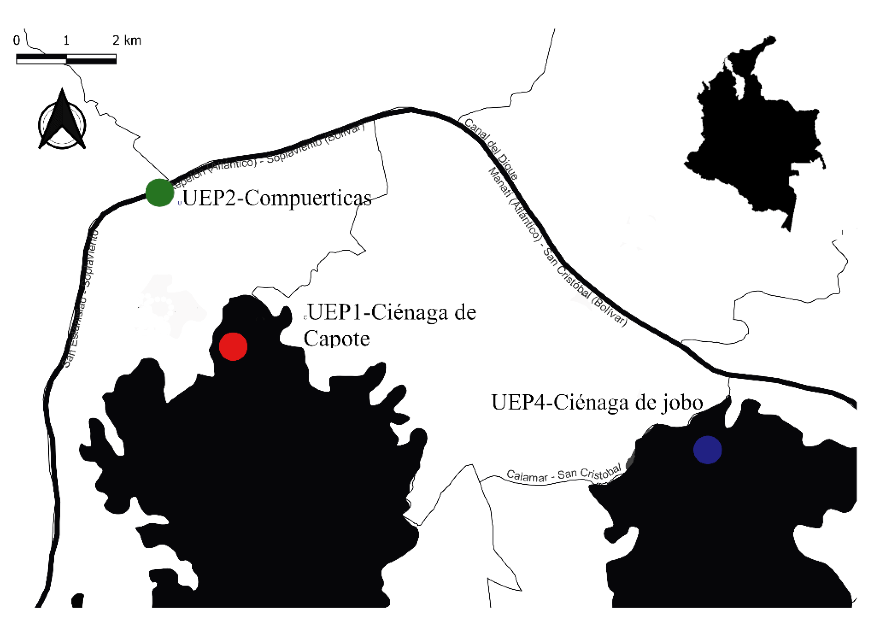
2.1. Study Area

The Canal del Dique, located in the Bolívar Department of Colombia, is an alluvial plain formed by a complex of wetlands composed of marshes that soften the Canal flow. The Canal is 113 km long from the town of Calamar to its mouth in Cartagena Bay [33].

2.2. Field Work

Field visits were carried out every other month for four days between December 2020 and October 2021, during which fresh C. kraussii specimens were collected by artisanal fishing with a cast net and gillnetting along the Canal del Dique and the marshes next to it.
The biological material was sacrificed and identified in situ using the field guide Colombian Andean Fish and the Catalogue of Continental Fishery Resources of Colombia [31,34]. Eighty C. kraussii specimens were analyzed from three locations being studied: UEP1—Ciénaga de Capote; UEP2—Canal del Dique/Compuerticas; and UEP4—Ciénaga del Jobo/San Cristóbal) (See Figure 1). There were 47 females and 33 males, the sex of which was determined by direct observation of gonads [35].

2.3. Geometric Morphometrics
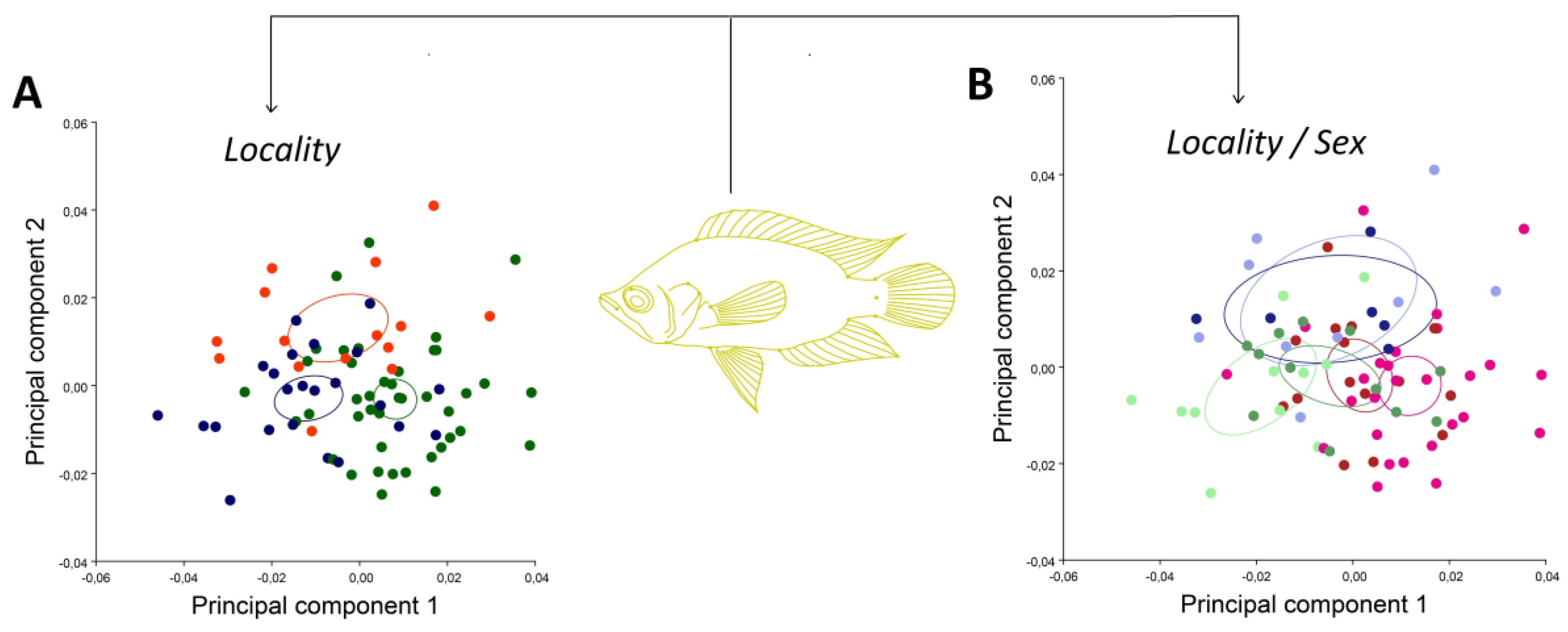
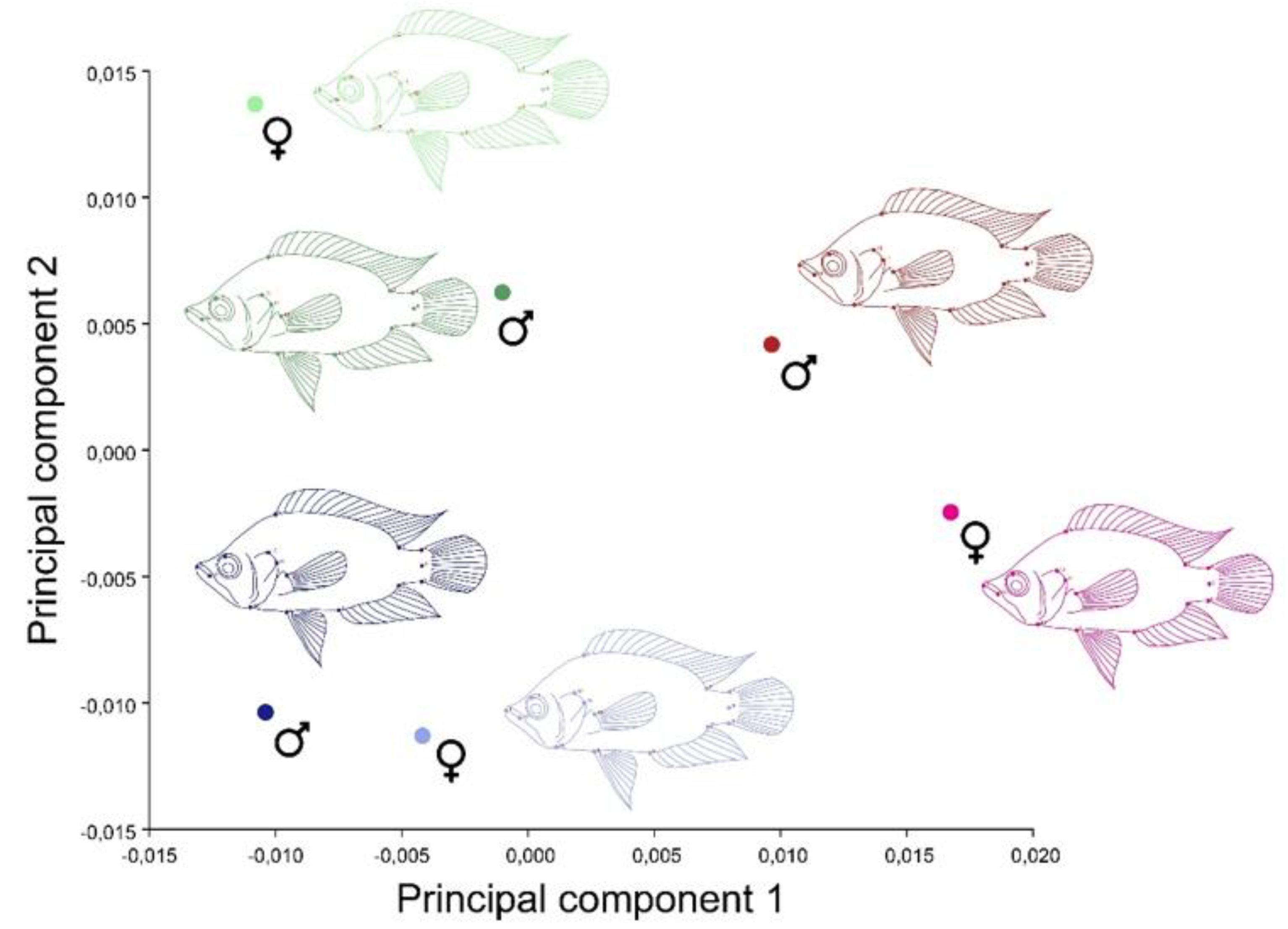
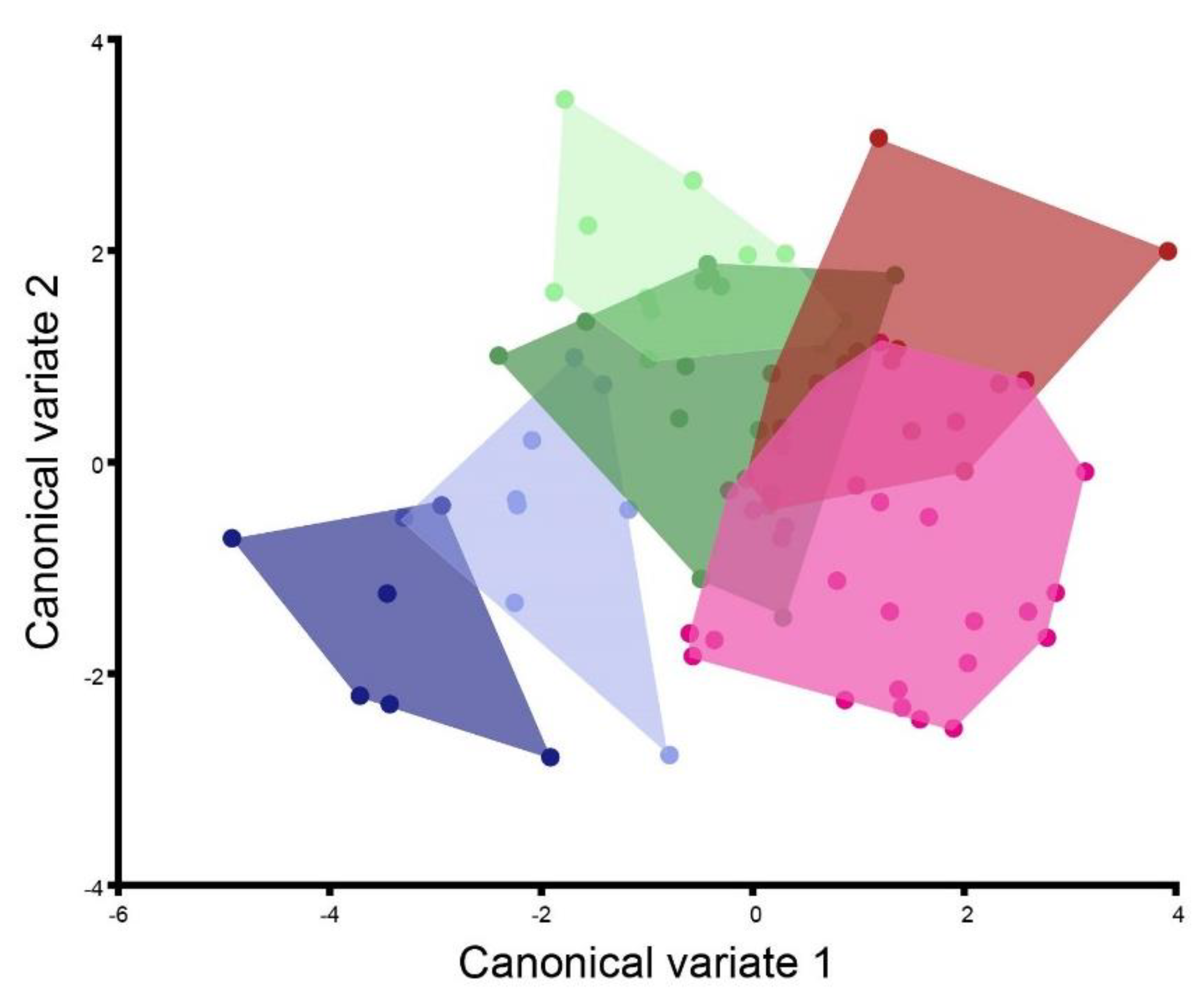
In order to obtain the images, individuals were fixed with pins and placed on a white Styrofoam base in an anterior–posterior position with fins extended. Additionally, a scale ruler was used to establish the scale when the picture was taken. The photographs were taken with a high-resolution FUJIFILM camera with 24 Megapixels. Landmarks were established following Corti and Crosetti [36], Soria et al. [37] and Aguirre and Jiménez-Prado [38] criteria, and they were digitized and converted into coordinates in a bidimensional plane using the tpsDig2 [39] software. In total, 15 landmarks were identified (Table 1), as shown in Figure 2.
By means of a Generalized Procrustes Analysis (GPA), the Cartesian coordinates derived from the landmark positions were processed. The GPA superimposes the resulting configurations of all the specimens analyzed, fitting them to a unitary centroid size and eliminating rotation and translation of configurations [14]. The GPA is based on the minimum squares to obtain the optimal superimposition; that is, it averages the lack of fit of all the points, allowing the detection of differences between different configurations. The GPA calculates the average configuration, which summarizes the configurations of all landmarks so that the specimen shape can be described and compared [14,23,40,41,42]. In the process of determining the landmarks, the measurement error was calculated by digitizing twice the reference points in a sample of individuals and by means of a Procrustes ANOVA; the values of mean squares of individuals were compared to see if they were lower than the error [43,44]. A principal component analysis (PCA) was conducted using the covariance matrix of individual shape [45]. Then, a scatterplot was generated, where the first two shape dimensions were shown graphically. After that, in order to observe the average shape changes for the classifiers (sex and population), a PCA from the covariance matrix of the average combined classifier (population × sex) was performed. Then, the body shapes were superimposed onto each other. Finally, with the purpose of highlighting changes associated with the sexual dimorphism of shape between populations, a canonical variate analysis (CVA) was carried out using a combined sex and location classifier. CVA uses the Procrustes coordinates and produces a set of new variables, canonical variates (CVs), which successively account for the maximum amount of among-group variance relative to within-group variance. Moreover, the CVs are uncorrelated within and among groups. It is important to note that CVA is a discriminant analysis that maximizes variation between groups, creating a new shape axis.
To determine the existence of statistically significant differences in SShD, a permutation test was performed (10,000 permutations) using Mahalanobis distances (morphological distances resulting from a CVA). All analyses were conducted with the MorphoJ 1.07a software [46].

3. Results

The Procrustes ANOVA conducted to assess the measurement error showed that the mean square value of individuals was higher than the measurement error (MSerror: 0.0000068573 < MSindividual 0.000077336) (Table 2).
The PCA by location showed that the first three PCs accounted for 50.03% (PC1: 23.9; PC2: 14.3; PC3: 11.7) of shape variation in C. kraussii (see Figure 3A). Similarly, the PCA using the sex–location classifier did not reveal large differences in the shape variance between sexes, but it did show a trend in which they become separated by location (Figure 3B).
After averaging data, the average PCA (Figure 4) showed a slight shape variation in the body of males between locations; however, it presented slight variations, with a descending displacement towards the ventral area formed by landmarks 10, 11 and 12, which corresponds to the insertion of the pectoral fin, posterior edge of the operculum and the dorsal edge of the preoperculum, respectively.
Likewise, changes associated with the upward movement towards the dorsal area of landmark 8 were observed, corresponding to the first anal spine (see Figure 5A). On the other hand, female shape showed more disparity between locations, where UEP1 and UEP4 populations had a more compact body, although there was a difference because UEP1 females had a shorter caudal peduncle than UEP4 females. On the other hand, UEP2 females exhibited a longer and hydrodynamic shape, with a larger size of the operculum, which is a characteristic of fish in lotic environments (see Figure 5B).
The canonical variate analysis confirmed the presence of the SShD of the C. kraussii body, with a clear difference among locations (see Figure 6). Furthermore, these differences were significant after the permutation test using Mahalanobis distances (Table 3). The only comparison resulting in no significant shape differences was between UEP2 females and UEP2 males.

4. Discussion

The results of this study show that, although there is slight sexual dimorphism of the geometric shape at the species level in C. kraussii, clear intralocation sexual shape dimorphism (SShD) of the body can be observed. C. kraussii, along the subregion Canal del Dique, inhabits a great variety of environments, from lentic water bodies (marsh) to lotic environments (Canal del Dique course) [47]. It is also observed that males have a more conserved body shape, with slight variations among the locations studied. Conversely, there is a significant variation in the body shape of females, which may be conditioned by their habitat. Females from both UEP1 and UEP4 have more robust and compact bodies, both being in marsh environments characterized by quiet waters with low current. The main differentiation is that UEP4 females have a longer tail, which may be efficient for moving in an environment where, although the current is low, it is deeper than the shallow marsh present in UEP1. On the other hand, the body of females inhabiting UEP2 is significantly more acute and longer, and at the same time, their caudal peduncle leans towards the dorsal zone of the caudal fin. The UEP2 habitat is above Canal del Dique; therefore, it is an environment with a strong flow, so the adaptations of females inhabiting UEP2 seem to have occurred in order to have a more hydrodynamic body shape adapted to move freely and with low effort in waters flowing at higher speeds. A similar pattern was reported by Perazzo et al. [48], who found that individuals in lotic environments have a more hydrodynamic shape, as well as a larger operculum.
In this sense, the fact that the body shape of C. kraussii males does not have major variations among locations, unlike females of the same species, seems to show the presence of different selective stresses acting in a dissimilar manner between individuals of different sexes within the same species in an interpopulational manner. As described by Hedrick and Temeles [49], the presence of a conserved shape in males among populations would be an indication that the selective stress on males is mainly exerted by females by means of the sexual selection of mates, where certain traits are actively selected over others, causing the male body shape to then be more reserved, with a conserved body scheme between populations. Kodric-Brown and Nicoletto [50] observed a similar pattern in poeciliids such as Guppy (Poecilia reticulada) and mosquitofish (Gambusia holbrooki). They studied the effect of two components in male mating, the color and exhibition behavior, in the selection by females, and they found that females select their mates based on both static and dynamic traits, showing a preference for the more noticeable males. On the other hand, Ramos Salazar [51] studied the factors that have an influence on females’ preference for males in Gambusia holbrooki individuals, and they found that females seem to prefer males with a larger size. Another study that supports our result that females may be exerting a degree of sexual selection on males is the one published by Selz et al. [52], who determined that, in cichlid species, there is sexual selection by females at intra- and interspecific levels, revealing that the nuptial coloring is a sexual selection objective by females [53].
On the other hand, body shape variance in females seems to be determined by selective environmental stresses due to both abiotic environmental features and the stress exerted by predators and intersexual competence. Bolnick and Doebeli [54] stated that species with strong selective mating based on ecological traits are less likely to develop sexual dimorphism, which is evidenced in the results of this study, considering that C. kraussii does not show many differences between males and females; however, when splitting them by population, dimorphism becomes apparent. In the same manner, it is possible that female shape variation changes due to ecological selection, which, according to Hedrick and Temeles [49] and Herler et al. [5], can have a differential impact on both sexes, favoring dimorphic niches. Their hypothesis suggests that environmental selection acts mainly on females [55], results that were shown by geometric morphometrics in C. kraussii females. Badyaev [56] and Lima Filho et al. [4] suggested that physical and ecological conditions of environments can give rise to the evolution of sexual dimorphisms [57,58]. A similar pattern was observed by Stillwell and Fox [59], showing that temperature (an environmental ecological factor) induces sexual shape dimorphism in the seed beetle (Callosobruchus maculatus). Regarding fish, Laporte et al. [60] studied the ecology of sexual shape dimorphism in Salaria fluviatilis, finding that selective environmental stresses affect sexual selection and, therefore, the visualization of sexual dimorphism within the species.
In addition, it is important to mention that there have been very few sexual shape dimorphism studies using geometric morphometrics. In fish, the sexual dimorphism of shape was studied in Tule perch, Hysterocarpus traskii, and significant differences in perch shape were found using GM [61]. Another study was conducted by Saura et al. [62], who, by means of GM, evaluated the fluctuating asymmetry and sexual dimorphism in Channa striata individuals; these groups are clear examples of studies where the combination of high-resolution morphological tools was able to unravel traits in the life history of organisms.
Finally, this study shows evidence for the effects of different selective stresses acting in an asymmetric manner between sexes in C. kraussii, as determined by environmental variables along a continuous environment, although heterogeneous. These results are a valuable information source for understanding the adaptative, selective and microevolutionary processes that occur simultaneously along the Canal del Dique basin. Similarly, since C. kraussii is a species with value and commercial relevance, this study provides useful information for the creation of management and exploitation plans for this resource, emphasizing considerations about the effect of the morphological variability of females subject to different environmental characteristics, and at the same time, since they have a conserved body shape, the translocation of males between different populations may serve as a strategy to increase the genetic heritage, which is useful information for creating conservation strategies.

Author Contributions

Conceptualization, J.H., A.V.-L., A.B., D.A.-C. and H.A.B.; data curation, J.H.; formal analysis, J.H. and H.A.B.; funding acquisition, A.B.; investigation, J.H., A.V.-L., D.A.-C., M.J.S. and H.A.B.; methodology, J.H. and H.A.B.; project administration, A.B.; resources, A.B.; software, J.H.; supervision, H.A.B.; validation, J.H. and H.A.B.; visualization, J.H., M.J.S. and H.A.B.; writing—original draft, J.H., A.V.-L., D.A.-C. and H.A.B.; writing—review and editing, J.H., A.V.-L., A.B., D.A.-C., M.J.S. and H.A.B. All authors have read and agreed to the published version of the manuscript.

Funding

The authors thank the Gobernación de Bolívar for supporting the projects of the General System of Royalties, specifically Special Cooperation Agreement 24 of 2018 in Science and Technology No. 24 signed by the Gobernación de Bolívar, Universidad de Cartagena and Autoridad Nacional de Acuicultura y Pesca—AUNAP, through the financing of the project “Development of a program for the sustainability of the aquaculture production chain of the Zodes Dique of the Bolívar Department”—BPIN 2017000100088, of which the results presented here are part.

Institutional Review Board Statement

Ethical review and approval were waived for this study, due to the species under study is not a protected species and is very abundant in the area, moreover, it is widely distributed and traded in sites of interest for this research.

Data Availability Statement

Data will be available in supplementary methods.

Acknowledgments

The authors thank María Raquel Lazo de la Vega for proofreading the manuscript, as well as Juan Sebastian Dueñas for helping with the figures. JH and AB thank the research vice-rector of the Universidad de Cartagena for the resources received within the framework of the results of the internship scholarship stipulated in resolution No. 02142 of 2021. Additionally, the authors would like to thank the Department of Zoology at the Universidad de Concepción for the administrative paperwork in Chile.

Conflicts of Interest

The authors declare no conflict of interest.

References

  1. Andersson, M. Sexual Selection; Princeton University Press: Princeton, NJ, USA, 1994. [Google Scholar]
  2. Fairbairn, D.J.; Preziosi, R.F. Sexual Selection and the Evolution of Sexual Size Dimorphism in the Water Strider. Aquar. Remigis. Evol. 1996, 50, 1549–1559. [Google Scholar] [CrossRef] [PubMed]
  3. Rodríguez, J.; Rojas, G.C.; Castro, K.R. Selección de pareja y comportamiento sexual de los guppys (Poecilia reticulata). Orinoquia 2005, 9, 38–44. [Google Scholar]
  4. Lima-Filho, P.A.; Bidau, C.J.; Alencar, C.E.R.D.; Molina, W.F. Latitudinal influence on the sexual dimorphism of the marine fish Bathygobius soporator (Gobiidae: Teleostei). Evol. Biol. 2017, 44, 374–385. [Google Scholar] [CrossRef]
  5. Herler, J.; Kerschbaumer, M.; Mitteroecker, P.; Postl, L.; Sturmbauer, C. Sexual dimorphism and population divergence in the Lake Tanganyika cichlid fish genus Tropheus. Front. Zool. 2010, 7, 1–10. [Google Scholar] [CrossRef] [PubMed] [Green Version]
  6. Fairbairn, D.J. Allometry for sexual size dimorphism: Pattern and process in the coevolution of body size in males and females. Annu. Rev. Ecol. Syst. 1997, 28, 659–687. [Google Scholar] [CrossRef]
  7. Horne, C.R.; Hirst, A.G.; Atkinson, D. Selection for increased male size predicts variation in sexual size dimorphism among fish species. Proc. R. Soc. B 2020, 287, 20192640. [Google Scholar] [CrossRef] [Green Version]
  8. Benítez, H.A.; Sukhodolskaya, R.A.; Órdenes-Clavería, R.; Avtaeva, T.A.; Kushalieva, S.A.; Saveliev, A.A. Measuring the Inter and Intraspecific Sexual Shape Dimorphism and Body Shape Variation in Generalist Ground Beetles in Russia. Insects 2020, 11, 361. [Google Scholar] [CrossRef]
  9. Benítez, H.A. Sexual dimorphism using geometric morphometric approach. In Sexual Dimorphism; Moriyama, H., Ed.; Intech: Rijeka, Croatia, 2013; pp. 35–50. [Google Scholar]
  10. Serrano-Meneses, M.A.; Reyes Hernández, M.; Carrillo Muñoz, A.; Rivas, M. La Conducta Reproductiva y La Evolución Del Dimorfismo Sexual En Tamaño; R.-AJ Martínez-Gómez, M., Lucio, R.A., Eds.; Biología del Comportamiento: Aportaciones desde la Fisiología: Tlaxcala, México, 2014; pp. 285–298. [Google Scholar]
  11. Uba, K.I.N. Sexual shape dimorphism in the monomorphic fish Decapterus macrosoma (Teleostei: Carangidae). Comput. Ecol. Softw. 2019, 9, 134. [Google Scholar]
  12. Benítez, H.A.; Sanzana, M.-J.; Jerez, V.; Parra, L.E.; Hernandez, C.E.; Canales-Aguirre, C.B. Sexual Shape and Size Dimorphism in Carabid Beetles of the Genus Ceroglossus: Is Geometric Body Size Similar Between Sexes Due to Sex Ratio? Zool. Sci. 2013, 30, 289–295. [Google Scholar] [CrossRef] [Green Version]
  13. Adams, D.C.; Rohlf, F.J.; Slice, D.E. Geometric morphometrics: Ten years of progress following the ‘revolution’. Ital. J. Zool. 2004, 71, 5–16. [Google Scholar] [CrossRef] [Green Version]
  14. Rohlf, F.J.; Slice, D. Extensions of the Procustes methods for the optimal superimposition of landmarks. Syst. Zool. 1990, 39, 40–59. [Google Scholar] [CrossRef] [Green Version]
  15. Slice, D.E. Geometric morphometrics. In Annual Review of Anthropology; Annual Reviews: Palo Alto, CA, USA, 2007; Volume 36, pp. 261–281. [Google Scholar]
  16. Adams, D.C.; Rohlf, F.J.; Slice, D.E. A field comes of age: Geometric morphometrics in the 21st century. Hystrix-Ital. J. Mammal. 2013, 24, 7–14. [Google Scholar] [CrossRef]
  17. Villalobos-Leiva, A.; Benítez, H.A. Morfometría Geométrica y sus Nuevas Aplicaciones en Ecología y Biología Evolutiva. Parte 2. Int. J. Morphol. 2020, 38, 1818–1836. [Google Scholar] [CrossRef]
  18. Espinosa-Santana, N. Selección sexual en machos de Poecilia reticulata (gupis) ante hembras emparentadas. An. Univ. De Etol. 2010, 4, 54–62. [Google Scholar]
  19. Berns, C.M. The evolution of sexual dimorphism: Understanding mechanisms of sexual shape differences. In Sexual Dimorphism; Moriyama, H., Ed.; Intech: Rijeka, Croatia, 2013; pp. 1–16. [Google Scholar]
  20. Benítez, H.A.; Avaria-Llautureo, J.; Canales-Aguirre, C.B.; Jerez, V.; Parra, L.E.; Hernandez, C.E. Evolution of sexual size dimorphism and its relationship with sex ratio in carabid beetles of Genus Ceroglossus Solier. Curr. Zool. 2013, 59, 769–777. [Google Scholar] [CrossRef]
  21. Parker, G. The evolution of sexual size dimorphism in fish. J. Fish Biol. 1992, 41, 1–20. [Google Scholar] [CrossRef]
  22. Oliveira, R.F.; Ros, A.F.; Gonçalves, D.M. Intra-sexual variation in male reproduction in teleost fish: A comparative approach. Horm. Behav. 2005, 48, 430–439. [Google Scholar] [CrossRef]
  23. González, R.; Bermúdez Tobón, A. Determinación de dimorfismo sexual usando técnicas morfométricas en Rachycentron canadum (Perciformes: Rachycentridae) cultivados en cautiverio. Bol. Investig. Mar. Costeras. 2021, 50, 71–90. [Google Scholar]
  24. Brzozowski, F.; Roscoe, J.; Parsons, K.; Albertson, C. Sexually Dimorphic Levels of Color Trait Integration and the Resolution of Sexual Conflict in L ake M alawi Cichlids. J. Exp. Zool. Part B Mol. Dev. Evol. 2012, 318, 268–278. [Google Scholar]
  25. López Álvarez, L.E.; Benavides Morales, Y.C. Biología reproductiva de la Cachegua Trachelyopterus insignis (Steindachner, 1878) en la Ciénaga de Ayapel, Colombia; Laboratorio de Investigación Biológico Pesquera-LIBP-FMVZ-Dpto. Ciencias Acuícolas: Bogota, Colombia, 2021. [Google Scholar]
  26. Pastana, M.; Dagosta, F.; Esguícero, A. A new sexually dichromatic miniature Hyphessobrycon (Teleostei: Characiformes: Characidae) from the Rio Formiga, upper Rio Juruena basin, Mato Grosso, Brazil, with a review of sexual dichromatism in Characiformes. J. Fish Biol. 2017, 91, 1301–1318. [Google Scholar] [CrossRef]
  27. Solano-Peña, D.; Segura-Guevara, F.; Olaya-Nieto, C. Crecimiento y reproducción de la mojarra amarilla (Caquetaia kraussii Steindachner, 1878) en el embalse de Urrá, Colombia. Rev. MVZ Córdoba 2013, 18, 3525–3533. [Google Scholar] [CrossRef] [Green Version]
  28. Olaya-Nieto, C.; Martínez-González, Á.; Díaz-Sánchez, D.; Pérez-Doria, W.; Segura-Guevara, F.; Tordecilla-Petro, G. Relación longitud-peso de la mojarra amarilla Caquetaia kraussii en la Ciénaga de Ayapel, Colombia. Neiva: V Congreso Colombiano de Acuicultura. Braz. J. Anim. Environ. Res. 2021, 4, 6469–6484. [Google Scholar] [CrossRef]
  29. Vanega, H.D.G. Algunos aspectos biológicos y pesqueros de Caquetaia kraussii (Steindachner, 1878) en la cuenca media y baja del río Atrato, Chocó. Rev. Biodivers. Neotrop. 2017, 7, 14–21. [Google Scholar] [CrossRef] [Green Version]
  30. Olaya-Nieto, C.W.; Ubarnes-Coronado, G.M.; Ensuncho-Morales, J.E. Crecimiento y mortalidad de mojarra amarilla Caquetaia kraussii en la ciénaga Grande de Lorica, Colombia. Rev. Logos Cienc. Tecnol. 2014, 5, 202–212. [Google Scholar] [CrossRef]
  31. Maldonado-Ocampo, J.A.; Ortega-Lara, A.; Usma, J.S.; Galvis, G.; Villa-Navarro, F.A.; Vásquez, L.; Prada-Pedreros, S.; Ardila, C. Peces de los Andes de Colombia. Instituto de Investigación de Recursos Biológicos “Alexander Von Humboldt”. Bogotá Colomb. 2005, 1, 346. [Google Scholar]
  32. Guzmán, M.Á.; Olaya, M.; Lasso, C.A.; Morales Betancourt, M.A.; Santos Calderón, J.M.; Uribe Botero, B.; Castaño Uribe, C.; Sanclemente, X.; Fajardo Rodríguez, Z.; Ponce de León Chaux, E. Catálogos de los Recursos Pesqueros Continentales de Colombia; Ministerio de Ambiente, Vivienda y Desarrollo Territorial-Instituto de Investigación de Recursos Biológicos Alexander von Humboldt: Bogotá, Colombia, 2011. [Google Scholar]
  33. Ahumada Lagares, G.A.; Penso Martínez, L.D. Caracterización socioeconómica de la subregión del Canal del Dique. Aguaita 2014, 26, 37–61. [Google Scholar]
  34. Lasso, C.; Agudelo-Córdoba, E.; Jiménez-Segura, L.; Ramírez-Gil, H.; Morales-Betancourt, M.; Ajiaco-Martínez, R.; Paula-Gutiérrez, F.; Usma-Oviedo, J.; Muñoz, S.; Sanabria, A.I. Catálogo de los recursos pesqueros continentales de Colombia. Serie Editorial Recursos Hidrobiológicos y Pesqueros Amazonian migratory catfish Brachyplatystoma rousseauxii in the Iquitos Región, Peru. J. Fish Biol. 2011, 75, 2527–2551. [Google Scholar]
  35. Agudelo, E.; Ajiaco, R.; Álvarez, L.; Barreto, C.; Bessudo, S.; Borda, C.; Bustamante, C.; Caldas, J.; De la Hoz, J.; Díazgranados, M. Protocolo De Captura De Información Pesquera, Biológica y Socio-Económica En Colombia; MADR, DPA: Bogotá, Colombia, 2011; 80p. [Google Scholar]
  36. Corti, M.; Crosetti, D. Geographic variation in the grey mullet: A geometric morphometric analysis using partial warp scores. J. Fish Biol. 1996, 48, 255–269. [Google Scholar] [CrossRef]
  37. Soria-Barreto, M.; Rodiles-Hernández, R.; González-Díaz, A.A. Morfometría de las especies de Vieja (Cichlidae) en ríos de la cuenca del Usumacinta, Chiapas, México. Rev. Mex. De Biodivers. 2011, 82, 569–579. [Google Scholar] [CrossRef] [Green Version]
  38. Aguirre, W.E.; Prado, P.J. Guía Práctica de Morfometría Geométrica: Aplicaciones En La Ictiología; Pontificia Universidad Católica del Ecuador Sede Esmeraldas (PUCESE): Esmeraldas, Ecuador, 2018; 104p. [Google Scholar]
  39. Rohlf, F.J. TPSdig, v. 2.17. Digitize Landmarks and Outlines; Department of Ecology and Evolution, State University at Stony Brook: New York, NY, USA, 2013. [Google Scholar]
  40. Klingenberg, C.P. Morphometrics and the role of the phenotype in studies of the evolution of developmental mechanisms. Gene 2002, 287, 3–10. [Google Scholar] [CrossRef]
  41. Toro-Ibacache, M.V.; Soto, G.M.; Galdames, I.S. Geometric Morphometry and the Biologic Shapes Study: From the Descriptive Morphology to the Quantitative Morphology. Int. J. Morphol. 2010, 28, 977–990. [Google Scholar]
  42. Benítez, H.A.; Püschel, T.A. Modelando la Varianza de la Forma: Morfometría Geométrica Aplicaciones en Biología Evolutiva. Int. J. Morphol. 2014, 32, 998–1008. [Google Scholar] [CrossRef] [Green Version]
  43. Arnqvist, G.; Martensson, T. Measurement error in geometric morphometrics: Empirical strategies to assess and reduce its impact on measures of shape. Acta Zool. Acad. Sci. Hung. 1998, 44, 73–96. [Google Scholar]
  44. Fruciano, C. Measurment error in geometric morphometrics. Dev. Genes Evol. 2016, 226, 139–158. [Google Scholar] [CrossRef] [PubMed]
  45. Jolliffe, I.T. Principal Component Analysis, 2nd ed.; Springer: New York, NY, USA, 2002. [Google Scholar]
  46. Klingenberg, C.P. MorphoJ: An integrated software package for geometric morphometrics. Mol. Ecol. Resour. 2011, 11, 353–357. [Google Scholar] [CrossRef] [PubMed]
  47. Ordóñez, J.; Cubillos, E.; Forero, G. Balance hídrico y sedimentológico del Canal del Dique y sus efectos sobre la sedimentación de la Bahía de Cartagena. In Proceedings of the Third Regional Symposium in Hydraulics on Rivers, Universidad Nacional de Colombia, Sede Bógota-Laboratorios de Ensayos Hidraulicos, Ottawa, ON, Canada, 8–10 November 2022; p. 18. [Google Scholar]
  48. Perazzo, G.X.; Corrêa, F.; Salzburger, W.; Gava, A. Morphological differences between an artificial lentic and adjacent lotic environments in a characid species. Rev. Fish Biol. Fish. 2019, 29, 935–949. [Google Scholar] [CrossRef]
  49. Hedrick, A.V.; Temeles, E.J. The evolution of sexual dimorphism in animals—hypotheses and tests. Trends Ecol. Evol. 1989, 4, 136–138. [Google Scholar] [CrossRef]
  50. Kodric-Brown, A.; Nicoletto, P.F. Female choice in the guppy (Poecilia reticulata): The interaction between male color and display. Behav. Ecol. Sociobiol. 2001, 50, 346–351. [Google Scholar] [CrossRef]
  51. Ramos Salazar, A. Selección sexual en Gambusia holbrooki (Agassiz, 1859) (Pisces: Poeciliidae). An. Univ. De Etol. 2007, 1, 90–95. [Google Scholar]
  52. Selz, O.; Thommen, R.; Pierotti, M.; Anaya-Rojas, J.M.; Seehausen, O. Differences in male coloration are predicted by divergent sexual selection between populations of a cichlid fish. Proc. R. Soc. B Biol. Sci. 2016, 283, 20160172. [Google Scholar] [CrossRef]
  53. Maan, M.E.; Sefc, K.M. Colour variation in cichlid fish: Developmental mechanisms, selective pressures and evolutionary consequences. Semin. Cell Dev. Biol. 2013, 24, 516–528. [Google Scholar] [CrossRef] [PubMed] [Green Version]
  54. Bolnick, D.I.; Doebeli, M. Sexual dimorphism and adaptive speciation: Two sides of the same ecological coin. Evolution 2003, 57, 2433–2449. [Google Scholar] [CrossRef] [PubMed]
  55. Cox, R.M.; Calsbeek, R. Sexually Antagonistic Selection, Sexual Dimorphism, and the Resolution of Intralocus Sexual Conflict. Am. Nat. 2009, 173, 176–187. [Google Scholar] [CrossRef] [PubMed] [Green Version]
  56. Badyaev, A.V. Origin of the fittest: Link between emergent variation and evolutionary change as a critical question in evolutionary biology. Proc. R. Soc. B Biol. Sci. 2011, 278, 1921–1929. [Google Scholar] [CrossRef] [PubMed] [Green Version]
  57. Fairbairn, D.J. Allometry for sexual size dimorphism: Testing two hypotheses for Rensch’s rule in the water strider Aquarius remigis. Am. Nat. 2005, 166, S69–S84. [Google Scholar] [CrossRef]
  58. Blanckenhorn, W.U.; Stillwell, R.C.; Young, K.A.; Fox, C.W.; Ashton, K.G. When Rensch meets Bergmann: Does sexual size dimorphism change systematically with latitude? Evolution 2006, 60, 2004–2011. [Google Scholar] [CrossRef]
  59. Stillwell, R.C.; Fox, C.W. Environmental effects on sexual size dimorphism of a seed-feeding beetle. Oecologia 2007, 153, 273–280. [Google Scholar] [CrossRef]
  60. Laporte, M.; Berrebi, P.; Claude, J.; Vinyoles, D.; Pou-Rovira, Q.; Raymond, J.-C.; Magnan, P. The ecology of sexual dimorphism in size and shape of the freshwater blenny Salaria fluviatilis. Curr. Zool. 2018, 64, 183–191. [Google Scholar] [CrossRef] [Green Version]
  61. Parvis, E.S.; Coleman, R.M. Sexual Dimorphism and Size-Related Changes in Body Shape in Tule Perch (Family: Embiotocidae), a Native California Live-Bearing Fish. Copeia 2020, 108, 12–18. [Google Scholar] [CrossRef]
  62. Saura, R.B.; Falcasantos, G.; Munda, L.; Alimorong, M.; Hernando, B.J. Evaluation of fluctuating asymmetry and sexual dimorphism of Channa striata using landmark-based geometric morphometric analysis from Agusan Marsh and Lake Mainit in Caraga Region, Philippines. Nusant. Biosci. 2021, 13, 100–110. [Google Scholar] [CrossRef]
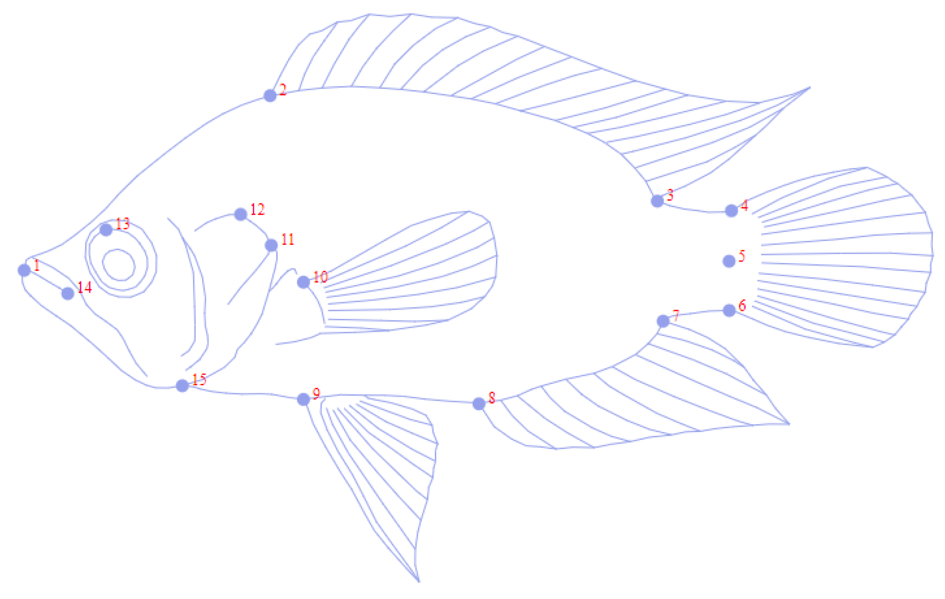
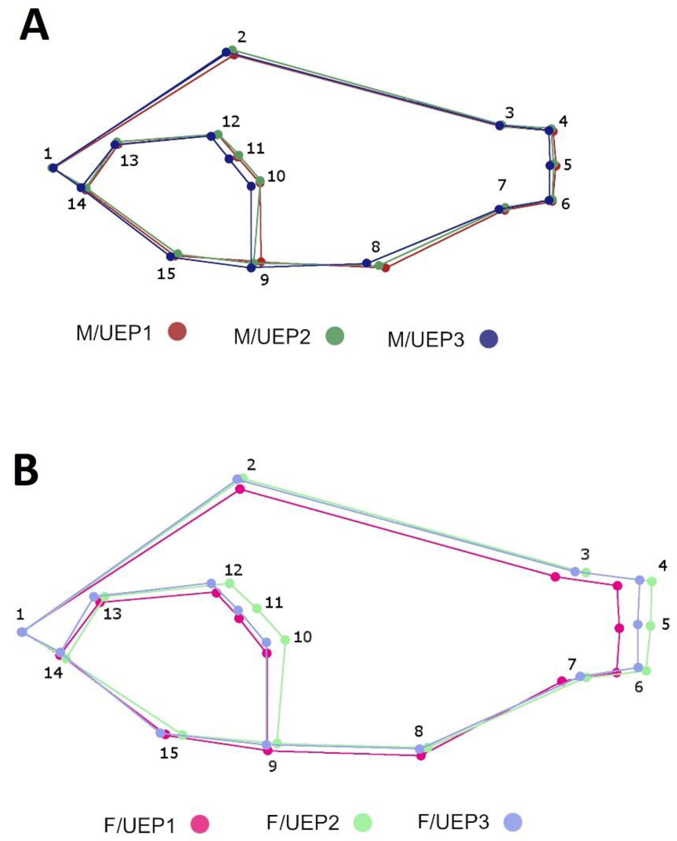
Figure 1. Location of Sampling Points. Zodes Dique (Canal del Dique Subregion: Red: UEP1, Green; UEP2; Blue: UEP4. Source: Author).
Fishes 07 00146 g001
Figure 2. Graphical representation of Caquetaia kraussi body shape with 15 landmarks.
Fishes 07 00146 g002
Figure 3. Principal component analysis of Caquetaia kraussi: (A): scatterplot of shape variation between three localities. Green: UEP1; orange: UEP4; and blue: UEP2. (B): Scatterplot of intralocality sexual shape dimorphism between three localities. Pink: female UEP1; red: male UEP1; light green: female UEP2; dark green: male UEP2; light blue: female UEP4; and dark blue: male UEP4. The confidence ellipses were computed from the mean shape using the criteria locality and locality/sex.
Fishes 07 00146 g003
Figure 4. Average principal component analysis of Caquetaia kraussi representing sexual shape dimorphism among localities. Pink: female UEP1; red: male UEP1; light green: female UEP2; dark green: male UEP2; light blue: female UEP4; and dark blue: male UEP4.
Fishes 07 00146 g004
Figure 5. Average shape superposition of Caquetaia kraussi represented by colored wireframe between sexes and localities. (A): Male Wireframe Overlay. red: male UEP1; dark green: male UEP2 and dark blue: male UEP4. (B): Female’s wireframe overlay. Pink: female UEP1; light green: female UEP2 and light blue: female UEP4.
Fishes 07 00146 g005
Figure 6. Canonical variate analysis of Caquetaia kraussi localities and sex in three populations in Colombia. Pink: female UEP1; red: male UEP1; light green: female UEP2; dark green: male UEP2; light blue: female UEP4; and dark blue: male UEP4.
Fishes 07 00146 g006
Table 1. Anatomical description of landmarks in C. kraussii.
N° LandmarksPosition
1Upper tip of the mouth
2First spine of the dorsal region
3Posterior insertion of dorsal fin
4Dorsal base of caudal fin
5Ventral insertion of caudal fin
6Ventral base of caudal fin
7Posterior insertion of anal fin
8First spine anal
9Anterior base of first pelvic fin ray
10Superior insertion of pectoral fin
11Posterior tip of operculum
12Dorsal border of preoperculum
13Middle of the eye
14Cleft of the upper lip
15Anterior margin of the cleithrum
Table 2. Procrustes ANOVA for digitizing measurement error of centroid size and shape of Caquetaia kraussii.
Centroid Size
EffectSSMSdfFP (param.)
Individual186.84775.0499383712.95<0.0001
Error 114.4301890.39000537
Shape, Procrustes ANOVA
EffectSSMSdfFP (param.)Pillai tr.P (param.)
Individual0.074397250.00007733696211.28<0.000119.65<0.0001
Error 10.006596690.000006857962
Table 3. Permutation test based on Mahalanobis distances and their corresponding p-values of the comparison between populations and sexes of C. kraussii.
H/UEP1H/UEP2H/UEP4M/UEP1M/UEP2
H/UEP23.1459
p-values:<0.0001
H/UEP43.35193.6005
p-values:<0.0001<0.0001
M/UEP12.23443.27293.9664
p-values:0.0001<0.0001<0.0001
M/UEP22.57262.0143.64652.7917
p-values:<0.00010.4495<0.00010.0002
M/UEP44.44695.03234.24345.14254.2772
p-values:<0.0001<0.00010.002<0.00010.0002
Publisher’s Note: MDPI stays neutral with regard to jurisdictional claims in published maps and institutional affiliations.

