Article

Hub Gene Identification and Heat-Stress-Related Transcriptional Regulation Mechanism in Cabbage (Brassica oleracea L.)

1
Faculty of Life Science, Tangshan Normal University, Tangshan 063000, China
2
Tangshan Key Laboratory of Cruciferous Vegetable Genetics and Breeding, Tangshan 063000, China
3
Faculty of Plantation and Agrotechnology, Universiti Teknologi MARA, Jasin Campus, Merlimau 77300, Melaka, Malaysia
*
Authors to whom correspondence should be addressed.
Horticulturae 2023, 9(9), 977; https://doi.org/10.3390/horticulturae9090977
Submission received: 24 July 2023 / Revised: 18 August 2023 / Accepted: 26 August 2023 / Published: 30 August 2023

Abstract

:
Cabbage is a heat-stress-sensitive cool-season crop. When exposed to high temperatures, cabbage plants can experience reduced growth, wilting, leaf yellowing, and premature bolting (the formation of a flowering stem). The regulatory mechanism controlling heat stress is poorly understood in cabbage. To investigate this mechanism, physiological changes and transcriptional profiling for different heat treatment times were analyzed in this study. The results showed that superoxide dismutase (SOD), catalase (CAT), and peroxidase (POD) activities were enhanced under heat stress. In particular, the increase in SOD and POD activities after 12 h of heat treatment was greater than that after 4 h of heat treatment. With increasing heat treatment duration, the leaf CAT activity and H2O2 content decreased after the initial increase. The electrolyte leakage and malondialdehyde (MDA) content dropped significantly, while the proline content increased. Alongside that, 7007 and 5537 upregulated genes were identified in the experimental group treated with heat stress for the 4 h and 12 h treatments, respectively. We found that 10,479 DEGs were shared in the heat stress treatment, of which 1241 were associated with the heat treatment time. By integrating the expression patterns and functional annotations of genes related to heat stress, we identified 15 hub genes that respond to heat stress in cabbage. Meanwhile, we had constructed a physiological to molecular model of cabbage response to long-term heat stress. These findings provide new insights for the comprehensive analysis of cabbage response to heat stress and genetic resources for breeding new varieties of cabbage with heat tolerance via genetic engineering.

1. Introduction

Global warming has been studied for many years as a scientific issue of great interest worldwide [1]. The United Nations Intergovernmental Panel on Climate Change (IPCC, http://www.ipcc.ch/), accessed on 1 March 2022, has released a new report stating that the warming of the climate system is expected to continue until mid-century. In the next 20 years, global temperature increases will reach or exceed 1.5 °C compared to the period of the industrial revolution. As global warming intensifies, our average temperatures will continue to rise in the future, and most plants will suffer the effects of high temperatures. Temperature is an important environmental factor affecting the growth, development, and function of plants [2]. With global warming, the frequency of periodic high-temperature weather is causing increasingly severe damage to crops [3,4]. The physiological damage caused to plants by high-temperature (HT) environments is known as heat stress [5]. Heat stress can hinder plant development, slow down growth rates, and even lead to plant death. It can also impair photosynthesis, affecting plant carbon metabolism and energy production [6]. The areas of damage exhibited by plants vary depending on the growth period during the heat stress exposure: heat stress at the seedling stage mainly affects plant height, the root system, and crown size [7]; and at flowering, it affects pollen activity, stigma surface activity, pollen germination, and pollen tube growth; Additionally, at fruiting, it not only leads to poor fruit set but also affects the overall productivity of the plants [8,9]. Heat stress similarly affects the physiological responses of plants. The biological membrane system is the center of heat damage [10]. The structure and function of the cellular protoplasmic membrane are first to be affected, leading to an increase in cell membrane permeability [11]. When plants are stressed by adversity, the balance of free radicals and reactive oxygen species in the body is also disrupted, leading to varying degrees of reactive oxygen species accumulation in the plant, which ultimately causes oxidative damage to the plant cells, thus activating the plant’s antioxidant defense system to adapt to or resist the damage caused by adversity [12]. In addition, heat stress affects otherwise normal physiological processes such as malondialdehyde (MDA), the accumulation of reactive oxygen species such as superoxide radicals (O2−), and hydroxyl radicals (-OH), causing disturbances in an otherwise balanced system as well as oxidative damage to lipids, nucleic acids, proteins, and cell membranes [13,14,15].
Recent studies have shown that the main transcription factors responding to heat stress include HSF, DREB, and MBF1, which have been confirmed in the transcript expression profiles of a variety of heat stress-affected plants. For example, analysis of the transcriptome of Lolium perenne in response to heat stress showed that HSF, AP2/EREBP, MYB, and bHLH transcription factor family genes were significantly up- or downregulated [16]. Analysis of transcriptome data from Capsella bursa-pastoris under heat and drought stress revealed that the main upregulated transcription factors were HSFs and DREBs (a subfamily of the AP2 transcription factor family) [17]. Studies have shown that candidate genes involved in plant hormone metabolism or signaling, including abscisic acid (ABA), growth hormone (IAA), ethylene (ET), and gibberellin (GA), are all regulated by heat stress. Many transcripts in response to salt and heat stress in Arabidopsis thaliana were found to be associated with the phytohormone abscisic acid [18]. One study found that oleuropein steroids (BRs) were also associated with heat stress, with BRI1–EMS–SUPPRESSOR 1 (bes1)-mediated defects in BR signaling exhibiting the most sensitive characteristics to heat stress. This study also highlighted the complex interactions between BR and ABA during heat acclimation [19]. Transcriptome analysis of Lolium temulentum under drought and heat stress revealed that among the hormones, growth hormone-related DEGs were most prevalent. DEGs associated with gibberellins, cytokinins, and ABA were also prevalent, with fewer DEGs associated with jasmonates and oleuropein steroids [20].
The impact of heat stress on cabbage (including heading cabbage and kale) is primarily manifested as restricted growth and development, leaf oxidation damage, hindered nutrient absorption and utilization, inhibited flower bud formation, and suppressed photosynthesis [21,22,23]. These effects can ultimately lead to reduced cabbage yield and quality. In recent years, there has been increasing attention paid to research investigating the molecular mechanisms underlying the heat stress response in cabbage. The integration of morphological characteristics, physiological changes, and gene expression patterns reveals the molecular regulation of heat stress response in cabbage involving the BoHSP70, BoSCL13, and BoDPB3-1 genes [24]. Studies have also found that the BolSGT1 gene positively regulates heat tolerance in cabbage. Furthermore, it plays a positive regulatory role in response to cold stress, salt stress, and drought stress [25]. The BoHsp70 and SCL13 genes are highly expressed in heat-tolerant cabbage, and these genes are also considered marker genes for screening heat-tolerant cabbage breeding [26]. Revealing the functions and regulatory mechanisms of heat tolerance genes in cabbage can help breed heat-tolerant varieties, improve yield, and enhance quality stability. Additionally, this research contributes to our understanding of plant response mechanisms to environmental stresses such as climate change, providing guidance for crop adaptation and sustainable agriculture.
The Brassicaceae family consists of over 372 genera and 4060 species, of which cabbage is the bulkier and more agronomically important species. Originating from the Mediterranean coast and the seashores of Northwestern Europe, cabbage has a rich variety and is considered a potential ancestor of many European vegetables. In this study, physiological and biochemical indicators of cabbage under high-temperature stress were measured in order to expect a comparison of the effects of different high and low temperatures on cabbage from a physiological perspective. In addition, this study focused on the molecular mechanisms of cabbage under heat stress using high-throughput sequencing technology in order to identify the pathways and genes that may be associated with heat stress more accurately, forming the basis for breeding new varieties of cabbage that are tolerant to high temperatures.

2. Materials and Methods

2.1. Plant Material and Treatment Methods

The leaves of seedlings were collected from the cabbage cultivar ‘Bomei 4′ for heat treatments. The experimental materials were cultivated under consistent growth conditions prior to treatment. The growth conditions were as follows: a temperature of 25 ℃, a light cycle of 8/16 h (light/dark), and a humidity of 70%. Cabbages cultivated until 3 weeks old were used for high-temperature treatment experiments. The high-temperature treatment conditions were set at 4 h at 35 °C (H4) and 12 h at 35 °C (H12), while the remaining conditions remained unchanged. Plant materials that did not undergo any treatment were used as the control group (CK) for the experiment. The experiment was divided into three groups, with each group consisting of ten cabbage plants with consistent growth. Each experimental group was replicated three times.

2.2. Determination of the Physiological and Biochemical Parameters of the Cabbage Leaves

The SOD assay employed the more stable and sensitive WST-8 method, which could react with the superoxide anion (O2−) produced by catalyzing xanthine oxidase (XO) to generate a water-soluble formazan dye [27]. POD and CAT activities were measured using the guaiacol and UV spectrophotometric methods, respectively [28]. The detection of H2O2 content utilized the reaction of H2O2 with titanium salt to generate a peroxide–titanium complex purple precipitate, which can be dissolved in concentrated sulfuric acid and then detected using a UV spectrophotometer at a maximum absorption peak at a 415 nm wavelength. The contents of malondialdehyde (MDA) and free proline were determined using the thiobarbiturate and colorimetric measurement methods, respectively [29,30].

2.3. Total RNA Extraction and Transcriptome Sequencing

Total RNA from young cabbage leaves was extracted using the RN38-EASYspin Plus Plant RNA Extraction Kit (Aidlab Biotechnologies, Beijing, China). The purity and concentration of the total RNA were determined using the NanoDrop 2000 spectrophotometer (Thermo Fisher Scientific, Cleveland, OH, USA), while the integrity of the total RNA was assessed using the Agilent 2100 bioanalyzer (Agilent Technologies, Santa Clara, CA, USA). High-quality total RNA that passed quality control was used to construct cDNA libraries. The library construction mainly involved magnetic bead enrichment of mRNA, random fragmentation of mRNA, synthesis and purification of cDNA, end-repair/dA-tailing of cDNA followed by adapter ligation, and polymerase chain reaction (PCR) enrichment [31]. The concentration of the library was measured using a Qubit 3.0 fluorometer (Thermo Fisher Scientific, USA), and the concentration needed to be above 1 ng/μL. Subsequently, the insert fragments of the library were analyzed using the Qsep400 high-throughput analysis system. Once the insert fragments met the expected criteria, the effective concentration of the library was quantified using the quantitative real-time polymerase chain reaction (Q-PCR) method. The qualified libraries were sequenced using the Illumina NovaSeq6000 sequencing platform.

2.4. Gene Expression Analysis

We processed raw data from the Illumina NovaSeq6000 sequencing platform by removing reads containing adapters, reads with an N ratio greater than 10%, and reads where more than 50% of the bases had a quality value (Q) less than or equal to 10. As a result, we obtained high-quality, clean reads. The clean reads were aligned to the cabbage reference genome (Brassica_oleracea.Braol_JZS_V2.0.genome) using HISAT2 software [32], a fast and accurate alignment method, to obtain the positional information of the reads on the reference genome. The StringTie v1.3.1 tool was used for transcriptome reconstruction to normalize sequencing depth and transcript length, resulting in the calculation of gene expression values as FPKM (fragments per kilobase per million) normalization [33]. The biological replicability of the samples was assessed using the Pearson correlation coefficient, while the sample variability was evaluated using principal component analysis (PCA).

2.5. Differential Expression Gene Identification and Functional Annotation

Based on FPKM values, DEGs were screened using DESeq2 software [34], with the screening criteria of false discovery rate (FDR) < 0.05 and absolute fold-change (FC) > 1.5. Additionally, a hypothesis testing probability (p-value) correction was performed, and genes with a corrected p-value < 0.05 were considered significant DEGs. The heatmap of the gene expression levels was visualized using the pheatmap package in the R programming language (https://cran.r-project.org/), accessed on 1 March 2022. The functions of DEGs were annotated based on the Kyoto Encyclopedia of Genes and Genomes (KEGG) and Gene Ontology (GO) databases [35,36].

2.6. qRT-PCR Validation of DEGs

Total RNA from cabbage leaves was extracted using the RNAprep Pure Plant Kit (TIANGEN, Beijing, China). The cDNA library was constructed using the FastKing RT kit (with gDNA) (TIANGEN, Beijing, China). The extraction of total RNA and construction of cDNA libraries were performed according to the instructions provided with the kit. Primer sequences for gene-specific qRT-PCR were designed using Premier 5 software (Table S1). We used a 25 μL reaction system of the SuperEeal PreMix Plus (SYBR Green) kit (TIANGEN, Beijing, China) to detect the relative expression levels of candidate genes. The actin gene in cabbage was used as an internal reference gene. The relative expression levels of candidate genes in response to heat stress in cabbage were calculated using the 2−ΔΔCt method [37].

3. Results

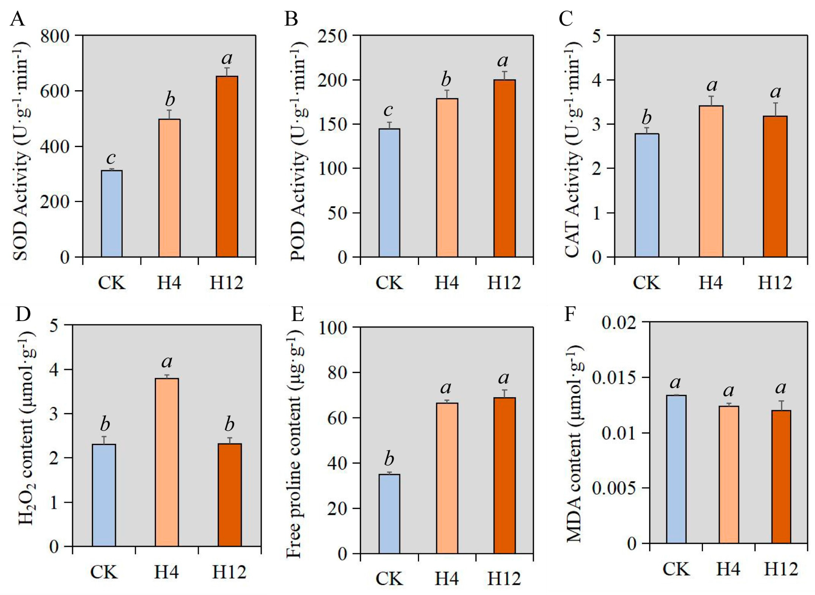
3.1. Studies on the Physiology of Cabbage under Heat Stress

Measuring plant physiological indicators under heat stress was significant because it helped assess the effects of heat stress on plant growth and productivity. Here, we measured the CAT, POD, and SOD activities, H2O2, free proline, and MDA content of cabbage under heat stress (Figure 1). The SOD, POD, and CAT activities were enhanced under heat stress. In particular, the increase in SOD and POD activities after 12 h of heat treatment was greater than that after 4 h of heat treatment. With increasing heat treatment duration, the leaf CAT activity and H2O2 content decreased after an initial increase. The content of free proline increased continuously with temperature treatment time, while the content of malondialdehyde showed a decreasing trend. Cabbage activated its antioxidant defense mechanisms to scavenge reactive oxygen species (ROS) generated due to heat stress and prevent oxidative damage to cellular structures. Moreover, it accumulated compatible solutes which acted as osmoprotectants and protected plant cells from damage caused by high temperatures. These physiological responses enabled plants to tolerate short-term heat stress conditions and avoid irreversible damage. However, prolonged exposure to high temperatures could cause permanent damage to plant tissues, leading to reduced growth, productivity, and even death.

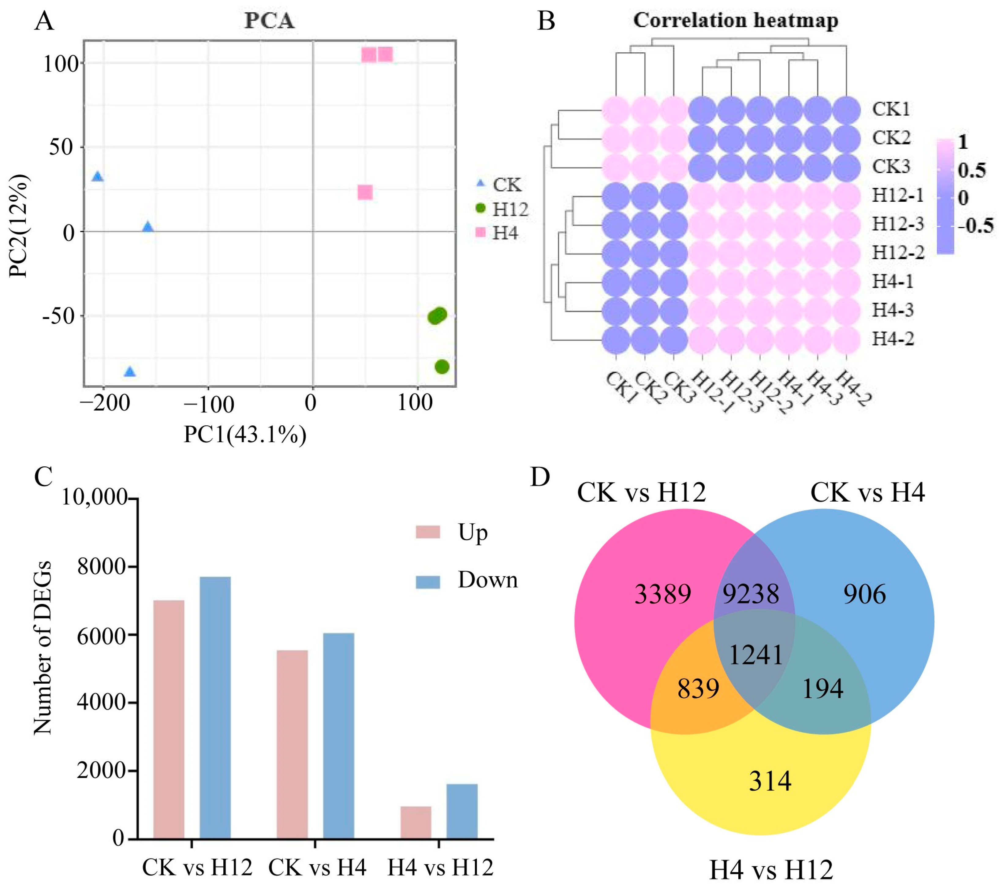
3.2. Transcriptome Sequencing Data Quality Control and Comparative Analysis

The Illumina NovaSeq6000 sequencing platform was used to sequence a large amount of raw data. After a series of quality controls, a total of 56.68 Gb of clean data was obtained from the nine transcriptomic libraries that were sequenced, and the filtered data (clean data) were counted to obtain information on the quality of the sequencing data, with the average number of clean reads being 21,047,089 and the GC content being relatively consistent, at around 47%. According to the assessment of the base quality, it was shown that the Q20 for each sample exceeded 97% and the Q30 exceeded 94.79% (Table S2). Therefore, the data results indicated that the transcriptome data was of high quality and could meet the requirements for subsequent analysis. The alignment efficiency of the reads from each sample to the cabbage reference genome ranged from 92.32% to 94.43%. Among them, an average of 88.99% of the data aligned only to a unique position in the cabbage reference genome, while 4.54% of the reads aligned to multiple locations in the cabbage reference genome (Table S3). Furthermore, relating to the data compared to the coding regions, an average of 82.08%, 3.65%, and 14.27% of the nine samples were compared to exons, introns, and spacer regions, respectively (Table S4). In summary, the majority of valid data obtained were for the gene transcripts of cabbage, and the data were predictive of gene expression levels in the cabbage genome.

3.3. Analysis of Differential Gene Expression in Cabbage under Heat Stress

In order to detect DEGs between different high-temperature treatments more accurately, we analyzed sample correlation analyses based on gene expression levels. We found that the PC1 dimension accounted for 9% and the PC2 dimension accounted for 89%. Further, the repeated samples were clustered based on the Euclidean distance method (Figure 2A). The Pearson correlation coefficient analysis also obtained similar results as the PCA analysis (Figure 2B). A total of 14,707 DEGs were identified in the experimental group treated with heat stress for 4 h, including 7007 upregulated genes and 7700 downregulated genes. Of the upregulated genes, 5537 and 6042 downregulated genes were identified in the experimental group treated with heat stress for 12 h, respectively. Taking heat stress treatment for 4 h as the control, 967 upregulated genes and 1621 downregulated genes were identified in the experimental group treated with heat stress for 12 h (Figure 2C). These results indicated that there was a close relationship between gene expression and treatment time in cabbage under heat stress. We found that 10,479 genes were shared in the heat stress treatment, and 1241 shared genes were associated with the heat treatment time (Figure 2D). By studying these DEGs, researchers can better understand how plants cope with heat stress at the molecular level and potentially develop new strategies to improve plant tolerance to high temperatures.

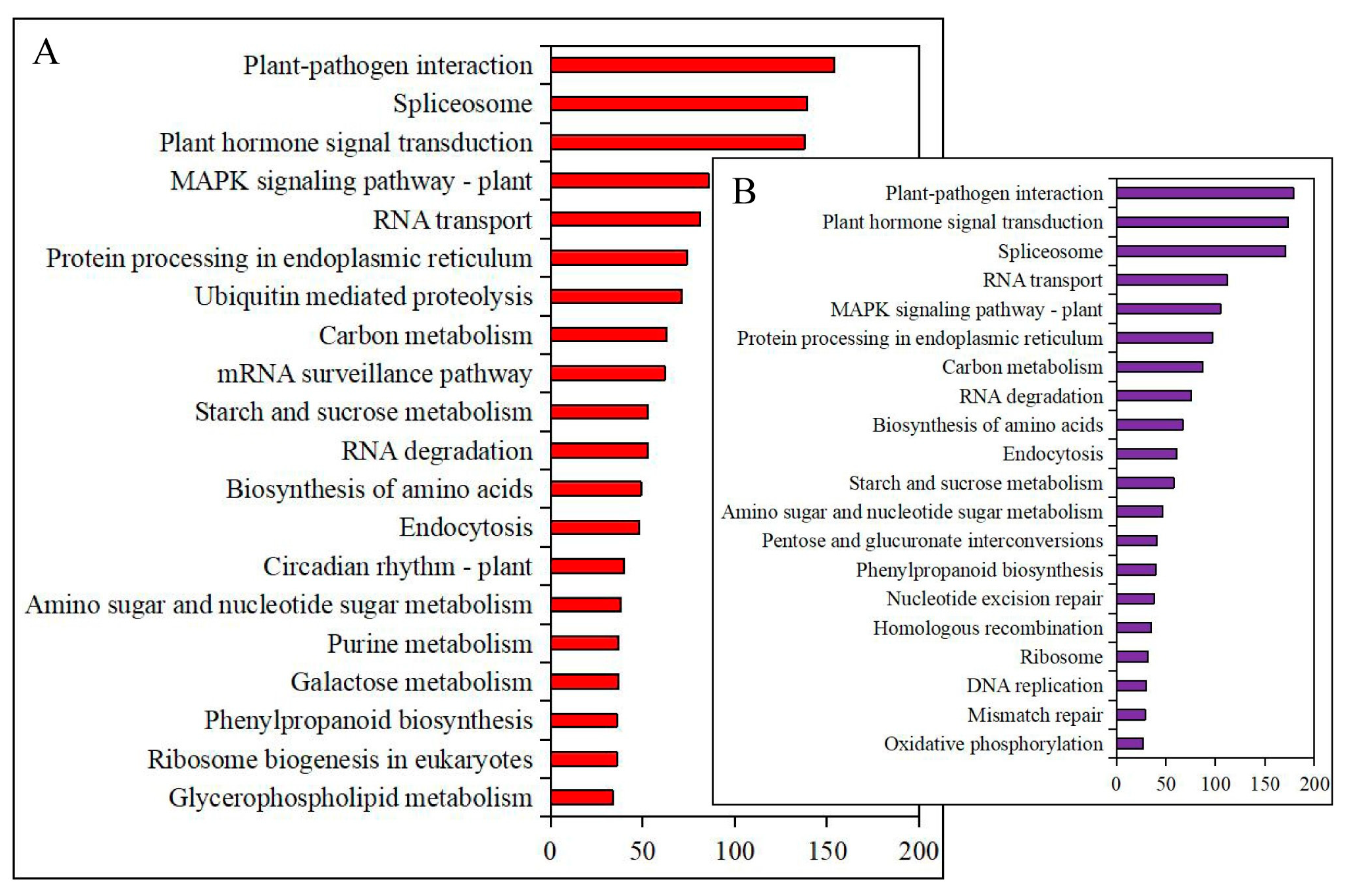
3.4. Annotation and Enrichment of Differential Gene Expression under Heat Stress

To gain further insights into gene function, DEGs were annotated based on multiple gene function annotation and enrichment databases: 14,331, 11,749, and 10,395 DEGs were annotated to the NR, GO, and KEGG databases in the CK vs. H12 group, respectively. At the same time, we retrieved information indicating that 11,294, 9205, and 8179 DEGs were annotated to the NR, GO, and KEGG databases in the CK vs. H4 group, respectively (Table S5).
In order to analyze the DEGs in cabbage leaves under different heat stresses, the detected differential genes were subjected to GO annotation to investigate their macroscopic distribution in terms of function. In CK vs. H4 and CK vs. H12, 37.77% and 67.41% of DEGs were annotated in the biological process term, 38.70% and 20.53% of DEGs were annotated in the cellular component term, and 23.54% and 12.06% of DEGs were annotated in the molecular function term, respectively. More genes were annotated in the biological process after 12 d of heat stress (Figure S1). To understand the GO entries that were significantly enriched compared to the whole genomic context, the differential gene sets for each subgroup were analyzed separately for biological processes, molecular functions, and cellular components through a hypergeometric test using the Cluster Profiler. The differential genes of CK vs. H4 and CK vs. H12 regarding the biological process were significantly enriched in ten types of translation: photosynthesis, response to water deprivation, photo protection, response to salt stress, one-carbon metabolic process, cell redox homeostasis, photosynthesis, light harvesting, carotenoid biosynthetic process, and auto phagosome organization. Among them, the differential genes in CK vs. H12 were more significantly enriched in photosynthesis, i.e., light harvesting. Moreover, the differential genes in CK vs. H12 were also enriched in protein folding, protein–chromophore linkage, photosystem II assembly, and regulation of the jasmonic acid-mediated signaling pathway, which were not involved in CK vs. H4 (Figure S2). For the differential genes in the cellular component, the differential genes in CK vs. H12 were enriched in photosystem I, chloroplast thylakoid membrane, and extrinsic components of the membrane types, which were more significant than in CK vs. H4 (Figure S3). In terms of molecular function, these genes were mainly enriched in structural constituents of ribosomes, metal ion binding, and NAD binding, among which the differential genes of CK vs. H12 were also significantly enriched in chlorophyll binding (Figure S4). In cabbage, all DEGs in the three comparison groups, CK vs. H4, CK vs. H12, and H4 vs. H12, were annotated to 134, 135, and 117 KEGG pathways, respectively. More DEGs in both CK vs. H4 and CK vs. H12 were clearly located within the plant–pathogen interaction, ribosome, plant hormone signal transduction, carbon metabolism, and the biosynthesis of amino acid pathways (Figure 3). More differential genes in H4 vs. H12 than in the other two groups were also distributed within the photosynthesis pathway.

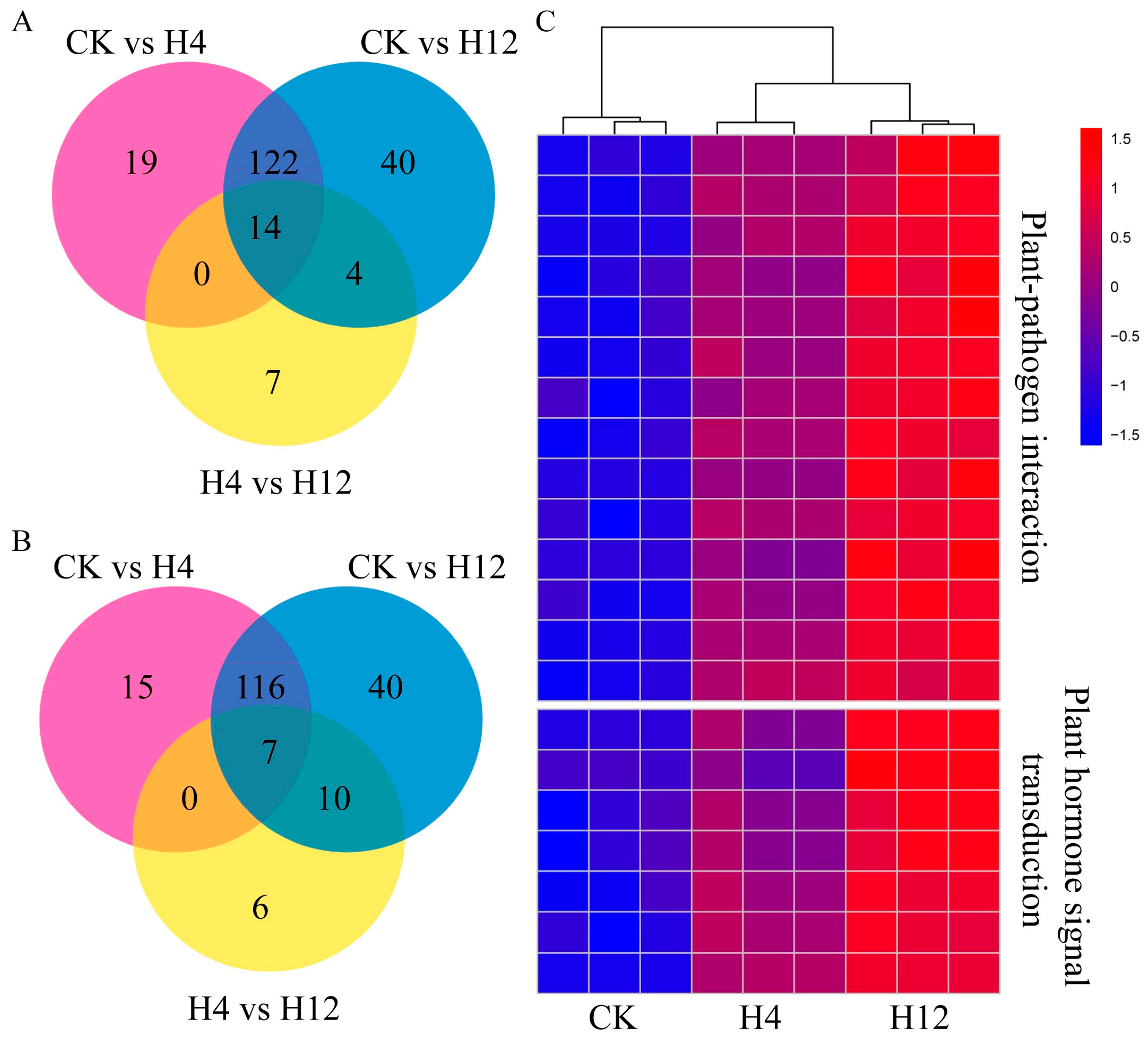
3.5. Analysis of Significantly Associated Genes in Pathways

A total of 34 DEGs were obtained in the metabolic pathways of plant–pathogen interactions after screening each group for log2FC ≤ −1 or ≥1 and at least one data point with FPKM not less than 20 (Table S6). A total of seven upregulated and twenty-five downregulated genes were available in CK vs. H4. A total of seven upregulated and twenty-seven downregulated genes were obtained in CK vs. H12. A total of six upregulated and sixteen downregulated genes were obtained in H4 vs. H12 (Table S6). We identified 14 shared upregulated genes in these three comparison groups, including two bHLH transcription factor genes, the calcium-dependent protein kinase gene (CPK), the pathogenesis-related gene transcriptional activator PTI6 gene (PTI6), the disease resistance protein RPS2 gene (RPS2), and the cathepsin F gene (CTSF). The UPA20 (BolC06g005890.2J) gene was the most abundantly expressed for the co-upregulated genes following heat stress (Figure 4B). The mitogen-activated protein kinase 1 gene (MAP2K1, MEK1) was only upregulated in CK vs. H4 and CK vs. H12 and held at normal levels in H4 vs. H12, suggesting that the MAP2K1, MEK1 gene may be a heat-sensing gene; however, it was not significantly expressed in the presence of sustained high temperatures (Table S7).
Here, we identified a total of 107 DEGs involved in plant hormone signaling pathways, with the majority of genes being shared during the response to heat stress treatment. Among them, 72 upregulated and 27 downregulated genes were responsive to 4 d of heat stress treatment, while 71 upregulated and 29 downregulated genes were responsive to 12 d of heat stress treatment. Of these, seven genes were common across all three groups: the DELLA protein gene (DELLA), the protein phosphatase 2C gene (PP2C, BolC01g051360.2J; PP2C-L, BolC01g051280.2J), the ABA responsive element binding factor gene (ABF), gibberellin receptor GID1 gene (GID1), the two-component response regulator ARR-B family gene (ARR-B), and the protein brassinosteroid insensitive 1 gene (BRI1), which were significantly upregulated (Figure 4C). The DELLA and ABF genes were expressed in greater numbers than other genes following heat stress (Figure 4B). The DELLA and GID1 proteins are both phytoalexin delivery signaling molecules, and ABF and PP2C were associated with phytoalexin signaling. The BRI1 enzyme is a leucine-rich repeat-like serine/threonine kinase located on the cell surface and is the receptor for phytoalexin end lipids. The genes gibberellin receptor (GID1, BolC06g035530.2J), two-component response regulator ARR-A family (ARR-A), and SAUR family protein (SAUR) were only upregulated in CK vs. H4, while the genes abscisic acid receptor PYR/PYL family (PYL) and protein brassinosteroid insensitive 1 (BRI1) were only upregulated in CK vs. H12 (Table S7).

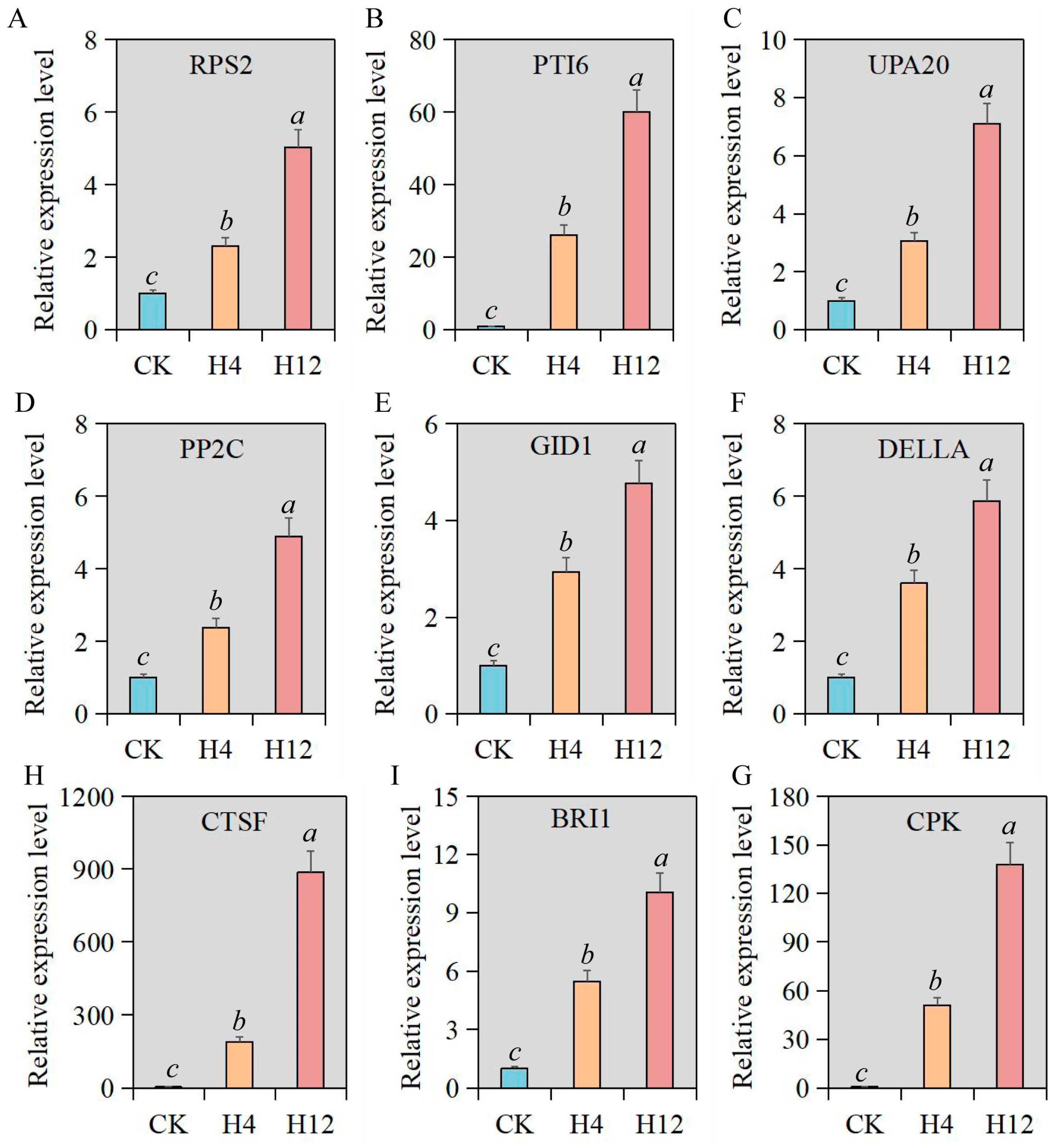
3.6. Verification of Relative Expression Levels of Key Genes under Heat Stress

By integrating the expression patterns and functional annotations of genes related to heat stress, we identified 15 hub genes that respond to heat stress in cabbage. The expression of 15 genes was significantly upregulated in response to heat stress. Meanwhile, the expression trend continuously increased with the duration of heat stress (Figure 5 and Figure S5). We found significant differences among the multiple differences in the expression levels of these genes during the response to heat stress treatment. Overall, the differentially expressed multiples of genes involved in plant hormone signal transduction pathways, such as ABF, PP2C, and BRI1 genes, were relatively small. The differential expression multiple of the ATPF1A gene was the most prominent, in which the differential expression multiples of 4 h and 12 h of heat treatment were nearly 600 and 1800 times, respectively. Among these genes, three groups of paralogous genes showed similar expression trends and differential expression multiples. This meant that these paralogous genes participated in the same biological process and performed similar functions. We found that cabbage response to heat stress was regulated by multiple genes, among which these 15 genes played a critical role in helping cabbage survive high temperatures.

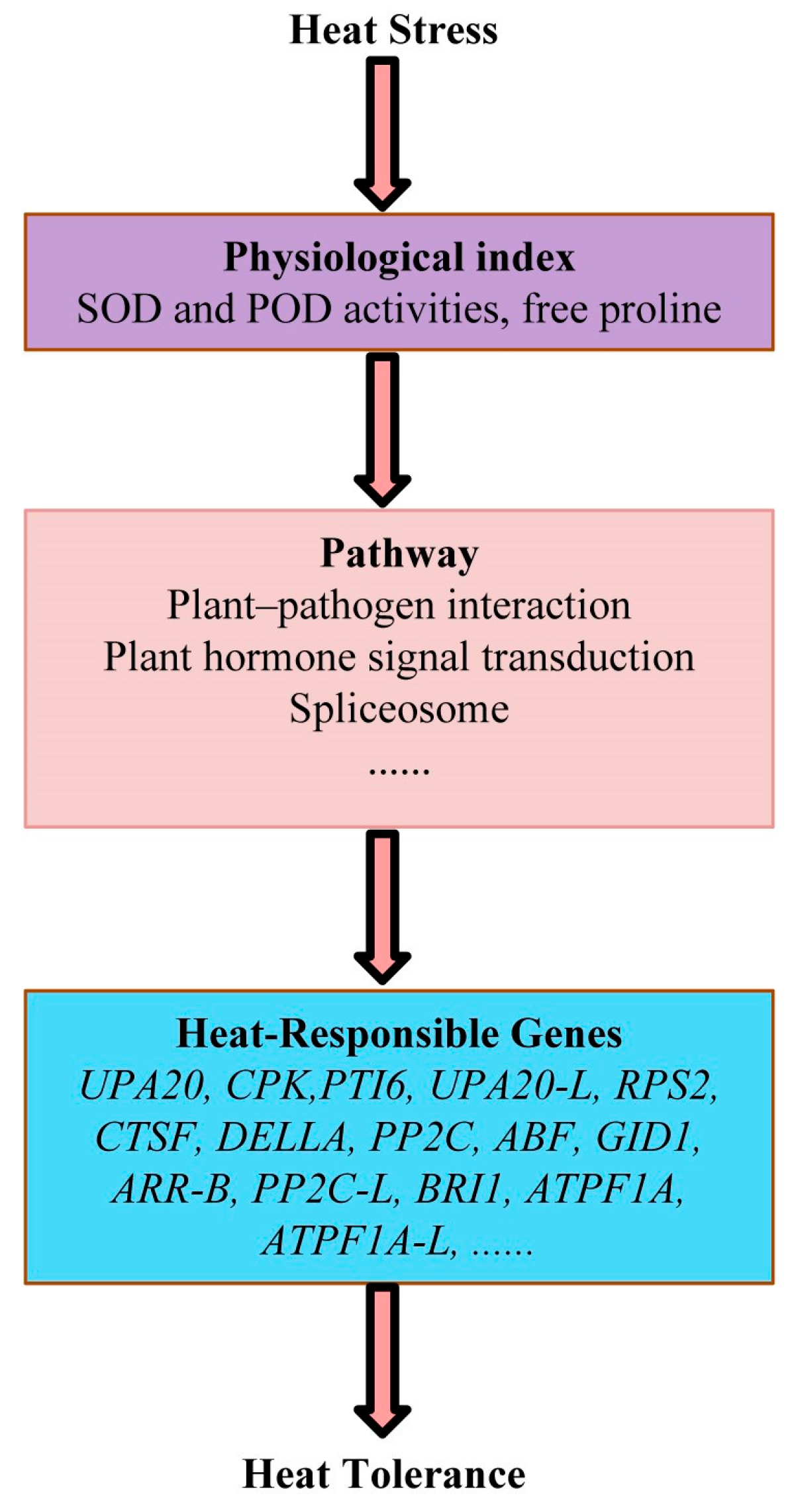
3.7. Physiological and Molecular Models of Cabbage Response to Long-Term Heat Stress

In order to carry out cabbage breeding more accurately and efficiently, we were particularly interested in the core factors involved in cabbage response to long-term heat stress, both at the physiological and molecular levels. Therefore, we integrated the patterns of physiological indicators, enrichment pathways of DEGs, and gene expression to establish physiological and molecular response models for cabbage under long-term heat stress. Changes in the activities of POD and SOD enzymes, as well as the content of free proline, were important physiological indicators of cabbage response to long-term heat stress. Plant hormone signaling transduction pathways and the plant–pathogen interaction pathway were significantly enriched with DEGs in response to long-term heat stress. Moreover, at least 15 genes have been identified as being upregulated in response to long-term heat stress in these two pathways (Figure 6). Based on this model, gradually expanding research content might serve as a starting point for comprehensively unraveling the mechanisms of cabbage response to long-term heat stress.

4. Discussion

High temperatures can inhibit plant growth and development [38]. The present study shows that the growth and development of cabbage are somewhat restricted under heat stress [39]. Plants under stress have increased cell membrane permeability, thus exhibiting extracellular leakage of intracellular electrolytes and an increase in the conductivity of tissue osmolytes [11]. Plants subjected to adversity stress can disrupt the equilibrium state of the white by-group reactive oxygen species in their own bodies, leading to varying degrees of reactive oxygen species accumulation in plants and ultimately causing oxidative damage to plant cells [12]. SOD, POD, and CAT play important roles in scavenging reactive oxygen species from plants. SOD converts O2− to H2O2, and CAT and POD convert H2O2 to H2O [40]. Some studies have shown that SOD activity shows a rapid increase at the beginning of heat stress. However, as the stress temperature increased, the plants suffered more damage and SOD activity decreased, which was related to the heat tolerance of the plant species [41]. In this study, when cabbage was subjected to heat stress at 35 °C, the overall activities of SOD, POD, and CAT enzymes showed an increasing trend. This indicated that the activities of these enzymes played an important role in regulating the physiological responses of plants to heat stress. The changes in free proline and MDA content during the response of plants to heat stress are also important indicators of plant tolerance [42,43]. In our study, the changes in free proline and MDA content during the response of cabbage to heat stress were consistent with the patterns observed in most plants. We found that exposure to a temperature of 35 ℃ subjected cabbage to high heat stress, but a 12 h treatment did not result in wilting or death of the plants. Additionally, physiological substances within the organism participate in regulating the response of cabbage to heat stress.
The method of identifying DEGs through transcriptome analysis has been widely used in studying the response of plants to high temperature stress [44]. In the process of responding to heat stress, 1720 and 6178 DEGs were identified in Clematis lanuginosa and Clematis crassifolia, respectively [45]. In the Brassica genus, the number of DEGs in response to heat stress is correlated with various factors, including cultivar, temperature, treatment duration, and tissue type [46,47,48,49]. In our study, 11,579 and 14,707 DEGs were identified in response to heat stress treatment for 4 and 12 h, respectively. Based on the changes in the number of DEGs, we speculated that the ‘Bomei 4′ cabbage variety was more sensitive to high temperature stress. Additionally, as the duration of heat stress treatment increased, the number of DEGs became more prominent. In addition, studies have found that gene expression in Brassica plants in response to heat stress is closely associated with epigenetic modifications such as methylation and acetylation [47,50,51]. This indicates that the response of cabbage to heat stress involves a complex molecular regulatory network. Investigating whether the expression patterns of cabbage genes and their response to the duration of heat stress are regulated by epigenetic inheritance would be a valuable research topic in the future.
Functional annotation and enrichment of DEGs are commonly used approaches to study the molecular mechanisms underlying important traits in plants. Heat stress is currently a hot topic of research in the Brassica genus, as it affects reproduction, floral progression, organogenesis, postharvest seedlings, and vegetative growth [47]. The functional enrichment of DEGs in response to heat stress has also gained considerable attention. In multiple studies focusing on the heat-stress-induced important traits in cabbage, we have observed that the functional enrichment of DEGs consistently highlights pathways related to the plant hormone signaling pathway, plant–pathogen interactions pathway, MAPK signaling pathway, phenylpropanoid biosynthesis, and biosynthesis of amino acids pathway [46,47]. Typically, heat stress in plants is primarily determined by extremely high temperatures and the duration of sustained high temperatures, which are the main external factors causing damage [47]. In this study, DEGs in cabbage, whether in response to 4 h or 12 h of heat stress, were significantly enriched in multiple biological processes, including the plant hormone signaling pathways and the plant–pathogen interactions pathway. At the same time, we found that seven DEGs belonging to the plant hormone signaling pathway and 14 DEGs belonging to the plant–pathogen interactions pathway exhibited a sustained increase in expression levels in response to heat stress.
The identification and functional characterization of key genes involved in plant response to high heat stress are crucial for heat tolerance breeding in plants. In recent years, multiple species of the Brassica genus have undergone whole-genome sequencing. Rapid progress has been made in the identification and analysis of gene families related to heat stress response in the Brassica genus, including the HSP, APX, MKK, and MAPK gene families [52,53,54,55]. Utilizing the transcriptome and other technological approaches has also enabled the identification of key genes involved in plant responses to heat stress, including the CPK, ABF, and PYL genes, among others [47,56,57]. In our study, we identified 15 genes that showed differential upregulation in response to heat stress, including the GID, UPA20, CTSF, and ATPF1A genes, which have not been previously reported in studies on heat stress response in the Brassica genus. We also observed that the expression levels of these 15 genes continued to increase with prolonged exposure to heat stress. This finding provides valuable genetic resources for breeding heat-tolerant cabbage varieties.

Supplementary Materials

The following supporting information can be downloaded at: https://www.mdpi.com/article/10.3390/horticulturae9090977/s1, Figure S1: GO enrichment of DEGs. A. GO enrichment of DEGs in CK vs. H4; B. GO enrichment of DEGs in CK vs. H12; Figure S2: GO enrichment of DEGs in biological processes: a histogram. A. Histogram of the biological process GO enrichment of DEGs in CK vs. H4; B. Histogram of the biological process GO enrichment of DEGs in CK vs. H12; Figure S3: GO enrichment on DEGs in the cellular component: a histogram. A. Histogram of the cellular component GO enrichment of DEGs in CK vs. H4; B. Histogram of the cellular component GO enrichment of DEGs in CK vs. H12; Figure S4: GO enrichment of DEGs in molecular functions: a histogram. A. Histogram of the molecular functions GO enrichment of DEGs in CK vs. H4; B. Histogram of the molecular functions GO enrichment of DEGs in CK vs. H12; Figure S5: Expression patterns of the hub genes under artificial high-temperature treatment. CK represents the control group (without any treatment); H4 represents heat treatment at 35 °C for 4 h; H12 represents heat treatment at 35 °C for 12 h. Table S1: List of all primers involved in this study; Table S2: Sequencing data statistics; Table S3: Sequence alignment of the sample sequencing data with selected reference genomes; Table S4: Sequencing of the sample reads distribution in different regions; Table S5: Statistics on the number of differentially expressed genes annotated; Table S6: Statistical analysis of KEGG enrichment results for differentially expressed genes; Table S7: Detailed statistics of key genes.

Author Contributions

Conceptualization, Q.L. and S.A.; methodology, Q.L.; validation, Q.L. and G.Z.; formal analysis, Q.L., G.Z. and Y.Z.; investigation, G.Z.; resources, Q.L.; data curation, Y.Z.; writing—original draft preparation, Q.L.; writing—review and editing, G.Z., Y.Z., L.G., Y.W., X.Y. and S.A.; visualization, Q.L.; supervision, S.A.; project administration, Q.L.; funding acquisition, Q.L. All authors have read and agreed to the published version of the manuscript.

Funding

This research was funded by the S&T Program of Hebei, grant number 21326341D; the Natural Science Foundation of Hebei, grant number C2022105004; the S&T Program of Tangshan, grant number 22130201H; and the Colleges and Universities in the Hebei Province Science and Technology Research Project, grant number QN2023066.

Data Availability Statement

The raw sequence data reported in this study have been deposited in the National Center for Biotechnology Information (NCBI) under accession number PBJNA961297.

Conflicts of Interest

The authors declare no conflict of interest.

References

  1. Mitchell, J.F. Prediction and detection of anthropogenic climate change. Int. Geophys. 2002, 83, 152–164. [Google Scholar] [CrossRef]
  2. Morison, J.; Lawlor, D. Interactions between increasing CO2 concentration and temperature on plant growth. Plant Cell Environ. 1999, 22, 659–682. [Google Scholar] [CrossRef]
  3. Min, L.; Li, Y.; Hu, Q.; Zhu, L.; Gao, W.; Wu, Y.; Ding, Y.; Liu, S.; Yang, X.; Zhang, X. Sugar and auxin signaling pathways respond to high-temperature stress during anther development as revealed by transcript profiling analysis in cotton. Plant Physiol. 2014, 164, 1293–1308. [Google Scholar] [CrossRef]
  4. Li, B.; Gao, K.; Ren, H.; Tang, W. Molecular mechanisms governing plant responses to high temperatures. J. Integr. Plant Biol. 2018, 60, 757–779. [Google Scholar] [CrossRef]
  5. Puteh, A.; Thuzar, M.; Mondal, M.; Abdullah, N.P.B.; Halim, M. Soybean [Glycine max (L.) Merrill] seed yield response to high temperature stress during reproductive growth stages. Aust. J. Crop. Sci. 2013, 7, 1472–1479. [Google Scholar]
  6. Shah, F.; Huang, J.; Cui, K.; Nie, L.; Shah, T.; Chen, C.; Wang, K. Impact of high-temperature stress on rice plant and its traits related to tolerance. J. Agric. Sci. 2011, 149, 545–556. [Google Scholar] [CrossRef]
  7. Veiseth, S.V.; Rahman, M.A.; Yap, K.L.; Fischer, A.; Egge-Jacobsen, W.; Reuter, G.; Zhou, M.M.; Aalen, R.B.; Thorstensen, T. The SUVR4 histone lysine methyltransferase binds ubiquitin and converts H3K9me1 to H3K9me3 on transposon chromatin in Arabidopsis. PLoS Genet. 2011, 7, e1001325. [Google Scholar] [CrossRef] [PubMed]
  8. Cho, J. Double fertilization in Oryza sativa L. and development of the endosperm with special reference to the aleurone layer. Bull. Natl. Inst. Agric. Sci. 1956, 6, 61–101. [Google Scholar]
  9. Jagadish, S.V.; Muthurajan, R.; Oane, R.; Wheeler, T.R.; Heuer, S.; Bennett, J.; Craufurd, P.Q. Physiological and proteomic approaches to address heat tolerance during anthesis in rice (Oryza sativa L.). J. Exp. Bot. 2010, 61, 143–156. [Google Scholar] [CrossRef]
  10. Niu, Y.; Xiang, Y. An Overview of Biomembrane Functions in Plant Responses to High-Temperature Stress. Front. Plant Sci. 2018, 9, 915. [Google Scholar] [CrossRef] [PubMed]
  11. He, Z.; He, C.; Zhang, Z.; Zou, Z.; Wang, H. Changes of antioxidative enzymes and cell membrane osmosis in tomato colonized by arbuscular Mycorrhizae under NaCl stress. Colloids Surf. B Biointerfaces 2007, 59, 128–133. [Google Scholar] [CrossRef] [PubMed]
  12. Gao, Q.; Zhang, L. Ultraviolet-B-induced oxidative stress and antioxidant defense system responses in ascorbate-deficient vtc1 mutants of Arabidopsis thaliana. J. Plant Physiol. 2008, 165, 138–148. [Google Scholar] [CrossRef] [PubMed]
  13. McClung, C.R.; Davis, S.J. Ambient thermometers in plants: From physiological outputs towards mechanisms of thermal sensing. Curr. Biol. CB 2010, 20, R1086–R1092. [Google Scholar] [CrossRef]
  14. Suzuki, N.; Koussevitzky, S.; Mittler, R.; Miller, G. ROS and redox signalling in the response of plants to abiotic stress. Plant Cell Environ. 2012, 35, 259–270. [Google Scholar] [CrossRef] [PubMed]
  15. Ding, Y.; Yang, S. Surviving and thriving: How plants perceive and respond to temperature stress. Dev. Cell 2022, 57, 947–958. [Google Scholar] [CrossRef] [PubMed]
  16. Wang, K.; Liu, Y.; Tian, J.; Huang, K.; Shi, T.; Dai, X.; Zhang, W. Transcriptional Profiling and Identification of Heat-Responsive Genes in Perennial Ryegrass by RNA-Sequencing. Front. Plant Sci. 2017, 8, 1032. [Google Scholar] [CrossRef] [PubMed]
  17. Bhardwaj, A.R.; Joshi, G.; Kukreja, B.; Malik, V.; Arora, P.; Pandey, R.; Shukla, R.N.; Bankar, K.G.; Katiyar-Agarwal, S.; Goel, S.; et al. Global insights into high temperature and drought stress regulated genes by RNA-Seq in economically important oilseed crop Brassica juncea. BMC Plant Biol. 2015, 15, 9. [Google Scholar] [CrossRef]
  18. Suzuki, N.; Bassil, E.; Hamilton, J.S.; Inupakutika, M.A.; Zandalinas, S.I.; Tripathy, D.; Luo, Y.; Dion, E.; Fukui, G.; Kumazaki, A.; et al. ABA Is Required for Plant Acclimation to a Combination of Salt and Heat Stress. PLoS ONE 2016, 11, e0147625. [Google Scholar] [CrossRef]
  19. Setsungnern, A.; Muñoz, P.; Pérez-Llorca, M.; Müller, M.; Thiravetyan, P.; Munné-Bosch, S. A defect in BRI1-EMS-SUPPRESSOR 1 (bes1)-mediated brassinosteroid signaling increases photoinhibition and photo-oxidative stress during heat stress in Arabidopsis. Plant Sci. Int. J. Exp. Plant Biol. 2020, 296, 110470. [Google Scholar] [CrossRef]
  20. Martin, R.C.; Kronmiller, B.A.; Dombrowski, J.E. Transcriptome Analysis of Lolium temulentum Exposed to a Combination of Drought and Heat Stress. Plants 2021, 10, 2247. [Google Scholar] [CrossRef]
  21. Song, X.; Hu, J.; Wu, T.; Yang, Q.; Feng, X.; Lin, H.; Feng, S.; Cui, C.; Yu, Y.; Zhou, R.; et al. Comparative analysis of long noncoding RNAs in angiosperms and characterization of long noncoding RNAs in response to heat stress in Chinese cabbage. Hortic. Res. 2021, 8, 48. [Google Scholar] [CrossRef] [PubMed]
  22. Sun, X.; Feng, D.; Liu, M.; Qin, R.; Li, Y.; Lu, Y.; Zhang, X.; Wang, Y.; Shen, S.; Ma, W.; et al. Single-cell transcriptome reveals dominant subgenome expression and transcriptional response to heat stress in Chinese cabbage. Genome Biol. 2022, 23, 262. [Google Scholar] [CrossRef] [PubMed]
  23. Ahmed, W.; Li, R.; Xia, Y.; Bai, G.; Siddique, K.H.M.; Zhang, H.; Zheng, Y.; Yang, X.; Guo, P. Comparative Analysis of miRNA Expression Profiles Between Heat-Tolerant and Heat-Sensitive Genotypes of Flowering Chinese Cabbage Under Heat Stress Using High-Throughput Sequencing. Genes 2020, 11, 264. [Google Scholar] [CrossRef]
  24. Moradpour, M.; Abdullah, S.N.A.; Namasivayam, P. The Impact of Heat Stress on Morpho-Physiological Response and Expression of Specific Genes in the Heat Stress-Responsive Transcriptional Regulatory Network in Brassica oleracea. Plants 2021, 10, 1064. [Google Scholar] [CrossRef]
  25. Shanmugam, A.; Thamilarasan, S.K.; Park, J.I.; Jung, M.Y.; Nou, I.S. Characterization and abiotic stress-responsive expression analysis of SGT1 genes in Brassica oleracea. Genome 2016, 59, 243–251. [Google Scholar] [CrossRef]
  26. Park, H.J.; Jung, W.Y.; Lee, S.S.; Song, J.H.; Kwon, S.Y.; Kim, H.; Kim, C.; Ahn, J.C.; Cho, H.S. Use of heat stress responsive gene expression levels for early selection of heat tolerant cabbage (Brassica oleracea L.). Int. J. Mol. Sci. 2013, 14, 11871–11894. [Google Scholar] [CrossRef]
  27. Yan, J.; Ma, H.; Lai, X.; Wu, J.; Liu, A.; Huang, J.; Sun, W.; Shen, M.; Zhang, Y. Artemisinin attenuated oxidative stress and apoptosis by inhibiting autophagy in MPP(+)-treated SH-SY5Y cells. J. Biol. Res. 2021, 28, 6. [Google Scholar] [CrossRef]
  28. Huan, X.; Wang, X.; Zou, S.; Zhao, K.; Han, Y.; Wang, S. Transcription Factor ERF194 Modulates the Stress-Related Physiology to Enhance Drought Tolerance of Poplar. Int. J. Mol. Sci. 2023, 24, 788. [Google Scholar] [CrossRef]
  29. Abrahám, E.; Hourton-Cabassa, C.; Erdei, L.; Szabados, L. Methods for determination of proline in plants. Methods Mol. Biol. 2010, 639, 317–331. [Google Scholar] [CrossRef] [PubMed]
  30. Sankar, A.N.A.M.S. Root growth, malonyldialdehyde content and antioxidant enzyme activities in response to low phosphorus supply in rice (Oryza sativa L.) genotypes. Res. Crops 2014, 15, 746–753. [Google Scholar]
  31. Cannon, J.T.; Kocot, K.M. Phylogenomics Using Transcriptome Data. Methods Mol. Biol. 2016, 1452, 65–80. [Google Scholar] [CrossRef]
  32. Kim, D.; Langmead, B.; Salzberg, S.L. HISAT: A fast spliced aligner with low memory requirements. Nat. Methods 2015, 12, 357–360. [Google Scholar] [CrossRef]
  33. Pertea, M.; Pertea, G.M.; Antonescu, C.M.; Chang, T.C.; Mendell, J.T.; Salzberg, S.L. StringTie enables improved reconstruction of a transcriptome from RNA-seq reads. Nat. Biotechnol. 2015, 33, 290–295. [Google Scholar] [CrossRef]
  34. Love, M.I.; Huber, W.; Anders, S. Moderated estimation of fold change and dispersion for RNA-seq data with DESeq2. Genome Biol. 2014, 15, 550. [Google Scholar] [CrossRef] [PubMed]
  35. Kanehisa, M.; Goto, S. KEGG: Kyoto encyclopedia of genes and genomes. Nucleic Acids Res. 2000, 28, 27–30. [Google Scholar] [CrossRef] [PubMed]
  36. Consortium, T.G.O. The Gene Ontology Resource: 20 years and still GOing strong. Nucleic Acids Res. 2019, 47, D330–D338. [Google Scholar] [CrossRef]
  37. Livak, K.J.; Schmittgen, T.D. Analysis of relative gene expression data using real-time quantitative PCR and the 2(-Delta Delta C(T)) Method. Methods 2001, 25, 402–408. [Google Scholar] [CrossRef]
  38. Wahid, A.; Gelani, S.; Ashraf, M.; Foolad, M.R. Heat tolerance in plants: An overview. Environ. Exp. Bot. 2007, 61, 199–223. [Google Scholar] [CrossRef]
  39. Alsamadany, H.; Alzahrani, Y.; Shah, Z.H. Physiomorphic and molecular-based evaluation of wheat germplasm under drought and heat stress. Front. Plant Sci. 2023, 14, 1107945. [Google Scholar] [CrossRef] [PubMed]
  40. Dias da Silva, L.; de Santana, T.; Priscila, S.; Laviola, B.; Costa, M.; Almeida, A.-A.; Gomes, F. Abscisic acid-mediated stomatal closure and antioxidant defenses in Jatropha curcas L. seedlings submitted to moderate water deficit. Afr. J. Agric. Res. 2016, 11, 2806–2816. [Google Scholar] [CrossRef]
  41. Zhao, X.; Huang, L.J.; Sun, X.F.; Zhao, L.L.; Wang, P.C. Differential Physiological, Transcriptomic, and Metabolomic Responses of Paspalum wettsteinii Under High-Temperature Stress. Front. Plant Sci. 2022, 13, 865608. [Google Scholar] [CrossRef]
  42. Wang, C.; Wen, D.; Sun, A.; Han, X.; Zhang, J.; Wang, Z.; Yin, Y. Differential activity and expression of antioxidant enzymes and alteration in osmolyte accumulation under high temperature stress in wheat seedlings. J. Cereal Sci. 2014, 60, 653–659. [Google Scholar] [CrossRef]
  43. Puangbut, D.; Jogloy, S.; Vorasoot, N.; Songsri, P. Photosynthetic and physiological responses to drought of Jerusalem artichoke genotypes differing in drought resistance. Agric. Water Manag. 2022, 259, 107252. [Google Scholar] [CrossRef]
  44. Bashir, K.; Matsui, A.; Rasheed, S.; Seki, M. Recent advances in the characterization of plant transcriptomes in response to drought, salinity, heat, and cold stress. F1000Research 2019, 8, 18424. [Google Scholar] [CrossRef]
  45. Qian, R.; Hu, Q.; Ma, X.; Zhang, X.; Ye, Y.; Liu, H.; Gao, H.; Zheng, J. Comparative transcriptome analysis of heat stress responses of Clematis lanuginosa and Clematis crassifolia. BMC Plant Biol. 2022, 22, 138. [Google Scholar] [CrossRef]
  46. Zhang, L.; Dai, Y.; Yue, L.; Chen, G.; Yuan, L.; Zhang, S.; Li, F.; Zhang, H.; Li, G.; Zhu, S.; et al. Heat stress response in Chinese cabbage (Brassica rapa L.) revealed by transcriptome and physiological analysis. PeerJ 2022, 10, e13427. [Google Scholar] [CrossRef]
  47. Kourani, M.; Mohareb, F.; Rezwan, F.I.; Anastasiadi, M.; Hammond, J.P. Genetic and Physiological Responses to Heat Stress in Brassica napus. Front. Plant Sci. 2022, 13, 832147. [Google Scholar] [CrossRef]
  48. Wang, A.; Hu, J.; Huang, X.; Li, X.; Zhou, G.; Yan, Z. Comparative Transcriptome Analysis Reveals Heat-Responsive Genes in Chinese Cabbage (Brassica rapa ssp. chinensis). Front. Plant Sci. 2016, 7, 939. [Google Scholar] [CrossRef]
  49. Quan, J.; Li, X.; Li, Z.; Wu, M.; Zhu, B.; Hong, S.B.; Shi, J.; Zhu, Z.; Xu, L.; Zang, Y. Transcriptomic Analysis of Heat Stress Response in Brassica rapa L. ssp. pekinensis with Improved Thermotolerance through Exogenous Glycine Betaine. Int. J. Mol. Sci. 2023, 24, 6429. [Google Scholar] [CrossRef]
  50. Liu, G.; Wang, J.; Hou, X. Transcriptome-Wide N(6)-Methyladenosine (m(6)A) Methylome Profiling of Heat Stress in Pak-choi (Brassica rapa ssp. chinensis). Plants 2020, 9, 80. [Google Scholar] [CrossRef]
  51. Liu, G.; Xia, Y.; Liu, T.; Dai, S.; Hou, X. The DNA Methylome and Association of Differentially Methylated Regions with Differential Gene Expression during Heat Stress in Brassica rapa. Int. J. Mol. Sci. 2018, 19, 1414. [Google Scholar] [CrossRef]
  52. Wang, Z.; Wan, Y.; Meng, X.; Zhang, X.; Yao, M.; Miu, W.; Zhu, D.; Yuan, D.; Lu, K.; Li, J.; et al. Genome-Wide Identification and Analysis of MKK and MAPK Gene Families in Brassica Species and Response to Stress in Brassica napus. Int. J. Mol. Sci. 2021, 22, 544. [Google Scholar] [CrossRef]
  53. Ma, L.; Qi, W.; Bai, J.; Li, H.; Fang, Y.; Xu, J.; Xu, Y.; Zeng, X.; Pu, Y.; Wang, W.; et al. Genome-Wide Identification and Analysis of the Ascorbate Peroxidase (APX) Gene Family of Winter Rapeseed (Brassica rapa L.) Under Abiotic Stress. Front. Genet. 2021, 12, 753624. [Google Scholar] [CrossRef]
  54. Verma, D.; Lakhanpal, N.; Singh, K. Genome-wide identification and characterization of abiotic-stress responsive SOD (superoxide dismutase) gene family in Brassica juncea and B. rapa. BMC Genom. 2019, 20, 227. [Google Scholar] [CrossRef]
  55. Wang, L.; Liu, F.; Ju, L.; Xue, B.; Wang, Y.; Wang, D.; Hou, D. Genome Structures and Evolution Analysis of Hsp90 Gene Family in Brassica napus Reveal the Possible Roles of Members in Response to Salt Stress and the Infection of Sclerotinia sclerotiorum. Front. Plant Sci. 2022, 13, 854034. [Google Scholar] [CrossRef]
  56. Zhang, H.; Liu, W.Z.; Zhang, Y.; Deng, M.; Niu, F.; Yang, B.; Wang, X.; Wang, B.; Liang, W.; Deyholos, M.K.; et al. Identification, expression and interaction analyses of calcium-dependent protein kinase (CPK) genes in canola (Brassica napus L.). BMC Genom. 2014, 15, 211. [Google Scholar] [CrossRef]
  57. Verma, V.; Ravindran, P.; Kumar, P.P. Plant hormone-mediated regulation of stress responses. BMC Plant Biol. 2016, 16, 86. [Google Scholar] [CrossRef]
Figure 1. Various physiological indicators of cabbage leaves under heat stress. (A), SOD activity; (B), POD activity; (C), CAT activity; (D), H2O2 content; (E), free proline content; (F), MDA content. Significant differences (a–c) over the bars indicate variations at a significance level of p < 0.05, determined through Tukey’s multiple comparison test.
Horticulturae 09 00977 g001
Figure 2. Analysis of differential gene expression. (A), principal component analysis plot; (B), heat map of expression correlation between two samples; (C), histogram of differential gene statistics; (D), Venn diagram of the differential gene set.
Horticulturae 09 00977 g002
Figure 3. KEGG enrichment of DEGs. (A). KEGG enrichment of DEGs in CK vs. H4. (B). KEGG enrichment of DEGs in CK vs. H12.
Horticulturae 09 00977 g003
Figure 4. Differential gene expression. (A), the number of DEGs in the plant–pathogen interaction pathway; (B), the number of DEGs in the plant hormone signal transduction pathway; (C), a heat map of shared DEGs in the plant–pathogen interaction pathway and the plant hormone signal transduction pathway.
Horticulturae 09 00977 g004
Figure 5. Expression patterns of hub genes under artificial high-temperature treatment. (AG), Relative gene expression levels. CK represents the control group (without any treatment); H4 represents heat treatment at 35 °C for 4 h; H12 represents heat treatment at 35 °C for 12 h. Significant differences (a–c) over the bars indicate variations at a significance level of p < 0.05, determined through Tukey’s multiple comparison test.
Horticulturae 09 00977 g005
Figure 6. A physiological to molecular model of cabbage response to long-term heat stress.
Horticulturae 09 00977 g006
Disclaimer/Publisher’s Note: The statements, opinions and data contained in all publications are solely those of the individual author(s) and contributor(s) and not of MDPI and/or the editor(s). MDPI and/or the editor(s) disclaim responsibility for any injury to people or property resulting from any ideas, methods, instructions or products referred to in the content.

