Article

Morpho-Anatomical and Physiological Assessments of Cryo-Derived Pineapple Plants (Ananas comosus var. comosus) after Acclimatization

by
Ariel Villalobos-Olivera
1,
José Carlos Lorenzo-Feijoo
2,
Nicolás Quintana-Bernabé
1,
Michel Leiva-Mora
3,
Jean Carlos Bettoni
4 and
Marcos Edel Martínez-Montero
1,*
1
Facultad de Ciencias Agrícolas, Universidad de Ciego de Ávila Máximo Gómez Báez, Ciego de Ávila 65200, Cuba
2
Laboratory for Plant Breeding and Conservation of Genetic Resources, Bioplant Centre, University of Ciego de Ávila, Ciego de Ávila 65200, Cuba
3
Departamento de Agronomía, Facultad de Ciencias Agropecuarias, Universidad Técnica de Ambato (UTA-DIDE), Campus Querochaca, Cantón Cevallos Vía a Quero, Sector El Tambo-La Universidad, Cevallos 1801334, Tungurahua, Ecuador
4
Independent Researcher, 35 Brasil Correia Street, Videira 89560510, SC, Brazil
*
Author to whom correspondence should be addressed.
Horticulturae 2023, 9(7), 841; https://doi.org/10.3390/horticulturae9070841
Submission received: 21 May 2023 / Revised: 17 July 2023 / Accepted: 20 July 2023 / Published: 24 July 2023

Abstract

:
Studies on the morpho-physiology of cryo-derived pineapple plants after acclimatization have been quite limited. Therefore, in the present study, the morpho-anatomical and physiological characteristics of cryo-derived Ananas comosus var. comosus ‘MD-2’ plants after acclimatization were investigated. Plants obtained from cryopreserved and non-cryopreserved shoot tips, as well as in vitro stock cultures (control), showed similar morphological development (viz. plant height, number of leaves, D leaf length, D leaf width, D leaf area, diameter of stem base, number of roots, plant fresh weight and plant dry weight) to conventionally micropropagated and non-cryopreserved plants. The pineapple plantlets developed efficient anatomical leaf structures that allowed them to adapt to the transition process from in vitro to ex vitro. In all groups of plants, the content of water and chlorophylls (a, a + b, a/b) decreased during the first 15 days of acclimatization and then remained constant until the end of the evaluation. The mesophilic succulence index increased to its maximum value after 15 days, then decreased and remained constant up to 45 days. Although physiological indicators fluctuated during the 45 days of acclimatization, no differences were observed in any of the indicators evaluated when plantlets obtained from cryopreserved shoot tips were compared with controls. The results of the plants from cryopreserved shoot tips show that they switched from C3 to Crassulacean acid metabolism, which denoted metabolic stability during acclimatization.

1. Introduction

The conservation of biological material through cryopreservation is now considered to be the safest and most cost-effective strategy for the long-term storage of plant genetic resources [1,2,3,4]. In the case of important edible horticultural crops like pineapples (Ananas comosus var. comosus), which are vegetatively propagated and where crosses between varieties produce botanical seeds that are highly heterozygous, their seeds are of limited interest for the conservation of specific gene combinations [5]. The cryopreservation of shoot tips is the most relevant strategy for the long-term conservation of the pineapple crop. This is because true-to-type, virus-free plants can be regenerated directly from cryopreserved shoot tips [2,3,4]. Once cryopreserved, shoot tips are stored in a state where cellular divisions and metabolic processes are halted, and theoretically, plant materials can be preserved without genetic alteration for an indefinite period of time [6].
Cryopreservation methods have already been developed for pineapple shoot tips [5,7,8,9,10,11,12,13]. The vitrification-based cryopreservation method is the most widely applied for cryopreserving pineapple shoot tips [5,10,11,12,13]. In this method, precultured shoot tips are exposed to a highly concentrated plant vitrification solution (PVS) before being directly immersed in liquid nitrogen (LN) [2,12,13]. To date, the droplet-vitrification technique has been shown to be the most effective cryopreservation method across diverse genotypes. Droplet-vitrification uses ultra-fast cooling and warming rates of shoot tips, an important requirement for successful cryopreservation protocols [14,15]. Souza et al. [10,11] described a successful droplet-vitrification protocol that was applied to 16 genotypes of Ananas, both wild and cultivated, belonging to four botanical varieties. In this procedure, shoot tips were precultured for 48 h in preculture medium containing 0.3 M sucrose and then transferred to aluminum foil in 4–5 μL of plant vitrification solution 2 (PVS2) and treated at 0 °C for 45 min before being directly submerged in LN. This procedure resulted in shoot tip regrowth ranging from 44% to 86% across the 16 pineapple genotypes [10].
A. comosus var. comosus ‘MD-2’ is a hybrid cultivar of interest to the agricultural industry due to its desirable characteristics, commercial potential and ability to meet the demands of the global market [16]. In this sense, the Bioplantas Center (www.bioplantas.cu; accessed on 10 July 2023) in Cuba initiated the development of a technological innovation project in 2010 to generalize the micropropagation of the ‘MD-2’ pineapple, promote the creation of donor plant banks in different regions of the country and introduce cryopreservation protocols into the practice.
Micropropagation involves growing plants in vitro under conditions that restrict gas exchange, maintain high humidity and low light and use a sucrose-based medium, which may interfere with photosynthesis [17,18]. In addition, the different steps during cryopreservation protocols may impose chemical or physical stresses to the plant tissues which would cause somaclonal, genetic and epigenetic variations in regenerates in addition to poor shoot tip regrowth due to reactive oxygen species (ROS) formation [19,20,21]. Treatments such as shoot tip excision, preculture, cryoprotection, dehydration, the freeze–thaw cycle and acclimatization have all been known to impose ROS-induced oxidative stress [21,22,23]. The overproduction of ROS is highly reactive and toxic and can cause lipid peroxidation, protein denaturation, alterations in nucleic acids and membrane disruptions that may lead to programmed cell death [24,25]. Therefore, it is necessary to not only ensure shoot tip viability after cryopreservation, but also the true-to-type of the regenerants [18,19,20,21].
Several research papers have mentioned success in acclimating cryo-derived pineapple plants [10,26,27,28]. However, there have been only a few studies on assessments of morpho-anatomical characteristics in cryo-derived pineapple plants during/after acclimatization [26,28]. Furthermore, these studies do not describe the metabolic changes in cryo-derived plants during the transition from the in vitro to the ex vitro environment. It is known that pineapple plants undergo a shift from C3 to Crassulacean acid metabolism (CAM) when adapting to ex vitro conditions [29,30]. Therefore, this study investigates in detail, for the first time, the morpho-anatomical and physiological characteristics of cryo-derived pineapple plants during acclimatization. The results reported here support the use of the droplet-vitrification cryopreservation procedure described by Souza et al. [10] and modified by Villalobos et al. [12] for the establishment of cryopreserved pineapple gene bank collections.

2. Materials and Methods

Plant material and stock cultures
Tissue-cultured plants of A. comosus var. comosus ‘MD-2’ were obtained from the Bioplantas Center of the Universidad de Ciego de Ávila Máximo Gómez Báez (www.unica.cu; accessed on 6 July 2023) and used in this study. In vitro stock cultures were propagated and maintained in active growth on Murashige and Skoog (MS) medium [31] supplemented with 1.0 mg∙L−1 thiamine, 0.1 mg∙L−1 myo-inositol, 0.3 mg∙L−1 1-naphthaleneacetic acid (NAA), 30 g∙L−1 sucrose, 2.1 mg∙L−1 6-benzylaminopurine (BAP) and 6.5 g∙L−1 agar at pH 5.7 [32]. Cultures were placed in glass vessels (55 × 95 mm) and incubated in a growth chamber at 25 ± 2 °C with a 16 h photoperiod with a photosynthetic flux density of 75 ± 3 μmol∙s−1∙m−2. Subculture in fresh culture medium was performed every 45 days. After 45 days of subculture and before cryopreservation, shoots were preconditioned for 45 days in a liquid MS culture medium containing 100 mg∙L−1 myoinositol, 1.0 mg∙L−1 thiamine and 30 g∙L−1 sucrose at a pH of 5.7, as defined by Villalobos-Olivera et al. [12].
Shoot tip cryopreservation
To cryopreserve pineapple shoot tips, the droplet-vitrification method was performed as described by Souza et al. [10], with some modifications according to the studies of Martínez-Montero et al. [5,9]. Specifically, plant vitrification solution 3 (PVS3; solution consisting of MS + 50% (w/v) sucrose + 50% (w/v) glycerol) [33] was used instead of PVS2 (solution consisting of MS + 30% (w/v) glycerol, 15% (w/v) ethylene glycol + 15% (w/v) dimethylsulfoxide + 0.4 M sucrose) [34]. Shoot tips (1 mm length, 1 mm width) with three- or four-leaf primordia were excised from 45-day-old in vitro stock cultures and incubated on MS culture medium supplemented with 100 mg∙L−1 myoinositol, 1.0 mg∙L−1 thiamine, 10 g∙L−1 sucrose and 6.5 g∙L−1 agar at pH 5.7 for 24 h at 25 ± 2 °C under dark conditions.
Pretreatment with glycerol and sucrose: Next, 10 shoot tips with an average fresh weight of 5 mg were placed in each polypropylene cryovial with a volume of 2.0 mL. The fresh weight of each shoot tip was estimated using the rule of three, based on the proportionality of a shoot tip’s weight to the weight of 20 shoot tips measured using an analytical balance (Sartorius Entris 64-1S, Germany). Immediately thereafter, the cryovials were filled with liquid MS culture medium containing 2 M glycerol and 0.4 M sucrose for 20 min at 25 ± 2 °C.
Treatment with PVS3 vitrification solution: The cryovials containing the shoot tips were poured onto the surface of 9 cm diameter Petri dishes, which had filter paper moistened with 5 mL of PVS3 solution precooled to 0 °C. The Petri dishes were placed on the surface of an ice bath to dehydrate the shoot tips for 60 min. Individual shoot tips were then placed on aluminum foil strips (7 mm × 20 mm × 50 µm) in droplets of 5 µL PVS3, with five shoot tips per slide.
Immersion in LN: After the end of the PVS3 dehydration time, the aluminum foil strips containing the shoot tips were plunged in LN. After LN exposure for a few seconds, the foils with shoot tips were transferred into 2 mL cryovials filled with LN and maintained in LN for 10 h.
Thawing: the aluminum foil strips with shoot tips were warmed quickly by inverting the aluminum foil strips into unloading solution (MS culture medium + 1 M sucrose) at 25 ± 2 °C for 5 min.
In vitro recovery after cryopreservation: Shoot tips were placed in 55 × 95 mm glass vessels with solid MS culture medium containing 1.0 mg∙L−1 NAA, 1.0 mg∙L−1 BAP, 10 g∙L−1 sucrose and 6.5 g∙L−1 agar at pH 5.7 and cultured for 7 weeks at 25 ± 2 °C in darkness. They were then grown in the same conditions as the in vitro stock cultures.
Acclimatization conditions
Plants with a height greater than 5 cm, 5 to 8 functional leaves and a fresh weight greater than 4.5 g were considered as optimal to be transferred to the ex vitro phase. Plants were dipped in the preventive fungicide Previcur Energy® (Bayer Cropscience) at a concentration of 1 mL∙L−1 for 5 min. To determine the effectiveness of acclimatization in pineapple plants from cryopreserved apices, two different conditions were established. The first condition consisted of plastic trays with four 0.5 cm Ø drainage holes, containing 90 cm3 of red ferric soil and filter cake (1:1). The second condition involved black polyethylene bags with a substrate volume of 400 cm3. Both conditions were performed according to Pino et al. [35]. The substrate mixture consisted of typical red ferralitic soil and filter cake (from sugarcane) (1:1) (v/v), which had been previously sieved. Table 1 shows the chemical properties of the substrate used in the acclimatization phase.
Irrigation was performed daily using microsprinklers twice a day, at 9:00 a.m. and 2:00 p.m., for 10 min. The microsprinklers used were from an atomizer located at a height of 1.80 m above the ground, a feature that allows water to reach the plants in the form of a mist to maintain moisture and temperature [35].
During the first 45 days, the plants were cultivated in a greenhouse with a ceiling that allowed the passage of 25% of sunlight, environmental conditions of 80 ± 3% of relative humidity and 26.5 °C (using a digital thermo-hygrometer TECPEL®, model DTM-303, Spain), with natural light and photoperiod and a photosynthetic flux density of 250 ± 30 µmol∙m−2∙s−1 (using a digital Lux/Light Meter, model FT-710 Faithfull, China) and atmospheric conditions with a CO2 concentration between 375 and 400 μmol∙mol−1 according to Pino et al. [30].
During this stage, the experimental treatments were established as follows: control plants that were micropropagated in a conventional manner (Microp), plants derived from shoot tips that were cryoprotected but not cryopreserved (non-cryopreserved) and plants derived from cryopreserved shoot tips (Cryo).
Morpho-anatomical and physiological assessments
After 45 days of acclimatization, the morpho-anatomical and physiological indicators of 50 plants from non-cryopreserved, cryopreserved and micropropagated plants were determined. Plant height (cm), number of leaves, length and width of leaf D (cm) (the leaf of the best physiological characteristics), leaf area D (cm2), diameter of stem base of the plant (cm), number of roots and plant fresh weight and dry weight (g) were evaluated.
For anatomical analysis, the procedures were performed as specified by Ebel et al. [36]. For leaf D, sections were made in the middle part to perform the anatomical analysis. The sections were fixed in 70% alcohol, formaldehyde and acetic acid (90:5:5) for 12 h and then freehand cross sections of 15–25 µm thickness were made with a razor blade. These were stained with safranin for 3 min and then with toluidine blue for 5 min. Visualization was performed using an inverted biological microscope (model NIB-100, China) in conjunction with a digital camera (model HDCE-50B, China).
For the physiological indicators, the mesophilic succulence index (MSI) was determined using the ratio between the aquifer and the photosynthetic tissue of the plants according to Rodríguez-Escriba et al. [29]. This assessment was performed on days 0, 15, 30 and 45 of acclimatization using the following equation: MSI = WC [Cfls (a + b)] − 1.
Chlorophyll contents (Cfls) a, b, a + b and a/b ratio were determined on slices from the central area of leaf D (diameter of 0.78 cm), as suggested by Rodríguez-Escriba et al. [29]. The water content (WC) of the slices was determined by the difference between the fresh weight and the dry weight of the samples after 72 h of drying in an oven at 60 °C.
Gas exchange rate: Fifty plants, both non-cryopreserved and cryopreserved, were sampled, and transpiration rate and CO2 assimilation were determined at 12:00 p.m. and 12:00 a.m. as described by Rodríguez-Escriba et al. [29]. To determine water use efficiency, CO2 assimilation was divided by transpiration rate.
Organic acid: Fifty samples of 1 g were taken from the center of the largest plant leaves of cryopreserved and non-cryopreserved plants at 12:00 p.m. and 12:00 a.m. of each experimental treatment. For these samples, 10 mL of 50% (v/v) ethanol was added and incubated in a thermal bath at 90 °C for 20 min. Then, the liquid phase was separated from the solid phase and acid–base evaluation was performed using 0.79 g∙L−1 NaOH and 1 mg∙L−1 phenolphthalein as indicators, as described by Rodríguez-Escriba et al. [29]. Organic acid content was expressed in µmol H+∙g−1 leaf fresh weight.
Statistical analysis
For data analysis, IBM SPSS version 22 was used. First, data from each treatment in each experiment were shown to meet the assumptions of normal distribution and homogeneity of variances; second, the Kolmogorov–Smirnov and Levene tests were both used with p ≤ 0.05. Parametric tests were performed (one-way ANOVA and bifactorial). In addition, Tukey’s HSD test with p ≤ 0.05 was performed for the ANOVA, which showed significant differences. First, for this analysis, data were transformed in percentages according to the function y’ = 2 arcsine (y/100)½.

3. Results

In general, the regeneration of shoot tips after cryoprotection treatment and cryopreservation exceeded 98%. Furthermore, all of the ‘MD-2’ pineapple plantlets developed from shoot tips in three variants (i.e., plants regenerated from cryopreserved shoot tips, conventionally micropropagated plants and non-cryopreserved plants) were successfully rooted in vitro and survived the acclimatization process.

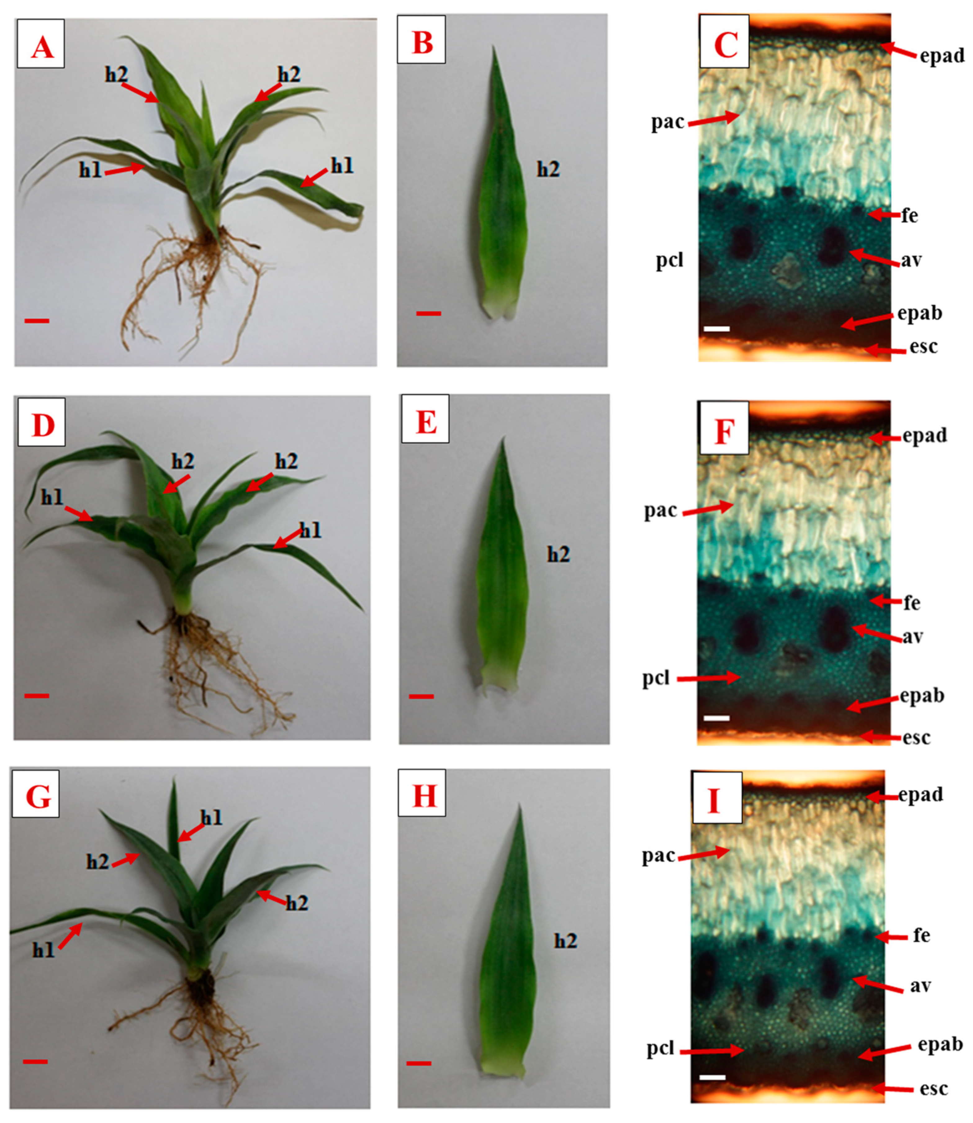
3.1. Morphological Variables

Table 2 shows a morphological comparison of the ‘MD-2’ pineapple plants regenerated from micropropagation or cryopreservation via droplet-vitrification (before or after LN immersion) and acclimatized (45 days) under different conditions (plastic trays or black polyethylene bags). Firstly, it is noteworthy that all measured variables, including plant height, number of leaves, leaf dimensions (length, width, area), stem base diameter, number of roots and fresh and dry weights of plants, were comparable between cryopreserved and non-cryopreserved plants, as well as in vitro stock cultures. Secondly, the plants acclimatized in black polyethylene bags exhibited the highest results in terms of morphological variables, except for leaf length and stem base diameter, where no statistical differences were observed. Therefore, for the subsequent research, the acclimatization process using black polyethylene bags was selected.

3.2. Morpho-Anatomical Characteristics

Figure 1 shows that no visual differences were observed between different treatments after acclimatization. After 45 days of ex vitro acclimatization, the pineapple plants in all three groups had similar root and leaf systems (Figure 1A,D,G) and displayed functional leaves and roots (Figure 1A–C). The leaves were thick and erect, with a distinct adaxial epidermis, aquifer parenchyma, chlorophyll parenchyma, abaxial epidermis and scales (Figure 1B,E,H). Extra axillary fibers and vascular rods were also clearly visible (Figure 1C,F,I).

3.3. Physiological Indicators

Although the variations in physiological indicators (Table 3) occurred along the 45 days of acclimatization, no differences were observed in any indicator evaluated when comparing plantlets recovered from cryopreserved shoot tips with the controls (micropropagated and non-cryopreserved), without LN exposure. Water content, chlorophyll a, chlorophylls a + b and a/b ratio decreased in the first 15 days and then started to increase after 30 days and reached their maximum value after 45 days. The chlorophyll b increased after 15 days and remained constant until the end of the assessment. The mesophilic succulence index reached its maximum value on day 15, and then decreased and remained constant until the end of the assessment.

3.4. Gas Exchange Rate and Organic Acid Levels

The gas exchange values found here confirm the natural expression of CAM metabolism in the plants of the three groups (Table 4). The gas exchange rate and organic acid levels were similar in plants from micropropagated, non-cryopreserved and cryopreserved shoot tips, and there was no difference between treatments. The highest gas exchange rate occurred at 12:00 a.m., while values were below the average at 12:00 p.m. The highest concentration of organic acids was also found at 12:00 a.m. (about 52 µmol H+ g−1 fresh weight).

4. Discussion

In this study, we report for the first time in Cuba the successful processes of in vitro propagation, cryopreservation and acclimatization of certified ‘MD2’ pineapples. This procedure provides an important tool to support in vitro gene banks and the long-term conservation of pineapple genetic resources. The availability of a cryopreservation method for pineapples may also facilitate the use of cryotherapy methods to eradicate viruses [37,38,39,40].
We found that cryopreservation using droplet-vitrification did not negatively affect the morphological characteristics of ‘MD-2’ pineapple plants, as they were comparable to those of non-cryopreserved plants and in vitro stock cultures (Table 2). Similar results of morphological characterization were also observed in the preliminary research of our group in two other pineapple cultivars ‘Red Spanish Florencia’ and ‘Hybrid 54′ (Smooth Cayenne/Red Spanish) after shoot tip cryopreservation and 45 days of acclimatization [27]. Similar morphological indicators were also observed in potatoes [2], shallots [41], apples [42], artichokes [43] and grapevines [44] between the cryopreserved plants and the control.
During the first few days after transplanting micropropagated plants under acclimatization conditions, relatively slow plant growth across all treatments was observed. This is likely due to the altered growth conditions and the shift in photosynthetic capacity [45], which is a common phenomenon observed in many plant species, including pineapple [30,35]. According to Villalobos et al. [18] and González et al. [46], pineapple plants grown in vitro develop a leaf system with autotrophic morphological structures during the initial days of ex vitro acclimatization. This helps plants to restore their photosynthetic machinery, allowing them to adapt to the low relative humidity conditions of ex vitro environments [29].
Previous studies by Hu et al. [47], Souza et al. [10] and Villalobos-Olivera et al. [12] described in vitro pineapple plants regenerated from shoot tip cryopreservation, but the adaptation of in vitro to ex vitro acclimatization conditions was not addressed. The success of in vitro propagation systems depends on effective acclimatization and this step is also critical for the establishment of seedlings under field conditions [48,49,50].
The acclimatization of pineapple plants in black polyethylene bags (size 400 cm3) produced the best morphological results when compared to the use of plastic trays (size 90 cm3), which is a novel finding (Table 2). The size of containers used for acclimatization presents a challenge in balancing optimal growing conditions and economic profitability in large-scale propagation [51]. While a reduced container size can provide significant economic benefits by lowering maintenance costs per unit of area, it can also negatively impact root development and photosynthetic activity, thereby affecting plantlet growth [52]. Furthermore, several studies have shown that small container size can restrict root growth and nutrient uptake, leading to reduced carbohydrate accumulation in leaves and an imbalanced source/sink ratio, which in turn can result in reduced shoot growth [53,54]. Kim et al. [55] demonstrated that the biggest-sized container was favorable for plant growth, as it showed higher plant height, leaf number, leaf area, fresh weight and dry weight for the growth of tissue-culture-propagated apple rootstock plants. Therefore, further studies on acclimatization based on industry requirements are needed.
The characteristics of morpho-anatomical development (Figure 1) correspond to those described by Pino et al. [35] and González et al. [46] during 45 days of ex vitro acclimatization of micropropagated pineapple plants. This reflects the adaptation process of leaf cells to the different environmental conditions, in which pineapple plantlets developed more functional leaf structures to adapt them to the transition process from in vitro to ex vitro [50,56].
Pineapple leaves formed in vitro have a short, club-shaped stem which narrow and strongly curved leaves grow around [30]. These leaves serve as a source of carbonaceous substances to meet metabolic needs and maintain plant adaptation during transitional stress in the first week under ex vitro conditions [30,45,46]. The newly developed leaves that adapted to ex vitro conditions (Figure 1B,E,H) exhibited a growth pattern with a more vertical orientation and increased width, which is indicative of greater metabolic efficiency and physiological adaptation to the specific environmental conditions [44]. These leaves exhibited a perfectly defined anatomical structure (Figure 1C,F,I), similar to that previously observed by Villalobos-Olivera et al. [28] and González et al. [46] in acclimatized pineapple plants from in vitro systems. These leaves exhibited the aquifer parenchyma, a specialized structure of plants with CAM metabolism that allows for higher efficiency in water use [36,57,58].
In the first phase of ex vitro acclimatization, pineapple plants switch from a C3 metabolism to a CAM metabolism to adapt to the increased temperature and light intensity [30,58,59,60]. The decrease in water content, chlorophyll a, chlorophylls a + b and a/b ratio in the first 15 days and the increase after 15 days could be related to the expression of the CAM metabolism (Table 3). The decrease in the water content of the leaves could be related to the loss of water vapor due to the inability of the stomata to function [29,58]. The increase in water content after 30 days could be related to further stomatal regulation [61]. This regulation aims to prevent water loss in the form of water vapor during respiration [29]. In addition, the plants of all groups develop aquifer tissue to store water.
The decrease in chlorophyll a in the first 15 days in plants might be related to tissue degradation during transition stress in the first week under ex vitro conditions due to increased temperature and light intensity [29]. Chlorophylls are sensitive indicators of the metabolic state of plants, and the decrease in their content reflects physiological disturbances or situations of biotic or abiotic stress [13]. Chlorophyll b increased consistently until the end of the evaluation. Working on the seedlings of four tropical woody species (Bignoniaceae) under stress conditions (low light intensity), Kitajima and Hogan [62] observed that while the chlorophyll a content decreased, the chlorophyll b content increased. This result is due to an adaptive response of the plant, in which chlorophyll b captures photons and transfers them to chlorophyll a to compensate for its function [63]. These observations could explain the decrease in chlorophyll a and increase in chlorophyll b in pineapple plants from cryopreserved shoot tips and controls (micropropagated and non-cryopreserved), as well as the results of Villalobos et al. [18]. Moreover, the fact that the chlorophyll a/b ratio remained at values above 1 can be understood as a typical photosynthetic plasticity response [29].
As for the mesophilic succulence index, the results found here are in agreement with those reported by Rodríguez-Escriba et al. [29] in pineapple plants under water stress situations. These authors reported that the increase in mesophilic succulence index was due to higher metabolic functionality. This increase was related to a decrease in chlorophylls and water content during the first 15 days under water stress conditions. The increase in the index indicates a better relationship between the hydric tissue and photosynthesis [64,65]. The plants in this study have values greater than one after 15 days of acclimatization, indicating the expression of CAM metabolism.
Gas exchange performed by plants under the three conditions after 45 days of acclimatization is characteristic of higher photosynthetic efficiency (Table 4). Pineapple plants exhibit C3 metabolism under optimal growth conditions and show CAM metabolism as an adaptation mechanism to biotic and abiotic stresses [66,67]. The CAM plants perform gas exchange at night [60,64,68]. The studied plants perform transpiration, stomatal conductance, CO2 assimilation and water use efficiency at night time (12:00 a.m.), which is characteristic of CAM metabolism.
The highest concentration of organic acids was also found at night time (12:00 a.m.). This increase in organic acids during the night could be related to the accumulation of CO2 in the form of malic acid in the vacuole [29], and during the day, malic acid is decarboxylated by phosphoenolpyruvate carboxylase (PEPC) [65]. According to Wang et al. [69] and Males and Griffiths [64], stomatal opening in CAM is related to increased relative humidity and decreased transpiration gradient in response to partial pressure of water vapor in the growing environment. Plants with these metabolic traits are more efficient in water use because they open their stomata at night and at cooler times of the day [70,71,72].
Overall, acclimatization is a complex process that involves the coordination of multiple physiological and biochemical responses to environmental stressors, including oxidative stress [73]. Changes in gene expression and cellular signaling pathways are important mechanisms through which plants acclimatize to environmental stresses, including oxidative stress. These mechanisms enable plants to adapt and maintain their development, even under stressful conditions [74,75].

5. Conclusions

The findings of the study have several potential implications for the cultivation of ‘MD-2’ pineapple plants. Firstly, the successful cryopreservation of ‘MD-2’ pineapple shoots using the droplet-vitrification method ensures the long-term conservation of pineapple germplasm, which may be useful for future propagation and breeding programs. Secondly, the morpho-anatomical and physiological characteristics of cryopreserved ‘MD-2’ pineapple plants were comparable to those of non-cryopreserved plants, indicating that cryopreservation does not negatively impact the growth and development of the plants, thus suggesting that this method may be ready for implementation in pineapple gene bank collections. Finally, we observed that the acclimatization of ‘MD-2’ pineapple plants in black polyethylene bags produced the best plant development, suggesting that this method could be used to improve plant propagation efficiency in commercial nurseries.

Author Contributions

Conceptualization, A.V.-O. and M.E.M.-M.; experimental design, A.V.-O., J.C.L.-F., N.Q.-B. and M.E.M.-M.; data collection, A.V.-O. and N.Q.-B.; statistical analysis, A.V.-O., J.C.L.-F. and N.Q.-B.; methodology, M.L.-M. preparation of the tables, writing—original paper preparation, A.V.-O.; writing—review and editing, M.L.-M., J.C.B. and M.E.M.-M.; visualization, all authors; project administration, A.V.-O. and M.E.M.-M.; funding acquisition, J.C.B. and M.E.M.-M. All authors have read and agreed to the published version of the manuscript.

Funding

This research received no external funding.

Data Availability Statement

The data presented in this study are available on request from the corresponding author.

Acknowledgments

The authors thank the Centro de Bioplantas and the Faculty of Agricultural Sciences of the Universidad de Ciego de Ávila Máximo Gómez Báez (Cuba), for their support in carrying out the research.

Conflicts of Interest

The authors declare no conflict of interest.

References

  1. Jiroutová, P.; Sedlák, J. Cryobiotechnology of plants: A hot topic not only for gene banks. Appl. Sci. 2020, 10, 4677. [Google Scholar] [CrossRef]
  2. Bettoni, J.C.; Bonnart, R.; Volk, G. Challenges in implementing plant shoot tip cryopreservation technologies. Plant Cell Tissue Organ Cult. 2021, 144, 21–34. [Google Scholar] [CrossRef]
  3. Bettoni, J.C.; Mathew, L.; Pathirana, R.; Wiedow, C.; Hunter, D.A.; McLachlan, A. Eradication of Potato Virus S, Potato Virus A, and Potato Virus M from infected in vitro-grown potato shoots using in vitro therapies. Front. Plant Sci. 2022, 13, 1431. [Google Scholar] [CrossRef]
  4. Zhang, A.; Wang, M.-R.; Li, Z.; Panis, B.; Bettoni, J.C.; Vollmer, R. Overcoming challenges for shoot tip cryopreservation of root and tuber crops. Agronomy 2023, 13, 219. [Google Scholar] [CrossRef]
  5. Martinez-Montero, M.E.; Gonzalez-Arnao, M.T.; Engelmann, F. Cryopreservation of tropical plant germplasm with vegetative propagation-review of sugarcane (Saccharum spp.) and pineapple (Ananas comusus (L.) Merrill) cases. In Current Frontiers in Cryopreservation; Katkov, I., Ed.; IntechOpen: London, UK, 2012; pp. 360–387. [Google Scholar] [CrossRef] [Green Version]
  6. Engelmann, F. Use of biotechnologies for the conservation of plant biodiversity. In Vitro Cell. Dev. Biol.-Plant 2011, 47, 5–16. [Google Scholar] [CrossRef]
  7. Gonzalez-Arnao, M.T.; Ravelo, M.M.; Urra-Villavicencio, C.; Martinez-Montero, M.M.; Engelmann, F. Cryopreservation of pineapple (Ananas comosus) apices. CryoLetters 1998, 19, 375–382. [Google Scholar]
  8. Gamez-Pastrana, R.; Martinez-Ocampo, Y.; Beristain, C.; Gonzalez-Arnao, M. An improved cryopreservation protocol for pineapple apices using encapsulation-vitrification. CryoLetters 2004, 25, 405–414. [Google Scholar]
  9. Martinez-Montero, M.E.; Martinez, J.; Engelmann, F.; Gonzalez-Arnao, M. Cryopreservation of pineapple (Ananas comosus (L.) Merr) apices and calluses. Acta Hortic. 2005, 666, 127–131. [Google Scholar] [CrossRef]
  10. Souza, F.V.D.; Kaya, E.; de Jesus Vieira, L.; de Souza, E.H.; de Oliveira Amorim, V.B.; Skogerboe, D. Droplet-vitrification and morphohistological studies of cryopreserved shoot tips of cultivated and wild pineapple genotypes. Plant Cell Tissue Organ Cult. 2016, 124, 351–360. [Google Scholar] [CrossRef]
  11. Souza, F.V.D.; de Souza, E.H.; Kaya, E.; de Jesus Vieira, L.; da Silva, R.L. Cryopreservation of pineapple shoot tips by the droplet vitrification technique. Plant Cell Cult. Protoc. 2018, 1815, 269–377. [Google Scholar] [CrossRef]
  12. Villalobos-Olivera, A.; Rodríguez, J.M.; Bernabé, N.Q.; Souza, F.V.D.; Olmedo, J.G.; Martinez-Montero, M.E. Effect of temperature on pre-conditioning pineapple in vitro donor plants for cryopreservation protocol of shoot apices. Acta Hortic. 2019, 1239, 113–120. [Google Scholar] [CrossRef]
  13. Villalobos-Olivera, A.; García-Brizuela, J.; Olaru, S.; Rodriguez-Ayerbe, P.; Martinez-Montero, M. A dynamical systems approach for pineapple cryopreservation analysis. IFAC-PapersOnLine 2019, 52, 263–268. [Google Scholar] [CrossRef]
  14. Sakai, A.; Engelmann, F. Vitrification, encapsulation-vitrification and droplet-vitrification: A review. CryoLetters 2007, 28, 151–172. [Google Scholar] [PubMed]
  15. Panis, B.; Piette, B.; Swennen, R. Droplet vitrification of apical meristems: A cryopreservation protocol applicable to all Musaceae. Plant Sci. 2005, 168, 45–55. [Google Scholar] [CrossRef]
  16. Thalip, A.A.; Tong, P.S.; Ng, C. The MD2 “Super Sweet” pineapple (Ananas comosus). UTAR Agric. Sci. J. 2015, 1, 14–17. [Google Scholar]
  17. Lakho, M.A.; Jatoi, M.A.; Solangi, N.; Abul-Soad, A.A.; Qazi, M.A.; Abdi, G. Optimizing in vitro nutrient and ex vitro soil mediums-driven responses for multiplication, rooting, and acclimatization of pineapple. Sci. Rep. 2023, 13, 1275. [Google Scholar] [CrossRef]
  18. Villalobo, A.; González, J.; Santos, R.; Rodríguez, R. Morpho-physiological changes in pineapple plantlets [Ananas comosus (L.) Merr.] during acclimatization. Ciência Agrotecnol. 2012, 36, 624–630. [Google Scholar] [CrossRef] [Green Version]
  19. Harding, K. Genetic integrity of cryopreserved plant cells: A review. CryoLetters 2004, 25, 3–22. [Google Scholar]
  20. Martinez-Montero, M.E.; Harding, K. Cryobionomics: Evaluating the concept in plant cryopreservation. In Plant Omics: The Omics of Plant Science; Barh, D., Khan, M., Davies, E., Eds.; Springer: New Delhi, India, 2015; pp. 655–682. [Google Scholar] [CrossRef]
  21. Wang, B.; Li, J.W.; Zhang, Z.B.; Wang, R.R.; Ma, Y.L.; Blystad, D.R. Three vitrification-based cryopreservation procedures cause different cryo-injuries to potato shoot tips while all maintain genetic integrity in regenerants. J. Biotechnol. 2014, 184, 47–55. [Google Scholar] [CrossRef]
  22. Czégény, G.; Mátai, A.; Hideg, É. UV-B effects on leaves—Oxidative stress and acclimation in controlled environments. Plant Sci. 2016, 248, 57–63. [Google Scholar] [CrossRef] [Green Version]
  23. Karim, M.F.; Johnson, G.N. Acclimation of photosynthesis to changes in the environment results in decreases of oxidative stress in Arabidopsis thaliana. Front. Plant Sci. 2021, 12, 683968. [Google Scholar] [CrossRef]
  24. Wang, M.R.; Bi, W.; Shukla, M.R.; Ren, L.; Hamborg, Z.; Blystad, D.R. Epigenetic and genetic integrity, metabolic stability, and field performance of cryopreserved plants. Plants 2021, 10, 1889. [Google Scholar] [CrossRef]
  25. Ren, L.; Wang, M.-R.; Wang, Q.-C. ROS-induced oxidative stress in plant cryopreservation: Occurrence and alleviation. Planta 2021, 124, 254. [Google Scholar] [CrossRef] [PubMed]
  26. Villalobos-Olivera, A.; Entensa, Y.; Martínez, J.; Escalante, D.; Quintana, N.; Souza, F.V. Storage of pineapple shoot tips in liquid nitrogen for three years does not modify field performance and fruit quality of recovered plants. Acta Physiol. Plant. 2022, 44, 65. [Google Scholar] [CrossRef]
  27. Villalobos-Olivera, A.; Ferreira, C.F.; Yanes-Paz, E.; Lorente, G.Y.; Souza, F.V.; Engelmann, F.; Martínez-Montero, M.E.; Lorenzo, J.C. Inter simple sequence repeat (ISSR) markers reveal DNA stability in pineapple plantlets after shoot tip cryopreservation. Vegetos 2022, 35, 360–366. [Google Scholar] [CrossRef]
  28. Villalobos-Olivera, A.; Nápoles, L.; Mendoza, J.R.; Escalante, D.; Martínez, J.; Concepción, O.; Zevallos, B.E.; Martínez-Montero, M.E.; Cejas, I.; Engelmann, F.; et al. Exposure of pineapple shoot tips to liquid nitrogen and cryostorage do not affect the histological status of regenerated plantlets. Rom. Biotechnol. Lett. 2019, 24, 1061–1066. [Google Scholar] [CrossRef]
  29. Rodríguez-Escriba, R.C.; Rodríguez, R.; López, D.; Lorente, G.Y.; Pino, Y.; Aragón, C.E. High light intensity increases CAM expression in MD-2 micro-propagated pineapple plants at the end of acclimatization stage. Am. J. Plant Sci. 2015, 6, 3109–3125. [Google Scholar] [CrossRef] [Green Version]
  30. Aragón, C.; Pascual, P.; González, J.; Escalona, M.; Carvalho, L.; Amancio, S. The physiology of ex vitro pineapple (Ananas comosus L. Merr. var MD-2) as CAM or C3 is regulated by the environmental conditions: Proteomic and transcriptomic profiles. Plant Cell Rep. 2013, 32, 1807–1818. [Google Scholar] [CrossRef]
  31. Murashige, T.; Skoog, F. A revised medium for rapid growth and bio assays with tobacco tissue cultures. Physiol. Plant. 1962, 15, 473–497. [Google Scholar] [CrossRef]
  32. Nápoles, L.; Cid, M.; Hernández, L.; Alvares, Y.; Zamora, V.; Lorente, G.L.; Rodríguez, R.; Concepción, O. Scale up of in vitro plant production of ‘MD2’ pineapple free of Pineapple mealybug wilt-associated virus-1, -2 and -3 for introduction to productive scale in Cuba. Acta Hortic. 2019, 1039, 121–128. [Google Scholar] [CrossRef]
  33. Nishizawa, S.; Sakai, A.; Amano, Y.; Matsuzawa, T. Cryopreservation of asparagus (Asparagus officinalis L.) embryogenic suspension cells and subsequent plant regeneration by vitrification. Plant Sci. 1993, 91, 67–73. [Google Scholar] [CrossRef]
  34. Sakai, A.; Kobayashi, S.; Oiyama, I. Cryopreservation of nucellar cells of navel orange (Citrus sinensis Osb. var. brasiliensis Tanaka) by vitrification. Plant Cell Rep. 1990, 9, 30–33. [Google Scholar] [CrossRef]
  35. Pino, Y.; Concepción, O.; Santos, R.; González, J.; Rodríguez, R. Effect of previcur (r) energy fungicide on MD-2 pineapple (Ananas comosus var. comosus) plantlets during the acclimatization phase. Pineapple News 2014, 21, 24–26. [Google Scholar]
  36. Ebel, A.I.; Itati Giménez, L.; González, A.M.; Alayón Luaces, P. Evaluación morfoanatómica de hojas “D” de piña (Ananas comosus (L.) Merr. var. comosus) en respuesta a la implantación de dos sistemas de cultivo en Corrientes, Argentina. Acta Agronómica 2016, 65, 390–397. [Google Scholar] [CrossRef]
  37. Bettoni, J.C.; Fazio, G.; Carvalho Costa, L.; Hurtado-Gonzales, O.P.; Rwahnih, M.A.; Nedrow, A.; Volk, G.M. Thermotherapy followed by shoot tip cryotherapy eradicates latent viruses and apple hammerhead viroid from in vitro apple rootstocks. Plants 2022, 11, 582. [Google Scholar] [CrossRef] [PubMed]
  38. Guerra, P.A.; de Souza, E.H.; de Andrade, E.C.; Max, D.D.A.S.; de Oliveira, R.S.; Souza, F.V.D. Comparison of shoot tip culture and cryotherapy for eradication of ampeloviruses associated with Pineapple mealybug wilt in wild varieties. In Vitro Cell. Dev. Biol.-Plant 2020, 56, 903–910. [Google Scholar] [CrossRef]
  39. Wang, M.R.; Bi, W.L.; Bettoni, J.C.; Zhang, D.; Volk, G.M.; Wang, Q.C. Shoot tip cryotherapy for plant pathogen eradication. Plant Pathol. 2022, 71, 1241–1254. [Google Scholar] [CrossRef]
  40. Bettoni, J.C.; Marković, Z.; Bi, W.; Volk, G.M.; Matsumoto, T.; Wang, Q.-C. Grapevine shoot tip cryopreservation and cryotherapy: Secure storage of disease-free plants. Plants 2021, 10, 2190. [Google Scholar] [CrossRef]
  41. Wang, M.R.; Hamborg, Z.; Slimestad, R.; Elameen, A.; Blystad, D.R.; Haugslien, S. Assessments of rooting, vegetative growth, bulb production, genetic integrity and biochemical compounds in cryopreserved plants of shallot. Plant Cell Tissue Organ Cult. 2021, 144, 123–131. [Google Scholar] [CrossRef]
  42. Bettoni, J.C.; Souza, J.A.; Volk, G.M.; Dalla Costa, M.; da Silva, F.N.; Kretzschmar, A.A. Eradication of latent viruses from apple cultivar ‘Monalisa’ shoot tips using droplet-vitrification cryotherapy. Sci. Hortic. 2019, 250, 12–18. [Google Scholar] [CrossRef]
  43. Tavazza, R.; Lucioli, A.; Benelli, C.; Giorgi, D.; D’aloisio, E.; Papacchioli, V. Cryopreservation in artichoke: Towards a phytosanitary qualified germplasm collection. Ann. Appl. Biol. 2013, 163, 231–241. [Google Scholar] [CrossRef]
  44. Wang, Q.; Mawassi, M.; Li, P.; Gafny, R.; Sela, I.; Tanne, E. Elimination of grapevine virus A (GVA) by cryopreservation of in vitro-grown shoot tips of Vitis vinifera L. Plant Sci. 2003, 165, 321–327. [Google Scholar] [CrossRef]
  45. Van Huylenbroeck, J.; Piqueras, A.; Debergh, P. Photosynthesis and carbon metabolism in leaves formed prior and during ex vitro acclimatization of micropropagated plants. Plant Sci. 1998, 134, 21–30. [Google Scholar] [CrossRef]
  46. González, G.Y.L.; Rodríguez-Escriba, R.C.; Martínez, R.E.I.; Pérez, L.S.; Borroto, Y.L.D.; Sánchez, R.R. Response of ‘MD-2’ pineapple plantlets (Ananas comosus var. comosus) to a controlled release fertilizer during the acclimatization stage. Pineapple News 2017, 23, 37–40. [Google Scholar]
  47. Hu, W.H.; Liu, S.F.; Liaw, S.I. Long-term preconditioning of plantlets: A practical method for enhancing survival of pineapple (Ananas comosus (L.) Merr.) shoot tips cryopreserved using vitrification. CryoLetters 2015, 36, 226–236. [Google Scholar]
  48. Hamid, N.S.; Tan, B.C.; Khalid, N.; Osman, N.; Jalil, M. Adaptive features of in vitro-derived plantlets of MD2 pineapple during acclimatization process. Indian J. Hortic. 2020, 77, 595–602. [Google Scholar] [CrossRef]
  49. Agbidinoukoun, A.; Ouikoun, G.C.; Mikpon, T.; Kamade, G.T.; Badou, B.T.; Aïsso, R.C. Influence of watering solution and phenotype on the growth of in vitro propagated pineapple (Smooth Cayenne Cultivar) plantlets during acclimatization. Agric. Sci. 2021, 12, 1215–1230. [Google Scholar] [CrossRef]
  50. Souza, J.A.; Bettoni, J.C.; Dalla Costa, M.; Baldissera, T.C.; dos Passos, J.F.M.; Primieri, S. In vitro rooting and acclimatization of ‘Marubakaido’ apple rootstock using indole-3-acetic acid from rhizobacteria. Commun. Plant Sci. 2022, 12, 16–23. [Google Scholar] [CrossRef]
  51. Mežaka, I.; Kļaviņa, D.; Kaļāne, L.; Kronberga, A. Large-Scale In Vitro Propagation and Ex Vitro Adaptation of the Endangered Medicinal Plant Eryngium maritimum L. Horticulturae 2023, 9, 271. [Google Scholar] [CrossRef]
  52. Bouzo, C.A.; Favaro, J.C. Container size effect on the plant production and precocity in tomato (Solanum lycopersicum L.). Bulg. J. Agric. Sci. 2015, 21, 325–332. [Google Scholar]
  53. Durand, M.; Mainson, D.; Porcheron, B.; Maurousset, L.; Lemoine, R.; Pourtau, N. Carbon source–sink relationship in Arabidopsis thaliana: The role of sucrose transporters. Planta 2018, 247, 587–611. [Google Scholar] [CrossRef] [Green Version]
  54. White, A.C.; Rogers, A.; Rees, M.; Osborne, C.P. How can we make plants grow faster? A source–sink perspective on growth rate. J. Exp. Bot. 2016, 67, 31–45. [Google Scholar] [CrossRef] [PubMed]
  55. Kim, J.K.; Shawon, M.R.A.; An, J.H.; Yun, Y.J.; Park, S.J.; Na, J.K.; Choi, K.Y. Influence of substrate composition and container size on the growth of tissue culture propagated apple rootstock plants. Agronomy 2021, 11, 2450. [Google Scholar] [CrossRef]
  56. Bettoni, J.C.; Dalla Costa, M.; Gardin, J.P.P.; Kretzchmar, A.A.; Souza, J.A. In vitro propagation of grapevine cultivars with potential for South of Brazil. Am. J. Plant Sci. 2015, 6, 1806–1815. [Google Scholar] [CrossRef] [Green Version]
  57. Demarco, P.; Gómez Herrera, M.D.; Gonzalez, A.; Alayón Luaces, P. Effects of foliar versus soil water application on ecophysiology, leaf anatomy and growth of pineapple. Fruits 2020, 75, 44–51. [Google Scholar] [CrossRef]
  58. de Paula Alves, J.; Pinheiro, M.V.M.; Corrêa, T.R.; Alves, G.L.; dos Santos Marinho, T.R.; Batista, D.S. Morphophysiology of Ananas comosus L. Merr during in vitro photomixotrophic growth and ex vitro acclimatization. In Vitro Cell. Dev. Biol.-Plant 2023, 59, 106–120. [Google Scholar] [CrossRef]
  59. Barberis, I.M.; Cárcamo, J.M.; Cárcamo, J.I.; Albertengo, J. Phenotypic plasticity in Bromelia serra Griseb.: Morphological variations due to plant size and habitats with contrasting light availability. Rev. Bras. Biociências 2017, 15, 3–10. [Google Scholar]
  60. Couto, T.R.; Silva, J.R.; Moraes, C.R.O.; Ribeiro, M.S.; Netto, A.T.; Carvalho, V.S. Photosynthetic metabolism and growth of pineapple (Ananas comosus L. Merr.) cultivated ex vitro. Theor. Exp. Plant Physiol. 2016, 3, 333–339. [Google Scholar] [CrossRef]
  61. Gotoh, E.; Oiwamoto, K.; Inoue, S.I.; Shimazaki, K.-I.; Doi, M. Stomatal response to blue light in crassulacean acid metabolism plants Kalanchoe pinnata and Kalanchoe daigremontiana. J. Exp. Bot. 2019, 70, 1367–1374. [Google Scholar] [CrossRef] [Green Version]
  62. Kitajima, K.; Hogan, K.P. Increases of chlorophyll a/b ratios during acclimation of tropical woody seedlings to nitrogen limitation and high light. Plant Cell Environ. 2003, 26, 857–865. [Google Scholar] [CrossRef]
  63. Mitchell, A.; Arnott, J. Effects of shade on the morphology and physiology of amabilis fir and western hemlock seedlings. New For. 1995, 10, 79–98. [Google Scholar] [CrossRef]
  64. Males, J.; Griffiths, H. Stomatal biology of CAM plants. Plant Physiol. 2017, 174, 550–560. [Google Scholar] [CrossRef] [PubMed] [Green Version]
  65. Wang, L.; Han, S.; Wang, S.; Li, W.; Huang, W. Morphological, photosynthetic, and CAM physiological responses of the submerged macrophyte Ottelia alismoides to light quality. Environ. Exp. Bot. 2022, 202, 105002. [Google Scholar] [CrossRef]
  66. Ceusters, N.; Borland, A.M.; Ceusters, J. How to resolve the enigma of diurnal malate remobilisation from the vacuole in plants with crassulacean acid metabolism? New Phytol. 2021, 229, 3116–3124. [Google Scholar] [CrossRef]
  67. Ceusters, N.; Valcke, R.; Frans, M.; Claes, J.E.; Van den Ende, W.; Ceusters, J. Performance index and PSII connectivity under drought and contrasting light regimes in the CAM orchid Phalaenopsis. Front. Plant Sci. 2019, 10, 1012. [Google Scholar] [CrossRef] [Green Version]
  68. Marques, A.R.; Duarte, A.A.; de Souza, F.A.; de Lemos-Filho, J.P. Does seasonal drought affect C3 and CAM tank-bromeliads from campo rupestre differently? Flora 2021, 282, 151886. [Google Scholar] [CrossRef]
  69. Wang, H.; Wang, X.Q.; Zeng, Z.L.; Yu, H.; Huang, W. Photosynthesis under fluctuating light in the CAM plant Vanilla planifolia. Plant Sci. 2022, 317, 111207. [Google Scholar] [CrossRef]
  70. Qiu, S.; Xia, K.; Yang, Y.; Wu, Q.; Zhao, Z. Mechanisms Underlying the C3–CAM Photosynthetic Shift in Facultative CAM Plants. Horticulturae 2023, 9, 398. [Google Scholar] [CrossRef]
  71. Heyduk, K. Evolution of Crassulacean acid metabolism in response to the environment: Past, present, and future. Plant Physiol. 2022, 190, 19–30. [Google Scholar] [CrossRef]
  72. Hu, R.; Zhang, J.; Jawdy, S.; Sreedasyam, A.; Lipzen, A.; Wang, M.; Ng, V.; Daum, C.; Keymanesh, K.; Liu, D.; et al. Comparative genomics analysis of drought response between obligate CAM and C3 photosynthesis plants. J. Plant Physiol. 2022, 277, 153791. [Google Scholar] [CrossRef]
  73. Nievola, C.C.; Carvalho, C.P.; Carvalho, V.; Rodrigues, E. Rapid responses of plants to temperature changes. Temperature 2017, 4, 371–405. [Google Scholar] [CrossRef] [PubMed]
  74. Baťková, P.; Pospíšilová, J.; Synková, H. Production of reactive oxygen species and development of antioxidative systems during in vitro growth and ex vitro transfer. Biol. Plant. 2008, 52, 413–422. [Google Scholar] [CrossRef]
  75. Chandra, S.; Bandopadhyay, R.; Kumar, V.; Chandra, R. Acclimatization of tissue cultured plantlets: From laboratory to land. Biotechnol. Lett. 2010, 32, 1199–1205. [Google Scholar] [CrossRef] [PubMed]
Figure 1. Morpho-anatomical characteristics of pineapple plants of ‘MD-2’ micropropagated control (AC), regenerated from cryoprotected (DF) and cryoprotected and cryopreserved (GI) 45 days after acclimatization. (A,D,G) Plants after 45 days of acclimatization, (B,E,H) leaf D of plants at the end of the acclimatization period and (C,F,I) cross section of leaves D of plants at the end of the acclimatization period. h1: in vitro leaves, h2: ex vitro leaves, epad: adaxial epidermis, pac: aquifer parenchyma, fe: extraaxillary fibers, av: vascular bar, pcl: chlorophyll parenchyma, epab: abaxial epidermis, esc: scales. The bar represents the 1 cm dimension for (A,D,G,B,E,H) and the 100 µm dimension for (C,F,I).
Horticulturae 09 00841 g001
Table 1. Chemical properties of the substrate used for ex vitro acclimatization.
ComponentsCaOK2OP2O5OMECpH
mg∙L−1%mS∙cm−1
Soil + filter cake211.32108.861107.931.41.077.10
Abbreviations: CaO, calcium oxide; K2O, potassium oxide; P2O5, phosphorus pentoxide; OM, organic matter; EC, electrical conductivity.
Table 2. Morphological comparison of ‘MD-2’pineapple plants regenerated from micropropagation or cryopreservation via droplet-vitrification before or after liquid nitrogen (LN) immersion, and acclimatized (45 days) in black polyethylene bags or plastic trays.
VariablesPlastic TraysBlack Polyethylene BagsAverageSE
MicropNon-CryoCryoMicropNon-CryoCryo
Plant height (cm)10.48 b10.52 b10.53 b11.98 a11.97 a11.99 a11.24±0.04
Number of leaves8.26 b8.24 b8.21 b9.20 a9.23 a9.21 a8.65±0.04
D leaf length (cm)9.11 a9.16 a9.16 a9.14 a9.13 a9.12 a9.14±0.05
D leaf width (cm)1.55 b1.42 b1.54 b1.60 a1.63 a1.62 a1.56±0.08
D leaf area (cm2)6.88 b6.82 b6.75 b7.02 a7.01 a7.03 a6.94±0.07
Diameter of stem base (cm)1.32 a1.29 a1.28 a1.36 a1.35 a1.36 a1.32±0.04
Number of roots10.88 b11.02 b10.96 b12.24 a12.17 a12.31 a11.59±0.14
Plant fresh weight (g)10.48 b10.52 b10.46 b10.66 a10.62 a10.66 a10.48±0.06
Plant dry weight (g)1.83 b1.82 b1.80 b1.86 a1.86 a1.86 a1.83±0.03
Abbreviations: SE: standard error of the mean. Microp: plants that were micropropagated in a conventional manner. Non-cryo: plants derived from shoot tips that were cryoprotected but not cryopreserved. Cryo: plants derived from cryopreserved shoot tips. Values labeled with the same letters in each column were not significantly different at p < 0.05 using Tukey’s mean separation test. Each data point represents the mean for n = 50.
Table 3. Ex vitro acclimatization effects on D leaf water content, chlorophyll a, chlorophyll b, chlorophyll a + b, chlorophyll a/b ratio and mesophilic succulence index of pineapple plantlets.
Evaluation Time
(Days)
D Leaf Water Content
(g H2O cm−2)
Chlorophyll a
(µg Chlorophylls cm−2
of D Leaf)
Chlorophyll b
(µg Chlorophylls cm−2
of D Leaf)
Chlorophyll (a + b)
(µg Chlorophylls cm−2
of D Leaf)
Chlorophylls a/bMesophilic SUCULENCE Index
(g H2O mg−1 Chlorophylls)
MicropNon-CryoCryoMicropNon-CryoCryoMicropNon-CryoCryoMicropNon-CryoCryoMicropNon-CryoCryoMicropNon-CryoCryo
00.053 b0.052 b0.052 b30.97 b30.38 b30.92 b16.23 b16.38 b16.15 b47.27 c47.12 c47.21 c1.89 a1.87 a1.90 a0.88 c0.90 c0.89 c
150.045 c0.044 c0.044 c20.77 c21.02 c20.98 c19.14 ab19.27 ab19.12 ab39.91 b39.78 b39.88 b1.08 b1.07 b1.06 b1.88 a1.89 a1.87 a
300.048 b0.049 b0.047 b21.14 c21.12 c21.19 c20.18 b19.88 b19.95 b40.99 b40.20 b40.52 b1.09 b1.10 b1.08 b1.54 b1.52 b1.53 b
450.081 a0.080 a0.080 a33.47 a33.56 a33.54 a24.27 a24.14 a24.19 a58.05 a58.02 a57.98 a1.88 a1.89 a1.87 a1.44 b1.42 b1.42 b
SE±0.012±0.12±0.15±0.78±0.02±0.032
Abbreviations: SE: standard error of the mean. Microp: plants that were micropropagated in a conventional manner. Non-cryo: plants derived from shoot tips that were cryoprotected but not cryopreserved. Values labeled with the same letters in each column were not significantly different at p < 0.05 using Tukey’s mean separation test. Each data point represents the mean for n = 50.
Table 4. Evaluation of gas exchange rate and organic acid levels in leaf D of ex vitro acclimatized pineapple plants after 45 days.
IndicatorMicropNon-CryoCryo
12:00
a.m.
12:00
p.m.
12:00
a.m.
12:00
p.m.
12:00
a.m.
12:00
p.m.
SE
D leaf transpiration rate
(µmol H2O m−2 s−1)
1.97 a0.02 b1.96 a0.03 b1.95 a0.04 b±0.01
D leaf stomatal conductance
(µmol H2O m−2s−1)
0.10 b58.22 a0.26 b58.57 a0.25 b58.56 a±0.25
D leaf CO2 assimilation
(µmol CO2 m−2 s−1)
8.26 a0.01 b8.20 a0.07 b8.21 a0.06 b±0.06
D leaf CO2 assimilation percentage (%)99.87 a0.12 b99.15 a0.84 b99.27 a0.72 b±0.71
D leaf water use efficiency
(µmol CO2 µmol−1 H2O)
4.19 a0.14 b4.18 a0.13 b4.20 a0.15 b±0.01
D leaf organic acid levels
(µmol H+ g−1 fresh weight)
52.58 a6.22 b52.46 a6.34 b52.47 a6.33 b±0.12
Abbreviations: SE: standard error of the mean. Microp: plants that were micropropagated in a conventional manner. Non-cryo: plants derived from shoot tips that were cryoprotected but not cryopreserved Cryo: plants derived from cryopreserved shoot tips. Values labeled with different letters within each column differed significantly at p < 0.05 according to Tukey’s mean separation test. Each data point represents the mean for n = 50.
Disclaimer/Publisher’s Note: The statements, opinions and data contained in all publications are solely those of the individual author(s) and contributor(s) and not of MDPI and/or the editor(s). MDPI and/or the editor(s) disclaim responsibility for any injury to people or property resulting from any ideas, methods, instructions or products referred to in the content.

