Essay

Rapid and Efficient Regeneration of Rhododendron decorum from Flower Buds

College of Agriculture, Guizhou University, Guiyang 550000, China
*
Author to whom correspondence should be addressed.
Horticulturae 2023, 9(2), 264; https://doi.org/10.3390/horticulturae9020264
Submission received: 14 January 2023 / Revised: 11 February 2023 / Accepted: 12 February 2023 / Published: 15 February 2023

Abstract

:
Rhododendron decorum is a woody species with high ornamental and medical value. Herein, we introduce a novel in vitro regeneration method for R. decorum. We used flower buds to develop an efficient and rapid plant regeneration protocol. Sterile flower buds of R. decorum of a 2 cm size were used as explants to study the effects of the culture medium and plant growth regulators on the callus induction and adventitious shoot differentiation, proliferation, and rooting. According to the results, the optimal medium combination for callus induction was WPM + 1 mg/L TDZ + 0.2 mg/L NAA, and its induction rate reached 95.08%. The optimal medium combination for adventitious shoot differentiation from the callus was WPM + 0.5 mg/L TDZ + 0.1 mg/L NAA, and its differentiation rate reached 91.32%. The optimal medium combination for adventitious shoot proliferation was WPM + 2 mg/L ZT + 0.5 mg/L NAA, for which the proliferation rate reached 95.32% and the proliferation coefficient reached 9.45. The optimal medium combination for rooting from adventitious shoots was WPM + 0.1 mg/L NAA + 1 mg/L IBA, and its rooting rate reached 86.90%. The survival rates of the rooted regenerated plantlets exceeded 90% after acclimatization and transplantation. This regeneration system has the advantages of being simple and highly efficient, and it causes little damage to the shoots of the mother plants, laying a foundation for the plantlet propagation, genetic transformation, and new-variety breeding of R. decorum.

Graphical Abstract

1. Introduction

Rhododendron decorum Franch. is a woody ornamental plant species that belongs to the subgenus Hymenanthes of Rhododendron of Ericaceae. It is distributed in southwest China and northeast Myanmar, and it grows under forests at an altitude of 1000–3700 m. Its inflorescence is huge, white, and graceful, with fragrance and late flowering, which make the plant popular. This plant is suited to a cool and humid climate and humus-rich and slightly acidic soil. It requires sunlight, but it is not resistant to strong sunlight [1,2]. In addition to its high ornamental value, R. decorum also has high medicinal value, and its roots, branches, and leaves are all traditional medicinal materials for local people in China [3]. Its corolla is rich in various amino acids, polysaccharides, minor elements, and other substances, and is a good food resource for locals [4,5,6,7]. In recent years, given the indiscriminate exploitation of its wild resources, the habitat of R. decorum has been seriously damaged, and its resources are being increasingly endangered [8]. Thus, its propagation and conservation are urgently needed.
The species of the subgenus Hymenanthes are difficult to propagate by cuttings, and particularly R. decorum [9]. The species of this subgenus are mainly propagated by seeds, but the seeds are small, the germination rate is low, the seedling growth cycle is long, and the traits of the seed seedlings are prone to variation [10]. Tissue culture technology can effectively solve these problems. The establishment of a tissue regeneration system not only provides an efficient technique for the propagation of R. decorum, but it also serves as an important prerequisite for genetic transformation and gene function verification studies [11,12,13,14]. Previous studies on R. decorum have mainly focused on genetic polymorphism [15,16], medicinal components [17,18], interspecific hybridization [19,20], and mycorrhizal fungi [21,22]. However, an efficient tissue regeneration system has not been reported.
Therefore, the effects of the flower bud culture medium and plant growth regulators on the callus induction and adventitious shoot differentiation, proliferation, and rooting of R. decorum were studied to provide a basis for its efficient propagation, further research on breeding, and related genetic studies.

2. Materials and Methods

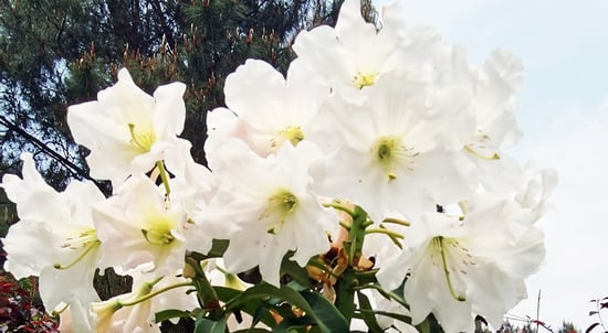
2.1. Plant Materials

The flower buds of R. decorum were collected from a rhododendron nursery in Guizhou Province, China, on 10 December 2021. The flower buds were soaked in tap water for 2 h. The outer sepals were removed and rinsed five times for 1 min with tap water, and the surfaces were dried with tissue paper. Then, the flower buds were sterilized with 75% alcohol (v/v) for 1 min, washed with sterile water five times, sterilized with 5% sodium hypochlorite (v/v) for 10 min and/or 0.1% mercuric chloride (v/v) for 10–30 min to compare the efficiency of the sterilization, and finally rinsed five times for 1 min with sterile distilled water. All the remaining sepals were finally removed. The white 2 cm sized sterile flower buds in the medium for tissue culture and their contamination and survival rates were observed after 2 weeks and were determined using the following equation: contamination rate (%) = number of flower buds contaminated/total number of flower buds × 100%; survival rate (%) = number of flower buds that survived/total number of flower buds × 100%.

2.2. Culture Medium and Conditions

The uniform flower bud explants were cultured vertically on the media. The culture media included Murashige and Skoog (MS) [23], Woody Plant Medium (WPM) [24], and Driver and Kuniyuki Walnut (DKW) [25]. Agar as the gelling agent (7 g/L) and sucrose (30 g/L) were added to each medium, and 15 g/L of sucrose was added to the rooting culture. The pH values of the media were adjusted to 5.8 with 1 N NaOH or 1 N HCl, and the media were autoclaved at 121 °C for 20 min. The callus induction culture was incubated in artificial climate chambers without light at 25 ± 1 °C, and the other cultures were incubated in artificial climate chambers under a 12 h light cycle with a light intensity of 20–30 µmol·m−2·s−1 at 25 ± 1 °C. All chemicals and reagents used in this study were purchased from Solarbio Company, Beijing, China.

2.3. Callus Induction

The flower buds were placed on the basal media MS, WPM, and DKW in the dark for the selection of the suitable basal medium for callus induction. Then, after 2 weeks, the flower buds were placed on a basal WPM containing different combinations of thidiazuron (TDZ) (0.1, 0.5, and 1 mg/L) and naphthaleneacetic acid (NAA) (0.1, 0.2, and 0.5 mg/L) in the dark for the screening of the best suitable combination of plant growth regulators for callus induction. All induction rates were determined after 30 days of the culture of the flower buds with the following equation: callus induction rate (%) = induction of the number of flower buds from the callus/total number of flower buds × 100%.

2.4. Shoot Induction

Calluses were transferred to a WPM containing different plant growth regulator combinations of TDZ (0.1, 0.5, and 1 mg/L) and NAA (0.1, 0.2, and 0.5 mg/L) under light conditions for adventitious shoot induction. All induction rates were determined after 30 days of the culture of the calluses with the following equation: induction rate of adventitious shoots (%) = number of callus-induced adventitious shoots/total number of calluses × 100%.

2.5. Shoot Proliferation

Adventitious shoots up to 1 cm high with two intact leaves were defined as effective shoots. They were transferred to the WPM containing different combinations of zeatin (ZT) (1, 2, and 3 mg/L) and NAA (0.1, 0.2, and 0.5 mg/L) for shoot proliferation. All induction rates were determined after 30 days of culture of the shoots with the following equation: adventitious shoot proliferation rate (%) = number of shoot-induced effective shoots/total number of shoots × 100%; proliferation coefficient = number of proliferating shoots/total number of shoots.

2.6. Cytological Observation

The cytological changes during callus induction, callus proliferation, and the differentiation of complete adventitious shoots were observed on paraffin sections prepared every 20 days of culture. The shoots were initially fixed in FAA solution (formalin, acetic acid, and 70% ethanol in a 1:1:13 ratio) for 24 h, dehydrated in the tertiary butyl alcohol series, and embedded in liquid paraffin [26]. The transverse sections of a 5–10 μm thickness were sliced using a rotary microtome (Erma, Yoshikawa, Japan). The sections were stained with 1% (w/v) safranin and mounted in glycerin. The specimens were microphotographed with a Leica DM 750 LED microscope, as described.

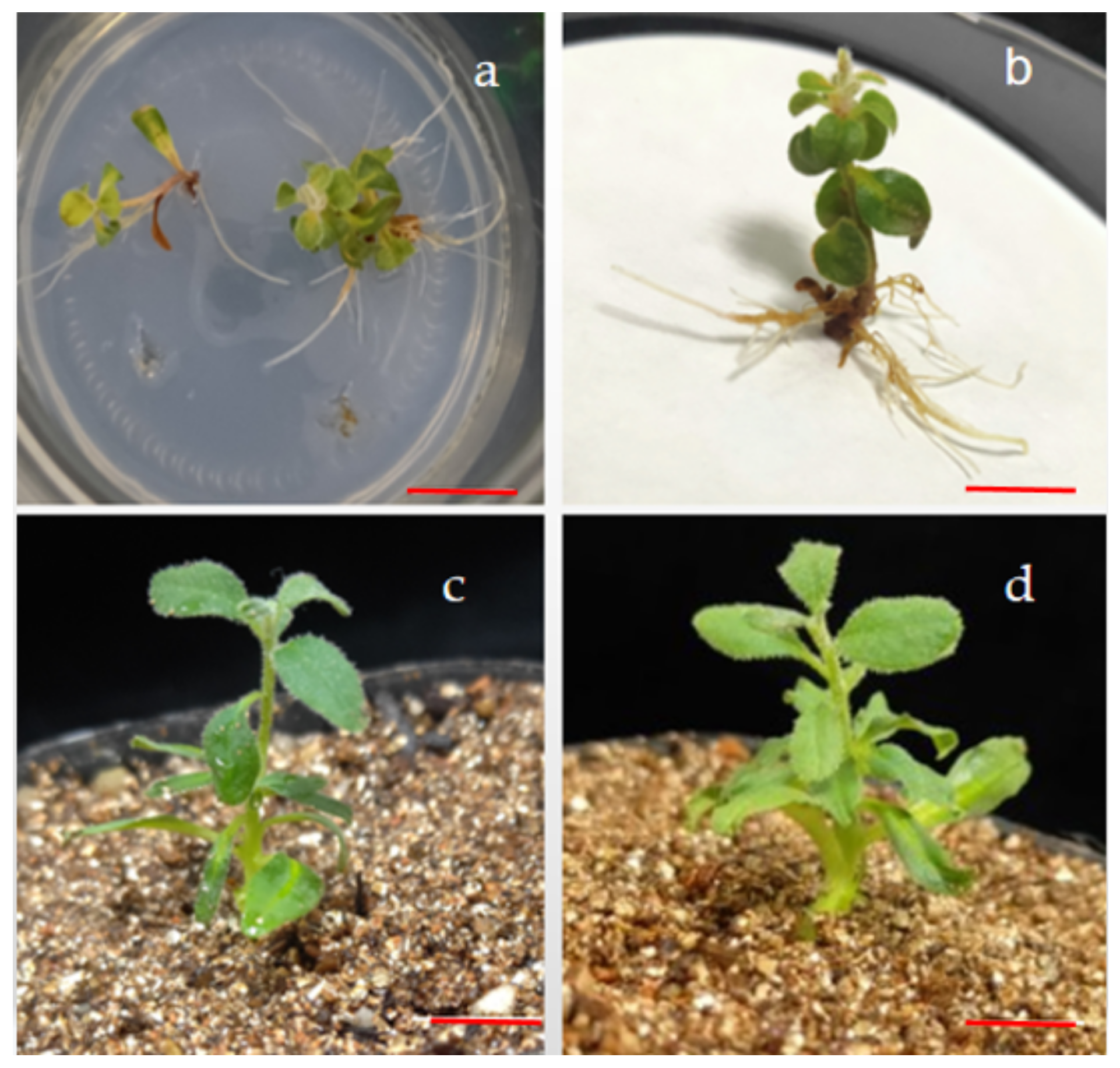
2.7. Rooting and Acclimatization

After the adventitious shoot proliferation culture, adventitious shoots of approximately 2 cm in height were cultured vertically on a WPM for rooting. Effective shoots were transferred to WPM medium combinations of indole-3-butyric acid (IBA) (0.5 and 1 mg/L) and NAA (0.1 and 0.5 mg/L) for rooting. The rooting status was recorded after 30 days. Regenerated plantlets with six fully developed leaves were transferred from an artificial climate and incubated under natural light for 5–7 days. The tissue culture bottles were opened for 2 days for the refinement of the seedlings. Plantlets were transferred to pots containing a sterilized substrate (vermiculite:peat soil = 3:1) at 121 °C for 1 h, and then covered with polyethylene film. Water was sprayed daily to create saturated conditions of relative humidity. The plantlets remained in a growth room at 24 ± 1 °C under a 16 h light cycle at a 35 µmol·m−2·s−1 photosynthetic photon flux density, provided by fluorescent lamps. After 4 weeks of acclimatization, the survival rate of the regenerated plantlets was determined using the following equation: rooting rate (%) = number of rooting shoots/numbers of transplanted shoots × 100%; survival rate (%) = number of surviving regenerated plants/total number of transplanted regenerated plants × 100%.

2.8. Statistical Analysis of Data

In the regeneration experiments, one flower bud was cultured on the medium of each bottle, and 20 flower buds were cultured for each experiment with three replicates. The results are presented as means ± standard errors (SEs). The mean and SE values were assessed using Microsoft Excel 2019. IBM SPSS Statistics v26 (Armonk, NY, USA) was used for the variance analyses. The significance of the differences among the mean values was assessed using Duncan’s multiple range test at p ≤ 0.05. The results are presented as the mean ± SE of three replicates.

3. Results

3.1. Sterilization of Flower Buds

The results of the different combinations of sterilization methods showed that sodium hypochlorite (5%) or mercury chloride (0.1%) resulted in a high rate of contamination and the low survival rate of the explants, whereas the combination of sodium hypochlorite and mercury chloride resulted in a lower rate of contamination than that of sodium hypochlorite and mercury chloride alone (Table 1). The sterilization combination of 5% sodium hypochlorite for 10 min followed by 0.1% mercury chloride for 30 min yielded the best result.

3.2. Effects of Different Media on Callus Induction

Calluses could be induced and grew well in the flower buds on the WPM, DKW, and MS media without plant growth regulators, but the induction rate varied significantly (Table 2). Two weeks after the flower buds were cultured in the different basal media, the bases of the flower buds began to swell and produce white calluses. No differences in the sizes or colors of the calluses were observed. The induction rates in the WPM and DKW media were significantly higher than the induction rate in the MS medium, and the WPM medium had the highest induction rate at 93.97%.

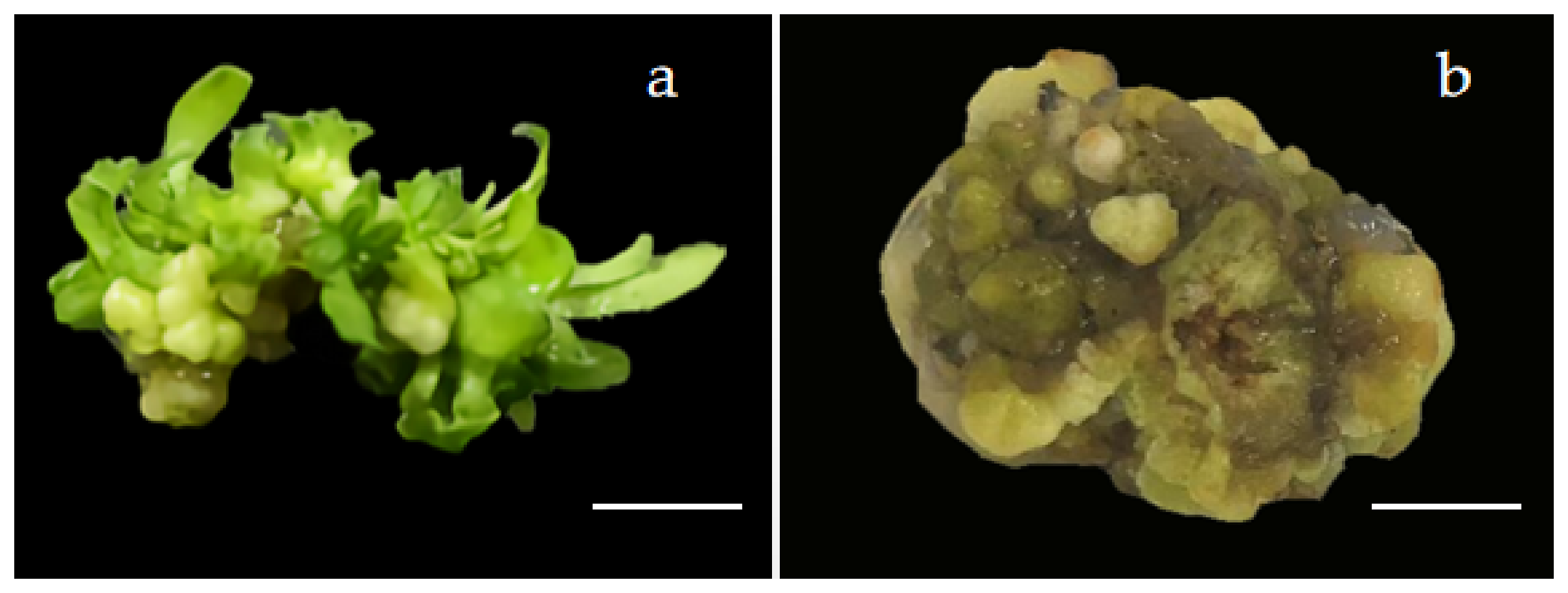
3.3. Effect of Plant Growth Regulators on Callus Induction

TDZ and NAA were used to induce the R. decorum calluses, and the induction effects are shown in Table 3. After the callus induction in the medium supplemented with plant growth regulators for 2 weeks in the dark, the flower bud bases expanded and produced white and soft calluses. The best callus induction combination was WPM + 1 mg/L TDZ + 0.2 mg/L NAA, and the induction rate was 95.08%. The induction rate increased with the TDZ and NAA concentrations, but the rate decreased when the concentration exceeded the optimal concentration (1 mg/L TDZ and 0.2 mg/L NAA).

3.4. Effects of Different Plant Growth Regulators on Adventitious Shoot Induction

The calluses began to differentiate adventitious shoots by culture on the shoot induction media after 2 weeks (Table 4). The best induction rate was 91.32% on the medium combination WPM + 0.5 mg/L TDZ + 0.1 mg/L NAA. The induction rate initially increased, and it then decreased with the increasing TDZ concentrations. In addition, when the TDZ concentration was low, a high concentration of NAA improved the induction rate of the adventitious shoots, and when the concentration of TDZ was high, a low concentration of NAA was more suitable for the adventitious shoot induction. Although the concentration of the growth regulator exceeded the optimal value, the growth rate of the adventitious shoots decreased with their increasing induction rate, but the adventitious shoot leaves were bright green and healthy. When cultured under a light environment, most of the calluses differentiated into adventitious shoots, and a few calluses did not, which exhibited browning and eventually died (Figure 1).

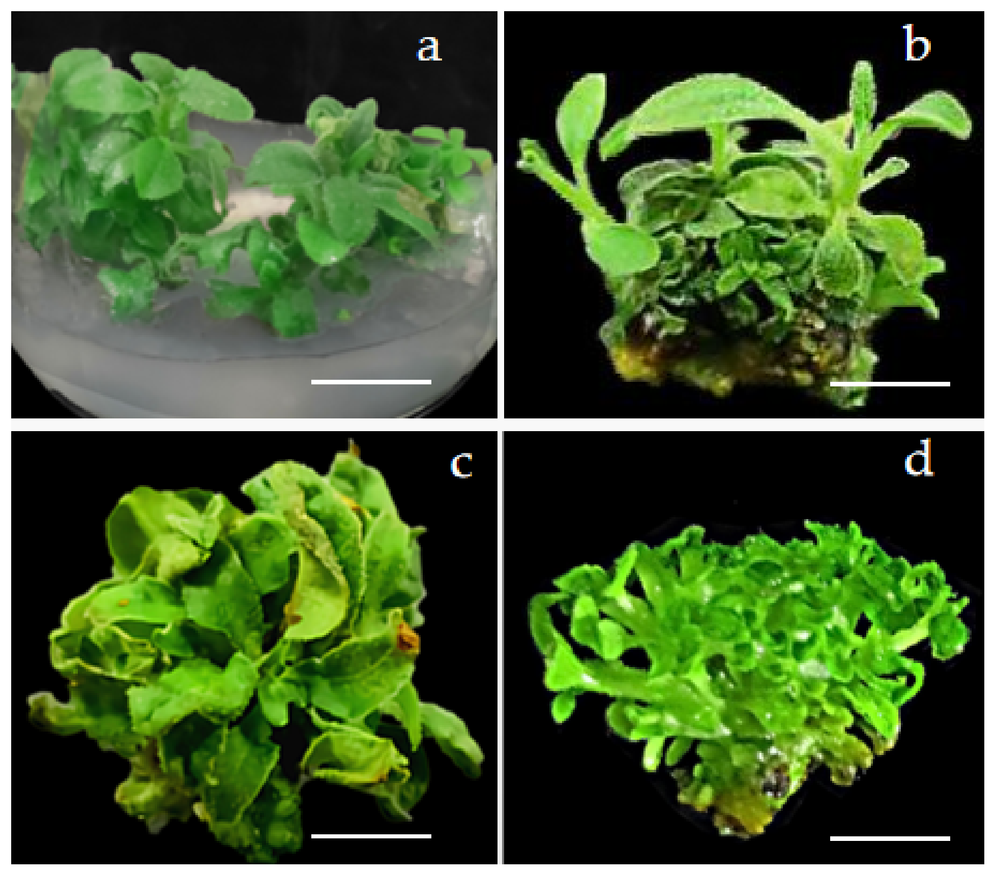
3.5. Effects of Different Plant Growth Regulators on Adventitious Shoot Proliferation

After 1 week of culture, adventitious shoots began to proliferate. Low and high concentrations of NAA and ZT produced adventitious shoots with low proliferation coefficients and weak growth shoots with abnormal leaves (Figure 2). The combination of 0.5 mg/L NAA and 2 mg/L ZT resulted in the best adventitious shoot proliferation rate of 95.32% and a proliferation coefficient of 9.42 (Table 5). The shoot leaves were fresh green, the shoot stems were strong, and the growth rate was fast. The deformed adventitious shoots produced by proliferation had a slow growth rate and death during the culture process.

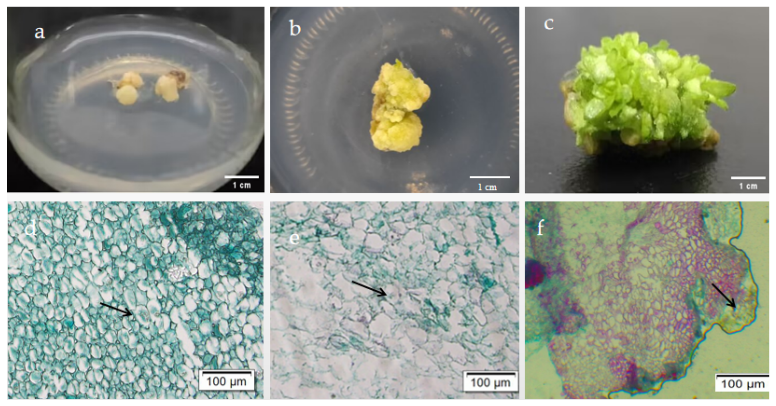
3.6. Cytological Observation of Shoot Regeneration

Cytological observation at three stages of the adventitious shoot formation from the calluses showed that white and fluffy calluses formed at the bases of the shoots after 20 days of culture (Figure 3a,d). The cells divided rapidly and were closely arranged. After 40 days of culture, the proliferation of the calluses was completed, and the cells continued to divide and expand (Figure 3b,e). After 60 days of culture, adventitious shoots differentiated from the calluses, which are clearly shown on the paraffin section (Figure 3c,f), suggesting that the shoot regeneration from the calluses was indirect.

3.7. Rooting and Acclimatization

The adventitious shoots were rooted in a medium containing NAA and IBA. The results are shown in Table 6. Adventitious roots were induced after 30 days of culture (Figure 4). The best medium combination for rooting was WPM + 0.1 mg/L NAA + 1 mg/L IBA, with a rooting rate of 86.9%. A high concentration of IBA and low concentration of NAA induced healthy adventitious roots, the rooting speed was fast, and no calluses appeared at the bases of the adventitious shoots (Figure 4a,b). However, a high concentration of NAA induced slow rooting with fewer roots and a large number of calluses at the base. In addition, after 30 days of transplantation, the survival rates of the rooted plantlets were higher than 90%, and the plantlets were in good growth condition (Figure 4c,d).

4. Discussion

There are 8–10 small flower buds in the racemes of R. decorum before blossom. Hence, its buds can be used as explants for developing tissue culture systems. Wang et al. [27] reported that the performance of a tissue culture using the flower buds of Rhododendron hybrids cn.’Dr. Tjebbe’ as explants had a low contamination rate and high induction rate. The use of flower buds as explants neither harms the mother plant nor causes mutation because small flower buds are wrapped in sepals, which are easy to sterilize and do not easily brown [28]. These features may be the reasons that the survival rates of the flower buds subjected to sterilization treatment reached 100% in this study.
In the process of tissue culture, the basal medium is the main source of the nutrients required by the explants, and the appropriate basal medium type is essential for the rapid and healthy growth and development of explants from different plant species [29,30,31]. Different parts of the same genotype can have different growth responses to a certain basal medium [32,33,34]. Therefore, the screening of the basal medium is beneficial to the smooth progress of the tissue culture. For example, in the rapid propagation of Vaccinium ashei Reade, which is a species belonging to Ericaceae, the basal medium DKW is more suitable for the rapid propagation culture of the stem segments than WPM or MS [35]. By contrast, in the present study, WPM was the most suitable basal medium for culturing the flower buds of R. decorum, which is a species that belongs to the same family, followed by DKW and MS. This difference may be caused by the low nitrogen demand of R. decorum. WPM, with a low nitrogen content, met this demand [36,37], whereas the DKW and MS media, with relatively high contents of nitrogen, did not [38].
Plant growth regulators are minor natural compounds that are produced in plant metabolism, and they regulate the growth and development processes of plants [39,40]. In tissue culture, plant growth regulators play a key regulatory role in the formation of calluses and the induction and proliferation of adventitious shoots. This effect is affected by the concentration and type of growth regulator, as well as by the interaction between growth regulators. A reasonable ratio of growth regulators is especially crucial for the induction and proliferation of adventitious shoots; thus, the formulas of the growth regulators in tissue culture systems vary among [41,42]. In the tissue culture process of woody plants, the commonly used plant growth regulators are TDZ, NAA, IBA, and kinetin [43,44]. However, different species have different degrees of sensitivity to plant growth regulators. TDZ is currently considered to be one of the most active cytokinins, with good induction effects on calluses and adventitious shoots, and it is widely used in the regeneration processes of R. calophytum [45], R. delavayi [46], and Rhododendron ‘Fragrantissimum Improved’ [47]. Similarly, in the present study, the plant growth regulator combination 0.2 mg/L NAA + 0.5 mg/L TDZ was the most suitable for the induction of the adventitious shoots of the flower buds of R. decorum, and the adventitious shoots grew rapidly and healthily.
Proliferation culture is an indispensable step in the process of tissue culture, and the proliferation coefficient can reflect the speed and efficiency of the propagation in vitro and is an important index for estimating the total production of plantlets. Previous studies have shown that ZT is the most ideal exogenous hormone to induce differentiation and proliferation during the tissue culture process of Rhododendron [48]. When the ZT concentration was extremely high, the adventitious shoot proliferation rate and proliferation coefficient were also increased, but the adventitious shoots were prone to elongation, thinness, and deformity, and they were hyperhydric [49].
Rooting induction is another important step in the establishment of a rapid propagation system in vitro. IBA and NAA are commonly used for the rooting induction of Rhododendrons species plantlets, which have a high rooting rate and multiple and strong roots, as well as a high survival rate after transplantation. For example, Elmongy et al. [50] found that 2 mg/L of IBA was suitable for the rooting of two azalea cultivars: ‘Mingchao’ and ‘Zihudie’. Almeida found that 1 mg/L of IBA + 2 mg/L of NAA was suitable for the rooting of R. ponticum [51]. In this study, NAA and IBA were found to be suitable for inducing the rooting of R. decorum adventitious shoots. The results showed that high concentrations of NAA resulted in slow rooting and weak roots, whereas high concentrations of IBA were conducive to the induction of root development and root health. A high concentration of IBA was conducive to the induction of root development, and the root system was developed and strong. The root systems of the regenerated plants were thick, and the survival rate of transplantation was high.
Tissue culture involves the induction, proliferation, rooting, and transplanting of plant materials within a sterile and controlled environment. In this study, the flower buds of R. decorum were used for regeneration. The protocol was as follows: (1) the induction of calluses using sterilized flower buds in WPM + 1 mg/L TDZ + 0.2 mg/L NAA in the dark; (2) the induction of adventitious shoots using fluffy calluses in WPM + 0.5 mg/L TDZ + 0.1 mg/L NAA under a 12 h light cycle; (3) the proliferation of adventitious shoots using shoots up to 1 cm in WPM + 2 mg/L ZT + 0.5 mg/L NAA under a 12 h light cycle; (4) the rooting of adventitious shoots using shoots up to 2 cm in WPM + 0.1 mg/L NAA + 1 mg/L IBA under a 12 h light cycle; (5) the transplanting of the rooted plantlets after acclimation under a 16 h light cycle.

5. Conclusions

This study established an in vitro propagation system for R. decorum through indirect organogenesis using its flower buds as explants by screening the influencing factors, such as different sterilization method, basal medium, and plant growth regulator combinations. Tissue culture using flower buds as explants has the advantages of thorough sterilization, efficient and rapid regeneration, and it causes little damage to the mother plants. This regeneration system is simple and highly efficient, and it lays a foundation for the plantlet propagation, genetic transformation, and new-variety breeding of R. decorum.

Author Contributions

Conceptualization, H.W. and F.L.; methodology, H.W.; validation, Q.A.; formal analysis, H.W., Q.A. and F.L.; investigation, H.W., Q.A., H.L. and F.L.; data curation, H.W. and H.L.; writing, H.W., H.L., Q.A. and H.L.; visualization, Q.A. and H.L.; supervision, H.L.; project administration, H.L.; funding acquisition, H.L. All authors have read and agreed to the published version of the manuscript.

Funding

This research was funded by the National Natural Science Foundation of China (32260415).

Data Availability Statement

Not applicable.

Conflicts of Interest

The authors declare no conflict of interest.

References

  1. Flora of China Editorial Committee; Chinese Academy of Sciences. Flora of China; Science Press: Beijing, China, 1994; pp. 16–17. [Google Scholar]
  2. Min, T.L. A revision of subgenus Hymenanthes (Rhododendron L.) in Yunnan and Xizang. Plant Divers. 1984, 6, 1. [Google Scholar]
  3. Wang, B.; Zhou, L.Y.; Xia, H.M. Impacts of sucrose, boric acid and Ca+ on pollen germination of Rhododendron decorum Franch. Jiangsu Agric. Sci. 2021, 49, 129–133. [Google Scholar]
  4. Zhu, Y.-X.; Zhang, Z.-X.; Yan, H.-M.; Lu, D.; Zhang, H.-P.; Li, L.; Liu, Y.-B.; Li, Y. Antinociceptive Diterpenoids from the Leaves and Twigs of Rhododendron decorum. J. Nat. Prod. 2018, 81, 1183–1192. [Google Scholar] [CrossRef] [PubMed]
  5. Rateb, M.E.; Hassan, H.; Arafa, E.-S.; Jaspars, M.; Ebel, R. Decorosides A and B, Cytotoxic Flavonoid Glycosides from the Leaves of Rhododendron decorum. Nat. Prod. Commun. 2014, 9, 473–476. [Google Scholar] [CrossRef] [PubMed] [Green Version]
  6. Zhu, Y.-X.; Zhang, Z.-X.; Zhang, H.-P.; Chai, L.-S.; Li, L.; Ma, S.-G.; Li, Y. A new ascorbic acid derivative and two new terpenoids from the leaves and twigs of Rhododendron decorum. J. Asian Nat. Prod. Res. 2019, 21, 579–586. [Google Scholar] [CrossRef]
  7. Shi, Y.; Zhou, M.; Zhang, Y.; Fu, Y.; Li, J.; Yang, X. Poisonous delicacy: Market-oriented surveys of the consumption of Rhododendron flowers in Yunnan, China. J. Ethnopharmacol. 2021, 265, 113320. [Google Scholar] [CrossRef]
  8. Zhang, S.; Dang, Z.; Zhang, L.Y. Research on seeds aseptic germination and seedling growth condition of Rhododendron decorum Franch. North. Hortic. 2014, 305, 77–80, (In Chinese with English Abstract). [Google Scholar]
  9. Lin, L.-C.; Wang, C.-S. Influence of Light Intensity and Photoperiod on the Seed Germination of Four Rhododendron Species in Taiwan. Pak. J. Biol. Sci. 2017, 20, 253–259. [Google Scholar] [CrossRef] [Green Version]
  10. Giri, C.C.; Shyamkumar, B.; Anjaneyulu, C. Progress in tissue culture, genetic transformation and applications of biotechnology to trees: An overview. Trees 2003, 18, 115–135. [Google Scholar] [CrossRef]
  11. Yavuz, D.Ö. Optimization of Regeneration Conditions and In Vitro Propagation of Sideritis Stricta Boiss & Heldr. Int. J. Biol. Macromol. 2016, 90, 59–62. [Google Scholar] [CrossRef]
  12. Nada, S.; Chennareddy, S.; Goldman, S.; Rudrabhatla, S.; Potlakayala, S.D.; Josekutty, P.; Deepkamal, K. Direct Shoot Bud Differentiation and Plantlet Regeneration from Leaf and Petiole Explants of Begonia tuberhybrida. Hortscience 2011, 46, 759–764. [Google Scholar] [CrossRef] [Green Version]
  13. Zhang, H.P.; Wang, H.B.; Wang, L.Q.; Bao, G.H.; Qin, G.W. A new 1,5-seco grayanotoxane from Rhododendron decorum. J. Asian Nat. Prod. Res. 2005, 7, 87–90. [Google Scholar] [CrossRef]
  14. Long, Y.; Yang, Y.; Pan, G.; Shen, Y. New Insights Into Tissue Culture Plant-Regeneration Mechanisms. Front. Plant Sci. 2022, 13, 926752. [Google Scholar] [CrossRef]
  15. Xue, Q.W.; Yuan, H.; Chun, L.L. Assessing the genetic consequences of flower-harvesting in Rhododendron decorum Franchet (Ericaceae) using microsatellite markers. Biochem. Syst. Ecol. 2013, 50, 296–303. [Google Scholar]
  16. Wang, X.-Q.; Huang, Y.; Long, C.-L. Isolation and Characterization of Twenty-four Microsatellite Loci for Rhododendron decorum Franch. (Ericaceae). Hortscience 2009, 44, 2028–2030. [Google Scholar] [CrossRef] [Green Version]
  17. Jin, H.Z.; Chen, G.; Li, X.F.; Shen, Y.H.; Yan, S.K.; Zhang, L.; Yang, M.; Zhang, W.D. Flavonoids from Rhododendron decorum. Chem. Nat. Compd. 2009, 45, 85–86. [Google Scholar] [CrossRef]
  18. Zhang, W.; Jin, H.; Chen, G.; Li, X.; Yan, S.; Zhang, L.; Shen, Y.; Yang, M. A new grandame diterpenoid from Rhododendron decorum. Fitoterapia 2008, 79, 602–604. [Google Scholar] [CrossRef]
  19. Zha, H.-G.; Milne, R.I.; Sun, H. Morphological and molecular evidence of natural hybridization between two distantly related Rhododendron species from the Sino-Himalaya. Bot. J. Linn. Soc. 2008, 156, 119–129. [Google Scholar] [CrossRef] [Green Version]
  20. Li, Q.; Li, H.E.; Yang, L.; Guo, Q.; Fu, Y.; Huang, J. Asymmetric hybridization origin of Rhododendron ageratum (Ericaceae) in Guizhou, China. Phytotaxa 2021, 510, 197–212. [Google Scholar] [CrossRef]
  21. Sun, L.; Pei, K.; Wang, F.; Ding, Q.; Bing, Y.; Gao, B.; Zheng, Y.; Liang, Y.; Ma, K. Different distribution patterns between putative ercoid mycorrhizal and other fungal as-semblages in roots of Rhododendron decorum in the Southwest of China. PLoS ONE 2018, 7, e49867. [Google Scholar]
  22. Tian, W.; Zhang, C.; Qiao, P.; Milne, R. Diversity of culturable ericoid mycorrhizal fungi of Rhododendron decorum in Yunnan, China. Mycologia 2011, 103, 703–709. [Google Scholar] [CrossRef] [PubMed]
  23. Murashige, T.; Skoog, F. A Revised Medium for Rapid Growth and Bioassays with Tobacco Tissue Cultures. Physiol. Plant. 1962, 15, 473–497. [Google Scholar] [CrossRef]
  24. McCown, B.H. Woody Plant Medium (WPM)-a mineral nutrient formulation for microculture for woody plant species. Hortscience 1981, 16, 453. [Google Scholar]
  25. Driver, J.A.; Kuniyuki, A.H. In Vitro Propagation of Paradox Walnut Rootstock. Hortscience 1984, 19, 507–509. [Google Scholar] [CrossRef]
  26. Zaytseva, Y.G.; Poluboyarova, T.V.; Novikova, T.I. Effects of thiazine on in vitro morphogenic response of Rhododendron sichotense Pojark. and Rhododendron catawbiense cv. Grandiflorum leaf explants. In Vitro Cell. Dev. Biol. Plant 2016, 52, 56–63. [Google Scholar] [CrossRef]
  27. Wang, W.Q.; Xiao, J.Z.; Li, Z.M.; Hu, J.; Bai, X. Study on the bud tissue culture and establishment of optimization system of Rhododendron. J. Hebei Norm. Univ. Sci. Technol. 2012, 26, 17–22, (In Chinese with English Abstract). [Google Scholar]
  28. Tomsone, S.; Gertnere, D. In vitro Shoot Regeneration from Flower and Leaf Explants in Rhododendron. Biol. Plant. 2003, 46, 463–465. [Google Scholar] [CrossRef]
  29. Nobuaki, M.; Shinsaku, T. Effects of medium components and shear conditions on the formation and growth of adventitious bud derived from hairy roots of Atropa belladonna L. Environ. Control Biol. 2012, 50, 393–406. [Google Scholar]
  30. Debnath, S.C. Propagation of Vaccinium in vitro: A review. Int. J. Fruit Sci. 2007, 6, 47–71. [Google Scholar] [CrossRef]
  31. Mohammed, A.; Chiruvella, K.K.; Namsa, N.D.; Ghanta, R.G. An efficient in vitro shoot regeneration from leaf petiolar explants and ex vitro rooting of Bixa orellana L.—A dye yielding plant. Physiol. Mol. Biol. Plants 2015, 21, 417–424. [Google Scholar] [CrossRef] [Green Version]
  32. Tanmayee, M.; Arvind, G.; Arnab, S. Somatic embryogenesis and genetic fidelity study of micropropagated medicinal species, Canna indica. Horticulture 2015, 1, 3–13. [Google Scholar]
  33. Nowakowska, K.; Pińkowska, A.; Siedlecka, E.; Pacholczak, A. The effect of cytokinins on shoot proliferation, biochemical changes and genetic stability of Rhododendron ‘Kazimierz Odnowiciel’ in the in vitro cultures. Plant Cell Tissue Organ Cult. PCTOC 2022, 149, 675–684. [Google Scholar] [CrossRef]
  34. Blazich, F.A.; Giles, C.G.; Haemmerle, C.M. Micropropagation of Rhododendron chapmani. J. Environ. Hortic. 1986, 4, 26–29. [Google Scholar] [CrossRef]
  35. Komakech, R.; Kim, Y.-G.; Kim, W.J.; Omujal, F.; Yang, S.; Moon, B.C.; Okello, D.; Rahmat, E.; Kyeyune, G.N.; Matsabisa, M.G.; et al. A Micropropagation Protocol for the Endangered Medicinal Tree Prunus africana (Hook f.) Kalkman: Genetic Fidelity and Physiological Parameter Assessment. Front. Plant Sci. 2020, 11, 548003. [Google Scholar] [CrossRef]
  36. Zhou, J.; Liu, Y.; Wu, L.; Zhao, Y.; Zhang, W.; Yang, G.; Xu, Z. Effects of Plant Growth Regulators on the Rapid Propagation System of Broussonetia papyrifera L. Vent Explants. Forests 2021, 12, 874. [Google Scholar] [CrossRef]
  37. Bell, R.L.; Srinivasan, C.; Lomberk, D. Effect of nutrient media on axillary shoot proliferation and preconditioning for adven-titious shoot regeneration of pears. In Vitro Cell. Dev. Biol. Plant 2009, 45, 708–721. [Google Scholar] [CrossRef]
  38. Tang, Q.; Guo, X.; Zhang, Y.; Li, Q.; Chen, G.; Sun, H.; Wang, W.; Shen, X. An optimized protocol for indirect organogenesis from root explants of Agapanthus praecox subsp. orientalis ‘Big Blue’. Horticulture 2022, 8, 715. [Google Scholar] [CrossRef]
  39. Poothong, S.; Reed, B.M. Modeling the effects of mineral nutrition for improving growth and development of micro propagated red raspberries. Sci. Hortic. 2014, 165, 132–141. [Google Scholar] [CrossRef]
  40. Carlín, A.P.; Tafoya, F.; Alpuche-Solis, A.; Pérez-Molphe-Balch, E. Effects of different culture media and conditions on biomass production of hairy root cultures in six Mexican cactus species. In Vitro Cell. Dev. Biol. Plant 2015, 51, 332–339. [Google Scholar] [CrossRef]
  41. Nic-Can, G.I.; Loyola-Vargas, V.M. The role of the auxins during somatic embryogenesis. In Somatic Embryogenesis: Fundamental Aspects and Applications; Springer: Cham, Switzerland, 2016; pp. 171–182. [Google Scholar]
  42. Debnath, S.C.; McRae, K.B. An efficient adventitious shoot regeneration system on excised leaves of micro propagated lin-gonberry (Vaccinium vitisidaea L.). J. Hortic. Sci. Biotechnol. 2002, 77, 744–752. [Google Scholar] [CrossRef]
  43. Zheng, M.; Yang, H.; Yang, E.; Zou, X.; Chen, X.; Zhang, J. Efficient in vitro shoot bud proliferation from cotyledonary nodes and apical buds of Moringa oleifera Lam. Ind. Crops Prod. 2022, 187, 115394. [Google Scholar] [CrossRef]
  44. Ahmad, Z.; Yadav, V.; Shahzad, A.; Emamverdian, A.; Ramakrishnan, M.; Ding, Y. Micropropagation, encapsulation, physiological, and genetic homogeneity assessment in Casuarina equisetifolia. Front. Plant Sci. 2022, 13, 905444. [Google Scholar] [CrossRef] [PubMed]
  45. Coste, A.; Vlase, L.; Halmagyi, A.; Deliu, C.; Coldea, G. Effects of plant growth regulators and elicitors on production of secondary metabolites in shoot cultures of Hypericum hirsutum and Hypericum maculatum. Plant Cell Tissue Organ Cult. PCTOC 2011, 106, 279–288. [Google Scholar] [CrossRef]
  46. Luo, L.; Bai, J.; Chen, C.; Chen, X.; Chen, K.; Chen, F. Study on plantlet regeneration for blade segments and the physiological trait for heat tolerance in the callus of Rhododendron calophytum. J. Bot. Northwest China 2014, 34, 1377–1382, (In Chinese with English Abstract). [Google Scholar]
  47. Tian, G.; Peng, L.C.; Qu, S.P.; Wang, J.; Zhao, Z.; Li, S.; Jie, W.; Guan, W. Studies on the adventitious bud induction from in vitro leaves of Rhododendron delavayi var. delavayi and sociological observation on the bud formation. J. Hortic. 2020, 47, 2019–2026, (In Chinese with English Abstract). [Google Scholar]
  48. Hebert, C.J.; Touchell, D.H.; Ranney, T.G.; LeBude, A.V. In vitro shoot regeneration and polyploid induction of Rhododendron ‘Fra-grantissimum Improved’. Hortic. Sci. 2010, 45, 801–804. [Google Scholar]
  49. Wei, X.; Chen, J.; Zhang, C.; Wang, Z. In vitro shoot culture of Rhododendron fortunei: An important plant for bioactive phytochemicals. Ind. Crops Prod. 2018, 126, 459–465. [Google Scholar] [CrossRef]
  50. Elmongy, M.S.; Cao, Y.; Zhou, H.; Xia, Y. Root development enhanced by using indole-3-butyric acid and naphthalene acetic acid and associated biochemical changes of in vitro Azalea micro shoots. J. Plant Growth Regul. 2018, 37, 813–825. [Google Scholar] [CrossRef]
  51. Almeida, R.; Gonçalves, S.; Romano, A. In vitro micropropagation of endangered Rhododendron ponticum L. subsp. baeticum (Boissier & Reuter) Handel-Mazzetti. Biodivers. Conserv. 2005, 14, 1059–1069. [Google Scholar] [CrossRef]
Figure 1. Adventitious shoots induced within 30 days: (a) adventitious shoots of callus differentiation; (b) browning callus. Bars = 1.0 cm.
Horticulturae 09 00264 g001
Figure 2. Adventitious shoot proliferation of calluses on different medium combinations: (a) adventitious shoots with high proliferation coefficient containing green and normal leaves or (d) hyperhydric; (b) adventitious shoots with low proliferation coefficient containing green normal leaves or (c) yellow-green and abnormal leaves. Scale bars = 1.0 cm.
Horticulturae 09 00264 g002
Figure 3. Observation on paraffin section of shoot regeneration of R. decorum: (a) white fluffy callus; (b) proliferation of fluffy calluses; (c) differentiation of adventitious shoots from calluses; (d) arrow shows cells that divided rapidly after 20 days of culture; (e) arrow shows cells that divided and expanded after 40 days of culture; (f) arrow shows adventitious shoots that differentiated from callus after 60 days of culture. Bars = 100 μm.
Horticulturae 09 00264 g003
Figure 4. Roots and acclimatization of rooted plantlets: (a) roots in WPM medium after 30 days of culture; (b) rooted plantlets; (c,d) acclimatized plantlets from ex vitro rooting after 30 days. Bars = 1.0 cm.
Horticulturae 09 00264 g004
Table 1. Effects of different sterilization methods on explants.
Sodium Hypochlorite
(min)
Mercury Chloride
(min)
Contamination Rate
(%)
Survival Rate
(%)
100100.00 ± 0.00 a0.00 ± 0.00 g
01065.84 ± 2.84 b34.16 ± 1.54 f
02046.12 ± 3.03 c53.88 ± 2.08 e
03034.67 ± 4.04 d65.33 ± 3.48 d
101016.63 ± 1.42 e83.37 ± 2.02 c
10205.72 ± 1.81 f94.28 ± 0.69 b
10300.00 ± 0.00 g100.00 ± 0.00 a
Different letters in same column are significantly different at p < 0.05 (DMRT).
Table 2. Effects of basal media on callus induction rate of R. decorum flower buds.
MediumInduction Rate (%)
WPM93.97 ± 1.51 a
DKW85.72 ± 3.31 b
MS65.25 ± 1.93 c
Different letters in same column are significantly different at p < 0.05 (DMRT).
Table 3. Effects of TDZ and NAA on callus induction.
TDZ (mg/L)NAA (mg/L)Induction Rate (%)
0.10.136.67 ± 1.53 f
0.10.251.02 ± 5.29 e
0.10.554.66 ± 3.51 e
0.50.165.73 ± 1.52 d
0.50.272.33 ± 3.79 c
0.50.578.67 ± 0.58 c
10.185.66 ± 2.08 b
10.295.08 ± 1.02 a
10.578.45 ± 5.13 c
Different letters in same column are significantly different at p < 0.05 (DMRT).
Table 4. Effects of TDZ and NAA on adventitious shoot induction.
TDZ (mg/L)NAA (mg/L)Induction Rate (%)
0.10.146.05 ± 2.45 f
0.10.261.21 ± 4.35 d
0.10.574.00 ± 2.36 c
0.50.191.32 ± 2.36 a
0.50.284.02 ± 1.56 b
0.50.573.07 ± 4.36 c
10.158.33 ± 0.57 d
10.255.53 ± 0.88 d
10.551.45 ± 2.03 e
Different letters in same column are significantly different at p < 0.05 (DMRT).
Table 5. Effects of NAA and ZT on adventitious shoot proliferation.
NAA
(mg/L)
ZT
(mg/L)
Proliferation Rate (%)Proliferation CoefficientGrowth Status
0.1166.34 ± 3.06 d4.67 ± 0.47 fLeaves were green and healthy, shoots were small
0.2174.03 ± 3.12 c5.57 ± 0.15 eLeaves were green and healthy, shoots were small
0.5176.67 ± 3.21 c7.47 ± 0.36 cLeaves were green and healthy, shoots were stout
0.1281.47 ± 6.51 b8.67 ± 0.11 aLeaves were green and healthy, shoots were stout
0.2288.76 ± 4.51 a8.97 ± 0.44 aLeaves were green and healthy, shoots were stout
0.5295.32 ± 2.56 a9.42 ± 0.27 bLeaves were green and healthy, shoots were stout
0.1389.32 ± 4.14 a4.21 ± 0.31 bLeaves were yellow-green and small, with a few abnormal leaves
0.2384.34 ± 2.65 b2.64 ± 0.09 cLeaves were yellow-green and hyperhydric, with many abnormal leaves
0.5374.43 ± 2.52 c1.95 ± 0.21 eLeaves were yellow-green and hyperhydric, with many abnormal leaves
Different letters in same column are significantly different at p < 0.05 (DMRT).
Table 6. Effects of NAA and IBA on adventitious shoot rooting.
NAA (mg/L)IBA (mg/L)Rooting Rate (%)
0.10.559.33 ± 4.09 b
0.1186.90 ± 2.97 a
0.50.521.53 ± 2.49 d
0.5142.67 ± 2.84 c
Different letters in same column are significantly different at p < 0.05 (DMRT).
Disclaimer/Publisher’s Note: The statements, opinions and data contained in all publications are solely those of the individual author(s) and contributor(s) and not of MDPI and/or the editor(s). MDPI and/or the editor(s) disclaim responsibility for any injury to people or property resulting from any ideas, methods, instructions or products referred to in the content.

