Article

Studies on the Identification of Resistance to Fusarium oxysporum (Schlecht.) in Different Genetic Backgrounds of Asparagus officinalis (L.) and Its Defense Responses

Julius Kuehn-Institute (JKI), Federal Research Centre for Cultivated Plants, Institute for Breeding Research on Horticultural Crops, Erwin-Baur-Strasse 27, D-06484 Quedlinburg, Germany
*
Author to whom correspondence should be addressed.
Horticulturae 2023, 9(2), 158; https://doi.org/10.3390/horticulturae9020158
Submission received: 21 December 2022 / Revised: 13 January 2023 / Accepted: 24 January 2023 / Published: 27 January 2023
(This article belongs to the Special Issue Advances in Asparagus Production, Genomics and Breeding)

Abstract

:
Due to the restricted genetic diversity among current asparagus cultivars, wild relatives are of particular interest as a source of resistance. In this study, seventeen cultivars and wild relatives were tested for their resistance to Fusarium oxysporum in the seedling test. Two isolates differing in virulence (single spore lines with high and low virulence) were used for testing. Most of the tested genotypes showed high infestation, whereby Asparagus aethiopicus caused no browning of the roots or growth depression. This wild relative was evaluated for its defence response after infection with F. oxysporum. For comparison, the wild relative Asparagus densiflorus and Asparagus officinalis cv. Thielim were tested. Like A. densiflorus, A. aethiopicus showed accumulation of hydrogen peroxide as a defence mechanism, while Asparagus officinalis cv. Thielim showed no storage of hydrogen compounds.

1. Introduction

Asparagus officinalis is cultivated on about 1.55 million ha worldwide, with the highest production in China, Europe, and Peru [1]. The human use of asparagus species other than A. officinalis is limited. Once mentioned as medicinal plants, some are now used as ornamentals, e.g., Asparagus plumosus. Only a few are still important medicinal plants, such as Asparagus racemosus in India [2]. Other wild species are considered a delicacy and are collected from the wild [2]. Cultivation efforts were only moderately successful [3]. Asparagus crown and root rot, caused by pathogenic soil fungi of the genus Fusarium are considered to be the main component of asparagus decline syndrome. Several Fusarium species are present in asparagus fields, but not all of them are pathogens of asparagus [4,5]. According to the study of Andrzejak and Janowska [4], only 50% of the collected field isolates are pathogens of asparagus. In infested plants, mainly F. oxysporum, F. culmorum, F. proliferatum [4,6,7], and F. redolens [6,8,9] were found. F. oxysporum is the most common and widespread species [5,6]. Studies have shown that the older the asparagus plantation, the higher the Fusarium infestation [4,6]. This can be explained by its ability to survive for a long time in plant debris and soil [6,10,11,12], which makes its control very difficult [6,11]. There are some studies on the use of fungicides and biological control agents to control Fusarium crown and root rot, but the results are mostly inconclusive or contradictory, especially in field trials. To make matters worse, the pathogen is ubiquitous [13,14,15]. Therefore, breeding asparagus cultivars resistant to F. oxysporum is an important strategy to control root and crown rot in asparagus production. The viability of F. oxysporum is enhanced by higher humidity and higher temperatures [16]. This is particularly important in the context of climate change. Studies by Delgado-Baquerizo et al. [17] indicate that temperature is one of the most important environmental factors for the global spread of potential plant pathogens and that their numbers are likely to increase in most regions. To date, resistance to F. oxysporum has been experimentally confirmed only in one wild species. Resistance to A. densiflorus was first mentioned by Lewis and Shoemaker in 1964 and has been validated several times [18,19,20,21,22]. He et al. [19] demonstrated hypersensitive cell death. However, this resistance has not yet been transferred into cultivars due to strong crossing barriers [23]. To date, several attempts to produce interspecific hybrids between A. officinalis and asparagus wild relatives have been described. A. officinalis × A. tenuifolius, A. officinalis × A. maritimus, and A. officinalis × A. prostratus are examples of interspecific F1 hybrids [24,25,26]. Many other crosses, such as A. officinalis × A. densiflorus, A. officinalis × A. stipularis, and A. officinalis × A. acutifolius have failed [24,27,28,29]. Despite these difficulties, the wild relatives are of particular interest, because of the restricted genetic diversity in recent asparagus cultivars [30,31]. The aim of this study was to find resistance to F. oxysporum using a seedling resistance assay and to investigate the defence mechanisms in more detail.

2. Materials and Methods

2.1. Fungal Isolates and Cultures

Two single-spore lines of Fusarium oxysporum were used for resistance tests, both are pathogenic and produce visual symptoms of damage but differ in virulence (Foxy II and Foxy III). For comparison, Foxy II expresses low and Foxy III high virulence [18]. The original material was isolated from asparagus spears in experimental fields at the JKI in Quedlinburg (Germany) in 2011. All isolates used in this study were cultured on a sterile potato dextrose agar (PDA) for 14 days at 22 ± 2 °C in the dark. The addition of 200 mg/L streptomycins to PDA suppressed contaminants without affecting the growth of F. oxysporum. For the microscopic determination of hypersensitive reaction, only Foxy III was used, which was prepared by the same method.

2.2. Plant Material and Growing Conditions

Eleven asparagus cultivars, two landraces, and four wild relatives were tested for their resistance to Foxy II and Foxy III (Table 1). Asparagus seeds were surface disinfected with 3% sodium hypochlorite for 3 min and rinsed three times in sterile distilled water for 1 min. The disinfected seeds were soaked in water at 36 °C for 24 h, then sown in autoclaved sand, and cultivated at 20 °C. Seedlings with a shoot length between 4 and 10 cm and a well-developed main root with first lateral roots were selected. The roots were cleaned of sand before use in the seedling resistance test. For the microscopic determination of hypersensitive response, seedlings from the same lot of A. densiflorus, A. aethiopicus, and A. officinalis cv. Thielim were grown under the conditions described above.
The results of the distance analysis were obtained from the modified data set of Nothnagel et al. [32]. For this work, they were re-analysed in a new combinations.

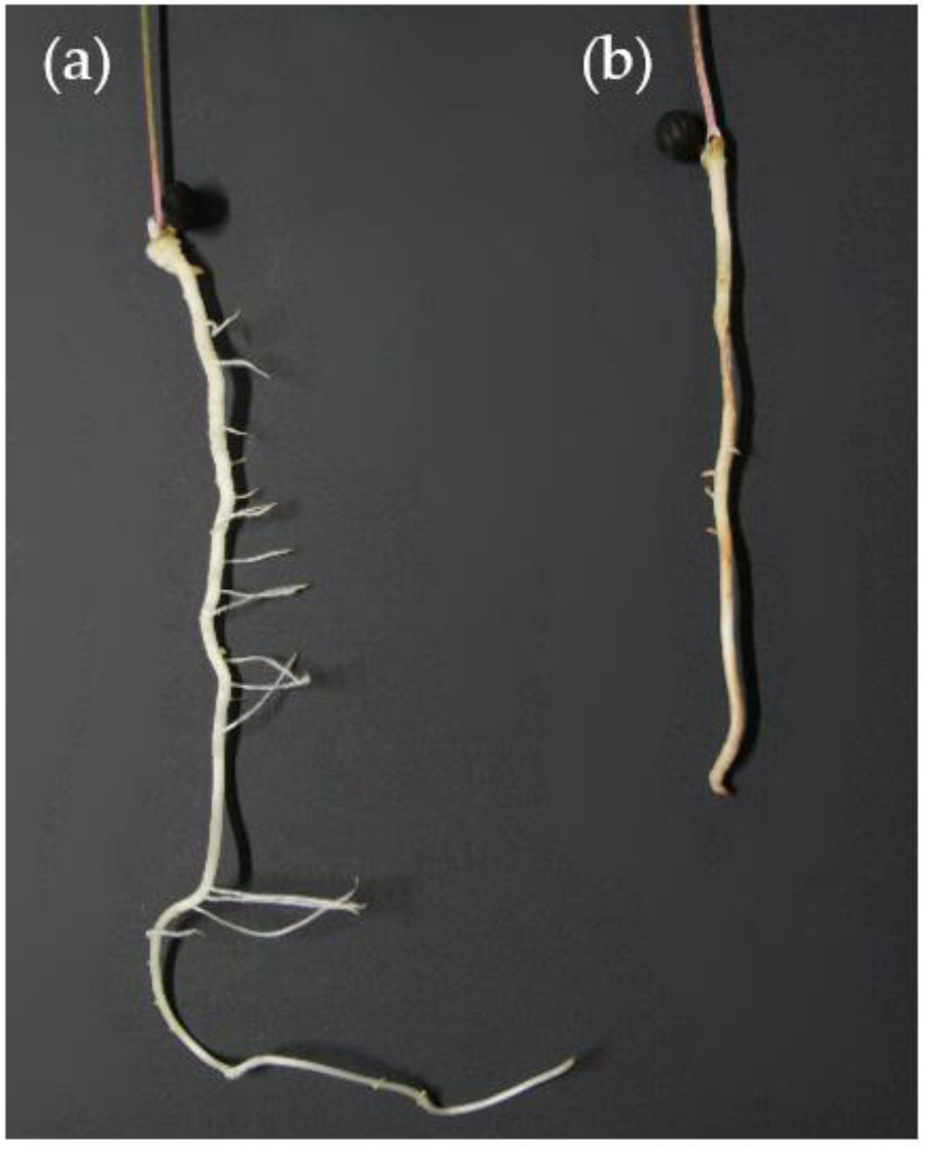
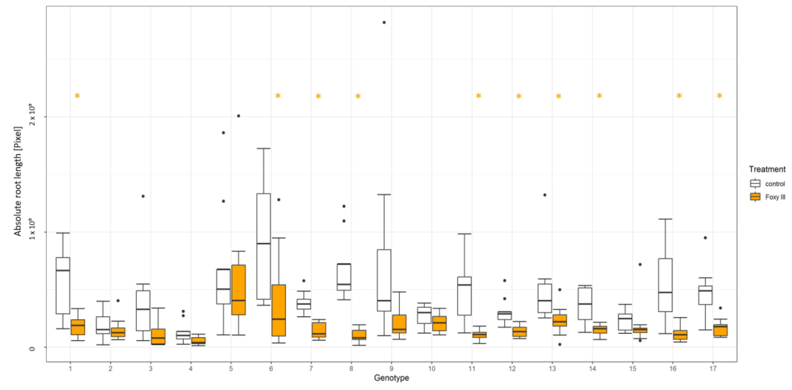
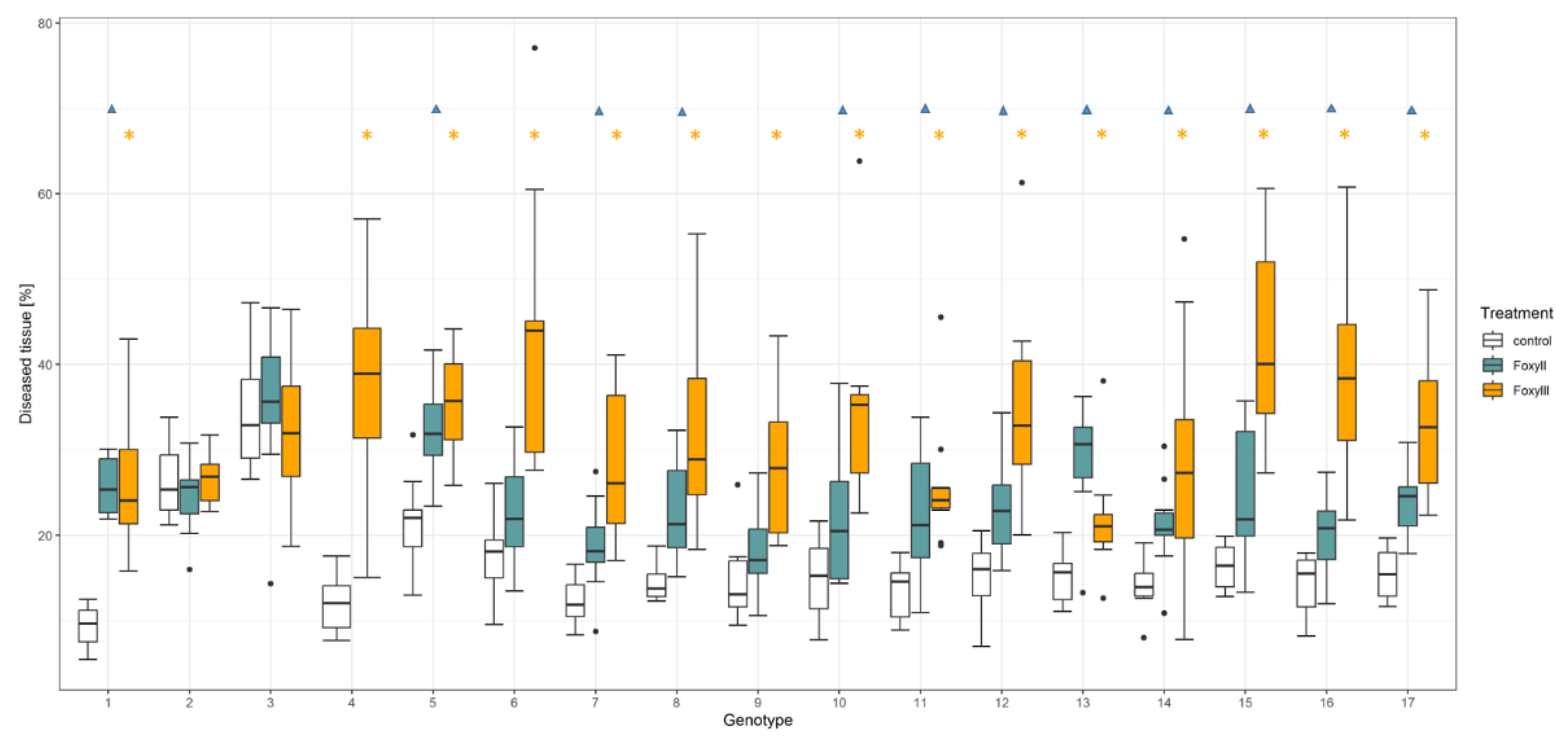
2.3. Seedling Resistance Test

Resistance to Fusarium oxysporum was tested on the basis of post-infection symptoms, according to Kathe et al. [18], with some modifications. Fungal conidia were harvested by scraping the spores with distilled water and a Drigalski spatula. The roots were immersed for 5 min in the conidial suspension (1 × 10⁶ conidia/mL) and carefully shaken. The plants were then incubated for 14 days at 22 °C with a photoperiod of 16 h in covered square bioassay dishes (245 × 245 × 24 mm) on moistened filter paper. Disease symptoms such as brown lesions and asparagus root reduction were evaluated using LemnaTec Scanalyzer PL (LemnaTec GmbH, Aachen, Germany). The software was calibrated on a symptom basis to distinguish between the healthy and infected root tissue. The pixels of the calibrated classes used for statistical evaluation were measured. In addition to absolute root length, the browning of roots was also studied as a symptom. To measure the root length, lines were drawn through each root (see Figure S1). Finally, to determine the absolute root length, the pixels of these lines were summed.
A complete screening was performed with the genotypes listed in Table 1. Because very little material was available for some genotypes, especially wild species, and old landraces, and the germination rate of these genotypes was also very low in some cases, there were some failures. These are listed in Table S1. To demonstrate that one screening provides clear information about the susceptibility or resistance of a genotype, a second screening was performed for a portion of the genotype (partial screening). To demonstrate this, Table S2 in Supplementary Material compares the complete screening with the partial screening for four genotypes.

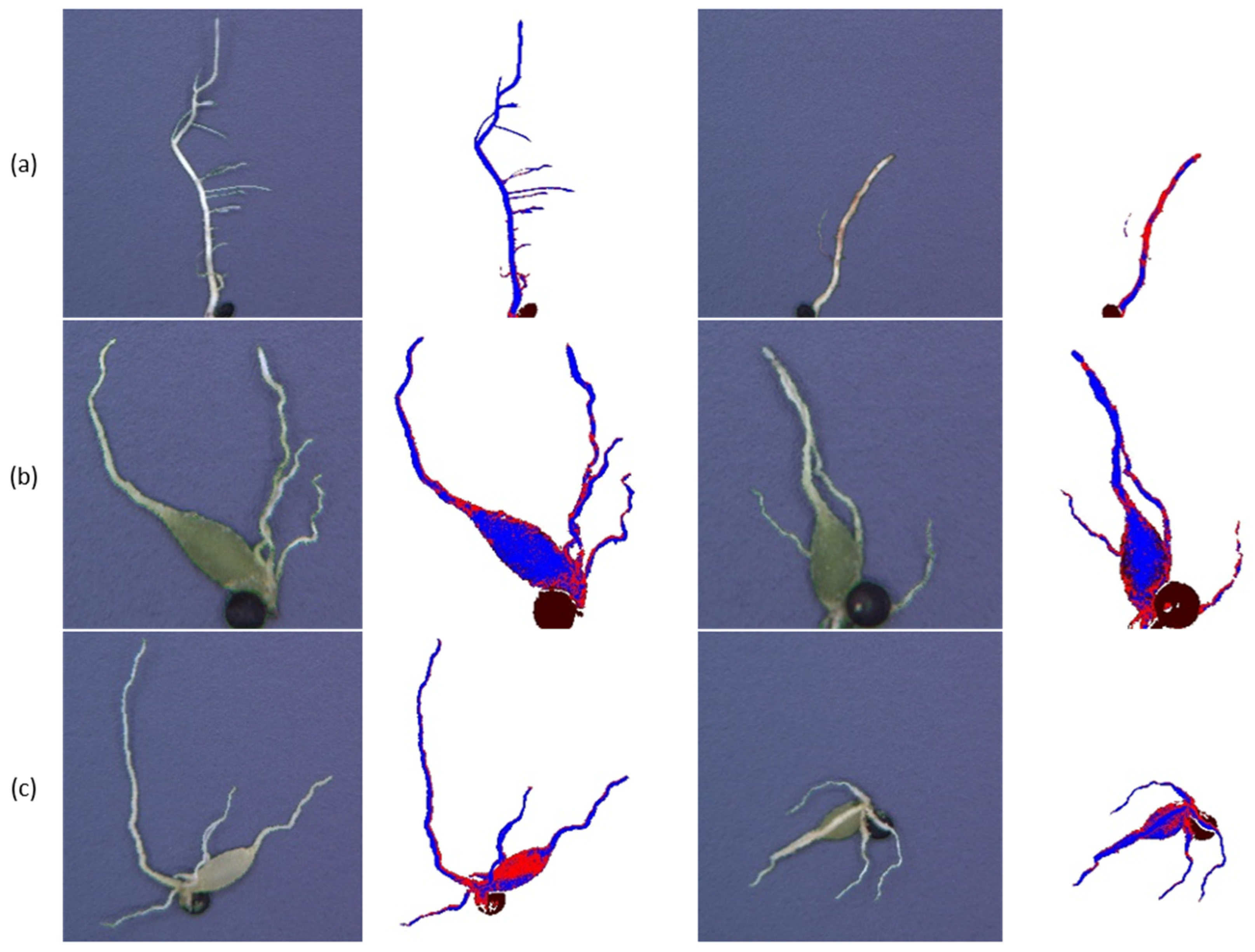
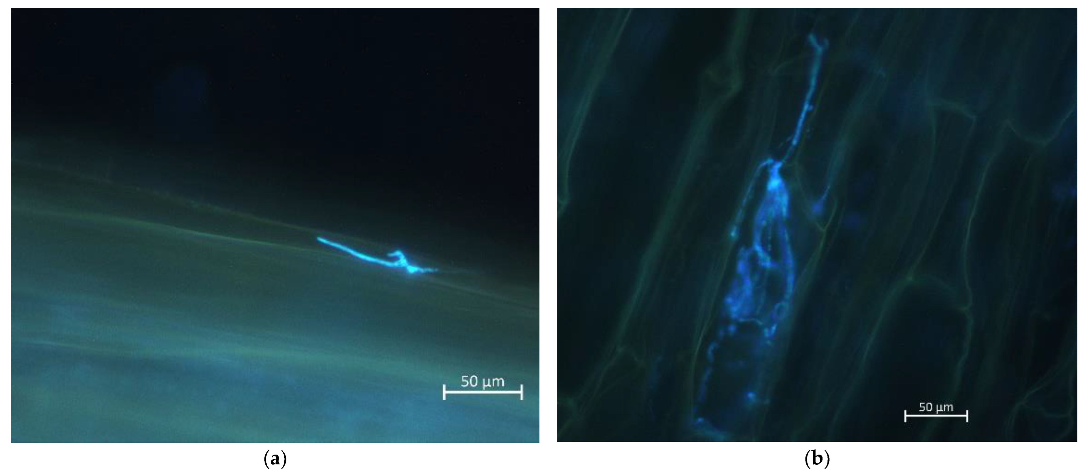
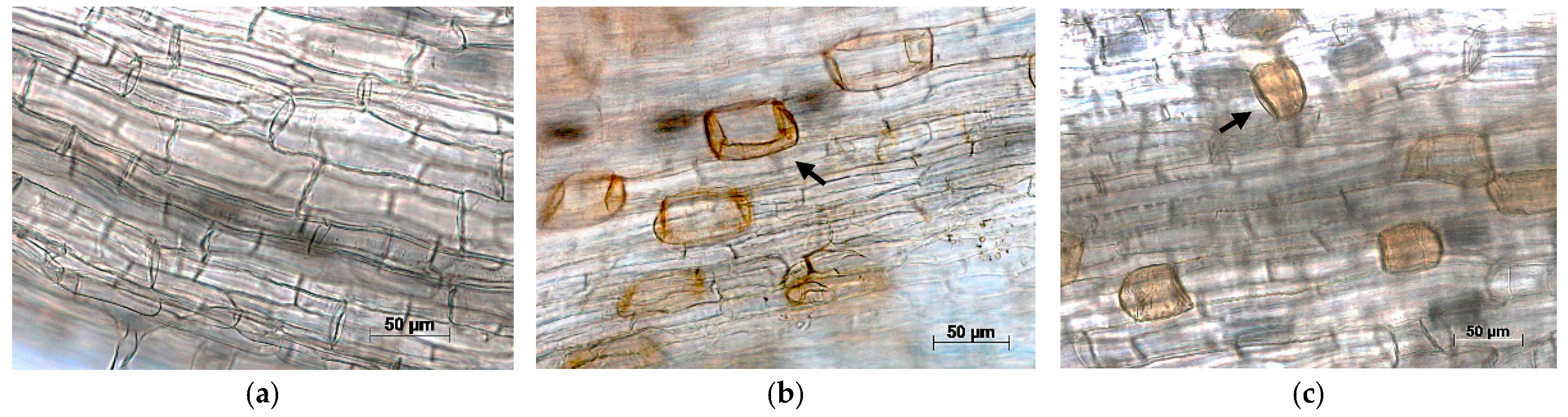
2.4. Staining Procedure and Microscopic Determination of Hypersensitive Response

A. densiflorus, A. aethiopicus, and cv. Thielim were tested for hydrogen peroxide accumulation in response to infection with F. oxysporum. Three seedlings per Asparagus species were infected. According to He et al. [19], the hyphal-sandwich root inoculation was used for inoculation. Between two- and ten week old seedlings were cleaned of sand and blotted dry. The roots were placed between two pieces of PDA containing a hyphae of Foxy III. Uninoculated PDA strips were used as the controls. For incubation, the treated seedlings were placed in a moistened and covered square BioAssay dish (245 × 245 × 24 mm) in a growth cabinet at 22 °C with a 16-h photoperiod. The method described by Serfling et al. [33] was used for staining. The roots of the test plants were sampled immediately before inoculation and after 8, 12, 24, 48, and 72 h of incubation. A 3,3’-Diaminobenzidine (DAB) solution was used to detect hydrogen peroxide (H2O2) accumulation in the inoculated roots. Calcofluor white M2R was used to stain the fungal structures. Three 1 cm root pieces per asparagus species were tested and examined under the microscope.
The microscopy of the roots and fungal structures was performed using the Axioskop 50 microscope equipped with an Axiocam 305 color and the ZEN 3.0 (blue edition) software package (Carl Zeiss AG, Jena, Germany). Fungal structures stained with Calcofluor white M2R were observed using a filter set 02 (excitation filter G 365, beam splitter FT 395, and barrier filter LP 420). The evaluation of the DAB staining was performed using the Axio Imager.A1 microscope equipped with an Axiocam MRc and the AxioVision SE64 Rel. 4.9 software package (Carl Zeiss AG, Jena, Germany).

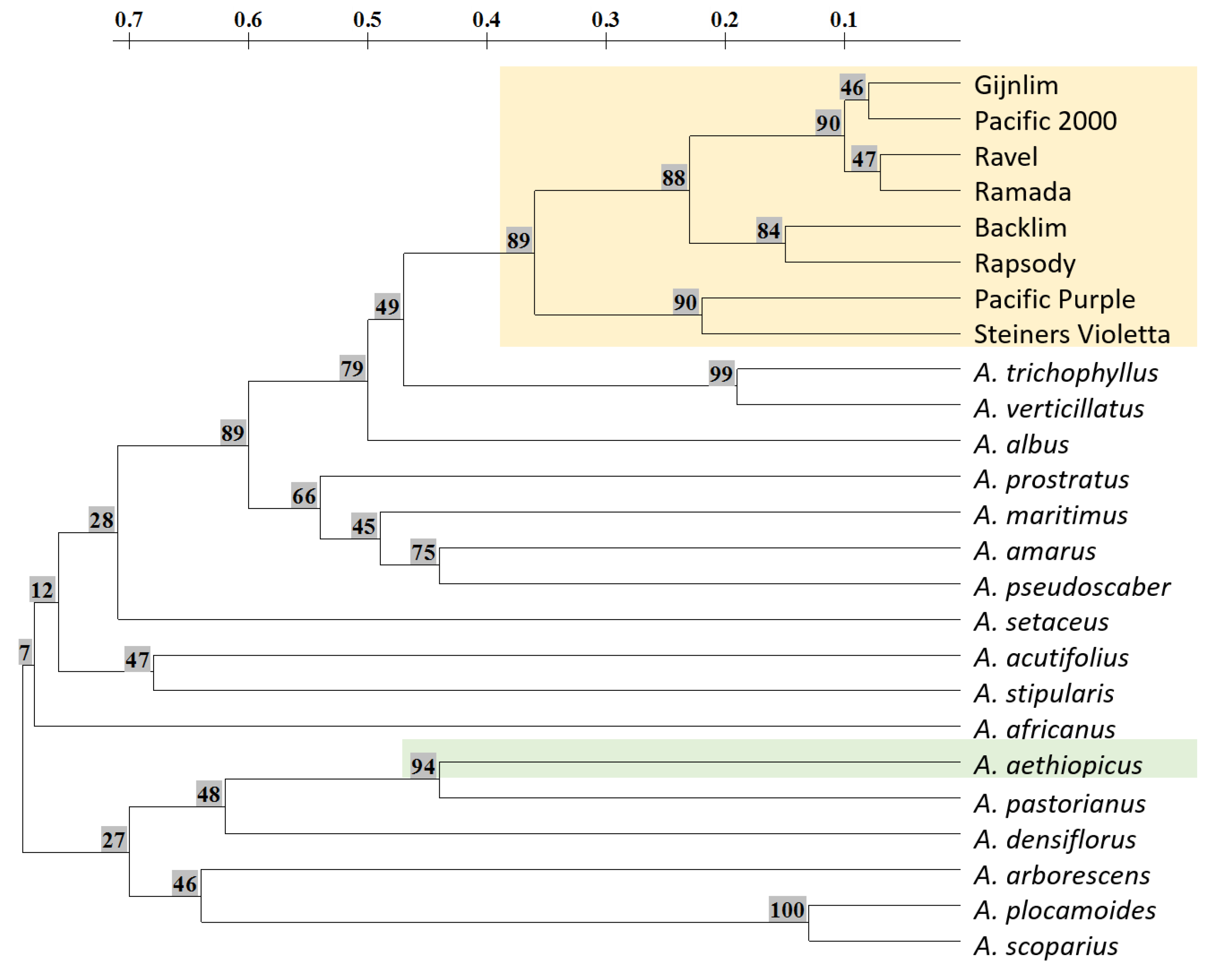
2.5. SSR Markers and Distance Analysis

The total genomic DNA was isolated from 100 mg juvenile cladophyll tissues of one plant per accession according to the protocol of Porebski et al. [34] and finally adjusted to 10 ng/μL. For SSR analysis, 25 PCR primer pairs (Table S3) were used, as described by Mercati et al. [35], along with a universal labelling strategy according to Oetting et al. [36]. The unlabelled forward primer extended by a 19 bp M13 sequence was combined with the fluorescence-labelled M13 universal primer and the reverse primer of an original length. Amplifications were performed in 6 µL total volume, containing 12.5 ng plant DNA, 1 × concentrated buffer solution (with MgCl2 and dNTPs), 0.8 µM primers, and 0.2 Units MyTaq DNA polymerase (Bioline, London, GB). A GeneAmp PCR System 9700 thermocycler (Applied Biosystems, Forster City, CA, USA) was programmed as follows: 94 °C for 5 min, 35 cycles of 94 °C for 30 s, 55 °C for 30 s, 72 °C for 1 min, followed by a final step of 72 °C for 7 min. The amplification products of two independent PCR amplifications with IRDye700 and IRDye800 labelled M13 primers were mixed. DNA fragments were separated using an LI-COR 4300 automatic sequencer (LI-COR Biosciences, Lincoln, NE, USA). The fragment sizes were calculated by comparison with IRDye700 and IRDye800 labelled 50–350 bp size ladder. Polymorphic bands were recorded as a 1/0 matrix and used for distance analysis according to Nei and Li [37] with the program Treecon (Scanalytics, Milwaukee, WI, USA). Cluster analysis was performed using the unweighted pair group method with the arithmetic mean (UPGMA) algorithm. The confidence level for each branch point was determined by bootstrap analysis with 100 iterations.

2.6. Statistical Analysis

The program R version 4.1.2 (R Foundation for Statistical Computing, Vienna, Austria) was used for the statistical analysis of the percentage data of diseased tissue and pixel data of absolute root lengths. When a normal distribution and homogeneity of variance were present, an independent sample t-test was used to test for significant differences between the control and infected plants (p < 0.05). If this was not the case, the Mann-Whitney–U test was used.

3. Results

3.1. Resistance Test

A total of 17 genotypes were tested, including four wild relatives, two landraces, and eleven cultivars. As part of the seedling resistance test, the root reduction and root browning after infection with F. oxysporum were examined.
Both used Foxy isolates were pathogenic to asparagus, with different virulence. As described by Kathe et al. [18], Foxy II was less virulent than Foxy III.
The roots of plants infected with F. oxysporum (Foxy II and Foxy III) showed reduced root growth compared to uninfected plants. In addition, lateral root growth was also obviously reduced. In the following, only the results for Foxy III are shown (Figure 1 and Figure 2). Foxy II has a similar effect on the root length but to a lesser extent. There are no significant differences between the control and Foxy III in the wild relatives A. densiflorus and A. aethiopicus, in the landrace CGN25609, and the A. officinalis cvs. Leistungsauslese, Schwetzinger Meiserschuss, and Ivancicky (Figure 2). All other genotypes show a significant difference between the absolute root length of control and Foxy III (*, Figure 2).
Infection with F. oxysporum was indicated mainly by elongated brown spots and by the discoloration of entire root sections. In severe infection symptoms, purple exudates were released sometimes on the filter paper, and/or the roots disintegrated partially. In rare cases, a white mycelium formed on the infected root. Differences in virulence between Foxy II and Foxy III were indicated by visibly fewer browning symptoms and a smaller infected area in Foxy II.
The wild species A. aethiopicus and A. densiflorus differ from the other genotypes tested. Both show thickened shoot roots as seedlings that turn green after 14 days of incubation (Figure 3b,c). This resulted in an increased discoloration rate due to the evaluation method. However, the images in Figure 3b,c show that neither the control nor the infected seedlings showed any signs of infection. For comparison, the susceptible A. officinalis cv. Thielim is shown in Figure 3a. This obviously shows the difference between the uninfected and infected root and the typical browning symptoms.
All tested cultivars showed susceptibility to both F. oxysporum isolates (Foxy II and Foxy III). However, there were some differences between the cultivars. For example, A. officinalis cv. Schwetzinger Meisterschuss had a significantly higher susceptibility than A. officinalis cv. Argenteuil (Figure 4, Genotype 15 and 7). The severity of infection ranged from 9% to 42% for Foxy II and 13% to 77% for Foxy III among the tested cultivars (Figure 4).
In addition, the wild relatives A. amarus and A. stipularis showed susceptibility to both F. oxysporum isolates after two weeks of incubation. For the wild relatives A. densiflorus and A. aethiopicus, there was no significant difference on average between the infected root and the control (Figure 4, Genotype 2 and 3). However, not all tested plants of these genotypes were free of symptoms. Of the 40 A. aethiopicus seedlings tested, three showed visible signs of infection. In addition, of the 10 A. densiflorus seedlings tested, two showed visible symptoms.
One screening already provided a meaningful result on the susceptibility or resistance of the genotypes tested. Although there were significant differences between the complete and partial resistance screening in some comparisons (Table S2), it was found that the susceptibility or resistance statement never changed for the genotypes tested twice.

3.2. Microscopic Analyses

After the resistance test showed a resistance of A. aethiopicus to F. oxysporum, the exact defence mechanism was investigated. Since it is known that A. densiflorus reacts with a hypersensitive response to invading fungi [19], this mechanism was tested for A. aethiopicus. To verify that the fungus had indeed invaded the root, the fungus was stained with the fluorescent dye Calcoflour White. As can be seen in Figure 5, F. oxysporum invades and spreads in the cells of susceptible A. officinalis cv. Thielim.
Staining with 3,3’-Diaminobenzobenzidine (DAB) shows that hydrogen peroxide (H2O2) has accumulated in the root cells of A. densiflorus and A. aethiopicus at 8 hpi. The stained areas increase with incubation time. A difference in the rate of H2O2 accumulation was observed. Thus, the storage of H2O2 in A. densiflorus WAS apparently faster than in A. aethiopicus (Figure 6, arrows in Figure 6b,c). By contrast, no DAB-stained areas were found in the susceptible genotype A. officinalis cv. Thielim (Figure 6a).

3.3. Phylogenetic Relationship

A new potential source of resistance was identified in A. aethiopicus. Because of the strong crossing barriers mentioned in the introduction, the determination of the genetic distance is important to develop strategies for resistance transmission. Twenty-five microsatellite loci, distributed over all ten A. officinalis chromosomes, were used to estimate the genetic distance of 26 Asparagus accessions. On average, 8.9 (3–17) alleles were amplified per tested SSR. The 222 polymorphic bands were recorded as a 1/0 matrix and used for cluster analysis. Two main clusters, which represent the Eurasian clade and the African clade, corresponded to their geographic origin.
The results showed that A. densiflorus and A. aethiopicus are closely related in a subgroup and that both wild relatives have a large genetic distance from the A. officinalis cultivars, forming their own subclade (Figure 7).

4. Discussion

Our study identified a new potential source of resistance to F. oxysporum in A. aethiopicus. In the seedling resistance test, this wild relative showed almost no susceptibility to the Fusarium isolates used (Foxy II and III). Moreover, it reacted rapidly after inoculation with F. oxysporum (Foxy III) with an accumulation of hydrogen peroxide.
As reported in the results, there was a problem with the increase in the discoloration rate in the wild species A. aethiopicus and A. densiflorus due to the method of evaluation (Figure 3). This was problematic for comparing these wild species to the other genotypes. Because the digital image analysis was performed with the same configuration, camera settings, and symptom definition for all genotypes. Changing these settings in favour of the wild species resulted in a significant misinterpretation of the other genotypes. Therefore, the described configuration was used even though it misinterpreted the fact that the wild species were infected.
In the resistance test, the browning of roots proved to be a better selection criterion than the reduction in root length. It is known that F. oxysporum has a negative effect on root growth [5,38]. This was also shown in our study. However, in our experiment, it was shown that this was not a suitable trait for the selection of resistant and susceptible genotypes. Some genotypes that were classified to be susceptible in the resistance test did not show a significant reduction in root lengths, such as landrace CGN25609 and A. officinalis cv. Leistungsauslese and A. officinalis cv. Schwetzinger Meisterschuss (Figure 2). Mostly this was because they had a high diversity in root growth even when uninfected.
All tested A. officinalis cultivars were susceptible in the resistance test, as were the wild relatives A. amarus and A. stipularis (Figure 4). The cultivars showed little variation in susceptibility, which was manifested by a more or less severe browning of the roots. This probably reflects the restricted gene pool of asparagus, especially in cultivars [30,31]. In contrast, the wild relatives A. densiflorus and A. aethiopicus showed resistance to both tested Fusarium oxysporum isolates. In the case of A. densiflorus, this result was consistent with previous studies [19,20]. Both wild species react rapidly after infection with a hypersensitive response, which is shown microscopically by an accumulation of H2O2 (Figure 6b,c). A difference in the rate of H2O2 accumulation was observed; the storage of H2O2 is apparently faster in A. densiflorus than in A. aethiopicus. According to our results, the resistance of the two wild species was based on the same mechanism. However, it is not yet possible to say anything more definite to verify these preliminary observations. For this, it needs further detailed tests. The rapid production of ROS, including H2O2, in response to infection, is a typical feature of resistance in plants [39]. This leads to rapid and localized cell death, which inhibits the spread of F. oxysporum to healthy tissues [19,39].
To better assess and strategically transfer resistance to cultivars, it is important to determine the genetic distance. For the distance analysis, more wild species were used than in the resistance test to present the most accurate analysis of relationships. This was conducted in order to see if the classical crossing and embryo rescue were promising or if protoplast fusion will be necessary. This fact is demonstrated by the interspecific hybrids of Ito et al. [27], who found a correlation between phylogenetic relativity and the potential for interspecific hybridization [27]. The results of the close relationship between A. aethiopicus and A. densiflorus indicate that A. aethiopicus, similar to A. densiflorus, is very difficult to cross-breed [28,29]. Therefore, it is important to search for other sources of resistance. This is because the probability of transferring resistance to F. oxysporum increases with multiple opportunities. In particular, if resistance was found in more closely related wild species, inclusive of native wild A. officinalis, the probability of a successful cross would increase significantly. These analyses support the goal of transferring Fusarium resistance into cultivated asparagus.
The next important step is to develop a greenhouse test to analyse resistance behaviour at later plant stages. The aim is to determine whether resistance in the seedling also occurs in the adult plant. The development of an in vitro assay is also needed to find as many sources of resistance to F. oxysporum as possible. This would especially facilitate the testing of wild species, which are sometimes very difficult and time-consuming to propagate.

Supplementary Materials

The following supporting information can be downloaded at: https://www.mdpi.com/article/10.3390/horticulturae9020158/s1, Table S1: Overview of the resistance tests carried out in the complete and partial screening. x represents complete runs (control, Foxy II, and Foxy III with 10 seedlings each). Deviations are defined more precisely. Table S2: Resistance test against Fusarium oxysporum isolates Foxy II and Foxy III, 14 dpi. Comparison of the percentage of diseased tissue measured in complete and partial resistance screening for A. amarus, A. aethiopicus, A. officinalis cv. Ravel, and A. officinalis cv. Start. Asterisks behind the average and SE show significant differences between the complete and partial resistance screening (p < 0.05). Δ indicates significance between control and Foxy II or Foxy III for each genotype (p < 0.05). Table S3: SSR primer used for the distance analysis. Figure S1: Analysis of root length evaluated with LemnaTec Scanalyzer PL. A. officinalis cv. Grolim 14 dpi. (a) Control; (b) Foxy III.

Author Contributions

Conceptualization, J.J., H.B., J.K. and T.N.; methodology, J.J. and H.B.; investigation, J.J.; writing—original draft preparation, J.J.; writing—review and editing, J.J., H.B., J.K. and T.N.; visualization, J.J.; supervision, J.K. and T.N.; project administration, J.K. and T.N. All authors have read and agreed to the published version of the manuscript.

Funding

This research received no external funding.

Institutional Review Board Statement

Not applicable.

Informed Consent Statement

Not applicable.

Data Availability Statement

Rating and marker data can be obtained from us on personal request for scientific use.

Acknowledgments

We thank Ute Fuhrmann, Astrid Bruchmüller and Karla Müller for the excellent technical assistance.

Conflicts of Interest

The authors declare no conflict of interest.

References

  1. FAO. FAOSTAT. Available online: https://www.fao.org/faostat/en/#home (accessed on 16 September 2022).
  2. Bharati, J.; Kumar, S. Shatavari (Asparagus racemosus). In Phytobiotics and Animal Production; Singh, P.K., Ed.; International Books & Periodical Supply Service: Delhi, India, 2019; pp. 567–590. [Google Scholar]
  3. Benincasa, P.; Tei, F.; Rosati, A. Plant Density and Genotype Effects on Wild Asparagus (Asparagus acutifolius L.) Spear Yield and Quality. HortScience 2007, 42, 1163–1166. [Google Scholar] [CrossRef] [Green Version]
  4. Andrzejak, R.; Janowska, B. The occurrence of Fusarium species in white spears of Asparagus officinalis. Not. Bot. Horti Agrobot. 2021, 49, 12559. [Google Scholar] [CrossRef]
  5. Blok, W.J.; Bollen, G.J. Fungi on roots and stem bases of asparagus in The Netherlands: Species and pathogenicity. Eur. J. Plant Pathol. 1995, 101, 15–24. [Google Scholar] [CrossRef]
  6. Brizuela, A.M.; de La Lastra, E.; Marín-Guirao, J.I.; Gálvez, L.; de Cara-García, M.; Capote, N.; Palmero, D. Fusarium Consortium Populations Associated with Asparagus Crop in Spain and Their Role on Field Decline Syndrome. J. Fungi 2020, 6, 336. [Google Scholar] [CrossRef]
  7. Logrieco, A.; Doko, B.; Moretti, A.; Frisullo, S.; Visconti, A. Occurrence of Fumonisin B1 and B2 in Fusarium proliferatum Infected Asparagus Plants. J. Agric. Food Chem. 1998, 46, 5201–5204. [Google Scholar] [CrossRef]
  8. de La Lastra, E.; Marín-Guirao, J.I.; López-Moreno, F.J.; Soriano, T.; de Cara-García, M.; Capote, N. Potential inoculum sources of Fusarium species involved in asparagus decline syndrome and evaluation of soil disinfestation methods by qPCR protocols. Pest Manag. Sci. 2021, 77, 4749–4757. [Google Scholar] [CrossRef]
  9. Weber, Z.; Karolewski, Z.; Irzykowska, L.; Knaflewski, M.; Kosiada, T. Occurrence of Fusarium species in spears of Asparagus (Asparagus officinalis). Phytopathol. Pol. 2007, 45, 9–15. [Google Scholar]
  10. Apaza, W.; Talledo, A.; Casas, A. Reaction of 10 Asparagus cultivars to infested soil with Fusarium oxysporum f.sp. asparagi and Meloidogyne incognita. Acta Hortic. 2018, 1223, 233–238. [Google Scholar] [CrossRef]
  11. Fiume, F.; Fiume, G. Field response of some asparagus varieties to rust, Fusarium crown root rot, and violet root rot. Commun. Agric. Appl. Biol. Sci. 2003, 68, 659–671. [Google Scholar]
  12. Blok, W.J.; Bollen, G.J. Inoculum sources of Fusarium oxysporum f.sp. asparagi in asparagus production. Ann. Appl. Biol. 1996, 128, 219–231. [Google Scholar]
  13. Elmer, W.H. Management of Fusarium crown and root rot of asparagus. Crop Prot. 2015, 73, 2–6. [Google Scholar] [CrossRef]
  14. Reid, T.C.; Hausbeck, M.K.; Kizilkaya, K. Use of Fungicides and Biological Controls in the Suppression of Fusarium Crown and Root Rot of Asparagus Under Greenhouse and Growth Chamber Conditions. Plant Dis. 2002, 86, 493–498. [Google Scholar] [CrossRef] [PubMed] [Green Version]
  15. Reid, T.C.; Hausbeck, M.K.; Kizilkaya, K. Effects of Sodium Chloride on Commercial Asparagus and of Alternative Forms of Chloride Salt on Fusarium Crown and Root Rot. Plant Dis. 2001, 85, 1271–1275. [Google Scholar] [CrossRef] [PubMed]
  16. Brizuela, A.M.; Lalak-Kańczugowska, J.; Koczyk, G.; Stępień, Ł.; Kawaliło, M.; Palmero, D. Geographical Origin Does Not Modulate Pathogenicity or Response to Climatic Variables of Fusarium oxysporum Associated with Vascular Wilt on Asparagus. J. Fungi 2021, 7, 1056. [Google Scholar] [CrossRef]
  17. Delgado-Baquerizo, M.; Guerra, C.A.; Cano-Díaz, C.; Egidi, E.; Wang, J.-T.; Eisenhauer, N.; Singh, B.K.; Maestre, F.T. The proportion of soil-borne pathogens increases with warming at the global scale. Nat. Clim. Change 2020, 10, 550–554. [Google Scholar] [CrossRef]
  18. Kathe, L.; Krämer, R.; Budahn, H.; Pillen, K.; Rabenstein, F.; Nothnagel, T. Development of a bioassay to assess resistance to Fusarium oxysporum (Schlecht.) in asparagus (Asparagus officinalis L.). J. Phytopathol. 2019, 167, 558–566. [Google Scholar] [CrossRef]
  19. He, C.; Hsiang, T.; Wolyn, D.J. Activation of defense responses to Fusarium infection in Asparagus densiflorus. Eur. J. Plant Pathol. 2001, 107, 473–483. [Google Scholar] [CrossRef]
  20. Stephens, C.T.; De Vries, R.M.; Sink, K.C. Evaluation of Asparagus Species for resistance to Fusarium oxysporum f. sp. asparagi and F. moniliforme. HortScience 1989, 2, 365–368. [Google Scholar] [CrossRef]
  21. Stephens, C.T.; Wade, E.H. An In Vitro Assay to Evaluate Sources of Resistance in Asparagus spp. to Fusarium Crown and Root Rot. Plant Dis. 1988, 72, 334–337. [Google Scholar] [CrossRef]
  22. Lewis, G.D.; Shoemaker, P.B. Presence of Fusarium oxysporum f.asparagi on asparagus seed and Fusarium resistance in plant introduction lines of asparagus. Phytopathology 1964, 54, 128. [Google Scholar]
  23. Plath, S.; Krämer, R.; Lantos, E.; Nothnagel, T. Breeding programs to transmit Asparagus virus 1 resistance. Acta Hortic. 2018, 1223, 17–24. [Google Scholar] [CrossRef]
  24. Falavigna, A.; Alberti, P.; Casali, P.E.; Toppino, L.; Huaisong, W.; Mennella, G. Interspecific hybridization for asparagus breeding in Italy. Acta Hortic. 2008, 776, 291–298. [Google Scholar] [CrossRef]
  25. McCollum, G.D. Asparagus densiflorus cultivars Sprengeri and Myers cross-pollinations with A. officinalis and other species. Asparagus Res. Newsl. 1988, 6, 1–10. [Google Scholar]
  26. Bozzini, A. Interspecific hybridization and experimental mutagenesis in the genetic improvement of asparagus. Genet. Agrar. 1962, 16, 212–218. [Google Scholar]
  27. Ito, T.; Ochiai, T.; Fukuda, T.; Ashizawa, H.; Kanno, A.; Kameya, T.; Sonoda, T. Potential of interspecific hybrids in the genus Asparagus. Acta Hortic. 2008, 776, 279–284. [Google Scholar] [CrossRef]
  28. Marcellán, O.; Camadro, E. Formation and development of embryo and endosperm in intra- and inter-specific crosses of Asparagus officinalis and A. densiflorus cv. Sprengeri. Sci. Hortic. 1999, 81, 1621–1625. [Google Scholar] [CrossRef]
  29. Marcellán, O.N.; Camadro, E.L. Self- and cross-incompatibility in Asparagus officinalis and Asparagus densiflorus cv. Sprengeri. Can. J. Bot. 1996, 74, 1621–1625. [Google Scholar] [CrossRef]
  30. Mercati, F.; Riccardi, P.; Harkess, A.; Sala, T.; Abenavoli, M.R.; Leebens-Mack, J.; Falavigna, A.; Sunseri, F. Single nucleotide polymorphism-based parentage analysis and population structure in garden asparagus, a worldwide genetic stock classification. Mol. Breed. 2015, 35, 59. [Google Scholar] [CrossRef]
  31. Knaflewski, M. Genealogy asparagus cultivars. Acta Hortic. 1996, 415, 87–92. [Google Scholar] [CrossRef]
  32. Nothnagel, T.; Budahn, H.; Krämer, I.; Schliephake, E.; Lantos, E.; Plath, S.; Krämer, R. Evaluation of genetic resources in the genus Asparagus for resistance to Asparagus virus 1 (AV-1). Genet. Resour. Crop Evol. 2017, 64, 1873–1887. [Google Scholar] [CrossRef]
  33. Serfling, A.; Templer, S.E.; Winter, P.; Ordon, F. Microscopic and Molecular Characterization of the Prehaustorial Resistance against Wheat Leaf Rust (Puccinia triticina) in Einkorn (Triticum monococcum). Front. Plant Sci. 2016, 7, 1668. [Google Scholar] [CrossRef] [PubMed] [Green Version]
  34. Porebski, S.; Bailey, L.G.; Baum, B.R. Modification of a CTAB DNA extraction protocol for plants containing high polysaccharide and polyphenol components. Plant Mol. Biol. Report. 1997, 15, 8–15. [Google Scholar] [CrossRef]
  35. Mercati, F.; Riccardi, P.; Leebens-Mack, J.; Abenavoli, M.R.; Falavigna, A.; Sunseri, F. Single nucleotide polymorphism isolated from a novel EST dataset in garden asparagus (Asparagus officinalis L.). Plant Sci. 2013, 203, 115–123. [Google Scholar] [CrossRef] [PubMed]
  36. Oetting, W.S.; Lee, H.K.; Flanders, D.J.; Wiesner, G.L.; Sellers, T.A.; King, R.A. Linkage analysis with multiplexed short tandem repeat polymorphisms using infrared fluorescence and M13 tailed primers. Genomics 1995, 30, 450–458. [Google Scholar] [CrossRef]
  37. Nei, M.; Li, W.H. Mathematical model for studying genetic variation in terms of restriction endonucleases. Proc. Natl. Acad. Sci. USA 1979, 76, 5269–5273. [Google Scholar] [CrossRef] [Green Version]
  38. Blok, W.J.; Zwankhuizen, M.J.; Bollen, G.J. Biological Control of Fusarium oxysporum f.sp. asparagi by Applying Non-pathogenic Isolates of F. oxysporum. Biocontrol Sci. Technol. 1997, 7, 527–542. [Google Scholar] [CrossRef]
  39. Pontier, D.; Balagué, C.; Roby, D. The hypersensitive response. A programmed cell death associated with plant resistance. C. R. L’académie Sci.-Ser. III-Sci. Vie 1998, 321, 721–734. [Google Scholar] [CrossRef]
Figure 1. A. officinalis cv. Ramada 14 dpi with F. oxysporum. (a) Control; (b) Foxy III.
Horticulturae 09 00158 g001
Figure 2. Comparison of absolute root growth 14 dpi with Foxy III of the 18 asparagus genotypes (Table 1) in pixel. * Indicates significance between control (white) and Foxy III (orange) for each genotype (p < 0.05).
Horticulturae 09 00158 g002
Figure 3. Analysis of diseased tissue caused by F. oxysporum (Foxy III, 14 dpi) using the digital image analysing system of LemnaTec. Left is the control and right is the infected seedlings, each with the original image and the generated colour class image. (a) A. officinalis cv. Thielim; (b) A. aethiopicus; (c) A. densiflorus.
Horticulturae 09 00158 g003
Figure 4. Resistance test against Fusarium oxysporum isolates Foxy II and Foxy III, 14 dpi. Percentage of diseased tissue for the 17 asparagus genotypes (Table 1) shown is the complete screening. * Indicates significance between the control and Foxy III for each genotype (p < 0.05). Δ Indicates significance between control and Foxy II for each genotype (p < 0.05).
Horticulturae 09 00158 g004
Figure 5. Fungal structures (Foxy III) in the susceptible genotype A. officinalis cv. Thielim 72 hpi. Fungal cell walls stained using Calcofluor White M2R. (a) Growing hypha; (b) Mycelium spreading from a cell.
Horticulturae 09 00158 g005
Figure 6. DAB stain of hydrogen peroxide on F. oxysporum-infected roots. Arrows in (b,c) mark enhanced hydrogen peroxide concentration stained by 3,3 Diaminobenzidine (a) A. officinalis cv. Thielim 12 hpi; (b) A. densiflorus 12 hpi; (c) A. aethiopicus 12 hpi.
Horticulturae 09 00158 g006
Figure 7. Determination of the phylogenetic distance between A. aethiopicus (green) and A. officinalis (cultivars; yellow) using 222 bands from SSR analysis. Numbers at the branches indicate bootstrap values in percent.
Horticulturae 09 00158 g007
Table 1. Asparagus species evaluated for resistance to Fusarium oxysporum.
CodeAsparagus
Species
Cultivar/
Wild Relative 1
Seed
Origin 2
AccessionGeographic Origin
1A. amarus DC.Wild relative (Pop.)CRA Italy
2A. aethiopicus L.Wild relative (Pop.)GG3333-3Malaga
3A. densiflorus (Kunth) JessopWild relative (Pop.)ISR Israel
4A. stipularis
Forssk.
Wild relative (Pop.)IBZ Ibiza
5A. officinalis L.Landrace (Pop.)WURCGN25609Armenia
6A. officinalis L.Landrace (Pop.)WURCGN25611Armenia
7A. officinalis L.cv. Argenteuil (OP)RZ French
8A. officinalis L.cv. Grolim (F1)LIM The Netherlands
9A. officinalis L.cv. Ivancicky (OP)IPKASP 1unknown
10A. officinalis L.cv. Leistungsauslese (OP)DS Germany
11A. officinalis L.cv. Ramada (F1)SWS Germany
12A. officinalis L.cv. Ramires (F1)SWS Germany
13A. officinalis L.cv. Ravel (F1)SWS Germany
14A. officinalis L.cv. Ruhm von Braunschweig (OP)IPKASP 31Germany
15A. officinalis L.cv. Schwetzinger
Meisterschuss (OP)
SWS Germany
16A. officinalis L.cv. Start (OP)IPKASP 3unknown
17A. officinalis L.cv. Thielim (F1)LIM The Netherlands
1 Pop.: Population, OP: Open pollinated, F1: Hybrid. 2 CRA: Research Institute for Vegetable Crops (Italy), ISR: Volcani Centre Bet Gagan (Israel), LIM: Limgroup B.V. (The Netherlands), SWS: Süd-West-Saat (Germany), RZ: Rijk Zwaan Zaadteelt en Zaadhandel B.V. (The Netherlands), IBZ: ornamental and wild asparagus from Ibiza (Spain), IPK: Leibniz Institute of plant genetics and crop plant research (Germany), DS: Deutsche Spargelzucht (Germany), GG: Gruson Gewächshäuser Magdeburg (Germany), WUR: Wageningen University & Research.
Disclaimer/Publisher’s Note: The statements, opinions and data contained in all publications are solely those of the individual author(s) and contributor(s) and not of MDPI and/or the editor(s). MDPI and/or the editor(s) disclaim responsibility for any injury to people or property resulting from any ideas, methods, instructions or products referred to in the content.

