Article

Antioxidant and Cytotoxic Activities of Leaf and Stem Extracts of Barleria albostellata C.B. Clarke

by
Serisha Gangaram
1,*,
Yougasphree Naidoo
1,
Yaser Hassan Dewir
2,
Moganavelli Singh
1,
Aliscia Nicole Daniels
1,
Katalin Magyar-Tábori
3 and
Nóra Mendler-Drienyovszki
3
1
School of Life Sciences, Westville Campus, University of KwaZulu-Natal, Private Bag X54001, Durban 4000, South Africa
2
Plant Production Department, College of Food and Agriculture Sciences, King Saud University, Riyadh 11451, Saudi Arabia
3
Research Institute of Nyíregyháza, Institutes for Agricultural Research and Educational Farm (IAREF), University of Debrecen, P.O. Box 12, 4400 Nyíregyháza, Hungary
*
Author to whom correspondence should be addressed.
Horticulturae 2023, 9(11), 1226; https://doi.org/10.3390/horticulturae9111226
Submission received: 12 October 2023 / Revised: 3 November 2023 / Accepted: 8 November 2023 / Published: 13 November 2023

Abstract

:
Barleria albostellata C.B. Clarke (Acanthaceae) is a plant native to South Africa and relatively few studies have been performed on it. Species in this genus are known for their ethnopharmacological and phyto-medicinal values. In this study, the total flavonoid and phenolic contents and the antioxidant and cytotoxic activities of hexane, chloroform, and methanol extracts were evaluated at five different concentrations (15, 30, 60, 120, and 240 μg/mL). The antioxidant activity of the extracts of B. albostellata was assessed in vitro using the 2,2′-diphenyl-1-picrylhydrazyl (DPPH) scavenging and ferric reducing antioxidant power (FRAP) assays, while the phenolic content was determined using a Folin–Ciocalteu assay. The extracts’ cytotoxicity was established using a 3-[(4,5-dimethylthiazol-2-yl)-2,5-diphenyl tetrazolium bromide] (MTT) assay in human embryonic kidney (HEK293), cervical cancer (HeLa), and breast adenocarcinoma (MCF-7) cell lines. Methanolic leaf extracts had the highest total flavonoid content (42.39 ± 1.14 mg GAE/g DW) compared to other solvents. Additionally, the total phenolic content was the greatest in the methanol leaf extract (6.05 ± 0.09 GAE/g DW), followed by the methanol stem extracts (2.93 ± 0.73 GAE/g DW). The methanolic leaf and stem extract concentrations needed for 50% inhibition (DPPH) were 16.95 µg/mL and 14.27 µg/mL, respectively, whereas for FRAP, the reducing powers of all extracts were considerably lower than the ascorbic acid standard. The IC50 values of extracts tested in the three cell lines were >63 µg/mL. According to the findings of our study, the leaves and stems of B. albostellata are rich in several bioactive compounds that may be a possible source of natural antioxidants and may have the potential to treat certain diseases. The extraction of the bioactive compounds from the leaves and stems of B. albostellata using bioassay-guided fractionation and the assessment of their safety will be essential for further investigations into this species in the search of potential novel therapeutic drug leads. To the best of our knowledge, this is the first report of the cytotoxic activities of leaf and stem extracts of Barleria albostellata.

1. Introduction

Reactive oxygen species (ROS) are free radicals (FRs) that are generated incessantly by an organism’s routine consumption of oxygen [1], either by exogenous stimuli [2] or endogenously [3]. There is considerable evidence indicating that an imbalance between the formations of these FRs can trigger a pathological condition called oxidative stress [4,5]. Reactive oxygen species can destroy DNA, lipids and proteins [3,6] and are linked to more than 100 diseases including inflammation [7], neurodegenerative disorders [8,9], and carcinogenesis [10,11]. Molecules known as antioxidants are applied by the human body to counteract these FRs (superoxide, hydroxyl, peroxyl, and nitric oxide radicals), thereby repairing radical damage by initiating cell regeneration [11,12,13]. The human body has an intrinsic antioxidant mechanism, and various biological functions such as anti-aging, antimutagenic, and anti-carcinogenic responses originate from this property [14,15].
Scientists have shown a keen interest in comprehending the utilization of antioxidants in the preservation of human well-being and in the mitigation and remedy of ailments [11,16]. Society is moving away from using synthetic antioxidants due to their adverse reactions, including carcinogenicity. Their long-term toxicological effects on humans have increased over the years, thus creating a demand for natural antioxidants, specifically from plants, for use in the food, cosmetic, and pharmaceutical sectors [17,18,19,20]. Naturally occurring antioxidants found either in raw plant extracts or in their chemical constituents are effective in preventing the destructive processes caused by oxidative stress [21,22]. Studies on plants have indicated the presence of various antioxidants such as flavonoids, phenolics, proanthocyanidins, and tannins [23,24]. These secondary metabolites alleviate oxidative stress by scavenging FRs [25]. Even though the toxicity profile of most medicinal plants has not been comprehensively evaluated, it is commonly accepted that medicines produced from plants are safer and cheaper than their synthetic counterparts [26,27]. A novel approach in finding effective anticancer drugs may be found in the development of agents with an antioxidant action that can overcome resistance and suppress the adverse effects caused by oxidative stress in cancer cells [28,29,30]. Therefore, plant-derived drug candidates with potent antioxidant activities may be ideal anticancer agents.
As stated by the World Health Organization (WHO), the key cause of morbidity and mortality is cancer, with approximately 8 million cancer-related deaths and 14 million new cases in 2012 [31] and 9.6 million deaths in 2018 [32]. However, this number is expected to further increase to 75 million prevalent cases, 27 million incident cases, and 17 million cancer-associated deaths by 2030 [33]. In South Africa, over one hundred thousand cancer cases are reported each year. The most predominant cancers found amid South African men are lung, prostate, esophageal, and colorectal cancers and Kaposi sarcoma, while in women, the most predominant cancers are cervical, breast, and colorectal cancers, melanoma, and Kaposi sarcoma [34,35].
Regardless of the therapeutic developments made in understanding the procedures involved in carcinogenesis, cancer has turned out to be one of the most critical medical problems [36]. Despite efforts to increase awareness, early prognosis, and novel medicinal interventions, the incidence of drug resistance, the high costs of treatments, and the increased reports of secondary toxicity of anticancer synthetic drugs have delayed the progress made [36,37]. Furthermore, current chemotherapeutic drugs bring about adverse drug reactions such as musculoskeletal pain, nausea, vomiting, headache, anorexia, gastritis, oral ulceration, diarrhea, constipation, alopecia, and neuropathy [38], resulting in additional counteractive treatments which further increase the overall cost of therapy. As a result, many patients in developed and developing countries depend purely on phytochemicals and plant extracts to combat cancer [39].
Cancer is amongst the most challenging human diseases, creating an increase in scientific and commercial interest for the discovery of novel anticancer agents from plant sources [40]. Since ancient times, humans have acquired knowledge on the use of medicinal plants [41,42]. Traditional medicinal plants are frequently used in the treatment of cancer for many people in Africa, due to their lower income or spatial distance from the urban treatment centers [43]. Approximately 60% of medical drugs used in the treatment of cancer are isolated from natural products [44,45]. This includes chemotherapeutic drugs such as etoposide phosphate, homoharringtonine, phenolics, podophyllum lignans, Taxus diterpenes, terpenoids, vinblastine, and vincristine [29,46,47,48,49].
Investigations into the ethnopharmacological use of medicinal plants in cancer treatment have been reviewed using scientific databases [50], but the identification of unknown plants for medical use in cancer is still an important topic. The main approaches used in the selection of plant species in cancer drug discovery include random screening, information on the chemotaxonomy of the plant, and ethnopharmacological knowledge [51,52]. Several studies have investigated the effect of plant extracts as anticancer agents, due to their low toxicity and few side effects [49,53,54]. Therefore, the search for medicinal plants with natural antioxidant and anticancer properties as safer treatment alternatives with fewer side effects is crucial. One such genus, Barleria (Acanthaceae), has displayed great potential for antioxidant and anticancer activity [55,56,57,58].
Barleria albostellata (Acanthaceae) C.B. Clarke, also known as ‘grey Barleria’, is an evergreen shrub of South Africa [59] and is broadly distributed from Limpopo, Gauteng, and Mpumalanga to KwaZulu-Natal [59,60]. This plant is recognized for its medicinal properties [61], as phytochemical compounds extracted from its leaves and stems, such as flavonoids, iridoids, phenolics, gallotannins, and proanthocyanidins, display a wide range of antibacterial activities and anti-inflammatory properties. In traditional medicine, there are no documented reports on the use of B. albostellata; however, several reports have been published on the anti-inflammatory, analgesic, antitumor, antileukemic, anti-hyperglycemic, anti-amoebic, antibiotic, and virucidal activities of species within the genus Barleria [61,62,63,64,65,66,67,68]. Specifically, B. greenii and B. prionitis display potent antibacterial and anti-inflammatory properties, and previously isolated bioactive compounds such as iridoids, quercetin, and quinones may be responsible for such activities [61]. Thus, the genus Barleria has great medicinal potential. The primary objectives of this investigation were to assess the antioxidant efficacy of extracts derived from the leaves and stems of Barleria albostellata using various assays and to examine the cytotoxicity of these extracts on specific mammalian cancer cell lines, namely HEK293, HeLa, and MCF-7. To our current knowledge, there are no existing published records regarding the antioxidant and anticancer properties of the extracts obtained from Barleria albostellata.

2. Materials and Methods

2.1. Plant Materials

Leaf and stem samples were procured in the month of September (Spring) from B. albostellata at the University of KwaZulu-Natal, Westville campus, situated in Durban, South Africa. These samples were subsequently preserved in the Ward Herbarium of the University of KwaZulu-Natal, Life Sciences, Westville campus, with the accession number 7973000.

2.2. Preparation of Extract

Leaf and stem samples were dehydrated in an oven set at a temperature of 35 °C for a duration of 2 weeks in preparation for the extraction process. A mechanical blender, specifically the Russel Hobbs model RHB315, was employed to pulverize the desiccated plant materials into a fine powder. The resulting powdered material was then subjected to sequential extraction within Soxhlet extraction apparatus, utilizing hexane, chloroform, and methanol solvents (MERCK). A quantity of 10 g of crushed leaves was subjected to boiling in a round-bottom flask containing 100 mL of the appropriate solvent, namely hexane, at a temperature of 40 °C for a duration of 3 h. The resultant solution obtained from this extraction was then subjected to filtration through No. 1 filter paper manufactured by Whatman®, and subsequently retained for further analysis. The extraction procedure was performed in multiple replicates. The aforementioned procedure was followed for each subsequent solvent extraction process, specifically the sequential extractions of chloroform and methanol [69].

Evaporation and Concentration

In a dark fume-hood, all extracts were left to evaporate at room temperature. To prevent the substance from reacting with the ambient air, the dried extracts were stored in sealed, labelled glass jars. The following equation was used to calculate the percentage yield of each extract:
Extract   Yield   % = Weight   of   dried   extract   ( g ) Weight   of   plant   material   ( g ) × 100

2.3. Total Flavonoid, Total Phenolic Content, and In Vitro Antioxidant Assay

2.3.1. Estimation of Total Flavonoid Content

The total flavonoid content was measured using a modified version of the assay [70]. A 96-well microtiter plate (F-Bottom, Greiner Bio-One, Kremsmünster, Austria) was filled with about 25 μL of each extract of varying concentrations (15, 30, 60, 120, and 240 µg/mL). Amounts of 100 μL of ultrapure water and 7.5 μL of 5% (w/v) sodium nitrite (NaNO2) were added to all extracts. After 5 min, 7.5 μL of 10% (w/v) aluminum chloride (AlCl3) was dispensed into the reaction mixture of each extract, then the mixture was allowed to stand for 6 min. Finally, all extracts were thoroughly mixed with 50 μL of 1M sodium hydroxide (NaOH) and 60 μL of ultrapure water. The absorbance was determined at 510 nm against a blank via a Synergy HTX Multi-mode reader (Bio. Tek Instruments Inc., Winooski, VT, USA). The total flavonoid content was determined via a quercetin standard curve and the results were expressed as mg quercetin equivalents (QE) per gram of dry weight (DW) using the following formula:
C tf = C V m
Ctf = Total flavonoid content (mg/g) in quercetin equivalent.
C = Concentration of quercetin acquired from the calibration curve in mg/mL.
V = Volume of extract in mL.
m = Mass of extract in gram.

2.3.2. Estimation of Total Phenolic Content

The total phenolic content was determined using a Folin–Ciocalteu assay [71]. A total amount of 150 μL of 10% diluted Folin–Ciocalteu reagent and 120 μL of 0.7 M sodium carbonate (Na2CO3) was added to each extract (30 μL) in various concentrations (15, 30, 60, 120, and 240 µg/mL) in a 96-well microtiter plate (F-Bottom, Greiner Bio-One). The microtiter plate was placed on a mechanical shaker (VEVOR Orbital Rotator Shaker) and incubated for 30 min at room temperature. The absorbance for each well was read at 765 nm using a Synergy HTX Multi-mode reader (Bio. Tek Instruments Inc., Winooski, VT, USA). These results were displayed as mg of gallic acid equivalents (GAE) per gram of dry weight (DW) using the formula below:
C tp = C V m
Ctp = Total phenolic content (mg/g) in GAE (gallic acid) equivalent.
C = Concentration of gallic acid acquired from the calibration curve in mg/mL.
V = Volume of extract in mL.
m = Mass of extract in gram.

2.3.3. DPPH Scavenging Activity

The radical scavenging activity of 2,2′-diphenyl-1-picrylhydrazyl (DPPH) (MERCK) was described [72]. In total, 50 µL of 0.1 mM DPPH was placed in methanol and added to 100 µL of each extract at different concentrations in a 96-well microtiter plate (F-Bottom, Greiner Bio-One) and mixed thoroughly. The plate was left to incubate in the dark for 30 min at room temperature (24 °C). An ascorbic acid standard was used, and the absorbance was read at 517 nm using the Synergy HTX Multi-mode reader (BioTek Instruments Inc., Winooski, VT, USA). To assess the in vitro antioxidant activity, the IC50 value (which is the inhibitory concentration at 50%) was calculated. This value can be obtained by graphically displaying the percentage inhibition data, plotting it as a function of the logarithmic scale of the concentration (inhibition curve). The extracts’ scavenging abilities were calculated using the following equation:
DPPH   Scavenging   activity   % = ( Abs control Abs sample ) Abs control × 100
Abs = Absorbance.
Absorbance of DPPH and methanol: Abscontrol.
Absorbance of DPPH radical + sample (standard or compound): Abssample.

2.3.4. Ferric (Fe3+) Reducing Antioxidant Power (FRAP) Assay

The ferric reducing antioxidant power of the extracts was determined using the method in [73] with modifications. A 96-well microtiter plate (F-Bottom, Greiner Bio-One) was filled with about 50 µL of each extract at different concentrations (15, 30, 60, 120, and 240 µg/mL). Amounts of 50 µL of 0.2 M sodium phosphate buffer (pH 6.6) and 100 μL of 1% potassium ferricyanide (MERCK) were added to each extract, which was mixed thoroughly. The solution was incubated at 50 °C for 30 min. Amounts of 10 µL of 0.1% iron (III) chloride (FeCl3) (MERCK), 50 μL of distilled water, and 50 μL of 10% trichloroacetic acid (MERCK) were added to each solution and thoroughly mixed to halt the reaction. The resultant solution was allowed to settle for 10 min before measuring the absorbance at 700 nm using a Synergy HTX Multi-mode reader (BioTek Instruments Inc., Winooski, VT, USA). Using the following formula, the results were presented as a percentage of the absorbance of the extract to that of gallic acid:
%   inhibition = Abs sample Abs garlic   acid × 100
Abs = Absorbance

2.4. In Vitro Cytotoxicity/MTT Assays

2.4.1. Preparation of Sample

Hexane, chloroform, and methanol leaf and stem extracts were dissolved in 10% dimethyl sulfoxide (DMSO) (MERCK) in different concentrations of 100, 50, 25, 12.5, 6.25, and 3.125 mg/mL. Prepared samples were placed in 3 mL Eppendorf Tubes® (MERCK, Rahway, NJ, USA) and stored at 4 °C until further use.

2.4.2. Cell Cultures

The cytotoxicity of the extracts was evaluated in human embryonic kidney (HEK293), cervical cancer (HeLa), and breast adenocarcinoma (MCF-7) cells. Cryopreserved cells were acquired from the American Type Culture Collection (ATCC), Manassas, VA, USA. All experimental work performed on cell cultures was carried out in a sterile class II biohazard hood. Cell lines were cryopreserved and stored in a −80 °C Nuaire biofreezer. Before analysis, cells were removed and thawed by rapidly placing them in a 37 °C water bath. Cell suspensions were transferred aseptically into centrifuge tubes and centrifuged (Eppendorf benchtop centrifuge) at 1000 rpm for 5 min. Thereafter, the supernatant of each suspension was discarded. The remaining pellet (cells) was then re-suspended in 1 mL of complete sterile medium (Eagle’s Minimum Essential Medium (EMEM)) with the addition of 1% antibiotics (100 units/mL penicillin, 100 μg/mL streptomycin) (MERCK) and 10% fetal bovine serum (FBS) (MERCK). Each cell suspension was transferred and grown in a 25 cm2 tissue culture flask containing 4 mL of complete sterile medium. Subsequently, cells were incubated (Thermo-Electron Corporation, Waltham, MA, USA) at 37 °C (comprising 5% CO2) and observed daily via an inverted microscope (Nikon TMS-F 6V, Tokyo, Japan). The medium was changed routinely until the cells reached confluency [74].

2.4.3. MTT (Cell Viability) Assay Protocol

The cells’ metabolic activity and their ability to reduce MTT (3-[(4,5-dimethylthiazol-2-yl)-2,5-diphenyl tetrazolium bromide]) to formazan via the succinate-tetrazolium reductase system was measured via the MTT assay [75]. Cells were treated with trypsin (Trypsin-EDTA, Sigma, St. Louis, MO, USA), seeded at an average density of 20,000 cells/well into 96-well microtiter plates, and thereafter incubated at 37 °C overnight, permitting cell adhesion. The growth medium was then substituted with fresh medium (EMEM + 10% FBS + 1% antibiotics) (MERCK) [74], and cells were treated with different concentrations of the B. albostellata extracts (15, 30, 60, 120, and 140 µg/mL) and incubated at 37 °C for 48 h. The growth medium in each well was then aspirated, followed by the addition of 100 µL of medium comprising 10 µL of MTT solution (5 mg/mL in phosphate-buffered saline (PBS) solution) and thereafter incubated for 4 h at 37 °C. To ensure that the formazan crystals had dissolved, the medium containing MTT was then removed and substituted with 100 µL of DMSO. The amount of these crystals present is suggestive of cellular viability [76]. Upon addition of DMSO, the solution changed in color to purple. The absorbance of each extract was recorded at 570 nm using a Mindray M-R-96A microplate reader (Vacutec, Hamburg, Germany), with DMSO as a blank. Positive controls (containing cells only) were recorded as 100% survival [74]. This assay was performed in triplicate and graphs generated via Microsoft Excel 2019™ were used to calculate the concentration at which 50% cell death was achieved (IC50). The viability of the cell lines was directly related to the absorbance. The percentage cell survival was calculated using the equation below:
%   cell   survival = Average   optical   density   of   control   cells   only Average   optical   density   of   treated   cells × 100

2.5. Statistical Analysis

Experimental analyses were performed in triplicate. Values were displayed as mean ± standard deviation and subjected to statistical analysis using R statistical computing software, 2020, version 3.6.3. Data were statistically analyzed using a One-Way Analysis of Variance (ANOVA) followed by Tukey’s honest significant difference multiple range post hoc tests. Data were expressed as means, significant at the p < 0.05 level.

3. Results and Discussion

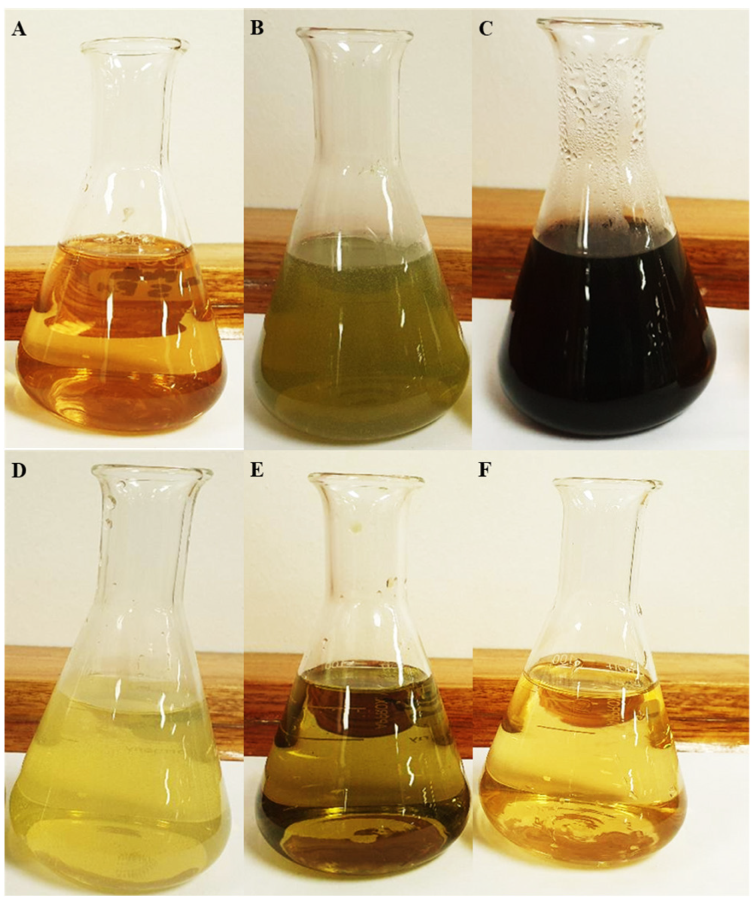
3.1. Percentage Yield of Extracts of B. albostellata

The methanolic leaf extract had significantly the highest percentage yield (16.78%), followed by the methanolic stem extract (9.38%) (Table 1), while the lowest yield (1.39%) was observed in the hexane leaf extract. This implies that the percentage yield of the phytochemical compounds in B. albostellata leaf extracts was higher than in the stem extracts. In addition, this suggests that the leaf extracts may include more polar compounds, while the stem may have fewer non-polar compounds. Thus, the obtained yield indicated that the polarity of the various solvents correlated with the pharmacological value of the plants [77,78]. The extracts (hexane, chloroform, and methanol) had different colors (Table 1) (Figure 1), while upon evaporation of the solvent, the hexane extracts were oily, while chloroform and methanol extracts dried to a hard, sticky solid.

3.2. Evaluating the Total Flavonoid and Total Phenolic Content of Extracts

One antioxidant assay cannot demonstrate all aspects of the natural product activities. This is because a plant’s antioxidant capabilities can be linked to several various mechanisms and pathways [79]. Flavonoids are secondary metabolites with a low molecular weight and are found throughout the plant kingdom. This phytochemical compound is produced by several plants in high quantities. The concentrations of flavonoids evaluated at 240 ug/mL in the extracts, measured as quercetin equivalents, are presented in Table 2. The highest total flavonoid content was observed in the methanolic leaf extract (42.39 ± 1.14 mg QE/g DW) compared to other solvents (Table 2). This may indicate that more flavonoids could be extractable with these solvents. The flavonoid content of all extracts of B. albostellata was satisfactory; this could be partially responsible for the observed different pharmacological activities of the extracts. Significantly, the lowest flavonoid content was found in the hexane leaves (11.22 ± 0.22 mg QE/g DW). Amoo et al. found the greatest and lowest flavonoid content in the leaves of B. greenii and B. albostellata stems, respectively [80]. With the exception of the hexane leaf extracts, the total flavonoid contents observed in the leaves and stems of B. albostellata were comparable (Table 2). The overall flavonoid content increased as the polarity increased from hexane (non-polar) to methanol (polar) (Table 2). The nature or quality of the flavonoid, aside from its quantity, in the leaf and stem extracts may change its therapeutic potential [80]. According to another study [81], the leaves of B. prionitis contained 6-hydroxyflavone, a naturally occurring flavonoid that is a promising drug candidate for treating anxiety-like disorders. Flavonoids have a wide range of biochemical and pharmacological properties, of which the most notable are their antioxidant, antimicrobial, and anti-inflammatory activities [82]. The antioxidant activity of flavonoids is attributed to numerous different mechanisms, including FR scavenging and enzyme inhibition, that cause free radical (FR) generation and chelation of metal ions [83]. Flavonoids, due to their structure, can scavenge nearly all known ROS [84].
Phenolic compounds are the most widely spread secondary metabolite in the plant kingdom. These compounds have the potential to be a natural antioxidant as well as an efficient radical scavenger [85]. Phenolic compounds react with active oxygen FRs, for instance, superoxide anions and hydroxyl and lipid peroxyl radicals [86]. These composites exhibit a diverse range of biological and chemical activities, including FR scavenging traits [87]. Table 3 presents the total phenolic content assessed at 240 µg/mL in each of the evaluated plant extracts. The highest level of total phenolics was found in methanol leaf extracts (6.05 ± 0.09 GAE/g DW), followed by the methanol stem extracts (2.93 ± 0.73 GAE/g DW). This may imply that more phenolic compounds could be extractable using these solvents. Amoo et al. reported similar results; the total phenolic content observed in the methanolic leaves of B. albostellata was greater (5.27 ± 0.324 mg GAE/g DW) than that of the methanolic stem extracts (3.76 ± 0.084 mg GAE/g DW) [80]. They found the greatest phenolic content in the leaves of B. prionitis, B. greenii, and B. albostellata compared to other plant parts [80]. According to Table 3, leaf extracts contained higher phenol contents than those of stems. Other similar findings showed that the amount of phenols detected in the leaves was higher than in the stems of B. prionitis [88], whereas Kumari et al. reported that B. lupulina stem extracts contained more phenols than those of leaves [89]. The ability of phenolic compounds to behave as hydrogen donors, singlet oxygen quenchers, and reducing agents is a key element in their antioxidant action [90]. Manian et al. [90] suggested the FR scavenging activity of extracts may be linked to the nature of the phenolic compounds present, therefore assisting in the hydrogen donating ability/electron transfer [91]. The therapeutic use of this compound could assist in the control of FR disorders such as inflammation, heart disease, stroke, cancer, and diabetes mellitus [92]. Phenolic compounds are regarded as more potent antioxidants than carotenoids in vitro and vitamins C and E [78].

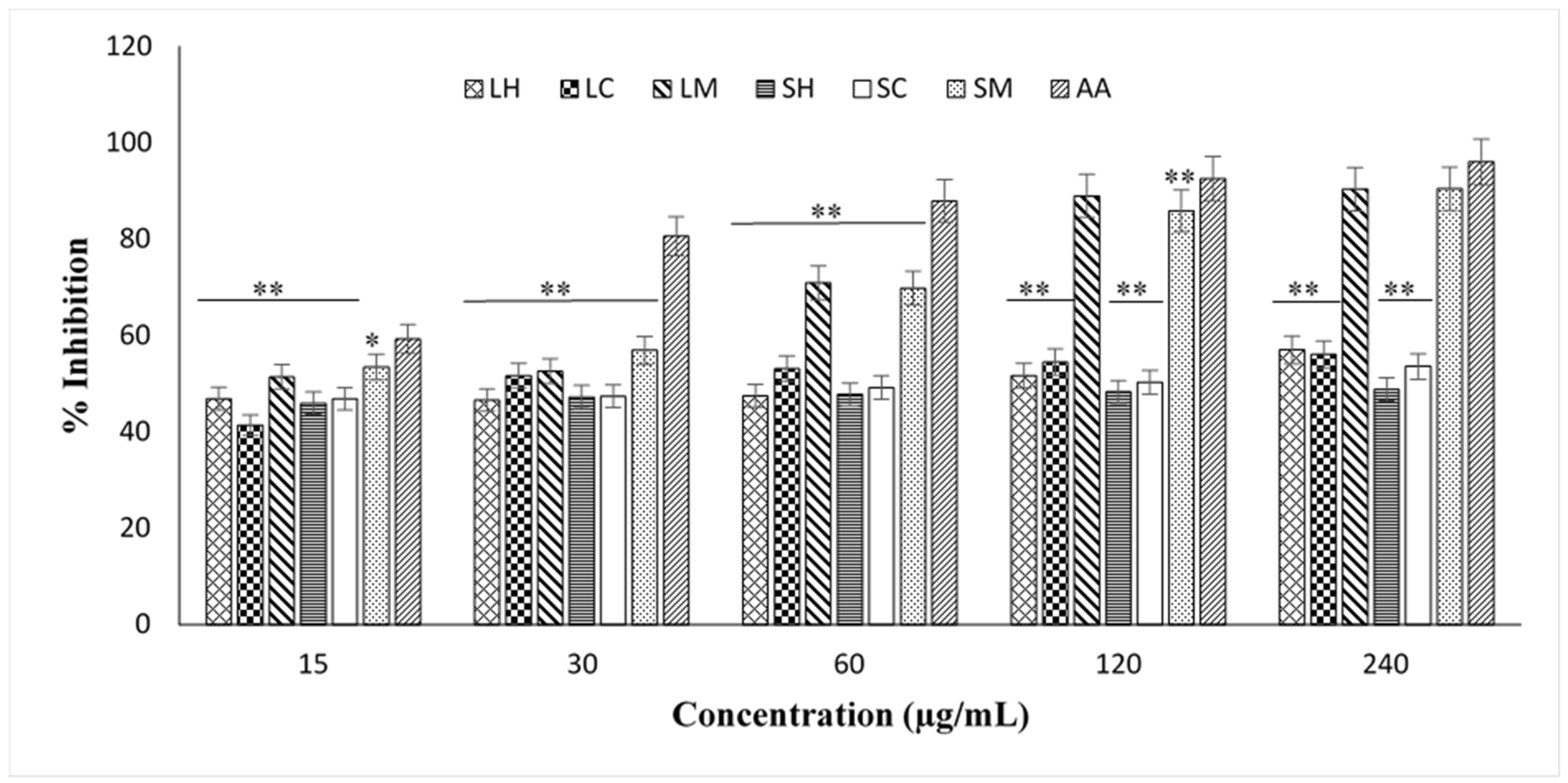
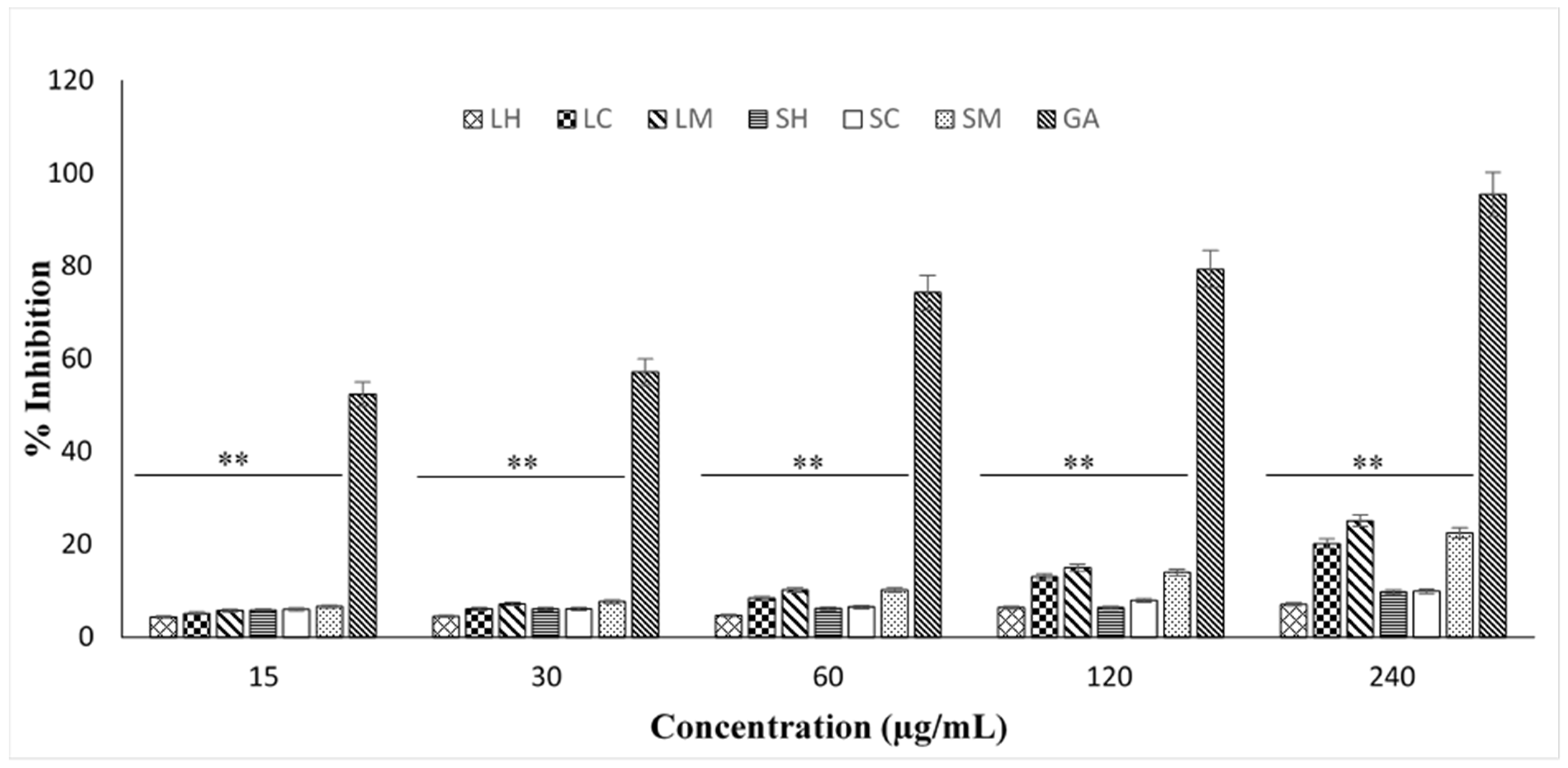
3.3. Antioxidant Screening of Extracts Using DPPH and FRAP Assays

The DPPH FR scavenging activity was evaluated by the decrease in absorbance at 516 nm, which is induced by antioxidants [93]. This assay is not specific to any precise class of antioxidants, and therefore provides the general antioxidant capacity of the extract [94]. Figure 2 presents the percentage FR scavenging activity of extracts of the leaves and stems of B. albostellata. The radical scavenging activities present in extracts of the leaves and stems were compared with ascorbic acid as a standard. The radical scavenging activity of the extracts was studied by their ability to reduce DPPH (stable radical) and any molecule that may donate a hydrogen or electron to DPPH [95]. The electron-donating ability of B. albostellata is most commonly determined using DPPH FR scavenging tests due to their reliability. For all extracts, there was a dose-dependent change in radical scavenging activities. Overall, in all extracts, with increasing concentration, there was an increase in the DPPH radical scavenging activity (Figure 2). A statistical analysis showed all extracts had significantly different activities across all concentrations (p < 0.05) when compared to ascorbic acid, at 15–240 µg/mL.
Methanolic leaf and stem extracts had a more effective radical scavenging activity than the hexane and chloroform plant extracts, with inhibitions of 90.37% and 90.43%, respectively. Dose-dependent radical scavenging activities were also observed in the methanolic extracts of different parts of B. prionitis, B. greenii, and B. albostellata [80]. These solutions had changed color from purple to a faded solution. A purple-colored solution visible in the DPPH assay accepts electrons, which then converts to a discolored solution. The point of the color change is linked to the effectiveness and concentration of antioxidants present [96]. The amount of discoloration indicates the FR scavenging action [97]. The scavenging activity of the methanolic extracts compared with the standard ascorbic acid suggests that the leaves and stems of B. albostellata are also an effective scavenger of FRs. Higher radical scavenging activity values were recorded at lower IC50 values (Table 4). Vasanth et al. [98] found the maximum DPPH radical scavenging activity at 100 μg/mL in ethanol and petroleum ether leaf extracts of B. cristata, with percentage inhibition values of 76.01 and 70.57, respectively.
The concentration of the methanolic leaf and stem extracts needed for 50% inhibition (IC50) was 16.95 µg/mL and 14.27 µg/mL, respectively. These results were compared with the IC50 value of ascorbic acid for the leaves (4.03 µg/mL) and stems (1.50 µg/mL) (Table 3). The radical scavenging activity of DPPH is influenced by the polarity of the medium, the chemical structure of the scavenger, the pH of the reaction, the concentration of the sample, and the reaction time [99]. Free radical reactions are linked in the pathology of several diseases such as cancer, Alzheimer’s disease, and inflammation [100]. Kumari et al. [89] studied the DPPH radical scavenging activity of the methanolic leaf and stem extracts of B. lupulina. These authors found IC50 values of the methanol leaf and stem extracts of 48.86 µg/mL and 60.82 µg/mL, respectively. Overall, the results obtained in this study indicated that the chloroform and methanol extracts displayed good radical scavenging activities, which were low when compared to the standard ascorbic acid.
The FRAP assay is based on an electron transfer reaction [101]. The presence of antioxidants (reductants) in the tested extracts leads to the reduction of ferricyanide/ferric ion compounds to their ferrous form via distinctive formation of ‘Perl’s Prussian blue’ and is measured spectrophotometrically [102]. According to Huang et al. [101], the degree of color variation is directly proportional to the concentrations of antioxidants present in the extracts. Figure 3 and Table 5 illustrate the reducing power of extracts from B. albostellata. All assessed extracts demonstrated an overall dose-dependent response. As the extract concentration increased, the reducing power decreased. The extracts’ reducing powers were significantly lower than the ascorbic acid standard (Figure 3). Similar results were observed for the different parts of B. prionitis, B. greenii, and B. albostellata [80]. A statistical analysis indicated that all extracts had significantly different activities across all concentrations (p < 0.05) compared to ascorbic acid, 15–240 µg/mL.
The leaf and stem hexane extracts displayed the lowest reducing power (Figure 3). Similar results were observed in leaf and stem hexane extracts of B. prionitis [103]. Low-to-moderate reducing powers were also observed in ethanol and petroleum ether leaf extracts of B. cristata [98]. The results in Figure 2 suggest the presence of antioxidant compounds with electron-donating abilities in the various extracts, which this assay is recognized to measure semi-quantitatively [104,105]. These compounds may be present in smaller amounts or in an impure form, which could be responsible for the low activity exhibited by the extracts [80].

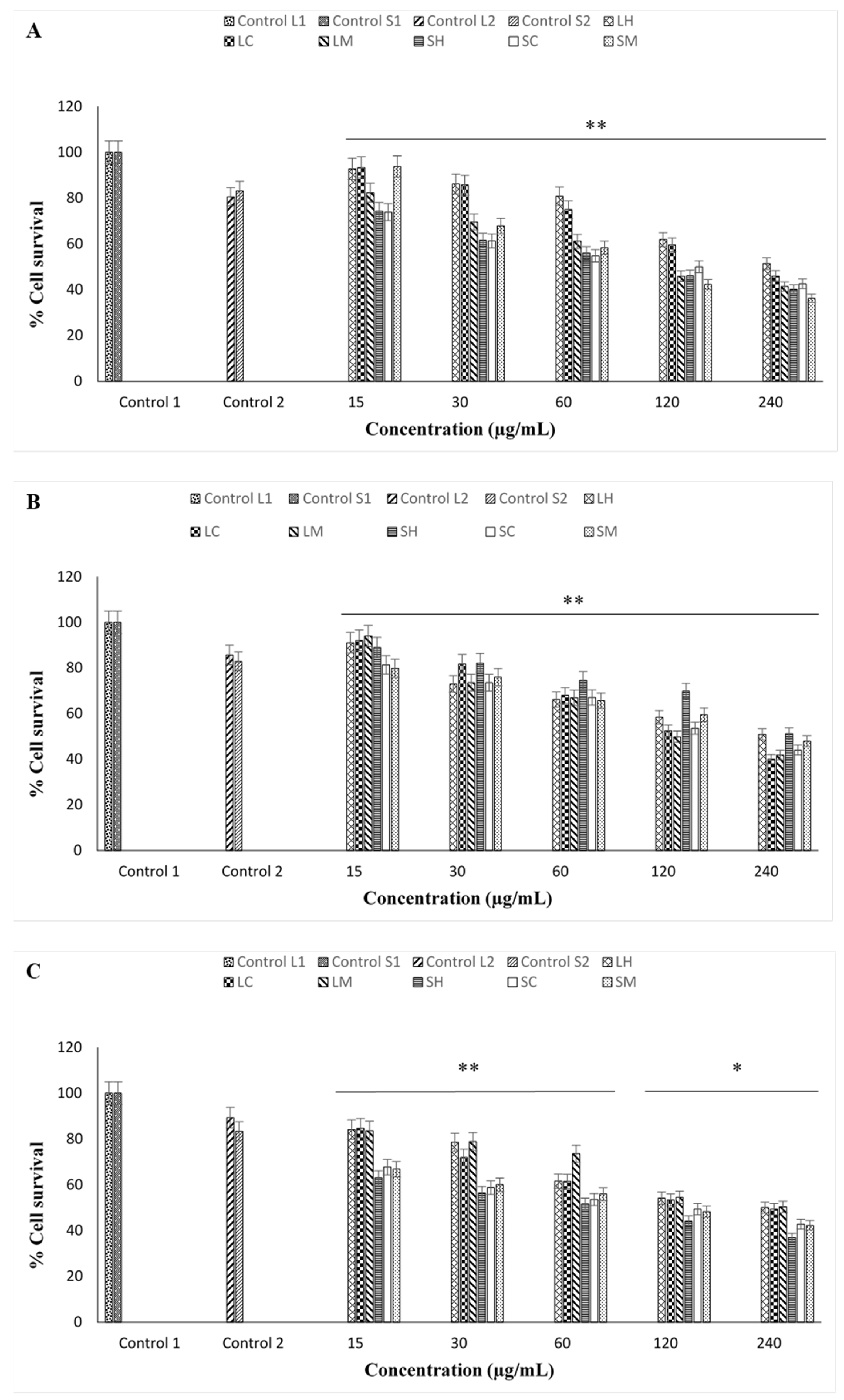
In Vitro Cytotoxicity Effect of Extracts of B. albostellata

New therapeutic approaches against cancerous cell lines can mediate the initiation of apoptosis [106]. The MTT cellular viability assay depends on the viable cells’ mitochondrial metabolic capacity [79]. As displayed in Figure 4A–C, the percentage cell survival for all extracts was dose-dependent. The results indicate the anti-proliferative effect decreases with increases in the concentration of the extract. All extracts at various concentrations showed a low-to-moderate cytotoxicity, which was lowest when treated with the methanolic stem extract (Figure 4A). Extracts demonstrated moderate cytotoxicities at high concentrations (240 µg/mL). The highest cellular viability for the HeLa cells was observed at 15 µg/mL of the methanol leaf extract (Figure 4B), while the lowest viability was observed at 240 µg/mL with the chloroform leaf extract (Figure 4B). For MCF-7 cells, the percentage cell viability was greatest at 15 µg/mL for the chloroform leaf extract, and lowest at 240 µg/mL for the methanol stem extract (Figure 4C). At varying concentrations, the cellular viability was >35% for all extracts (Figure 4A–C). As the concentration of each extract increased, its toxicity become more significant in each cell line. This slow decrease in cellular viability in all extracts may be due to the occurrence of some compounds that can inhibit cell proliferation [107].
Sawarkar et al. [108] evaluated the cytotoxicity of ethanolic extracts of B. prionitis and B. grandiflora. These authors observed a 50% cytotoxicity for human dermal fibroblast and human gingival fibroblast cell lines at >1000 μg/mL. Cytotoxic agents found in extracts may destroy the cell membrane, leading to cell lysis, cause necrosis, or induce apoptosis by triggering many biochemical mechanisms [109]. Additionally, the differences in cytotoxicity among the various extracts of B. albostellata may be attributed to the level of antioxidants present or related to the inhibitory effects through other signaling pathways [110]. Furthermore, it is well recognized that the temperature, solvent type, methods, and time of extraction can disturb the extraction of phytochemical compounds [111].
Statistical analyses indicated extracts across all concentrations had significantly different activities (p < 0.05). The IC50 values of the extracts in the three mammalian cell lines are represented in Table 6. In the HEK293 and HeLa cells, a low cytotoxic activity was observed for all extracts. The hexane leaf extract had the highest IC50 value of 294.44 µg/mL for HEK293, an indication of poor cytotoxicity. Additionally, a low cytotoxicity for HeLa was observed for the hexane stem extract (IC50 value of 376.70 µg/mL). A low-to-moderate cytotoxicity was observed in the hexane stem extracts (HEK293) and methanolic leaf extracts (HeLa), with IC50 values of 95.28 µg/mL and 98.86 µg/mL, respectively. The flavonoids found in these extracts may be responsible for their cytotoxicity. There is ongoing evidence that several flavonoids exert anticancer activity, although the mechanisms responsible for this effect have not been fully explained [112].
In MCF-7 cells, a low cytotoxicity was observed in the methanol leaf extracts (IC50 value of 239.88 µg/mL), whereas moderate cytotoxic levels were observed for the hexane stem extracts (IC50 value of 63.10 µg/mL). According to Manapradit et al. [113], the highest cytotoxicity of the butanolic leaf extracts of B. strigosa was found in HeLa and MCF-7 cells. Kumari and Dubey [57] treated Hep G2 cells with both aqueous and ethanolic leaf extracts of B. lupulina and observed growth inhibition, cell shrinkage, vacuolation, and cell lysis due to the extracts. The cytotoxicity observed in both HeLa and MCF-7 may be due to the presence of several glycoside, anthraquinone, saponin, flavonoid, and phenolic compounds. The isolated compounds were 6-hydroxyflavones, barlerin, acetyl barlerin, luteolin-7-O-β-Dglucoside, shanziside methyl ester, 6-O-trans-p-coumaroyl-8-O-acetyl shanziside methyl ester, anthraquinone,1,3,6,8-tetramethoxy-2,7-dimethyl anthraquinone, prioniside A, prioniside B, prioniside C, balarenone, and pipataline. These compounds found in B. prionitis and B. grandiflora displayed a potent cytotoxicity against human gingival fibroblast cell lines and human dermal fibroblast cell lines. Therefore, it is possible that any cytotoxic effects induced in the cancer cells could be due to the active phytochemical compounds found in the various extracts of the plant.
The antioxidant activity observed from flavonoids in Barleria is due to numerous different mechanisms, such as scavenging of FRs, inhibition of enzymes that cause free radical (FR) generation, and chelation of metal ions [83]. The flavonoids observed in the leaves and stems of B. albostellata may differ in their therapeutic potential. Other species of Barleria contain a naturally occurring flavonoid, 6-hydroxyflavone, which is a promising drug candidate for treating anxiety-like disorders. Phenols found in the methanolic extracts of B. albostellata have the potential to be a natural antioxidant and have the ability to act as an efficient radical scavenger [85]. Various authors have reported the greatest phenolic content in the leaves of species of Barleria compared to other plant parts [80,88]. The evaluated extracts may protect against FRs and oxidative damage occurring in various pathological mechanisms. The in vitro cytotoxicity observed in the extracts of B. albostellata may be due to the presence of flavonoids and phenols and the antioxidant activity in the different parts of this species. Additionally, the differences in cytotoxicity among the various extracts of B. albostellata may be attributed to the level of antioxidants present or related to the inhibitory effects through other signaling pathways [110].

4. Conclusions

Overall, the results of this study highlight the therapeutic potential of Barleria albostellata. The present study evaluated the antioxidant and cytotoxicity of B. albostellata, which can be useful in establishing its therapeutic value. All extracts, to some degree, presented good antioxidant properties. Methanolic leaf extracts had the highest total flavonoid content (42.39 ± 1.14 mg GAE/g DW) compared to other solvents. The total phenolic content was greatest in the methanol leaf extracts (6.05 ± 0.09 GAE/g DW), followed by the methanol stem extracts (2.93 ± 0.73 GAE/g DW). The methanolic leaf and stem extract concentrations needed for 50% inhibition (DPPH) were 16.95 µg/mL and 14.27 µg/mL. Methanolic leaf and stem extract concentrations required for 50% inhibition (DPPH) were 16.95 µg/mL and 14.27 µg/mL, respectively, whereas for FRAP, the reducing power of all extracts was considerably lower than the ascorbic acid standard. This could suggest that the evaluated extracts of B. albostellata may protect against the FRs and oxidative damage occurring in various pathological mechanisms. Antioxidants in the human body are essential for controlling the damaging consequences of FRs. The link between radical-scavenging agents in extracts and their cytotoxicity in cancer cells could be beneficial to data-screening projects that explore natural products with cytotoxicity potential. The IC50 values of extracts tested in the three cell lines were >63 µg/mL. The observed in vitro cytotoxicity may be due to the presence of flavonoids and phenols and the antioxidant activity in the different parts of this species. The isolation of specific bioactive compounds from the leaves and stems through bioassay-guided fractionation and the evaluation of their safety will be necessary in the further exploration of this species for new potential therapeutic drug leads. This could perhaps aid in underpinning the precise compounds responsible for the various pharmacological activities. To the best of our knowledge, this is the first report on the cytotoxic activities of leaf and stem extracts of Barleria albostellata. The findings from this study significantly contribute to the advancement of natural compounds for potential use in the healthcare sector.

Author Contributions

Conceptualization, S.G. and Y.N.; methodology, S.G. and Y.N.; formal analysis, S.G., Y.N. and M.S.; investigation, S.G., Y.N., M.S. and A.N.D.; data curation, S.G., Y.N., Y.H.D., M.S. and A.N.D.; writing—original draft preparation, S.G., Y.N. and A.N.D.; writing—review and editing, S.G., Y.N., Y.H.D., M.S., K.M.-T. and N.M.-D.; validation, S.G., Y.N., Y.H.D., M.S., A.N.D., K.M.-T. and N.M.-D.; visualization, Y.H.D., M.S., K.M.-T. and N.M.-D.; supervision, Y.N., Y.H.D. and M.S.; project administration, Y.N.; funding acquisition, Y.N. and Y.H.D. All authors have read and agreed to the published version of the manuscript.

Funding

The authors are thankful to the National Research Foundation (Grant No. 137957) South Africa and Researchers Supporting Project number (RSP-2023R375), King Saud University, Riyadh, Saudi Arabia.

Data Availability Statement

All data used to support the findings of this study are available from the corresponding author upon request.

Acknowledgments

The authors are thankful to the National Research Foundation (Grant No. 137957), South Africa. The authors acknowledge Researchers Supporting Project number (RSP-2023R375), King Saud University, Riyadh, Saudi Arabia.

Conflicts of Interest

The authors declare no conflict of interest.

References

  1. Tiwari, A.K. Antioxidants: New-generation therapeutic base for treatment of polygenic disorders. Curr. Sci. 2004, 86, 1092–1102. [Google Scholar]
  2. Juan, C.A.; Pérez de la Lastra, J.M.; Plou, F.J.; Pérez-Lebeña, E. The chemistry of reactive oxygen species (ROS) Revisited: Outlining their role in biological macromolecules (DNA, Lipids and Proteins) and induced pathologies. Int. J. Mol. Sci. 2021, 22, 4642. [Google Scholar] [CrossRef] [PubMed]
  3. Ziech, D.; Franco, R.; Georgakilas, A.G.; Georgakila, S.; Malamou-Mitsi, V.; Schoneveld, O.; Pappa, A.; Panayiotidis, M.I. The role of reactive oxygen species and oxidative stress in environmental carcinogenesis and biomarker development. Chem.-Biol. Interact. 2010, 188, 334–339. [Google Scholar] [CrossRef] [PubMed]
  4. Folorunsho, A.A.; Oluwafunke, A.B.; David, K.B.; Olayemi, A.A. Age-related changes in the expression of heat shock protein 70 and 90 on the gastric mucosa during gastric ulcer healing. Pharm. Biosci. J. 2018, 6, 1–10. [Google Scholar] [CrossRef]
  5. Jamshidi-Kia, F.; Wibowo, J.P.; Elachouri, M.; Masumi, R.; Salehifard-Jouneghani, A.; Abolhasanzadeh, Z.; Lorigooini, Z. Battle between plants as antioxidants with free radicals in human body. J. Herbmed Pharmacol. 2020, 9, 191–199. [Google Scholar] [CrossRef]
  6. Yan, L.L.; Zaher, H.S. How do cells cope with RNA damage and its consequences? J. Biol. Chem. 2019, 294, 5158–15171. [Google Scholar] [CrossRef]
  7. Mangge, H.; Becker, K.; Fuchs, D.; Gostner, J.M. Antioxidants, inflammation and cardiovascular disease. World J. Cardiol. 2014, 6, 462–477. [Google Scholar] [CrossRef]
  8. Gandhi, S.; Abramov, A.Y. Mechanism of oxidative stress in neurodegeneration. Oxidative Med. Cell. Longev. 2012, 2012, 428010. [Google Scholar] [CrossRef]
  9. Madireddy, S.; Madireddy, S. Protection from the pathogenesis of neurodegenerative disorders, including Alzheimer’s disease, amyotrophic lateral sclerosis, Huntington’s disease, and Parkinson’s diseases, through the mitigation of reactive oxygen species. J. Neurosci. Neurol. Disord. 2020, 3, 148–161. [Google Scholar] [CrossRef]
  10. Li, S.; Tan, H.Y.; Wang, N.; Zhang, Z.J.; Lao, L.; Wong, C.W.; Feng, Y. The role of oxidative stress and antioxidants in liver diseases. Int. J. Mol. Sci. 2015, 16, 26087–26124. [Google Scholar] [CrossRef]
  11. Majumder, D.; Nath, P.; Debnath, R.; Maiti, D. Understanding the complicated relationship between antioxidants and carcinogenesis. J. Biochem. Mol. Toxicol. 2020, 35, e22643. [Google Scholar] [CrossRef] [PubMed]
  12. Jaouad, B.; Torsten, B. Exogenous antioxidants-Doubleedged swords in cellular redox state. Oxidative Med. Cell. Longev. 2010, 3, 28–37. [Google Scholar]
  13. Bhattacharya, S. Reactive oxygen species and cellular defense system. In Free Radicals in Human Health and Disease; Rani, V., Yadav, U.C.S., Eds.; Springer: New Delhi, India, 2015; p. 25. [Google Scholar]
  14. Göçer, H.; Gülçin, İ. Caffeic acid phenethyl ester (CAPE): Correlation of structure and antioxidant properties. Int. J. Food Sci. Nutr. 2011, 62, 821–825. [Google Scholar] [CrossRef] [PubMed]
  15. Gülcin, I. Antioxidant activity of food constituents: An overview. Arch. Toxicol. 2012, 86, 345–391. [Google Scholar] [CrossRef] [PubMed]
  16. Halliwell, B.; Gutteridge, J.M.C. Formation of thiobarbituric acid reactive substances from deoxyribose in the presence of iron salts: The role of superoxide and hydroxyl radicals. FEBS Lett. 1981, 128, 347–352. [Google Scholar] [CrossRef]
  17. García-Alonso, M.; de Pascual-Teresa, S.; Santos-Buelga, C.; Rivas-Gonzalo, J.C. Evaluation of the antioxidant properties of fruits. Food Chem. 2004, 84, 13–18. [Google Scholar] [CrossRef]
  18. Ferreira, A.; Proença, C.; Serralheiro, M.L.M.; Araujo, M.E.M. The in vitro screening for acetylcholinesterase inhibition and antioxidant activity of medicinal plants from Portugal. J. Ethnopharmacol. 2006, 108, 31–37. [Google Scholar] [CrossRef]
  19. Sylvie, D.D.; Anatole, P.C.; Cabral, B.P.; Veronique, P.B. Comparison of in vitro antioxidant properties of extracts from three plants used for medical purpose in Cameroon: Acalypha racemosa, Garcinia lucida and Hymenocardia lyrata. Asian Pac. J. Trop. Biomed. 2014, 4, S625–S632. [Google Scholar] [CrossRef]
  20. Kapadiya, D.B.; Dabhi, B.K.; Aparnathi, K.D. Spices and herbs as a source of natural antioxidants for food. Int. J. Curr. Microbiol. Appl. Sci. 2016, 5, 280–288. [Google Scholar] [CrossRef]
  21. Zengin, G.; Aktumsek, A.; Guler, G.O.; Cakmak, Y.S.; Yildiztugay, E. Antioxidant Properties of Methanolic Extract and Fatty Acid Composition of Centaurea urvillei DC. subsp. hayekiana Wagenitz. Rec. Nat. Prod. 2011, 5, 123–132. [Google Scholar]
  22. Hassan, W.; Noreen, H.; Rehman, S.; Gul, S.; Amjad Kamal, M.; Paul Kamdem, J.; Zaman, B.; BT da Rocha, J. Oxidative stress and antioxidant potential of one hundred medicinal plants. Curr. Top. Med. Chem. 2017, 17, 1336–1370. [Google Scholar] [CrossRef] [PubMed]
  23. Saeed, N.; Khan, M.R.; Shabbir, M. Antioxidant activity, total phenolic and total flavonoid contents of whole plant extracts Torilis leptophylla L. BMC Complement. Altern. Med. 2012, 12, 221. [Google Scholar] [CrossRef]
  24. Adebiyi, O.E.; Olayemi, F.O.; Ning-Hua, T.; Guang-Zhi, Z. In vitro antioxidant activity, total phenolic and flavonoid contents of ethanol extract of stem and leaf of Grewia carpinifolia. Beni-Suef Univ. J. Basic Appl. Sci. 2017, 6, 10–14. [Google Scholar] [CrossRef]
  25. Madikizela, B.; McGaw, L.J. In vitro cytotoxicity, antioxidant, and anti-inflammatory activities of Pittosporum viridiflorum Sims and Hypoxis colchicifolia Baker used traditionally against cancer in Eastern Cape, South Africa. South Afr. J. Bot. 2019, 126, 250–255. [Google Scholar] [CrossRef]
  26. Vongtau, H.O.; Abbah, J.; Chindo, B.A.; Mosugu, O.; Salawu, A.O.; Kwanashie, H.O.; Gamaniel, K.S. Central inhibitory effects of the methanol extract of Neorautanenia mitis root in rats and mice. Pharm. Biol. 2005, 43, 113–120. [Google Scholar] [CrossRef]
  27. Oluyemi, K.A.; Okwuonu, U.C.; Baxter, D.G.; Oyesola, T. Toxic effects of methanolic extract of Aspilia africana leaf on the estrous cycle and uterine tissues of Wistar rats. Int. J. Morphol. 2007, 25, 609–614. [Google Scholar] [CrossRef]
  28. Verpoorte, R. Pharmacognosy in the new millennium: Lead finding and biotechnology. J. Pharm. Pharmacol. 2000, 52, 253–262. [Google Scholar] [CrossRef]
  29. Cragg, G.M.; Newman, D.J. Plants as a source of anti-cancer agents. J. Ethnopharmacol. 2005, 100, 72–79. [Google Scholar] [CrossRef]
  30. Tauchen, J.; Huml, L.; Bortl, L.; Doskocil, I.; Jarosova, V.; Marsik, P.; Frankova, A.; Clavo Peralta, Z.M.; Chuspe Zans, M.E.; Havlik, J.; et al. Screening of medicinal plants traditionally used in Peruvian Amazon for in vitro antioxidant and anticancer potential. Nat. Prod. Res. 2019, 33, 2718–2721. [Google Scholar] [CrossRef]
  31. Forman, D.; Ferlay, J. The Global and Regional Burden of Cancer. In World Cancer Report 2014; Stewart, B.W., Wild, C.P., Eds.; International Agency for Research on Cancer: Lyon, France, 2014; pp. 7250–7257. [Google Scholar]
  32. World Health Organization (WHO). Latest Global Cancer Data: Cancer Burden Rises to 18.1 Million New Cases and 9.6 Million Deaths in 2018; International Agency for Research on Cancer: Lyon, France, 2018. [Google Scholar]
  33. Adeloye, D.; David, R.A.; Aderemi, A.V.; Iseolorunkanmi, A.; Oyedokun, A.; Iweala, E.E.; Omoregbe, N.; Ayo, C.K. An estimate of the incidence of prostate cancer in Africa: A systematic review and meta-analysis. PLoS ONE 2016, 11, e0153496. [Google Scholar] [CrossRef]
  34. Madhuri, S.; Pandey, G. Some anticancer medicinal plants of foreign origin. Curr. Sci. 2009, 96, 779–783. [Google Scholar]
  35. CANSA. South African Cancer Statistics. 2017. Available online: https://www.cansa.org.za/files/2017/03/Fact-Sheet-Top-Ten-Cancers-per-Population-Group-in-SA-NCR-2012-web-Feb-2017.pdf (accessed on 16 May 2020).
  36. Singh, S.; Sharma, B.; Kanwar, S.S.; Kumar, A. Lead Phytochemicals for anticancer drug development. Front. Plant Sci. 2016, 7, 1667. [Google Scholar] [CrossRef] [PubMed]
  37. Niraula, S.; Amir, E.; Vera-Badillo, F.; Seruga, B.; Ocana, A.; Tannock, I.F. Risk of incremental toxicities and associated costs of new anticancer drugs: A meta-analysis. J. Clin. Oncol. 2014, 32, 3634–3642. [Google Scholar] [CrossRef] [PubMed]
  38. Singh, S.; Singh, P.K. Pattern and impact of drugs targeted toward toxicity amelioration in patients receiving cancer chemotherapy. Perspect. Clin. Res. 2018, 9, 23. [Google Scholar] [CrossRef] [PubMed]
  39. Shukla, S.; Mehta, A. Anticancer potential of medicinal plants and their phytochemicals: A review. Braz. J. Bot. 2015, 38, 199–210. [Google Scholar] [CrossRef]
  40. Mbaveng, A.T.; Kuete, V.; Efferth, T. Potential of Central, Eastern and Western Africa medicinal plants for cancer therapy: Spotlight on resistant cells and molecular targets. Front. Pharmacol. 2017, 8, 343. [Google Scholar] [CrossRef]
  41. El-Seedi, H.R.; Burman, R.; Mansour, A.; Turki, Z.; Boulos, L.; Gullbo, J.; Göransson, U. The traditional medical uses and cytotoxic activities of sixty-one Egyptian plants: Discovery of an active cardiac glycoside from Urginea maritima. J. Ethnopharmacol. 2013, 145, 746–757. [Google Scholar] [CrossRef]
  42. Ouelbani, R.; Bensari, S.; Mouas, T.N.; Khelifi, D. Ethnobotanical investigations on plants used in folk medicine in the regions of Constantine and Mila (North-East of Algeria). J. Ethnopharmacol. 2016, 194, 196–218. [Google Scholar] [CrossRef]
  43. Kabbaj, F.Z.; Meddah, B.; Cherrah, Y.; El, M.; Faouzi, A. Ethnopharmacological profile of traditional plants used in Morocco by cancer patients as herbal therapeutics. Phytopharmacology 2012, 2, 243–256. [Google Scholar]
  44. Reddy, L.A.; Odhav, B.; Bhoola, K.D. Natural products for cancer prevention: A global perspective. Pharmacol. Ther. 2003, 99, 1–13. [Google Scholar] [CrossRef]
  45. Gordaliza, M. Natural products as leads to anticancer drugs. Clin. Transl. Oncol. 2007, 9, 767–776. [Google Scholar] [CrossRef] [PubMed]
  46. Tan, G.; Gyllenhaal, C.; Soejarto, D.D. Biodiversity as a source of anticancer drugs. Curr. Drug Targets 2006, 7, 265–277. [Google Scholar] [CrossRef] [PubMed]
  47. Kaur, R.; Kapoor, K.; Kaur, H. Plants as a source of anticancer agents. J. Nat. Prod. Plant Resour. 2011, 1, 119–124. [Google Scholar]
  48. Atanasov, A.G.; Waltenberger, B.; Pferschy-Wenzig, E.M.; Linder, T.; Wawrosch, C.; Uhrin, P.; Temml, V.; Wang, L.; Schwaiger, S.; Heiss, E.H.; et al. Discovery and resupply of pharmacologically active plant-derived natural products: A review. Biotechnol. Adv. 2015, 33, 1582–1614. [Google Scholar] [CrossRef]
  49. Newman, D.J.; Cragg, G.M. Natural products as sources of new drugs from 1981 to 2014. J. Nat. Prod. 2016, 79, 629–661. [Google Scholar] [CrossRef]
  50. Tariq, A.; Sadia, S.; Pan, K.; Ullah, I.; Mussarat, S.; Sun, F.; Abiodun, O.O.; Batbaatar, A.; Li, Z.; Song, D.; et al. A systematic review on ethnomedicines of anti-cancer plants. Phytother. Res. 2017, 31, 202–264. [Google Scholar] [CrossRef]
  51. Alonso-Castro, A.J.; Villarreal, M.L.; Luis, A.; Olivo, S.; Gomez-Sanchez, M.; Dominguez, F.; Carranca, A.G. Mexican medicinal plants used for cancer treatment: Pharmacological, phytochemical and ethnobotanical studies. J. Ethnopharmacol. 2011, 133, 945–972. [Google Scholar] [CrossRef]
  52. Solowey, E.; Lichtenstein, M.; Sallon, S.; Paavilainen, H.; Solowey, E.; Lorberboum-Galski, H. Evaluating medicinal plants for anticancer activity. Sci. Writ. J. 2014, 2014, 721402. [Google Scholar] [CrossRef]
  53. Jain, R.; Jain, S.K. Screening of in vitro cytotoxic activity of some medicinal plants used traditionally to treat cancer in Chhattisgarh state, India. Asian Pac. J. Trop. Biomed. 2011, 1, S147–S150. [Google Scholar] [CrossRef]
  54. Mulla, S.K.; Swamy, P. Anticancer activity of ethanol and polyphenol extracts of Portulaca quadrifida Linn. on human colon cancer cell lines. Int. J. Pharma Bio Sci. 2012, 3, 488–498. [Google Scholar]
  55. Manglani, N.; Vaishnava, S.; Dhamodaran, P.; Sawarkar, H. In vitro and in vivo anticancer activity of leaf extract of Barleria grandiflora. Int. J. Pharm. Pharm. Res. 2014, 6, 7072. [Google Scholar]
  56. Choudhury, S.M.; Maity, P.; Bepari, M. Combined mixtures of Calotropis gigantea latex and Barleria lupulina leaf extracts ameliorate Dalton’s Ascitic Lymphoma induced cell Proliferation. Int. J. Eng. Sci. Res. Technol. 2015, 2, 2394–3386. [Google Scholar]
  57. Kumari, R.; Dubey, R.C. Phytochemical analysis and antibacterial and cytotoxic properties of Barleria lupulina Lindl. Extracts. J. Plant Pathol. Microbiol. 2016, 7, 2. [Google Scholar] [CrossRef]
  58. Panchal, P.K.; Meena, S.K.; Singh, K.; Sharma, N. Anticancer and antimicrobial potential of Barleria prionitis leaves ethanol extract. Int. J. Pharm. Pharm. Sci. 2018, 10, 100–103. [Google Scholar] [CrossRef]
  59. Froneman, W.; Le Roux, L.N. Barleria albostellata. 2007. Available online: http://pza.sanbi.org/barleria-albostellata (accessed on 2 February 2019).
  60. Balkwill, M.J.; Balkwill, K. A preliminary analysis of distribution patterns in a large, pantropical genus, Barleria L. (Acanthaceae). J. Biogeogr. 2002, 25, 95–110. [Google Scholar] [CrossRef]
  61. Amoo, S.O.; Finnie, J.F.; Van Staden, J. In vitro pharmacological evaluation of three Barleria species. J. Ethnopharmacol. 2009, 121, 274–277. [Google Scholar] [CrossRef]
  62. Yosook, C.; Panpisutchai, Y.; Chaichana, S.; Santisuk, T.; Reutrakul, V. Evaluation of anti-HSV-2 activities of Barleria lupulina and Clinacanthus nutans. J. Ethnopharmacol. 1999, 67, 179–187. [Google Scholar] [CrossRef]
  63. Wang, B.U.; Wu, M.; Perchellet, E.M.; Mcllvain, C.J.; Sperfslage, B.J.; Huang, X.; Tamura, M.; Stephany, H.A.; Hua, D.H.; Perchellet, J.P. A synthetic triptycene bisquinone, which blocks nucleoside transport and induces DNA fragmentation, retains its cytotoxic efficacy in daunorubicin-resistant HL-60 cell lines. Int. J. Oncol. 2001, 19, 1169–1178. [Google Scholar] [CrossRef]
  64. Jassim, S.A.A.; Naji, A.M. Novel antiviral agents: A medicinal plant perspective. J. Appl. Microbiol. 2003, 95, 412–427. [Google Scholar] [CrossRef]
  65. Suba, V.; Murugesan, T.; Arunachalam, G.; Mandal, S.C.; Saha, B.P. Anti-diabetic potential of Barleria lupulina extract in rats. Phytomedicine 2004, 11, 202–205. [Google Scholar] [CrossRef]
  66. Suba, V.; Murugesan, T.; Kumaravelrajan, R.; Mandal, S.C.; Saha, B.P. Antiinflammatory, analgesic and antiperoxidative efficacy of Barleria lupulina Lindl. extract. Phytother. Res. 2005, 19, 695–699. [Google Scholar] [CrossRef] [PubMed]
  67. Chomnawang, M.T.; Surassmo, S.; Nukoolkarn, V.S.; Gritsanapan, W. Antimicrobial effects of Thai medicinal plants against acne-inducing bacteria. J. Ethnopharmacol. 2005, 101, 330–333. [Google Scholar] [CrossRef] [PubMed]
  68. Shukla, S.; Gunjegaokar, S.M. Pharmacognostical and pharmacological profiling of Barleria prionitis Linn. J. Biol. Sci. Med. 2018, 4, 41–50. [Google Scholar]
  69. Gangaram, S.; Naidoo, Y.; Dewir, Y.H.; Singh, M.; Lin, J.; Murthy, H.N. Phytochemical Composition and Antibacterial Activity of Barleria albostellata CB Clarke Leaf and Stem Extracts. Plants 2023, 12, 2396. [Google Scholar] [CrossRef]
  70. Arruda, H.S.; Pereira, G.A.; de Morais, D.R.; Eberlin, M.N.; Pastore, G.M. Determination of free, esterified, glycosylated and insoluble-bound phenolics composition in the edible part of araticum fruit (Annona crassiflora Mart.) and its by-products by HPLC-ESI-MS/MS. Food Chem. 2018, 245, 738–749. [Google Scholar] [CrossRef] [PubMed]
  71. Liu, Q.; Yao, H. Antioxidant activities of barley seeds extracts. Food Chem. 2007, 102, 732–737. [Google Scholar] [CrossRef]
  72. Braca, A.; Sortino, C.; Politi, M.; Morelli, I.; Mendez, J. Antioxidant activity of flavonoids from Licania licaniaeflora. J. Ethnopharmacol. 2002, 79, 379–381. [Google Scholar] [CrossRef]
  73. Benzie, I.F.; Strain, J.J. The ferric reducing ability of plasma (FRAP) as a measure of “antioxidant power”: The FRAP assay. Anal. Biochem. 1996, 239, 70–76. [Google Scholar] [CrossRef]
  74. Daniels, A.N.; Singh, M. Sterically stabilized siRNA: Gold nanocomplexes enhance c-MYC silencing in a breast cancer cell model. Nanomedicine 2019, 14, 1387–1401. [Google Scholar] [CrossRef]
  75. Mosman, T. Rapid colourimetric assay for cellular growth and survival: Application to proliferation and cytotoxicity assays. J. Immunol. Methods 1983, 65, 55–63. [Google Scholar] [CrossRef]
  76. Vinken, M.; Blaauboer, B.J. In vitro testing of basal cytotoxicity: Establishment of an adverse outcome pathway from chemical insult to cell death. Toxicol. Vitr. 2017, 39, 104–110. [Google Scholar] [CrossRef]
  77. Abubakar, E.M.; Misau, S.; Modibbo, S.; Bala, G.L. Percentage yield and acute toxicity of the plant extracts of Ceiba pentandra grown in Bauchi State, North Eastern Nigeria. J. Pharmacogn. Phytochem. 2017, 6, 1777–1779. [Google Scholar]
  78. Chintalapani, S.; Swathi, M.S.; Mangamoori, L.N. Phytochemical screening and in vitro antioxidant activity of whole plant extracts of Sesuvium portulacastrum L. Asian J. Pharm. Clin. Res. 2018, 11, 1–6. [Google Scholar] [CrossRef]
  79. Grauzdytė, D.; Pukalskas, A.; Viranaicken, W.; El Kalamouni, C.; Venskutonis, P.R. Protective effects of Phyllanthus phillyreifolius extracts against hydrogen peroxide induced oxidative stress in HEK293 cells. PLoS ONE 2018, 13, e0207672. [Google Scholar] [CrossRef]
  80. Amoo, S.O.; Ndhlala, A.R.; Finnie, J.F.; Van Staden, J. Antifungal, acetylcholinesterase inhibition, antioxidant and phytochemical properties of three Barleria species. South Afr. J. Bot. 2011, 77, 435–445. [Google Scholar] [CrossRef]
  81. Ren, L.; Wang, F.; Xu, Z.; Chan, W.M.; Zhao, C.; Xue, H. GABAA receptor subtype selectivity underlying anxiolytic effect of 6-hydroxyflavone. Biochem. Pharmacol. 2010, 79, 1337–1344. [Google Scholar] [CrossRef] [PubMed]
  82. Middleton, E., Jr.; Kandaswami, C.; Theoharides, T.C. The effects of plant flavonoids on mammalian cells: Implications for inflammation, heart disease, and cancer. Pharmacol. Rev. 2000, 52, 673–751. [Google Scholar] [PubMed]
  83. Sawarkar, H.A.; Kashyap, P.P.; Kaur, C.D. RBC Haemolysis prevention and antioxidant activity of Barleria prionitis. Chiang Mai J. Sci. 2018, 45, 888–896. [Google Scholar]
  84. Maryam, Z.; Farrukh, A.; Iqbal, A. The in vitro antioxidant activity and total phenolic content of four Indian medicinal plants. Int. J. Pharm. Pharm. Sci. 2009, 1, 88–95. [Google Scholar]
  85. Kapoor, A.; Shukla, S.; Kaur, R.; Kumar, R.; Lehra, K.S.; Kapoor, S. Preliminary phytochemical screening and antioxidant activity of whole plant of Barleria prionitis Linn. Int. J. Adv. Pharm. Biol. Chem. 2014, 3, 410–419. [Google Scholar]
  86. Afanasiev, I.B.; Dorozhko, A.I.; Brodshi, A.V.; Kostyak, V.A.; Potaporitch, A.I. Chelating and free radical scavenging mechanisms of inhibitory action of rutin and quercetin in lipid peroxidation. Biochem. Pharmacol. 1989, 38, 1763–1769. [Google Scholar] [CrossRef] [PubMed]
  87. Makhafola, T.J.; Elgorashi, E.E.; McGaw, L.J.; Verschaeve, L.; Eloff, J.N. The correlation between antimutagenic activity and total phenolic content of extracts of 31 plant species with high antioxidant activity. BMC Complement. Altern. Med. 2016, 16, 490. [Google Scholar] [CrossRef] [PubMed]
  88. Jaiswal, S.K.; Dubey, M.K.; Das, S.; Verma, A.R.; Rao, C.V. A comparative study on total phenolic content, reducing power and free radical scavenging activity of aerial parts of Barleria prionitis. Int. J. Phytomed. 2010, 2, 155–159. [Google Scholar] [CrossRef]
  89. Kumari, R.; Kumar, S.; Kumar, A.; Goel, K.K.; Dubey, R.C. Antibacterial, antioxidant and immuno-modulatory properties in extracts of Barleria lupulina Lindl. BMC Complement. Altern. Med. 2017, 17, 484. [Google Scholar] [CrossRef]
  90. Samak, G.; Shenoy, R.P.; Manjunatha, S.M.; Vinayak, K.S. Superoxide and hydroxyl radical scavenging actions of botanical extracts of Wagetea spicata. Food Chem. 2009, 115, 631–634. [Google Scholar] [CrossRef]
  91. Manian, R.; Anusuya, N.; Siddhuraju, P.; Manian, S. The antioxidant activity and free radical scavenging potential of two different solvent extracts of Camellia sinensis (L.) O. Kuntz, Ficus bengalensis L. and Ficus racemosa L. Food Chem. 2008, 107, 1000–1007. [Google Scholar] [CrossRef]
  92. Ghasemzadeh, A.; Ghasemzadeh, N. Flavonoids and phenolic acids: Role and biochemical activity in plants and human. J. Med. Plants Res. 2011, 5, 6697–6703. [Google Scholar] [CrossRef]
  93. Manjula, M.S.; Ganthi, A.S. In-vitro antioxidant and anti-inflammatory potential of ethanol extracts (root and aerial parts) of Barleria noctiflora. Ann. Plant Sci. 2018, 7, 1997–2001. [Google Scholar]
  94. Tepe, B.; Donmez, E.; Unlu, M.; Candan, F.; Daferera, D.; Vardar-Unlu, G.; Polissiou, M.; Sokmen, A. Antimicrobial and antioxidative activities of the essential oils and methanol extracts of Salvia cryptantha (Montbret et Aucher ex Benth.) and Salvia multicaulis (Vahl). Food Chem. 2004, 84, 519–525. [Google Scholar] [CrossRef]
  95. Patel, R.; Patel, A.; Desai, S.; Nagee, A. Study of secondary metabolites and antioxidant properties of leaves, stem and root among Hibiscus rosa-sinensis cultivars. Asian J. Exp. Biol. Sci. 2012, 3, 719–725. [Google Scholar]
  96. Herrera-Calderon, O.; Alvarado-Puray, C.; Arroyo-Acevedo, J.L.; Rojas-Armas, J.P.; Chumpitaz-Cerrate, V.; Hañari-Quispe, R.; Valenzuela-Herrera, R. Phytochemical screening, total phenolic content, antioxidant, and cytotoxic activity of five Peruvian plants on human tumor cell lines. Pharmacogn. Res. 2018, 10, 161–165. [Google Scholar] [CrossRef]
  97. Kumar, S.; Sandhir, R.; Ojha, S. Evaluation of antioxidant activity and total phenol in different varieties of Lantana camara leaves. BMC Res. Notes 2014, 7, 560. [Google Scholar] [CrossRef] [PubMed]
  98. Vasanth, S.; Bupesh, G.; Siva Vijayakumar, T.; Balachandar, V.; Rajan Gunasekaran, D. Evaluation of in vitro antidiabetic and antioxidant potential of Barleria cristata leaves extracts. Asian J. Pharm. Clin. Res. 2018, 11, 287–290. [Google Scholar] [CrossRef]
  99. Sujatha, A.P.; Doss, A.; Muthukumarasamy, S.; Mohan, V.R. Study of antioxidant activity of Barleria courtrallica. Res. J. Life Sci. Bioinform. Pharm. Chem. Sci. 2018, 4, 513–521. [Google Scholar]
  100. Houghton, P.J.; Howes, M.-J.; Lee, C.C.; Steventon, G. Uses and abuses of in vitro tests in ethnopharmacology: Visualizing an elephant. J. Ethnopharmacol. 2007, 110, 391–400. [Google Scholar] [CrossRef] [PubMed]
  101. Huang, D.; Ou, B.; Prior, R.L. The chemistry behind antioxidant capacity assays. J. Agric. Food Chem. 2005, 53, 1841–1856. [Google Scholar] [CrossRef] [PubMed]
  102. Chung, Y.C.; Chang, C.T.; Chao, W.W.; Lin, C.F.; Chou, S.T. Antioxidative activity and safety of the 50% ethanolic extract from red bean fermented by Bacillus subtilis IMR-NK1. J. Agric. Food Chem. 2002, 50, 2454–2458. [Google Scholar] [CrossRef]
  103. Sharma, P.; Sharma, G.N.; Shrivastava, B.; Jadhav, H.R. Evaluation of antioxidant potential of Barleria prionitis leaf and stem. Am. J. Phytomedicine Clin. Ther. 2014, 2, 1177–1186. [Google Scholar]
  104. Amarowicz, R.; Pegg, R.B.; Rahimi-Moghaddam, P.; Barl, B.; Weil, J.A. Free-radical scavenging capacity and antioxidant activity of selected plant species from the Canadian prairies. Food Chem. 2004, 84, 551–562. [Google Scholar] [CrossRef]
  105. Rumbaoa, R.G.O.; Cornago, D.F.; Geronimo, I.M. Phenolic content and antioxidant capacity of Philippine sweet potato (Ipomoea batatas) varieties. Food Chem. 2009, 113, 1133–1138. [Google Scholar] [CrossRef]
  106. Motadi, L.R.; Choene, M.S.; Mthembu, N.N. Anticancer properties of Tulbaghia violacea regulate the expression of p53-dependent mechanisms in cancer cell lines. Sci. Rep. 2020, 10, 12924. [Google Scholar] [CrossRef] [PubMed]
  107. Gordanian, B.; Behbahani, M.; Carapetian, J.; Fazilati, M. In vitro evaluation of cytotoxic activity of flower, leaf, stem and root extracts of five Artemisia species. Res. Pharm. Sci. 2014, 9, 91–96. [Google Scholar] [PubMed]
  108. Sawarkar, H.A.; Kashyap, P.P.; Pandey, A.K.; Singh, M.K.; Kaur, C.D. Antimicrobial and cytotoxic activities of Barleria prionitis and Barleria grandiflora: A comparative study. Bangladesh J. Pharmacol. 2016, 11, 802–809. [Google Scholar] [CrossRef]
  109. Alonso-Carrillo, N.; de los Ángeles Aguilar-Santamaría, M.; Vernon-Carter, E.J.; Jiménez-Alvarado, R.; Cruz-Sosa, F.; Román-Guerrero, A. Extraction of phenolic compounds from Satureja macrostema using microwave-ultrasound assisted and reflux methods and evaluation of their antioxidant activity and cytotoxicity. Ind. Crops Prod. 2017, 103, 213–221. [Google Scholar] [CrossRef]
  110. Sammar, M.; Abu-Farich, B.; Rayan, I.; Falah, M.; Rayan, A. Correlation between cytotoxicity in cancer cells and free radical-scavenging activity: In vitro evaluation of 57 medicinal and edible plant extracts. Oncol. Lett. 2019, 18, 6563–6571. [Google Scholar] [CrossRef] [PubMed]
  111. Tuntiwachwuttikul, P.; Pancharoen, O.; Taylor, W.C. Iridoid glucosides of Barleria lupulina. Phytochemistry 1998, 49, 163–166. [Google Scholar] [CrossRef]
  112. Kopustinskiene, D.M.; Jakstas, V.; Savickas, A.; Bernatoniene, J. Flavonoids as anticancer agents. Nutrients 2020, 12, 457. [Google Scholar] [CrossRef] [PubMed]
  113. Manapradit, N.; Poeaim, S.; Charoenying, P. Cytotoxicity and antimicrobial activities of leaf extracts from Barleria strigosa. Int. J. Agric. Technol. 2015, 11, 551–561. [Google Scholar]
Figure 1. Extracts of B. albostellata. (A) Hexane leaves; (B) chloroform leaves; (C) methanol leaves; (D) hexane stems; (E) chloroform stems; (F) methanol stems.
Horticulturae 09 01226 g001
Figure 2. In vitro antioxidant activity (% inhibition DPPH) of crude extracts from the leaves and stems of B. albostellata (* p < 0.05 and ** p < 0.001 above each bar were considered statistically significant when comparing each extract to ascorbic acid at different concentrations, 15–240 µg/mL). Data are presented as means ± SD, n = 3, and displayed as a percentage of the control sample. LH—leaf hexane; LC—leaf chloroform; LM—leaf methanol; SH—stem hexane; SC—stem chloroform; SM—stem methanol; AA—ascorbic acid.
Horticulturae 09 01226 g002
Figure 3. In vitro antioxidant activity (% inhibition FRAP) of crude extracts from the leaves and stems of B. albostellata (** p < 0.001 above each bar were considered statistically significant when comparing each extract to gallic acid, at different concentrations, 15–240 µg/mL). Data are presented as means ± SD, n = 3, and displayed as a percentage of the control sample. LH—leaf hexane; LC—leaf chloroform; LM—leaf methanol; SH—stem hexane; SC—stem chloroform; SM—stem methanol; GA—gallic acid.
Horticulturae 09 01226 g003
Figure 4. In vitro cytotoxicity activity (% cell survival) of crude leaf and stem extracts of B. albostellata. (A) Human embryonic kidney (HEK293); (B) cervical cancer cells (HeLa), (C) breast adenocarcinoma (MCF-7) (* p < 0.05 and ** p < 0.001 were considered statistically significant within the different concentrations, 15–240 µg/mL). Data are presented as means ± SD, n = 3, and displayed as a percentage of the control sample. Control Leaves 1—cells only; Control Stems 1—cells only; Control Leaves 2—DMSO only; Control Stems 2—DMSO only; LH—leaf hexane; LC—leaf chloroform; LM—leaf methanol; SH—stem hexane; SC—stem chloroform; SM—stem methanol.
Horticulturae 09 01226 g004
Table 1. Percentage yield of the leaf and stem extracts of B. albostellata.
Crude ExtractLeavesStemLeavesStemLeavesStems
Dried Extract Yield (g)Percentage Yield (%)Color
Hexane0.1390.1941.391.94Dark yellowLight yellow
Chloroform0.2650.2192.652.19Dark greenLight green
Methanol1.6780.93816.789.38Dark brownLight yellow
Table 2. Total flavonoid content of the extracts of the leaves and stems of B. albostellata.
Crude ExtractsTotal Flavonoid (mg QE/g DW)
LeavesStem
Hexane11.22 ± 0.2231.79 ± 0.59
Chloroform34.38 ± 0.2836.20 ± 0.685
Methanol42.39 ± 1.1437.10 ± 0.95
Data displayed as means ± SD of three experiments.
Table 3. Total phenolic content of the extracts of the leaves and stems of B. albostellata.
Crude ExtractsTotal Phenols (mg GAE/g DW)
LeavesStem
Hexane1.15 ± 0.561.06 ± 0.03
Chloroform2.51 ± 0.271.25 ± 0.28
Methanol6.05 ± 0.092.93 ± 0.73
Data displayed as means ± SD of three experiments.
Table 4. IC50 values of the DPPH radical scavenging activity of the leaf and stem extracts of B. albostellata.
ExtractDPPH (µg/mL)
LeavesStems
Hexane61.53697.75
Chloroform 44.9973.38
Methanol16.9514.27
Ascorbic acid 4.031.50
Data displayed as means, n = 3, of triplicate determinations.
Table 5. IC50 values of the FRAP reducing power activity of the leaf and stem extracts of B. albostellata.
ExtractFRAP (µg/mL)
LeavesStems
Hexane1.20 × 10209.12 × 1017
Chloroform>90,0001.05 × 1015
Methanol>15,000>58,000
Gallic acid14.9614.90
Data displayed as means, n = 3, of triplicate determinations.
Table 6. IC50 values of the cytotoxicity activity of hexane, chloroform, and methanol leaf and stem extracts of B. albostellata.
ExtractsCytotoxicity (µg/mL)
Cell LinesLeavesStems
HEK293Hexane294.4495.28
Chloroform210.86110.15
Methanol116.1596.61
HeLaHexane220.80376.70
Chloroform143.55165.58
Methanol98.86232.27
MCF-7Hexane194.9863.10
Chloroform181.97102.33
Methanol239.88102.33
Data displayed as means, n = 3, of triplicate determinations.
Disclaimer/Publisher’s Note: The statements, opinions and data contained in all publications are solely those of the individual author(s) and contributor(s) and not of MDPI and/or the editor(s). MDPI and/or the editor(s) disclaim responsibility for any injury to people or property resulting from any ideas, methods, instructions or products referred to in the content.

