1. Introduction
Medicinal plants and their phytochemicals have been used for the treatment of various diseases for a long time [
1]. The identification of new plants with medicinal properties is of great importance, as approximately 60% of the new medicines produced since 1981 came from plants [
2]. Plant phytochemicals (e.g., phenolic compounds) have attracted attention as bioactive agents exhibiting strong antioxidant potential and are preferably used to treat various diseases to improve human health [
3]. Bioactive compounds generated by plants also have applications in the food industry, such as in the manufacture of beverages, flavorings, and food preservatives.
In the human body, antioxidants play a very important role in human health by transferring a hydrogen (H) atom or an electron (e−) to inactivate reactive species [
4]. The most used antioxidant activity assays are based on these two mechanisms: the H atom transfer (HAT) or the e− transfer (ET). HAT-based assays apply a competitive reaction scheme between antioxidant and substrate [
5]; meanwhile, ET-based assays measure the capacity of an antioxidant in the reduction of an oxidant agent (which changes color when reduced) [
5]. ET-based assays such as 2,2-Diphenyl-1-picrylhydrazyl (DPPH), the ferric reducing antioxidant power (FRAP), and 2,2′-azino-bis (3-ethylbenzthiazo-line-6-sulphonic) diammonium acid (ABTS) are the most used to measure the antioxidant capacity of plant extracts [
6].
Mexico has an ancestral tradition of using medicinal plants as first-aid remedies. However, research on medicinal plants is scarce in the north of the country [
7]. Northern Mexico is a desert rich in endemic flora, accompanied by ample local and traditional knowledge. Natural products of arid-zone plants represent an alternative to the search for new bioactive compounds. The production of phytochemical compounds in desert plants helps them survive in difficult climatic conditions, and these phytochemicals give plants important biological properties such as antioxidant, antimicrobial, and anticancer activities [
7].
Studies on plants used in traditional medicine in Coahuila state (northern Mexico) are limited to a few, such as
Larrea tridentata [
8] and
Lippia graveolens (Mexican oregano) [
9,
10]. To explore the full potential of the medicinal plants from the semi-desert of Viesca, Coahuila, our work group recently carried out an ethnobotanical study, finding that 77 medicinal plants are used for this purpose in the region [
7]. In this study, for the first time, the phenolic content of 23 of these plants will be measured, identifying the antioxidant potential and the compounds of the main plants. The determination of the antioxidant potential of plants used in traditional medicine can contribute to their safe use in local health systems and their valorization under the concept of bioeconomy. Therefore, the objective of this research was to investigate the phenolic content and antioxidant potential of plants used in traditional medicine in the arid zone of Viesca, Mexico.
2. Materials and Methods
2.1. Reagents
Ethanol, Folin–Ciocalteu reagent, Sodium hydroxide, gallic acid, Trolox, 2,2-diphenyl-1-pricryl-hydrazyl (DPPH), 2,2′-Azinobis-3-ethyl-benzo-thiazoline-6-sulfonic acid (ABTS), and FRAP reagents were purchased from Sigma Chemical Co. (St. Louis, MO, USA).
2.2. Plant Material
Twenty-three medicinal plants were collected in Viesca, Coahuila, Mexico (24°45′–25°39′ north latitude and 102°28′–103°30′ west longitude) and Parras, Coahuila, Mexico (102°11′10″ west longitude and 25°26′27″ north latitude) (
Table 1). The scientific names of the plants were confirmed using
https://www.theplantlist.org/ (accessed on 1 August 2023). These plants were selected based on an ethnobotanical study carried out by Torres et al. [
7]. These plants were identified by specialists of the Research Center and Ethnobiological Garden of the Universidad Autonoma de Coahuila (CIJE-UAdeC) and the Botanical Reference Center of the Universidad Autonoma Agraria Antonio Narro (CREB-UAAAN).
2.3. Obtaining Plant Extracts
The plant parts used in this study were selected based on a previous ethnobotanical study by our research group [
7]. Approximately 500 g of the selected parts of the plants (
Table 1) were dried in the absence of light for two days at an average temperature of 30 °C at the Ethnobiological Garden and Research Center located in Viesca, Coahuila. Subsequently, the dried plants were transported to the Phytopharmacology laboratory of the Autonomous University of Coahuila. Immediately, the plants were ground with a laboratory mill (Nixtamatic, NG-02) until a homogeneous powder was obtained (particle size less than 200 µm). Samples were stored in packages for dehydrated products (aluminum bag).
2.4. Evaluation of the Phenolic Content of 23 Plants
The extraction was evaluated in two stages: In the first stage, a solid–liquid extraction of the 23 plants was carried out with 50% ethanol at a ratio of 1:25 g/mL, and the extraction method used was traditional maceration in a shaker (Premiere, TX, USA) at 180 rpm stirring at room temperature (28–30 °C) for 2 h. Subsequently, the samples were centrifuged (ThermoFisher, Waltham, MA, USA) for 4 min at 10,000 rpm. The extracts were stored at 4 °C for subsequent phenolic quantification [
11].
2.5. Evaluation of Extraction Methods
In the second stage, the three plants that presented the highest polyphenol content were subjected to extraction, evaluating two methods: ultrasound-assisted extraction (UAE) (Fisher Scientific, Waltham, MA, USA) and maceration (Premiere, TX, USA) with three solid/liquid ratios (1:10, 1:25, and 1:50 g/mL), using the same solvent and centrifugation conditions as in the first stage. The extraction in both methods was carried out for 30 min at 30 °C. The extracts from stage 2 were evaluated for antioxidant activity, phenol content, and the identification of their components by HPLC, as described below.
2.5.1. Quantification of Phenols
The content of phenolic compounds in the extracts was estimated by the Folin–Ciocalteu colorimetric method, according to Blois [
12], with the modifications of Magalhães et al. [
13] (change sodium carbonate solution for sodium hydroxide to reduce the reaction time). Briefly, 300 µL of 0.2 N Folin’s reagent was added to 300 µL of the plant extracts. Subsequently, 600 µL of 0.35 M sodium hydroxide solution was added. The samples were incubated for 5 min at room temperature, and the absorbance at 760 nm was measured in a spectrophotometer (Velab, Pharr, TX, USA). The concentration of phenolic compounds was calculated according to the calibration curve of gallic acid in Equation (1).
The content of phenolic compounds was expressed as milligrams of gallic acid equivalent per gram of dry weight of plant material (mg GAE/g).
2.5.2. Antioxidant Activity Assay
DPPH Assay
The DPPH radical scavenging activity of each extract solution was analyzed using the method reported by Mensor et al. [
14]. An amount of 2.5 mL of extract from the plants was mixed with 1 mL of methanolic DPPH• 0.3 mM. The reaction mixtures were incubated for 30 min in the dark at room temperature, and the reduction of radical DPPH was measured at 518 nm (Velab, Pharr, TX, USA). Results were expressed as milligram Trolox equivalent per gram of dry weight of plant material (mg TE/g).
FRAP Assay
Ferric reducing antioxidant power assay was performed as follows. The FRAP reagent was prepared in a 1:1:10 ratio of 10 mM TPTZ, 20 mM FeCl3, and 0.3 mM acetate buffer with a pH of 3.6, respectively. The mixture was prepared and incubated for 10 min at 37 °C prior to use. An amount of 1425 µL of FRAP reagent was mixed with 75 µL of plant extract, the samples were incubated at 37 °C for 30 min in the dark, and the absorbance was measured in a spectrophotometer (Velab, TX, USA) at 593 nm. Values were expressed as milligram Trolox equivalents per gram of dry weight of plant material (mg TE/g).
ABTS Assay
The ABTS radical cation (ABTS•+) scavenging activity of the sample was measured using the method reported by Re et al. [
15]. ABTS•+ was generated by oxidation of 7 mmol/L ABTS with 2.45 mmol/L potassium persulfate after incubation in the dark (12 h). The freshly prepared ABTS•+ solution was diluted with ethanol (1:40) to obtain an absorbance of 0.70 ± 0.02 at 734 nm in a spectrophotometer (Velab, TX, USA). An amount of 10 μL of plant extracts and 1 mL of ABTS•+ were incubated for 1 min in the dark, and the absorbance was then recorded at 734 nm (Velab, TX, USA). The values of the ABTS assay were expressed in a similar way to those in the DPPH assay (mg TE/g).
2.6. RP-HPLC-ESI-MS Analysis
The analyses by RP-HPLC were performed on a Varian HPLC system [
16]. An amount of 4 mg of dry extract was diluted in 1 mL of ethanol and filtered (0.2 µm) before injection. A liquid chromatograph ion trap mass spectrometer (Varian 500-MS IT Mass Spectrometer, Palo Alto, CA, USA) equipped with an electro spray ion source was used. Samples were injected into a Denali C18 column (150 mm × 2.1 mm, 3 mm, Grace, USA). The eluents were formic acid (0.2%,
v/
v; A solvent) and acetonitrile (B solvent) [
16]. All MS experiments were carried out in negative mode [M−H]−1. Nitrogen was used as nebulizing gas and helium as damping gas. The ion source parameters were as follows: spray voltage was 5.0 kV, and capillary voltage and temperature were 90.0 V and 350 °C, respectively. Data were collected and processed using MS Workstation software (V6.9). Samples were first analyzed in full scan mode acquired in the
m/
z range 50–2000 [
16].
2.7. Statistical Analysis
All studies were performed in triplicate. The influence of the extraction method and solvent-to-solid ratio was investigated using a full factorial design for each medicinal plant (2 extraction processes and 3 solid/liquid ratios). Statistical analysis was performed with the JMP software, trial version 16.2.0. Data were analyzed using a Tukey test (p ˂ 0.05) to detect significant differences between treatments. The Student’s t-test was run to determine significant differences (p < 0.05) in the two extraction methods evaluated.
3. Results
3.1. Evaluation of the Phenolic Content of 23 Plants
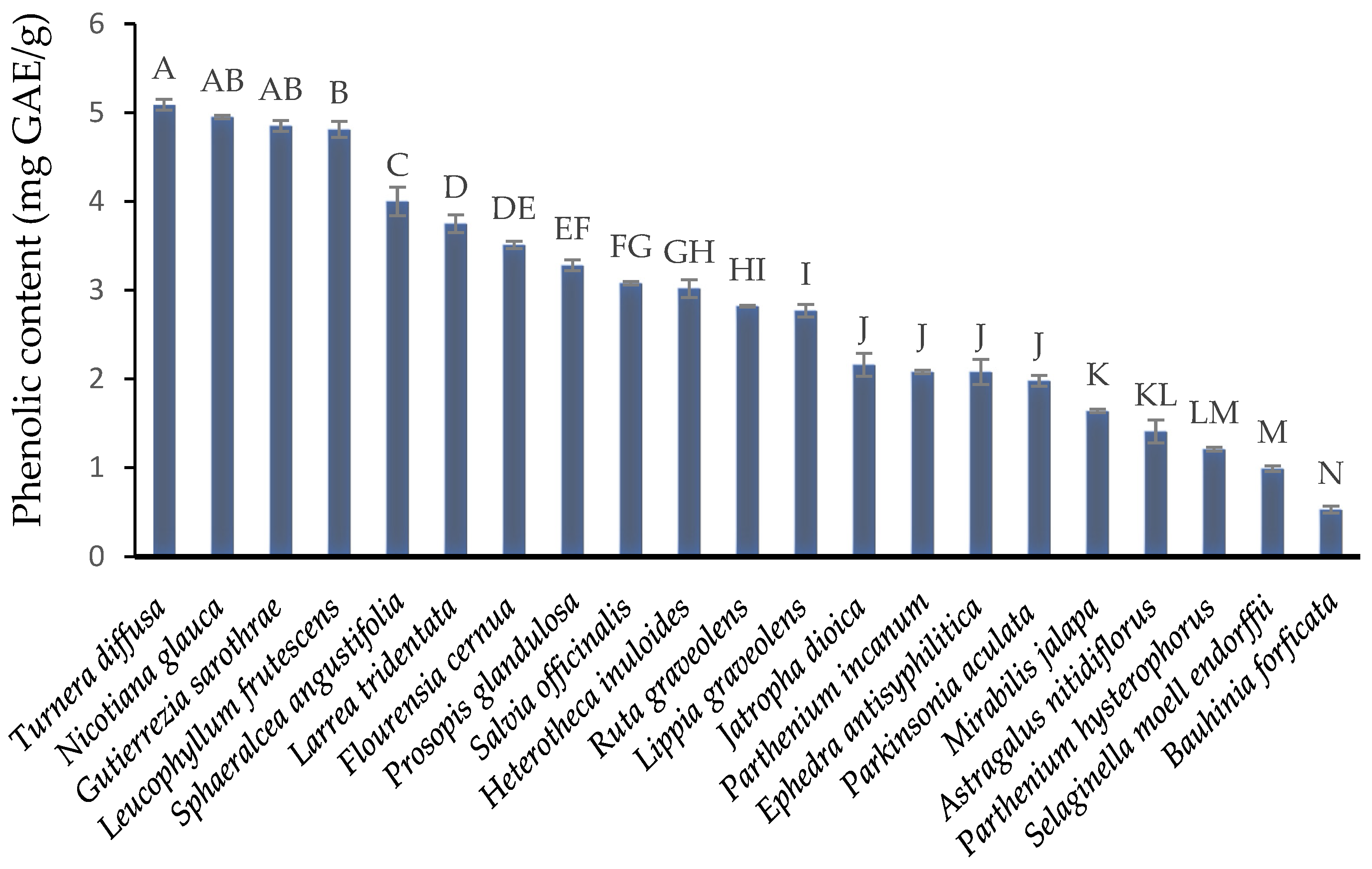
The phenolic content of a plant is an indicator of its functional potential.
Figure 1 shows the polyphenol content of 23 plants used in traditional medicine in Viesca, Coahuila. The plants
Turnera diffusa (5.09 ± 0.06 mg GAE/g),
Nicotiana glauca (4.95 ± 0.02 mg GAE/g),
Gutierrezia sarothrae (4.85 ± 0.06 mg GAE/g), and
Leucophyllum frutescens (4.81 ± 0.09 mg GAE/g) present the highest content of phenolic compounds (extracts from these plants were selected for the following studies). The
N. glauca plant was omitted from subsequent analyses due to previous cytotoxicity reports. The values obtained for
T. diffusa are similar to those previously reported (8 mg GAE/g) [
17]. The variation in phenolic content may be due to environmental and agronomic factors. The phenolic content could not be compared with other studies due to the limited information available.
The order of other plants is presented in continuation: Sphaeralcea angustifolia (4 ± 0.16 mg GAE/g), Larrea tridentata (3.75 ± 0.1 mg GAE/g), Fluorensia cernua (3.51 ± 0.04 mg GAE/g), Prosopis glandulosa (3.28 ± 0.06 mg GAE/g), Salvia officinalis (3.0 ± 0.02 mg GAE/g), Heterotheca inuloides (3.02 ± 0.1 mg GAE/g), Ruta graveolens (2.82 ± 0.01 mg GAE/g), Lippia graveolens (2.77 ± 0.07 mg GAE/g), Jatropha dioica (2.16 ± 0.13 mg GAE/g), Parthenium incanum (2.08 ± 0.02 mg GAE/g), Ephedra antisyphilitica (2.08 ± 0.14 mg GAE/g), Parkinsonia aculata (1.98 ± 0.06 mg GAE/g), Mirabilis jalapa (1.64 ± 0.02 mg GAE/g), Astragalus nitidiflorus (1.41 ± 0.13 mg GAE/g), Parthenium hysterophorus (1.21 ± 0.02 mg GAE/g), Selaginella moell endorfi (0.99 ± 0.03 mg GAE/g), Bauhinia forficata (0.53 ± 0.04 mg GAE/g). This investigation classifies plants for the first time by their phenolic content; these studies are very valuable for future studies in ethnopharmacology.
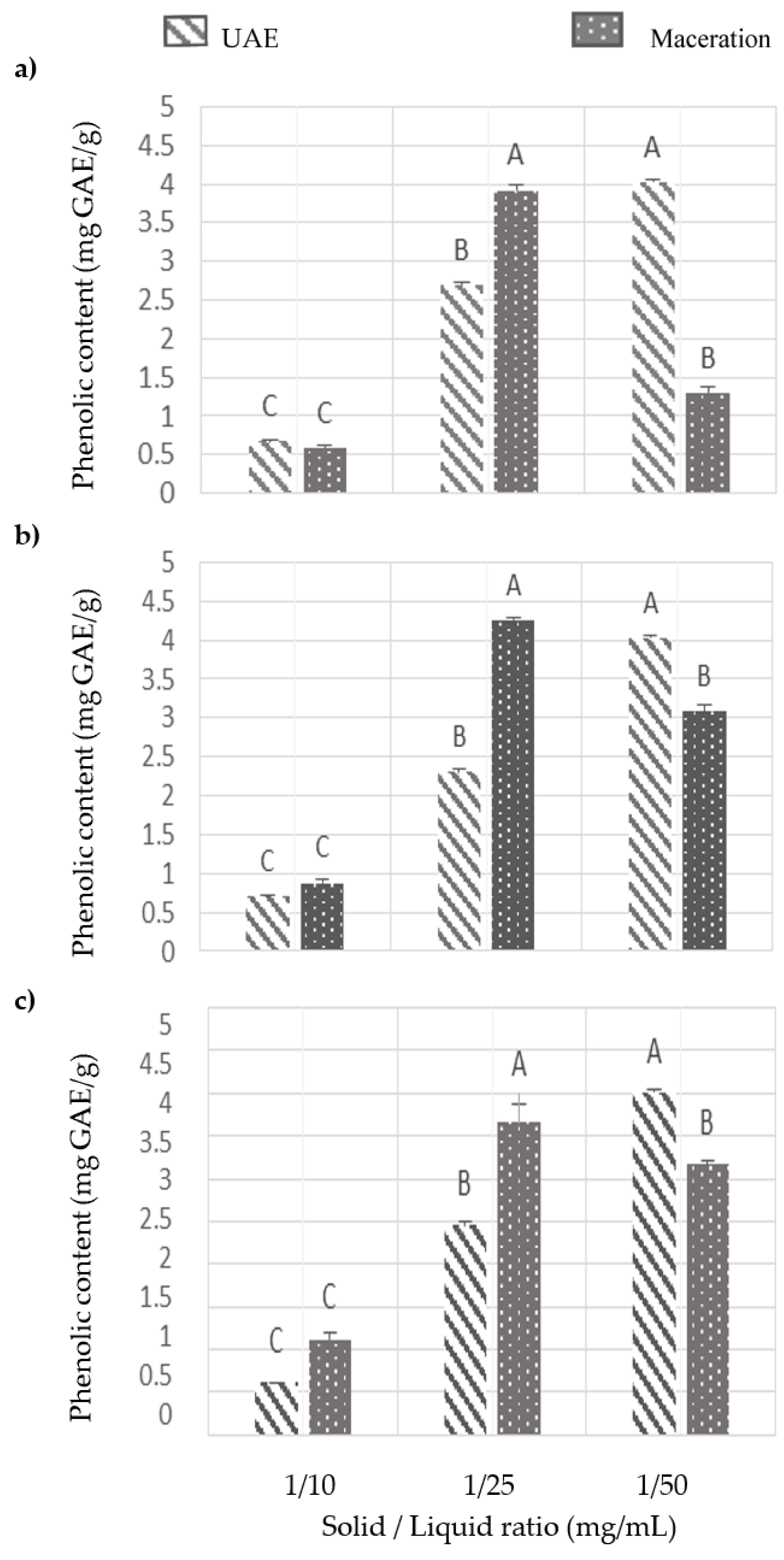
3.2. Evaluation of Extraction Methods
Figure 2 shows the effector of the solid/liquid ratio and the extraction method on the phenolic content of
T. diffusa,
G. sarothrae, and
L. frutescens. At the 1:10 concentration for the three plants studied, there is no significant difference with the UAE or maceration methods. In the solid/liquid ratio of 1:25, the maceration method presented the highest values (4.2 ± 0.04 mg GAE/g) of phenols for the three plants; the values were statistically significant. However, in the solid/liquid ratio of 1:50, the opposite effect was present; statistically, UAE was the method that presented the highest values of phenols (4 ± 0.02 mg GAE/g).
According to the results, when the solute concentration is high, 1:10 or 1:25 mg/mL, the maceration method is more suitable, but when the dilution is higher (1:50 mg/mL), the emerging UAE technology is more appropriate. This behavior is attributed to the principle of mass transfer in a solid–liquid extraction. In the solid/liquid ratio of 1:10, the decrease in phenol content is attributed to a phenomenon of saturation of the solvent; in saturation, the cellular phenomenon of diffusion stops and there is a stabilization rate. According to the principles of mass transfer, the driving force during transfer is the concentration gradient between the solid and the bulk of the liquid. This gradient is greater when a higher solvent-to-solid ratio is used. The interactions of the extracted compounds with the solvent can modify the activity coefficients and therefore, the solubility of the compounds. Similar results on the effect of the solid/liquid ratio on the extraction of phenolic compounds have been reported [
11,
18]. The main driving force of UAE is acoustic cavitation, which reduces particle size and facilitates mass transfer in less concentrated solutions. UAE is effective at low concentrations by improving solvent accessibility through the implosion of bubbles on the surface of the plant matrix. According to the results obtained, the UAE generates extracts rich in phenolic compounds with a smaller amount of plant material. This is very important for future studies where the efficiency of extraction methods is evaluated [
19]. In summary, the conventional maceration method can be used with high concentrations of material (this is more in line with the method used by local communities to prepare the plant). The UAE method cannot be discarded since it is more efficient with low concentrations of plant material.
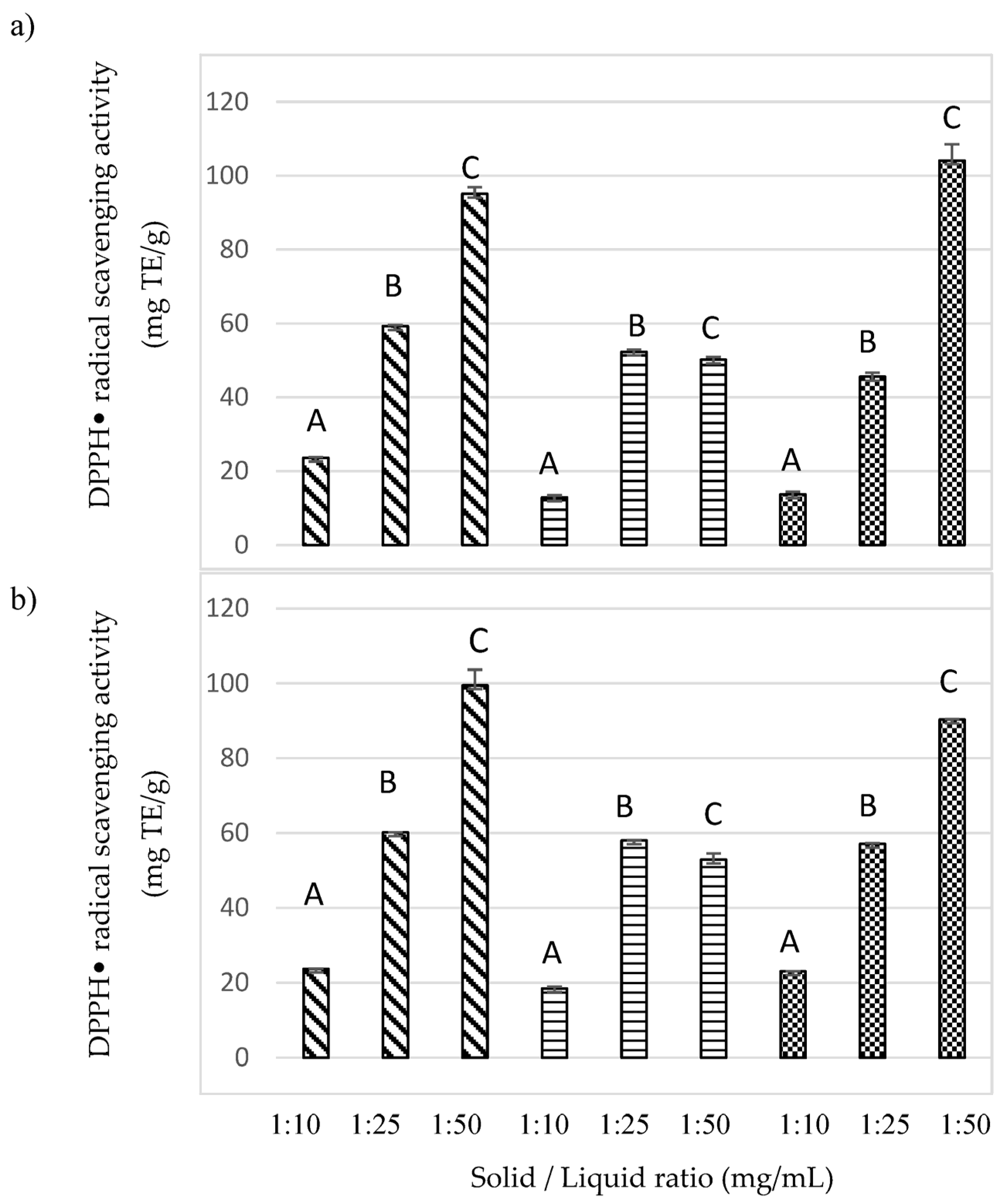
3.2.1. Antioxidant Activity Assay
DPPH Assay
In the present study, extracts from
T. diffusa,
G. sarothrae, and
L. frutescens were analyzed for their DPPH• scavenging activity, and the results are shown in
Figure 3a for the UAE method and
Figure 3b for the maceration method. The effect of three solid/liquid ratios (1:10, 1:25, and 1:50 mg/mL) was also evaluated. According to the results, in the two extraction methods evaluated,
T. diffusa and
G. sarothrae present greater antioxidant activity at a solid/liquid ratio of 1:50 mg/mL. At this concentration, there is a statistically significant difference with respect to the other two concentrations (
p < 0.05).
G. sarothrae presented the highest antioxidant activity in both methods at a concentration of 1:25 mg/mL (
p < 0.05). This result is attributed to a modification in the activity of the extracted compounds in
G. sarothrae during the extraction with the solvent [
20].
T. diffusa and
L. frutescens present the highest values of antioxidant activity with the maceration method at 100 and 57 mg TE/g, respectively. However, the UAE of
G. sarothrae presented the highest antioxidant activity of the three plants evaluated (103 mg TE/g). The results show the extracts of the three plants present high antioxidant activity. Additionally, such high free radical scavenging properties of the extracts are higher than those reported in hydroalcoholic extracts of medicinal plants, such as
Lavandula stoechas L. (58 mg TE/g) and
Aesculus indica (Wall. ex Cambess.) Hook (46 mg TE/g) [
3].
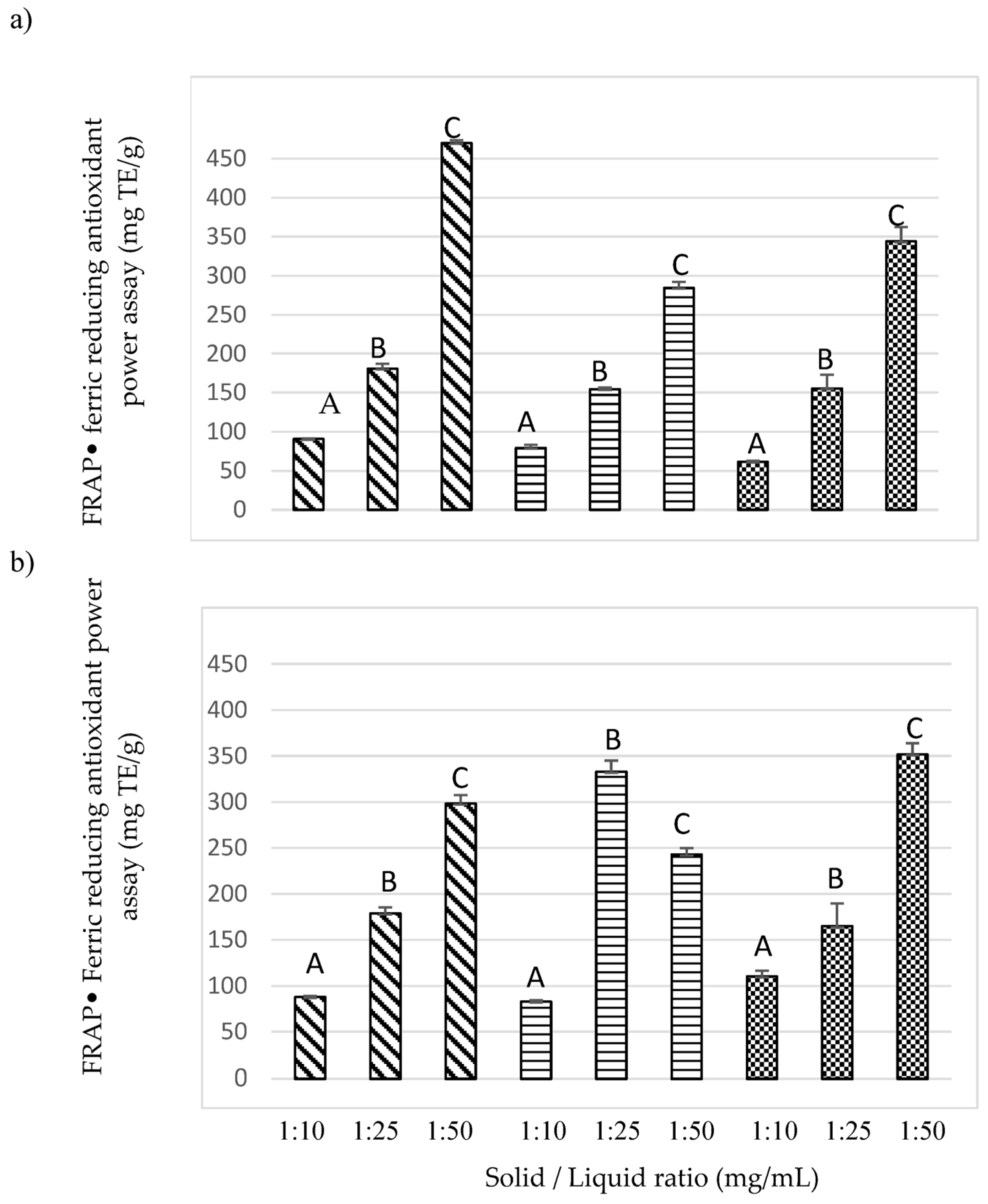
FRAP Assay
The results of the ferric reducing antioxidant power assay are presented in
Figure 4. The
T. diffusa and
G. sarothrae plants presented a similar trend to the results of the DPPH method; the solid/liquid ratio of 1:50 g/mL significantly showed greater antioxidant activity compared to the ratios of 1:10 and 1:25 mg/mL.
T. diffusa (in the solid/liquid ratio of 1:50 g/mL) presented the highest activity with the UAE extraction method (469 mg TE/g) compared to extraction by maceration (300 mg TE/g). On the other hand, in
G. sarothrae, no difference was found between the extraction methods evaluated (350 mg TE/g).
L. frutescens significantly presented the highest antioxidant activity with the maceration extraction method at a solid/liquid ratio of 1:25 g/mL (332 mg TE/g); this behavior was similar to the DPPH analysis. The results determined in the present study were superior to those determined for hydroalcoholic extracts from medicinal plants such as
Tephrosia purpurea (L.) Pers (6 mg TE/g),
Lavandula stoechas L. (9 mg TE/g),
Aesculus indica (Wall. ex Cambess.) Hook. (47 mg TE/g),
Iris ensata Thunb. (177 mg TE/g),
Kalanchoe pinnata (Lam.) Pers. (206 mg TE/g) [
3]., and
Reum especiforme (83 mg TE/g) (R. spiciforme is known for its anticancer properties [
21]).
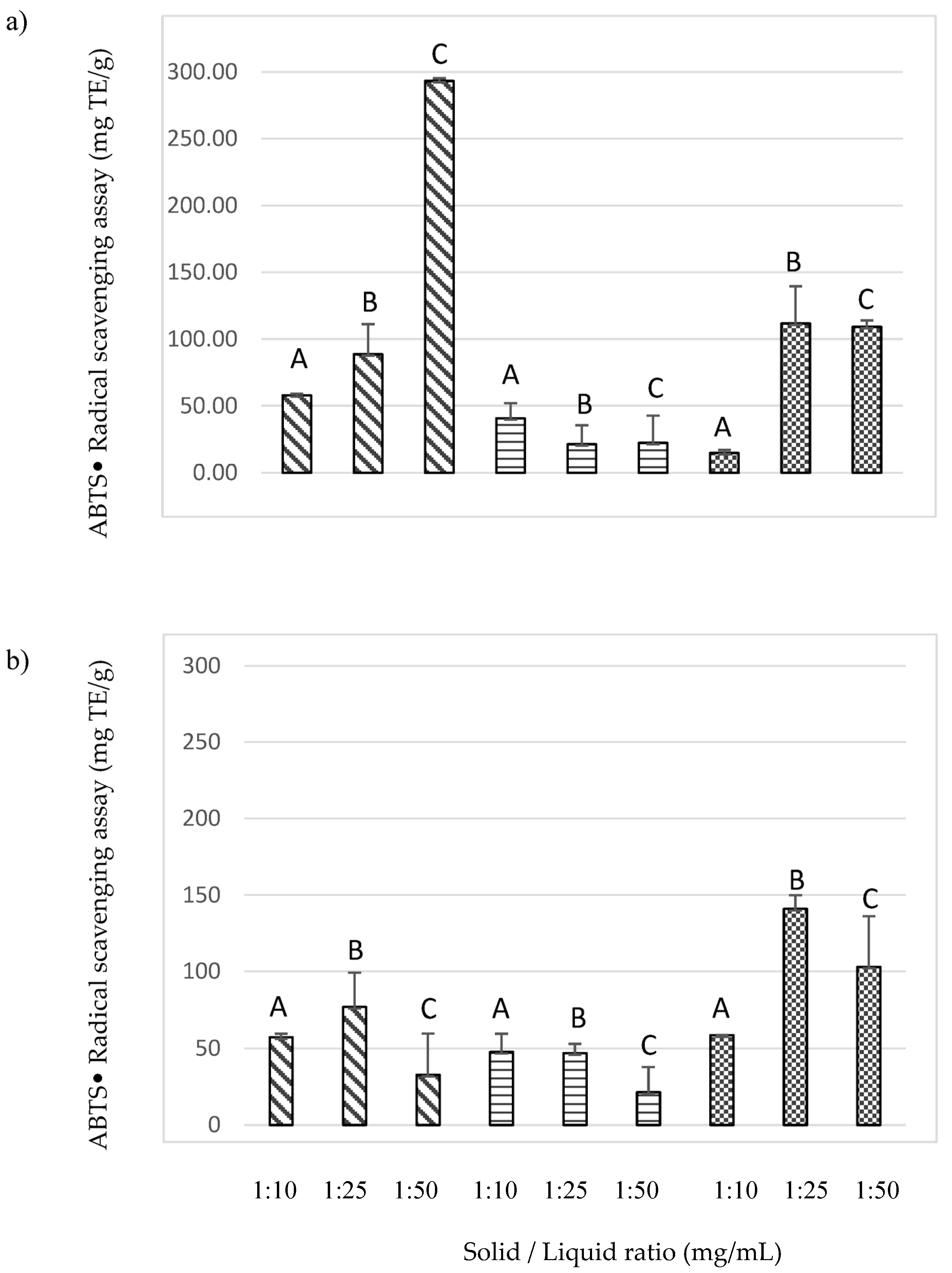
ABTS Assay
The ABTS assay was also used to investigate the antioxidant activity of the plants
T. diffusa,
G. sarothrae, and
L. frutescens.
T. diffusa presented significantly higher antioxidant activity (300 mgTE/g) with a solid/liquid ratio of 1:50 (mg TE/mL) and using UAE (
Figure 5). This behavior for this plant is similar to that presented previously with the DPPH and FRAP methods.
L. frutescens and G. sarothrae showed greater antioxidant activity with the maceration extraction method. L. frutescens significantly presented the greatest activity at the solid/liquid ratio of 1:10 g/mL (50 mg TE/mL), and G. sarothrae significantly showed the greatest antioxidant activity at the solid/liquid ratio of 1:25 (141 mg TE/mL). For both plants, the values were lower than the maximum reached by T. diffusa (300 mg TE/g). The observed differential scavenging activities of the extracts against the DPPH• and ABTS•+ systems demonstrate the presence of different compounds and different chemical features (see HPLC-MS analysis). This phenomenon can also be explained by the different affinities of the evaluated methods. Unlike DPPH•, ABTS•+ has an affinity with water-soluble compounds and with lipophilic compounds since it is soluble in both aqueous and organic solvents.
3.3. Analysis of the Extraction Methods Evaluated
Table 2 shows a summary of the efficiency between the UAE and maceration methods. In the three antioxidant activity assays evaluated,
T. diffusa with a solid/liquid ratio of 1:50 g/mL only showed a significant difference in the DPPH• assay and the highest activity was obtained for the maceration method. For
L. frutescens with a solid/liquid ratio of 1:25 g/mL, maceration was the extraction method that significantly showed the highest antioxidant activity. Finally, for
G. sarothrae using a solid/liquid ratio of 1:50 g/mL, the method that significantly presented greater antioxidant activity was UAE.
The maceration method was the most suitable for extracting antioxidant compounds from T. diffusa and L. frutescens, while UAE was more efficient for G. sarothrae.
3.4. RP-HPLC-ESI-MS Analysis
The bioactive compounds present in the plants
G. sarothrae,
T. diffusa, and
L. frutescens were identified by RP-HPLC-ESI-MS (
Table 3).
For T. diffusa, we identified 14 molecules that can be classified in different families, such as hydroxycinnamic acids, methoxycinnamic acids, and flavones. 24-Methylcholesterol ferulate, Caffeoyl tartaric acid, Sinensetin, 3-p-Coumaroylquinic acid, 4-p-Coumaroylquinic acid, and p-Coumaric acid 4-O-glucoside were the major component of polyphenolic extracts from T. diffusa.
Eleven molecules were identified for
L. frutescens belonging to the hydroxycinnamic acids, methoxycinnamic acids, flavones, proanthocyanidin dimers, methoxyflavones, anthocyanins and tyrosols families. Apigenina 7-O-diglucurónido 3 and 4-DHPEA-EA were the major components of polyphenolic extracts from the
L. frutescens. Apigenins were identified in similar studies [
22]. Apigenin has been recognized in traditional medicine due to the fact that it has many farmacologic activities, like 17-β-hidroxiesteroide deshidrogenasa enzyme inhibition [
23,
24].
Four molecules were found for G. sarothrae, and these molecules belong to flavonoids, lignans, and phenolic acids.
4. Conclusions
To summarize our findings, we have reported for the first time the phenol contents of twenty-three medicinal plants used in the traditional medicine of Viesca, Coahuila, Mexico. T. diffusa, L. frutescens, and G. sarothrae are the richest sources of phenolic compounds and were selected to subsequently study their antioxidant potential by three different assays. Two extraction methods and three solid/liquid ratios were evaluated in the three plants. The three plants presented high antioxidant activity, the solid/liquid ratio presented a significant influence on their antioxidant activity, and the solid/liquid ratio of 1:50 g/mL presented the highest activity values for T. diffusa and G. sarothrae. L. frutescens presented the highest values of antioxidant activity with a solid/liquid ratio of 1:25 mg/mL. The maceration method was the most suitable for extracting antioxidant compounds from T. diffusa and L. frutescens, while UAE was more efficient for G. sarothrae. The HPLC-MS analysis showed the presence of 14 molecules for T. diffusa, 11 molecules for L. frutescens, and 4 molecules for G. sarothrae. The medicinal plants used in Viesca, Mexico, have a high antioxidant potential and can be a source of natural antioxidants with applications in medicine and the food industry. In conclusion, the plants used in traditional medicine from Viesca, Mexico, have great potential for the development of natural products.
Author Contributions
Conceptualization, A.R.-M. and C.T.-L.; methodology, A.R.-M. and C.T.-L.; software, J.A.A.-V.; validation, N.R.-G., E.F.-L. and J.A.A.-J.; investigation, M.P.-W., A.R.-M. and C.T.-L.; resources, C.T.-L., J.A.A.-V. and J.A.A.-J.; data curation, C.T.-L.; writing—original draft preparation, M.P.-W.; writing—review and editing C.T.-L.; supervision, A.R.-M. and C.T.-L.; project administration, C.T.-L.; funding acquisition, C.T.-L. All authors have read and agreed to the published version of the manuscript.
Funding
This research was funded by the Secretary of Public Education of Mexico (SEP) (PRODEP: UACOAH-PTC-533) and the Mexican Council for Humanities, Science, and Technology CONAHCYT (Proyecto RENAJEB-2023-17).
Data Availability Statement
The data presented in this study are available on request from the corresponding author.
Acknowledgments
The authors are especially thankful to Engr. Alma Leticia Espinoza (CIJE-UAdeC) and the local people region for their collaboration, and we would like to thank Engr. Juan Carlos Chavarria (CIJE-UAdeC) and MSc. Eduardo Blanco Contreras (CREB-UAAAN) for their valuable contribution in plant identification.
Conflicts of Interest
The authors declare no conflict of interest.
References
- Contreras-Esquivel, J.C.; Cano-González, C.N.; Ascacio-Valdes, J.; Aguirre-Joya, J.A.; Aguillón-Gutierrez, D.; Breccia, J.; Espinoza-Perez, J.D.; Aguilar, C.N.; Torres-León, C. Polyphenolic-Rich Extracts from the Leaves of Ilex Paraguariensis and Larrea Divaricata and Their Antioxidant and AntiCOVID-19 Potential. Biotecnia 2023, 25, 61–66. [Google Scholar] [CrossRef]
- Newman, D.J.; Cragg, G.M. Natural Products as Sources of New Drugs over the Nearly Four Decades from 01/1981 to 09/2019. J. Nat. Prod. 2020, 83, 770–803. [Google Scholar] [CrossRef]
- Rao, H.; Rao, I.; Saeed, L.; Aati, H.Y.; Aati, S.; Zeeshan, M.; ur Rehman Khan, K. Phytochemical Analysis and Bioactivity Assessment of Five Medicinal Plants from Pakistan: Exploring Polyphenol Contents, Antioxidant Potential, and Antibacterial Activities. Saudi J. Biol. Sci. 2023, 30, 103783. [Google Scholar] [CrossRef] [PubMed]
- Cömert, E.D.; Gökmen, V. Evolution of Food Antioxidants as a Core Topic of Food Science for a Century. Food Res. Int. 2018, 105, 76–93. [Google Scholar] [CrossRef] [PubMed]
- Huang, D.; Boxin, O.U.; Prior, R.L. The Chemistry behind Antioxidant Capacity Assays. J. Agric. Food Chem. 2005, 53, 1841–1856. [Google Scholar] [CrossRef] [PubMed]
- Chaves, N.; Santiago, A.; Alías, J.C. Quantification of the Antioxidant Activity of Plant Extracts: Analysis of Sensitivity and Hierarchization Based on the Method Used. Antioxidants 2020, 9, 76. [Google Scholar] [CrossRef]
- Torres-León, C.; Rebolledo Ramírez, F.; Aguirre-Joya, J.A.; Ramírez-Moreno, A.; Chávez-González, M.L.; Aguillón-Gutierrez, D.R.; Camacho-Guerra, L.; Ramírez-Guzmán, N.; Hernández Vélez, S.; Aguilar, C.N. Medicinal Plants Used by Rural Communities in the Arid Zone of Viesca and Parras Coahuila in Northeast Mexico. Saudi Pharm. J. 2023, 31, 21–28. [Google Scholar] [CrossRef]
- Martins, S.; Amorim, E.L.C.; Sobrinho, T.J.S.P.; Saraiva, A.M.; Pisciottano, M.N.C.; Aguilar, C.N.; Teixeira, J.A.; Mussatto, S.I. Antibacterial Activity of Crude Methanolic Extract and Fractions Obtained from Larrea Tridentata Leaves. Ind. Crops Prod. 2013, 41, 306–311. [Google Scholar] [CrossRef]
- Bautista-Hernández, I.; Aguilar, C.N.; Martínez-Ávila, G.C.G.; Ilina, A.; Torres-León, C.; Verma, D.K.; Chávez González, M.L. Phenolic Compounds and Antioxidant Activity of Lippia graveolens Kunth Residual Leaves Fermented by Two Filamentous Fungal Strains in Solid-State Process. Food Bioprod. Process. 2022, 136, 24–35. [Google Scholar] [CrossRef]
- Bautista-Hernández, I.; Aguilar, C.N.; Martínez-ávila, G.C.G.; Torres-León, C.; Ilina, A.; Flores-Gallegos, A.C.; Kumar Verma, D.; Chávez-González, M.L. Mexican Oregano (Lippia graveolens Kunth) as Source of Bioactive Compounds: A Review. Molecules 2021, 26, 5156. [Google Scholar] [CrossRef]
- Castro-López, C.; Ventura-Sobrevilla, J.M.; González-Hernández, M.D.; Rojas, R.; Ascacio-Valdés, J.A.; Aguilar, C.N.; Martínez-Ávila, G.C.G. Impact of Extraction Techniques on Antioxidant Capacities and Phytochemical Composition of Polyphenol-Rich Extracts. Food Chem. 2017, 237, 1139–1148. [Google Scholar] [CrossRef] [PubMed]
- Blois, M. Antioxidant Determinations by the Use of a Stable Free Radical. Nature 1958, 181, 1199–1200. [Google Scholar] [CrossRef]
- Magalhães, L.M.; Santos, F.; Segundo, M.A.; Reis, S.; Lima, J.L.F.C. Rapid Microplate High-Throughput Methodology for Assessment of Folin-Ciocalteu Reducing Capacity. Talanta 2010, 83, 441–447. [Google Scholar] [CrossRef] [PubMed]
- Mensor, L.; Menezes, F.; Leitão, G.; Reis, A.; dos Santos, T.; Coube, C.; Leitão, S. Screening of Brazilian Plant Extracts for Antioxidant Activity by the Use of DPPH Free Radical Method. Phytother. Res. 2001, 15, 127–130. [Google Scholar] [CrossRef]
- Re, R.; Pellegrini, N.; Proteggente, A.; Pannala, A.; Yang, M.; Rice, C. Antioxidant Activity Applying an Improved ABTS Radical Cation Decolorization Assay. Free Radic. Biol. Med. 1999, 26, 1231–1237. [Google Scholar] [CrossRef] [PubMed]
- Ascacio-Valdés, J.A.; Aguilera-Carbó, A.F.; Buenrostro, J.J.; Prado-Barragán, A.; Rodríguez-Herrera, R.; Aguilar, C.N. The Complete Biodegradation Pathway of Ellagitannins by Aspergillus Niger in Solid-State Fermentation. J. Basic Microbiol. 2016, 56, 329–336. [Google Scholar] [CrossRef]
- Soriano-Melgar, L.d.A.A.; Alcaraz-Meléndez, L.; Méndez-Rodríguez, L.C.; Puente, M.E.; Rivera-Cabrera, F.; Zenteno-Savín, T. Antioxidant Responses of Damiana (Turnera diffusa Willd) to Exposure to Artificial Ultraviolet (UV) Radiation in an In Vitro Model: Part I; UV-C Radiation. Nutr. Hosp. 2014, 29, 1109–1115. [Google Scholar] [CrossRef]
- Pinelo, M.; Rubilar, M.; Jerez, M.; Sineiro, J.; Núñez, M.J. Effect of Solvent, Temperature, and Solvent-to-Solid Ratio on the Total Phenolic Content and Antiradical Activity of Extracts from Different Components of Grape Pomace. J. Agric. Food Chem. 2005, 53, 2111–2117. [Google Scholar] [CrossRef]
- Torres-León, C.; Rojas, R.; Serna-Cock, L.; Belmares-Cerda, R.; Aguilar, C.N. Extraction of Antioxidants from Mango Seed Kernel: Optimization Assisted by Microwave. Food Bioprod. Process. 2017, 105, 188–196. [Google Scholar] [CrossRef]
- Jun, X. Caffeine Extraction from Green Tea Leaves Assisted by High Pressure Processing. J. Food Eng. 2009, 94, 105–109. [Google Scholar] [CrossRef]
- Shahar, B.; Dolma, N.; Chongtham, N. Phytochemical Analysis, Antioxidant Activity and Identification of Bioactive Constituents from Three Wild Medicinally Important Underutilized Plants of Ladakh, India Using GCMS and FTIR Based Metabolomics Approach. Food Humanit. 2023, 1, 430–439. [Google Scholar] [CrossRef]
- Sarris, J.; McIntyre, E.; Camfield, D.A. Plant-Based Medicines for Anxiety Disorders, Part 1: A Review of Preclinical Studies. CNS Drugs 2013, 27, 207–219. [Google Scholar] [CrossRef] [PubMed]
- Eun, J.C.; Kim, G.H. Apigenin Induces Apoptosis through a Mitochondria/Caspase-Pathway in Human Breast Cancer MDA-MB-453 Cells. J. Clin. Biochem. Nutr. 2009, 44, 260–265. [Google Scholar] [CrossRef]
- Hasegawa, E.; Nakagawa, S.; Sato, M.; Tachikawa, E.; Yamato, S. Effect of Polyphenols on Production of Steroid Hormones from Human Adrenocortical NCI-H295R Cells. Biol. Pharm. Bull. 2013, 36, 228–237. [Google Scholar] [CrossRef] [PubMed]
| Disclaimer/Publisher’s Note: The statements, opinions and data contained in all publications are solely those of the individual author(s) and contributor(s) and not of MDPI and/or the editor(s). MDPI and/or the editor(s) disclaim responsibility for any injury to people or property resulting from any ideas, methods, instructions or products referred to in the content. |
© 2023 by the authors. Licensee MDPI, Basel, Switzerland. This article is an open access article distributed under the terms and conditions of the Creative Commons Attribution (CC BY) license (https://creativecommons.org/licenses/by/4.0/).


 ,
, 


 T. diffusa,
  L. frutescens, and
  G. sarothrae extracts obtained by (a) UAE and (b) maceration. The different letter indicates significant differences (p < 0.05) according to Tukey’s multiple range test.

 T. diffusa,
  L. frutescens, and
  G. sarothrae extracts obtained by (a) UAE and (b) maceration. The different letter indicates significant differences (p < 0.05) according to Tukey’s multiple range test.

 T. diffusa,
  L. frutescens, and
  G. sarothrae extracts obtained by (a) UAE and (b) maceration. The different letter indicates significant differences (p < 0.05) according to Tukey’s multiple range test.

