Article

UVA Enhanced Promotive Effects of Blue Light on the Antioxidant Capacity and Anthocyanin Biosynthesis of Pak Choi

1
Institute of Urban Agriculture, Chinese Academy of Agricultural Sciences, Chengdu 610213, China
2
National Chengdu Agricultural Science and Technology Center, Chengdu 610213, China
3
School of Food and Biological Engineering, Chengdu University, Chengdu 610106, China
4
College of Materials and Energy, South China Agricultural University, Guangzhou 510642, China
*
Author to whom correspondence should be addressed.
These authors contributed equally to this work.
Horticulturae 2022, 8(9), 850; https://doi.org/10.3390/horticulturae8090850
Submission received: 15 August 2022 / Revised: 10 September 2022 / Accepted: 14 September 2022 / Published: 17 September 2022

Abstract

:
Anthocyanins are widely common natural antioxidants and represent an important economic feature in vegetables, but the potential response of UVA–blue co-irradiation on the anthocyanin biosynthesis of pak choi is not clear. Here, we investigated the effects of the supplement of four doses of UVA to blue light on growth, metabolites and the anthocyanin biosynthesis of two cultivars of pak choi. The results revealed that supplementing UVA light to blue light positively affected the growth of the pak choi and elevated the soluble protein content and antioxidant capacity. Especially, when compared with a monochromatic blue light, the anthocyanin synthesis was enhanced with an increase in UVA light strength, which reached a peak value at the strength of 10 μmol·m−2·s1. Further study revealed that the UVA–blue co-irradiation enhanced the transcription of partial light-induced and anthocyanin structural genes. The intraspecific difference in the expression patterns of MYB1 and PAP1 were observed in these two tested cultivars. MYB1 was significantly up-regulated in red-leaf pak choi, but down-regulated in purple-leaf pak choi. On the contrary, PAP1 was significantly up-regulated in purple-leaf pak choi, but down-regulated in red-leaf pak choi. To sum up, this study established an efficient pre-harvest lighting strategy to elevate the economic value of pak choi.

1. Introduction

Pak choi (Brassica rapa var chinensis) is a member of the cruciferous vegetables and is extensively cultivated and consumed in Japan and China [1] Pak choi contains a large amount of beneficial antioxidants and nutrients (such as carotenoids, vitamins, flavonoids and minerals). In recent years, red- and purple-leaf pak choi have received a great amount of attention due to their graceful appearance and high nutritional value [2]. With the improvement in consumption standards, red- and purple-leaf pak choi have attracted more attention from the public than the green-leaf cultivars, which is largely attributed to the high level of health-promoting anthocyanins in mature leaves [1,2].
Anthocyanins are specialized water-soluble flavonoids derived from the phenylpropanoids pathway, and are widely distributed in plant vacuoles [3]. These abundant vacuolar pigments have been shown to play a vital role in providing visual cues (e.g., red, blue, purple and pink) to attract pollinators and defend against hunters [3,4]. As antioxidant chemicals, anthocyanins are beneficial in the treatment against cancer and cardiovascular disease [5,6]. The biosynthesis of anthocyanins was usually considered to be an anti-stress response to environmental adverse events (e.g., drought, low temperature, high-intensity light and ultraviolet radiation) in plants, which further indicates that anthocyanins play a defensive role in cell stress [7,8,9].
In higher plants, anthocyanin biosynthesis is carried out through the flavonoid metabolic pathway (Figure 1), which mainly occurs on the surface of the endoplasmic reticulum (ER) [10]. Anthocyanins are catalyzed stepwise by diverse enzymes coded by structural genes, including phenylalanine ammonia lyase (PAL), chalcone synthase (CHS), chalcone isomerase (CHI), flavanonoe-3-hydroxylase (F3H), dihydroflavonol 4-reductase (DFR), anthocyanidin synthase (ANS) and UDP-glucose: flavonoid 3-O-glucosyltransferase (UFGT) [11,12]. Anthocyanins are initially formed in the ER and ultimately transported to the vacuoles. Glutathione S-transferase (GST) serves as a pivotal porter during the transmembrane transportation of anthocyanins, and a series of homologous genes were identified in Arabidopsis [10], Zea mays [13] and Petunia hybrida [14]. With the exception of the genes involved in anthocyanin biosynthesis, some groups of transcription factors (TFs), mostly including the R2R3 MYB domain protein (R2R3-MYB), basic helix–loop–helix (bHLH) and conserved WD40, TF repeats have been found to interact as an MBW complex, activating the transcription of structural genes [3,15,16]. Several studies have reported that anthocyanin biosynthesis-related genes (e.g., CHS, F3′H, DFR, ANS/LDOX, UFGTs and GSTs) were primarily regulated by the MYB domain protein 2 (MYB2) and transparent testa 8 (TT8) in Brassicaceae crops [17,18]. Furthermore, MYB2 and TT8 TFs were confirmed as regulators in the stimulation of the anthocyanin synthesis in purple pak choi [19]. To be able to breed fruits and vegetables that are rich in anthocyanins, it is necessary to investigate their regulatory mechanisms and how they are induced by environmental factors.
Light is regarded as an indispensable environmental factor for plants. Blue light is essential in sustaining the normal photosynthesis of a plant and its existent dose response [20]. Moreover, blue light can induce the opening of stomata and the flowering and biosynthesis of photosynthetic pigments in plants [21,22]. Ultraviolet (UV) radiation is regarded as a stress signal involved in inducing the accumulation of anthocyanins in plants as a result of different environmental factors, having been observed in tomato [23], turnip [24] and lettuce [25]. UVA accounts for 95% of natural UV light, and knowledge regarding the effects of UVA on plants is limited [26]. Compared with UVB and UVC, UVA has been widely used in horticulture cultivation as an effective environmental management strategy [27]. Previous experiments found that the adequate supplementation of UVA (10~20 μmol·m−2·s−1) was beneficial for biomass production and the nutritive quality of leafy vegetables and fruit [26,28,29]. As the signal transduction of blue light and UVA in plants is mediated by the same photoreceptors, there exists an interactive relationship between them. It has been reported that supplemental blue light resulted in enhanced plant tolerance to UV irradiation in Arabidopsis [30]. Moreover, blue light could also stimulate epidermal UVA absorbance and flavonoid accumulation in pea plants [31]. Especially, UVA enhanced radish anthocyanin accumulation in hydrogen-rich water. This means that UVA plays an important role in inducing anthocyanin biosynthesis as a result of other environmental factors [9].
Cryptochromes (CRYs) mediate plants’ ability to sense UVA from the solar spectrum (Figure 1), while simultaneously sensing blue light and being vital for regulating anthocyanin accumulation, hypocotyl elongation and leaf and cotyledon expansion [21,32]. There generally exist two CRYs with redundantly functional diversification in higher plants [33]. In Arabidopsis, CRY1 mediates photomorphogenesis in response to high light, while CRY2 specifically transduces low light [34]. The over-expression of CRY1 from Brassica napus results in hypocotyl growth inhibition and anthocyanin accumulation dependent on light intensity [35]. Constitutively, photomorphogenic 1 (COP1) is a RING-finger-type ubiquitinated E3 ligase and a negative regulator of photomorphogenesis, which is activated by nuclear proteins (e.g., suppressor of phytochrome 1, SPA1) to mediate the degradation of elongated hypocotyl 5 (HY5) via the 26S proteasome pathway [36,37]. CRYs regulate photomorphogenesis by directly and indirectly inhibiting COP1-SPA activity [36,38]. HY5 is characterized as an accelerator that binds directly to the promoters of light-inducible genes, including regulatory and structural genes, enhancing their abundance to accumulate anthocyanins [38,39]. As a result, enhancing the production of anthocyanins with blue and UVA radiation is a feasible method according to molecular data.
Therefore, to investigate the physiological effects and gene expression profiles underlying anthocyanin biosynthesis in red- and purple-leaf edible leaves under blue and UVA light, we put forward an efficient strategy improving their nutritive quality during crop production while maintaining normal growth. This study aims to expand our understanding of the mechanisms of light-induced anthocyanin accumulation in red- and purple-leaf pak choi at both metabolic and transcriptional levels, and raise an efficient and ecological strategy for pak choi quality promotion.

2. Materials and Methods

2.1. Plant Materials and Growth Conditions

The experiment was conducted in an artificially lit plant growth chamber (11.8 m × 2.13 m × 2.72 m) (Chengdu Academy of Agriculture and Forestry Sciences, Sichuan, China). Pak choi seeds (Brassica rapa var chinensis) were purchased from Jingyan Yinong (Beijing) Seed Industry Technology Co., Ltd. (Beijing, China). Additionally, seeds of red- and purple-leaf pak choi were sown in sponge cubes (2 × 2 × 2 cm). After the expansion of the third true leaf, seedings were transferred into hydroponic containers (12 seedings per container and 30 L volume, three replicates per treatment, total of 24 containers) under white light at an intensity of 100 μmol·m−2·s1 with a 16 h light/8 h dark photoperiod. The environmental conditions were maintained at 25/22·°C (day/night), 65% to 85% relative humidity (RH) and 400 μmol·mol−1 CO2 level. Hoagland’s solution was refreshed every 5 days, 1.3~1.4 mS·m−1 of the electrical conductivity (EC) and 5.8~6.0 of pH were maintained and the pH was adjusted with H3PO4 (liquid, 85% in water) and KOH (liquid, 1 mol/L). After 15 days, pak choi at the ten-leaf stage were used for subsequent experiments.

2.2. Light Treatment

Adjustable LED lamps (Chenghui Equipment Co., Ltd., Guangzhou, China), including white light, blue light (λ = 450 ± 10 nm) and UVA (λ = 385 ± 10 nm), were selected as light sources [40]. The light intensity of each treatment was adjusted with a photometer (Apogee SS-110, Logan, UT, USA). The distance between the light source and upper side of plant canopy was controlled at approximately 35 cm. Four light treatments were tested for 5 days in this experiment, and the intensities of UVA and blue light were adjusted to 0/50 (T0), 5/45 (T5), 10/40 (T10) and 20/30 (T20), keeping the total intensity of each light treatment at 50 μmol·m−2·s−1 (Table S1). The photoperiod of this study was maintained at 16 h light/8 h dark. To further clarify the gene expression profiles of anthocyanin biosynthesis under UVA–blue light co-irradiation, and to avoid the interference of the dark cycle of the photoperiod, another experiment was conducted under continuous irradiation. Two light treatments were continuously illuminated for 48 h, with T0 (blue light) as the control and T10 (blue + UVA light) as the experimental treatment.

2.3. Growth Measurement and Sampling

After 5-day-long experimental treatments, the fresh and dry weight of edible parts (five plants) were weighed with an analytical balance (FA2004, Shanghai, China). Fresh leaf samples were frozen with liquid nitrogen and stored at −80 °C for further testing. In the preliminary experiment, we found that the phenotype of sixth leaves was obvious compared with other leaves; therefore, the sixth fresh leaf tissues of red- and purple-leaf pak choi were separately collected in 50 mL centrifugal tubes (sterile and DNase/RNase-free specialty) and frozen in liquid nitrogen after 0 h, 12 h, 24 h and 48 h continuous light treatments.

2.4. Measurement of Chlorophyll (chl) Content

For the chlorophyll content determination, improved spectrophotometric methods with the mixture (ethanol/acetone v/v, 1:1) were used [41]. Fresh leaf (0.1 g) was mixed with 15 mL of extraction solution, and the tested solution was place in the dark for 12 h until the leaf turned white. The absorbance of the extracting solution was read at wavelengths (nm) of 645 and 663 using an UV spectrophotometer (MAPADA, UV-1200, Shanghai, China). The chl content (mg/g FW) was calculated as follows: chl a = (12.7 × OD663 − 2.69 × OD645) × extraction solution volume/0.1 g; chl b = (22.9 × OD645 − 4.86 × OD663) × extraction solution volume/0.1 g; total chl = (chl a + chl b) × extraction solution volume/0.1 g.

2.5. Measurement of Soluble Sugar

To determine the soluble sugar content, a mixture of 0.5 g frozen powder and 10 mL distilled water was maintained in a 95 °C water bath for 0.5 h (extracted twice). An invariant general volume of 25 mL extracting solution was prepared using separation and a constant volume, and the solution was determined through sulfuric acid anthrone colorimetric method at 630 nm [42].

2.6. Measurement of Soluble Protein

Soluble protein was measured according to the method established by Blakesley and Boezi [43]. Frozen powder (0.5 g) was mixed up with 5 mL distilled water and centrifuged for 10 min at 4 °C and 12,000× g. Then, 0.1 mL of a supernatant, 5 mL of Coomassie brilliant blue G-250 solution and 0.9 mL of distilled water were adequately mixed. The OD value of the soluble protein was assayed at 595 nm after 2 min.

2.7. Measurement of Nitrate Content

The nitrate content of pak choi was measured using the slightly modified technique reported by Cataldo et al. [44]. Frozen tissue powder (0.5 g) was added to 10 mL deionized water and boiled for 0.5 h. Then, the supernatant was separated and diluted to a constant volume with deionized water (25 mL). In total, 0.5 mL of tested solution was combined with 0.4 mL of 5% (w/v) salicylic acid (prepared with concentrated sulfuric acid) and 9.5 mL of 8% (w/v) NaOH solution. After the tested solution was cooled, the content of nitrate was assayed at 410 nm based on the nitrate standard curve.

2.8. Measurement of Anthocyanin Content

The anthocyanin content of pak choi was measured according to a previously described method with slight modifications [45]. Frozen leaf tissue (0.5 g) was immersed in methanol containing 0.1% hydrochloric acid (v/v) for the anthocyanin extraction, and left to stand for 20 min in the dark. After being centrifuged at 4 °C and 12,000× g for 15 min, the OD value of the extraction solution was measured at 510 nm and 700 nm in buffers with pH values of 1.0 and 4.5, respectively. Total anthocyanins were calculated with the followed formula: A = (A510 nm − A700 nm) PH 1.0 − (A510 nm − A700 nm) PH 4.5, with a molar extinction coefficient of cyanidin-3-glucoside of 29,600.

2.9. The 2,2-Diphenyl-1-Picrylhydrazyl (DPPH) Radical Scavenging Rate Measurement

The measurement of the DPPH radical scavenging rate was carried out according to Liu et al. [46]. Frozen powder (0.5 g) was adequately mixed with a certain amount of absolute ethanol and the mixture solution was diluted to 9 mL with deionized water. After shaking and leaving to stand in a dark room for 0.5 h, the extract solution was centrifuged at 3000× g for 10 min and the OD value of supernatant was read at 517 nm. The rate of DPPH radical scavenging was computed as follows: DPPH (%) = [1 − (Ai − Aj)/Ac] × 100%, where Ai is the absorbance of the mixture of extract and DPPH solution, Aj is the absorbance of the extract and Ac is the absorbance of the DPPH solution.

2.10. Antioxidant Capacity Measurement

The total antioxidant capacity was assessed by determining the ferric-reducing antioxidant power (FRAP) [47]. A mixture of 3.6 mL working solution (20 mM FeCl3 solution, 0.3 M acetate buffer, 10 mM TPTZ solution v/v/v, 1:1:10) and 0.1 mL extract was prepared and then the absorbance at 593 nm was determined. A standard curve of 0~1.5 mM FeSO4 was determined for the final value calculation and showed as oxidized mmol/L Fe2+.

2.11. RNA Extraction and Quantitative Real-Time PCR (qRT-PCR) Analyses

Frozen powder from leaf tissue (50~100 mg) was used for RNA isolation with a Trizol kit (Takara), according to the manufacturer’s instructions. RNA samples (~4 μg) were used for the first-strand cDNA synthesis with an oligo (dT) primer (Moloney Murine Leukemia Virus Reverse Transcriptase, M-MuLV RT). 2×SG Fast qPCR Master Mix (SYBR Green 1, MgCl2 and dNTP mix) was adopted for qRT-PCR. All reactions (20 μL) consisted of 9 μL cDNA (<20 ng template DNA), 0.5 μL of each primer (10 μM) and 10 μL 2×SG Fast qPCR Master Mix. qRT-PCR was performed using a CFX96 real-time PCR system (Bio-Rad, Hercules, CA, USA) with the following procedures: 3 min at 95 °C followed by 40 cycles of 95 °C for 3 s and 60 °C for 30 s. The primers of pak choi used for qRT-PCR were designed with Primer 5.0 and are shown in Table S2. The gene expression was computed with the 2−ΔΔCt approach against the Actin gene (Bra022356).

2.12. Statistical Analysis

To determine the relationship between the blue and UVA light treatments in physiological and molecular responses of two cultivars of pak choi, different ratios of blue light and UVA were considered as experimental treatments to analyze physiological and molecular data in this study. By means of the SPSS 25.0 software, one-way ANOVA was manipulated to show all of the data as means + standard error (SE), and a Duncan’s multiple test was used to determine the statistical significance (p < 0.05). Additionally, forms and graphs were created in Microsoft Word 2010 and Origin 2018.

3. Results

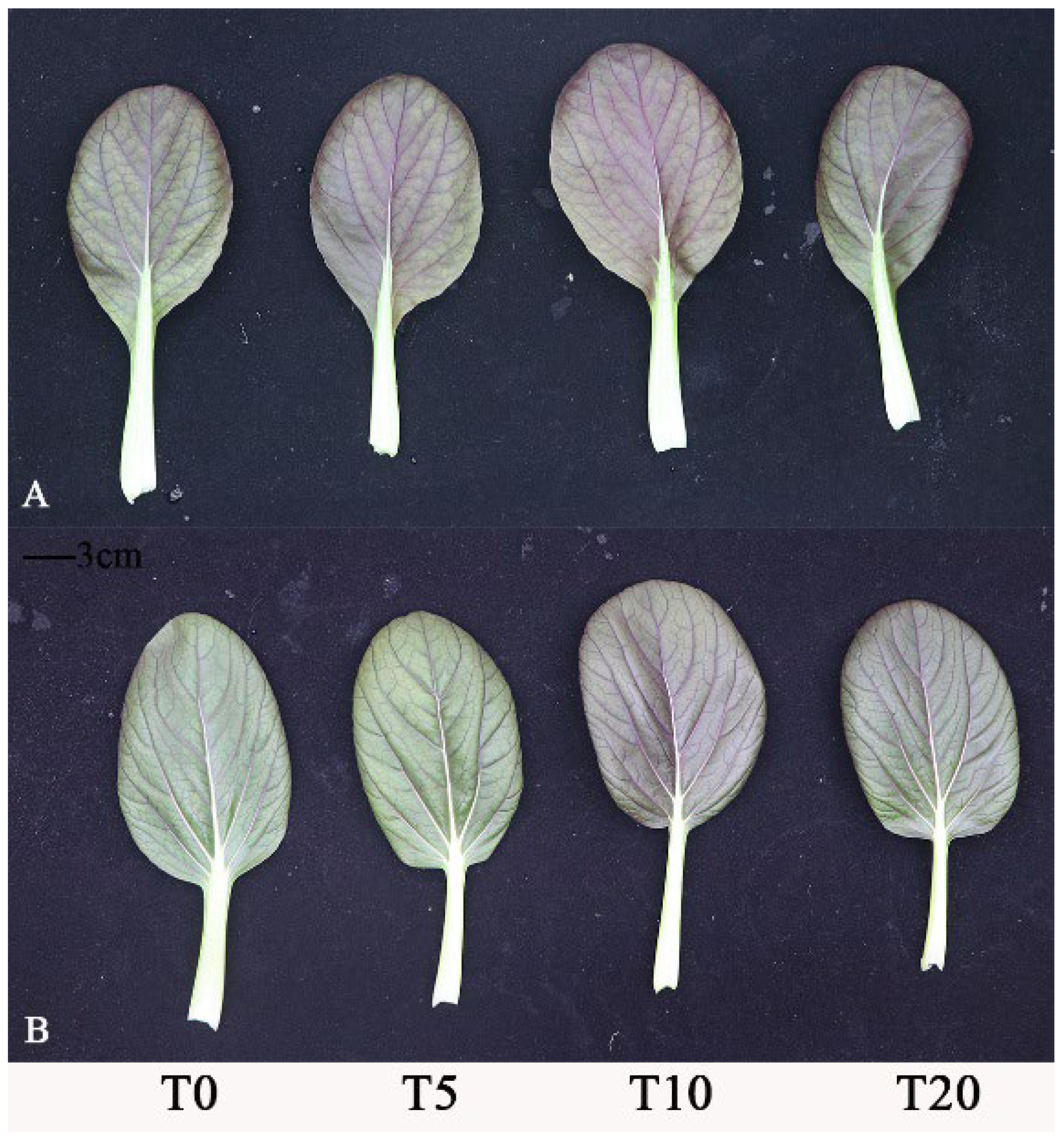
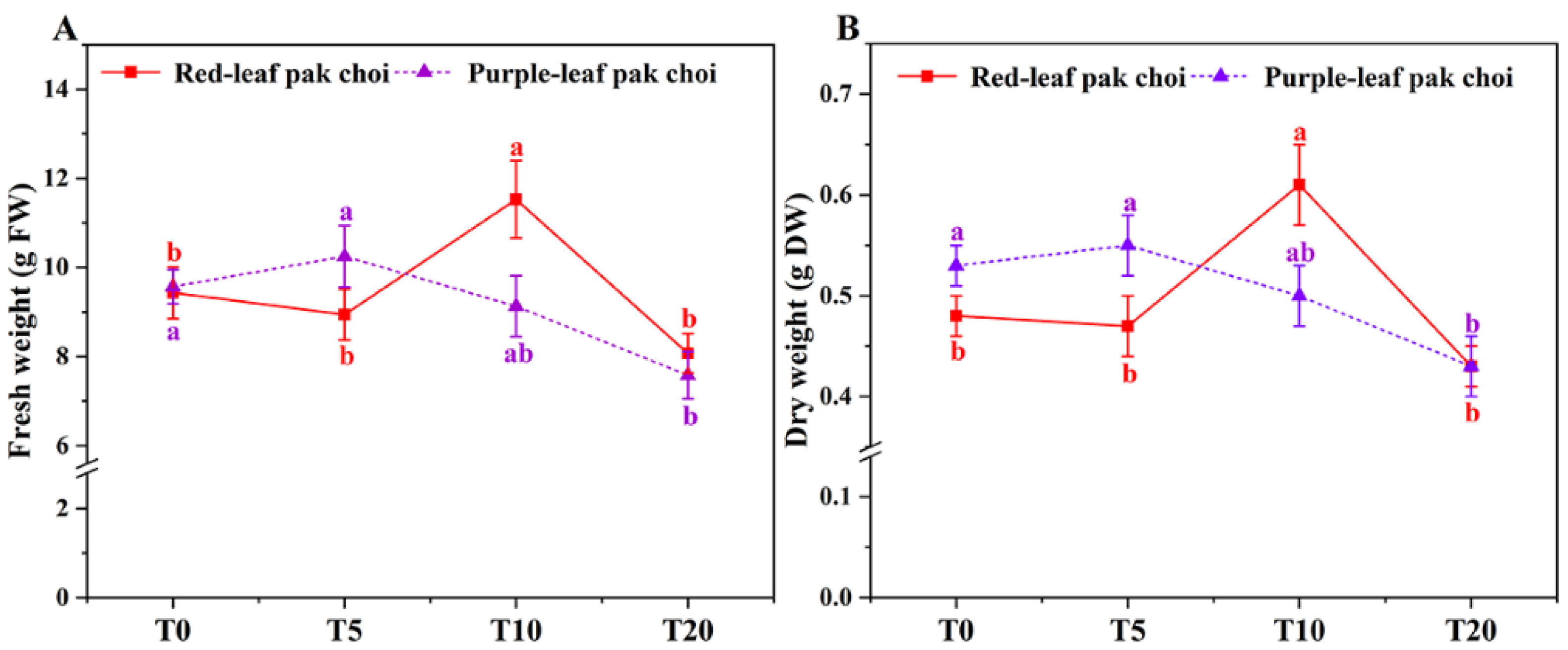
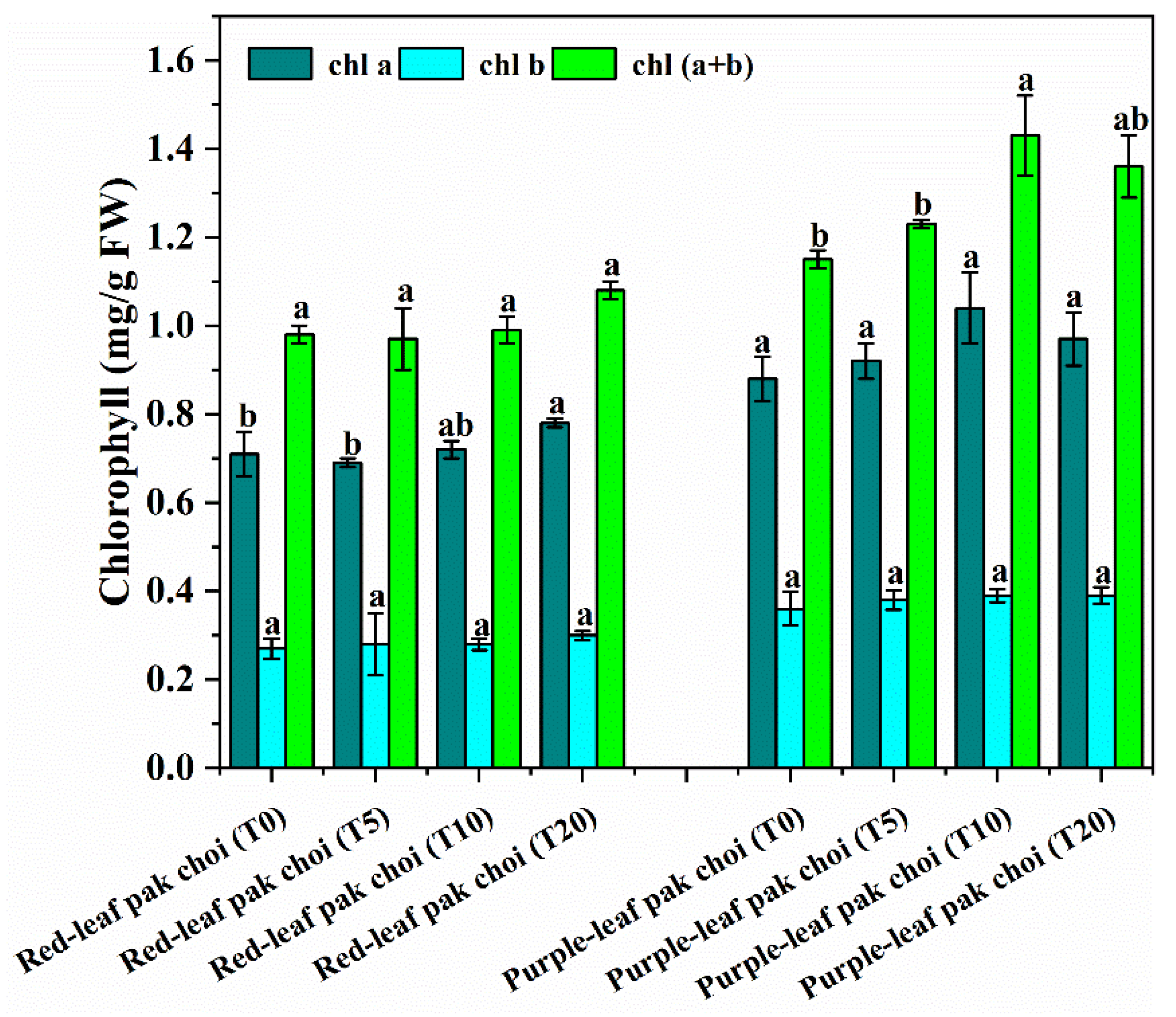
3.1. Morphology, Leaf Coloration and Growth of Pak Choi under UVA–Blue Co-Irradiation

Plants exposed to UVA radiation often exhibit changes in morphological traits and leaf colors. Distinct morphological properties in two cultivars of pak choi were observed under four combinations of blue and UVA light (Figure S1 and Figure 2). Compared with T0 (control), the plant canopies of the two tested cultivars appeared to be more compact under the co-irradiation of blue and UVA light (Figure S1). Meanwhile, UVA supplementation tended to increase the coloration of the leaf, and, interestingly, the deepest color was found under T10 rather than T20 (Figure 2). On the other hand, UVA supplementation also exerted impacts on the biomass production of pak choi. The highest fresh weight (Figure 3A) and dry weight (Figure 3B) from red-leaf pak choi were achieved in T10, significantly increased by 22.27% compared to the control (p < 0.05). In purple-leaf pak choi, although T5 resulted in a slightly increased biomass compared with T0, this discrepancy was not significant (Figure 3).

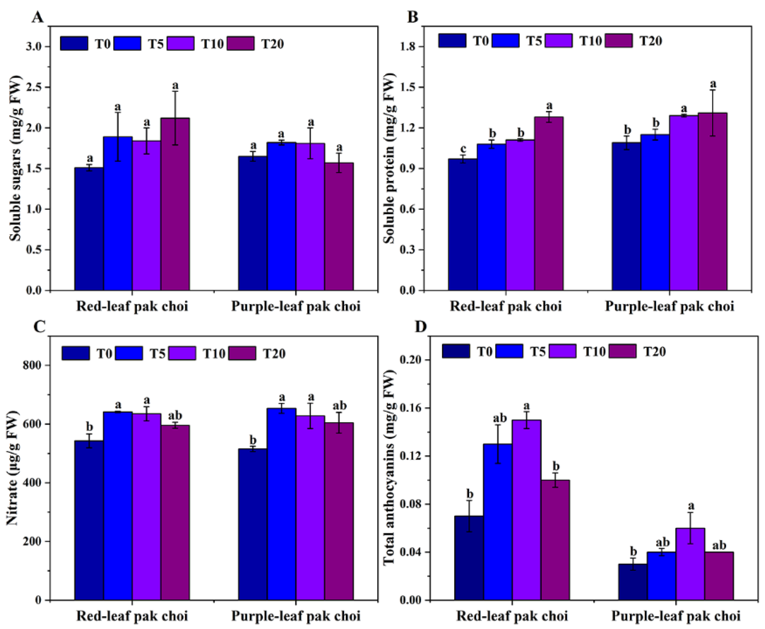
3.2. Phytochemical Variation of Pak Choi under UVA–Blue Co-Irradiation

As a widely cultivated commercial crop, nutritive quality is an important economic trait of pak choi. Therefore, nutrition-related substances were quantified under the tested light environments (Figure 4 and Figure 5). As shown in Figure 4, chl b and total chl contents in red-leaf pak choi under four combinations of blue and UVA light exhibited no significant differences, and the highest chl a content was achieved in T20, which was 9.86% higher than in T0 (p < 0.05) (Figure 4). In purple-leaf pak choi, no significant differences were detected on the contents of chl a and chl b under different treatments; however, the highest total chl content was achieved in T10, which was 18.26% higher than the control (p < 0.05) (Figure 4).
Soluble sugar and soluble protein are another two important nutritional components. The results revealed that UVA–blue co-irradiation exerted no significant impact on the soluble sugar content of red- and purple-leaf pak choi compared with monochromatic blue light (Figure 5A). However, soluble protein was largely influenced by UVA strength and exhibited the same pattern in two tested cultivars (Figure 5B). The soluble protein content increased with the increase in UVA strength, and the highest values were both found in T20 as 1.28 mg/g and 1.36 mg/g, respectively (Figure 5B).
Supplementing UVA light was positive on nitrate accumulation compared with the control (Figure 5C). However, no significant differences in nitrate content from two varieties of pak choi were observed among T5, T10 and T20 (Figure 5C). Two pak choi cultivars exhibited similar anthocyanin accumulation patterns under four tested light treatments (Figure 5D). UVA supplementation was favorable for anthocyanin accumulation in pak choi. However, a high UVA ratio tended to limit the further accumulation of anthocyanins, as shown in T20. The highest anthocyanin contents of 0.15 mg/g and 0.06 mg/g in two tested cultivars were both found in T10, interestingly consistent with that of leaf coloration (Figure 2 and Figure 5D).

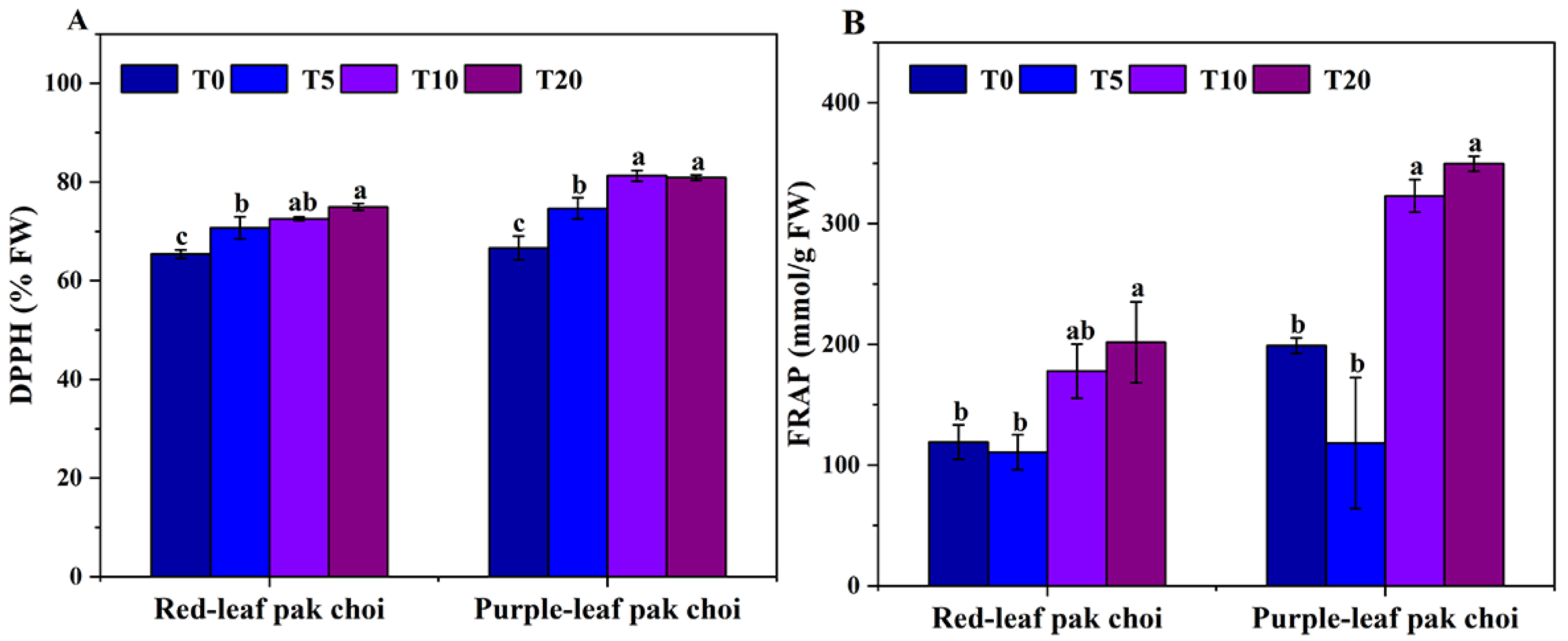
3.3. Antioxidant Capacity of UVA–Blue Co-Irradiated Pak Choi

Adding UVA to blue light exerted significant impacts on the antioxidant capacity of pak choi (Figure 6). Generally speaking, the DPPH radical scavenging rate and FRAP of both cultivars showed higher values in T10 and T20 than the control (p < 0.05) (Figure 6). T20 achieved the highest DPPH radical scavenging rate in red-leaf pak choi, 14.60% higher than T0 (p < 0.05) (Figure 6A). Similarly, purple-leaf pak choi displayed a higher DPPH radical scavenging rate in T10 and T20, 21.99% and 21.42% higher than T0, respectively (p < 0.05). On the other hand, two pak choi cultivars exhibited the highest FRAP under T20, higher by 69.36% in red-leaf pak choi and 75.72% in purple-leaf pak choi than the control (p < 0.05) (Figure 6B). In total, the antioxidant capacity of two tested pak choi cultivars under UVA–blue light co-irradiation was enhanced.

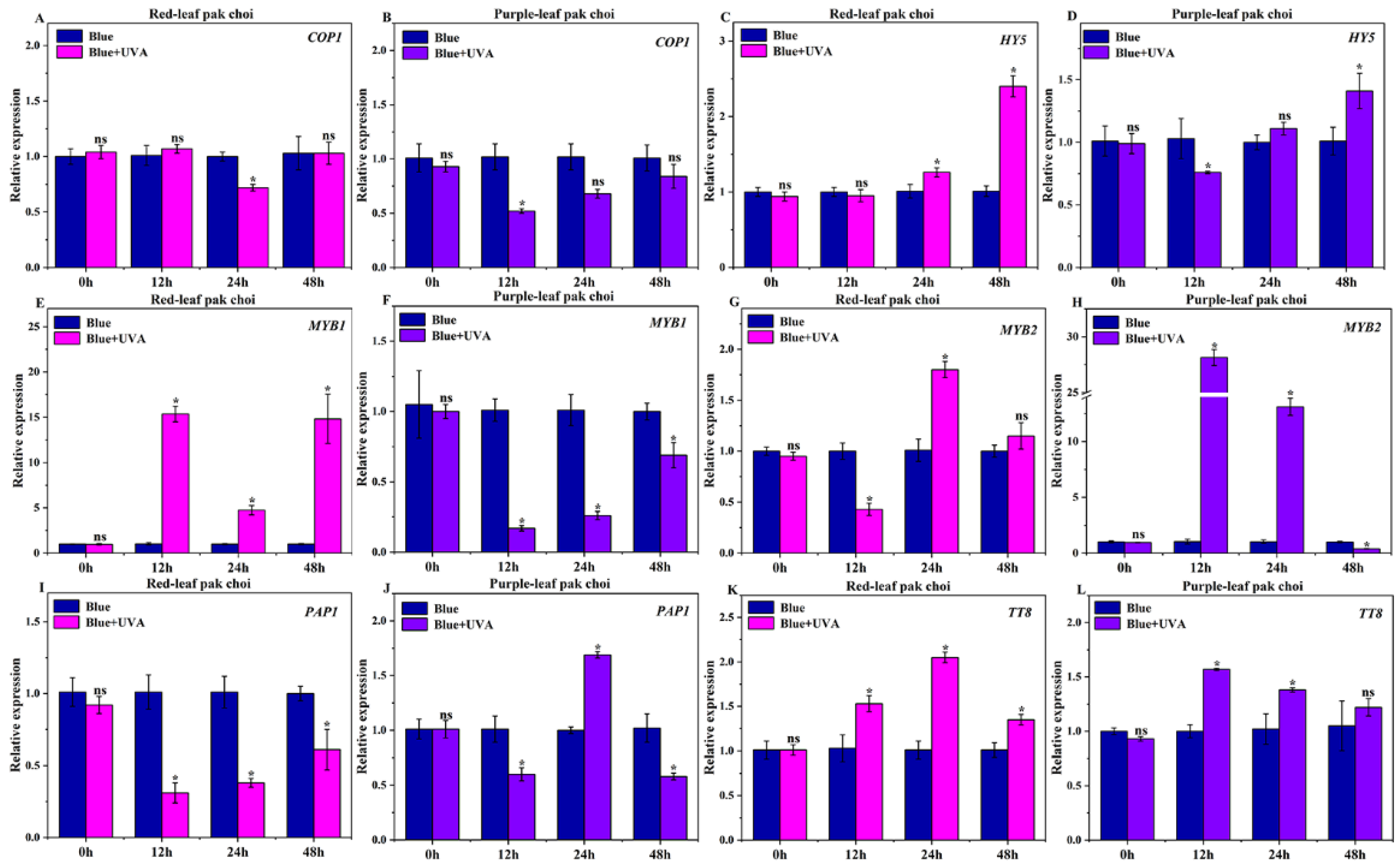
3.4. Effects of UVA–Blue Co-Irradiation on the Expression of Photoreceptor Genes

Plants sense blue/UVA light through cryptochromes, which are involved in mediating multiple growth and developmental responses [35]. For understanding the molecular response of these two tested materials, the expression patterns of CRY1 and CRY2 under continuous light treatments of T10 and T0 were tested (Figure 7). Compared with the control, the expression of CRY1 (Figure 7A) and CRY2 (Figure 7C) in the red cultivar was significantly up-regulated after continuous 24 h co-irradiation (p < 0.05); however, no significant differences were observed at other time points. For the purple-leaf pak choi, the expression levels of CRY1 (Figure 7B) and CRY2 (Figure 7D) under T10 were significantly higher after 12 h continuous irradiation compared with T0 (p < 0.05), which indicated a quicker response than red-leaf pak choi. Overall, the expressions of CRY1 and CRY2 were up-regulated by UVA–blue light co-irradiation compared with monochromatic blue light irradiation. Meanwhile, CRY1 and CRY2 were regulated with different patterns, and CRY2 seemed to be more sensitive when pak choi encountered UVA light.

3.5. Effects of UVA–Blue Co-Irradiation on the Expression of Regulatory Genes

To gain information about anthocyanin accumulation in transcription factors, the expressions of genes encoding COP1, HY5, MYB1, MYB2, PAP1 and TT8 were analyzed in our study (Figure 8). In Arabidopsis, COP1 is considered to be a central switch for light-inducible gene expression, which is the most widely investigated suppressor of photomorphogenesis [36]. Compared with the control, the expressions of COP1 in red-leaf pak choi cultivars were significantly down-regulated at 24 h continuous irradiation with UVA–blue light (p < 0.05) (Figure 8A,B). Additionally, the expressions of COP1 in purple-leaf pak choi were significantly down-regulated at 12 h (p < 0.05) (Figure 8A,B). HY5 is an activator of photomorphogenesis belonging to the basic leucine zipper (bZIP) family of TFs, and is regarded to have an important role in photomorphogenesis and anthocyanin transcriptional regulation in a light-dependent manner in Arabidopsis [39]. The expression of HY5 in both tested cultivars was significantly elevated after 48 h irradiation in UVA–blue light (p < 0.05) and the red-leaf pak choi tended to have a stronger response (Figure 8C,D).
The late biosynthesis genes (LBGs) of anthocyanin in Arabidopsis were mainly regulated by R2R3-MYB TFs, which is related to the formation of the MBW complex [16]. MYB1 in two tested cultivars showed expression profiles of different trends (Figure 8E,F). Compared with the control, UVA–blue irradiation tended to significantly elevate the expression of MYB1 in the red variety of pak choi, and decreased that in purple pak choi (p < 0.05). For the expression of MYB2, however, UVA–blue irradiation exerted a positive effect on both cultivars, and purple-leaf pak choi was fiercely affected (Figure 8G,H), which indicated that MYB1 and MYB2 possibly served as different roles in the regulation of anthocyanin accumulation in the tested cultivars. The significantly decreased expression of PAP1 in red-leaf pak choi was observed from 12 h to 48 h under continuous UVA–blue light (p < 0.05) (Figure 8I). The expression of PAP1 in purple-leaf pak choi was significantly increased at 24 h (p < 0.05); however, the down-regulated expression of that at 12- and 48 h continuous UVA–blue light was observed (p < 0.05) (Figure 8J). Regarding bHLH TFs as members of the MBW complex, TT8 plays a pivotal role in modulating the biosynthesis of proanthocyanidin and anthocyanin [14]. In our work, the abundance of TT8 in two tested cultivars was significantly up-regulated after 12 h continuous irradiation with UVA–blue light compared with the control (p < 0.05) (Figure 8K,L).

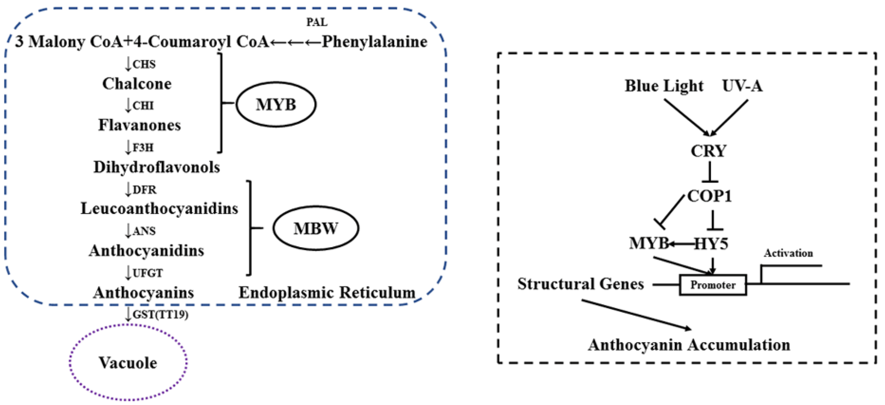
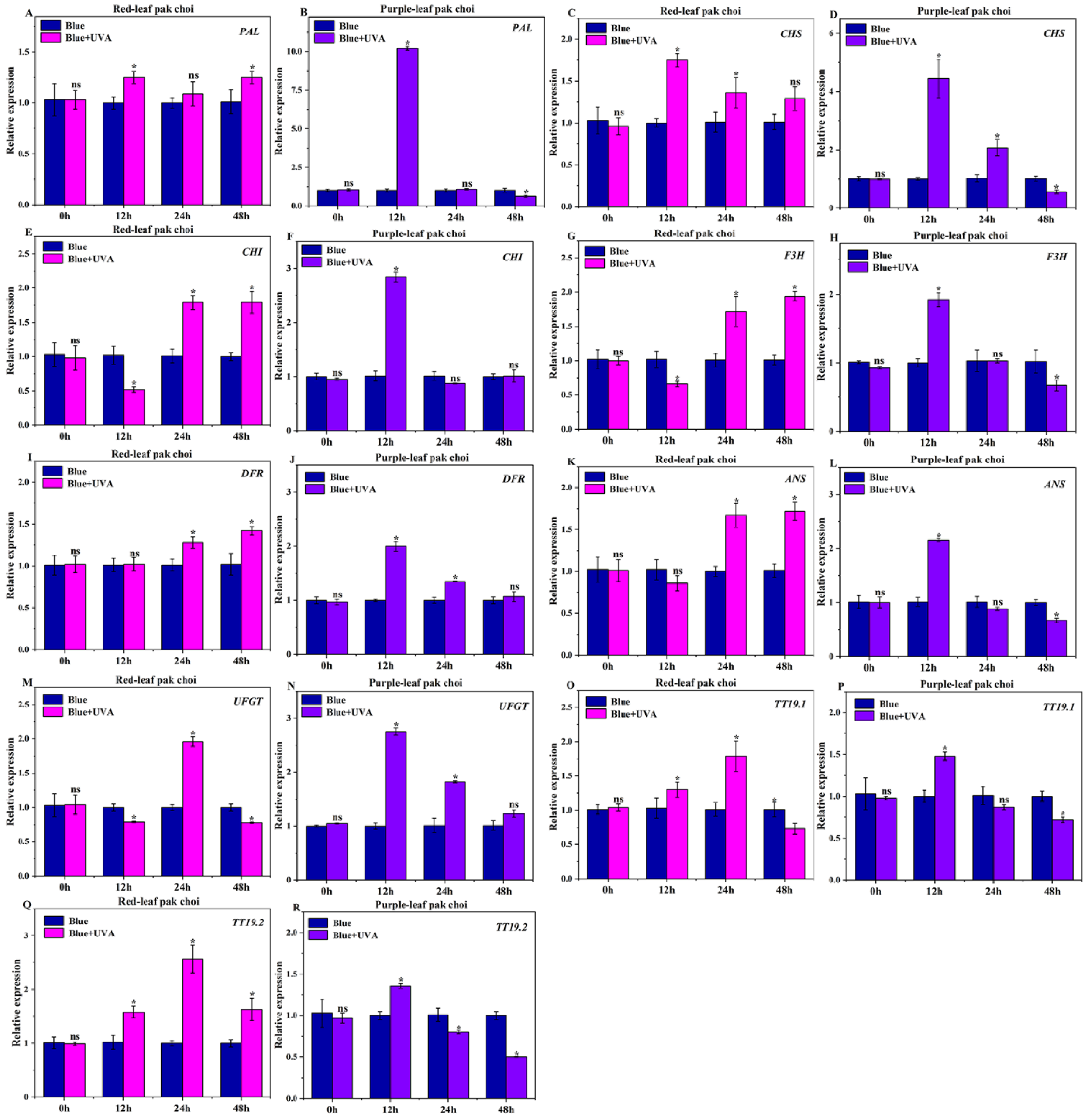
3.6. Effects of UVA–Blue Co-Irradiation on the Expression of Anthocyanin Structural Genes

To reveal the molecular mechanism of anthocyanin accumulation under UVA–blue co-irradiation, expression profiles of anthocyanin biosynthesis-related structural genes were determined (Figure 9). Anthocyanins are synthesized through the phenylpropanoid pathway, whose precursor is catalyzed by PAL in the cytoplasm [23]. In our study, the significantly increased expression of PAL in the tested cultivars was observed at 12 and 48 h compared with the control, respectively (p < 0.05) (Figure 9A). However, the purple-leaf pak choi displayed an obviously higher expression of PAL than that under the control at 12 h (p < 0.05) (Figure 9B).
For genes involved in early anthocyanin biosynthesis (EBGs), different expression patterns were observed in two pak choi cultivars (Figure 9C–H). Compared with the control, UVA–blue irradiation tended to increase the expression of CHS in the red and purple cultivars (p < 0.05) (Figure 9C,D). Abundances of CHI and F3H were both elevated in the tested cultivars and exhibited identical patterns in different cultivars (p < 0.05) (Figure 9E–H). In red-leaf pak choi, expressions of CHI and F3H were both significantly down-regulated at 12 h (p < 0.05) and up-regulated at 24 and 48 h (p < 0.05), while in purple-leaf pak choi, CHI and F3H both performed significantly higher in expression at 12 h (p < 0.05). For the LBGs involved in anthocyanin accumulation (including DFR, ANS and UFGT), the UVA–blue irradiation exerted positive effects on the expression level compared with the control, and slightly different expression patterns of them were observed in two cultivars (Figure 9I–N).
In both vesicle trafficking and transporter-mediated systems, TT19 as a glutathione S-transferase is characterized as an implement for facilitating the efficient transportation of anthocyanins from the ER to the vacuole [10]. In this study, the expressions of the main genes (TT19.1, TT19.2) coding transport proteins were detected (Figure 9O–R). Compared with the control, the expression of TT19.1 and TT19.2 in red-leaf pak choi under blue–UVA irradiation was significantly up-regulated between 12 and 24 h (p < 0.05) (Figure 9O,Q). However, purple-leaf pak choi exhibited an elevated expression level at 12 h (Figure 9P,R) (p < 0.05).

4. Discussion

4.1. Adding UVA to Blue Light Can Increase the Biomass by Affecting Plant Morphology

Multiple light signals transduced by the photoreceptors of plants are integrated in the initiation of the process leading to photomorphogenesis [48]. Cryptochromes and phototropins are vital photoreceptors of blue and UVA light, and participate in the shaping of plant morphological characteristics [49]. Meanwhile, it has been confirmed that UVA plays a critical role in shaping the morphological appearance of cucumber and tomato [26,50]. In our experiment, adding UVA to blue light tended to result in more compact phenotypes of pak choi compared with monochromatic blue light (Figure S1). Compact phenotypes of plants probably allow for more light to penetrate the canopy, contributing to increased light interception, leading to the enhancement of biomass. Generally speaking, in contrast to other wavebands (e.g., far-red, red, blue and UVB), the effects of UVA radiation on the growth of plants are less known and are often controversial [32]. Interestingly, a certain amount of UVA (T10) was beneficial for the biomass accumulation of the tested red-leaf pak choi. Micro-doses of UVA were also reported to increase the biomass production of tomato seedlings, which was mainly attributed to the elevated light intercepting ability of leaves [29]. In purple-leaf pak choi, T5 slightly increased the biomass with no significance, while the biomass in T20 was significantly decreased, indicating that a relatively high UVA intensity was negative for the growth of pak choi (Figure 3). Similar results were also reported by Chen et al. [28], that high UVA (40 μmol·m−2·−1) led to biomass loss in lettuce. High UVA radiation doses might cause decreases in the photosynthetic activity due to excessive reactive oxygen species (ROS) [32]. On the other hand, the high UVA treatment signified that the intercepted light energy might be insufficient to support the normal growth of pak choi, as blue light is characterized to be in the photosynthetic activation spectrum. Therefore, the proper use of UVA is a promising strategy for crop cultivation, and the differential physiological responses between varied genotypes should be considered simultaneously.

4.2. Adding UVA to Blue Light Can Enhance Antioxidant Ability and Anthocyanin Accumulation

Energy, enzymes and substrates are necessary for the synthesis of phytochemicals in plants. In higher plants, it is vital to evaluate photosynthetic pigments, soluble sugars, soluble proteins and nitrates for the carbon and nitrogen metabolism. It is particularly essential to understand the biosynthesis of anthocyanins from the perspective of physiological changes. Sugars affect the expression profiles of genes related to anthocyanin formation by acting as endogenous triggers [51]. There were practically insignificant effects on chlorophyll and soluble sugars of pak choi among the four light treatments (Figure 4 and Figure 5A). Compared with the control, soluble proteins, nitrate and anthocyanins all showed varied degrees of elevation under the co-irradiation of blue light and UVA (Figure 5B–D).
Blue light is a positive environmental factor for anthocyanin biosynthesis in plants. In this study, the co-irradiation of UVA–blue light led to the further accumulation of anthocyanins compared with monochromatic blue light (Figure 5D). In the turnip ‘Tsuda’, UVA specifically induced anthocyanin biosynthesis, and had a much more obvious effect on anthocyanin accumulation than blue light [24]. The highest content of anthocyanins in both cultivars of pak choi was achieved in the T10 treatment (Figure 5D), which was in accordance with the coloration of the leaves in both cultivars (Figure 2). Zhang et al. [4] reported that UVA promoted anthocyanin production in radishes in hydrogen-rich water. However, a higher UVA ratio tended to inhibit the biosynthesis of anthocyanins as shown in T20, which was similar to the effect of UVA light on the biomass production of pak choi. As anthocyanins are effective antioxidants in plants, the antioxidant ability of pak choi was simultaneously enhanced (Figure 6). Our results showed that the antioxidant capacity of tested cultivars was positively related to UVA strength and reached the highest value at T20 (Figure 5B and Figure 6) and disagreed with the accumulate pattern of anthocyanin, indicating that UVA irradiation may increase the content of other bio-antioxidants that were not measured in this study (Figure 5D). The flavonoid metabolism pathway in the process of anthocyanin biosynthesis is an indispensable pathway, leading to the production of numerous flavonoids, involving a relationship between their competition [10]. Therefore, it is possible that more substrates and enzymes were used in the synthesis of other flavonoids, making the antioxidant capacity at T20 greater than T10 (Figure 5B and Figure 6).

4.3. Adding UVA to Blue Light Can Affect Gene Abundance to Enhance Anthocyanin Accumulation

Our preliminary study found that an effective combination of UVA–blue light (T10) significantly enhanced anthocyanin levels (p < 0.05). To understand the gene expression profiles of anthocyanin accumulation in pak choi under continuous UVA–blue co-irradiation, we performed a qRT-PCR analysis for photoreceptors and regulatory and structural genes. The majority of the tested genes performed distinct expression patterns in the two tested pak choi cultivars (Figure 7, Figure 8 and Figure 9). Cryptochromes are specific sensing photoreceptors of blue–UVA light, and play vital roles in regulating multiple plant growth and developmental processes. In this study, the expression level of CRY1 and CRY2 was significantly elevated due to the UVA–blue co-irradiation, as compared to monochromatic blue light (Figure 7). The physiologically inactive monomer states of CRY stability existed in darkness, and photoexcited CRY molecules in response to blue–UVA light perception underwent homodimerization and became physiologically active [34,36,52]. The knock-down of CRY1a with RNAi decreased the anthocyanin levels, while over-expressing CRY2 in transgenic tomato plants showed elevated anthocyanin accumulation [53]. Meanwhile, Liu et al. [8] found that the over-expression of CRY1a increased the anthocyanin content when exposed to blue light. However, there are few studies on the molecular response of CRYs under UVA irradiation. Hence, our results indicated CRY1 and CRY2 were more sensitive in perceiving UVA than blue light, and may contributed to anthocyanin accumulation. Moreover, the expression profiles of CRY1 and CRY2 were different at diverse time points in response to light, possibly related to pak choi cultivars.
COP1 is another important molecular regulator and functions at a central position of light-signaling networks [36,37]. After the activation of the CRY homodimer or oligomers, they prevent the formation of the COP1/SPA complex to inhibit the ubiquitylation and degradation of TFs, such as HY5 [36,54]. We found that both pak choi cultivars under continuous UVA–blue light exhibited an up-regulated expression of COP1 at 24 h compared to the control (Figure 8A,B). It has been reported that photoexcited CRYs interacting with SPA indirectly inhibit the activation of the COP1/SPA E3 ligase complex [35]. In Arabidopsis, CRY2 suppresses the activity of COP1 by interacting with SPA1 is a blue-light-dependent fashion [33]. Moreover, CRY1 and CRY2 C terminal domains also interact directly with COP1 [37], contributing to HY5 stabilization and accumulation, and leading to light morphogenesis [30,36]. Our study showed that the expression of CRYs from two varieties of pak choi exposed to UVA–blue light was significantly higher than that of the control at 24 h (Figure 7), which could cause the down-regulation of COP1. (Figure 8). On the other hand, COP1 and HY5 are sensitive in response to light or darkness, and their sensitivities to perceive light quality seemingly differ due to species differences [36,39]. Several experiments revealed that HY5 could directly and indirectly affect anthocyanin accumulation by binding to the promoter of the DNA domain, including MYB75 (PAP1), CHS and DFR [55]. Meanwhile, the silencing of SIHY5 in the over-expression of CRY1a in tomato reduced the accumulation of anthocyanin [8]. As shown in our results, the expression of HY5 in red-leaf pak choi was significantly up-regulated between 24 h and 48 h; nevertheless, the expression of HY5 in purple-leaf pak choi exhibited up-regulated expression at 48 h upon exposure to continuous UVA–blue light radiation (Figure 8D), which indicated that COP1 and HY5 work as important intermediates during the promoting biosynthesis of anthocyanins caused by UVA–blue light co-irradiation.
The R2R3-MYB TFs have been identified and characterized in numerous plants, and have been found to be activators and repressors of structural genes participating in the progress of the anthocyanin metabolism [16]. Until recently, some R2R3-MYB TFs were verified to respond to light signals and to form the MBW regulatory complex involved in the regulation of anthocyanin biosynthesis [18]. Despite this notion, the investigation of bHLH and WD40 partners has not yet been clarified in light-induced anthocyanin accumulation. In this study, compared with purple-leaf pak choi, the expression of MYB1 in red-leaf pak choi was extremely up-regulated when irradiated with continuous UVA–blue light, while the expression of MYB2 in purple-leaf pak choi reacted more severely than that seen in red-leaf pak choi (Figure 8E–H). Furthermore, the expression of MYB1 and PAP1 showed contrasting expression patterns in the tested cultivars. It was reported that solitary R2R3-MYBs may only have limited impact on anthocyanin biosynthesis, and some studies have suggested that the cooperation of R2R3-MYBs and bHLH could promote anthocyanin accumulation [55]. Simultaneously, both cultivars showed significantly higher expressions of TT8 when treated under UVA–blue light co-irradiation (p < 0.05) (Figure 8K,L). Overall, MYB1, MYB2, PAP1 and TT8 functioned as positive regulators in response to UVA–blue-light-induced anthocyanin biosynthesis, and the main function factors in the tested cultivars were different, which may be attributed to the genetic difference.
To better understand the expression pattern of genes involved in anthocyanin biosynthesis under UVA–blue co-irradiation, we systematically investigated the expression level of these genes. The results revealed that the structural genes of the anthocyanin biosynthesis pathway overall showed an increased expression level in both tested pak choi cultivars (Figure 9). PAL was the first rate-limiting enzyme of the phenylpropanoid metabolism, and the abundance of PAL in pak choi was significantly increased after 12 h when exposed to UVA–blue light. In UVA-treated tomato, it was reported that the expression of PAL was up-regulated in a time-dependent manner [23]. Additionally, the transcripts of the EBGs (e.g., CHS, CHI and F3H) were manipulated by R2R3-MYB transcriptional regulators. However, the LBGs (e.g., DFR, ANS and UFGT) were modulated by the MBW ternary complex [18]. As the results show in Figure 9, compared with the control, the transcript levels of CHS, CHI, F3H, DFR, ANS, UFGT, TT19.1 and TT19.2 were greatly up-regulated in continuous UVA–blue-light-grown red and purple-leaf pak choi. Consequently, we speculated that light-induced regulatory genes (MYB1, MYB2, PAP1 and TT8) stimulated anthocyanin accumulation in pak choi through the regulation of structural genes involved in anthocyanin biosynthesis (Figure 8 and Figure 9).

5. Conclusions

UVA–blue light co-irradiation comprehensively affected the morphological traits and anthocyanin biosynthesis in pak choi. Meanwhile, UVA positively affected the biomass enhancement of pak choi under blue light. In this study, compared to other treatments, T10 was beneficial for anthocyanin and biomass accumulation in pak choi. Furthermore, molecular data revealed that light-induced regulatory genes play a vital role in the regulation of anthocyanin biosynthesis. Overall, UVA–blue light co-irradiation could provide a reference strategy for improving the quality of vegetables.

Supplementary Materials

The following supporting information can be downloaded at: https://www.mdpi.com/article/10.3390/horticulturae8090850/s1, Table S1: the exhibition of UVA (385 nm) and blue (460 nm) light treatments, Table S2: a list of primer sequences for the qRT-PCR analysis, Figure S1: effects of blue and UVA light on the canopies of red- (A) and purple-leaf (B) pak choi. T0, T5, T10 and T20 mean 0 UVA with 50 B, 5 UVA with 45 B, 10 UVA with 40 B and 20 UVA with 30 B (μmol·m−2·s−1), respectively.

Author Contributions

Conceptualization, J.H. and Y.X.; methodology, J.H. and X.L., software, J.H. and P.M.; validation, J.H., X.L. and Y.X.; formal analysis, Z.B.; investigation, Y.Z.; resources, Q.Y.; data curation, S.W.; writing—original draft preparation, J.H., X.L. and Y.X.; writing—review and editing, Y.X., B.L. and W.L.; visualization, Y.Z.; supervision, Q.Y.; project administration, Y.X., Y.Z. and Q.Y.; funding acquisition, Y.X. All authors have read and agreed to the published version of the manuscript.

Funding

This work was financially supported by the National Key Research and Development Program (No. 2020YFB0407902), the Central Public-interest Scientific Institution Basal Research Fund of China (No. 34-IUA-03; No. 34-IUA-01), the Elite Youth Program of the Chinese Academy of Agricultural Sciences (Yinjian Zheng) and the Local Financial Funds of National Agricultural Science and Technology Center, Chengdu, China (No. NASC2020AR10; No. NASC2021PC03).

Institutional Review Board Statement

Not applicable.

Informed Consent Statement

Not applicable.

Data Availability Statement

All data can be found in our manuscript and Supplemental Materials. If necessary, data are available by contact with the first author.

Acknowledgments

We sincerely thank the Chengdu Academy of Agricultural and Forestry Sciences for providing us with the experimental site and equipment.

Conflicts of Interest

The authors declare no conflict of interest.

References

  1. Harbaum, B.; Hubbermann, E.M.; Zhu, Z.; Schwarz, K. Free and bound phenolic compounds in leaves of pak choi (Brassica campestris L. ssp. chinensis var. communis) and Chinese leaf mustard (Brassica juncea Coss). Food Chem. 2008, 110, 838–846. [Google Scholar] [CrossRef]
  2. Björkman, M.; Klingen, I.; Birch, A.N.; Bones, A.M.; Bruce, T.J.; Johansen, T.J.; Meadow, R.; Mølmann, J.; Seljåsen, R.; Smart, L.E.; et al. Phytochemicals of Brassicaceae in plant protection and human health—Influences of climate, environment and agronomic practice. Phytochemistry 2011, 72, 538–556. [Google Scholar] [CrossRef]
  3. Chaves-Silva, S.; dos Santos, A.L.; Chalfun-Júnior, A.; Zhao, J.; Peres, L.E.; Benedito, V.A. Understanding the genetic regulation of anthocyanin biosynthesis in plants—Tools for breeding purple varieties of fruits and vegetables. Phytochemistry 2018, 153, 11–27. [Google Scholar] [CrossRef]
  4. Saigo, T.; Wang, T.; Watanabe, M.; Tohge, T. Diversity of anthocyanin and proanthocyanin biosynthesis in land plants. Curr. Opin. Plant Biol. 2020, 55, 93–99. [Google Scholar] [CrossRef]
  5. Amiot, M.J.; Riva, C.; Vinet, A. Effects of dietary polyphenols on metabolic syndrome features in humans: A systematic review. Obes. Rev. 2016, 17, 573–586. [Google Scholar] [CrossRef]
  6. Khoo, H.E.; Azlan, A.; Tang, S.T.; Lim, S.M. Anthocyanidins and anthocyanins: Colored pigments as food, pharmaceutical ingredients, and the potential health benefits. Food Nutr. Res. 2017, 61, 1361779. [Google Scholar] [CrossRef]
  7. Gu, K.-D.; Wang, C.-K.; Hu, D.-G.; Hao, Y.-J. How do anthocyanins paint our horticultural products? Sci. Hortic. 2019, 249, 257–262. [Google Scholar] [CrossRef]
  8. Liu, C.-C.; Chi, C.; Jin, L.-J.; Zhu, J.; Yu, J.-Q.; Zhou, Y.-H. The bZip transcription factor HY5 mediates CRY1a -induced anthocyanin biosynthesis in tomato. Plant Cell Environ. 2018, 41, 1762–1775. [Google Scholar] [CrossRef]
  9. Zhang, X.; Wei, J.; Tian, J.; Li, N.; Jia, L.; Shen, W.; Cui, J. Enhanced anthocyanin accumulation of immature radish microgreens by hydrogen-rich water under short wavelength light. Sci. Hortic. 2019, 247, 75–85. [Google Scholar] [CrossRef]
  10. Sun, Y.; Li, H.; Huang, J.-R. Arabidopsis TT19 functions as a carrier to transport anthocyanin from the cytosol to tonoplasts. Mol. Plant 2012, 5, 387–400. [Google Scholar] [CrossRef] [PubMed]
  11. Chen, L.; Hu, B.; Qin, Y.; Hu, G.; Zhao, J. Advance of the negative regulation of anthocyanin biosynthesis by MYB transcription factors. Plant Physiol. Biochem. 2019, 36, 178–187. [Google Scholar] [CrossRef]
  12. Saito, K.; Yonekura-Sakakibara, K.; Nakabayashi, R.; Higashi, Y.; Yamazaki, M.; Tohge, T.; Fernie, A.R. The flavonoid biosynthetic pathway in Arabidopsis: Structural and genetic diversity. Plant Physiol. Biochem. 2013, 72, 21–34. [Google Scholar] [CrossRef] [PubMed]
  13. Marrs, K.A.; Alfenito, M.R.; Lloyd, A.M.; Walbot, V. A glutathione S-transferase involved in vacuolar transfer encoded by the maize gene Bronze-2. Nature 1995, 375, 397–400. [Google Scholar] [CrossRef] [PubMed]
  14. Larsen, E.S.; Alfenito, M.R.; Briggs, W.R.; Walbot, V. A carnation anthocyanin mutant is complemented by the glutathione S-transferases encoded by maize Bz2 and petunia An9. Plant Cell Rep. 2003, 21, 900–904. [Google Scholar] [CrossRef]
  15. Lloyd, A.; Brockman, A.; Aguirre, L.; Campbell, A.; Bean, A.; Cantero, A.; Gonzalez, A. Advances in the MYB–bHLH–WD repeat (MBW) pigment regulatory model: Addition of a WRKY factor and co-option of an anthocyanin MYB for betalain regulation. Plant Cell Physiol. 2017, 58, 1431–1441. [Google Scholar] [CrossRef]
  16. Naing, A.H.; Kim, C.K. Roles of R2R3-MYB transcription factors in transcriptional regulation of anthocyanin biosynthesis in horticultural plants. Plant Mol. Biol. 2018, 98, 1–18. [Google Scholar] [CrossRef]
  17. He, Y.; Chen, Q.; Shu, C.; Yang, M.; Zhou, E. Colletotrichum truncatum, a new cause of anthracnose on Chinese flowering cabbage (Brassica parachinensis) in China. Trop. Plant Pathol. 2016, 41, 183–192. [Google Scholar] [CrossRef]
  18. Lim, S.-H.; Kim, D.-H.; Kim, J.K.; Lee, J.-Y.; Ha, S.-H. A radish basic helix-loop-helix transcription factor, RsTT8 acts a positive regulator for anthocyanin biosynthesis. Front. Plant Sci. 2017, 8, 1917. [Google Scholar] [CrossRef] [PubMed]
  19. Tang, B.; Li, L.; Hu, Z.; Chen, Y.; Tan, T.; Jia, Y.; Xie, Q.; Chen, G. Anthocyanin accumulation and transcriptional regulation of anthocyanin biosynthesis in purple pepper. J. Agric. Food Chem. 2020, 68, 12152–12163. [Google Scholar] [CrossRef]
  20. Hogewoning, S.; Trouwborst, G.; Maljaars, H.; Poorter, H.; Van Ieperen, W.; Harbinson, J. Blue light dose-responses of leaf photosynthesis, morphology, and chemical composition of Cucumis sativus grown under different combinations of red and blue light. J. Exp. Bot. 2010, 61, 3107–3117. [Google Scholar] [CrossRef]
  21. Huché-Thélier, L.; Crespel, L.; Le Gourrierec, J.; Morel, P.; Sakr, S.; Leduc, N. Light signaling and plant responses to blue and UV radiations—Perspectives for applications in horticulture. Environ. Exp. Bot. 2016, 121, 22–38. [Google Scholar] [CrossRef]
  22. Nakai, A.; Tanaka, A.; Yoshihara, H.; Murai, K.; Watanabe, T.; Miyawaki, K. Blue LED light promotes indican accumulation and flowering in indigo plant, Polygonum tinctorium. Ind. Crop. Prod. 2020, 155, 112774. [Google Scholar] [CrossRef]
  23. Guo, J.; Wang, M.H. Ultraviolet A-specific induction of anthocyanin biosynthesis and PAL expression in tomato (Solanum lycopersicum L.). Plant Growth Regul. 2010, 62, 1–8. [Google Scholar] [CrossRef]
  24. Zhou, B.; Li, Y.; Xu, Z.; Yan, H.; Homma, S.; Kawabata, S. Ultraviolet A-specific induction of anthocyanin biosynthesis in the swollen hypocotyls of turnip (Brassica rapa). J. Exp. Bot. 2007, 58, 1771–1781. [Google Scholar] [CrossRef] [PubMed]
  25. Li, Q.; Kubota, C. Effects of supplemental light quality on growth and phytochemicals of baby leaf lettuce. Environ. Exp. Bot. 2009, 67, 59–64. [Google Scholar] [CrossRef]
  26. Zhang, Y.; Kaiser, E.; Zhang, Y.; Zou, J.; Bian, Z.; Yang, Q.; Li, T. UVA radiation promotes tomato growth through morphological adaptation leading to increased light interception. Environ. Exp. Bot. 2020, 176, 104073. [Google Scholar] [CrossRef]
  27. Neugart, S.; Schreiner, M. UVB and UVA as eustressors in horticultural and agricultural crops. Sci. Hortic. 2018, 234, 370–381. [Google Scholar] [CrossRef]
  28. Chen, Y.; Fanourakis, D.; Tsaniklidis, G.; Aliniaeifard, S.; Yang, Q.; Li, T. Low UVA intensity during cultivation improves the lettuce shelf-life, an effect that is not sustained at higher intensity. Postharvest Biol. Technol. 2021, 172, 111376. [Google Scholar] [CrossRef]
  29. Kang, S.; Zhang, Y.; Zhang, Y.; Zou, J.; Yang, Q.; Li, T. Ultraviolet-A radiation stimulates growth of indoor cultivated tomato (Solanum lycopersicum) seedlings. HortScience 2018, 53, 1429–1433. [Google Scholar] [CrossRef]
  30. Tissot, N.; Ulm, R. Cryptochrome-mediated blue-light signalling modulates UVR8 photoreceptor activity and contributes to UV-B tolerance in Arabidopsis. Nat. Commun. 2020, 11, 1–10. [Google Scholar] [CrossRef] [Green Version]
  31. Siipola, S.M.; Kotilainen, T.; Sipari, N.; Morales, L.O.; Lindfors, A.V.; Robson, T.M.; Aphalo, P.J. Epidermal UV-A absorbance and whole-leaf flavonoid composition in pea respond more to solar blue light than to solar UV radiation. Plant Cell Environ. 2014, 38, 941–952. [Google Scholar] [CrossRef]
  32. Verdaguer, D.; Jansen, M.A.; Llorens, L.; Morales, L.O.; Neugart, S. UV-A radiation effects on higher plants: Exploring the known unknown. Plant Sci. 2017, 255, 72–81. [Google Scholar] [CrossRef]
  33. Wang, Q.; Zuo, Z.; Wang, X.; Liu, Q.; Gu, L.; Oka, Y.; Lin, C. Beyond the photocycle—how cryptochromes regulate photoresponses in plants? Curr. Opin. Plant Biol. 2018, 45, 120–126. [Google Scholar] [CrossRef]
  34. Lin, C.; Shalitin, D. Cryptochrome structure and signal transduction. Annu. Rev. Plant Biol. 2003, 54, 469–496. [Google Scholar] [CrossRef]
  35. Sharma, P.; Chatterjee, M.; Burman, N.; Khurana, J.P. Cryptochrome 1 regulates growth and development in Brassica through alteration in the expression of genes involved in light, phytohormone and stress signalling. Plant Cell Environ. 2013, 37, 961–977. [Google Scholar] [CrossRef]
  36. Han, X.; Huang, X.; Deng, X.W. The photomorphogenic central repressor COP1: Conservation and functional diversification during evolution. Plant Commun. 2020, 1, 100044. [Google Scholar] [CrossRef]
  37. Podolec, R.; Ulm, R. Photoreceptor-mediated regulation of the COP1/SPA E3 ubiquitin ligase. Curr. Opin. Plant Biol. 2018, 45, 18–25. [Google Scholar] [CrossRef]
  38. Jiang, M.; Ren, L.; Lian, H.; Liu, Y.; Chen, H. Novel insight into the mechanism underlying light-controlled anthocyanin accumulation in eggplant (Solanum melongena L.). Plant Sci. 2016, 249, 46–58. [Google Scholar] [CrossRef]
  39. Gangappa, S.N.; Botto, J.F. The multifaceted roles of HY5 in plant growth and development. Mol. Plant 2016, 9, 1353–1365. [Google Scholar] [CrossRef]
  40. Mao, P.; Duan, F.; Zheng, Y.; Yang, Q. Blue and UV-A light wavelengths positively affected accumulation profiles of healthy compounds in pak-choi. J. Sci. Food Agric. 2020, 101, 1676–1684. [Google Scholar] [CrossRef]
  41. Arnon, D.I. Copper enzymes in isolated chloroplasts. Polyphenoloxidase in Beta vulgaris. Plant Physiol. 1949, 24, 1–15. [Google Scholar] [CrossRef]
  42. Kohyama, K.; Nishinari, K. Effect of soluble sugars on gelatinization and retrogradation of sweet potato starch. J. Agric. Food Chem. 1991, 39, 1406–1410. [Google Scholar] [CrossRef]
  43. Blakesley, R.W.; Boezi, J.A. A new staining technique for proteins in polyacrylamide gels using Coomassie brilliant blue G250. Anal. Biochem. 1977, 82, 580–582. [Google Scholar] [CrossRef]
  44. Cataldo, D.A.; Maroon, M.; Schrader, L.E.; Youngs, V.L. Rapid colorimetric determination of nitrate in plant tissue by nitration of salicylic acid. Commun. Soil Sci. Plant Anal. 1975, 6, 71–80. [Google Scholar] [CrossRef]
  45. Huang, C.; Yu, B.; Teng, Y.; Su, J.; Shu, Q.; Cheng, Z.; Zeng, L. Effects of fruit bagging on coloring and related physiology, and qualities of red Chinese sand pears during fruit maturation. Sci. Hortic. 2009, 121, 149–158. [Google Scholar] [CrossRef]
  46. Liu, D.; Guo, Y.; Wu, P.; Wang, Y.; Golly, M.K.; Ma, H. The necessity of walnut proteolysis based on evaluation after in vitro simulated digestion: ACE inhibition and DPPH radical-scavenging activities. Food Chem. 2020, 311, 125960. [Google Scholar] [CrossRef]
  47. Dinis, T.C.; Madeira, V.M.; Almeida, L.M. Action of phenolic derivatives (acetaminophen, salicylate, and 5-aminosalicylate) as inhibitors of membrane lipid peroxidation and as peroxyl radical scavengers. Arch. Biochem. Biophys. 1994, 315, 161–169. [Google Scholar] [CrossRef]
  48. Kurepin, L.V.; Pharis, R.P. Light signaling and the phytohormonal regulation of shoot growth. Plant Sci. 2014, 229, 280–289. [Google Scholar] [CrossRef]
  49. Christie, J.M.; Blackwood, L.; Petersen, J.; Sullivan, S. Plant flavoprotein photoreceptors. Plant Cell Physiol. 2014, 56, 401–413. [Google Scholar] [CrossRef]
  50. Qian, M.; Rosenqvist, E.; Flygare, A.-M.; Kalbina, I.; Teng, Y.; Jansen, M.A.K.; Strid, Å. UV-A light induces a robust and dwarfed phenotype in cucumber plants (Cucumis sativus L.) without affecting fruit yield. Sci. Hortic. 2015, 263, 109110. [Google Scholar] [CrossRef]
  51. Teng, S.; Keurentjes, J.; Bentsink, L.; Koornneef, M.; Smeekens, S. Sucrose-specific induction of anthocyanin biosynthesis in Arabidopsis requires the MYB75/PAP1 gene. Plant Physiol. 2005, 139, 1840–1852. [Google Scholar] [CrossRef] [PubMed]
  52. Lian, H.; Xu, P.; He, S.; Wu, J.; Pan, J.; Wang, W.; Xu, F.; Wang, S.; Pan, J.; Huang, J.; et al. Photoexcited cryptochrome 1 interacts directly with G-Protein β subunit AGB1 to regulate the DNA-binding activity of HY5 and photomorphogenesis in Arabidopsis. Mol. Plant 2018, 11, 1248–1263. [Google Scholar] [CrossRef] [PubMed]
  53. Giliberto, L.; Perrotta, G.; Pallara, P.; Weller, J.L.; Fraser, P.D.; Bramley, P.M.; Fiore, A.; Tavazza, M.; Giuliano, G. Manipulation of the blue light photoreceptor cryptochrome 2 in tomato affects vegetative development, flowering time, and fruit antioxidant content. Plant Physiol. 2005, 137, 199–208. [Google Scholar] [CrossRef] [PubMed]
  54. Shin, D.H.; Choi, M.; Kim, K.; Bang, G.; Cho, M.; Choi, S.-B.; Choi, G.; Park, Y.-I. HY5 regulates anthocyanin biosynthesis by inducing the transcriptional activation of the MYB75/PAP1 transcription factor in Arabidopsis. FEBS Lett. 2013, 587, 1543–1547. [Google Scholar] [CrossRef]
  55. Feng, S.; Sun, S.; Chen, X.; Wu, S.; Wang, D.; Chen, X. PyMYB10 and PyMYB10.1 interact with bHLH to enhance anthocyanin accumulation in pears. PLoS ONE 2015, 10, e0142112. [Google Scholar] [CrossRef]
Figure 1. The pathway of anthocyanin biosynthesis and anthocyanin-related signal transduction induced by blue and UVA light.
Horticulturae 08 00850 g001
Figure 2. Effects of blue and UVA light on leaves of red- (A) and purple-leaf (B) pak choi. T0, T5, T10 and T20 mean 0 UVA with 50 B, 5 UVA with 45 B, 10 UVA with 40 B and 20 UVA with 30 B (μmol·m2·s1), respectively.
Horticulturae 08 00850 g002
Figure 3. Effects of blue and UVA light on the fresh weight (A) and dry weight (B) of red- and purple-leaf pak choi. T0, T5, T10 and T20 mean 0 UVA with 50 B, 5 UVA with 45 B, 10 UVA with 40 B and 20 UVA with 30 B (μmol·m2·s1), respectively. Different letters mean statistical differences determined with Duncan’s multiple test at p < 0.05 among treatments. Error bars indicate ± SE (n = 3).
Horticulturae 08 00850 g003
Figure 4. Effects on the chl a, chl b and total chl of red- and purple-leaf pak choi under blue and UVA light. T0, T5, T10 and T20 mean 0 UVA with 50 B, 5 UVA with 45 B, 10 UVA with 40 B and 20 UVA with 30 B (μmol·m2·s1), respectively. Different letters mean statistical differences determined with Duncan’s multiple test at p < 0.05 among treatments. Error bars indicate ± SE (n = 3).
Horticulturae 08 00850 g004
Figure 5. Effects of blue and UVA light on the soluble sugars (A), soluble protein (B), nitrate (C) and total anthocyanins (D) of red- and purple-leaf pak choi. T0, T5, T10 and T20 mean 0 UVA with 50 B, 5 UVA with 45 B, 10 UVA with 40 B and 20 UVA with 30 B (μmol·m2·s1), respectively. Different letters mean statistical differences determined with Duncan’s multiple test at p < 0.05 among treatments. Error bars indicate ± SE (n = 3).
Horticulturae 08 00850 g005
Figure 6. Effects of blue and UVA light on DPPH radical scavenging rate (A) and FRAP (B) of red- and purple-leaf pak choi. T0, T5, T10 and T20 mean 0 UVA with 50 B, 5 UVA with 45 B, 10 UVA with 40 B and 20 UVA with 30 B (μmol·m2·s1), respectively. Different letters mean statistical differences determined with Duncan’s multiple test at p < 0.05 among treatments. Error bars indicate ± SE (n = 3).
Horticulturae 08 00850 g006
Figure 7. qRT-PCR analysis results of cryptochrome 1 (CRY1) (A,B) and cryptochrome 2 (CRY2) (C,D) in red- and purple-leaf pak choi. Detection of qRT-PCR analysis at four time points under blue and blue + UVA continuous illumination for 48 h. ‘ns’ means non-significant and ‘*’ represents statistically significant differences (p < 0.05). Error bars indicate ± SE (n = 3).
Horticulturae 08 00850 g007
Figure 8. qRT-PCR analysis results of anthocyanin regulatory genes in red- and purple-leaf pak choi. These genes were abbreviated as follows: constitutively photomorphogenic 1 (COP1) of red- (A) and purple-leaf (B) pak choi; elongated hypocotyl5 (HY5) of red- (C) and purple-leaf (D) pak choi; MYB domain protein 1(MYB1) of red- (E) and purple-leaf (F) pak choi; MYB domain protein 2 (MYB2) of red- (G) and purple-leaf (H) pak choi; production of anthocyanin pigment 1 (PAP1) in red- (I) and purple-leaf (J) pak choi; transparent testa 8 (TT8) of red- (K) and purple-leaf (L) pak choi. Detection of quantitative RT-PCR analysis at four time points under blue and blue + UVA light continuous illumination for 48 h. ‘ns’ means non-significant and ‘*’ represents statistically significant differences (p < 0.05). Error bars indicate ± SE (n = 3).
Horticulturae 08 00850 g008
Figure 9. qRT-PCR analysis results of anthocyanin structural genes in red- and purple-leaf pak choi. These genes were abbreviated as follows: phenylalanine ammonia lyase (PAL) of red- (A) and purple-leaf (B) pak choi; chalcone synthase (CHS) of red- (C) and purple-leaf (D) pak choi; chalcone isomerase (CHI) of red- (E) and purple-leaf (F) pak choi; flavanone 3-hydroxylase (F3H) of red- (G) and purple-leaf (H) pak choi; dihydroflavonol 4-reductase (DFR) of red- (I) and purple-leaf (J) pak choi; anthocyanidin synthase (ANS) of red- (K) and purple-leaf (L) pak choi; UDP-glucose flavonoid 3-O-glucosyltransferase (UFGT) of red- (M) and purple-leaf (N) pak choi; transparent testa19.1 (TT19.1) of red- (O) and purple-leaf (P) pak choi; transparent testa19.2 (TT19.2) of red- (Q) and purple-leaf (R) pak choi. Detection of quantitative RT-PCR analysis at four time points under blue and blue + UVA light continuous illumination for 48 h. ‘ns’ means non-significant and ‘*’ represents statistically significant differences (p < 0.05). Error bars indicate ± SE (n = 3).
Horticulturae 08 00850 g009
Publisher’s Note: MDPI stays neutral with regard to jurisdictional claims in published maps and institutional affiliations.

