Article

Phytochemical Screening and Biological Activities of Diospyros villosa (L.) De Winter Leaf and Stem-Bark Extracts

by
Oluwatosin Temilade Adu
1,
Yougasphree Naidoo
1,
Johnson Lin
2,
Temitope Samson Adu
3,
Venkataramegowda Sivaram
4,5,
Yaser Hassan Dewir
6,* and
Antar Nasr El-Banna
7,8
1
School of Life Sciences, University of KwaZulu-Natal, Westville Campus, Private Bag X54001, Durban 4000, South Africa
2
Department of Microbiology, School of Life Sciences, College of Agriculture, Engineering and Science, University of KwaZulu-Natal, Private Bag X54001, Durban 4000, South Africa
3
Department of Physiological Sciences, Obafemi Awolowo University, Ile Ife 220005, Nigeria
4
Laboratory of Biodiversity and Apiculture, Department of Botany, Bangalore University, Bangalore 560056, India
5
V Sivaram Research Foundation, Vijayanagar, Bangalore 560056, India
6
Plant Production Department, College of Food & Agriculture Sciences, King Saud University, Riyadh 11451, Saudi Arabia
7
Federal Research Centre for Cultivated Plants, Julius Kühn-Institut (JKI), 38104 Braunschweig, Germany
8
Genetics Department, Faculty of Agriculture, Kafrelsheikh University, Kafr El-Sheikh 33516, Egypt
*
Author to whom correspondence should be addressed.
Horticulturae 2022, 8(10), 945; https://doi.org/10.3390/horticulturae8100945
Submission received: 30 August 2022 / Revised: 4 October 2022 / Accepted: 6 October 2022 / Published: 14 October 2022
(This article belongs to the Special Issue Biological Activities of Medicinal and Aromatic Plants)

Abstract

:
This study aimed to evaluate the phytochemical components, antioxidant capacity, and antimicrobial effects of Diospyros villosa (L.) De Winter leaves and stem bark. The extracts were obtained using different media (methanol, chloroform, and hexane). The DPPH and FRAP methods were used to assess the antioxidant activity and the Folin–Ciocalteu method was used to determine the total phenolic contents of the crude extracts. The antimicrobial effects of the extracts against five pathogenic bacteria were determined using the MIC, MBC, and agar-well diffusion methods. Flavonoids, alkaloids, and phenols were identified in the D. villosa extracts. The mean concentrations of the methanolic leaf and stem-bark extracts against DPPH providing 50% inhibition were 9.53 ± 0.25 μg·mL−1 and 9.52 ± 0.30 μg·mL−1, respectively. In addition, the total phenolic content within the test range of concentrations was found to be 28.45 ± 0.50 mg of gallic acid equivalent per g of sample extract [mg·g−1 (GAE)] (methanolic leaf extract) and 4.88 ± 0.36 mg·g−1 (GAE) (methanolic stem-bark extract). The methanolic leaf extracts further showed promising antimicrobial activity against Pseudomonas aeruginosa, Klebsiella pneumonia, Staphylococcus aureus, and methicillin-resistant Staphylococcus aureus with inhibition zones of 18.0 ± 0.58, 23.5 ± 0.58, 20.0 ± 0.88, and 17.0 ± 2.0 mm, respectively which were comparable to the control (gentamicin and streptomycin). The results suggest that bioactive compounds are abundant in D. villosa leaves and stem bark and could serve as a potential source of natural antioxidants as well as an antibacterial agent for the treatment of pathogenic bacterial infections.

1. Introduction

The accumulation of free radicals at high levels is attributed to many pathological conditions and chronic diseases in humans [1]. In such situations, humans rely on plant sources of antioxidants for life maintenance and therapeutic measures. Certain research studies have further focused on various plant species that can circumvent the effects of oxidative damage incurred by the generation of free radicals [2,3,4]. High concentrations of antioxidants in some plant species serve to scavenge free radicals [5], whereas the identified antioxidants, flavonoids, and phenolic compounds have a wide range of structural features and descriptions as well as characteristics and biological consequences [6]. Bioactive compounds are also recognized for other multiple biological effects including their antimicrobial/bactericidal properties [7,8].
Rasheed et al. [9] indicated that Diospyros lotus contains both gallic acid (C7H6O5) and quercetin (C15H10O7). Diospyros montana has also been reported to contain 8-hydroxydiospyrin (C22H14O6) [10]. These active compounds have diverse defensive responses against different microbial strains through the generation of hydrogen peroxide [11] and alteration of the permeability of the microbial membrane [12,13]. Following these reports, the idea of investigating a novel South African plant for its antimicrobial activity and generation of free radicals comes to mind. Such plants may stand a better chance of alleviating the generation of free radicals and microbes in the disease state.
Diospyros villosa (L.) De Winter is an African plant that occurs naturally in southern parts of the continent. D. villosa is a perennial, bushy evergreen plant with a height range of 1–4 m. The leaves are chartaceous, drying dull brown above and much paler beneath. The dimensional length and breadth of the leaf lamina are on average 3 cm and 1.5–6.5 cm, respectively. The shape of the leaves is always obovate but sometimes appears oblong. The leaf apex is usually broadly rounded and slightly emarginated and sometimes obtuse. The leaf base often has a cordate or round shape. The roots of the plant are used locally as toothbrushes and to treat oral infections [14]. This report gives an insight into the concept of this study in such a way as to provide scientific evidence for the medicinal use of the D. villosa plant in the pathogenesis of infection in the oral cavity. Escherichia coli was reported to be among the top pathogens that manifest in infections, which further result in cases of diarrhoeal illness [15]. Other aerobic Gram-negative bacteria such as Pseudomonas aeruginosa and Klebsiella pneumonia have been reported as opportunistic bacteria that further contribute to dental caries and sinusitis in orthodontic patients [16,17]. Gram-positive bacteria such as Staphylococcus aureus have been reported to co-exist with other microbial strains in the inflamed cavities of immunocompromised individuals and further induce bacterial reactivation in the infected cells [18]. Vellappally et al. [19] established that streptococci were not the only Gram-positive bacteria responsible for bloodstream infections and that S. aureus and methicillin-resistant S. aureus (MRSA) were often isolated in the hollow cavities of the body. In fact, the scourge MRSA accounted for almost 20–30% of all cases of oral infections [19].
At present, no studies are using D. villosa plant extracts against these bacteria strains. Hence, this study was geared toward making a significant contribution to the current search for ways to combat microbial strains involved in microbial infections and perhaps a contribution to existing knowledge of the antioxidant and antimicrobial properties of D. villosa extracts. To this end, the study intended to investigate the probable effects of D. villosa leaf and stem-bark extracts on identified Gram-negative and Gram-positive bacteria strains in close association with human body infections.

2. Materials and Methods

2.1. Chemicals

1,1-Diphenyl-2-picrylhydrazyl (DPPH), gallic acid, ethanoic acid, Folin–Ciocalteu, gentamicin, and streptomycin were procured from Sigma-Aldrich (St. Louis, MO, USA). Mueller Hilton agar media, hexane, methanol, and chloroform were purchased from Merck Chemical Co. (Durban, South Africa).

2.2. Plant Collection

Fresh samples of D. villosa (mature leaves and stem bark) were collected in April and August of 2019 in KwaZulu-Natal, Durban, South Africa (29°84′33.6″ S, 31°4′12″ E). The plant was identified and a voucher specimen was deposited in the Herbarium (01/18257) at the School of Life Sciences, University of KwaZulu-Natal. The collected plant parts (leaves and stem bark) were air-dried at room temperature for 45 days and crushed into a fine powder. The powdered samples were kept in a cool, dry place for extraction purposes.

2.3. Plant Extraction

Powdered samples of the plant weighing 8 g were heated to a temperature of 40 °C for 15 min with 100 mL of 95% methanol in a round-bottom flask attached to a Soxhlet apparatus. The crude extract was retained and the process was repeated thrice. Successive extractions using chloroform and hexane, respectively, were carried out at 30 min intervals. The condensate was further evaporated to dryness under reduced pressure at 40 °C in a rotary evaporator. The crude extract was stored at 4 °C and used within 48 h for further tests. The extraction yield (%) = W e i g h t   o f   t h e   d r y   e x t r a c t   ( g ) W e i g h t   o f   d r y   s a m p l e   u s e d   f o r   t h e   e x t r a c t o n   ( g )

2.4. Qualitative Phytochemical Tests

The qualitative phytochemical constituent screening of the different extracts of the leaves and stem bark obtained from D. villosa was conducted using standard qualitative protocols [20,21,22,23].

2.5. Gas Chromatography–Mass Spectrometry (GC–MS) Analysis

The extracts were filtered using a Whatman No. 4 and subsequently via a 0.22 µL membrane filter. A GCMS-QP2010 Plus (Shimadzu, Tokyo, Japan) with a capillary chromatography column (30 cm × 0.25 mm ID × 0.25 µm film thickness of 5% phenylmethylsiloxane) was used for the GC–MS analysis of the extracts. At first, the instrument was fixed at a temperature of 50 °C, sustained for 1.5 min, then increased to a temperature of 200 °C using a pace of 4 °C min−1. The temperature was further increased to a temperature of 300 °C using a pace of 10 °C min−1 for 7 min. The injector and interface temperatures were 240 and 220 °C, respectively. The helium flow rate was maintained at a rate of 1.2 mL min−1, and 2 µL of each of the crude extracts was dissolved using different solvents (≥99%, GC grade, Sigma-Aldrich) and filtered through a 0.22 µm filter and subsequently injected into the ‘splitless’ mode system. The range and the total running time for the spectral scan were 40–500 m/z and 30 min, respectively. A comparison of each mass spectrum with the data published in Adams [24] and the Mass Spectral Search Program database of the National Institute of Standards and Technology, Washington, DC, USA, aided the identification of each component.

2.6. Fourier Transform Infrared (FT–IR) Analysis

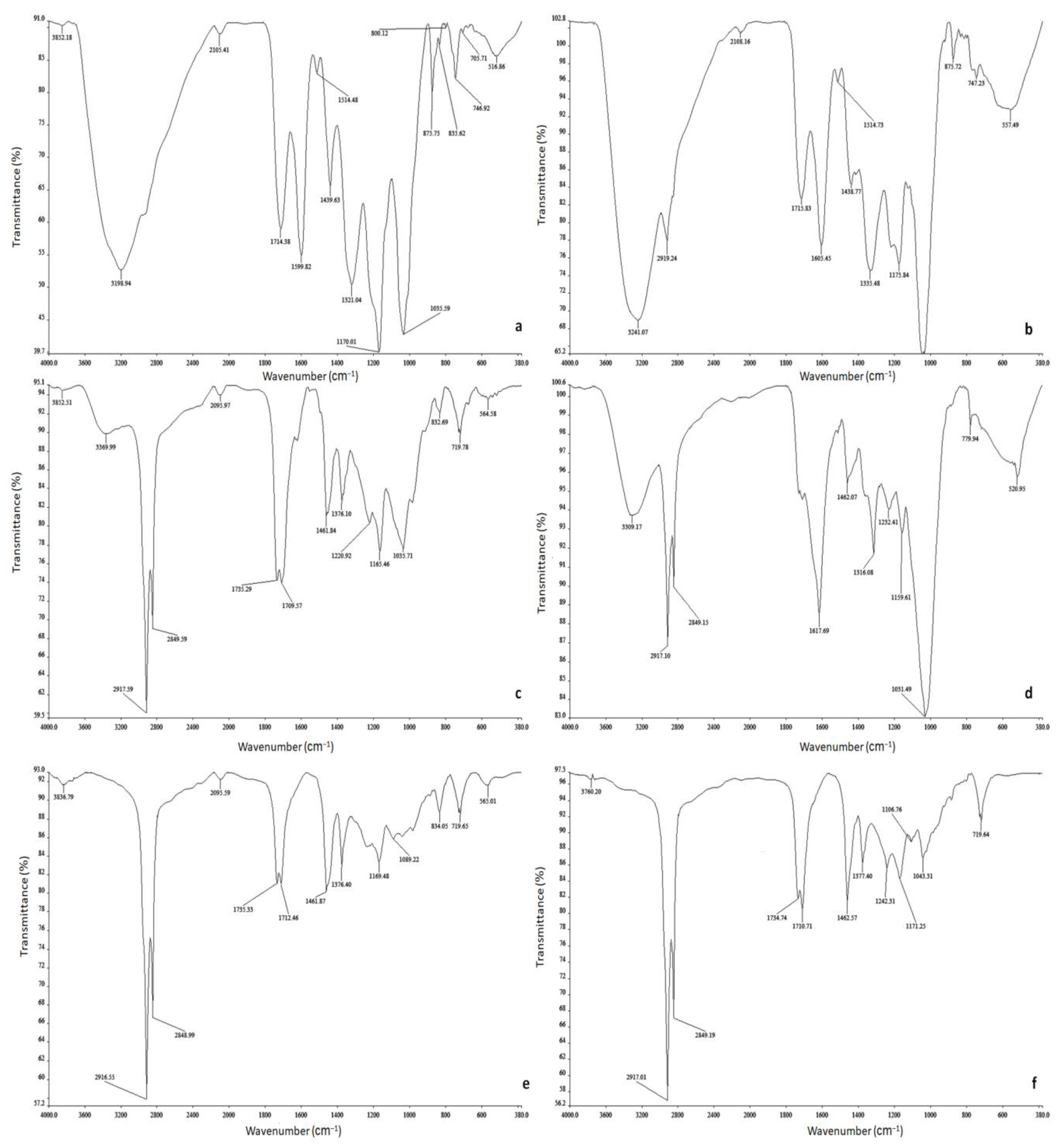
The FT–IR analysis was conducted to characterize the functional groups in the D. villosa leaf and stem-bark extracts that are responsible for the biochemical and molecular potential of the plants using a spectrophotometer (Perkin Elmer 100 FT–IR, Waltham, MA, USA). The spectra were examined and imaged at a range of 4000–400 cm−1 with a resolution of 4 cm−1. KBr was used as a standard to analyze the samples by dispersing them uniformly in a matrix of dry KBr.

2.7. Sensitivity Test of Leaves and Stem-Bark Extracts of D. villosa on Test Microorganisms

The antimicrobial activities of the methanolic, chloroformic, and hexanolic leaf and stem-bark extracts of D. villosa were investigated using the agar-well diffusion method. The discs were prepared using a Whatman No. 1 and obtained by punching and placing them in vials, which were further sterilized in an oven at 150 °C for 15 min. The test microorganisms were reactivated on the nutrient agar broth and further incubated at 37 °C overnight. The test microorganisms were also standardized at an optical density of 0.1 at 625 nm using a UV-vis spectrophotometer (Agilent Cary 60 Spectr., Santa Clara, CA, USA). Following this, 0.2 mL of the standardized test culture was added to 20 mL of molten Muller–Hilton agar and homogenized. This was then poured into sterile plates and allowed to solidify. Thereafter, the wells were aseptically bored into the inoculated Muller–Hilton agar plates using a 6mm sterile cork borer. The test solutions of extracts (100 µL) at graded concentrations of 0.625, 1.25, 2.5, 5, and 10 mg mL−1 already dissolved in 10% DMSO were then placed into each designated well on the plate, ensuring that there was no spillage. A standardized amount (10 µg·mL−1) of gentamicin and streptomycin was introduced into the residual wells on the plate, which was used as a control for the Gram-negative and Gram-positive bacteria strains, respectively. The plates were then allowed to diffuse at room temperature for 1hr in the medium and eventually incubated at 37 °C for 16–18 h in an incubator. The clear zones of inhibition were noted, measured, and recorded in millimeters. Each extract’s activity was tested in triplicate.

2.8. Antioxidant Activity

2.8.1. DPPH Scavenging Activity

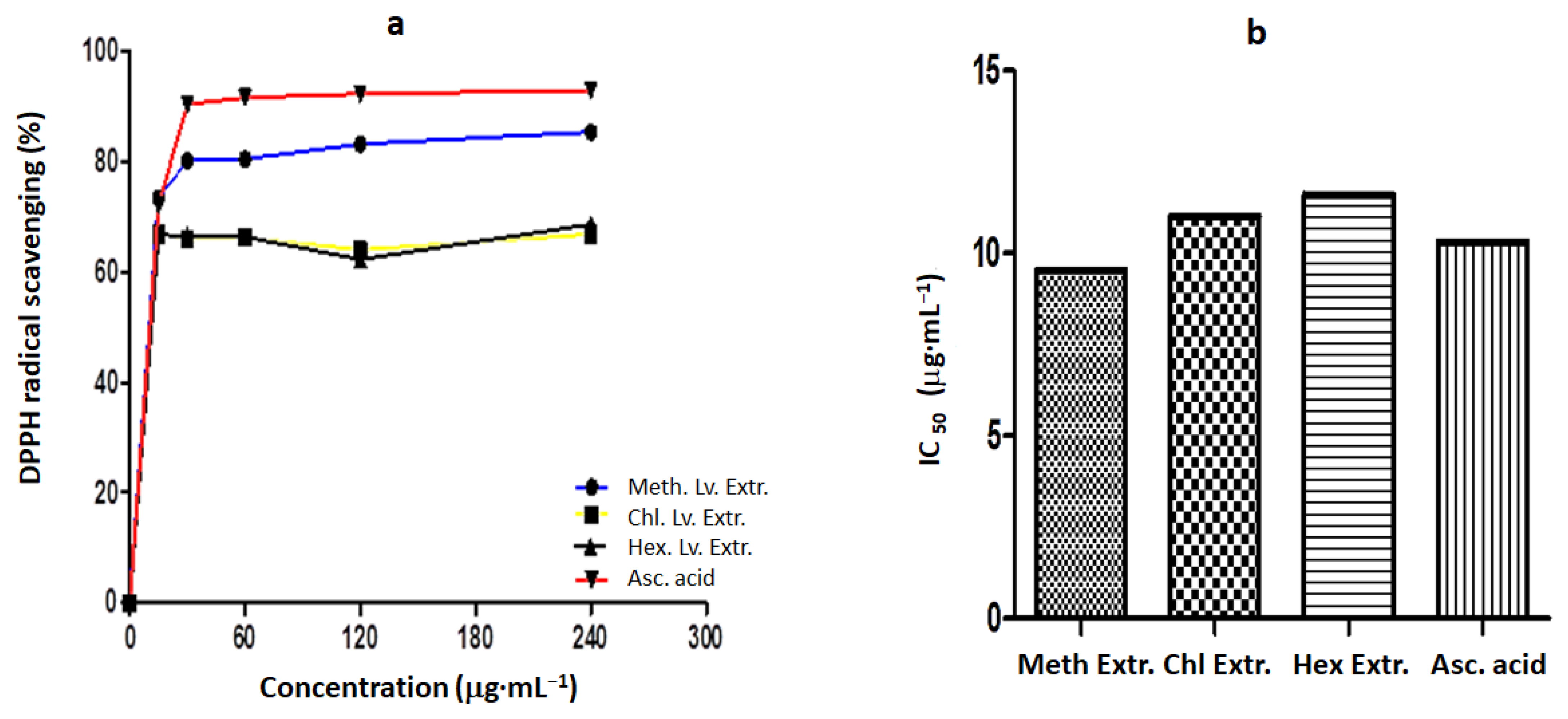
The free radical scavenging activity of the extracts was determined by DPPH (1, 1-diphenyl-2-picrylhydrazyl) radicals [25]. An aliquot of 3 mL of 0.004% DPPH solution in 95% ethanol and 0.1 mL of each plant extract at concentrations of 15, 30, 60, 120, and 240 μg·mL−1 were mixed. The mixture was thoroughly mixed and allowed to sit for 30 min at room temperature. The procedure was repeated for the ascorbic acid (control). The decolorization of DPPH was ascertained by quantifying the absorbance at 517 nm. The control was prepared using 0.1 mL of each constituent and double-distilled water as a replacement for the plant extract or ascorbic acid. The percentage expression of DPPH radical scavenging activity by the plant extracts was calculated as thus: A b s o r b a n c e   o f   C o n t r o l A b s o r b a n c e   o f   T e s t   s a m p l e s A b s o r b a n c e   o f   C o n t r o l × 100 .

2.8.2. Ferric-Reducing Antioxidant Potential (FRAP) Assay

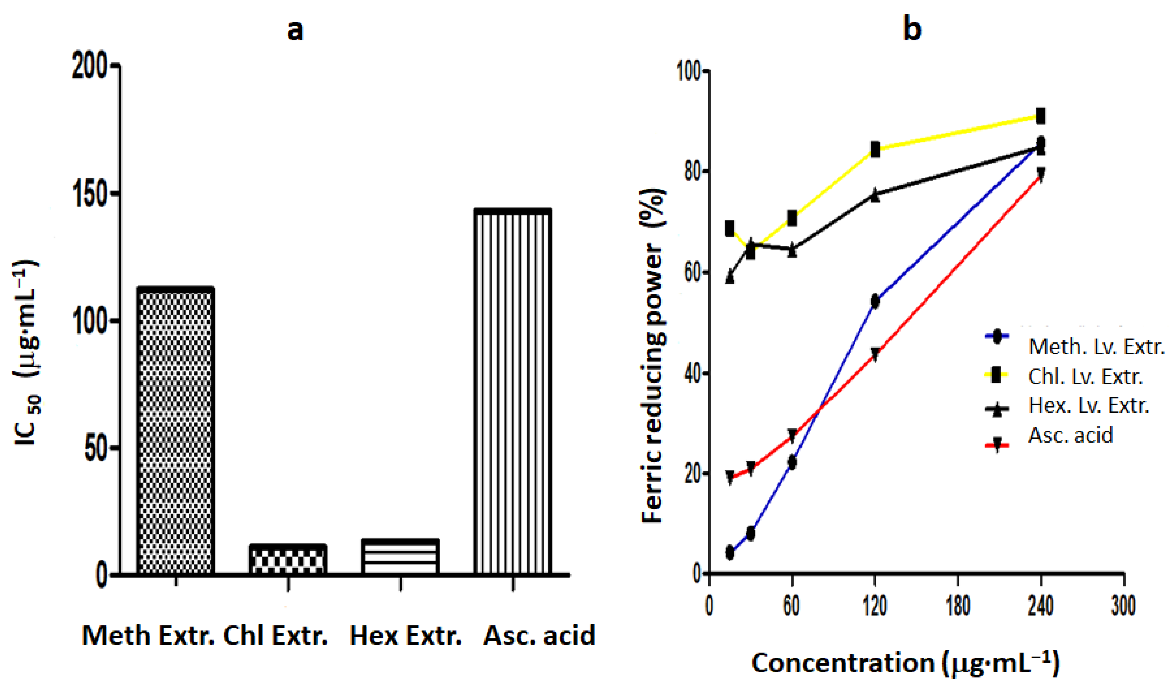
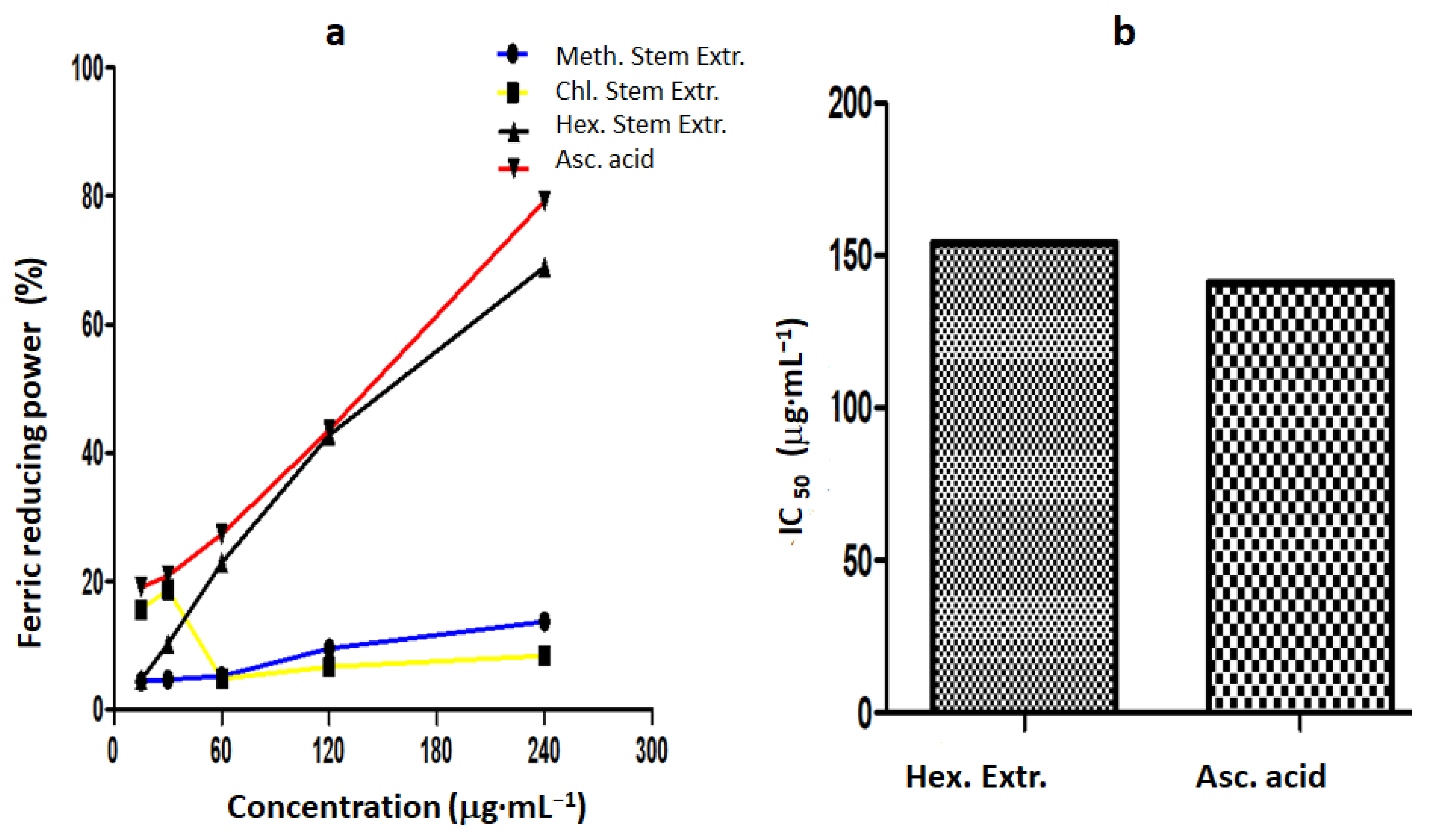
The FRAP assay was conducted as previously described by Juntachote and Berghofer [26]. Multiple graded concentrations of the extract (15, 30, 60, 120, and 240 μg·mL−1) of the extract (1 mL each) were added to 2.5 mL of phosphate buffer (0.2M, pH 6) and 2.5 mL of potassium ferricyanide (1% w/v). The resulting admixture was incubated for 20 min at a temperature of 50 °C. Then, 2.5 mL of 10% trichloroacetic acid was added to the mixture. A quantity of 2.5 mL from each mixture was further diluted twice with deionized water, and 0.5 mL of 0.1% (w/v) FeCl3 was added. The absorbance was later determined at 700 nm after 30 min. The positive control used was ascorbic acid. The half-maximal inhibitory concentration (IC50) was calculated from the graph of absorbance against the concentrations of the extracts. The results were generated as thus:
Scavenging   Effect   ( % ) = [ A b s o r b a n c e   o f   C o n t r o l A b s o r b a n c e   o f   s a m p l e   A b s o r b a n c e   o f   c o n t r o l   ]   ×   100 .

2.8.3. Total Phenolic Content (TPC)

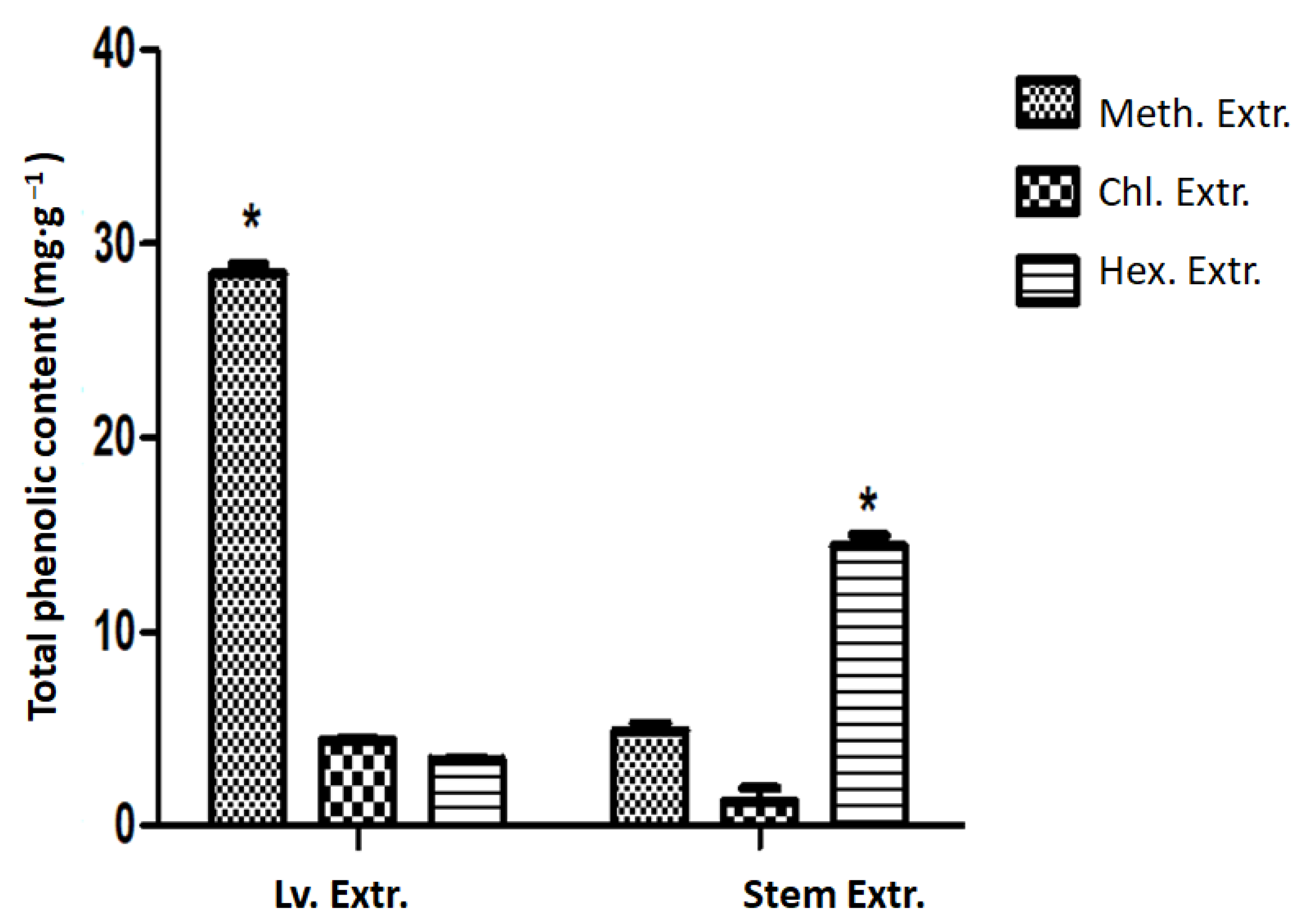
TPC was investigated using the Folin–Ciocalteu colorimetric method with slight adjustments [27]. A volume of 0.1 mL of each extract was mixed thoroughly with 3 mL of distilled water and 0.5 mL of Folin–Ciocalteu reagent was also added to each sample extract. The mixture was allowed to sit at room temperature for 3 min and 2 mL of 20% sodium carbonate was added. The mixture was further incubated for 30 min at room temperature. The total phenolic content was measured at 725 nm using a spectrophotometer. Gallic acid was used as the positive control. The total phenol values were expressed as mg of gallic acid equivalents (GAE)/g of dry sample extracts.

2.9. Test Micro-Organisms

The bacteria strains used were identified as American-type collection culture strains obtained from the School of Pharmacy and Pharmacological Sciences, University of KwaZulu-Natal. Three Gram-negative bacteria, namely E. coli (ATCC 35218), P. aeruginosa (ATCC 27853), and K. pneumoniae (ATCC 700603), and 2 Gram-positive bacteria, S. aureus (ATCC 33591) and MRSA (ATCC 43300), were used in this study.

2.9.1. Determination of Minimum Inhibitory Concentration (MIC) of the D. villosa Extracts on Microorganisms

The MIC of the extracts was determined using the method described by Akinpelu and Onakoya [28]. The extract was diluted in two folds and a portion of 2 mL of the extracts at different concentrations was mixed with 18 mL of sterilized molten Mueller–Hilton agar in order to achieve a final concentration regime of 0.313 mg·mL−1 to 0.01 mg·mL−1. The resulting medium was transferred into sterile Petri dishes and allowed to set. The dry surface of the medium was achieved before streaking with 18 h-old standardized bacterial cultures. The plates were then incubated at 37 °C for 48 h and later scrutinized for either the absence or presence of growth. The MIC was taken as the lowest concentration that prevents bacterial growth.

2.9.2. Determination of Minimum Bactericidal Concentration (MBC) of the Extracts on Test Microorganisms

The minimum bactericidal concentration of the extract was determined according to Ferrazzano et al. [29]. The inoculum was taken on the line of streaks without visible growth in the MIC assay and subcultured on freshly prepared nutrient agar and incubated at 37 °C for 48 h. The plates were later scrutinized for either the absence or presence of growth. The lowest concentration of the extracts with no indication of any growth on a new set of plates was considered as the MBC of the extracts.

2.10. Statistical Analysis

The results were expressed as means ± SE. Statistical analysis was performed using Graph Pad Prism 5 (Graph Pad Software Inc., San Diego, CA, USA). All outcomes were compared with the control using both one-way and two-way analysis of variance (ANOVA) followed by a Bonferroni post hoc analysis. Effects were considered statistically significant at p value ≤ 0.05.

3. Results

3.1. Phytochemical Analysis

The percentage extract yield of D. villosa leaves and stem bark are represented in Table 1. It was found that the methanol extract in the leaves produced the highest yield of phytochemicals, at about 10.8%, whereas the chloroform and hexane extracts in the leaves yielded 8.4% and 7.1%, respectively. Similarly, the methanol extract in the stem bark produced a yield of 9.2%, whereas the chloroform and hexane extracts yielded 7.9% and 10.3%, respectively.
The results of the qualitative phytochemical assessment of the D. villosa leaves and stem-bark extracts are shown in Table 2. Phytochemical screening showed the presence of alkaloids, terpenoids, and phenols in the methanol extracts of D. villosa leaves. In addition, the presence of flavonoids was further observed in the methanol extracts of D. villosa stem bark. In addition, terpenoids, flavonoids, and phenols were shown to be present in both the chloroformic and hexanolic stem extracts of D. villosa. All the extracts from the plant’s leaves and stem cumulatively contained steroids, alkaloids, carbohydrates, and saponins.

3.2. Gas Chromatography–Mass Spectrometry (GC–MS) Analysis

The GC–MS analysis of both the leaves and stem of D. villosa showed the presence of certain bio-active compounds such as n-hexadecanoic acid, phytol, palmitoleic acid, eicosanoic acid and its derivatives, ascorbic acid and its derivatives, etc. (Table 3, Table 4, Table 5, Table 6, Table 7 and Table 8). The retention time (RT), compound name, molecular formula, and peak height (%) are presented in Table 3, Table 4, Table 5, Table 6, Table 7 and Table 8.

3.3. Antimicrobial Effects

The zone of inhibition of D. villosa leaf and stem-bark extracts at 10 mg·mL−1 was significantly higher compared to the control (p < 0.05), except for the activity of the methanol stem-bark extract against E. coli, where the zone of inhibition at 10 mg·mL−1 was lower compared to the control (Table 9). At a concentration of 5 mg·mL−1, the zones of inhibition of the methanol leaf extract as observed against S. aureus, methicillin-resistant S. aureus, and K. pneumoniae were found to be higher compared to the control (streptomycin and gentamicin) (Table 9). The zone of inhibition of the methanol stem-bark extract at 5 mg·mL−1 as observed against S. aureus was significantly higher compared to the control (p < 0.05).
There was an effect of the chloroform extract of D. villosa leaves on S. aureus. The zone of inhibition of the chloroform leaf extract against K. pneumoniae was found to be higher compared to the control (gentamicin) at a concentration of 10 mg·mL−1 (Table 9). At all concentrations, the zone of inhibition of the hexane leaf extract against K. pneumoniae was lower compared to the control (gentamicin). Meanwhile, the zone of inhibition against K. pneumoniae at a concentration of 10 mg·mL−1 was higher compared to the control (Table 9). The zones of inhibition at all other doses were lower compared to the control.
Following the serial dilution of 0.625 mg·mL−1 in the lowest concentration of the extract, all the bacterial strains were observed to be resistant to the activities of the methanolic leaf extract of D. villosa at a concentration of 0.01 mg·mL−1 (Table 10). In addition, the same trend was observed with the methanol stem-bark extract of the plant at 0.01 mg·mL−1. It was also observed that the bacterial strains were sparred as there was no reactivity at a concentration of 0.01 mg·mL−1.

3.4. DPPH Radical Scavenging Activity

The free radical scavenging activity and IC50 values of the leaf and stem-bark extracts are summarized in Figure 1 and Figure 2, respectively. The simultaneous occurrence of a greater percentage of radical scavenging activity and lower IC50 values indicated excellent antioxidant activity. Out of all the different leaf extracts, the methanol leaf extract showed higher scavenging activity (85.29%) compared to the other extracts, whereas the ascorbic acid at the same concentration showed 92.82%, which are somewhat close to each other (Figure 1a). In addition, the methanol leaf extract showed excellent DPPH radical scavenging activity with an IC50 value of 9.53 μg·mL−1 compared to that of the ascorbic acid (10.3 μg·mL−1) (Figure 1b).
In addition, both the chloroform and hexane leaf extracts showed weak antioxidant behavior with higher IC50 values of 10.7 μg·mL−1 and 11.8 μg·mL−1, respectively, compared to that of the ascorbic acid. The methanol and hexane stem-bark extracts also showed high scavenging activity at 85.76% and 87.22%, respectively, but these were not as high as the ascorbic acid (92.82%) (Figure 2a). The IC50 of both the methanol (9.53 μg·mL−1) and hexane (9.53 μg·mL−1) stem-bark extracts were lower compared to the ascorbic acid (10.0 μg·mL−1) (Figure 2b). Meanwhile, the chloroform stem-bark extract indicated a higher IC50 compared to the ascorbic acid.

3.5. Ferric-Reducing Antioxidant Potential

The ferric-reducing potential and IC50 values of the leaf and stem-bark extracts of D. villosa are summarized in Figure 3 and Figure 4, respectively. Among the different leaf extracts, the hexanic leaf extract showed high scavenging activity (85.2%), whereas the ascorbic acid indicated 79.3% at the same concentration (Figure 3a). The chloroform and methanol leaf extracts showed a higher reducing power compared to the ascorbic acid. Similarly, the methanol leaf extract showed ferric-reducing power with an IC50 value of 112 μg·mL−1 compared to that of the ascorbic acid (143 μg·mL−1) (Figure 3b). On the other hand, both the chloroform and hexane leaf extracts showed excellent antioxidant behavior with IC50 values of 11.0 μg·mL−1 and 13.3 μg·mL−1, respectively, compared to that of the ascorbic acid (Figure 3b). The methanol and hexane stem-bark extracts also showed high scavenging activity at 85.8% and 87.22%, respectively, but these were not as high as the ascorbic acid (92.8%) (Figure 4a). The IC50 of both methanol and chloroform stem extracts was lower compared to that of the ascorbic acid (141.0 μg·mL−1) (Figure 4b). Similarly, the hexane stem-bark extract further indicated a higher IC50 compared to the ascorbic acid.

3.6. Total Phenolic Content

The TPC in the leaves and stem bark of D. villosa was estimated and analyzed. One-way analysis of variance showed that there was a significant difference in the total phenol contents in D. villosa leaves and stem bark F(2, 6) = 225.8, p ≤ 0.001. The highest TPC in D. villosa leaves was found in the methanol extract (28.45 ± 0.50) mg gallic acid equivalent per gram of dry weight. Meanwhile, the highest TPC in the stem bark was found in the hexane extract (14.40 ± 0.58) mg gallic acid equivalent per gram of dry weight (Figure 5).

3.7. Fourier Transform Infrared (FT–IR) Spectral Analysis

The FT–IR spectra of the D. villosa leaf and stem-bark extracts using methanol, chloroform, and hexane as the media for extraction are presented in Figure 6. The spectra analysis revealed the vibrational frequencies of the various functional groups observed to be present in the crude extracts. Absorption peaks with a broad range of 2500–3300 cm−1 were characteristic of the hydroxyl (O-H) group, particularly from carboxylic acid (Figure 6a,b). The peaks around 1650–1750 cm−1 were assigned to the carbonyl (C=O) group. The stretching peaks at 1599 and 1605 cm−1 occurred due to the presence of (C≡N) nitrile. The C-H (hydrocarbon) stretches appeared at 2927 cm−1 (Figure 6c,d). The peak at 2849 cm−1 was meant to further characterize the (C-H) stretching vibration. The strong peaks absorbed at 1735 and 1617 cm−1 (Figure 6c,d) were the (C=O) stretches in the aldehydes and ketones. The peaks at 3369 and 3309 cm−1 (Figure 6c,d) occurred due to the presence of the (O-H) functional group. In addition, the strong peaks at 2916 and 2848 cm−1, as well as at 2917 and 2849 cm−1 (Figure 6e,f), were due to the presence of the (C-H) functional group.

4. Discussion

Several studies have reported that herbal antioxidants act against free radicals [30,31,32]. The presence of secondary metabolites such as terpenoids, alkaloids, flavonoids, and phenolic compounds have been implicated as antioxidant factors in different plant materials [33]. It is interesting to note that the methanol extract of D. villosa (both leaves and stem bark) showed the presence of flavonoids and alkaloids. Following the qualitative phytochemical analysis, the methanol leaf extract presented with a color intensity to indicate the presence of terpenoids, flavonoids, and even phenolic compounds. It is without a doubt that the confirmed compounds (terpenoids, flavonoids, and phenols) could provide the justifiable underlying factors for the antioxidant activity of D. villosa. This confirms the findings of Echeverría et al. [34] that showed that the antioxidant activity of natural flavonoids of Chilean flora (Flora of Chile) is a result of the embedded hydroxyl group for the oxygenation-substitution pattern. The presence of a hydroxyl group in the D. villosa leaves and stem bark supports hydroxylation as the plant’s mechanism for exhibiting its antioxidant function and thereby conferring stability to free radicals. In addition, this study revealed the presence of alkaloids in the methanol leaf and stem-bark extracts only. Many alkaloids are potent antioxidants and are used for the treatment and/or management of skin cancer [35]. The detection of alkaloids in the plant extracts is consistent with Dangoggo et al. [36], who revealed that the aqueous extract of Diospyros mespiliformis (ebony diospiros) leaves was quite rich in alkaloids and may further be responsible for the effective inhibitory effect on DPPH, thereby helping in the detoxification of the generated radical oxygen species.
The GC–MS analysis indicted the existence of a number of phytocompounds such as n-hexadecanoic acid; phytol; palmitoleic acid; oxalic acid; 3,7,11,15-Tetramethyl-2-hexadecen-1-ol; acetate; alpha-cadinol; tau-Muurolol; eisosane; and vitamin E. The major compounds found in the methanol leaf extracts were 11, 14, 17-eicosatrienoic acid, methyl ester (15.19%), pentadecanoic acid, and methyl ester (29.89%), whereas in the methanol stem extracts, n-hexadecanoic acid (25.93%), hexadecanoic methyl ester (25.93%), and methyl 10-trans, 12-cis octadecadienoate (10.79%) were present. In the chloroform leaf extract, the major compounds were n-hexadecanoic acid (20.12%) and cis, cis, cis-7, 10, 13-Hexadecatrienal (14.36%), whereas phytol acetate (48.61%) and 3,7,11,15-tetramethyl-2-hexadecen-1-ol (15.54%) were found in the chloroform stem extract of D. villosa. Similarly, oxalic acid, cyclohexyl ethylester (22.48%), and n-hexadecanoic acid (11.77%) were found in the hexane leaf extract, whereas in the hexane stem extract, oxalic acid, cyclohexylpropyl ester (21.93%), n-hexadecanoic acid, and ethyl ester (13.98%) were found in high proportions. The high concentrations of valeric acid in the hexane extracts of the leaves and stem bark may be associated with the antioxidant activity of the extracts. These results support the findings of Vishwakarma et al. [37], where valeric acid isolated from Valeriana wallichii was scientifically proven to have anti-inflammatory properties by reducing lipid peroxidation and restoring glutathione levels in intracerebrovascular streptozotocin-induced neurodegeneration, and further suggests that it could be used in the management of inflammatory diseases.
Phytol is not just a diterpene compound but can also act as an anti-inflammatory, anticancer, antimicrobial, and diuretic. Phytol acetate as found in the D. villosa extracts was revealed to be in high concentrations and could be used as a novel class of pharmaceuticals as a therapeutic measure for rheumatoid arthritis and especially for chronic inflammatory diseases. This is further corroborated by Ogunlesi et al. [38], where phytol increased oxidative burst in vivo and further corrected the effect of the genetic polymorphism in the translational model of arthritis. Phytol may further be considered a novel class of drugs for treating chronic inflammatory diseases. In fact, phytol as an acyclic diterpene alcohol could be considered a precursor for the industrial synthesis of vitamin E [38]. Among the found compounds, n-hexadecanoic acid, hexadecanoic acid, and palmitoleic acid have antioxidant, hypocholesterolemic, nematicide, pesticide, and lubricating properties [39]. In addition, n-hexadecanoic acid and ethyl ester have antitumor, antifungal, and antibacterial properties. Hexadecanoic acid as found in D. villosa possesses antioxidant and haemolytic properties and is also an effective pesticide.
Similarly, Rathee et al. [40] reported that the methanolic extract of Mentha longifolia showed remarkable antioxidant activity via the reactive oxygen species scavenging efficacy and lipid peroxidation inhibition. In this study, the methanol extracts of D. villosa (both leaves and stem bark) showed notable antioxidant activity in comparison with the reference drug (ascorbic acid). This activity could further be associated with the presence of alkaloids and flavonoids as well as the phenolic content. The observed lower IC50 values of these extracts support the relevance of D. villosa leaves and stem bark as a potential organic source of antioxidants and hence they can be used for the prevention of free-radical-mediated diseases. Furthermore, the results of the DPPH radical scavenging ability showed that the methanol leaf and stem-bark extracts can prevent radical-induced oxidative damage. This is reflected in the phenol content in the methanol leaf and stem-bark extracts. Accordingly, Saeed et al. [41] established a correlation between the health benefits of polyphenolic-rich plants and their antioxidant properties, and the possible mechanism responsible for the phenolic activity could be the redox properties of its hydroxyl group.
The functional groups in any bio-organic compound influence the biological activities of the compound. The influence is a result of the contribution of the embedded functional groups to the inherent properties of the compounds such as solubility, stereochemistry, partition coefficient, acid–base properties, etc. All these proficiencies are believed to induce the metabolic extraction, absorption, distribution, and toxicity of bioactive molecules [42]. Therefore, the analysis of the functional group showed that it performs a dynamic function in identifying the physicochemical properties of the extracts. The detection of the functional groups, therefore, helped to assess the structure–function relationship of the bio-organic compound. In this study, the FT–IR spectral analysis of the leaf and stem-bark extracts of D. villosa showed the presence of phytochemicals carrying a hydrogen-bonded OH functional group. The functionality of most phenolic compounds such as tannins and flavonoids owes to the presence of a hydroxyl functional group [43].
Although the mechanism of antibacterial activity could not be ascertained in this study, it was however noted that higher zones of inhibition were produced by the graded doses of the methanol leaf extract compared to those of the stem bark. It is not a mere coincidence that the chloroform and hexane extracts showed antibacterial activity against K. pneumonia only. The presence of alkaloids in the methanol extracts may be identified as an additional key factor for the antibacterial activity of the D. villosa plant. This is supported by Bai et al. [39]’s evidence and confirms the antibacterial activity of the alkaloids as well as the mechanism of action through intercalation with bacterial DNA. There has been motivation and justification for the production of new antimicrobial agents to treat infections [44]. The newest trend shows that plant-based antimicrobial agents have high medicinal efficacy since they pose no hazardous threats to human life [45]. The fact that plant extracts produce zones of inhibition against different bacteria strains indicates their antimicrobial activity and further confirms their use as anti-infection agents. In addition, the production of zones of inhibition against both Gram-negative and Gram-positive bacteria shows their applicability for a wide spectrum of activity. The methanol extract of D. villosa leaves further indicates a higher zone of inhibition against S. aureus, P. aeruginosa, and K. pneumoniae compared to conventional antibiotics of high concentrations. Although the mechanisms of action of D. villosa are yet to be ascertained, there is no doubt that the chemical contents of the plants, such as phenols, flavonoids, and alkaloids, are much more likely to be responsible for antimicrobial activities. This is similar to Linuma et al. [46], where the flavonoid contents of the extract were revealed to be a typical phytochemical responsible for microbial inhibition. In addition, the methanol stem-bark extract of D. villosa further showed a reaction against these strains but it was not as high as the control drug. The observed reduction in the degree of inhibitory activity could be ascribed to the lower concentration of phenolic content in the stem-bark extract of D. villosa. Vaquero et al. [47] indicated that phenolic compounds possess high antibacterial effects. Similarly, Majhenič et al. [48] found that methanol extracts of Paullinia cupana (guarana) seed showed high antibacterial activity due to their high phenolic contents [48]. Hence, it can be said that D. villosa possesses exceptional phytochemicals that account for bacterial metabolism inhibition.

5. Conclusions

The present study indicated that the leaf and stem-bark extracts of D. villosa displayed the occurrence of flavonoids, alkaloids, phenols, and even terpenoids. The leaves of the plant further revealed strong antioxidant activity owing to the high concentration of total phenolic content as well as the absorption peaks with a wide range for the hydroxyl group. In addition, the plant showed strong antimicrobial activities against Gram-negative and Gram-positive bacteria strains with a minimum inhibitory concentration of 0.01 mg·mL−1. Therefore, this study suggests that the leaf and stem-bark extracts of D. villosa are a notable source of natural antioxidant and antimicrobial agents. It is expected that this study could lead to the design of bio-compounds that could be used to investigate novel and efficacious antioxidants as well as the antimicrobial agents of plant origin. Further research is needed to isolate the active molecules from the crude extract and also to evaluate in detail the in vivo biological activities of these isolated compounds.

Author Contributions

Conceptualization and methodology, O.T.A. and Y.N.; investigation, O.T.A., Y.N. and J.L.; formal analysis and data curation, O.T.A., Y.N. and J.L.; writing—original draft preparation, O.T.A., Y.N., J.L. and V.S.; writing—review and editing, T.S.A., V.S., Y.H.D. and A.N.E.-B.; validation and visualization, T.S.A., V.S., Y.H.D. and A.N.E.-B.; Supervision, Y.N. All authors have read and agreed to the published version of the manuscript.

Funding

The authors acknowledge the Researchers Supporting Project number (RSP-2021/375), King Saud University, Riyadh, Saudi Arabia.

Institutional Review Board Statement

Not applicable.

Informed Consent Statement

Not applicable.

Data Availability Statement

All data are presented within the article.

Acknowledgments

The authors acknowledge the Researchers Supporting Project number (RSP-2021/375), King Saud University, Riyadh, Saudi Arabia. The authors are thankful to the TWAS/National Research Foundation (NRF) for their financial support and the University of Kwa-Zulu-Natal, South Africa, for providing the research facilities for this work.

Conflicts of Interest

The authors declare no conflict of interest.

References

  1. Sugamura, K.; Keaney, J.F., Jr. Reactive oxygen species in cardiovascular disease. Free Radic. Biol. Med. 2011, 51, 978–992. [Google Scholar] [CrossRef] [PubMed] [Green Version]
  2. White, B.L.; Howard, L.R.; Prior, R.L. Polyphenolic composition and antioxidant capacity of extruded cranberry pomace. J. Agric. Food Chem. 2009, 58, 4037–4042. [Google Scholar] [CrossRef] [PubMed]
  3. Ali, M.; Cheng, Z.; Ahmad, H.; Hayat, S. Reactive oxygen species (ROS) as defenses against a broad range of plant fungal infections and case study on ROS employed by crops against Verticillium dahliae wilts. J. Plant Interact. 2018, 13, 353–363. [Google Scholar] [CrossRef] [Green Version]
  4. Das, S.K.; Prusty, A.; Samantaray, D.; Hasan, M.; Jena, S.; Patra, J.K.; Samanta, L.; Thatoi, H. Effect of Xylocarpus granatum Bark Extract on amelioration of Hyperglycaemia and oxidative stress associated complications in STZ-induced diabetic mice. Evid.-Based Complement. Altern. Med. 2019, 2019, 8493190. [Google Scholar] [CrossRef] [PubMed] [Green Version]
  5. Pandey, K.B.; Rizvi, S.I. Plant polyphenols as dietary antioxidants in human health and disease. Oxidative Med. Cell. Longev. 2009, 2, 270–278. [Google Scholar] [CrossRef] [PubMed] [Green Version]
  6. Balasundram, N.; Sundram, K.; Samman, S. Phenolic compounds in plants and agri-industrial by-products: Antioxidant activity, occurrence, and potential uses. Food Chem. 2006, 99, 191–203. [Google Scholar] [CrossRef]
  7. Jagani, S.; Chelikani, R.; Kim, D.S. Effects of phenol and natural phenolic compounds on biofilm formation by Pseudomonas aeruginosa. Biofouling 2009, 25, 321–324. [Google Scholar] [CrossRef] [PubMed]
  8. Borras-Linares, I.; Stojanovic, Z.; Quirantes-Pine, R.; Arraez-Roman, D.; Švarc-Gajic, J.; Fernadez-Gutierrez, A.; Segura-Carretero, A. Rosmarinus officinalis leaves as a natural source of bioactive compounds. Int. J. Mol. Sci. 2014, 15, 20585–20606. [Google Scholar] [CrossRef] [PubMed] [Green Version]
  9. Rasheed, K.; Zhang, X.J.; Luo, M.T.; Zheng, Y.T. Anti-HIV-1 activity of phenolic compounds isolated from Diospyros lotus fruits. Phytopharmacology 2012, 3, 199–207. [Google Scholar]
  10. Rauf, A.; Uddin, G.; Patel, S.; Khan, A.; Halim, S.A.; Bawazeer, S.; Ahmad, K.; Muhammad, N.; Mubarak, M.S. Diospyros, an under-utilized, multi-purpose plant genus: A review. Biomed. Pharmacother. 2017, 91, 714–730. [Google Scholar] [CrossRef]
  11. Kumar, S.; Abedin, M.M.; Singh, A.K.; Das, S. Role of Phenolic Compounds in Plant-Defensive Mechanisms. In Plant Phenolics in Sustainable Agriculture; Springer: Berlin/Heidelberg, Germany, 2020; pp. 517–532. [Google Scholar]
  12. Gorniak, I.; Bartoszewski, R.; Kroliczewski, J. Comprehensive review of antimicrobial activities of plant flavonoids. Phytochem. Rev. 2019, 18, 241–272. [Google Scholar] [CrossRef]
  13. Singh, A.K.; Singla, P. Root Phenolics Profile Modulates Microbial Ecology of Rhizosphere. In Plant Phenolics in Sustainable Agriculture; Springer: Singapore, 2020; pp. 555–578. [Google Scholar]
  14. Cirera, J.; Da Silva, G.; Serrano, R.; Gomes, E.; Duarte, A.; Silva, O. Antimicrobial activity of Diospyros villosa root. Planta Med. 2010, 76, P454. [Google Scholar] [CrossRef]
  15. Brando, R.; Miliwebsky, E.; Bentancor, L.; Deza, N.; Baschkier, A.; Ramos, M.; Fernandez, G.; Meiss, R.; Rivas, M.; Palermo, M. Renal damage and death in weaned mice after oral infection with Shiga toxin 2-producing Escherichia coli strains. Clin. Exp. Immunol. 2008, 153, 297–306. [Google Scholar] [CrossRef]
  16. Kitada, K.; De Toledo, A.; Oho, T. Increase in detectable opportunistic bacteria in the oral cavity of orthodontic patients. Int. J. Dent. Hyg. 2009, 7, 121–125. [Google Scholar] [CrossRef] [PubMed]
  17. Flynn, T.R. Evidence-Based Principles of Antibiotic Therapy. In Evidence-Based Oral Surgery; Springer International Publishing: Cham, Switzerland, 2019. [Google Scholar] [CrossRef]
  18. Vila, T.; Sultan, A.S.; Montelongo-Jauregui, D.; Jabra-Rizk, M.A. Oral candidiasis: A disease of opportunity. J. Fungi 2020, 6, 15. [Google Scholar] [CrossRef] [PubMed] [Green Version]
  19. Vellappally, S.; Divakar, D.D.; Al Kheraif, A.A.; Ramakrishnaiah, R.; Alqahtani, A.; Dalati, M.; Anil, S.; Khan, A.A.; Harikrishna Varma, P. Occurrence of vancomycin-resistant Staphylococcus aureus in the oral cavity of patients with dental caries. Acta Microbiol. Immunol. Hung. 2017, 64, 343–351. [Google Scholar] [CrossRef] [Green Version]
  20. Harborne, J.B. Phenolic compounds. In Phytochemical Methods; Springer International Publishing: Cham, Switzerland, 1973; pp. 33–88. [Google Scholar]
  21. Treare, G.; Evans, W. Pharmacognosy, 17th ed.; Bahive Tinal: London, UK, 1985; p. 149. [Google Scholar]
  22. Sofowora, A. Recent trends in research into African medicinal plants. J. Ethnopharmacol. 1993, 38, 197–208. [Google Scholar] [CrossRef]
  23. Evans, W. Trease and Evans’ Pharmacognosy, Hartcourt Brace and Company; Asia Pvt. Ltd.: Singapore, 1997; pp. 226–227. [Google Scholar]
  24. Adams, R.P. Identification of Essential Oil Components by Gas Chromatography/Mass Spectrometry; Allured Publishing Corporation: Carol Stream, IL, USA, 2007. [Google Scholar]
  25. Barros, L.; Falcao, S.; Baptista, P.; Freire, C.; Vilas-Boas, M.; Ferreira, I.C. Antioxidant activity of Agaricus sp. mushrooms by chemical, biochemical and electrochemical assays. Food Chem. 2008, 111, 61–66. [Google Scholar] [CrossRef]
  26. Juntachote, T.; Berghofer, E. Antioxidative properties and stability of ethanolic extracts of Holy basil and Galangal. Food Chem. 2005, 92, 193–202. [Google Scholar] [CrossRef]
  27. Shao, Y.; Xu, F.; Sun, X.; Bao, J.; Beta, T. Identification and quantification of phenolic acids and anthocyanins as antioxidants in bran, embryo and endosperm of white, red and black rice kernels (Oryza sativa L.). J. Cereal Sci. 2014, 59, 211–218. [Google Scholar] [CrossRef]
  28. Akinpelu, D.; Onakoya, T. Antimicrobial activities of medicinal plants used in folklore remedies in south-western. Afr. J. Biotechnol. 2006, 5, 1078–1081. [Google Scholar]
  29. Ferrazzano, G.F.; Scioscia, E.; Sateriale, D.; Pastore, G.; Colicchio, R.; Pagliuca, C.; Cantile, T.; Alcidi, B.; Coda, M.; Ingenito, A. In vitro antibacterial activity of pomegranate juice and peel extracts on cariogenic bacteria. Biomed. Res. Int. 2017, 2017, 2152749. [Google Scholar] [CrossRef] [Green Version]
  30. Wangia, C.O.; Orwa, J.A.; Muregi, F.W.; Kareru, P.G.; Cheruiyot, K.; Kibet, J. Comparative anti-oxidant activity of aqueous and organic extracts from Kenyan Ruellia lineari-bracteolata and Ruellia bignoniiflora. Eur. J. Med. Plants 2016, 17, 1–7. [Google Scholar] [CrossRef]
  31. Dintcheva, N.T.; Arrigo, R.; Baiamonte, M.; Rizzarelli, P.; Curcurto, G. Concentration-dependent anti-/pro-oxidant activity of natural phenolic compounds in bio-polyesters. Polym. Degrad. Stab. 2017, 142, 21–28. [Google Scholar] [CrossRef]
  32. Tatipamula, V.B.; Killari, K.N.; Ketha, A.; Sastry, V.G. Taxithelium napalense acts against free radicals and diabetes mellitus. Bangladesh J. Pharmacol. 2017, 12, 197–203. [Google Scholar] [CrossRef] [Green Version]
  33. Muchirah, P.N.; Waihenya, R.; Muya, S.; Abubakar, L.; Ozwara, H.; Makhokha, A. Characterization and anti-oxidant activity of Cucurbita maxima Duchesne pulp and seed extracts. J. Phytopharm. 2018, 7, 134–140. [Google Scholar] [CrossRef]
  34. Echeverria, J.; Opazo, J.; Mendoza, L.; Urzua, A.; Wilkens, M. Structure-activity and lipophilicity relationships of selected antibacterial natural flavones and flavanones of Chilean flora. Molecules 2017, 22, 608. [Google Scholar] [CrossRef] [PubMed] [Green Version]
  35. Ahsan, H.; Reagan-Shaw, S.; Eggert, D.M.; Tan, T.C.; Afaq, F.; Mukhtar, H.; Ahmad, N. Protective effect of sanguinarine on ultraviolet B-mediated damages in SKH-1 hairless mouse skin: Implications for prevention of skin cancer. Photochem. Photobiol. 2007, 83, 986–993. [Google Scholar] [CrossRef] [PubMed]
  36. Dangoggo, S.; Hassan, L.; Sadiq, I.; Manga, S. Phytochemical analysis and antibacterial screening of leaves of Diospyros mespiliformis and Ziziphus spina-christi. J. Chem. Eng. 2012, 1, 31–37. [Google Scholar]
  37. Vishwakarma, S.; Goyal, R.; Gupta, V.; Dhar, K.L. GABAergic effect of valeric acid from Valeriana wallichii in amelioration of ICV STZ induced dementia in rats. Rev. Bras. Farmacogn. 2016, 26, 484–489. [Google Scholar] [CrossRef] [Green Version]
  38. Ogunlesi, M.; Okiei, W.; Ofor, E.; Osibote, A.E. Analysis of the essential oil from the dried leaves of Euphorbia hirta Linn (Euphorbiaceae), a potential medication for asthma. Afr. J. Biotechnol. 2009, 8, 7042–7050. [Google Scholar]
  39. Bai, L.P.; Zhao, Z.Z.; Cai, Z.; Jiang, Z.H. DNA-binding affinities and sequence selectivity of quaternary benzophenanthridine alkaloids sanguinarine, chelerythrine, and nitidine. Bioorg. Med. Chem. 2006, 14, 5439–5445. [Google Scholar] [CrossRef]
  40. Rathee, J.S.; Hassarajani, S.A.; Chattopadhyay, S. Antioxidant activity of Mammea longifolia bud extracts. Food Chem. 2006, 99, 436–443. [Google Scholar] [CrossRef]
  41. Saeed, N.; Khan, M.R.; Shabbir, M. Antioxidant activity, total phenolic and total flavonoid contents of whole plant extracts Torilis leptophylla L. BMC Complement. Altern. Med. 2012, 12, 221. [Google Scholar] [CrossRef] [Green Version]
  42. Olivares-Vicente, M.; Barrajon-Catalan, E.; Herranz-Lopez, M.; Segura-Carretero, A.; Joven, J.; Encinar, J.A.; Micol, V. Plant-derived polyphenols in human health: Biological activity, metabolites and putative molecular targets. Curr. Drug Metab. 2018, 19, 351–369. [Google Scholar] [CrossRef] [Green Version]
  43. Diaz, P.; Jeong, S.C.; Lee, S.; Khoo, C.; Koyyalamudi, S.R. Antioxidant and anti-inflammatory activities of selected medicinal plants and fungi containing phenolic and flavonoid compounds. Chin. Med. 2012, 7, 26. [Google Scholar] [CrossRef] [Green Version]
  44. Fair, R.J.; Tor, Y. Antibiotics and bacterial resistance in the 21st century. Perspect. Med. Chem. 2014, 6, 25–64. [Google Scholar] [CrossRef] [Green Version]
  45. Anand, U.; Jacobo-Herrera, N.; Altemimi, A.; Lakhssassi, N. A comprehensive review on medicinal plants as antimicrobial therapeutics: Potential avenues of biocompatible drug discovery. Metabolites 2019, 9, 258. [Google Scholar] [CrossRef] [Green Version]
  46. Iinuma, M.; Tsuchiya, H.; Sato, M.; Yokoyama, J.; Ohyama, M.; Ohkawa, Y.; Tanaka, T.; Fujiwara, S.; Fujii, T. Flavanones with potent antibacterial activity against methicillin-resistant Staphylococcus aureus. J. Pharm. Pharmacol. 1994, 46, 892–895. [Google Scholar] [CrossRef]
  47. Vaquero, M.R.; Alberto, M.R.; De Nadra, M.M. Antibacterial effect of phenolic compounds from different wines. Food Control 2007, 18, 93–101. [Google Scholar] [CrossRef]
  48. Majhenic, L.; Škerget, M.; Knez, Ž. Antioxidant and antimicrobial activity of guarana seed extracts. Food Chem. 2007, 104, 1258–1268. [Google Scholar] [CrossRef]
Figure 1. Comparison of DPPH radical scavenging (a) IC50 values of D. villosa (%) of D. villosa leaf extracts in DPPH scavenging assay (b). Chl = Chloroform; Hex = Hexane, Meth = Methanol; Lv. = leaves; Extr. = Extract.
Horticulturae 08 00945 g001
Figure 2. Comparison of DPPH radical (a) IC50 values of D. villosa stem scavenging (%) of D. villosa stem-bark extracts in DPPH radical scavenging assay (b). Chl. = Chloroform; Hex = Hexane, Meth. = Methanol; Lv. = leaves; Extr. = Extract; Asc. Acid = Ascorbic acid.
Horticulturae 08 00945 g002
Figure 3. Comparison of ferric-reducing power (a) IC50 values for D. villosa extracts of D. villosa leaf extract in ferric-reducing antioxidant potential (b). Chl. = Chloroform; Hex = Hexane, Meth. = Methanol; Lv. = leaves; Extr. = Extract; Asc. Acid= Ascorbic acid.
Horticulturae 08 00945 g003
Figure 4. Comparison of ferric-reducing power in (%) of D. villosa stem-bark extract (a), IC50 values for D. villosa stem ferric-reducing antioxidant potential (b). Chl. = Chloroform; Hex = Hexane, Meth. = Methanol; Extr. = Extract; Asc. Acid = Ascorbic acid.
Horticulturae 08 00945 g004
Figure 5. Total phenol content of the solvent extracts of both D. villosa leaves and stem bark. F(2, 6) = 225.8, p ≤ 0.0001. * (methanol vs. chloroform), p < 0.05. * (hexane vs. methanol), p < 0.05. Values are expressed as means ± SEM; n = 3/group. Chl. = Chloroform; Hex = Hexane, Meth. = Methanol; Lv. = leaves; Extr. = Extract.
Horticulturae 08 00945 g005
Figure 6. FT–IR spectra of crude plant extracts of D. villosa (a) crude leaf methanol extract; (b) crude stem-bark methanol extract; (c) crude leaf chloroform extract; (d) crude stem-bark chloroform extract; (e) crude leaf hexane extract; (f) crude stem-bark hexane extract at room temperature (24 °C).
Horticulturae 08 00945 g006
Table 1. Yield of extracts of D. villosa leaves and stem bark.
Leaf Extract Yield (%)Stem-Bark Extract Yield (%)
Methanol10.89.2
Chloroform8.47.9
Hexane7.110.3
Table 2. Phytochemical screening of the methanol, chloroform, and hexane extracts of both the leaves and stem bark of D. villosa.
Name of TestLeavesStem Bark
MethanolChloroformHexaneMethanolChloroformHexane
AlkaloidsMayer’s test+++--+++--
Wagner’s test-++-++
ProteinDragendorff-++-++
CarbohydratesBenedict’s+--+--
SteroidsLieberman-Burchard test+++++++
CoumarinsNaOH test++--++--
SaponinFoam test+++++++++
FlavonoidsLead acetate+++--+++-++
TerpenoidsSalowski’s+++--+++++
PhenolsFeCl3 test+++--+++-
+ denotes presence; ++ denotes moderate presence; +++ denotes abundant presence; - denotes absence.
Table 3. Compounds identified in the chloroform extracts of D. villosa leaves by GC–MS spectral analysis.
S/NRTCompound NameMolecular FormulaHeight (%)
1.3.8011-Butene, 3-chloro-2-methyl-C5H9cl1.08
2.4.2541, 1-Dimethyl-3-chloropropanolC5H11clO0.87
3.4.296Pentanoic acid, 2-hydroxy-4-methyl-(S)-C6H12O37.45
4.5.2521-Butene,2,3,3-trimethylC7H140.93
5.10.45411-MethyldodecanolC13H28O1.14
6.10.55711-MethyldodecanolC13H28O1.21
7.10.65611-MethyldodecanolC13H28O1.02
8.11.830VanillinC8H8O30.28
9.12.39710-MethylnonadecaneC20H420.67
10.12.545EicosaneC20H420.53
11.12.680Heptadecanoic acid, heptadecyl esterC34H68O20.25
12.12.869Phenol, 2, 4-bis(1,1-dimethylethyl)-C14H22O5.54
13.12.9431-Dodecanol, 2-hexyl-C18H38O0.87
14.13.015EicosaneC20H421.12
15.13.1251-Dodecanol, 2-hexyl-C18H38O0.98
16.13.2222(4H)-Benzofuranone, 5,6,7,7a-tetrahydro-4,4C11H16O22.52
17.13.585Fumaric acid, ethyl 2-methyl allyl esterC10H14O41.19
18.13.697Decane, 2,3,7-trimethyl-C13H280.33
19.14.193Trans-3(10)-Caren-2-olC10H16O1.04
20.14.340Decane, 2,3,7-trimethyl-C13H280.33
21.14.4292,4a,8,8-Tetramethyldecahydrocyclopropa[d]nC15H261.23
22.14.5122-Butanone, 4-(2,6,6-trimethyl-1-cyclohexen-1C13H22O1.02
23.14.677Oxalic acid, 6-ethyloct-3-yl heptyl esterC19H36O40.39
24.14.7491-{2-[3-(2-Acetyloxiran-2-yl)-1,1-dimethyl propyl]cycloprop-2-enyl} ethanoneC14H20O31.27
25.14.98111-MethyldodecanolC13H28O0.68
26.15.30411-MethyldodecanolC13H28O0.67
27.15.440Tetradecanoic acidC14H28O23.13
28.15.55011-MethyldodecanolC13H28O0.80
29.15.6851-Dodecanol, 2-OctylC20H42O0.52
30.15.8796-Methyl-cyclodec-5-enolC11H20O3.88
31.16.320Phytol, acetateC22H42O24.14
32.16.4102-Pentadecanone,6,10,14-trimethyl-C18H36O3.77
33.16.6793,7,11,15-Tetramethyl-2-hexadecen-1-olC20H40O0.91
34.16.761Pentadecanoic acidC15H30O20.48
35.16.9703,7,11,15-Tetramethyl-2-hexadecen-1-olC20H40O1.41
36.17.5385,9,13-Pentadecatrien-2-one,6,10,14-trimethyC18H30O0.64
37.17.741Hexadecanoic acid,methyl esterC17H34O21.74
38.18.168cis-13-Eicosenoic acidC20H38O20.63
39.18.753n-Hexadecanoic acidC16H32O221.20
40.21.024Eicosanoic acidC20H40O20.40
41.21.795Methyl 10-trans,12-cis-octadecadienoateC19H34O20.45
42.21.991Methyl 8,11,14-heptadecatrienoateC18H30O21.22
43.22.537PhytolC20H40O5.21
44.22.935cis,cis,cis-7,10,13-HexadecatrienalC16H26O14.36
45.24.190Methyl 5,13-docosadienoateC23H42O20.41
Table 4. Compounds found in the chloroform extracts of D. villosa stem by GC–MS analysis.
S/NRTCompound NameMolecular FormulaHeight (%)
1.10.830Octadecanoic acidC18H36O21.48
2.10.925Octadecanoic acidC18H36O23.05
3.10.991Undecanoic acidC11H22O23.04
4.11.050n-Decanoic acidC10H20O22.71
5.13.890Disulfide,di-tert-dodecylC24H50S21.63
6.14.524Phytol,acetateC22H42O20.85
7.16.326Phytol,acetateC22H42O248.61
8.16.6823,7,11,15-Tetramethyl-2-hexadecen-1-olC20H40O10.00
9.16.9763,7,11,15-Tetramethyl-2-hexadecen-1-olC20H40O15.54
10.18.5861-(+)-Ascorbic acid 2,6-dihexadecanoateC38H68O82.79
11.19.546EicosaneC20H423.17
12.20.2336,8a-Epidoxy-4a-methyl-2-oxo-3,4,4a,5,6,7,8C10H14O40.87
13.22.405E,E,Z-1,3,12-Nonadecatriene-5,14-diolC19H34O21.92
14.22.4352-(2-vinyloxy-ethoxy)-cyclohexanolC10H18O31.60
15.22.620PhytolC20H40O2.76
Table 5. Compounds identified in the methanol extracts of D. villosa leaves by GC–MS spectral analysis.
S/NRTCompound NameMolecular FormulaHeight (%)
1.3.971TolueneC7H82.05
2.4.349TetrachloroethyleneC2cl42.33
3.13.240Nonanedioic acid, dimethyl esterC11H20O41.24
4.13.931Ethyl-1-thio-beta-d-glucopyranosideC8H16O5S0.66
5.14.956Methyl-21-methyldocosanoateC24H48O21.12
6.16.131Carda-4,20(22)-dienolide,3-(6-deoxy-3-O-methyl)C30H44O90.68
7.16.185Carda-4,20(22)-dienolide,3-(6-deoxy-3-O-methyl)C30H44O90.67
8.16.325Phytol acetateC22H42O29.80
9.16.430CyclohexadecanoneC16H30O1.10
10.16.6833,7,11,15-Tetramethyl-2-hexadecen-1-olC20H40O4.98
11.16.785Pseudosmilagenin bis(3,5-dinitrobenzoate)C41H48N4O130.52
12.16.9723,7,11,15-Tetramethyl-2-hexadecen-1-olC20H40O7.28
13.17.295Acetamide,N-methyl-N-[4-(3-hydroxypyrrolidinyl)-2-butynylC11H18N2O20.20
14.17.4491-oxaspiro(2,5)octan-4-one,2,2,6-trimethyl-transC10H16O20.65
15.17.669Methylhexadec-9-enoateC17H32O22.60
16.17.756Pentadecanoic acid,14-methyl-,methyl esterC17H34O229.89
17.17.925Methyl 2,3,4-tri-O-acetyl-6,7-di-O-methyl-beta-D-glucoheptopyranosideC16H26O100.26
18.18.635n-Hexadecanoic acidC16H32O27.65
19.18.8251,1,1,4,7,7,7-Heptamethyl-4-vinyltrisilethyleneC13H32Si30.56
20.21.8209,12-Octadecadienoic acid, methyl esterC19H34O26.48
21.22.02311,14,17-Eicosatrienoic acid, methyl esterC21H36O215.19
22.22.964Heptadecanoic acid, 16-methyl-, methyl esterC19H38O21.86
23.23.450Acetamide, N-(4-piperidinylmehyl)-C8H16N2O0.32
24.23.6203-[3-[1-Aziridinyl]propoxy]-2,5-dimethylpyrazC11H17N3O1.01
25.23.6413-Tridecen-1-yne,(Z)-C13H221.01
Table 6. Compounds identified in the methanol extracts of D. villosa stem by GC–MS spectral analysis.
S/NRTCompound NameMolecular FormulaHeight (%)
1.3.965TolueneC7H82.71
2.4.343TetrachloroethyleneC2cl43.17
3.13.4252,2,6,7-Tetramethyl-10-oxatricyclo [4,3,1,0 (1,6)-decan-5-olC13H22O20.34
4.14.339tau-MuurololC15H26O2.41
5.14.462A-CardinolC15H26O3.28
6.15.8582-MethyltetracosaneC25H521.53
7.16.325Phytol, acetateC22H42O20.99
8.17.766Hexadecanoic acid, methyl esterC17H34O226.89
9.17.905Benzenepropanoic acid, 3,5-bis(1,1-dimethylethyl)-4-hydroxy-methyl esterC18H28O33.35
10.18.030Borane, diethyl(1-ethyl-2-methyl-1-butenyl)-(Z)C11H23B0.73
11.18.608n-Hexadecanoic acidC16H32O225.93
12.19.0402,7-dithiatricyclo [4,3,1,0(3,8)] decane,10-bromoC8H11BrS20.40
13.19.146Ethyl 15-methyl-hexadecanoateC19H38O21.51
14.21.825Methyl 10-trans, 12-cis-octadecadienoateC19H34O210.79
15.22.0376-Octadecenoic acid, methyl ester, (Z)-C19H36O28.20
16.22.2236-Octadecenoic acid, methyl ester, (Z)-C19H36O21.47
17.22.957Heptadecanoic acid, 16-methyl-, methyl esterC19H38O22.01
18.23.4691-[3-Aminopropyl]-2[1H]-pyridoneC8H12N2O0.91
19.23.640Bicyclo [3,3,2] decan-9-oneC10H16O2.59
20.23.7101H-Inden-1-one,octahydro-7a-methyl-,transC10H16O0.81
Table 7. Compounds found in the hexane extracts of D. villosa leaves by GC–MS analysis.
S/NRTCompound NameMolecular FormulaHeight (%)
1.3.8363-HexanoneC6H12O0.86
2.3.8852-HexanoneC6H12O1.43
3.3.985Cyclopentanol,1-methyl-C6H12O1.03
4.4.639Cyclopentanone,3-methyl-C6H12O1.42
5.5.4252-Pentanethiol,2-methyl-C5H14S6.21
6.5.678Valeric acid,2-ethoxyethyl esterC9H18O310.80
7.5.924Pentanoic acid,2-propenyl esterC8H14O24.33
8.6.0612-Furanmethanol, tetrahydro-5-methyl-C6H12O23.55
9.6.308Oxalic acid, cyclohexyl ethyl esterC10H16O222.48
10.6.6112-Pentene,4,4-dimethyl-,(E)-C7H142.55
11.6.6502-Pentene,4,4-dimethyl-,(E)-C7H142.36
12.6.740Phosphorus dibromide, cyclohexyl-C6H11Br2P0.86
13.10.4582-Isopropyl-5-methyl-1-heptanolC11H24O0.41
14.10.6592-Isopropyl-5-methyl-1-heptanolC11H24O0.26
15.11.765Octadecanoic acidC18H36O20.29
16.11.845Tetradecanoic acidC14H28O20.41
17.11.948Octadecanoic acidC18H36O21.00
18.12.1595,9-Undecadien-2-one,6,10-dimethyl-,(E)C13H22O20.45
19.12.874Phenol,2,4-bis(1,1-dimethylethyl)-C14H22O1.39
20.13.022Hexadecanoic acid, butyl esterC20H40O21.87
21.13.155Dodecane,4-methyl-C13H280.40
22.13.2222(4H)-Benzofuranone,5,6,7,7a-tetrahydro-4,4C11H16O20.99
23.15.473Tetradecanoic acidC14H28O21.85
24.16.336Phytol,acetateC22H42O1.57
25.16.4322-Pentadecanone,6,10,14-trimethyl-C18H36O4.43
26.16.6903,7,11,15-Tetramethyl-2-hexadecen-1-olC20H40O0.27
27.16.798Pentadecanoic acidC15H30O20.23
28.16.9873,7,11,15-Tetramethyl-2-hexadecen-1-olC20H40O0.42
29.17.5545,9,13-Pentadecatrien-2-one,6,10,14-trimethylC18H30O0.41
30.17.675Oxirane,hexadecyl-C18H36O0.14
31.17.764Pentadecanoic acid,14-methyl-,methyl esterC17H34O21.99
32.18.204Palmitoleic acidC16H30O20.45
33.18.871n-Hexadecanoic acidC16H32O211.77
34.19.2352-Hexadecene,3,7,11,15-tetramethyl-,[R-[RC20H400.17
35.21.093Eicosanoic acidC20H40O20.26
36.21.8469,12-Octadecadienoic acid, methyl esterC19H34O20.70
37.22.052Methyl 8,11,14-heptadecatrienoateC18H30O21.40
38.22.579PhytolC20H40O2.52
39.23.187Butyl 9,12-OctadecadienoateC22H40O20.41
40.24.158cis,cis,cis-7,10,13-HexadecatrienalC16H26O5.65
Table 8. Compounds found in the hexane extracts of D. villosa stem by GC–MS spectral analysis.
S/NRTCompound NameMolecular FormulaHeight (%)
1.3.8812-HexanoneC6H12O1.08
2.3.979Cyclopentanol,1-methyl-C6H12O0.69
3.4.636Cyclopentanone,3-methyl-C6H10O1.07
4.5.4182-Pentanethiol,2-methyl-C6H14S5.96
5.5.669Valeric acid, 2-ethoxy ethyl esterC9H18O310.71
6.5.916Pentanoic acid, 2-propenyl esterC8H14O23.88
7.6.0542-Furanmethanol, tetrahydro-5-methyl-C6H12O23.59
8.6.299Oxalic acid, cyclohexyl propyl esterC11H18O421.93
9.6.6032-Pentene,4,4-dimethyl-,(Z)-C7H142.26
10.6.6442-Pentene,4,4-dimethyl-,(Z)-C7H142.04
11.6.733Cyclohexane,nitroC6H11NO20.77
12.12.679Heptadecane,2,6,10,15-tetramethyl-C21H440.54
13.12.868Phenol,2,4-bis(1,1-dimethyl ethyl)-C14H22O1.30
14.13.3651,6,10-Dodecatrien-3-ol,3,7,11-trimethyl,(E)C15H26O0.68
15.13.737Carophyllene oxideC15H24O1.96
16.14.01412-Oxabicyclo [9,1,0]dodeca-3-7-diene,1,5, 5,8-tetramethyl-[1R,3E,7E,11R]C15H24O0.69
17.14.1226-Isopropenyl-4,8a-dimethyl-1,2,3,5,6,7,8, 8a-octahydronaphthalene-2-olC15H24O0.50
18.14.156CubenolC15H26O0.39
19.14.313α-cadinolC15H26O2.44
20.14.429α-cadinolC15H26O3.86
21.14.684Octadecane,1-chloroC18H37cl0.62
22.14.8716-Isopropenyl-4,8a-dimethyl-1,2,3,5,6,7,8, 8a-octahydronaphthalene-2-olC15H24O0.38
23.15.450Tetradecanoic acidC14H28O21.32
24.16.327Phytol, acetateC22H42O21.73
25.16.4192-Pentadecanone,6,1014-trimethyl-C18H36O2.36
26.16.545Cyclopentadecanone,2-hydroxyC15H28O20.31
27.16.6833,7,11,15-Tetramethyl-2-hexadecen-1-olC20H40O0.36
28.16.780Pentadecanoic acidC15H30O20.53
29.16.9793,7,11,15-Tetramethyl-2-hexadecen-1-olC20H40O0.55
30.17.753Pentadecanoic acid, 14-methyl-,methyl esterC17H34O22.41
31.18.921n-Hexadecanoic acid, ethyl esterC16H32O213.98
32.19.155Hexadecanoic acid, ethyl esterC18H36O20.32
33.19.3152-BromotetradecaneC14H29Br0.20
34.21.079Heptadecanoic acidC17H34O20.44
35.21.8289,12-Octadecadienoic acid, methyl esterC19H34O20.65
36.22.0568-Octadecenoic acid, methyl esterC19H36O20.72
37.22.600PhytolC20H40O0.91
38.22.971MethylstearateC19H38O20.17
39.23.8401,E-11,Z-13-OctadecatrieneC18H322.69
40.24.1256-Octadecenoic acid,(Z)-C18H34O23.04
Table 9. Zone of inhibition (mm) of the graded doses of D. villosa leaf and stem-bark extracts against bacteria strains.
Bacteria StrainMeth. Lv. Extr. Conc.(mg·mL−1)Meth. Stem Extr. Conc (mg·mL−1)
1052.51.250.625Control1052.51.250.625Control
E. coli18.3 ± 0.6719 ± 019.5 ± 0.519.67 ± 1.280.90 ± 0.1021 ± 3.0816.3 ± 0.8815 ± 0.8813.3 ± 1.0512.3 ± 1.050.8 ± 023.3 ± 0.77
P. aeruginosa18.0 ± 0.5813.0 ± 0.5812.0 ± 1.5313.33 ± 0.880.90 ± 011.67 ± 0.7714.3 ± 0.6711.0 ± 0.580.9 ± 010.67 ± 0.330.9 ± 0.110.33 ± 0.29
S. aureus20.3 ± 0.8815.3 ± 0.3310 ± 1.1512.33 ± 0.670.83 ± 0.1015 ± 1.1514.0 ± 0.5811.3 ± 0.8812.50 ± 0.5010.33 ± 0.330.90 ± 05.45 ± 4.55
MRSA17 ± 215.5 ± 1.5012.67 ± 0.6711.67 ± 0.880.2 ± 017.67 ± 1.4516 ± 015.33 ± 0.8813.33 ± 0.3311.33 ± 0.670.63 ± 0.0919 ± 1.15
K. pneumonia23.50 ± 0.5021.5 ± 0.5016.67 ± 3.3313.67 ± 1.2010.67 ± 5.4619.3 ± 2.9120.33 ± 1.3317.33 ± 1.8617.0 ± 2.0811.67 ± 0.880.73 ± 0.1221.67 ± 1.20
Chl. Lv. Extr. Conc (mg·mL−1) Chl. Lv. Extr. Conc (mg·mL−1)
1052.51.250.625Control1052.51.250.625Control
K. pneumonia24.50 ± 0.522 ± 1.021.50 ± 1.5020 ± 1.015.0 ± 2.020 ± 020.33 ± 1.3320 ± 018.0 ± 0.5816.33 ± 1.3314.33 ± 2.3319 ± 0.88
Hex. Lv. Extr. Conc (mg·mL−1) Hex. Stem-bark Extr. Conc (mg·mL−1)
1052.51.250.625Control1052.51.250.625Control
K. pneumonia17.50 ± 0.5015 ± 2.014 ± 013 ± 1.00.85 ± 0.0519.5 ± 0.5024.0 ± 1.021.0 ± 2.020.5 ± 3.5015.50 ± 3.5013.50 ± 3.5021.50 ± 1.50
E. coli = Escherichia coli; P. aeruginosa = Pseudomonas aeruginosa; S. aureus = Staphylococcus aureus; MRSA = methicillin-resistant Staphylococcus aureus; K. pneumonia = Klebsiella pneumonia; Chl.= Chloroform; Hex = Hexane, Meth = Methanol; Lv = leaves; Extr. = Extract; Conc. = Concentration.
Table 10. Minimum inhibitory concentration (MIC) and minimum bactericidal concentration (MBC) of D. villosa leaf and stem-bark extracts against different bacterial strains.
Meth. Lv. Extr.Meth. Stem Extr.Chl. Lv. Extr.Chl. Stem Extr.
Conc. (mg·mL−1)Conc. (mg·mL−1)Conc. (mg·mL−1)Conc. (mg·mL−1)
Bacteria strainMICMBCMICMBCMICMBCMICMBC
K. pneumonia0.010.010.010.010.010.010.010.01
P. aeruginosa0.010.010.010.01
S. aureus0.010.010.010.01
MRSA0.010.010.010.01
E. coli0.010.010.010.01
P. aeruginosa = Pseudomonas aeruginosa; S. aureus = Staphylococcus aureus; MRSA = methicillin-resistant Staphylococcus aureus; K. pneumonia = Klebsiella pneumonia; Chl. = Chloroform; Hex = Hexane, Meth. = Methanol; Lv. = leaves; Extr. = Extract; Conc. = Concentration.
Publisher’s Note: MDPI stays neutral with regard to jurisdictional claims in published maps and institutional affiliations.

