Article

Phytochemical Screening, and Antibacterial and Antioxidant Activities of Mangifera indica L. Leaves

1
School of Life Sciences, University of KwaZulu-Natal, Westville Campus, Private Bag X54001, Durban 4000, South Africa
2
Plant Production Department, College of Food & Agriculture Sciences, King Saud University, Riyadh 11451, Saudi Arabia
3
Faculty of Science and Environment, School of Biological Sciences, University of Plymouth, Drake Circus PL4 8AA, UK
4
Phytome Life Sciences, Launceston PL15 7AB, UK
*
Author to whom correspondence should be addressed.
Horticulturae 2022, 8(10), 909; https://doi.org/10.3390/horticulturae8100909
Submission received: 30 August 2022 / Revised: 23 September 2022 / Accepted: 28 September 2022 / Published: 5 October 2022
(This article belongs to the Special Issue Biological Activities of Medicinal and Aromatic Plants)

Abstract

:
The bio-constituents of medicinal plants are greatly influenced by the environmental conditions and growing seasons. This study aimed to uncover the presence of different metabolites and to investigate the biological properties of the leaves of Mangifera indica during summer and winter seasons. The extract of M. indica leaves for summer and winter using different solvent extracts (hexane, chloroform, and methanol) showed the presence of phenols, flavonoids, tannins, terpenoids, alkaloids, phytosterol, saponins, steroids, and carbohydrates. Antibacterial activity of the methanolic leaf extracts for summer and winter were evaluated against the bacterial species Staphylococcus aureus (ATCC 43300) and Escherichia coli (ATCC 25922). For S. aureus (ATTC 43300), the summer crude extract displayed lower antibacterial activity than the control streptomycin, with zones of inhibition of 14.17 and 16.67 mm, respectively. Winter extracts had a zone of inhibition of 12 mm, while streptomycin had a 13.67 mm zone of inhibition. For E. coli (ATCC 25922), the summer crude extract displayed higher antibacterial activity than the control gentamycin, with zones of inhibition of 18.05 and 17.5 mm, respectively. The winter extracts had a zone of inhibition of 8.5 mm, while gentamycin had a 14.5 mm zone of inhibition. Antibacterial screening showed positive results for both seasons; however, summer extracts showed a more potent effect. The antioxidant screening was conducted using 2,2-diphenyl-1-picryl hydrazyl radical (DPPH) assay. Potent radical scavenging activity was exhibited for both summer and winter seasons with hexane and methanolic extracts for summer (IC50 of 19.53 μg/mL and 12.71 μg/mL, respectively) and winter (22.32 μg/mL and 14.35 μg/mL, respectively) in comparison to the control ascorbic acid, which produced an IC50 of 3.20 μg/mL. The summer leaf extracts had better radical scavenging IC50 capacity than winter extracts. In conclusion, hexane and methanolic extracts had significant antioxidant activity, while methanolic extracts exhibited antibacterial activity. Further studies are required against more strains of bacteria and cancer cell lines to test for potency.

1. Introduction

Herbal remedies from medicinal plants to cure and prevent several ailments differ between communities [1,2]. The advent of science into the search for antibiotics largely depends on some of these medicinal plants as raw materials [3]. For many years, medicine depended exclusively on flowers, barks, and the leaves of plants. Only recently have these carbon copy chemicals, which are identified in plants, been used to make synthetic drugs [4,5,6]. Due to the increase in human infections that are caused by bacteria, researchers have thus focused on medicinal plants for cheap and effective modes of therapy [4,5,6]. Because of the developed resistance to several antibiotics by microorganisms, the use of the extracts and compounds derived from medicinal plant as resistance against microorganisms has been increased [7]. Plant-based medicines are drawing significant interest due to their lower toxicity and minimal side effects [8]. The antioxidants potential of plant extracts and compounds have great values The antioxidant potential of plant extract compounds have great values since increased oxidative stress is considered a major contributing factor in the expansion and development of many high-risk diseases in humans [9,10].
The discovery of antibiotics has revolutionised modern medicine, transforming the methods used to treat many infectious diseases of human and animal origin [11]. Primarily, due to their indiscriminate use, antibiotic therapy has been globally jeopardised by the marked increase in antibacterial resistance among common bacterial pathogens [12]. This has led to an intense drive to find novel agents that can alleviate the rise of antibiotic resistance [13]. Since plants do not possess a developed cellular and biochemical immune system, it is suggested that they would have evolved intrinsic methods to overcome bacterial infections [3]. Thus, the inherent phytochemicals in medicinal plants may represent a valuable reservoir of therapeutic products that display varying effectiveness against bacteria, sometimes even at low concentrations [14]. In addition to having antimicrobial properties, plants have been examined for their role in disrupting the bacterial infection process [15,16]. Conventional antibiotics produce bactericidal and mutagenic action, an attribute that has culminated in the expansion of multi-resistant pathogens that are difficult to treat [17].
Many South Africans (up to 80%) rely on local medicines, mostly from plants, to manage their diseases and general healthcare needs [16]. Low-income communities are predicted to be hit the hardest by drug-resistant infections [18]. To address this knowledge gap, the plant species Mangifera indica L. belonging to the family Anacardiaceae was investigated to identify phytochemicals that could promote antibacterial and antioxidant activities. The medicinal properties of other species from the genus Mangifera have been reported to include antidiabetic [19], antiviral [20,21], antibacterial [19,22,23], anti-Alzheimer agent [24], antioxidants [25], and anti-cancer effects [21]. To date, little is known about the phytochemical and pharmacological activities of M. indica in South Africa. Thus, to incept the investigations in the current study, the phytochemical classes were identified, as well as antioxidant screening, which was performed using three solvents (hexane, chloroform, and methanol) of M. indica leaves for summer and winter, followed by the analyses of the antibacterial properties of M. indica leaves methanolic extract of summer and winter seasons using Gram-positive (Staphylococcus aureus) and Gram-negative (Escherichia coli) bacteria.

2. Materials and Methods

2.1. Collection and Identification of Plant Materials

The leaves of M. indica were collected from Durban, KwaZulu-Natal, South Africa (24°49′05″ S 30°56′46″ E). Summer samples were collected between December 2019 and March 2020, and winter samples were collected between June and August 2020. A voucher specimen (accession number: NU0092176) was deposited in the Ward Herbarium, School of Life Sciences (Biology), University of KwaZulu-Natal, Durban, South Africa.

2.2. Preparation of Leaf Extracts

A Waring blender was used to ground air-dried fresh leaves (dried to a consistent weight) into powder. Through raising polarity using chloroform (CHCl3), hexane (C6H14), and methanol (CH3OH), 10 grams for each season were subjected to a sequential extraction using a reflux extraction apparatus. The extracts were filtered through filter paper (Whatman no. 1. Whatman Limited, Maidstone, UK) after being cooled at room temperature. The extract was then subjected to drying at room temperature and then stored in labelled, airtight jars in the dark at 4 °C until further use. Thereafter, the yield of crude extract for each season was determined by the formula below [26]:
Extract   yield   ( % ) = W e i g h t   o f   d r i e d   e x t r a c t   ( g ) W e i g h t   o f   l e a f   m a t e r i a l   ( g ) × 100

2.3. Phytochemical Screening

The intensity of the colour reactions was illustrated by symbols: (−) for no observed changes and therefore negative result, (+) for low-intensity positive result, (++) for mild-intensity positive result, and (+++) for very high-intensity positive result. Preliminary phytochemical screening was carried out on the powdered material, which was chemically tested for the presence of alkaloids, phenols, tannins, flavonoids, quinones, terpenes, carbohydrates, proteins, and gums and mucilage using standard methods [27,28,29,30,31].

2.4. In Vitro Antibacterial Assay

The prepared crude extracts were subjected to antibacterial assays. The antibacterial activity of leaf samples were tested against 2 strains, Gram-negative E. coli (ATCC 25922) and Gram-positive S. aureus (ATCC 43300) bacterial strains. The strains were provided by Professor Johnson Lin, Department of Microbiology, University of KwaZulu-Natal, and kept at −80 °C in 75% glycerol. The crude extracts (methanol) from the summer and winter leaves of M. indica were dissolved in 10% dimethylsulfoxide (DMSO) to different concentrations of 10, 5, 2.5, 1.25, and 0.625 mg/mL.
Müller–Hinton agar (MHA) was prepared by suspending 38 g of the agar into 1 L of distilled water. The medium was mixed on a stirrer (15 min) and autoclaved at 121 °C for 1 h. The agar was poured into sterile Petri plates (90 mm) and was allowed to set at room temperature (23 °C). Both Gram-positive and Gram–negative bacteria from stock cultures were sub-cultured onto fresh nutrient broth (Merck, Darmstadt, Germany) and incubated for 24 h at 37 °C. The bacterial cultures were then further diluted using sterile nutrient broth to an OD of 0.08–0.1 at 625 nm using a UV–VIS spectrophotometer in order to result in a final concentration of approximately 1 × 10⁸ to 1 × 10⁹ bacterial cells/mL. The standardised cell culture was then allowed to dry at room temperature after it was swabbed evenly onto the Müller–Hinton agar (MHA) plates.
The in vitro antibacterial assay of the methanolic crude leaf extract for summer and winter was carried out using the agar well diffusion method described by Perez et al. [32] with slight modifications. Each bacterial strain was separately smeared uniformly over the surface of the MHA plates with a sterile cotton swab. Bore wells in the Petri plates were carried out using a 6 mm diameter improvised sterile cork-borer, and thereafter, 100 μL each of the prepared different concentrations (0.625, 1.25, 25, 5, and 10 mg/mL) of the crude extract, which were pipetted into the wells. The plates were incubated at 36 °C, the growth inhibition zones around the bored wells were recorded as positive results, and the diameters of growth inhibition were measured within 18–24 winter after incubation. Autoclaved sterile 10% dimethylsulfoxide (DMSO) was used as the negative control [26,33]. For positive control, 10 μg/mL of streptomycin (for Gram-positive bacteria) and gentamicin (for Gram-negative bacteria) and were applied. The tests were carried out in triplicate, and data were presented as mean ± standard deviation.

2.5. In Vitro Antioxidant Assay: DPPH Free Radical Scavenging Activity

The hydrogen donating ability of solvents was assessed applying a modified 2,2-diphenyl-1-picrylhydrazyl (DPPH) spectrophotometric assay following the procedures described by Braca et al. [34] with slight modifications. Briefly, 1 mL of plant extract with concentrations of a range of 15, 30, 60, 120, and 240 μg/mL was pipetted into a 96-well microplate in triplicate. After that, 150 μL of 0.3 mM DPPH solution was added. The microplate was incubated at 25 °C for 30 min under dark conditions. The absorbance of the solvents was recorded at 517 nm using the Synergy HTX Multi-mode reader, BioTek Instruments Inc. (Winooski, VT, USA), and the percentage of the free radical inhibition was used to express the free radical scavenging activity. Ascorbic acid was used as the standard. The IC50 value (the concentration of the antioxidant agent that gives rise to 50% inhibition of the oxidant) was derived from the inhibition curves by plotting the percentage inhibition against the concentration logarithmic scale. The free radical scavenging ability of pure compound was assessed using the equation below:
DPPH   scavenging   activity   ( % ) = ( A b s o r b a n c e   o f   c o n t r o l A b o r b a n c e   o f   s a m p l e A b s o r b a n c e   o f   c o n t r o l ) × 100
where
  • Abs control is the absorbance of DPPH and methanol;
  • Abs sample is the absorbance of the DPPH radical + sample.
Dose–response curves were plotted between the percentage of scavenging activity (y-axis) and the logarithmic transformation of the concentrations (x-axis) using Microsoft Office Excel, 2016. The IC50 value was calculated from the antilogarithmic values of the linear regression analysis. The IC50 is the concentration of extract that inhibits the formation of DPPH radicals by 50%.

2.6. Statistical Analysis

Statistical Package for the Social Sciences (SPSS) versions 26 and 27 was used for statistical analyses. Data are indicated as a mean of triplicate replicates (n = 3). For antioxidants, the mean values were subjected to Tukey significant difference (HSD) post hoc test at p ≤ 0.05 [35].

3. Results and Discussion

3.1. Percentage Yield and Phytochemical Screening

The methanolic extract of the leaves for summer and winter provided the highest recovery yield (27 and 23.3%, respectively), while the lowest were recorded from the crude hexane (8.2 and 5.5%, respectively) (Table 1). This implies that there are more polar compounds in the leaves of M. indica than non-polar compounds. Phyto-metabolites such as phenols, flavonoids, alkaloids, tannins, terpenes, terpenoids (steroids), triterpenes (cardiac glycosides), carbohydrates, proteins, and gums and mucilage were present in the crude hexane, chloroform, and methanol extracts (Table 2). Medicinal plants and their phyto-therapeutic components have an integral role in the advancement of modern medicine [11]. Over recent years, there has been a renewal of interest in traditionally used medicinal plants in southern Africa, propelling the exploration of these plants for new therapeutically active compounds [36]. This premise formed the foundation for the current study, i.e., to initialise investigations into the potential curative properties of the plant species M. indica. Medicinal plants and their phytocompounds dictate their therapeutic effectiveness [37]. The polarity and solvent used during the extraction process have an intense effect on the medicinal properties, including the yield of the resultant extracts due to the solubility of the phytocompounds in the various solvents [38]. The therapeutic potential of extracts was assessed by performing an array of phytochemical tests, which confirmed the presence of different compounds (Table 2) known to impart medicinal characteristics of the plant. The extract of M. indica leaves for both summer and winter showed the presence of phenols, flavonoids, tannin, terpenoids, alkaloids, phytosterol, saponins, steroids, and carbohydrates. These findings are similar to a study of phytochemicals conducted by Mohammed et al. [39], which showed the presence of flavonoids, steroids, alkaloids, and tannins in their aqueous, ethanol, and chloroform extracts [39].
In nature, carbohydrates are potentially the most common organic substance [40]. Carbohydrates are utilised in pharmacy for the preparation of sucrose; as binders for the preparation of tablets (lactose); and in anti-diarrhoea drugs (pectin), antacids, diuretic drugs (mannitol and sorbitol), etc. [40]. Gums are formed by the breakdown of plants’ cell walls and occur as a result of injury to the plant due to unfavourable conditions, such as drought [41]. Mucilage is formed within the cells of plants and is usually a normal metabolism product [41]. Therefore, gums and mucilage are regarded as pathological products and physiological products of plants, respectively [41]. Both gums and mucilage have several applications in pharmacy. They are utilised in medicine for their anti-inflammatory and anti-irritant activity in cough suppression. These hydrophilic (soluble in water) polymers are useful as tablet binders, coating agents in capsules, and disintegrants (agents added to tablet formulations to help break up the tablet) [42].
In this study, flavonoids, steroids, terpenoids, and gums and mucilage were shown to be present in all extracts for both summer and winter. Due to their anti-inflammatory properties, these phytochemicals are known to alleviate chest pains that are caused by respiratory diseases or infections [43]. Terpenoids have been discovered to be useful in preventing and treating several diseases, as well as in the management of cancer. They are well known to possess antimicrobial, antiparasitic, antifungal, antiviral, anti-inflammatory, and anti-allergenic properties. They are also used to help in the regulation of immune systems [44,45]. Steroids help calm airway inflammation in asthma [46] and possess cholesterol-reducing properties [47]. The leaf methanol extract tested positive for the majority of the phytochemicals tested for both summer and winter, whereas hexane and chloroform did not. This is because hexane is only regarded as a good solvent when dissolving a non-polar compound such as oils; however, using hexane to dissolve a polar compound would be highly ineffective [48]. On the other hand, methanol would be a much better option than hexane for dissolving polar compounds as methanol is polar and would interact with the polar compounds more easily [48]. From this study, it can be deduced that M. indica extracts have various medicinal values due to the presence of a variety of phytochemicals. As reported from previous studies [49,50,51,52], all the phytochemical compounds found in the leaves of this plant has been reported to have beneficial health effects on humans.

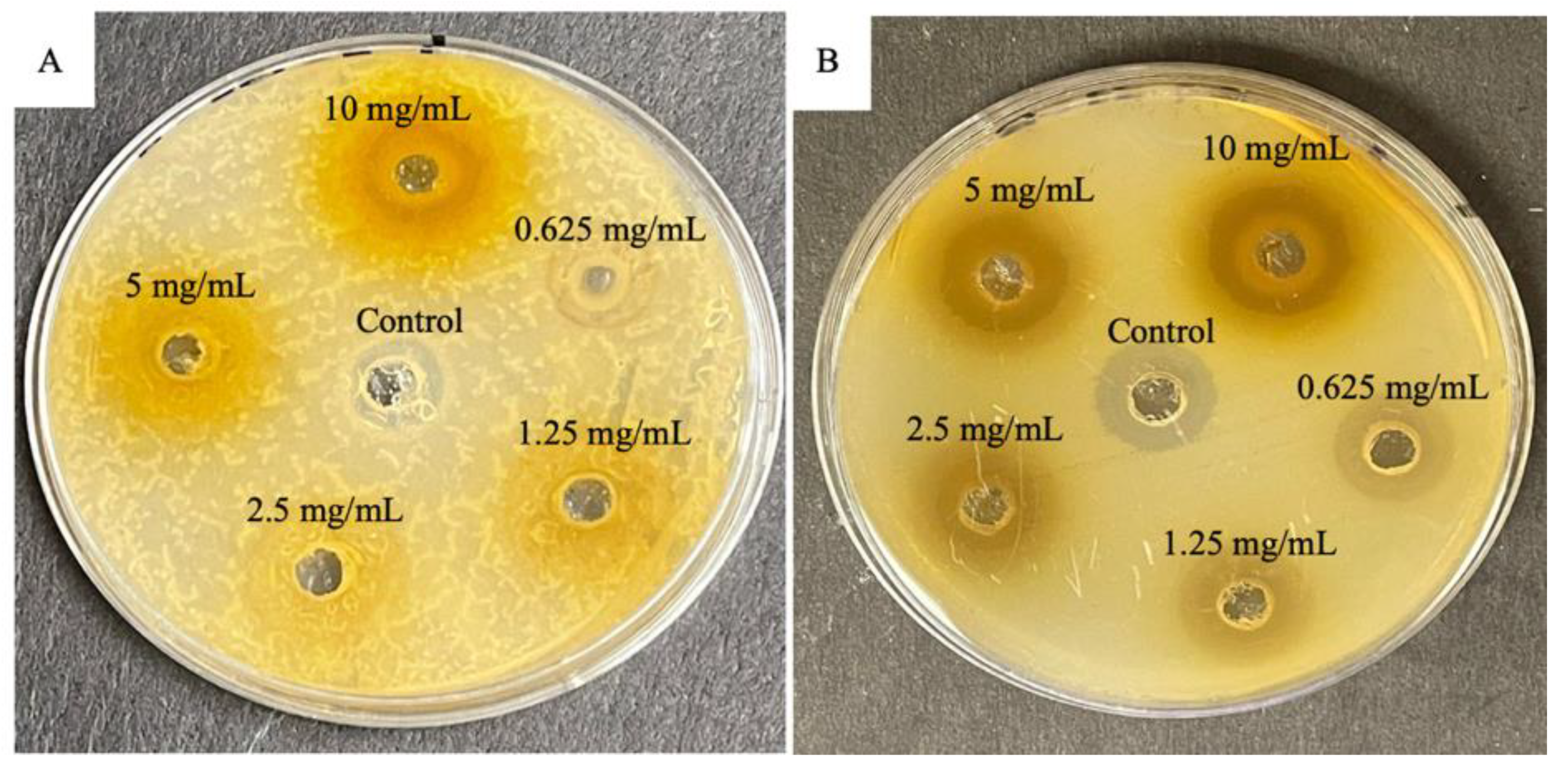
3.2. Antibacterial Activity

The antimicrobial activity of the methanolic leaf extracts for summer and winter of M. indica were evaluated against the bacterial species Staphylococcus aureus and Escherichia coli using the agar well diffusion method described by [32]. A total of 10 μg/mL of gentamicin (Gram-negative) and streptomycin (Gram-positive) were used as the positive controls. The experiments were carried out in triplicate, and data were expressed as mean ± standard deviation. The assessment of the antibacterial activity of the tested plant extracts are presented in Table 3 and as shown in Figure 1 and Figure 2.
The methanolic extracts of M. indica leaf demonstrated promising antibacterial activity against Gram-positive bacteria (Table 3). Staphylococcus aureus strains are associated with a plethora of diseases, such as mild skin and soft tissue infections, infective endocarditis, osteomyelitis, bacteraemia, and fatal pneumonia, with the multidrug-resistant strains presenting a severe challenge in hospital-acquired infections [53,54]. Consistent with the results obtained from the current investigations, ethnobotanical studies have documented that the fruits and leaves of M. indica can be used to treat skin diseases, including carbuncles, which are generally caused by S. aureus infections [55,56,57,58].
Scientific studies carried out by Shukla et al. [59] showed the anti-staphylococcal capacity of M. indica leaves and bark (which have the most concentrated amount of mangiferin) synergistically enhanced the susceptibility of S. aureus. These findings further support study by Mazlan et al. [60], where the authors reported an increase in the antimicrobial activity with the combinatory effect of mangiferin with antibiotics, nalidixic acid, ciprofloxacin, vancomycin, and tetracycline against S. aureus. Likewise, future studies targeting the synergistic interactions of M. indica stem bark and leaf extracts combined with antibiotics could potentially be developed into new anti-staphylococcal agents [7].
Interestingly, for S. aureus (ATCC 43300), the summer crude extract displayed higher antibacterial activity than the control streptomycin. The summer extract had a zone of inhibition of 14.17 ± 0.29 mm, while streptomycin had a 13.67 ± 0.76 mm zone of inhibition. Conversely, the winter extracts had a zone of inhibition of 12.33 ± 0.58 mm, while streptomycin had a 13.33 ± 0.76 mm zone of inhibition. Correspondingly, concentration plays a role in effectiveness as the concentration of crude extract decreased, as did the zones of inhibition. This was shown for both summer and winter samples. The results are in accordance with the comparison of antibacterial activity of leaves extracts of Mangifera altissima Mangifera indica, and Mangifera laurina, which showed that M. indica extracts were effective against all test concentrations; however, the potency was dependent on the concentration of crude extract [61]. It came as no surprise that for E. coli (ATCC 25922), the summer crude extract displayed higher antibacterial activity than the control gentamycin: the summer extract had a zone of inhibition of 18.5 ± 0.5 mm, while gentamycin had a 17.5 ± 1.5 mm zone of inhibition. The winter extracts had a zone of inhibition of 8.5 ± 1.32 mm, while gentamycin had a 14.5 ± 3.04 mm zone of inhibition. The weak activity of both the control gentamycin may indicate that this commercial antibiotic drug is slowly losing potency and is no longer capable of exerting an effect against E. coli (ATCC 25922) bacteria [62].
Krishnananda and Ramakrishna [61] conducted a comparative study of the antibacterial activity of different solvent extracts of each plant organ, revealing that the methanolic leaf, stem bark, and seed extracts had the most pronounced effect against the Gram-positive strains compared to the other solvent extracts of the respective plant organs. It can be deduced that methanol is an effective extracting solvent, effectively solubilising phytocompounds with antibacterial activity [63]. This can be supported by other studies that acquired similar results [19,64,65]. Overall, the summer extracts performed the best in the antibacterial assays, as these extracts showed notable antibacterial activity against Gram-negative and Gram-positive bacteria. A possible reason may be due to the winter Petri dishes having a more excessive bacterial growth than the summer samples. Another possible reason could be the seasonal change in the phytochemical composition, which play a role in the effectiveness in antibacterial activity [66]. This is shown in Table 2, where the ruthenium red test for gums and mucilage was shown to be more prevalent in summer than in winter season. Alternatively, the different solvent extracts of M. indica were only mildly effective against the tested Gram-negative pathogens from an antibacterial perspective. It is widely known that Gram-negative bacteria possess a formidable outer membrane (OM) that is composed of an asymmetric bilayer of phospholipids and lipopolysaccharides [67,68]. The OM of Gram-negative bacteria is the main reason for resistance to a wide range of antibiotics, including β-lactams, quinolones, colistins, and other antibiotics [69]. To circumvent such challenges, alternative strategies such as conjugating antibacterial agents to siderophore molecules that chelate iron (Fe3+) ions have been investigated [70]. Bacteria require Fe3+ for proliferation and may take up the conjugates of the siderophores and the antimicrobial agents into its periplasm or cytoplasm [71]. The mangiferin compound possesses a long, non-polar, hydrophobic alkyl saturated chain [72], potentially useful for membrane insertion [17]. This characteristic might afford the mangeferin compound to be an ideal siderophore-conjugating agent that could bypass the protective OM bilayer of the Gram-negative bacteria [73]. Future studies exploring these aspects together with the isolation of lead compounds from M. indica fruit and bark extracts may result in a greater potency against Gram-negative bacteria.
The antibacterial activity in the leaves of M. indica leaves can be attributed to the presence of steroids, tannins, flavonoids, and phenols. These compounds are good antifungal and antibacterial agents that may play a role in the antibacterial properties in the leaves of M. indica [19,64,65]. Though this study suggests antibacterial potential, and future studies should include more bacterial strains that will highlight the antibacterial activity on a broader scale and possibly motivate the use of M. indica in future drug developments.

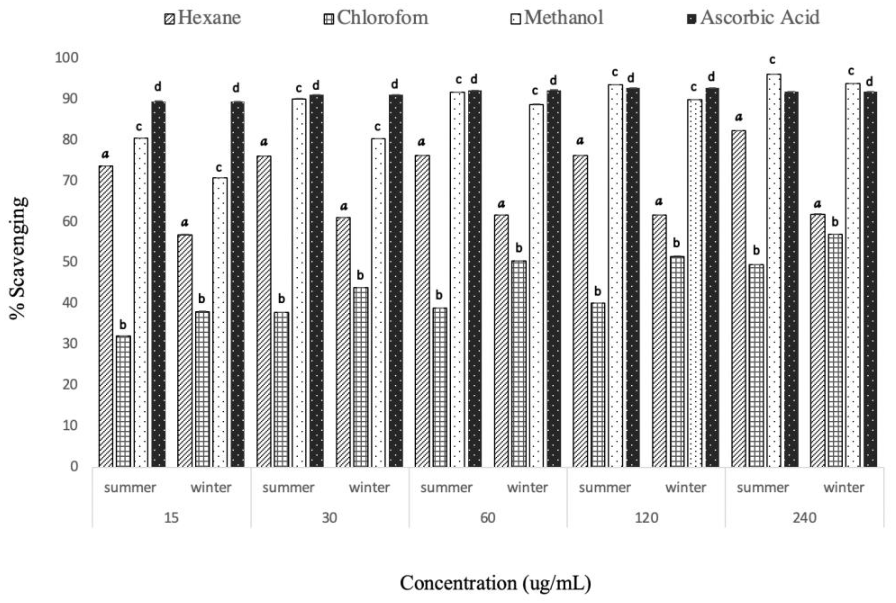
3.3. In Vitro Antioxidant Activity

Antioxidant activity involves intercepting reactive oxygen species to prevent or lessen the radicals and to produce less aggressive chemical species that are likely to cause tissue damage [74]. The usage of antioxidants has drawn much interest due to their ability to protect against the harmful effect caused by reactive oxygen species [75]. These solutions had discoloured from purple to a faded solution. A purple-coloured solution visible in the DPPH assay accepts electrons, which then transforms into a discoloured solution [14,69,76]. The point of colour change is linked to the effectiveness and concentration of antioxidants present [77]. The amount of discolouration indicates the free radical scavenging action [78]. The DPPH free radical scavenging activity was evaluated by the decrease in absorbance at 516 nm, which is induced by antioxidants [79]. This assay is not specific to any precise class of antioxidants and therefore provides the general antioxidant capacity of the extract [80]. The percentage free radical scavenging activity of crude extracts of the leaves of M. indica for summer and winter is presented in Figure 3. The radical scavenging activity of the crude extracts was studied by its ability to reduce DPPH (stable radical) and any molecule that may donate hydrogen or electron to DPPH [81]. The electron-donating ability of M. indica is most determined using DPPH free radical scavenging tests due to its reliability [38]. There was a dose-dependent change in radical scavenging activities for all crude extracts, i.e., in general all extracts, with increasing concentration, showing an increase in the DPPH radical scavenging activity (Figure 3). Statistical analysis indicated that all extracts had significantly different activity across all concentrations (p < 0.05) when compared to the ascorbic acid, 15–240 µg/mL.
The methanolic leaf extracts for summer and winter displayed effective radical scavenging activity in comparison with the hexane and chloroform plant extracts, with inhibition at 96.17 and 93.89%, respectively. Between summer and winter, summer exhibited the better radical scavenging activity. Dose–response radical scavenging activities were also observed in the methanolic extracts of different parts of Mangifera caesia, Mangifera pajang, and Mangifera linearifolia [82]. The scavenging activity of the methanolic extracts compared with the standard ascorbic acid suggests that the leaves of M. indica are also an effective scavenger of free radicals [49,83,84]. Free-radical reactions are linked to the pathology of several diseases such as cancer, Alzheimer’s disease, and inflammation [85]. Kumari et al. [86] observed the DPPH radical-scavenging activity of the methanolic leaf extracts of Mangifera linearifolia. The authors observed IC50 values of the methanol leaves as 48.86 µg/mL and hexane as 60.82 µg/mL. This trend corresponds with the current study, where methanol extracts had a better radical-scavenging activity than hexane. Overall, the results obtained in this study indicated that the hexane and methanolic extracts displayed good radical scavenging activity, which was a lower amount compared to the standard ascorbic acid (97.86%). The methanolic and hexane extracts for summer and winter showed the most convincing radical scavenging capacity with IC50 for summer extracts being 19.53, 70.23, and 12.71 μg/mL for hexane, chloroform, and methanol, respectively. The winter extracts were 22.3, 93.08, and 14.35 μg/mL for hexane, chloroform, and methanol, respectively, when compared to ascorbic acid, which had an IC50 of 3.202 μg/mL (Table 4). For the summer and winter extracts, hexane and methanol showed better scavenging capacity than chloroform. Overall, summer has a better scavenging capacity than winter with respect to the control. Several studies revealed that antioxidant activity was more potent for hexane and methanol extracts [75,76,87]. A study by Ocampo et al. [88] also revealed that the other parts of M. indica also exhibit significant antioxidant activity, as well as the leaves, which supports the findings in this study [88].
The antioxidant activity in the leaves of M. indica can be attributed to the presence of flavonoids, terpenes, and phenols. Phenolic compounds are strong antioxidants that can prevent the tissue damage caused by the free radicals [13]. The significant amount of phenol content in the extracts might be responsible for their antioxidant property [78,88,89]. The flavonoids act through the scavenging or chelating process and help in reducing the effect of antioxidants [78]. The flavonoid content in the extracts may also contribute to their antioxidant activity. Although this study suggests antioxidant potential, it is proposed that at least three antioxidant activity assays should be conducted to determine the antioxidant activity of a compound to increase the validity of the experiment [88]. Further investigations are also required to reveal other active constituents present. There are some advantages of using antimicrobial compounds of medicinal plants that include fewer side effects, better patient tolerance, relatively less expenses, and acceptance due to its long history of use and renewable place in nature [90]. On this basis, the search for new antibiotics continues with great urgency.

4. Conclusions

Mangifera indica leaf extract has shown biological properties that can be used to treat various diseases. Hexane and methanol extracts have significant antioxidant and antibacterial activities. This study revealed the presence of important phytoconstituents, i.e., alkaloids, flavonoids, phenols, saponins, steroids, and many more that may be responsible for their potent biological activities. Bioassay-guided fractionation, isolation of specific bioactive compounds from the leaves, and the evaluation of their safety will be necessary to further explore this species for potentially new therapeutic drug leads.

Author Contributions

Conceptualisation, A.M. and Y.N.; methodology, A.M. and Y.N.; formal analysis, A.M., Y.N. and Y.H.D.; investigation and data curation, A.M., Y.N. and Y.H.D.; validation, Y.H.D. and H.R.; visualisation, H.R.; writing—original draft preparation, A.M. and Y.N.; writing—review and editing, Y.H.D. and H.R.; supervision, Y.N. and Y.H.D. All authors have read and agreed to the published version of the manuscript.

Funding

The authors acknowledge Researchers Supporting Project (number RSP-2021/375), King Saud University, Riyadh, Saudi Arabia.

Institutional Review Board Statement

Not applicable.

Informed Consent Statement

Not applicable.

Data Availability Statement

All data are presented within the article.

Acknowledgments

The authors acknowledge the Researchers Supporting Project (number RSP-2021/375), King Saud University, Riyadh, Saudi Arabia.

Conflicts of Interest

The authors declare no conflict of interest.

References

  1. Sharif, M.D.M.; Banik, G.R. Status and utilization of medicinal plants in Rangamati of Bangladesh. Res. J. Agric. Biol. Sci. 2006, 2, 268–273. [Google Scholar]
  2. Kubmarawa, D.; Ajoku, G.A.; Enwerem, N.M.; Okorie, D.A. Preliminary phytochemical and antimicrobial screening of 50 medicinal plants from Nigeria. Afr. J. Biotechnol. 2007, 6, 90–1696. [Google Scholar]
  3. Lamboro, T.; Ketema, T.; Bacha, K. Prevalence and antimicrobial resistance in Salmonella and Shigella species isolated from outpatients, Jimma University Specialized Hospital, Southwest Ethiopia. Can. J. Infect. Dis. Med. Microbiol. 2016, 2016, 4210760. [Google Scholar] [CrossRef] [Green Version]
  4. Chatterji, D.; Petchiappan, A. Antibiotic resistance: Current perspectives. ACS Omega 2017, 2, 7400–7409. [Google Scholar]
  5. Tower, S.J.; Hetcher, W.J.; Myers, T.E.; Kuehl, N.J.; Taylor, M.T. Selective modification of tryptophan residues in peptides and proteins using a biomimetic electron transfer process. J. Am. Chem. Soc. 2020, 142, 9112–9118. [Google Scholar] [CrossRef] [PubMed]
  6. Streicher, J.M.; Liktor-Busa, E.; Keresztes, A.; LaVigne, J.; Largent-Milnes, T.M. Analgesic potential of terpenes derived from Cannabis sativa. Pharmacol. Rev. 2021, 73, 98–126. [Google Scholar]
  7. Dzotam, J.K.; Kuete, V. Antibacterial and antibiotic-modifying activity of methanol extracts from Six cameroonian food plants against multidrug-resistant enteric bacteria. BioMed Res. Int. 2017, 2017, 1583510. [Google Scholar] [CrossRef] [PubMed] [Green Version]
  8. Kumar, M.; Saurabh, V.; Tomar, M.; Hasan, M.; Changan, S.; Sasi, M.; Mekhemar, M. Mango (Mangifera indica L.) leaves: Nutritional composition, phytochemical profile, and health-promoting bioactivities. Antioxidants 2021, 10, 299. [Google Scholar] [CrossRef]
  9. Kasote, D.M.; Katyare, S.S.; Hegde, M.V.; Bae, H. Significance of antioxidant potential of plants and its relevance to therapeutic applications. Int. J. Biol. Sci. 2015, 11, 982–991. [Google Scholar] [CrossRef] [Green Version]
  10. Yoo, S.; Kim, K.; Nam, H.; Lee, D. Discovering health benefits of phytochemicals with integrated analysis of the molecular network, chemical Properties and ethnopharmacological evidence. Nutrients 2018, 10, 1042. [Google Scholar] [CrossRef] [Green Version]
  11. Aslam, B.; Wang, W.; Arshad, M.I.; Khurshid, M.; Muzammil, S.; Rasool, M.H.; Nisar, M.A.; Alvi, R.F.; Aslam, M.A.; Qamar, M.U.; et al. Antibiotic resistance: A rundown of a global crisis. Infect. Drug Resist. 2018, 11, 1645–1658. [Google Scholar] [CrossRef] [PubMed] [Green Version]
  12. Alam, S.B.; Mahmud, M.; Akter, R.; Hasan, M.; Sobur, A.; Nazir, K.N.H.; Noreddin, A.; Rahman, T.; El Zowalaty, M.E.; Rahman, M. Molecular detection of multidrug resistant Salmonella Species isolated from broiler farm in Bangladesh. Pathogens 2020, 9, 201. [Google Scholar] [CrossRef] [PubMed] [Green Version]
  13. Rupasinghe, H.P.V.; Balasuriya, N.; Wang, Y. Prevention of type 2 diabetes by polyphenols of fruits. In Nutritional Antioxidant Therapies: Treatments and Perspectives; Springer: Cham, Switzerland, 2017; pp. 447–466. [Google Scholar]
  14. Bouyahya, A.; Et-Touys, A.; Bakri, Y.; Talbaui, A.; Fellah, H.; Abrini, J.; Dakka, N. Chemical composition of Mentha pulegium and Rosmarinus officinalis essential oils and their antileishmanial, antibacterial and antioxidant activities. Microb. Pathog. 2017, 111, 41–49. [Google Scholar] [CrossRef] [PubMed]
  15. Khan, N.; Bano, A.; Zandi, P. Effects of exogenously applied plant growth regulators in combination with PGPR on the physiology and root growth of chickpea (Cicer arietinum) and their role in drought tolerance. J. Plant Interact. 2018, 13, 239–247. [Google Scholar] [CrossRef]
  16. Erhabor, C.; Erhabor, J.; McGaw, L. The potential of South African medicinal plants against microbial biofilm and quorum sensing of foodborne pathogens: A review. S. Afr. J. Bot. 2019, 126, 214–231. [Google Scholar] [CrossRef]
  17. Qais, F.A.; Shafiq, A.; Khan, H.M.; Husain, F.M.; Khan, R.A.; Alenazi, B.; Ahmad, I. Antibacterial effect of silver nanoparticles synthesized using Murraya koenigii (L.) against multidrug-resistant pathogens. Bioinorg. Chem. Appl. 2019, 2019, 4649506. [Google Scholar] [CrossRef] [Green Version]
  18. Tan, A.C.; Ashley, D.M.; López, G.Y.; Malinzak, M.; Friedman, H.S.; Khasraw, M. Management of glioblastoma: State of the art and future directions. CA A Cancer J. Clin. 2020, 70, 299–312. [Google Scholar] [CrossRef]
  19. Verma, P.; Verma, R.K. Succession of culturable microbes on rhizospheres soil of iron ore mined overburden dump in Dalli Rajhara, Durg, Chhattisgarh, India. Plant Arch. 2019, 19, 1179–1189. [Google Scholar]
  20. Guha, B.; Arman, M.; Islam, M.N.; Tareq, S.M.; Rahman, M.M.; Sakib, S.A.; Alqahtani, A.M. Unveiling pharmacological studies provide new insights on Mangifera longipes and Quercus gomeziana. Saudi J. Biol. Sci. 2021, 28, 183–190. [Google Scholar] [CrossRef]
  21. Tegen, D.; Dessie, K.; Damtie, D. Candidate Anti-COVID-19 Medicinal Plants from Ethiopia: A review of plants traditionally used to treat viral diseases. Evid.-Based Complement. Altern. Med. 2021, 2021, 6622410. [Google Scholar] [CrossRef]
  22. Ainane, A.; Cherroud, S.; El Kouali, M.; Abba, E.H.; Ainane, T. Chemical compositions, insecticidal and antimicrobial activities of two moroccan essential oils of Citrus limonum and Syzygium aromaticum. Pharmacol. J. 2020, 30, 190–199. [Google Scholar]
  23. Abdoul-Latif, F.M.; Elmi, A.; Merito, A.; Nour, M.; Risler, A.; Ainane, A.; Ainane, T. Essential Oils of Tagetes minuta and Lavandula coronopifolia from Djibouti: Chemical Composition, Antibacterial Activity and Cytotoxic Activity against Various Human Cancer Cell Lines. Int. J. Plant Biol. 2022, 13, 315–329. [Google Scholar] [CrossRef]
  24. Nuthakki, V.K.; Mudududdla, R.; Sharma, A.; Kumar, A.; Bharate, S.B. Synthesis and biological evaluation of indoloquinoline alkaloid cryptolepine and its bromo-derivative as dual cholinesterase inhibitors. Bioorganic Chem. 2019, 90, 103062. [Google Scholar] [CrossRef]
  25. Nafiqoh, N.; Zairin, M.; Lusiastuti, A.; Sarter, S.; Caruso, D.; Avarre, J.C. Antimicrobial properties against Aeromonas hydrophila and immunostimulant effect on Clarias gariepinus of Piper betle, Psidium guajava, and Tithonia diversifolia plants. Aquac. Int. 2020, 28, 1–13. [Google Scholar] [CrossRef]
  26. Akwu, N.A.; Naidoo, Y.; Singh, M. Cytogenotoxic and biological evaluation of the aqueous extracts of Grewia lasiocarpa: An Allium cepa assay. S. Afr. J. Bot. 2019, 125, 371–380. [Google Scholar] [CrossRef]
  27. Harborne, J.B. Photochemical Methods: A Guide to Modern Techniques of Plant Analysis; Chapman & Hall: London, UK, 1973. [Google Scholar]
  28. Trease, G.E.; Evans, W.C. Pharmacology, 11th ed.; Bailliere Tindall Ltd.: London, UK, 1978. [Google Scholar]
  29. Sofowora, A. Medicinal Plants and Traditional Medicines in Africa; Chichester John Wiley & Sons: New York, NY, USA, 1993. [Google Scholar]
  30. Tiwari, P.; Kumar, B.; Kaur, M.; Kaur, G.; Kaur, H. Phytochemical screening and extraction: A review. Int. Pharm. Sci. 2011, 1, 98–106. [Google Scholar]
  31. Karthikeyan, A.; Senthil, N.; Min, T. Nanocurcumin: A promising candidate for therapeutic applications. Front. Pharmacol. 2020, 11, 487. [Google Scholar] [CrossRef]
  32. Perez, C.; Paul, M.B.P. Antibiotic assay by agar-well diffusion method. Acta Biol. Med. Exp. 1990, 15, 113–115. [Google Scholar]
  33. Mani, M.; Okla, M.K.; Selvaraj, S.; Kumar, A.R.; Kumaresan, S.; Muthukumaran, A.; Elshikh, M.S. A novel biogenic Allium cepa leaf mediated silver nanoparticle for antimicrobial, antioxidant, and anticancer effects on MCF-7 cell line. Environ. Res. 2021, 198, 111199. [Google Scholar] [CrossRef]
  34. Braca, A.; Sortino, C.; Politi, M.; Morelli, I.; Mendez, J. Antioxidant activity of flavonoids from Licanialicaniaeflora. J. Ethnopharmacol. 2002, 79, 379–381. [Google Scholar] [CrossRef]
  35. Moradi, M.; Mateu Cronie, O.J. Inhomogeneous higher-order summary statistics for point processes on linear networks. Stat. Comput. 2020, 30, 1221–1239. [Google Scholar]
  36. Omokhua, A.G.; Abdalla, M.A.; Van Staden, J.; McGaw, L.J. A comprehensive study of the potential phytomedicinal use and toxicity of invasive Tithonia species in South Africa. BMC Complement. Altern. Med. 2018, 18, 272. [Google Scholar] [CrossRef] [PubMed] [Green Version]
  37. Oladeji, O. The characteristics and roles of medicinal plants: Some important medicinal plants in Nigeria. Nat. Prod. Indian J. 2016, 12, 102. [Google Scholar]
  38. Dowlath Hus, M.J.; Kumar, K.S.; DarulRaiyaan, G.I.; Khalith, S.B.M.; Sundarapandian, S.; Deivi, A.K. Effect of solvents on phytochemical composition and antioxidant activity of Cardiospermum halicacabum (L.) extracts. Pharmacogn. J. 2020, 12, 1241–1251. [Google Scholar] [CrossRef]
  39. Mohammed, A.H.; Náinna, S.Z.; Yusháu, M.; Salisu, B.; Adamu, U.; Garba, S.A. Phytochemical screening and antibacterial activity of Mangifera indica extracts. UMYU J. Microbiol. Res. 2016, 1, 23–28. [Google Scholar]
  40. Ruther, J.; Meiners, T.; Steidle, J.L.M. Allelochemical Reactions Involving Heterotrophic Microorganisms. Biogeochem. Inland Waters 2010, 22, 436–438. [Google Scholar]
  41. Geetha, B.; Gowda, K.P.; Kulkarni, G.T.; Badami, S. Microwave assisted fastextraction of mucilages and pectins. Indian J. Pharm. Educ. Res. 2009, 43, 260–265. [Google Scholar]
  42. Sungthongjeen, S.; Pitaksuteepong, T.; Somsiri, A.; Sriamornsak, P. Studies on pectins as potential hydrogel matrices for controlled-release drug delivery. Drug Dev. Ind. Pharm. 1999, 25, 1271–1276. [Google Scholar] [CrossRef]
  43. Babu, H.R.; Savithramma, N. Screening of secondary metabolites of underutilized species of Cyperaceae. Int. J. Pharm. Sci. Res. 2014, 24, 182–187. [Google Scholar]
  44. Rabi, T.; Bishayee, A. Terpenoids and breast cancer chemoprevention. Breast Cancer Res. Treat. 2009, 115, 223–239. [Google Scholar] [CrossRef]
  45. Yadav, R.N.S.; Agarwala, M. Phytochemical analysis of some medicinal plants. J. Phytol. 2011, 3, 10–14. [Google Scholar]
  46. Krishnaiah, D.; Devi, T.; Bono, A.; Sarbatly, R. Studies on phytochemical constituents of six Malaysian medicinal plants. J. Med. Plants Res. 2009, 3, 67–72. [Google Scholar]
  47. Shah, B.A.; Qazi, G.N.; Taneja, S.C. Boswellic acids: A group of medicinally important compounds. Nat. Prod. Rep. 2009, 26, 72–89. [Google Scholar] [CrossRef] [PubMed]
  48. Doss, A. Preliminary phytochemical screening of some Indian medicinal plants. Anc. Sci. Life 2009, 29, 12. [Google Scholar] [PubMed]
  49. Joona, K.; Sowmia, C.; Dhanya, K.P.; Divya, M.J. Preliminary phytochemical investigation of Mangifera indica leaves and screening of antioxidant and anticancer activity. RJPBCS 2013, 4, 1112–1118. [Google Scholar]
  50. Rakholiya, K.; Chanda, S. Pharmacognostic, Physicochemical and Phytochemical Investigation of Mangifera indica L. var. Kesar leaf. Asian Pac. J. Trop. Biomed. 2012, 2, 680–684. [Google Scholar] [CrossRef]
  51. Masud, P.G.M. Pharmacological activities of mango (Mangifera indica): A review. J. Pharmacogn. Phytochem. 2016, 5, 1–7. [Google Scholar]
  52. Kabir, Y.; Shekhar, H.U.; Sidhu, J.S. Phytochemical compounds in functional properties of mangoes. In Handbook of Mango Fruit; Wiley: Hoboken, NJ, USA, 2017; pp. 237–254. [Google Scholar]
  53. Guo, L.; Gong, S.; Wang, Y.; Sun, Q.; Duo, K.; Fei, P. Antibacterial Activity of Olive Oil Polyphenol Extract against Salmonella Typhimurium and Staphylococcus aureus: Possible Mechanisms. Foodborne Pathog. Dis. 2020, 17, 396–403. [Google Scholar] [CrossRef]
  54. Sengstock, C.; Lopian, M.; Motemani, Y.; Borgmann, A.; Khare, C.; Buenconsejo, P.J.S.; Köller, M. Structure-related antibacterial activity of a titanium nanostructured surface fabricated by glancing angle sputter deposition. Nanotechnology 2014, 25, 195101. [Google Scholar] [CrossRef]
  55. India, S.J.; Ray, P.; Manchanda, V.; Bajaj, J.; Chitnis, D.S.; Gautam, V.; Indian Network for Surveillance of Antimicrobial Resistance (INSAR) group. Methicillin resistant Staphylococcus aureus (MRSA) in India: Prevalence & susceptibility pattern. Indian J. Med. Res. 2013, 137, 363. [Google Scholar]
  56. Jeeva, S.; Johnson, M.; Aparna, J.S.; Irudayaraj, V. Preliminary phytochemical and antibacterial studies on flowers of selected medicinal plants. Int. J. Med. Aromat. Plants 2011, 1, 107–114. [Google Scholar]
  57. Choudhary, R.K.; Swarnkar, P.L. Antioxidant activity of phenolic and flavonoid compounds in some medicinal plants of India. Nat. Prod. Res. 2011, 25, 1101–1109. [Google Scholar] [CrossRef] [PubMed]
  58. Shukla, A.; Parmar, P.; Patel, B.; Goswami, D.; Saraf, M. Breaking Bad: Better call gingerol for improving antibiotic susceptibility of Pseudomonas aeruginosa by inhibiting multiple quorum sensing pathways. Microbiol. Res. 2021, 252, 126863. [Google Scholar] [CrossRef] [PubMed]
  59. Teka, A.; Rondevaldova, J.; Asfaw, Z.; Demissew, S.; Van Damme, P.; Kokoska, L.; Vanhove, W. In vitro antimicrobial activity of plants used in traditional medicine in Gurage and Silti Zones, south central Ethiopia. BMC Complement. Altern. Med. 2015, 15, 286. [Google Scholar] [CrossRef] [Green Version]
  60. Mazlan, N.A.; Azman, S.; Ghazali, N.F.; Yusri, P.Z.S.; Idi, H.M.; Ismail, M.; Sekar, M. Synergistic antibacterial activity of mangiferin with antibiotics against Staphylococcus aureus. Drug Invent. Today 2019, 12, 14–17. [Google Scholar]
  61. Krishnananda, K.K.; Shabaraya, A.R. Comparison of antibacterial activity of leaves extracts of Tectona grandis, mangifera Indica, and Anacardium Occidentale. Int. J. Pharmacol. Res. 2017, 9, 36–39. [Google Scholar]
  62. Diso, S.; Ali, M.; Mukhtar, S.; Garba, M. Antibacterial activity and phytochemical screening of Mangifera indica (Mango) stem and leaf extracts on clinical isolates of methicillin resistant Staphylococcus aureus. J. Adv. Med. Pharm. Sci. 2017, 13, 1–6. [Google Scholar] [CrossRef]
  63. Sanrawal, A.; Sushil, K. Antibacterial activity of MI leaves against drug resistant bacteria strain. Int. J. Adv. Res. 2013, 1, 82–86. [Google Scholar]
  64. Seleshe, S.; Kang, S.N. In vitro antimicrobial activity of different solvent extracts from Moringa stenopetala leaves. Prev. Nutr. Food Sci. 2019, 24, 70. [Google Scholar] [CrossRef]
  65. Cudjoe, E.; Donu, D.; Okonu, R.E.; Amponsah, J.A.; Amoah, L.E. The in vitro antiplasmodial activities of aqueous extracts of selected Ghanaian herbal plants. J. Parasitol. Res. 2020, 2020, 5041919. [Google Scholar] [CrossRef]
  66. Ramasar, R.; Naidoo, Y.; Dewir, Y.H.; El-Banna, A.N. Seasonal Change in Phytochemical Composition and Biological Activities of Carissa macrocarpa (Eckl.) A. DC. Leaf Extract. Horticulturae 2022, 8, 780. [Google Scholar] [CrossRef]
  67. Delcour, A.H. Outer membrane permeability and antibiotic resistance. Biochim. Et Biophys. Acta (BBA)-Proteins Proteom. 2009, 1794, 808–816. [Google Scholar] [CrossRef] [PubMed] [Green Version]
  68. Flaxman, S.; Mishra, S.; Gandy, A. Estimating the effects of non-pharmaceutical interventions on COVID-19 in Europe. Nature 2020, 584, 257–261. [Google Scholar] [CrossRef] [PubMed]
  69. Breijyeh, Z.; Jubeh, B.; Karaman, R. Resistance of gram-negative bacteria to current antibacterial agents and approaches to resolve it. Molecules 2020, 25, 1340. [Google Scholar] [CrossRef] [Green Version]
  70. Dauner, M.; Skerra, A. Scavenging bacterial siderophores with engineered lipocalin proteins as an alternative antimicrobial strategy. Chembiochem Eur. J. Chem. Biol. 2020, 21, 601–606. [Google Scholar] [CrossRef] [Green Version]
  71. Tavassoli-Kafrani, E.; Gamage, M.V.; Dumée, L.F.; Kong, L.; Zhao, S. Edible films and coatings for shelf-life extension of mango: A review. Crit. Rev. Food Sci. Nutr. 2020, 62, 2432–2459. [Google Scholar] [CrossRef] [PubMed]
  72. Zhang, J.; Terrones, M.; Park, C.R.; Mukherjee, R.; Monthioux, M.; Koratkar, N.; Bianco, A. Carbon science in status, challenges, and perspectives. Carbon 2016, 98, 708–732. [Google Scholar] [CrossRef]
  73. Sridharan, M.; Kamaraj, P.; Vennila, R.; Huh, Y.S.; Arthanareeswari, M. Bio-inspired construction of melanin-like polydopamine-coated CeO2 as a high-performance visible-light-driven photocatalyst for hydrogen production. New J. Chem. 2020, 44, 15223–15234. [Google Scholar] [CrossRef]
  74. Kumar, Y.; Kumar, V.S. Comparative antioxidant capacity of plant leaves and herbs with their antioxidative potential in meat system under accelerated oxidation conditions. J. Food Meas. Charact. 2020, 14, 3250–3262. [Google Scholar] [CrossRef]
  75. Mohan, C.G.; Deepak, M.; Viswanatha, G.L.; Savinay, G.; Hanumantharaju, V.; Rajendra, C.E.; Halemani, P.D. Anti-oxidant and anti-inflammatory activity of leaf extracts and fractions of Mangifera indica. Asian Pac. J. Trop. Med. 2013, 6, 311–314. [Google Scholar] [CrossRef] [Green Version]
  76. Itoh, K.; Matsukawa, T.; Okamoto, M.; Minami, K.; Tomohiro, N.; Shimizu, K.; Kajiyama, S.; Endo, Y.; Matsuda, H.; Shigeoka, S. In vitro antioxidant activity of Mangifera indica leaf extracts. J. Plant Stud. 2020, 9, 39. [Google Scholar] [CrossRef]
  77. Herrera, F.; Mitchell, J.D.; Pell, S.K.; Collinson, M.E.; Daly, D.C.; Manchester, S.R. Fruit morphology and anatomy of the Spondioid Anacardiaceae. Bot. Rev. 2018, 84, 315–393. [Google Scholar] [CrossRef] [PubMed] [Green Version]
  78. Kumar, S.; Sandhir, R.; Ojha, S. Evaluation of antioxidant activity and total phenol in different varieties of Lantana camara leaves. BMC Res. Notes 2014, 7, 560. [Google Scholar] [CrossRef]
  79. Manjula, M.S.; Ganthi, A.S. In-vitro antioxidant and anti-inflammatory potential of ethanol extracts (root and aerial parts) of Mangifera indica. Ann. Plant Sci. 2018, 7, 1997–2001. [Google Scholar]
  80. Tepe, B.; Eminagaoglu, O.; Akpulat, H.A.; Aydin, E. Antioxidant potentials and rosmarinic acid levels of the methanolic extracts of Salvia verticillata (L.) subsp. verticillata and S. verticillata (L.) subsp. amasiaca (Freyn & Bornm.) Bornm. Food Chem. 2007, 100, 985–989. [Google Scholar]
  81. Patel, S.; Wilson, R.; Dawood, A.; Foschi, F.; Mannocci, F. The detection of periapical pathosis using digital periapical radiography and cone beam computed tomography—Part 2: A 1year post treatment follow up. Int. Endod. J. 2012, 45, 711–723. [Google Scholar] [CrossRef]
  82. Amoo, S.O.; Finnie, J.F.; Van Staden, J. The role of meta-topolins in alleviating micropropagation problems. Plant Growth Regul. 2011, 63, 197–206. [Google Scholar] [CrossRef]
  83. Nithitanakool, S.; Pithayanukul, P.; Bavovada, R. Antioxidant and hepatoprotective activities of Thai mango seed kernel extract. Planta Med. 2009, 75, 1118–1123. [Google Scholar] [CrossRef] [Green Version]
  84. Dhital, K.S. Phytochemical screening and antioxidant activities of Mangifera indica leaves grown in temperate region of Nepal. J. Pharmacogn. Phytochem. 2017, 6, 205–209. [Google Scholar]
  85. Houghton, R.A.; Butman, D.; Bunn, A.G.; Krankina, O.N.; Schlesinger, P.; Stone, T.A. Mapping Russian forest biomass with data from satellites and forest inventories. Environ. Res. Lett. 2007, 2, 045032. [Google Scholar] [CrossRef]
  86. Kumari, A.; Parida, A.K.; Rangani, J.; Panda, A. Antioxidant activities, metabolic profiling, proximate analysis, mineral nutrient composition of Salvadora persica fruit unravel a potential functional food and a natural source of pharmaceuticals. Front. Pharmacol. 2017, 8, 61. [Google Scholar] [CrossRef] [PubMed] [Green Version]
  87. Born, M.; Carrupt, P.A.; Zini, R.; Bree, F.; Tillement, J.P.; Hostettmann, K. Electrochemical behavior and antioxidant activity of some natural polyphenols. Helv. Chim. Acta 1996, 79, 1147–1158. [Google Scholar] [CrossRef]
  88. Ocampo, E.T.M.; Libron, J.A.M.A.; Guevarra, M.L.D.; Mateo, J.M.C. Phytochemical screening, phenolic acid profiling and antioxidant activity analysis of peels from selected mango (Mangifera spp.) genotypes in the Philippines. Food Res. 2020, 4, 1116–1124. [Google Scholar] [CrossRef]
  89. Xu, S.; Li, J.L.; Zhang, X.Q.; Wei, H.; Cui, L.J. Effects of heat acclimation pretreatment on changes of membrane lipid peroxidation, antioxidant metabolites, and ultrastructure of chloroplasts in two cool-season turfgrass species under heat stress. Environ. Exp. Bot. 2006, 56, 274–285. [Google Scholar] [CrossRef]
  90. Vermani, K.; Garg, S. Herbal medicines for sexually transmitted diseases and AIDS. J. Ethnopharmacol. 2002, 80, 49–66. [Google Scholar] [CrossRef]
Figure 1. Plates displaying antibacterial activity of methanolic crude extract from the leaves of Mangifera indica L. for summer. (A) Gram-positive Staphylococcus aureus (ATCC 43300) bacteria against control streptomycin; (B) Gram-negative Escherichia coli (ATCC 25922) bacteria against control gentamycin.
Horticulturae 08 00909 g001
Figure 2. Plates displaying antibacterial activity of methanolic crude extract from the leaves of Mangifera indica L. for winter. (A) Gram-positive Staphylococcus aureus (ATCC 43300) bacteria against control streptomycin; (B): Gram-negative Escherichia coli (ATCC 25922) bacteria against control gentamycin.
Horticulturae 08 00909 g002
Figure 3. Mean and standard deviation of DPPH free radical scavenging activity of hexane, chloroform, and methanol from leaves of Mangifera indica L. at various concentrations for summer and winter seasons. (Two-way-ANOVA multivariate Tukey’s honest significant difference multiple range post hoc test p < 0.05 IBM SPSS version 25; each bar was considered statistically significant when comparing each extract to the ascorbic acid at different concentrations, 15–240 µg/mL.) Data are presented as means ± SD, n = 3, and are displayed as a percentage of the control sample. Different letters are significantly different at p < 0.05.
Horticulturae 08 00909 g003
Table 1. Percentage yield of the crude extract of leaves for summer and winter of Mangifera indica.
SolventSummer Leaves Winter Leaves Summer LeavesWinter Leaves
Dried Extract Yield (g)Percentage Yield (%)
Hexane0.820.458.25.5
Chloroform0.980.779.87.7
Methanol2.72.332723.3
Table 2. Phytochemical screening of Mangifera indica leaf extracts using different solvents: hexane, chloroform, and methanol for the summer and winter seasons.
TestSummer ExtractsWinter Extracts
HexaneChloroformMethanolHexaneChloroformMethanol
Alkaloid tests
(i)
Mayers test
++++++
(ii)
Wagners test
++++++
Phenols, tannins, flavonoids, and quinones tests
(i) Ferric chloride test++++++++
(ii) Lead acetate test ++++++++++++
(iii) Gelatin test+++++++++++
Flavonoid test
(i) Alkaline reagent test (alkaline hydrolysis) ++++++++++++
(ii) Acid hydrolysis (sulphuric acid) ++++++++++
Terpene test
(a) Saponins
(i) Foam test ++
(b) Steroids
(i) Liebermann–Burchard test or acetic anhydride test++++
(ii) Salkowski’s test ++++++
Carbohydrate test
(i) Molisch’s test ++++++
Protein test
(i) Ninhydrin test
(ii) Biuret test++++++++
Gum and mucilage test
(i) Precipitation test++++++
(ii) Ruthenium red test+++++++
(iii) Detection of resins ++
− Negative result; + mildly present; ++ distinctly present; +++ very strongly present.
Table 3. Antibacterial screening of the crude methanolic leaf extracts of Mangifera indica L. for summer and winter seasons against Gram-negative E. coli (ATCC 25922) and Gram-positive S. aureus (ATCC 43300) bacterial strains. Gentamicin (Gram-negative) and streptomycin (Gram-positive) were the positive controls.
Bacterial StrainConcentrations (mg/mL) and Diameter of Zone of Inhibition (mm)
0.6251.252.5510Control
Summer
Staphylococcus aureus (ATCC 43300)7.5 ± 0.58.83 ± 0.769.83 ± 07611.50 ± 1.3214.17 ± 0.2913.67 ± 0.76
Escherichia coli (ATCC 25922)9.17 ± 1.76 11.17 ± 1.0414.33 ± 0.5816.33 ± 1.1518.5 ± 0.517.5 ± 1.5
Winter
Staphylococcus aureus (ATCC 43300)6.83 ± 0.297.67 ± 0.588.83 ± 0.7610.67 ± 0.5812.33 ± 0.5813.33 ± 0.76
Escherichia coli (ATCC 25922)6.25 ± 0.356.67 ± 1.157.17 ± 1.157.67 ± 1.618.5 ± 1.3214.5 ± 3.04
Values presented are means ± standard deviation.
Table 4. IC50 values of hexane, chloroform, and methanolic extracts from the leaves of Mangifera indica, and ascorbic acid used for the DPPH assay.
Extraction Solvents (IC50—μg/mL)
SeasonsHexaneChloroformMethanol
Summer19.5370.1312.71
Winter22.3293.0814.35
Ascorbic acid3.20
Note: IC50 value represents the concentration of sample required to inhibit 50% of DPPH free radicals. The IC50 of the DPPH assay isolated from the crude hexane, chloroform, and methanolic extract of the leaves of Mangifera indica. Data are presented as mean, n = 3, of triplicate determinates.
Publisher’s Note: MDPI stays neutral with regard to jurisdictional claims in published maps and institutional affiliations.

