Article

Oral Administration of Animal and Plant Protein Mixture with Lactiplantibacillus plantarum IDCC 3501 Improves Protein Digestibility

1
School of Food Science and Biotechnology, Kyungpook National University, Daegu 41566, Republic of Korea
2
Ildong Bioscience, Pyeongtaek-si 17957, Republic of Korea
3
Ildong Pharmaceutical, Hwaseong-si 18849, Republic of Korea
4
Carl R. Woese Institute for Genomic Biology, The University of Illinois at Urbana-Champaign, Urbana, IL 61801, USA
*
Authors to whom correspondence should be addressed.
These authors contributed equally to this work.
Fermentation 2023, 9(6), 560; https://doi.org/10.3390/fermentation9060560
Submission received: 10 May 2023 / Revised: 9 June 2023 / Accepted: 10 June 2023 / Published: 12 June 2023
(This article belongs to the Special Issue Recent Trends in Probiotics and Gut Microbiome for Human Health)

Abstract

:
A combined usage of animal and plant proteins-mixture could aid to solve environmental and social problems arising from the use of animal protein alone, while also improving the taste and texture of plant protein. Protein mixtures could be a better protein source due to the high availability of amino acids in the body compared with single proteins. Consuming proteins with probiotics can provide more beneficial health effects by helping to hydrolyze protein and absorb amino acids in the body. In this study, coadministration of an animal and plant protein mixture with a high concentration of probiotics was investigated to increase protein digestibility and amino acids absorbability in a mice model. Lactiplantibacillus plantarum IDCC 3501, which has the maximum ability to hydrolyze a protein mixture, composed of soybean protein and milk protein, was selected, and the changes in mice (C57BL/6J, male, six weeks) were investigated after the coadministration of protein mixture and 5 × 10⁸ or 5 × 10⁹ CFU/mL of L. plantarum for eight weeks. Normal diet, high-protein diet (HPD), and HPD supplementing L. plantarum were separately administered to mice. Food and water consumption of the mice did not differ depending on diet type. Measurements of the serum concentrations of amino acids showed that the absorption of aspartate, glutamate, isoleucine, leucine, valine, and lysine increased when high concentrations of protein and probiotics were administered. Thus, high L. plantarum concentrations could be a protein diet supplementation to improve health by promoting the absorption of amino acids.

1. Introduction

Dietary protein is considered to be an essential nutrient because of its ability to modulate various metabolic activities, including controlling the balance of hormones [1] and enzymes [2], cell recovery [3], muscle synthesis [4], and immune control [5], in the human body. For a long time, humans have been consuming animal protein, with excellent nutrients from the milk or meat of livestock [4,6]. However, the manufacturing of animal proteins cannot be free from issues, such as a lack of water and breeding grounds [7] and climate changes induced by greenhouse gas emissions [8] contributed to by the increasing consumption of meat products per person (e.g., approximately a 102% increase from 2000 to 2050) [9,10]. Although substituting all animal protein with plant protein has been attempted, lysine deficiency and digestibility limitations [11] when eating only plant protein sources were proposed as the problem. The flavor and texture of plant protein are also not as good as those of animal protein [12], and manufacturing plant protein into animal-protein-like flavor and texture has not been easy [13]. Therefore, the production of protein mixture, animal protein partially replaced with plant protein or a combination of animal and plant protein, has been considered [14]. The application of protein mixture has been in the limelight as it overcomes limitations according to animal protein production, having both animal and plant protein advantages, such as higher amino acids availability [15,16,17,18]. Consumers also prefer a protein mixture compared to a single protein [19]. Nevertheless, the low digestibility of plant protein is still an ongoing problem to be solved.
The amino acids compositions in the protein and the host’s bioavailability of amino acids are the two fundamental factors in determining the nutritional value of protein [20]. In other words, it is important to increase the protein digestibility and the bioavailability of the derived amino acids. After consuming a protein source, the ingested protein is cut into polypeptides by pepsin in the stomach, with low pH and protease in the pancreas, and the polypeptides that progress down to the small intestine are further broken down into peptides and amino acids [21,22]. The dipeptides and tripeptides are absorbed into intestinal cells through peptide transporters and decomposed into amino acids in the cytoplasm [23], and the amino acids enter cells through several membrane-bound proteins and amino acids transporters in the small intestine [24]. However, some proteins that have not been perfectly digested in the small intestine move to the large intestine [25]. These undigested proteins and peptides are broken down by intestinal microorganisms to produce numerous microbial metabolites, and most free amino acids are assimilated by the gut microbiome [26], which can exert beneficial effects on the host by maintaining the amino acid balance between microorganisms and the host [27].
Consequently, the intake of probiotics could help intestinal microorganisms to proteolyze and increase their ability to absorb amino acids in intestinal epithelial cells [28]. Nonetheless, only a few studies have been conducted on increasing the absorption of amino acids through the coadministration of probiotics and proteins, except for those using a single type of proteins. For example, coadministration of lactobacillus and pea proteins or skim milk proteins increased amino acids plasma concentrations using a representative plant- or animal-based protein, respectively [29,30]. However, more proof of probiotics’ feasibility to provide improved digestion of proteins should be performed using protein mixtures, not using a single type protein. In addition, although probiotics have possessed high levels of proteolytic ability due to their abundant proteases [31,32], most studies have been limited to food applications, such as cheese fermentation and sausage manufacturing [31,33,34]. Hence, it is necessary to investigate the increases in the bioavailability of amino acids through the coadministration of protein mixture and probiotics with high proteolytic activity.
We conducted this study to investigate whether probiotic strains with excellent proteolytic activity could help to absorb amino acids in a mouse model with administration of a protein mixture and improve the health of the mice, such as body weight and muscle mass. Accordingly, the strain with the highest proteolytic activity was selected among Lactiplantibacillus, Lacticaseibacillus, and Lactococcus species. The protein mixture and the selected probiotic strain at two different concentrations of 5 × 10⁸ CFU/day or 5 × 10⁹ CFU/day were administered to the mice. After evaluating food and water consumption, the concentration of amino acids in the blood serum was examined, and the body weight and muscle mass of the mice were measured.

2. Materials and Methods

2.1. Bacterial Culture

Lactiplantibacillus plantarum IDCC 3501, Lactococcus lactis subsp. lactis IDCC 2301, and Lacticaseibacillus paracasei IDCC 2201 were obtained from Ildong Bioscience Co., Ltd. (Pyeongtaek-si, Gyeonggi-do, Republic of Korea) and anaerobically cultured in 14 mL of De man, Rogosa, and Sharpe (MRS; BD Difco, Frankin Lakes, NJ, USA) medium at 37 °C for 24 h without shaking. Probiotic strains were freeze-dried and sequentially coated following the previous study to utilize as diets of mice [35].

2.2. Proteolytic Activity

The basal medium, composed of 15 g/L of agar (Sigma Aldrich, St. Louis, MO, USA), 5 g/L of tryptone (BD Difco, Fankin Lakes, NJ, USA), 2.5 g/L of yeast extract (BD Difco), and 1 g/L of glucose (Sigma Aldrich), added with 28 g/L of skim milk protein (BD Difco), 28 g/L of pea protein (TATUA, Morrinsville, New Zealand), or 28 g/L of protein mixture (Himmune Protein Balance; Ildong Foodis Inc., Seoul, Republic of Korea) was used for investigating the proteolytic activity of probiotic strains. The protein mixture was composed of isolated soybean protein, bovine whey protein concentrate, bovine milk protein concentrate, bovine milk protein isolates, and goat whey protein concentrate. Each 10 µL aliquot of 108 CFU/mL probiotic strain was dropped on the agar plate, added with the protein, and cultured at 37 °C for 24 h [36,37]. After the incubation, the appearance of a clear zone surrounding the drop was considered positive, which implies that it has proteolytic activity. The diameter of the clear zone surrounding the colonies was determined using ImageJ software (version 1.8.0, NIH, Bethesda, MD, USA).

2.3. Genome-Wide Identification of Genes Encoding the Proteolytic System

The protein-coding sequences were predicted using Prokaryotic Genome Annotation Pipeline on NCBI [38], and functional annotation was achieved on the Rapid Annotation using Subsystems Technology server [39]. Furthermore, to compare the proteolytic genes of other L. plantarum strains, the complete genome sequences of L. plantarum WFSC1 (AL935263.2), L. plantarum 299v (NZ_LEAV01000004.1), and L. plantarum IDCC 3501 (CP031702.1) were obtained from the NCBI microbial genome database.

2.4. Mouse Experiment for Quantification of the Concentration of Amino Acids in Blood Serum and Measurement of Body and Muscle Weights of Mice

C57BL/6J male mice aged 6 weeks were obtained from Central Lab Animal Inc. (Seoul, Republic of Korea). Normal diet (ND) and high-protein diet (HPD; 40% protein) were supplied by Ildong Foodis Inc. (Seoul, Republic of Korea). Mice were maintained in an air-conditioned room with a 12 h/day/night schedule at 21 °C ± 2 °C and acclimatized to the experimental facility with free access to food and water for 1st week. A total of 48 mice were used in this study, and six mice were measured per group for one experiment in 4th week and 8th week, randomly divided into the following four groups: ND, HPD, HPD with a low concentration of L. plantarum IDCC 3501 (5 × 10⁸ CFU/day) (HPD + LPL), HPD with a high concentration of L. plantarum IDCC 3501 (5 × 10⁹ CFU/day) (HPD + HPL). The concentrations of macronutrients of ND (Teklad Global 18% Protein Rodent Diet; ENVIGO, Indianapolis, IN, USA) and HPD were shown in Table S1, and the composition of amino acids in normal diet (ND) was shown in Table S2, which could compare to the amino acids composition of high-protein diet in Table S3. Animal studies were approved by the Institutional Animal Care and Use Committee of Chungbuk National University (approval number: CBNUA-1687-22-02). Mice were administrated freeze-dried L. plantarum IDCC 3501 by oral gavage once a day. The amount of food and water consumption was investigated every week for 8 weeks.
Anaesthetized animals were euthanized due to exsanguination during blood sample collection. The blood sample was centrifuged at 600× g for 20 min for separating serum. The collected serum was stored at −70 °C in a deep freezer until further experiments. Free amino acids in the serum were analyzed using an automated amino acid analyzer L-8900 with a UV detector (Hitachi High-Technologies Corporation, Tokyo, Japan) and an ion-exchange column (# 2622PH column 4.6 × 60 mm).
The body weight of each mouse depending on the diet type was measured weekly. After 4th and 8th weeks, the mice were fasted for 6 h and consequently anesthetized with diethyl ether. Next, the thigh muscles were excised, rinsed with phosphate-buffered saline, and weighed.

2.5. Statistical Analyses

Data are expressed as mean ± standard deviation (SD). For statistical comparisons, one-way analysis of variance and Turkey’s multiple comparison tests were performed using GraphPad Prism 9.5.0.

3. Results

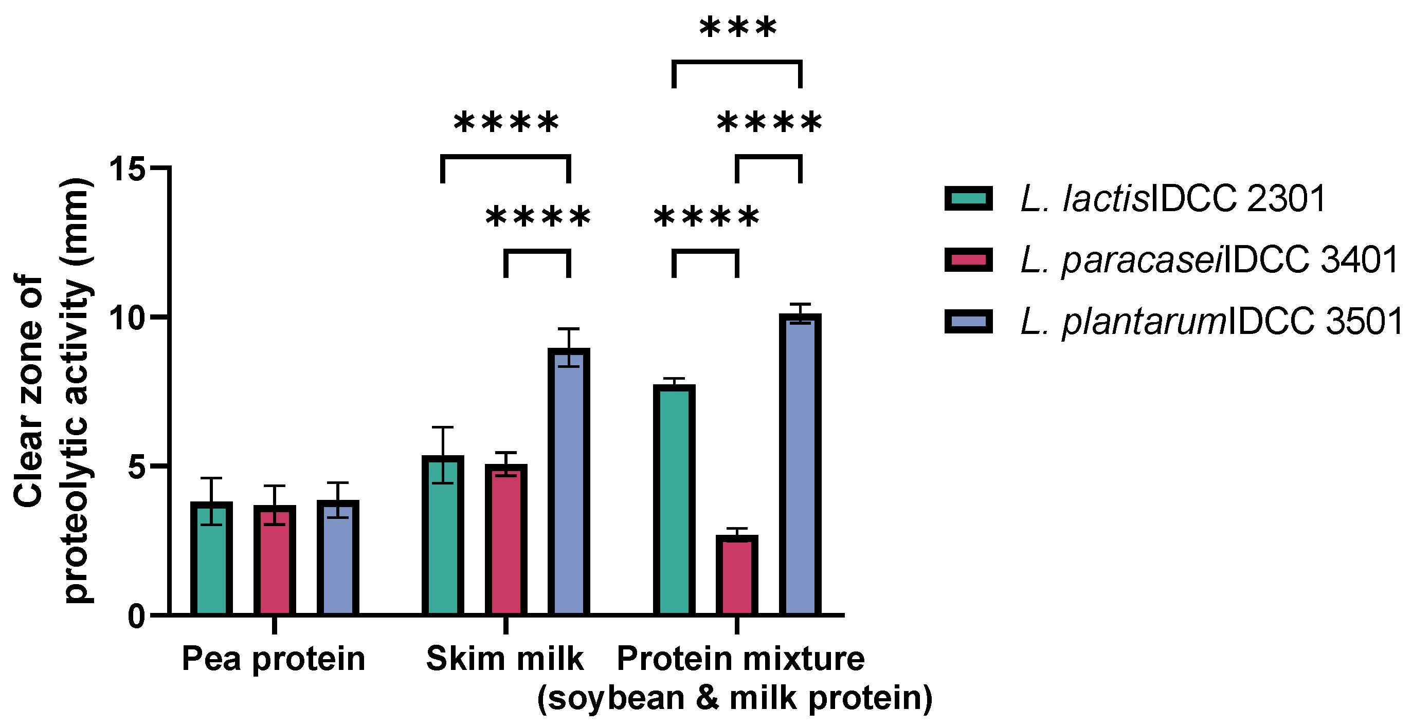
3.1. Proteolytic Activities of Probiotic Strains on Animal and Plant Protein Mixture

For selecting the probiotic strain with the best proteolytic activity, the proteolytic activities of probiotic strains L. lactis IDCC 2301, L. paracasei IDCC 3401, and L. plantarum IDCC 3501 were investigated on basal medium with plant-based protein (i.e., pea protein), animal-based protein (i.e., skim milk), and protein mixture (Figure 1). All the probiotic strains used in this study exhibited some proteolytic activities; however, the degree of activity was dependent on the type of strain as well as the protein source. Specifically, the radii of the clear zone in pea-protein-containing agar were 3.82 ± 0.64 mm for L. lactis, 3.69 ± 0.53 mm for L. paracasei, and 3.86 ± 0.47 mm for L. plantarum. The proteolytic activity on the plant protein showed no significant differences among these strains. However, in skim-milk-containing agar (i.e., skim milk or skim milk with pea protein mixture), L. plantarum exhibited the highest proteolytic activity, with the radii of the clear zone being 8.97 ± 0.51 mm for skim milk and 10.11 ± 0.26 mm for protein mixture. Other strains showed an approximately 5 mm clear zone in skim milk alone, and the radii of the clear zone were 7.74 ± 0.16 mm for L. lactis and 2.70 ± 0.17 mm for L. paracasei. L. plantarum showed the largest proteolytic clear zone of 10.11 ± 0.26 mm. Therefore, L. plantarum, showing the highest proteolytic activity, was selected to examine whether the strain could aid the absorption of amino acids through protein hydrolysis in the intestines of mice.
Accordingly, the potent proteolytic capacity of L. plantarum IDCC 3501 was investigated by genomic analysis. It is well known that various lactic acid bacteria exhibit comparable proteolytic capacity [40]. In the genome of L. plantarum IDCC 3501, six genes encoding the oligopeptides ABC transport (Opp) system, including three oligopeptide binding proteins (OppA), two oligopeptide transport system permeases (1 OppB and 1 OppC), and one nucleotide binding protein (OppD), were verified. Therefore, L. plantarum IDCC 3501 strain may display a strong ability of oligopeptide to transport the complete Opp system. Moreover, the presence of 16 Pep genes encoding peptidases, such as endo-, amino-, tri-, di-, and proline-specific peptidases, in the genome suggested the potentiality of the proteolytic system in L. plantarum IDCC 3501 (Table 1).

3.2. Simultaneous Oral Administration of Animal and Plant Protein Mixture with Different Concentrations of L. plantarum to Mice

3.2.1. Food and Water Consumption and Body Weight

After the coadministration of 40% (v/v) protein mixture and different concentrations of L. plantarum to the mice for 8 weeks, no significant difference was observed in the food and water intake depending on the type of diet (Figure S1). For instance, ND-fed mice had 4.08 ± 0.24 g food consumption with 3.35 ± 0.15 g water consumption in the 1st week and 4.34 ± 0.24 g food consumption with 4.13 ± 0.22 g water consumption after 8 weeks. However, HPD-fed mice showed slightly less food consumption and higher water consumption than ND-fed mice, irrespective of the addition of probiotics. For example, HPD-, HPD + LPL-, and HPD + HPL-fed mice had 3.50 ± 0.13, 3.37 ± 0.27, and 3.45 ± 0.24 g food consumption and 4.83 ± 0.15, 4.74 ± 0.05, and 4.65 ± 0.01 g water consumption after 8 weeks, respectively, which showed no specific pattern.
The body weight of mice fed with ND, HPD, HPD + LPL, and HPD + HPL was measured for 8 weeks (Figure S2). Similar to the results of food and water consumption, no significant differences were observed in the body weight of mice during the 8 weeks of experiments depending on the diet, showing a gradual increase from the 1st to 8th week with all diets. Dependent on the diet type, the body weight in the 1st week was comparable; however, HPD-, HPD + LPL-, and HPD + HPL-fed mice had slightly higher body weights than ND-fed mice in the 8th week (ND: 22.68 ± 0.90 g in the 1st week to 26.32 ± 1.35 g in the 8th week, HPD: 22.71 ± 0.87 g in the 1st week to 27.63 ± 1.26 g in the 8th week, HPD + LPL: 22.72 ± 0.85 g in the 1st week to 27.15 ± 1.45 g in the 8th week, and HPD + HPL: 22.71 ± 0.84 g in the 1st week to 26.67 ± 1.12 g in the 8th week). Thus, the change in amino acid concentration and muscle mass was further investigated as the intake of high protein and probiotics affected the mouse body weight.

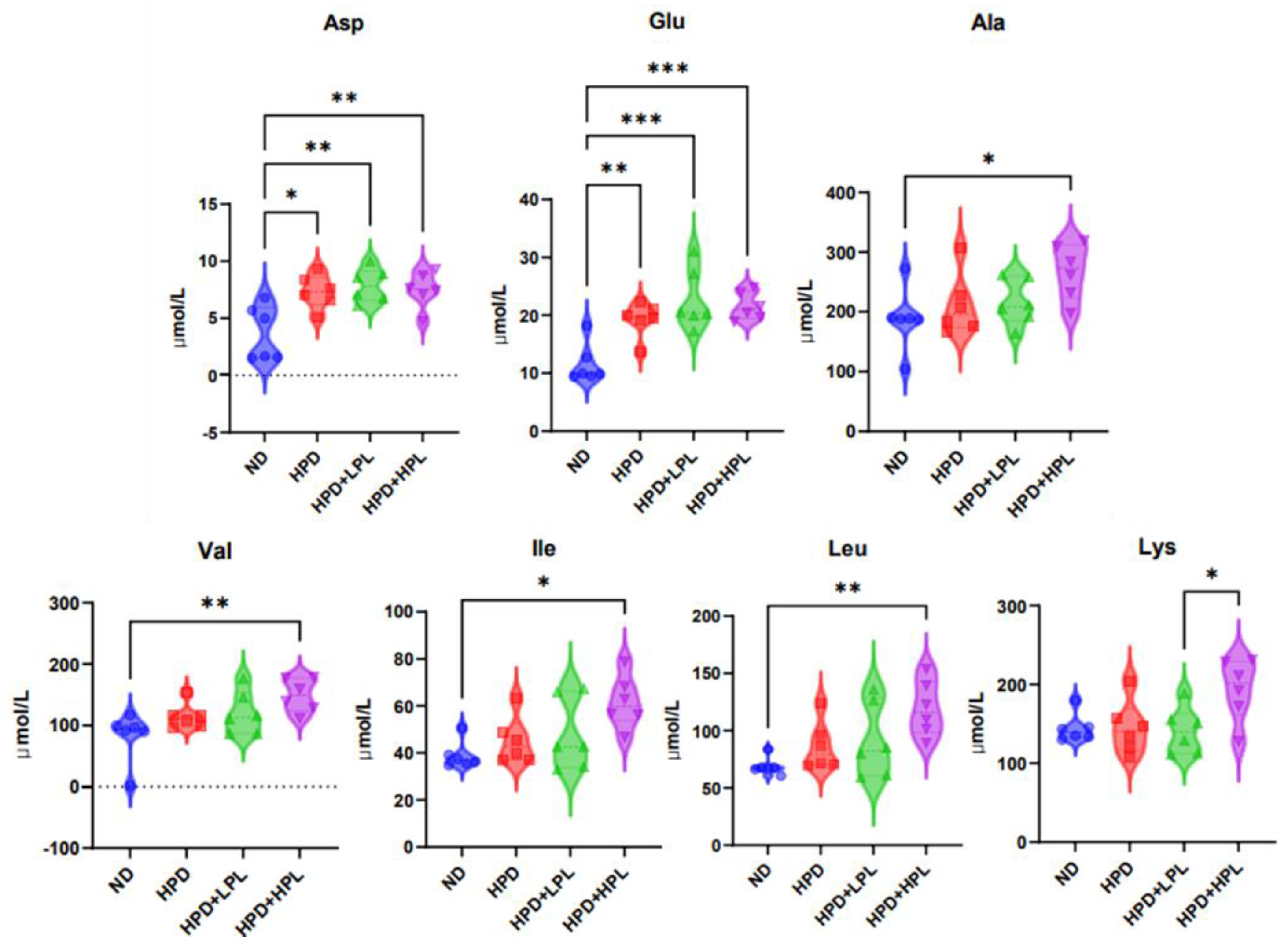
3.2.2. Concentration of Amino Acids in Blood Serum
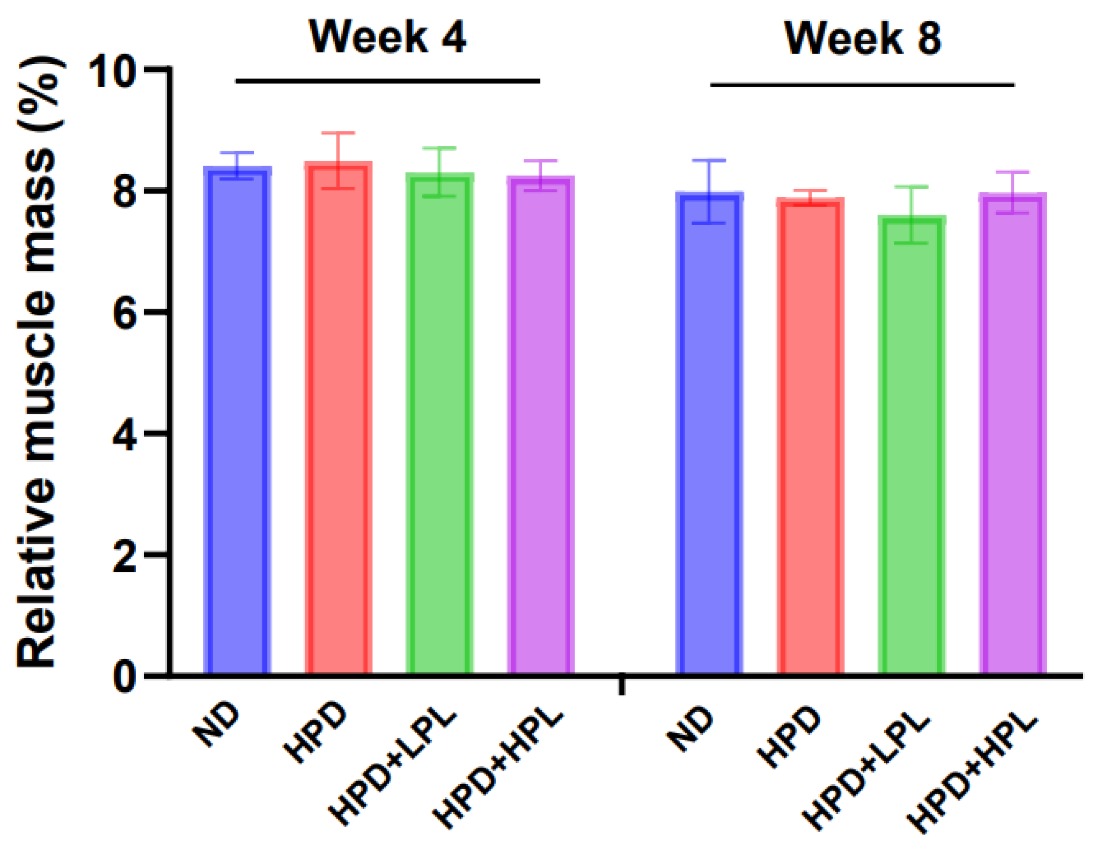
After the coadministration of protein mixture and L. plantarum to the mice, we investigated the changes in amino acid concentrations in the blood serum to assess whether L. plantarum helps the enhancement of protein hydrolysis or amino acid absorption in the mouse intestine (Table S4). The overall concentrations of 22 amino acids in the mouse serum were slightly reduced in the order of administrating ND, HPD, HPD + LPL, and HPD + HPL. According to the results of violin plots, the concentrations of seven amino acids (aspartate, glutamate, alanine, valine, isoleucine, leucine, and lysine) were significantly different between the diets (Figure 2). Aspartate and glutamate showed significantly high concentrations in the blood serum of each of HPD-, HPD + LPL-, and HPD + HPL-fed mice compared to those in ND-fed mice. However, there was no significant difference among HPD-fed mice, irrespective of the coadministration of probiotic strains. For instance, the concentration of aspartate in the serum of HPD-fed mice was 3.62 ± 2.08 µmol/L higher than that of ND-fed mice, and that of glutamate in HPD-fed mice was 7.67 ± 3.36 µmol/L higher than that in ND-fed mice. Moreover, the concentrations of aspartate and glutamate in mice fed with HPD + LPL were 4.17 ± 2.39 and 11.09 ± 7.73 µmol/L, respectively, higher than those in mice fed with ND, and the respective concentrations of aspartate and glutamate in mice fed with HPD + HPL were 3.79 ± 2.37 and 9.96 ± 4.13 µmol/L higher than those in ND-fed mice. These findings indicate that aspartate and glutamate could be absorbed well if a high concentration of protein is supplied irrespective of the concentration of L. plantarum.
Alanine, valine, isoleucine, and leucine showed significantly higher concentrations of 78.88 ± 77.89, 66.28 ± 34.67, 22.57 ± 14.34, and 50.61 ± 27.27 µmol/L, respectively, in the serum of HPD + HPL-fed mice than in the serum of ND-fed mice. As HPD-fed mice and HPD + LPL-fed mice showed no significant increases in the absorption of these amino acids compared with control mice, their absorption in the body could increase only when high-concentration protein and high-concentration probiotics are consumed together.
The uptake of lysine was significantly increased by 49.35 ± 39.39 µmol/L in HPD + HPL-fed mice compared with that in HPD + LPL-fed mice. Because lysine was absorbed differently depending on the concentration of probiotic strains in the body, a sufficient concentration of L. plantarum, such as 5 × 10⁹ CFU/day, was required. Therefore, when the appropriate concentration of probiotic strains was not supplemented, lysine could not be absorbed significantly even if a large amount of protein was supplied.

3.2.3. Muscle Mass

Muscle mass was investigated to determine whether the amino acids absorbed in the blood of mice can aid muscle synthesis (Figure 3). The relative muscle mass (%), which is the ratio of the absolute muscle mass to the total weight, showed no significant difference with all diets in the 4th and 8th weeks. The values of the relative muscle mass of ND-, HPD-, HPD + LPL-, and HPD + HPL-fed mice in the 4th week were 8.41% ± 0.22%, 8.50% ± 0.46%, 8.31% ± 0.40%, and 8.25% ± 0.24%, respectively, and, in the 8th week, the respective values were 7.99% ± 0.52%, 7.99% ± 0.13%, 7.60% ± 0.47%, and 7.97% ± 0.34%. Overall, the types of feed and the addition of probiotic strains did not result in considerable differences in mice growth patterns, such as food consumption, water uptake, weight gain, and muscle formation.

4. Discussion

Dietary protein consumption provides various benefits in terms of health, environment, and human preferences. A protein mixture using both animal and plant proteins could compensate for the limitations in flavor and texture of plant protein with enhanced bioavailability [41]. Proteolysis is a reaction in which the peptide bond of a protein is hydrolyzed into smaller peptides or amino acids [42]. In the body, the peptide bond of the protein is hydrolyzed by enzymes such as pepsin and protease released from the gastrointestinal tract, and then free amino acids are released in the small intestine [25]. However, the protein mixture abundantly contained different amino acids, such as glutamate, aspartate, leucine, and lysine, as shown in Table S3; sufficient proteolysis is essential for efficient utilization of the protein in the body. According to Figure 1, the proteolytic activity of the strains, particularly in pea-protein-containing agar, was relatively low and showed no differences depending on the probiotic strain. This might be because the hydrolysis of plant proteins was inhibited by the presence of compounds known as antinutritional factors [43,44].
In contrast, L. plantarum showed the highest protein degrading ability in animal-protein-containing agar since L. plantarum is a probiotic bacterium with excellent proteolytic activity due to its abundant protease [45,46]. According to Table 1, L. plantarum IDCC 3501 could demonstrate proteolytic activity due to genes related to 10 types of peptidases for proteolytic activity among a total of 12 functional cellular peptidases, such as PepO, PepE, PepB, PepN, PepC, PepT, PepD, PepQ, PepX, PepP, PepM, and PepXP [47]. Specifically, proteolysis of probiotics began to degrade protein to oligopeptides (Opp) by using cell envelope protease (CEP), and the Opp, ABC-type transporters of main transport systems [48], were broken down into smaller peptides by protease enzymes in a probiotic-specific transport system [49]. Although it will be difficult to directly break down the consumed protein by L. plantarum because of the acidic environment in the stomach, our results could be supported by a previous study showing that proteolytic activities in the stomach, intestine, and pyloric caeca of trout after the administration of L. plantarum were significantly increased compared with the control group [50], and there were increases in the concentrations of amino acids, dipeptides, and tripeptides in mice feces and plasma by the protein and probiotic diet [51]. This is because probiotic strains could affect the length and thickness of villi [52,53], and gut microbiota changed by probiotic supplementation could increase the absorption and metabolism of amino acids [54,55].
After the oral administration to mice, the amino acids consisting of the protein mixture (Table S3) were observed in the blood serum of mice (Table S4). This is consistent with previous research demonstrating an increase in the activity of digestive enzymes such as protease after the consumption of probiotics [56] and an increase in the absorption of amino acids in the body [28,29]. Aspartate and glutamate were significantly increased in HPD-fed mice and HPD + LPL-fed mice compared to those in ND-fed mice, probably because these nonessential amino acids that can maintain digestive function and act as major sources of energy in the small intestine [57] are sufficiently synthesized and utilized for the maximum growth of animals and humans [7,58,59] by aid of protein intake [59]. In addition, essential amino acids (EAA) and branched-chain amino acids (BCAA), such as aspartate, glutamate, alanine, valine, isoleucine, and leucine, showed significantly higher concentrations in HPD + HPL-fed mice than in control mice. Adequate absorption of EAAs is very important because a deficiency in essential amino acids inhibits protein synthesis in the body, specifically the skeletal muscle [60]. Moreover, valine, isoleucine, leucine, and lysine (EAAs) cannot be synthesized in the human body and have to be supplied from the diet [61]. As EAAs are available from incomplete proteins supplied from plant protein sources, a protein mixture could be an excellent source of EAAs compared with a single plant protein. Furthermore, isoleucine, leucine, and valine (BCAAs) are crucial nutrients and major metabolic energy sources for protein biosynthesis [62,63]. It has also been reported that BCAAs promote intestinal cell proliferation for activating the intestinal barrier function [64], increase the intestinal absorption of glucose [65], and are highly related to immunity [66], elevating their importance. In our study, lysine also showed a significantly high concentration in the blood serum of HPD + HPL-fed mice compared to that in control mice. Lysine plays a key role in protein synthesis [67], osteoblast proliferation [68], and the production of hormones and enzymes [69]. As lysine is the most easily deficient amino acid when consuming plant proteins [70], co-intakes of protein mixture and a high concentration of L. plantarum should increase the absorption rate of lysine. Therefore, the protein mixture could improve the absorption of EAAs, BCAAs, and lysine due to the high concentration of L. plantarum. Nevertheless, the body weight and muscle mass of HPD-, HPD + LPL-, or HPD + HPL-fed mice were not considerably affected by the absorption of amino acids in the serum. Since the amino acids concentrations in the body could be increased due to the improvement in peptide uptake [71], it could be interpreted that it takes longer for the absorbed amino acid to cause external changes, such as reduction in body weight. In addition, the difference in muscle mass according to the diet was not significant in both the 4th and 8th weeks. Although water consumption was not significantly different in this study, it seems that a high protein diet leads to more water intake in mice due to acidic properties, increasing the production of ammonia salts [72]. This is consistent with other reports of increased water consumption of mice administered a high-protein diet [73,74]. Thus, it appears to be difficult for the absorbed amino acids to directly assist muscle synthesis. These results could be explained by previous research showing that dietary intake of BCAAs does not stimulate muscle protein synthesis [75] or that slowly digesting proteins do not cause a rapid increase in muscle mass [76].
Therefore, oral administration of a protein mixture and a high concentration of L. plantarum can improve mice health by increasing the absorption of aspartate, glutamate, EAAs, BCAAs, and lysine in mouse blood serum, although diet cannot directly help to improve mouse muscle mass. Further studies are required to elucidate the mechanism underlying amino acid absorption according to the proteolytic activity of L. plantarum, as well as, whether as a form of probiotics, parabiotics (i.e., inactivated probiotics), or postbiotics (i.e., metabolic products secreted by probiotics) [77], it can provide more precise effects on protein hydrolysis.

5. Conclusions

L. plantarum exhibited the highest proteolytic activity in protein mixture. Oral coadministration of an animal and plant protein mixture supplementing a high concentration of L. plantarum promoted the absorption of seven amino acids, including EAAs and BCAAs, in the blood serum of mice. Our findings suggest that the administration of L. plantarum and a protein mixture could improve digestibility and thus the overall health of the body by increasing the absorption of useful amino acids in the blood.

Supplementary Materials

The following supporting information can be downloaded at: https://www.mdpi.com/2311-5637/9/6/560/s1, Table S1. The concentration of macronutrients of normal diet (ND) and high protein diet (HPD); Table S2. The composition of amino acids in normal diet (ND); Table S3. The content of constitutive amino acids in the mixture of animal and plant protein sample; Figure S1. Daily water and food intake. (a) Average daily food intake changes during the animal experiment. (b) Average daily water intake changes during the animal experiment. NC: Normal control group; HPD: High-protein diet group; HPD + LPL: group treated with high-protein diet with 5 × 10⁸ CFU/day L. plantarum IDCC 3501; HPD + HPL: group treated with high-protein diet with 5 × 10⁹ CFU/day L. plantarum IDCC 3501. Values are mean with SD; Figure S2. Body weight changes. Average body weight changes during the animal experiment. NC: Normal control group; HPD: High-protein diet group; HPD + LPL: group treated with high-protein diet with 5 × 10⁸ CFU/day L. plantarum IDCC 3501; HPD + HPL: group treated with high-protein diet with 5 × 10⁹ CFU/day L. plantarum IDCC 3501. Values are mean with SD; Table S4. The concentration of free amino acid profiles in the blood serum of mice. Values are mean with SD. Different superscript letters (a-b) indicate significant differences between the groups (p ≤ 0.05).

Author Contributions

H.J.J.: Conceptualization, Writing Original Draft; H.K.: Methodology, Visualization, Investigation; M.L.: Visualization, Investigation; J.M.: Methodology, Data curation; J.K.: Methodology, Data curation; J.Y.: Supervision, Investigation, Writing Original Draft; Y.H.J.: Supervision, Writing—review and editing, Project administration. All authors have read and agreed to the published version of the manuscript.

Funding

This work is supported by the Korea Institute of Planning and Evaluation for Technology in Food, Agriculture and Forestry (IPET) through the High Value-added Food Technology Development Program, funded by the Ministry of Agriculture, Food and Rural Affairs (MAFRA) (grant No. 321028-5).

Institutional Review Board Statement

Not applicable.

Informed Consent Statement

Not applicable.

Data Availability Statement

The data that support the findings of this study are available from the corresponding author upon reasonable request.

Acknowledgments

This study was performed in Ildong Bioscience and Kyungpook National University.

Conflicts of Interest

The researchers claim no conflict of interest.

References

  1. Rosenberg, M.E.; Swanson, J.E.; Thomas, B.L.; Hostetter, T.H. Glomerular and Hormonal Responses to Dietary Protein Intake in Human Renal Disease. Am. J. Physiol.-Ren. Physiol. 1987, 253, F1083–F1090. [Google Scholar] [CrossRef] [PubMed]
  2. Sans, M.D.; Crozier, S.J.; Vogel, N.L.; D’Alecy, L.G.; Williams, J.A. Dietary Protein and Amino Acid Deficiency Inhibit Pancreatic Digestive Enzyme MRNA Translation by Multiple Mechanisms. Cell. Mol. Gastroenterol. Hepatol. 2021, 11, 99–115. [Google Scholar] [CrossRef]
  3. Kosterlitz, H.W. The Effects of Changes in Dietary Protein on the Composition and Structure of the Liver Cell. J. Physiol. 1947, 106, 194–210. [Google Scholar] [CrossRef]
  4. Mamerow, M.M.; Mettler, J.A.; English, K.L.; Casperson, S.L.; Arentson-Lantz, E.; Sheffield-Moore, M.; Layman, D.K.; Paddon-Jones, D. Dietary Protein Distribution Positively Influences 24-h Muscle Protein Synthesis in Healthy Adults. J. Nutr. 2014, 144, 876–880. [Google Scholar] [CrossRef]
  5. Bounous, G.; Létourneau, L.; Kongshavn, P.A.L. Influence of Dietary Protein Type on the Immune System of Mice. J. Nutr. 1983, 113, 1415–1421. [Google Scholar] [CrossRef] [PubMed]
  6. Hoffman, J.R.; Falvo, M.J. Protein–Which Is Best? J. Sports Sci. Med. 2004, 3, 118–130. [Google Scholar]
  7. Hou, Y.; Yin, Y.; Wu, G. Dietary Essentiality of “Nutritionally Non-Essential Amino Acids” for Animals and Humans. Exp. Biol. Med. 2015, 240, 997–1007. [Google Scholar] [CrossRef] [PubMed]
  8. Rauw, W.M.; Rydhmer, L.; Kyriazakis, I.; Øverland, M.; Gilbert, H.; Dekkers, J.C.; Hermesch, S.; Bouquet, A.; Gómez Izquierdo, E.; Louveau, I.; et al. Prospects for Sustainability of Pig Production in Relation to Climate Change and Novel Feed Resources. J. Sci. Food Agric. 2020, 100, 3575–3586. [Google Scholar] [CrossRef]
  9. Boland, M.J.; Rae, A.N.; Vereijken, J.M.; Meuwissen, M.P.M.; Fischer, A.R.H.; van Boekel, M.A.J.S.; Rutherfurd, S.M.; Gruppen, H.; Moughan, P.J.; Hendriks, W.H. The Future Supply of Animal-Derived Protein for Human Consumption. Trends Food Sci. Technol. 2013, 29, 62–73. [Google Scholar] [CrossRef]
  10. Zarocostas, J. The UN Reports Global Asymmetries in Population Growth-The Lancet. Available online: https://www.thelancet.com/journals/lancet/article/PIIS0140-6736(22)01323-X/fulltext (accessed on 8 January 2023).
  11. Bohrer, B.M. An Investigation of the Formulation and Nutritional Composition of Modern Meat Analogue Products. Food Sci. Hum. Wellness 2019, 8, 320–329. [Google Scholar] [CrossRef]
  12. Rackis, J.J.; Sessa, D.J.; Honig, D.H. Flavor Problems of Vegetable Food Proteins. J. Am. Oil. Chem. Soc. 1979, 56, 262–271. [Google Scholar] [CrossRef]
  13. Mittermeier-Kleßinger, V.K.; Hofmann, T.; Dawid, C. Mitigating Off-Flavors of Plant-Based Proteins. J. Agric. Food Chem. 2021, 69, 9202–9207. [Google Scholar] [CrossRef] [PubMed]
  14. Alves, A.C.; Tavares, G.M. Mixing Animal and Plant Proteins: Is This a Way to Improve Protein Techno-Functionalities? Food Hydrocoll. 2019, 97, 105171. [Google Scholar] [CrossRef]
  15. Guidi, S.; Formica, F.A.; Denkel, C. Mixing Plant-Based Proteins: Gel Properties of Hemp, Pea, Lentil Proteins and Their Binary Mixtures. Food Res. Int. 2022, 161, 111752. [Google Scholar] [CrossRef]
  16. Jose, J.; Pouvreau, L.; Martin, A.H. Mixing Whey and Soy Proteins: Consequences for the Gel Mechanical Response and Water Holding. Food Hydrocoll. 2016, 60, 216–224. [Google Scholar] [CrossRef]
  17. Wu, C.; Wang, J.; Yan, X.; Ma, W.; Wu, D.; Du, M. Effect of Partial Replacement of Water-Soluble Cod Proteins by Soy Proteins on the Heat-Induced Aggregation and Gelation Properties of Mixed Protein Systems. Food Hydrocoll. 2020, 100, 105417. [Google Scholar] [CrossRef]
  18. Guyomarc’h, F.; Arvisenet, G.; Bouhallab, S.; Canon, F.; Deutsch, S.-M.; Drigon, V.; Dupont, D.; Famelart, M.-H.; Garric, G.; Guédon, E.; et al. Mixing Milk, Egg and Plant Resources to Obtain Safe and Tasty Foods with Environmental and Health Benefits. Trends Food Sci. Technol. 2021, 108, 119–132. [Google Scholar] [CrossRef]
  19. Saint-Eve, A.; Irlinger, F.; Pénicaud, C.; Souchon, I.; Marette, S. Consumer Preferences for New Fermented Food Products That Mix Animal and Plant Protein Sources. Food Qual. Prefer. 2021, 90, 104117. [Google Scholar] [CrossRef]
  20. Kies, C. Bioavailability: A Factor in Protein Quality. J. Agric. Food Chem. 1981, 29, 435–440. [Google Scholar] [CrossRef]
  21. Bhutia, Y.D.; Ganapathy, V. Chapter 47-Protein Digestion and Absorption. In Physiology of the Gastrointestinal Tract, 6th ed.; Said, H.M., Ed.; Academic Press: Cambridge, MA, USA, 2018; pp. 1063–1086. ISBN 978-0-12-809954-4. [Google Scholar]
  22. Sah, B.N.P.; McAinch, A.J.; Vasiljevic, T. Modulation of Bovine Whey Protein Digestion in Gastrointestinal Tract: A Comprehensive Review. Int. Dairy J. 2016, 62, 10–18. [Google Scholar] [CrossRef]
  23. Nielsen, C.U.; Brodin, B.; Jørgensen, F.S.; Frokjaer, S.; Steffansen, B. Human Peptide Transporters: Therapeutic Applications. Expert Opin. Ther. Pat. 2002, 12, 1329–1350. [Google Scholar] [CrossRef]
  24. Jahan-Mihan, A.; Luhovyy, B.L.; El Khoury, D.; Anderson, G.H. Dietary Proteins as Determinants of Metabolic and Physiologic Functions of the Gastrointestinal Tract. Nutrients 2011, 3, 574–603. [Google Scholar] [CrossRef]
  25. van der Wielen, N.; Moughan, P.J.; Mensink, M. Amino Acid Absorption in the Large Intestine of Humans and Porcine Models. J. Nutr. 2017, 147, 1493–1498. [Google Scholar] [CrossRef] [PubMed]
  26. Smith, E.A.; Macfarlane, G.T. Enumeration of Amino Acid Fermenting Bacteria in the Human Large Intestine: Effects of PH and Starch on Peptide Metabolism and Dissimilation of Amino Acids. FEMS Microbiol. Ecol. 1998, 25, 355–368. [Google Scholar] [CrossRef]
  27. Lin, R.; Liu, W.; Piao, M.; Zhu, H. A Review of the Relationship between the Gut Microbiota and Amino Acid Metabolism. Amino. Acids 2017, 49, 2083–2090. [Google Scholar] [CrossRef]
  28. Wang, J.; Ji, H. Influence of Probiotics on Dietary Protein Digestion and Utilization in the Gastrointestinal Tract. Curr. Protein Pept. Sci. 2019, 20, 125–131. [Google Scholar] [CrossRef] [PubMed]
  29. Jäger, R.; Zaragoza, J.; Purpura, M.; Iametti, S.; Marengo, M.; Tinsley, G.M.; Anzalone, A.J.; Oliver, J.M.; Fiore, W.; Biffi, A.; et al. Probiotic Administration Increases Amino Acid Absorption from Plant Protein: A Placebo-Controlled, Randomized, Double-Blind, Multicenter, Crossover Study. Probiotics Antimicro. Prot. 2020, 12, 1330–1339. [Google Scholar] [CrossRef]
  30. Kim, H.; Kim, J.; Lee, M.; Jeon, H.J.; Moon, J.S.; Jung, Y.H.; Yang, J. Increased Amino Acid Absorption Mediated by Lacticaseibacillus Rhamnosus IDCC 3201 in High-Protein Diet-Fed Mice. J. Microbiol. Biotechnol. 2023, 33, 511–518. [Google Scholar] [CrossRef]
  31. Bergamini, C.V.; Peralta, G.H.; Milesi, M.M.; Hynes, E.R. Growth, Survival, and Peptidolytic Activity of Lactobacillus Plantarum I91 in a Hard-Cheese Model. J. Dairy Sci. 2013, 96, 5465–5476. [Google Scholar] [CrossRef]
  32. Yamamoto, N.; Akino, A.; Takano, T. Antihypertensive Effect of the Peptides Derived from Casein by an Extracellular Proteinase from Lactobacillus Helveticus CP790. J. Dairy Sci. 1994, 77, 917–922. [Google Scholar] [CrossRef]
  33. Fadda, S.; Oliver, G.; Vignolo, G. Protein Degradation by Lactobacillus Plantarum and Lactobacillus Casei in a Sausage Model System. J. Food Sci. 2002, 67, 1179–1183. [Google Scholar] [CrossRef]
  34. Khalid, N.M.; Marth, E.H. Proteolytic Activity by Strains of Lactobacillus Plantarum and Lactobacillus Casei. J. Dairy Sci. 1990, 73, 3068–3076. [Google Scholar] [CrossRef]
  35. Bang, W.Y.; Kim, H.; Chae, S.A.; Yang, S.-Y.; Ban, O.-H.; Kim, T.-Y.; Kwon, H.-S.; Jung, Y.H.; Yang, J. A Quadruple Coating of Probiotics for Enhancing Intestinal Adhesion and Competitive Exclusion of Salmonella Typhimurium. J. Med. Food 2022, 25, 213–218. [Google Scholar] [CrossRef]
  36. Kazanas, N. Proteolytic Activity of Microorganisms Isolated from Freshwater Fish. Appl. Microbiol. 1968, 16, 128–132. [Google Scholar] [CrossRef]
  37. Wei, L.-H.; Xue, Q.-Y.; Wei, B.-Q.; Wang, Y.-M.; Li, S.-M.; Chen, L.-F.; Guo, J.-H. Screening of Antagonistic Bacterial Strains against Meloidogyne Incognita Using Protease Activity. Biocontrol Sci. Technol. 2010, 20, 739–750. [Google Scholar] [CrossRef]
  38. Tatusova, T.; DiCuccio, M.; Badretdin, A.; Chetvernin, V.; Nawrocki, E.P.; Zaslavsky, L.; Lomsadze, A.; Pruitt, K.D.; Borodovsky, M.; Ostell, J. NCBI Prokaryotic Genome Annotation Pipeline. Nucleic. Acids Res. 2016, 44, 6614–6624. [Google Scholar] [CrossRef]
  39. Aziz, R.K.; Bartels, D.; Best, A.A.; DeJongh, M.; Disz, T.; Edwards, R.A.; Formsma, K.; Gerdes, S.; Glass, E.M.; Kubal, M.; et al. The RAST Server: Rapid Annotations Using Subsystems Technology. BMC Genom. 2008, 9, 75. [Google Scholar] [CrossRef] [PubMed]
  40. Liu, M.; Bayjanov, J.R.; Renckens, B.; Nauta, A.; Siezen, R.J. The Proteolytic System of Lactic Acid Bacteria Revisited: A Genomic Comparison. BMC Genom. 2010, 11, 36. [Google Scholar] [CrossRef] [PubMed]
  41. Bryant, C.J. Plant-Based Animal Product Alternatives Are Healthier and More Environmentally Sustainable than Animal Products. Future Foods 2022, 6, 100174. [Google Scholar] [CrossRef]
  42. Raju, T.S. Proteolysis of Proteins. In Co- and Post-Translational Modifications of Therapeutic Antibodies and Proteins; John Wiley & Sons, Ltd.: Hoboken, NJ, USA, 2019; pp. 183–202. ISBN 978-1-119-05335-4. [Google Scholar]
  43. Sá, A.G.A.; Moreno, Y.M.F.; Carciofi, B.A.M. Food Processing for the Improvement of Plant Proteins Digestibility. Crit. Rev. Food Sci. Nutr. 2020, 60, 3367–3386. [Google Scholar] [CrossRef]
  44. Van Der Poel, A.F.B. Effect of Processing on Antinutritional Factors and Protein Nutritional Value of Dry Beans (Phaseolus Vulgaris L.). A Review. Anim. Feed. Sci. Technol. 1990, 29, 179–208. [Google Scholar] [CrossRef]
  45. Marathe, M.; Ghosh, J. Study of Proteinase Activity of Lactobacillus Plantarum NCIM 2083. Int. J. Genet. Mol. Biol. 2009, 1, 1–5. [Google Scholar]
  46. Strahinic, I.; Kojic, M.; Tolinacki, M.; Fira, D.; Topisirovic, L. The Presence of PrtP Proteinase Gene in Natural Isolate Lactobacillus Plantarum BGSJ3–18. Lett. Appl. Microbiol. 2010, 50, 43–49. [Google Scholar] [CrossRef]
  47. Rivett, A.J. The Multicatalytic Proteinase: Multiple Proteolytic Activities. J. Biol. Chem. 1989, 264, 12215–12219. [Google Scholar] [CrossRef] [PubMed]
  48. Doeven, M.K.; Kok, J.; Poolman, B. Specificity and Selectivity Determinants of Peptide Transport in Lactococcus Lactis and Other Microorganisms. Mol. Microbiol. 2005, 57, 640–649. [Google Scholar] [CrossRef]
  49. Christensen, J.E.; Dudley, E.G.; Pederson, J.A.; Steele, J.L. Peptidases and Amino Acid Catabolism in Lactic Acid Bacteria. Antonie Van Leeuwenhoek 1999, 76, 217–246. [Google Scholar] [CrossRef] [PubMed]
  50. Enferadi, M.H.N.; Mohammadizadeh, F.; Soltani, M.; Bahri, H.; Sheikhzadeh, N. Effects of Lactobacillus Plantarum on Growth Performance, Proteolytic Enzymes Activity and Intestine Morphology in Rainbow Trout. Turk. J. Fish. Aquat. Sci. 2018, 18, 351–356. [Google Scholar]
  51. Liu, Y.; Tian, X.; Daniel, R.C.; Okeugo, B.; Armbrister, S.A.; Luo, M.; Taylor, C.M.; Wu, G.; Rhoads, J.M. Impact of Probiotic Limosilactobacillus Reuteri DSM 17938 on Amino Acid Metabolism in the Healthy Newborn Mouse. Amino. Acids 2022, 54, 1383–1401. [Google Scholar] [CrossRef] [PubMed]
  52. Merrifield, D.L.; Harper, G.M.; Dimitroglou, A.; Ringø, E.; Davies, S.J. Possible Influence of Probiotic Adhesion to Intestinal Mucosa on the Activity and Morphology of Rainbow Trout (Oncorhynchus Mykiss) Enterocytes. Aquac. Res. 2010, 41, 1268–1272. [Google Scholar] [CrossRef]
  53. Pirarat, N.; Pinpimai, K.; Endo, M.; Katagiri, T.; Ponpornpisit, A.; Chansue, N.; Maita, M. Modulation of Intestinal Morphology and Immunity in Nile Tilapia (Oreochromis Niloticus) by Lactobacillus Rhamnosus GG. Res. Vet. Sci. 2011, 91, e92–e97. [Google Scholar] [CrossRef]
  54. Mardinoglu, A.; Shoaie, S.; Bergentall, M.; Ghaffari, P.; Zhang, C.; Larsson, E.; Bäckhed, F.; Nielsen, J. The Gut Microbiota Modulates Host Amino Acid and Glutathione Metabolism in Mice. Mol. Syst. Biol. 2015, 11, 834. [Google Scholar] [CrossRef]
  55. Dai, Z.-L. Amino Acid Metabolism in Intestinal Bacteria: Links between Gut Ecology and Host Health. Front. Biosci. 2011, 16, 1768. [Google Scholar] [CrossRef]
  56. Ziaei-Nejad, S.; Rezaei, M.H.; Takami, G.A.; Lovett, D.L.; Mirvaghefi, A.-R.; Shakouri, M. The Effect of Bacillus Spp. Bacteria Used as Probiotics on Digestive Enzyme Activity, Survival and Growth in the Indian White Shrimp Fenneropenaeus Indicus. Aquaculture 2006, 252, 516–524. [Google Scholar] [CrossRef]
  57. Wang, J.; Wang, N.; Qi, M.; Li, J.; Tan, B. Glutamine, Glutamate, and Aspartate Differently Modulate Energy Homeostasis of Small Intestine under Normal or Low Energy Status in Piglets. Anim. Nutr. 2022, 8, 216–226. [Google Scholar] [CrossRef] [PubMed]
  58. Choi, B.-H.; Coloff, J.L. The Diverse Functions of Non-Essential Amino Acids in Cancer. Cancers 2019, 11, 675. [Google Scholar] [CrossRef] [PubMed]
  59. Wu, G.; Wu, Z.; Dai, Z.; Yang, Y.; Wang, W.; Liu, C.; Wang, B.; Wang, J.; Yin, Y. Dietary Requirements of “Nutritionally Non-Essential Amino Acids” by Animals and Humans. Amino. Acids 2013, 44, 1107–1113. [Google Scholar] [CrossRef]
  60. Hou, Y.; Wu, G. Nutritionally Essential Amino Acids. Adv. Nutr. 2018, 9, 849–851. [Google Scholar] [CrossRef]
  61. Lopez, M.J.; Mohiuddin, S.S. Biochemistry, Essential Amino Acids. In StatPearls; StatPearls Publishing: Treasure Island, FL, USA, 2022. [Google Scholar]
  62. Harper, A.E.; Miller, R.H.; Block, K.P. Branched-Chain Amino Acid Metabolism. Annu. Rev. Nutr. 1984, 4, 409–454. [Google Scholar] [CrossRef]
  63. Zhou, H.; Yu, B.; Gao, J.; Htoo, J.K.; Chen, D. Regulation of Intestinal Health by Branched-Chain Amino Acids. Anim. Sci. J. 2018, 89, 3–11. [Google Scholar] [CrossRef]
  64. Yang, Z.; Huang, S.; Zou, D.; Dong, D.; He, X.; Liu, N.; Liu, W.; Huang, L. Metabolic Shifts and Structural Changes in the Gut Microbiota upon Branched-Chain Amino Acid Supplementation in Middle-Aged Mice. Amino. Acids 2016, 48, 2731–2745. [Google Scholar] [CrossRef]
  65. Doi, M.; Yamaoka, I.; Fukunaga, T.; Nakayama, M. Isoleucine, a Potent Plasma Glucose-Lowering Amino Acid, Stimulates Glucose Uptake in C2C12 Myotubes. Biochem. Biophys. Res. Commun. 2003, 312, 1111–1117. [Google Scholar] [CrossRef] [PubMed]
  66. Nakamura, I. Impairment of Innate Immune Responses in Cirrhotic Patients and Treatment by Branched-Chain Amino Acids. World J. Gastroenterol. 2014, 20, 7298–7305. [Google Scholar] [CrossRef]
  67. Lin, X.; Li, S.; Zou, Y.; Zhao, F.-Q.; Liu, J.; Liu, H. Lysine Stimulates Protein Synthesis by Promoting the Expression of ATB0,+ and Activating the MTOR Pathway in Bovine Mammary Epithelial Cells. J. Nutr. 2018, 148, 1426–1433. [Google Scholar] [CrossRef]
  68. Torricelli, P.; Fini, M.; Giavaresi, G.; Giardino, R.; Gnudi, S.; Nicolini, A.; Carpi, A. L-Arginine and L-Lysine Stimulation on Cultured Human Osteoblasts. Biomed. Pharmacother. 2002, 56, 492–497. [Google Scholar] [CrossRef]
  69. Naz, M.; Türkmen, M. Changes in the Digestive Enzymes and Hormones of Gilthead Seabream Larvae (Sparus Aurata, L. 1758) Fed on Artemia Nauplii Enriched with Free Lysine. Aquacult. Int. 2008, 17, 523. [Google Scholar] [CrossRef]
  70. Millward, D.J. The Nutritional Value of Plant-Based Diets in Relation to Human Amino Acid and Protein Requirements. Proc. Nutr. Soc. 1999, 58, 249–260. [Google Scholar] [CrossRef] [PubMed]
  71. Gangolli, S.D.; Simson, P.; Lis, M.T.; Cheng, B.; Crampton, R.F.; Matthews, D.M. Amino Acid and Peptide Uptake in Protein Absorption. Clin. Sci. 1970, 39, 18P. [Google Scholar] [CrossRef]
  72. Friedman, A.N. High-Protein Diets: Potential Effects on the Kidney in Renal Health and Disease. Am. J. Kidney Dis. 2004, 44, 950–962. [Google Scholar] [CrossRef]
  73. Vu, J.P.; Luong, L.; Parsons, W.F.; Oh, S.; Sanford, D.; Gabalski, A.; Lighton, J.R.; Pisegna, J.R.; Germano, P.M. Long-Term Intake of a High-Protein Diet Affects Body Phenotype, Metabolism, and Plasma Hormones in Mice. J. Nutr. 2017, 147, 2243–2251. [Google Scholar] [CrossRef]
  74. Shertzer, H.G.; Woods, S.E.; Krishan, M.; Genter, M.B.; Pearson, K.J. Dietary Whey Protein Lowers the Risk for Metabolic Disease in Mice Fed a High-Fat Diet. J. Nutr. 2011, 141, 582–587. [Google Scholar] [CrossRef]
  75. Wolfe, R.R. Branched-Chain Amino Acids and Muscle Protein Synthesis in Humans: Myth or Reality? J. Int. Soc. Sport. Nutr. 2017, 14, 30. [Google Scholar] [CrossRef] [PubMed]
  76. Overduin, J.; Guérin-Deremaux, L.; Wils, D.; Lambers, T.T. NUTRALYS ® Pea Protein: Characterization of in Vitro Gastric Digestion and in Vivo Gastrointestinal Peptide Responses Relevant to Satiety. Food Nutr. Res. 2015, 59, 25622. [Google Scholar] [CrossRef] [PubMed]
  77. Nataraj, B.H.; Ali, S.A.; Behare, P.V.; Yadav, H. Postbiotics-Parabiotics: The New Horizons in Microbial Biotherapy and Functional Foods. Microb. Cell. Fact. 2020, 19, 168. [Google Scholar] [CrossRef] [PubMed]
Figure 1. Proteolytic activity of L. lactis IDCC 2301, L. paracasei IDCC 3401, and L. plantarum IDCC 3501 in basal agar medium supplemented with different protein sources, such as pea protein, skim milk, or protein mixture. Data are expressed as means ± SDs of three independent experiments (***, p < 0.001; ****, p < 0.0001).
Fermentation 09 00560 g001
Figure 2. Seven amino acids as serum biomarkers for the effects of amino acid absorption in the high-protein diet mouse model treated with L. plantarum IDCC 3501. The concentration of seven amino acids in serum after HPD treatment or L. plantarum IDCC 3501 administration. NC: normal control group; HPD: high-protein diet group; HPD + LPL: group treated with high-protein diet with 5 × 10⁸ CFU/day L. plantarum IDCC 3501; HPD + HPL: group treated with high-protein diet with 5 × 10⁹ CFU/day L. plantarum IDCC 3501. Values are mean with SD (*, p < 0.05; **, p < 0.01; ***, p < 0.001).
Fermentation 09 00560 g002
Figure 3. Effects of L. plantarum IDCC 3501 administration on mouse thigh muscles. The relative muscle mass (%) was calculated as a percentage by dividing the body weight of the mouse by the total mass of the left and right leg muscles. NC: normal control group; HPD: high-protein diet group; HPD + LPL: group treated with high-protein diet with 5 × 10⁷ CFU/day L. plantarum IDCC 3501; HPD + HPL: group treated with high-protein diet with 5 × 10⁸ CFU/day L. plantarum IDCC 3501. Values are mean with SD.
Fermentation 09 00560 g003
Table 1. Proteolytic system genes present in three L. plantarum strains.
TypeGene NameAnnotationIDCC 3501299vWFCS1
Oligopeptides ABC transport (Opp) system OppAOligopeptide binding protein OppA333
OppBOligopeptide transport system permease protein OppB111
OppCOligopeptide transport system permease protein OppC111
OppDOligopeptide transport ATP binding protein OppD111
Number of oligopeptide-related genes666
EndopeptidasesPepOEndopeptidase PepO101
PepEDipeptidase E111
PepBGroup B oligopeptidase PepB222
AminopeptidasesPepNMembrane alanyl aminopeptidase111
PepCCysteine aminopeptidase222
TripeptidasesPepTTripeptide aminopeptidase111
DipeptidasesPepDDipeptidase444
Proline-specificPepQXaa-Pro dipeptidase232
PepXXaa-Pro dipeptidyl-peptidase111
PepPXaa-Pro aminopeptidase111
Number of peptide-related genes161616
Disclaimer/Publisher’s Note: The statements, opinions and data contained in all publications are solely those of the individual author(s) and contributor(s) and not of MDPI and/or the editor(s). MDPI and/or the editor(s) disclaim responsibility for any injury to people or property resulting from any ideas, methods, instructions or products referred to in the content.

