Article

Fermented (By Monascus purpureus or Aspergillus oryzae) and Non-Fermented Defatted Soybean Flour Extracts: Biological Insight and Mechanism Differences in Inflammatory Pain and Peritonitis

by
Marília F. Manchope
1,
Mariana M. Bertozzi
1,
Sergio M. Borghi
1,
Cíntia L. Handa
2,
Mariana A. Queiroz-Cancian
2,
Camila R. Ferraz
3,
Sandra S. Mizokami
1,
Stephanie Badaró-Garcia
1,
Ketlem C. Andrade
1,
Tiago H. Zaninelli
1,
Wilma A. Spinosa
2,
Sandra R. Georgetti
3,*,
Elza I. Ida
2,*,
Waldiceu A. Verri
1,* and
Rubia Casagrande
3,*
1
Department of Pathology, State University of Londrina, Rod. Celso Garcia Cid, Km 380, Londrina 86057-970, PR, Brazil
2
Department of Food Science and Technology, State University of Londrina, Rod. Celso Garcia Cid, Km 380, Londrina 86057-970, PR, Brazil
3
Department of Pharmaceutical Sciences, State University of Londrina, Av. Robert Koch 60, Londrina 86038-440, PR, Brazil
*
Authors to whom correspondence should be addressed.
Fermentation 2023, 9(2), 167; https://doi.org/10.3390/fermentation9020167
Submission received: 31 December 2022 / Revised: 7 February 2023 / Accepted: 8 February 2023 / Published: 11 February 2023

Abstract

:
Background: Monascus purpureus and Aspergillus oryzae have been used to ferment defatted soybean flour (DSF: DSFF-Mp and DSSF-Ao, respectively) extract, improving antioxidant availability and conversion of the glycosylated isoflavones to aglycones. The aim of the present study was to evaluate the biological activity of fermented and non-fermented DSF extracts in pain and inflammation, which has not yet been explored. Methods: Phenolic compounds of extracts were determined. Non-fermented DSF (DSF-Non), DSFF-Mp, and DSFF-Ao (10–100 mg/kg) were administrated i.p., 30 min before i.pl. or i.p. carrageenan stimulus. Mechanical and thermal hyperalgesia, edema, histopathology, leukocyte recruitment, and oxidative stress in the paw tissue, and inflammatory cell recruitment, NFκB activation, and cytokine production were assessed in the peritoneum. Stomach and kidney toxicity were evaluated. Results: DSF-Non, DSFF-Mp, and DSFF-Ao extracts inhibited mechanical and thermal hyperalgesia, paw edema, histopathology, neutrophil recruitment, and oxidative stress, as well as inhibited peritoneal leukocyte recruitment. DSF-Non increased IL-10, and DSFF-Ao reduced IL-33 levels. DSFF-Mp increased IL-10 and reduced IL-33 production, and NFκB activation in CD45+ cells, without inducing toxicity. Conclusions: The present data reveal for the first time that fermented/non-fermented DSF extracts are analgesic and anti-inflammatory, showing differences in the mechanism of action depending on fungi applied for fermentation.

1. Introduction

Natural products have been indicated for medical purposes contributing as sources of novel analgesics, anti-arthritic, and anti-inflammatory drugs [1]. Thus, supporting the significance of natural products in the development of novel compounds to ameliorate inflammation and pain. For centuries, soybeans have been consumed in Asia, and nowadays are often included in western diets, providing protein, oil, carbohydrates, isoflavones, and other nutrients [2]. Defatted soybean flour (DSF) is a by-product of soybean oil extraction and has been used for animal feed or to enrich food as a source of protein [3]. DSF and whole soybean flour present a similar isoflavone profile. Quite interestingly, DSF presents a higher concentration of isoflavones than the whole soybean flour [4]. Soybean is a major source of isoflavonoids in human diet, in which glycosidic isoflavones are the majority with minor concentrations of aglycone forms, that are more bioactive [5]. The composition of isoflavones in soy-based products depends on the type of food processing [6]. For instance, microbial fermentation enhances molecules availability by biochemical modification, conferring enhanced antioxidative capacity to final products [6,7]. Fungi of Monascus sp. are usually employed in fermentation processes to increase bioavailability of active molecules, therefore conferring to the final product enhanced bioactivity [8,9]. Aspergillus oryzae is a β-glucosidase producer (converting glycosylated isoflavones into aglycones), also widely used for soy sauce fermentation, which improves antioxidant capacity [10,11] and suggests an increase in biological activity. The DSF extract fermented by Monascus purpureus or A. oryzae have greater levels of aglycon isoflavones than the non-fermented DSF byproduct, and allow the increase in phenolic compounds, which also play an important role as antioxidant molecules [7,12].
Inflammation is a vital response orchestrated by the immune system against harmful stimuli, such as pathogens, injured cells and tissues, toxic compounds, or irradiation [13]. Pain is an unpleasant sensory and emotional experience associated with a real or potential tissue injury. During inflammation, nociceptive sensory neurons are sensitized, increasing the responsiveness to painful stimulus (hyperalgesia) through direct action of inflammatory mediators on their peripheral receptors. Cytokines and free radicals, including superoxide anion (O2) produced by resident cells or infiltrated leukocytes, such as neutrophils and monocytes, induce nociceptive sensory neuron sensitization, leading to hyperalgesia [14,15,16,17,18]. For instance, interleukin (IL)-33 is one of the first cytokines produced in carrageenan-induced inflammation and pain. IL-33/ST2 signaling orchestrates carrageenan inflammation since it leads to the production of other cytokines and prostaglandins [19,20,21], and directly activates sensory neurons inducing itch and pain [22]. Moreover, IL-33 is involved in leukocyte recruitment [19,23] and edema formation in inflammation [19]. On the other hand, IL-10 is a well-known anti-inflammatory and analgesic cytokine, counterbalancing the harmful impacts of inflammatory responses [24,25]. O2 and hydrogen peroxide also lead to nociceptor sensitization and activation [16,17,18,26]. O2 itself can induce pain, edema, and leukocyte recruitment [16]. Under homeostatic conditions, oxidative stress is controlled by antioxidant systems such as the enzyme superoxide dismutase (SOD) and the non-enzymatic compound reduced glutathione (GSH) [16,26].
Limited evidence has investigated whether DSF and its fermented extracts produce effective reduction of inflammation. Previous work demonstrated that an extract of soybean meal fermented with Aspergillus fumigatus F-993 or Aspergillus awamori FB-133 possesses higher antioxidant activity than the non-fermented extract in in vitro settings. Both fermented extracts also presented anticancer and anti-viral activities [27]. Moreover, soybean fermented with Monascus sp. was reported to reduce the levels of tumor necrosis factor (TNF)-α, IL-6 and cyclooxygenase-2 enzyme in an in vitro model of diabetic injury of β-pancreatic cells by alloxan [28]. Therefore, there is evidence on DSF and its fermented extracts activity against oxidative stress and inflammation markers in in vitro models resembling cancer, viral infection, and diabetes. However, whether DSF and its fermented extracts have activity in vivo in an experimental model of innate immunity inflammation and pain, and if there are differences in their activity and/or mechanism of action, remains, to our knowledge, to be investigated. Herein, we explored the analgesic and anti-inflammatory effects of non-fermented (DSF-Non) or fermented by M. purpureus (DSFF-Mp) or A. oryzae (DSFF-Ao) DSF extracts in carrageenan-induced inflammatory pain and peritonitis models in mice, aiming to reveal novel potential therapeutic activities of these products and their mechanisms of action.

2. Materials and Methods

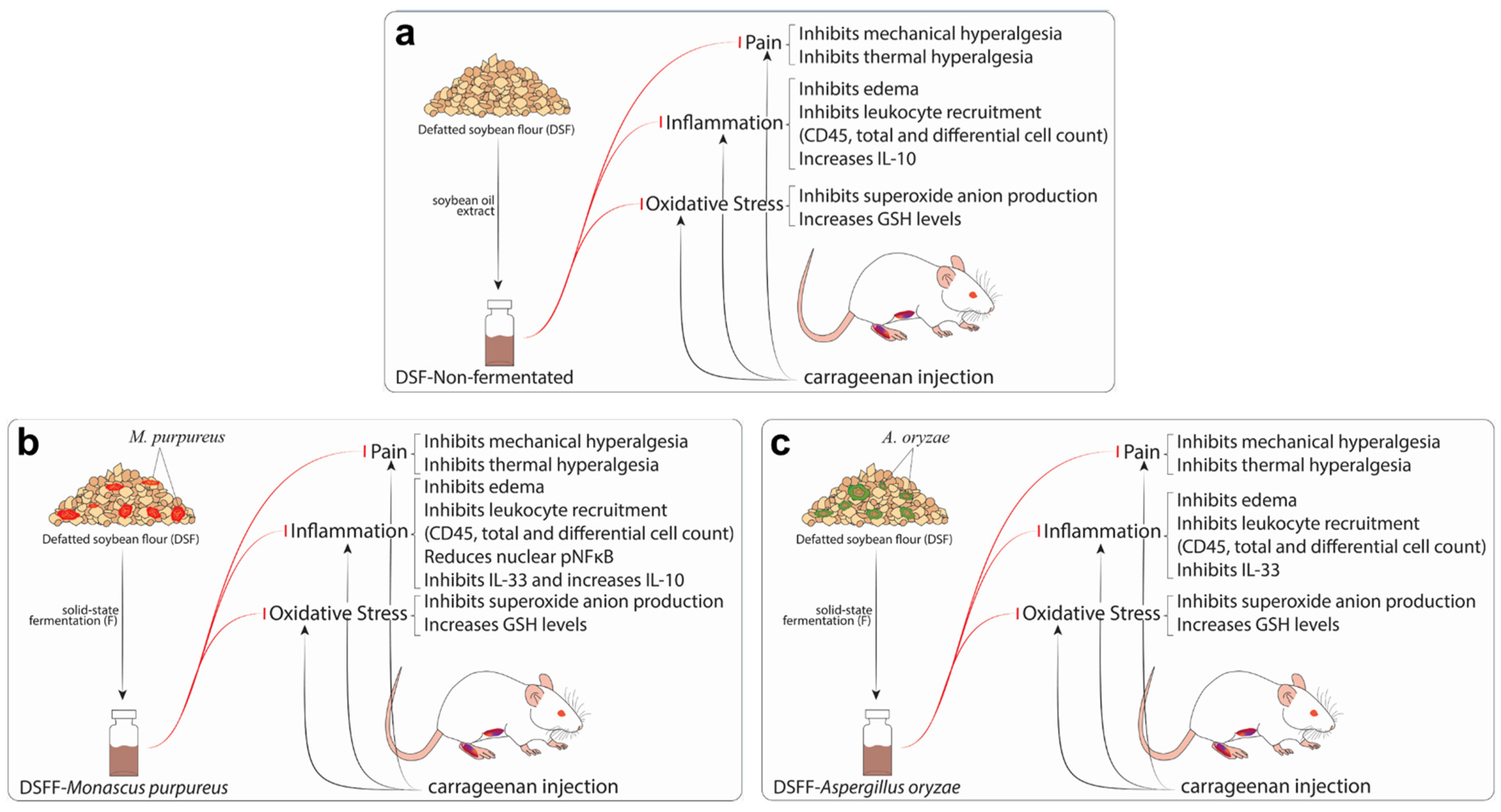
2.1. Defatted Soybean Fermentation by M. purpureus and A. ooryzae (DSFF-Mp/DSFF-Ao)

The DSF from commercial source was used as a substrate for solid-state fermentation. M. purpureus NRRL 1992 (GenBank: JQ614061.1) was donate by the Laboratory of Biochemistry and Applied Microbiology of the Institute of Food Science and Technology of the Federal University of Rio Grande do Sul (Porto Alegre, RS, Brazil). A. oryzae IOC 3999/1998 was donated by the Oswaldo Cruz Foundation (Fiocruz, Rio de Janeiro, Brazil). The solid-state fermentation of DSF by fungi was carried out for 5 days as described by Handa et al. [7]. Samples were immediately frozen, lyophilized (Christ Alpha 2–4 LD plus, Osterode am Harz, Germany), milled (Ika A11 basic, St. Louis, MO, USA), and stored at −20 °C until the analysis.

2.2. Determination of Total Phenolic Content and Isoflavones, and Soluble Sugar Composition of Extracts

Total phenolic content assessment was performed as described by Handa et al. [12] by extracting twice from the previously defatted samples. The extracts were filtered (Millex filter-H, 0.22 µm, Millipore, Billerica, MA, USA), and concentrated using the 40 °C, lyophilized, vacuum-packed, and the dried extract was stored at −20 °C until the analysis. It was used the colorimetric method of Folin–Ciocalteu to determine the total phenolic content [29], and a standard curve of gallic acid (GA) was used. The results were expressed as mg of GA equivalents/100 mL of each extract. The isoflavones were quantified by UHPLC (Waters, Milford, MA, USA) as described [7]. The results were expressed as μmol of isoflavones/100 mL of each extract. For the evaluation of soluble sugar composition, the lyophilized extracts were solubilized in ultra-pure water and subsequently filtered (Millex-GV, PVDF hydrophilic membrane, 0.22 µm, Millipore, Billerica, MA, USA). Aliquots of 10 µL were automatically injected into an ICS 5000 (Dionex Canada Ltd., Oakville, Canada) chromatograph equipped with a CarboPac® PA1 column (250 mm × 4 mm, 10 μm; Dionex/Thermo Fisher Scientific), CarboPac® PA1 guard column (50 mm × 4 mm, 10 µm), and a pulsed amperometric detector (PAD; Dionex/Thermo Fisher Scientific). The mobile phase was comprised of 90% solvent A (ultra-pure water) and 10% solvent B (200 mmol of NaOH/L of ultra-pure water) with isocratic elution for 52 min at 1 mL/min and 25 °C. At the end chromatographic run, a column washing step was performed with 200 mmol of NaOH/L of ultra-pure water for 10 min at 25 °C followed by column stabilization with 20 mmol of NaOH/L for 15 min. For the detection of sugars, a working gold electrode connected to a pH-Ag/AgCl reference electrode (Dionex/Thermo Scientific) was used to promote the oxidation of the sugars by means of a waveform (E = potential, t = duration): E1 = +0.1 V, t1 = 400 ms; E2 = −2.0 V, t2 = 20 ms; E3 = +0.6, t3 = 10 ms; and E4= −0.10, t4 = 70 ms. Chromeleon software 6.8 (Dionex/Thermo Scientific) was used for data acquisition. The sugar content was expressed as mg of sugar/100 mL of each extract.

2.3. Chemicals

The materials were obtained from the following sources as indicated: carrageenan from Santa Cruz Biotechnology (Dallas, TX, USA), indomethacin from Sigma Chemical Co. (St. Louis, MO, USA), and sodium diclofenac (Neutaren®) from Novartis Biociências S.A. (São Paulo, SP, Brazil). The DSF was purchased from BRF Brasil Foods S.A. (Curitiba, PR, Brazil). The chemical composition of the product was as follows: 8.95% moisture, 1.07% fat, 48.96% protein (N × 6.25), 5.98% ash, and 35.04% carbohydrates by the difference from the other constituents.

2.4. Animals

Pathogen-free male Swiss mice (20–25 g, 4–6 weeks) from Londrina State University (UEL), Paraná, Brazil, and male lysozyme (LysM)-enhanced green fluorescent protein (eGFP) C57BL6 background mice (20–25 g, 4–6 weeks) from Ribeirão Preto Medical School, São Paulo, Brazil, were used in this study. Mice were housed in standard clear plastic cages, with food and water ad libitum, under a 12:12 h light/dark cycle, and at a controlled temperature room (21 °C). Behavioral tests were performed always between 9 a.m. and 5 p.m. and acclimatization in the testing room at least 1 h before the experiments. Animal care and handling procedures were in accordance with the guidelines of the International Association for Study of Pain (IASP). This study was approved by the Animal Welfare Committee of the UEL (protocol number: 4584.2014.19). All efforts were made to minimize the number of animals used and their suffering.

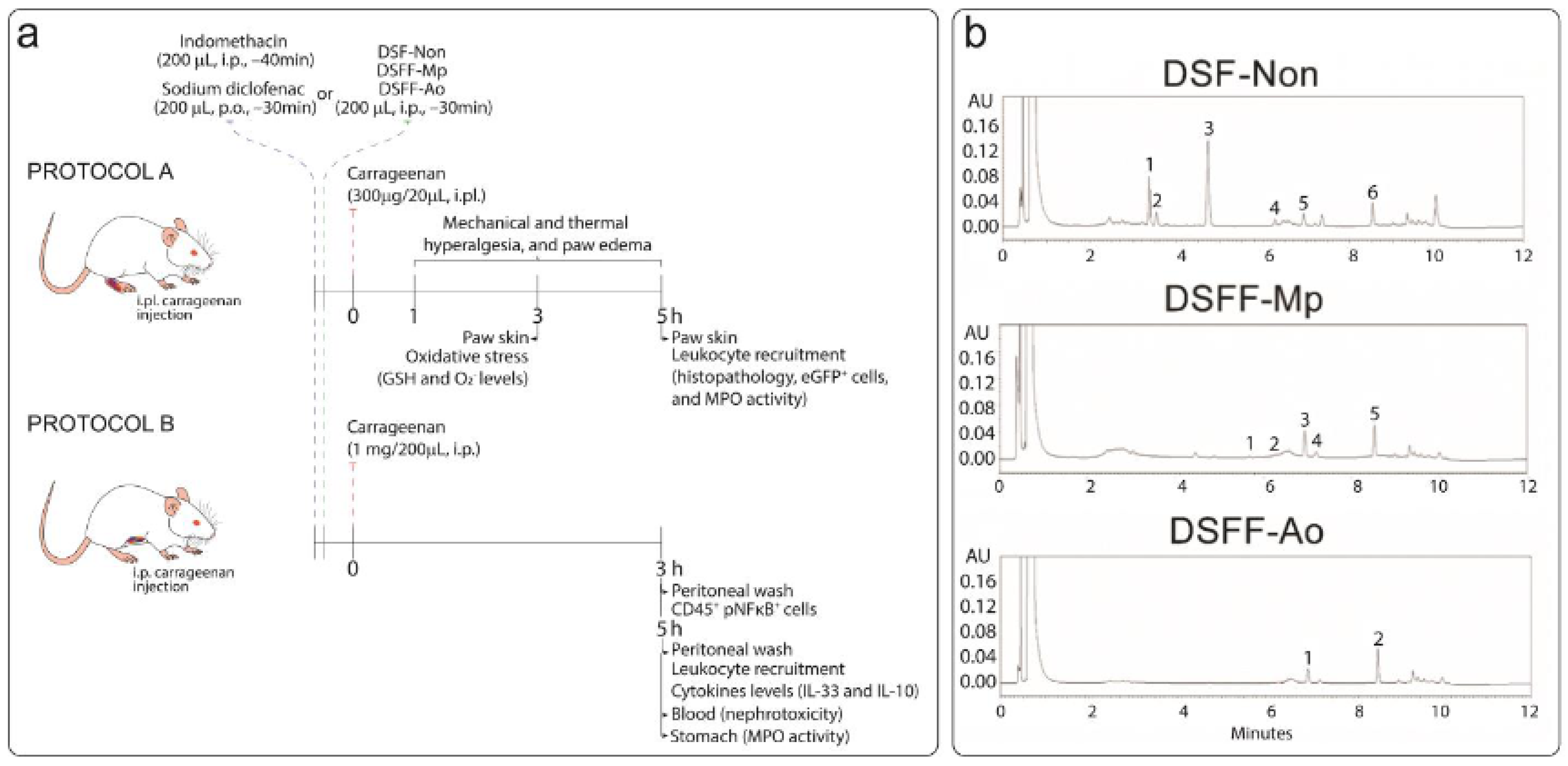
2.5. In Vivo Experimental Approaches

Mice were treated with DSF-Non, DSFF-Mp or DSFF-Ao extracts (10, 30 and 100 mg/kg, i.p., diluted in saline), 30 min before i.pl. (300 µg/20 µL) or i.p. (1 mg/cavity) injections of the flogistic agent, carrageenan. Mechanical hyperalgesia and thermal hyperalgesia, and paw edema were evaluated 1, 3, and 5 h after carrageenan i.pl. injection. Histopathology, LysM-eGFP+ cells fluorescence and myeloperoxidase (MPO) activity evaluations for the determination of leukocyte recruitment to the paw tissue, and to the peritoneum, were evaluated 5 h after carrageenan stimulus. GSH and superoxide anion levels assays were performed using paw tissue collected 3 h after carrageenan i.pl. injection for the evaluation of oxidative stress. IL-33 and IL-10 levels were evaluated 5 h after carrageenan i.p. injection. Recruited leukocytes in the peritoneum were collected 3 h after carrageenan stimulus to immunofluorescence assay staining for CD45, pNFkB and nucleus (DAPI). Stomach and blood samples were collected 5.5 h after the treatments with the extracts aiming the evaluation of possible stomach and kidney toxic effects. The doses of carrageenan and time periods for sample collection were based on previous studies from our group [30,31]. Indomethacin (5 mg/kg, i.p., diluted in tris/HCl buffer) or sodium diclofenac (200 mg/kg, p.o., diluted in saline) were used as positive controls. Figure 1a summarizes the experimental procedures applied in the present study.

2.6. Mechanical Hyperalgesia Evaluation

Mechanical hyperalgesia was assessed using an electronic version of von Frey filaments [16]. Mice were placed individually in acrylic cages with dimensions of 12 × 10 × 17 cm, composed of wire grid floors, 15–30 min before the start of testing. A tilted mirror below the grid provided a clear view of the animal’s hind paw. The test consists of evoking a hind paw flexion reflex using hand-held force transducer fitted coupled to a polypropylene tip (Electronic anesthesiometer; Insight, Ribeirao Preto, São Paulo, Brazil). The intensity of the pressure (in grams) applied by the experimenter at the moment of paw withdrawal is automatically recorded by the apparatus. The test was repeated until three subsequently consistent measurements were obtained. The mechanical hyperalgesia was evaluated before (baseline) and after carrageenan i.pl. stimulus. The results are expressed as delta (Δ) withdrawal threshold, obtained by subtraction of the baseline values from the measurements at each time point (1, 3, and 5 h after stimulus). Experimenters was always blinded to the experimental groups.

2.7. Thermal Hyperalgesia Test

Mice were placed on a hot plate apparatus (Insight, Ribeirao Preto, São Paulo, Brazil) at a temperature of 50 ± 1 °C [16]. The endpoint was characterized by the removal of the paw from the hot surface followed by clear paw flinching or licking behaviors performed by the animals. The latency time until the endpoint is automatically determined by the equipment in seconds. The maximum latency was set at 20 s to avoid tissue damage. The results are expressed by as means of latency (in seconds) of each experimental group at 1, 3, and 5 h after i.pl. carrageenan injection. Experimenters was always blinded to the experimental groups.

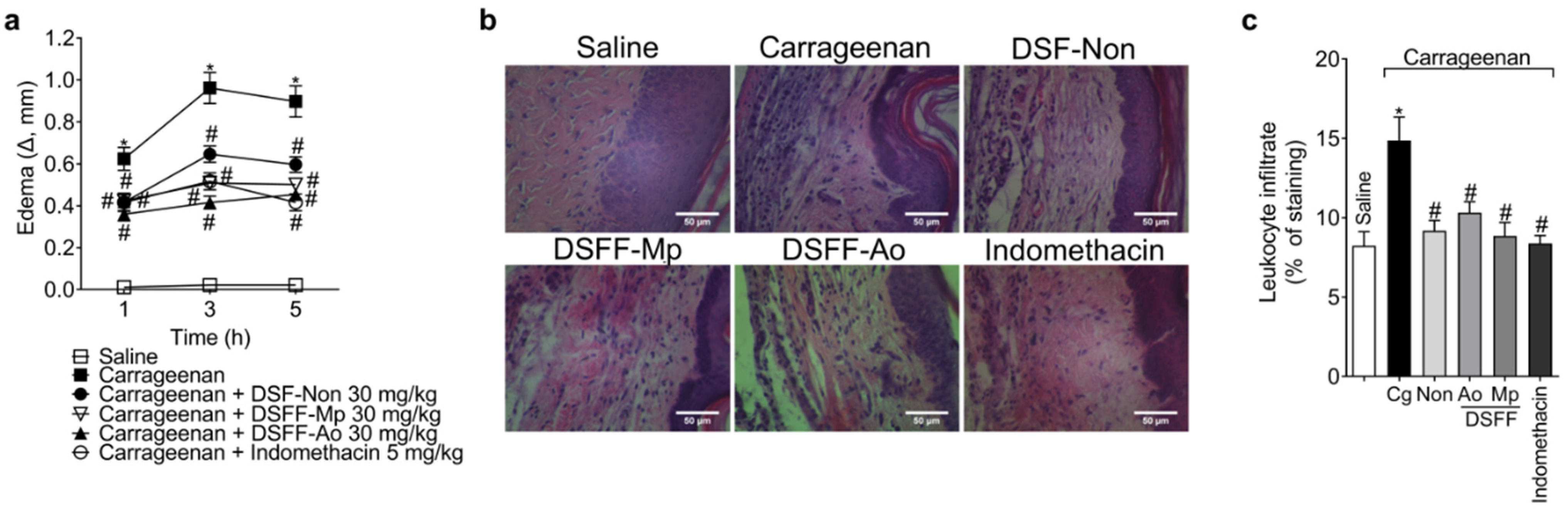
2.8. Paw Edema Assessment

Paw edema was measured using a caliper gauge 0–10 mm (Mitutoyo, Andover, Hampshire, UK). The results were expressed as the paw thickness (in mm). The results were obtained by subtracting the measurements obtained at each time point (1, 3, and 5 h after stimulus) from the baseline values.

2.9. Histopathology

Paw skin was collected 5 h after i.pl. carrageenan stimulus and fixed with 10% paraformaldehyde (PFA) by 24 h. After this initial step, samples underwent a process of embedding in paraffin. Tissue sections of 7 µm were stained with hematoxylin and eosin (H&E). Leukocyte infiltration into the cutaneous tissue were analyzed by a blinded experimenter for the treatments through digital images acquired under a conventional light microscope (40× objective). Digital images were analyzed using the ImageJ 1.44 software (http://rsb.info.nih.gov/ij/ accessed between 22 January 2020 and 21 May 2020) using color threshold tool prior to RGB image transformation, which were further used on threshold tool. Leukocyte recruitment was determined at 3,145,728 pixels area. The results are presented as leukocyte infiltrate (% of staining).

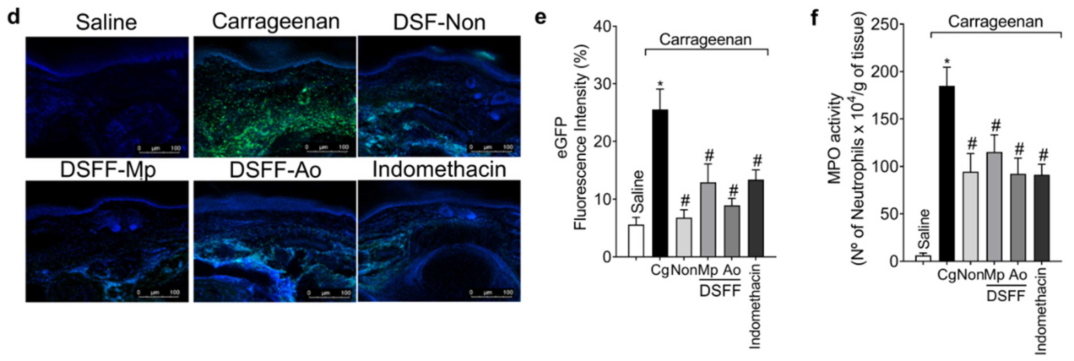
2.10. Paw Tissue Immunofluorescence

Samples of paw tissue were collected 5 h after carrageenan i.pl. stimulus and maintained in 4% PFA for 24 h, and then in 30% sucrose solution for a period of 72 h, before inclusion at optimum cutting temperature reagent (Tissue-Tek®, O.C.T. Compound, IA018, ProSciTech, Australia). Sections of 10 µm were stained for the nuclear marker Hoechst 33342 (1:500 dilution) and Fluoromount-G reagent (Thermo Fisher Scientific, Waltham, MA, USA) was put in slides to complete their assembly. Four slides per animal and four mice per group were used during the analysis. Confocal microscope (Leica TCS SP8, Leica, Wetzlar, Germany) was used to acquire images through 40× objective. Fluorescence intensity was quantified on the processed images (Leica EL6000 software, Wetzlar, Germany) by an investigator blinded to the treatment in randomly selected fields (one field per sample) as an indication of neutrophil/macrophage recruitment to the paw tissue. The results are presented as eGFP fluorescence intensity (%).

2.11. MPO Activity Assay

Neutrophil recruitment into the paw tissue and stomach were evaluated using the MPO activity colorimetric method assay [31]. Paw skin and stomach samples were collected 5 h after carrageenan stimulus and homogenized in an ice-cold 50 mM K2HPO4 buffer (pH 6.0) containing 0.5% hexadecyltrimethylammonium bromide (HTAB) using an ultra-Turrax® (IKA Labortechnik, Staufen, Germany). Homogenates were then centrifuged (16,100× g × 2 min × 4 °C), and the resulting supernatant was used for the determination of MPO activity spectrophotometrically at 450 nm. The MPO activity of samples was compared to a standard curve of neutrophils. The results are presented as number of neutrophils ×104/grams of tissues.

2.12. Evaluation of GSH and O2 Levels

Tissue was collected 3 h after carrageenan i.pl. injection and maintained at −80 °C until the evaluations of GSH and O2 levels [31]. For GSH assay, samples were homogenized (IKA Labortechnik, Staufen, Germany) in 0.02 M ethylenediamine tetraacetic acid (EDTA) buffer solution. The homogenates were then mixed with trichloroacetic acid (TCA) 50% and homogenized three times for 15 min using a vortex, followed by centrifugation (1500× g × 15 min × 4 °C). The supernatants were added to a 96-well microplate followed by addition of 200 μL of 0.2 M Tris-HCl buffer (pH 8.2) and 10 μL of 0.01 M dithiobisnitrobenzoic acid (DTNB). After 5 min, the absorbance was measured at 412 nm (Multiskan GO Microplate Spectrophorometer, Thermo Scientific, Vantaa, Finland). The values obtained were compared to a standard curve of GSH. The results are presented as GSH levels (nmol) per mg of protein of tissue. The O2 production was determined by the reduction of the redox dye nitroblue tetrazolium (NBT) reagent. Paw skin samples were homogenized (IKA Labortechnik, Staufen, Germany) with 500 μL of saline, and 50 μL of the homogenate were placed in a 96-well microplate, followed by the addition of 100 μL of NBT solution (1 mg/mL) and further incubation period for 1 h at room temperature. The supernatant was removed from the wells and the formazan precipitated was solubilized after the addition of 120 μL of 2 M KOH and 120 μL of dimethylsulfoxide (DMSO). The optical density was measured at 600 nm using a microplate spectrophotometer reader (Multiskan GO Microplate Spectrophotometer, Thermo Scientific, Vantaa, Finland). The results were presented as NBT reduction [optical density (OD)/mg of tissue].

2.13. Leukocyte Recruitment into Peritoneal Cavity

Peritoneal cavities were washed with 1 mL of phosphate-buffered saline (PBS) 5 h after carrageenan i.p. injection (1 mg/cavity) [32]. The total and differential number of leukocytes was determined in a Neubauer chamber diluted in Turk’s solution (2% acetic acid). Differential cell counts were performed using the Fast Panotic Kit for histologic analysis (Laborclin, Pinhais, Paraná, Brazil) to distinguish polymorphonuclear (PMNs) and mononuclear cells using a conventional light microscope (Olympus Optical Co., Hamburg, Germany). The results are presented as number of cells ×105 per cavity.

2.14. Cytokine Quantitation Procedure

For cytokine production assessment, peritoneal cavities were washed with 1 mL of PBS at 5 h after carrageenan i.p. administration. Samples were centrifuged (3000 rpm × 15 min × 4 °C) and the resultant supernatant were used to determine the levels of IL-33 and IL-10 by ELISA using eBioscience kits (eBioscience, San Diego, CA, USA). Initially, plates were coated overnight at 4 °C with an immunoaffinity-purified polyclonal antibody specifically for each cytokine. On the next day, recombinant murine for each cytokine standards at various dilutions and the samples were added in duplicate and incubated for an additional period of 2 h at room temperature. Subsequently, rabbit biotinylated immunoaffinity-purified antibodies were added, followed by another incubation at room temperature for 1 h. Avidin-HRP was then added to each well, and after 30 min, the plates were washed and the color reagent o-phenylenediamine was added. After blocking all reactions, measurements were performed spectrophotometrically at 450 nm (Multiskan GO Microplate Spectrophotometer, Thermo Fischer Scientific, Vantaa, Finland). The results were presented as picograms (pg) of per mL of cytokine.

2.15. Immunofluorescence Staining in Samples of Peritoneal Cavity Cells

Three hours after i.p. injection (1 mg/cavity) of carrageenan stimulus, peritoneal cavities were washed with 1 mL of phosphate-buffered saline (PBS) (30). Next, the collected samples were blocked with 5% bovine serum albumin (BSA) in phosphate buffered saline with 0.3% Triton 100×. Wash samples were then transferred to slides, and samples stained with mouse phospho-NFκB (1:200, cat. #sc-136548, Santa Cruz Bio-technology), followed by goat anti-mouse Alexa Fluor 647 (1:500, cat. #A-21236, Thermo Fisher Scientific), and the nuclei were stained with DAPI (1:500, cat. # 14285, Cayman Chemicals). In a subsequent approach, samples were stained with mouse CD45 PE/Cy5 (1:500, cat. #103110, Biolegend). Image acquisition and analysis of fluorescence intensity (CD45 and pNFκB/DAPI colocalization area (in m2) and CD45+ pNFκB/DAPI+ cell counts were performed using a confocal microscope (TSC SPB, Leica Microsystems, Manheim, Germany). The analyses reflect the average of evaluations performed in quadruplicate (four distinct fields in the sample). Scale bars are presented in each representative image contained in the results section. Analyses were always performed by a blinded experimenter and measured using confocal microscope software to provide quantitative data for the experiments.

2.16. Renal Function Tests

Blood samples were collected under isoflurane anesthesia (3%) by cardiac puncture 5.5 h after the treatments with the extracts or the nephrotoxic drug sodium diclofenac, and added into microtubes containing anticoagulant (EDTA, 5000 U/mL, Sigma Chemical Co., St. Louis, MO, USA). The plasma was separated by centrifugation (200× g, 10 min, 4 °C) and processed according to the manufacturer’s instructions for the determination of creatinine and urea levels (Labtest Diagnóstico S.A., Lagoa Santa, Minas Gerais, Brazil) as indicators of nephrotoxicity. Results are presented as mg/dL of plasma.

2.17. Statistical Analysis

Results are presented as mean ± SEM of 4–6 animals (informed in the figure caption) per group per experiment and are representative of two independent experiments. Two-way analysis of variance (ANOVA) followed by Tukey’s post hoc were used to compare groups and doses at all time points when responses were measured at different time points after stimulus injection. One-way ANOVA followed by Tukey’s post-hoc was performed in single time point experiments between experimental groups. Significance level was set at p < 0.05. All data analyses were performed using GraphPad Prism 6.0 (GraphPad Software, Inc., La Jolla, CA, USA).

3. Results

3.1. Characterization of DSF Extracts

Figure 1a presents a schematic representation of the experimental design, treatments, and parameters of analyses. Initially, the total phenolic content of DSF-Non extract was addressed (Figure 1b and Table 1). It is composed by six types of isoflavones: daidzin, glycitin, genistin, malonyl genistin, dadzein, and genistein, in a total amount of 82.043 ± 2.132 μmol of isoflavones/100 mL of extract (Figure 1b and Table 1). Next, we addressed the total phenolic and sugar contents of DSFF-Mp and DSFF-Ao extracts (Figure 1b, and Table 2 and Table 3, respectively). DSFF-Mp composition includes five types of isoflavone: malonyl daidzin, malonyl genistin, acetyl genistin, dadzein, and genistein, in a total amount of 76.040 ± 1.505 μmol of isoflavones/100 mL of extract (Figure 1b and Table 2). DSFF-Ao composition includes two types of isoflavone: dadzein and genistein, in a total amount of 38.326 ± 0.731 μmol of isoflavones/100 mL of extract (Figure 1b and Table 3). The full chromatographic profile is available elsewhere [33]. Additionally, the sugar content was determined for the two fermented extracts according to the five major sugars: glucose, fructose, sucrose, raffinose, and stachyose. The amount of sugar content of each fermented extract is shown in Table 4 (DSFF-Mp) and Table 5 (DSFF-Ao) as mg of sugar/100 mL of extract.

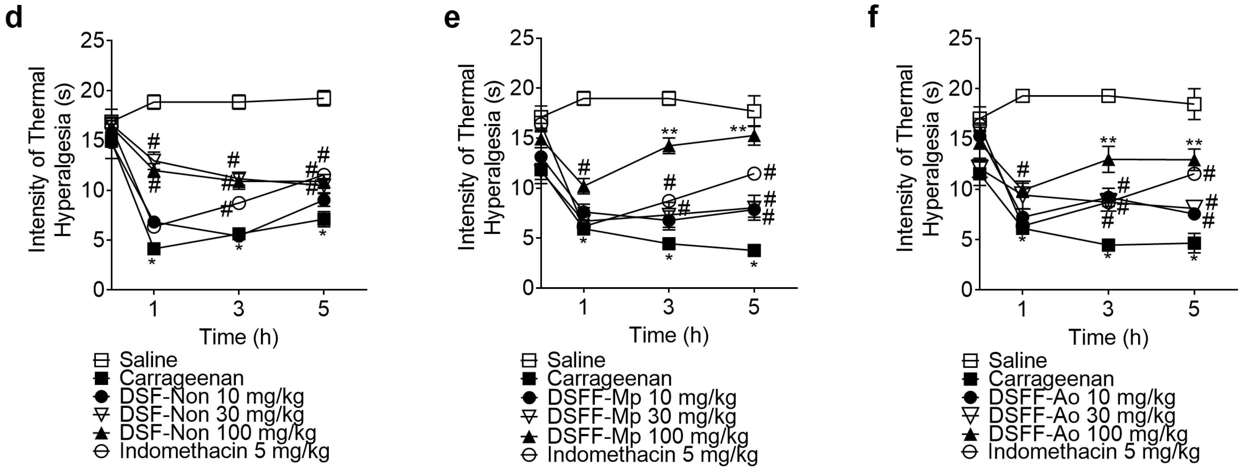
3.2. DSF-Non, DSFF-Mp, and DSFF-Ao Inhibited Carrageenan-Induced Mechanical Hyperalgesia and Thermal Hyperalgesia

Starting the in vivo experiments, a dose-response experiment for all extracts was performed with the same dose range (10, 30, and 100 mg/kg, i.p.) (Figure 2). For DSF-Non extract, the dose of 30 mg/kg inhibited carrageenan-induced mechanical hyperalgesia at all time points (1–5 h), showing an effect comparable to that of the positive control, whereas at the early time points (1 and 3 h), it had higher analgesic effect compared to the other doses. The dose of 10 mg/kg inhibited mechanical hyperalgesia at 3 and 5 h. The doses of 10 and 100 mg/kg presented a superposing profile with minor analgesic effect compared to the dose of 30 mg/kg, indicating a bell-shaped dose effect (Figure 2a). Regarding carrageenan-induced thermal hyperalgesia, the doses of 30 and 100 mg/kg inhibited thermal hyperalgesia at all time points (Figure 2d). For DSFF-Mp extract, the dose of 10 mg/kg did not inhibit mechanical hyperalgesia. The dose of 100 mg/kg inhibited it at 3 and 5 h. The dose of 30 mg/kg inhibited carrageenan-induced mechanical hyperalgesia at all time points with significant differences compared to the lower doses at 3 and 5 h, thus presenting a bell-shaped dose-effect and higher activity with the intermediary dose than the low (10 mg/kg) and high (100 mg/kg) doses (Figure 2b). In terms of thermal hyperalgesia, the dose of 100 mg/kg inhibited it at all time points with significant difference compared to the lower doses at 3 and 5 h. The dose of 30 mg/kg inhibited thermal hyperalgesia at 3 and 5 h, and the dose of 10 mg/kg, only inhibited thermal hyperalgesia at 5 h. No effect was detected by the doses of 10 and 30 mg/kg in the thermal hyperalgesia at 1 h (Figure 2e). For DSFF-Ao extract, 30 and 100 mg/kg doses inhibited mechanical hyperalgesia at all time points. The dose of 10 mg/kg had no effect upon mechanical hyperalgesia (Figure 2c). All doses of DSFF-Ao extract inhibited carrageenan-induced thermal hyperalgesia at 3 and 5 h, with the dose of 100 mk/kg presenting significant difference compared to lower doses at these time points. Only 100 mg/kg dose inhibited thermal hyperalgesia at all time points evaluated (Figure 2f). Indomethacin was used as a positive control and as expected had analgesics effects (Figure 2a–f). Considering the description of the results presented above, the dose of 30 mg/kg of all extracts was chosen for the following experiments, since it was the lowest dose that was able to inhibit the inflammatory pain parameters evaluated. Based on the above results we concluded that DSF non-fermented and fungi-fermented extracts have analgesics actions in carrageenan-induced inflammatory pain. In the following experiments, we intended to analyze the mechanisms of action by which these different extracts exert these actions to control inflammation and pain.

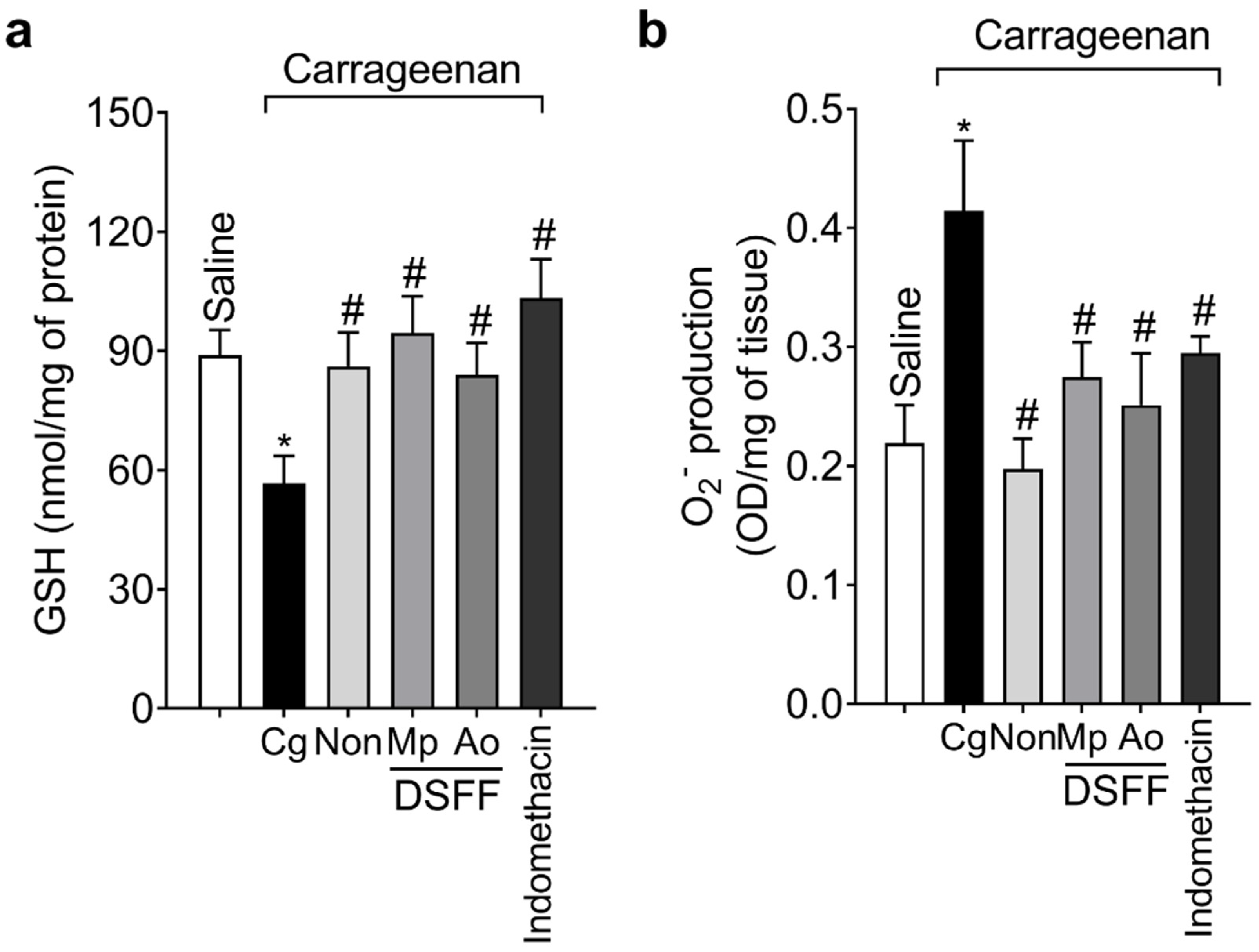
3.3. DSF-Non, DSFF-Mp, and DSFF-Ao Inhibited Carrageenan-Induced Edema, Leukocyte Recruitment, and Oxidative Stress in the Paw Tissue

In the next set of experiments, we evaluated whether the treatment with the extracts could reduce edema, infiltration of leukocytes (Figure 3), and oxidative stress (Figure 4) in the paw tissue. Carrageenan stimulus induced paw edema (Figure 3a) and leukocyte infiltration (histopathology; Figure 3b,c), which were significantly inhibited by DSF-Non, DSFF-Mp, and DSFF-Ao extracts. For a better understanding of recruited cell profile, LysM-eGFP+ cell fluorescence detection and MPO activity assay in the paw tissue were used since both parameters can be used to assess neutrophil and monocyte/macrophage counts [31,34]. All extracts inhibited the increased eGFP+ fluorescence and MPO activity induced by carrageenan indicating a reduction in the numbers of neutrophils and monocytes/macrophages (Figure 3d–f). Carrageenan injection also increased oxidative stress in the paw tissue observed through the reduction of GSH levels associated to the increase in the production of O2. This redox status was significantly reverted by the treatment with all extracts (Figure 4a,b). The control drug indomethacin inhibited all parameters (Figure 3 and Figure 4).

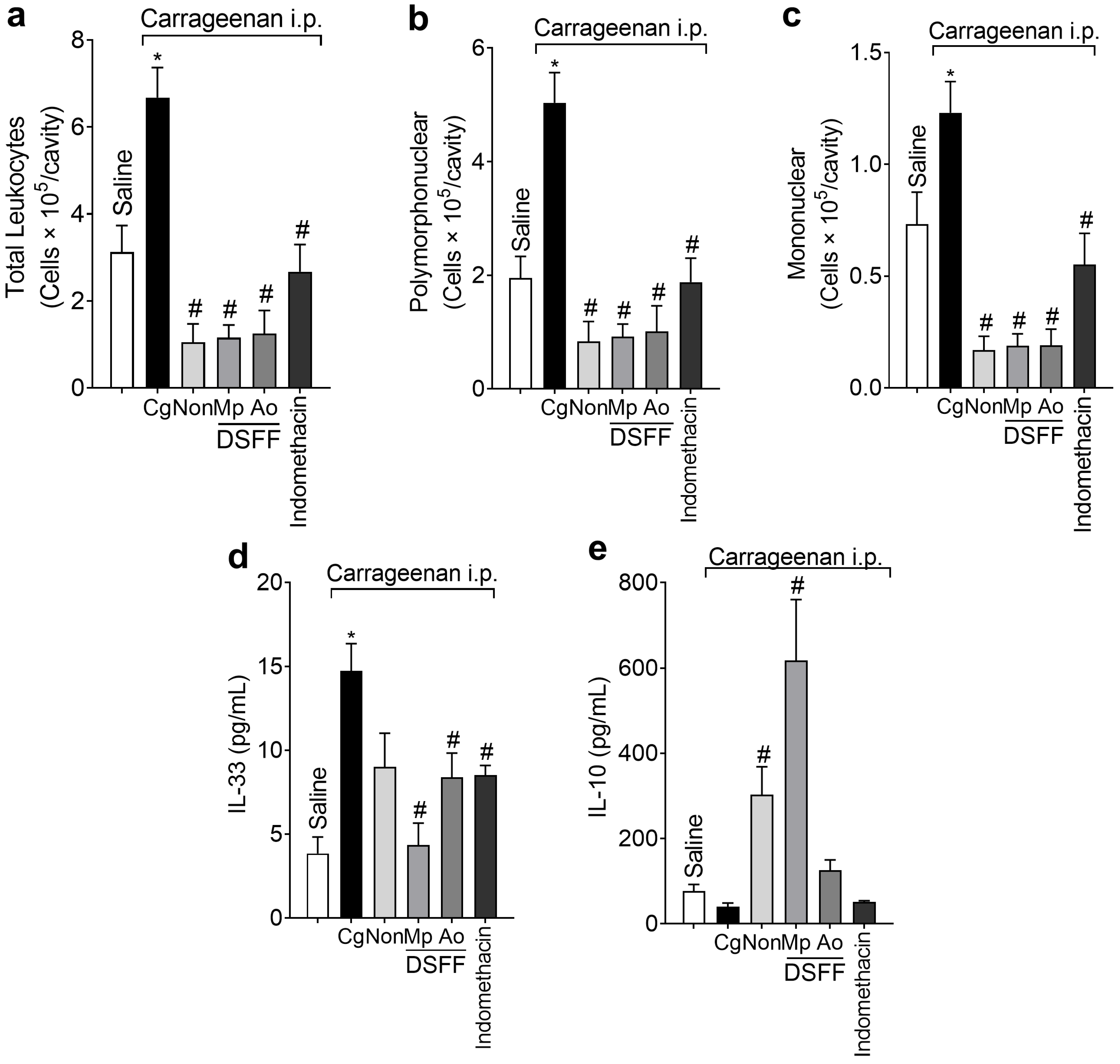
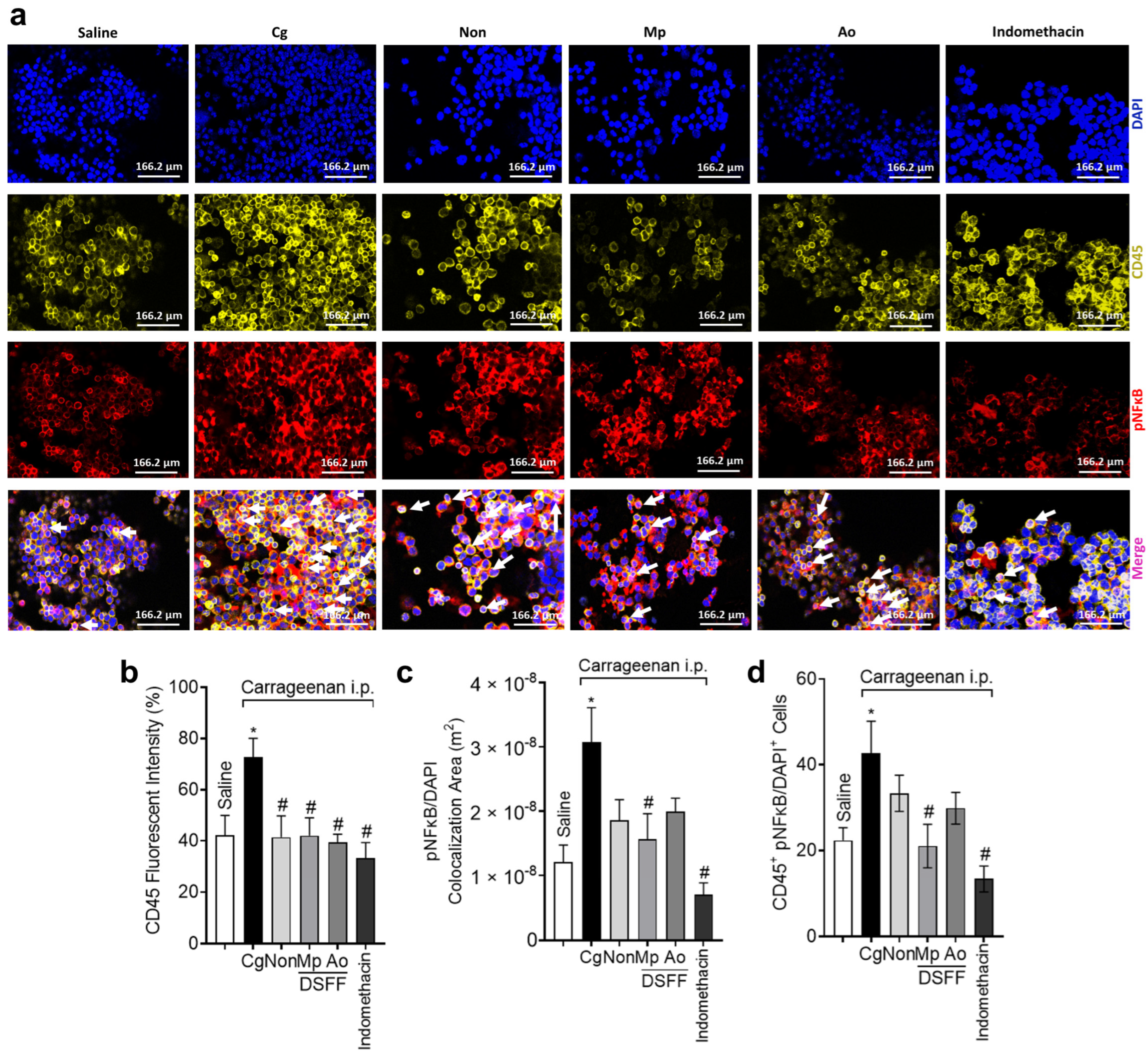
3.4. DSF-Non, DSFF-Mp, and DSFF-Ao Inhibited Leukocyte Recruitment and Differentially Modulated NFκB Activation in Peritoneal Leukocytes Recruited by Carrageenan

Treatments with DSF-Non, DSFF-Mp, and DSFF-Ao extracts inhibited total leukocytes (Figure 5a), polymorphonuclear (Figure 5b), and mononuclear (Figure 5c) cell infiltration into peritoneum triggered by carrageenan (Figure 5a–c). Interestingly, when the production of cytokines was evaluated, the mechanism of action of extracts was proven to be distinct. Carrageenan stimulus increased the production of the pro-inflammatory cytokine IL-33 without affecting the production of the anti-inflammatory cytokine IL-10. DSF-Non extract did not interfere with IL-33 production in peritoneal cavity while DSFF-Mp and DSFF-Ao extracts, and positive control indomethacin significantly inhibited its production (Figure 5d). Regarding IL-10 levels in the peritoneal cavity, DSF-Non and DSFF-Mp significantly increased its production, whereas DSFF-Ao and positive control indomethacin did not change the levels of IL-10 (Figure 5e). The pronounced trend of inducing IL-10 production by DSFF-Mp treatment compared to the DSF-Non extract was not significant. These data demonstrate that DSF-Non and fungi-fermented extracts possess anti-inflammatory properties in sterile peritonitis models by inhibiting leukocyte migration in an equivalent degree (Figure 5a–c), however, they do so with differential effects on cytokine production (Figure 5d–e). While DSF-Non acts by stimulating IL-10 and DSFF-Ao by inhibiting IL-33, DSFF-Mp acts in these two fronts, manifesting a broader spectrum of biological activity.
We further investigated potential mechanism differences among the three extracts in carrageenan peritonitis. We assessed whether the treatments with the extracts could reduce the recruitment of CD45+ hematopoietic cells, and next, whether they could target the nuclear levels of phosphorylated (p) NFκB (active form of the transcription factor NFkB) (Figure 6). DSF-Non, DSFF-Mp, and DSFF-Ao extracts all inhibited the recruitment of CD45+ cells triggered by carrageenan stimulus injection into the peritoneum (Figure 6a,b). This result agrees with the evidence that blood leukocytes of hematopoietic lineages are recruited to acute inflammatory foci [35,36]. Despite the treatments with DSF-Non and DSFF-Ao, extracts presented a trend towards inhibition relative to carrageenan group (e.g., p = 0.1595 and p = 0.2628, for pNFκB/DAPI colocalization area, respectively), only the DSFF-Mp extract was efficient in reducing the nuclear detection of pNFκB in leukocytes as demonstrated by diminished pNFκB/DAPI colocalization area (p = 0.0475) and CD45+ pNFκB/DAPI+ cell counts (p = 0.0294; Figure 6a,c,d). The positive control drug indomethacin also inhibited these parameters (Figure 6a–d).

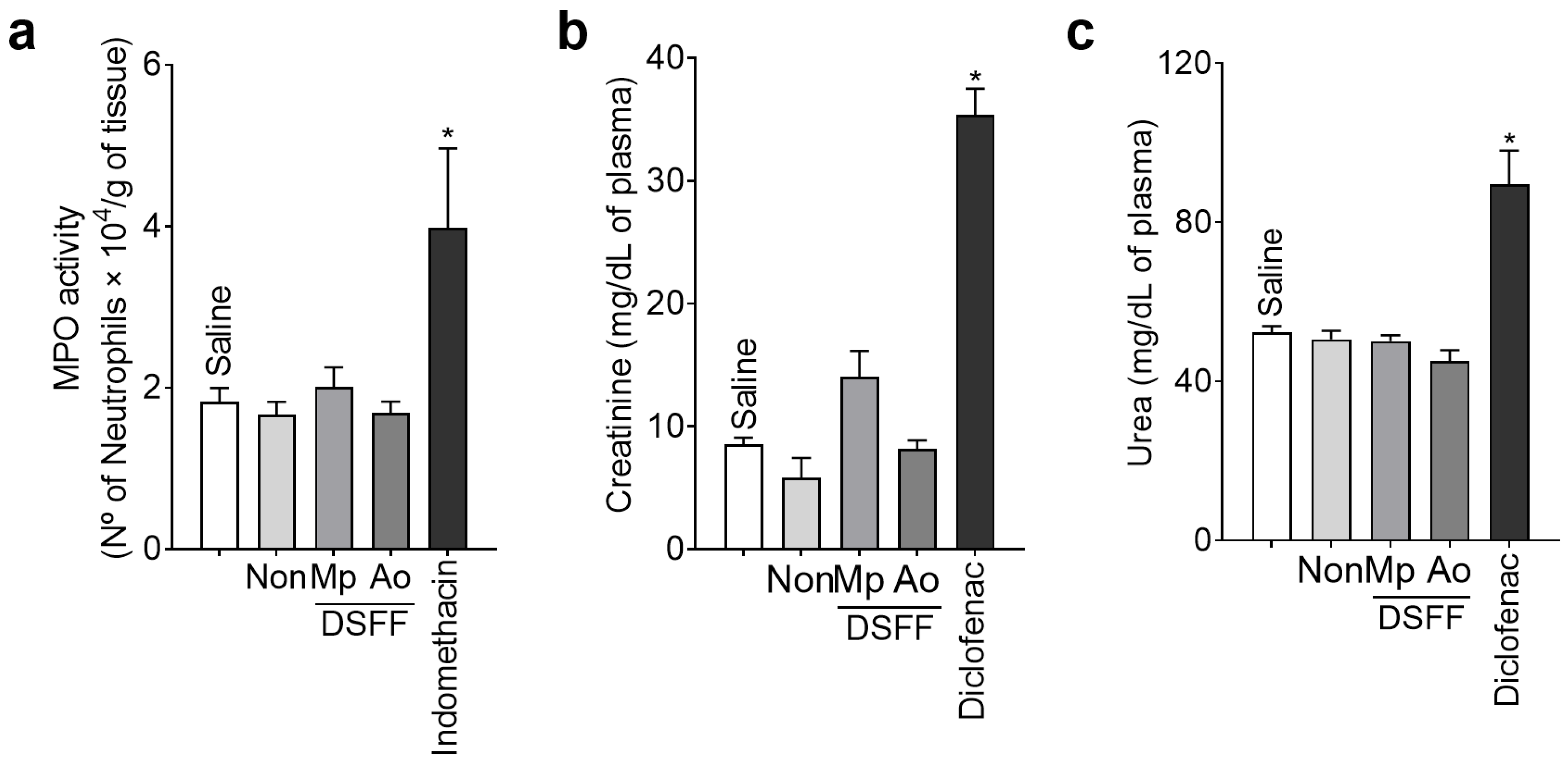
3.5. DSF-Non, DSFF-Mp, and DSFF-Ao Extracts Did Not Induce Stomach or Kidney Toxicity

The final approach of the study was to assess whether the extracts could cause damage to stomach and kidneys at a pharmacologically active dose (Figure 7). Stomach and kidneys are two major organs often affected by the adverse effects induced by non-steroidal anti-inflammatory drugs. DSF-Non, DSFF-Mp, and DSFF-Ao at the selected pharmacologically active dose (30 mg/kg) did not induce stomach or kidney toxicity, which contrasted with the positive control drugs, indomethacin (increased stomach MPO activity), and diclofenac (increased plasma creatinine and urea levels) (Figure 7), which are well-known harmful compounds to stomach and renal tissues, respectively.

4. Discussion

DSF is a by-product of soybean oil extraction. In the present work we demonstrate the analgesic, anti-inflammatory, and antioxidant properties of DSF-Non, DSSF-Mp and DSSF-Ao in carrageenan-induced inflammatory pain and peritonitis in mice. Importantly, these pharmacological effects were not accompanied by gastric or renal toxic effects, which are commonly detected upon treatment with non-steroidal anti-inflammatory drugs [37,38]. Carrageenan models of inflammatory pain are widely used to screen compounds and extracts with analgesic and anti-inflammatory activities as well as their mechanisms of action, which supports the importance of the present data [39,40,41,42].
LysM and MPO are expressed by granulocytes, such as neutrophils, and mononuclear cells such as macrophages [31,34]. Neutrophils produce prostanoids including prostaglandin E2 (PGE2), which sensitizes nociceptors causing hyperalgesia [43,44], and macrophages secrete a great variety of mediators that are chemoattractant to neutrophils and that also sensitize nociceptors [23]. Additionally, both are major sources of reactive oxygen species [16], that induce inflammatory pain [17,18]. Together with the identified analgesic effects, these data evidenced that non-fermented and fungi fermented DSF extracts are capable of inhibiting the classical innate immune response evoked by carrageenan that triggers inflammatory pain. Isoflavones are distributed in soybean as aglycones and glycosylated forms. Aglycone forms are considered to have better antioxidant activity [7]. The fermentation process of DSF contributed to the conversion of β-glucoside isoflavones (e. g. daidzin and genistin) into aglycones (daidzein and genistein, respectively) by means of β-glucosidase synthesized by M. purpureus and A. oryzae [7]. Daidzein and genistein have an important role in controlling excessive oxidative stress by inhibition of cytochrome c release and by regulating the reactive oxygen species-NF-kB positive feedback loop [45,46]. Genistein has high free radical scavenging capacity due to an additional hydroxyl group on its AC-ring [12]. Daidzein antioxidants properties may be improved by its metabolites O-desmethylangolensin (O-DMA) and equol [47]. Importantly, the solid-state fermentation process of soybeans also promotes the upgrade of phenolic antioxidants [48], and recently, optimum conditions using M. purpureus or A. oryzae aiming to improve the antioxidant activities were described [7]. Although part of the antioxidant properties of fermented extracts are dependent on isoflavones, other phenolic compounds may act as powerful antioxidants by transferring hydrogen atoms or single electrons [12]. Phenolic compounds are known to exhibit a scavenging effect for free radicals and metal-chelating ability [48]. Vanillic, ferulic, syringic, and chlorogenic acids are among the phenolic compounds in the whole soybean [12]. As we identified here, fermentation process using M. purpureus or A. oryzae increases both the final content of aglycones forms, including daidzein and genistein, and total phenolic compounds. Therefore, this food industry technology, in addition to promoting improvement in food quality, should be considered to produce potential antioxidant substances for the treatment of inflammation and inflammatory pain.
NFκB represents a crucial pro-inflammatory transcription factor involved in the regulation of several downstream pro-inflammatory genes as well as can be activated by pro-inflammatory cytokines and elevated oxidative reactions [49,50,51]. We observed that all extracts inhibited the migration of total leukocytes, neutrophils, and mononuclear cells into the peritoneum cavity. Furthermore, all extracts reduced the recruitment of CD45+ hematopoietic cells. However, only DSFF-Mp extract could significantly reduce the nuclear detection of pNFκB in CD45+ hematopoietic cells. This result highlights that although a trend was also observed with the other two DSF extracts, DSF fermentation by M. purpureus created an extract that has pNFkB as a molecular target in CD45+ hematopoietic cells, which did not occur with the other DSF extracts at the same degree. NFκB can be activated by IL-33 secreted in response to endogenous or exogenous molecules [52,53]. Neutrophils and macrophages are responsive to IL-33, which is chemoattractant to neutrophils and activates macrophages to produce inflammatory cytokines upon IL-33 signaling [23]. Accordingly, inhibiting IL-33 signaling (through soluble IL-33 receptor–sST2, or ST2 knockout) reduces pain, edema, and leukocyte recruitment [19,20]. Therefore, the effects conferred by treatment with fermented extracts might be explained by the reduction of IL-33. On the other hand, IL-10 is known to inhibit NFκB signaling [54,55]. The approximate 8-fold increase of IL-10 levels upon DSFF-Mp treatment when compared to the saline group was a striking find. This anti-inflammatory effect might explain the inhibition of nuclear pNFκB in CD45+ hematopoietic cells under DSFF-Mp treatment in peritonitis model. Evidence showed that IL-10 negatively regulates the IL-33/ST2 signaling axis in macrophages in vivo [56]. This information aligns with the present data. Moreover, IL-10 has a crucial role in limiting inflammation and inflammatory pain and limits the production of inflammatory cytokines [25,57]. For instance, IL-10 genetic deletion enhances pro-inflammatory cytokine production and the overall inflammatory response, including fever [58]. IL-10 also reduces leukocyte recruitment and hyperalgesia and oxidative stress triggered by inflammatory molecules [59,60,61]. Thus, increasing IL-10 levels might explain the analgesic and anti-inflammatory actions of DSF-Non. In turn, the activity of DSFF-Ao is related to inhibiting IL-33 production. However, among the three extracts, the DSFF-Mp could target IL-33 production and pNFkB as well as enhance IL-10 levels. Figure 8 summarizes and highlights the mechanistic differences among the DSF extracts tested in the present study.
Inflammation and pain are complex phenomena and there is a plethora of mechanisms that can be investigated and potentially be targeted by DSF extracts. To the best of our knowledge, this is the first study highlighting DSF-Non, DSFF-Mp, and DSFF-Ao extracts inhibiting pain and inflammation in carrageenan paw and peritoneal inflammatory responses as well as demonstrating differences in the mechanism of action among those extracts. It is of no less importance to note that the analgesic and anti-inflammatory activities of DSF extracts were not accompanied by adverse effects in major organs such as stomach and kidneys, which are known side effect targets of non-steroidal anti-inflammatory drugs. This study also raises the importance of further exploring by-products of food processing as sources of natural medicines.
Despite the novelty of the present study, it has limitations in addition to opening novel venues in this field. For instance, the carrageenan-triggered inflammation is a classical model to screen and determine the mechanism of action of novel drugs and extracts [42,62]. However, it is an acute inflammation model with characteristics of innate immune response [42], therefore, studies using chronic inflammation or adaptive immune responses might bring an additional understanding of DSF-Non, DSFF-Mp, and DSFF-Ao activity and mechanism of action. There are other pain contexts in addition to inflammation such as cancer pain, neuropathic pain, and drug-induced pain in which the activity of DSF extracts can be assessed considering the role of NFkB, IL-33, and IL-10 in those other types of pain [63,64,65]. Hence, considering these mechanisms of action, the biological effects of DSF extracts can be investigated in models of varied diseases such as asthma, rheumatoid arthritis, gout arthritis, wound healing, inflammatory bowel diseases, and atherosclerosis [66,67,68], with the potential of fostering their therapeutic applicability. Finally, translational investigation is also important. These are some examples of the limitations and potential follow-up outcomes of the present study.

5. Conclusions

In summary, to the best of our knowledge, we demonstrated for the first time that the treatment with extracts of non-fermented DSF or DSF extracts fermented by M. puprpureus or A. oryzae present analgesic and anti-inflammatory activities in carrageenan paw inflammation and peritonitis models. It is interesting that all three extracts reduce disease parameters triggered by carrageenan such as leukocyte recruitment, oxidative stress, mechanical hyperalgesia, and thermal hyperalgesia. In particular, despite a similar disease outcome, the three extracts have different mechanisms of action. DSF-Non induces IL-10 production, DSFF-Ao reduces IL-33 production, and DSFF-Mp induces IL-10 production, reduces IL-33 production and NFkB activation in CD45+ hematopoietic cells. Thereby, our experimental design could identify some mechanistic differences among DSF extracts, demonstrating that fermentation can be applied to shape the biological activities of DSF extracts. Thus, the present study advances in the understanding of how the fermentation of DSF extracts provide analgesic and anti-inflammatory effects. Taken together with the nutritional values of fermented defatted soy products [69], the present results unveil that they can be used for the treatment of pain and inflammation, which based on the present result, can be seen as medicines that still have the potential to be exploited to promote beneficial health effects.

Author Contributions

M.F.M., M.M.B., S.M.B., C.L.H., M.A.Q.-C., C.R.F., S.S.M., S.B.-G., K.C.A. and T.H.Z. performed experiments and analyzed the results. W.A.S., S.R.G. and E.I.I. contributed with fundamental reagents, analytical tools, and expert intellectual support for the study. W.A.V. and R.C. were responsible for the experimental design, funding acquisition, supervision and analyzed the results. M.F.M., S.M.B., W.A.V. and R.C. wrote the original draft of the manuscript. All authors have read and agreed to the published version of the manuscript.

Funding

This work was supported by Brazilian grants from Conselho Nacional de Desenvolvimento Científico e Tecnológico (CNPq) grant call Universal (#405027/2021–4; #427946/2018-2); CNPq senior research fellowship (#309633/2021-4; #307852/2019-9); PPSUS grant funded by Decit/SCTIE/MS intermediated by CNPq with support of Fundação Araucária and SESA-PR (agreement #041/2017); PRONEX grant supported by SETI/Fundação Araucária and MCTI/CNPq, and Governo do Estado do Paraná (agreement #014/2017); Fundação Nacional de Desenvolvimento do Ensino Superior Particular (FUNADESP; grant number #5301159) research fellowship and CAPES (finance code #001).

Institutional Review Board Statement

All procedures were performed in accordance with local, national, and international standards of ethics. Animal handling and welfare were approved by the Universidade Estadual de Londrina Ethics Committee for animal experimentation (CEUA-UEL, protocol number 4584.2014.19 approved on 30 April 2014). Animal studies are reported in compliance with the ARRIVE guidelines.

Informed Consent Statement

Not applicable.

Data Availability Statement

Authors should be contacted for data and materials requests.

Acknowledgments

The authors gratefully acknowledge the technical assistance of Maria Rosana F. De Paula. We thank the access free-of-charge to the equipment core facility CMLP-UEL (Central Multiusuário de Laboratórios de Pesquisa da Universidade Estadual de Londrina).

Conflicts of Interest

The authors declare no conflict of interest.

References

  1. Newman, D.J.; Cragg, G.M. Natural Products as Sources of New Drugs over the Nearly Four Decades from 01/1981 to 09/2019. J. Nat. Prod. 2020, 83, 770–803. [Google Scholar] [CrossRef] [PubMed]
  2. Sugano, M. (Ed.) Soy in Health and Disease Prevention, 1st ed.; CRC Press: Boca Raton, FL, USA, 2019. [Google Scholar]
  3. Villalobos Mdel, C.; Serradilla, M.J.; Martin, A.; Ordiales, E.; Ruiz-Moyano, S.; Cordoba Mde, G. Antioxidant and antimicrobial activity of natural phenolic extract from defatted soybean flour by-product for stone fruit postharvest application. J. Sci. Food Agric. 2016, 96, 2116–2124. [Google Scholar] [CrossRef] [PubMed]
  4. Genovese, M.I.; Barbosa, A.C.; Pinto Mda, S.; Lajolo, F.M. Commercial soy protein ingredients as isoflavone sources for functional foods. Plant Foods Hum. Nutr. 2007, 62, 53–58. [Google Scholar] [CrossRef] [PubMed]
  5. Izumi, T.; Piskula, M.K.; Osawa, S.; Obata, A.; Tobe, K.; Saito, M.; Kataoka, S.; Kubota, Y.; Kikuchi, M. Soy isoflavone aglycones are absorbed faster and in higher amounts than their glucosides in humans. J. Nutr. 2000, 130, 1695–1699. [Google Scholar] [CrossRef] [PubMed]
  6. Faraj, A.; Vasanthan, T. Soybean Isoflavones: Effects of Processing and Health Benefits. Food Rev. Int. 2004, 20, 51–75. [Google Scholar] [CrossRef]
  7. Handa, C.L.; Couto, U.R.; Vicensoti, A.H.; Georgetti, S.R.; Ida, E.I. Optimisation of soy flour fermentation parameters to produce beta-glucosidase for bioconversion into aglycones. Food Chem. 2014, 152, 56–65. [Google Scholar] [CrossRef]
  8. Pyo, Y.H.; Seong, K.S. Hypolipidemic effects of Monascus-fermented soybean extracts in rats fed a high-fat and -cholesterol diet. J. Agric. Food Chem. 2009, 57, 8617–8622. [Google Scholar] [CrossRef]
  9. Chiang, S.S.; Chang, S.P.; Pan, T.M. Osteoprotective effect of Monascus-fermented dioscorea in ovariectomized rat model of postmenopausal osteoporosis. J. Agric. Food Chem. 2011, 59, 9150–9157. [Google Scholar] [CrossRef]
  10. Zhang, X.-Y.; Chen, J.; Li, X.-L.; Yi, K.; Ye, Y.; Liu, G.; Wang, S.-F.; Hu, H.-L.; Zou, L.; Wang, Z.-G. Dynamic changes in antioxidant activity and biochemical composition of tartary buckwheat leaves during Aspergillus niger fermentation. J. Funct. Foods 2017, 32, 375–381. [Google Scholar] [CrossRef]
  11. Da Silva, L.; Celeghini, R.; Chang, Y. Effect of the fermentation of whole soybean flour on the conversion of isoflavones from glycosides to aglycones. Food Chem. 2011, 128, 640–644. [Google Scholar] [CrossRef]
  12. Handa, C.L.; de Lima, F.S.; Guelfi, M.F.; Georgetti, S.R.; Ida, E.I. Multi-response optimisation of the extraction solvent system for phenolics and antioxidant activities from fermented soy flour using a simplex-centroid design. Food Chem. 2016, 197, 175–184. [Google Scholar] [CrossRef] [PubMed]
  13. Medzhitov, R. Inflammation 2010: New adventures of an old flame. Cell 2010, 140, 771–776. [Google Scholar] [CrossRef] [PubMed]
  14. Peirs, C.; Seal, R.P. Targeting Toll-like receptors to treat chronic pain. Nat. Med. 2015, 21, 1251–1252. [Google Scholar] [CrossRef] [PubMed]
  15. Verri, W.A., Jr.; Cunha, T.M.; Parada, C.A.; Poole, S.; Cunha, F.Q.; Ferreira, S.H. Hypernociceptive role of cytokines and chemokines: Targets for analgesic drug development? Pharmacol. Ther. 2006, 112, 116–138. [Google Scholar] [CrossRef]
  16. Maioli, N.A.; Zarpelon, A.C.; Mizokami, S.S.; Calixto-Campos, C.; Guazelli, C.F.; Hohmann, M.S.; Pinho-Ribeiro, F.A.; Carvalho, T.T.; Manchope, M.F.; Ferraz, C.R.; et al. The superoxide anion donor, potassium superoxide, induces pain and inflammation in mice through production of reactive oxygen species and cyclooxygenase-2. Braz. J. Med. Biol. Res. 2015, 48, 321–331. [Google Scholar] [CrossRef] [PubMed]
  17. Khattab, M.M. TEMPOL, a membrane-permeable radical scavenger, attenuates peroxynitrite- and superoxide anion-enhanced carrageenan-induced paw edema and hyperalgesia: A key role for superoxide anion. Eur. J. Pharmacol. 2006, 548, 167–173. [Google Scholar] [CrossRef]
  18. Wang, Z.Q.; Porreca, F.; Cuzzocrea, S.; Galen, K.; Lightfoot, R.; Masini, E.; Muscoli, C.; Mollace, V.; Ndengele, M.; Ischiropoulos, H.; et al. A newly identified role for superoxide in inflammatory pain. J. Pharmacol. Exp. Ther. 2004, 309, 869–878. [Google Scholar] [CrossRef]
  19. Zarpelon, A.C.; Cunha, T.M.; Alves-Filho, J.C.; Pinto, L.G.; Ferreira, S.H.; McInnes, I.B.; Xu, D.; Liew, F.Y.; Cunha, F.Q.; Verri, W.A., Jr. IL-33/ST2 signalling contributes to carrageenin-induced innate inflammation and inflammatory pain: Role of cytokines, endothelin-1 and prostaglandin E2. Br. J. Pharmacol. 2013, 169, 90–101. [Google Scholar] [CrossRef]
  20. Verri, W.A., Jr.; Guerrero, A.T.; Fukada, S.Y.; Valerio, D.A.; Cunha, T.M.; Xu, D.; Ferreira, S.H.; Liew, F.Y.; Cunha, F.Q. IL-33 mediates antigen-induced cutaneous and articular hypernociception in mice. Proc. Natl. Acad. Sci. USA 2008, 105, 2723–2728. [Google Scholar] [CrossRef]
  21. Zarpelon, A.C.; Rodrigues, F.C.; Lopes, A.H.; Souza, G.R.; Carvalho, T.T.; Pinto, L.G.; Xu, D.; Ferreira, S.H.; Alves-Filho, J.C.; McInnes, I.B.; et al. Spinal cord oligodendrocyte-derived alarmin IL-33 mediates neuropathic pain. FASEB J. 2016, 30, 54–65. [Google Scholar] [CrossRef]
  22. Liu, B.; Tai, Y.; Achanta, S.; Kaelberer, M.M.; Caceres, A.I.; Shao, X.; Fang, J.; Jordt, S.E. IL-33/ST2 signaling excites sensory neurons and mediates itch response in a mouse model of poison ivy contact allergy. Proc. Natl. Acad. Sci. USA 2016, 113, E7572–E7579. [Google Scholar] [CrossRef]
  23. Verri, W.A., Jr.; Souto, F.O.; Vieira, S.M.; Almeida, S.C.; Fukada, S.Y.; Xu, D.; Alves-Filho, J.C.; Cunha, T.M.; Guerrero, A.T.; Mattos-Guimaraes, R.B.; et al. IL-33 induces neutrophil migration in rheumatoid arthritis and is a target of anti-TNF therapy. Ann. Rheum. Dis. 2010, 69, 1697–1703. [Google Scholar] [CrossRef] [PubMed]
  24. Iyer, S.S.; Cheng, G. Role of interleukin 10 transcriptional regulation in inflammation and autoimmune disease. Crit. Rev. Immunol. 2012, 32, 23–63. [Google Scholar] [CrossRef] [PubMed]
  25. Poole, S.; Cunha, F.Q.; Selkirk, S.; Lorenzetti, B.B.; Ferreira, S.H. Cytokine-mediated inflammatory hyperalgesia limited by interleukin-10. Br. J. Pharmacol. 1995, 115, 684–688. [Google Scholar] [CrossRef]
  26. Keeble, J.E.; Bodkin, J.V.; Liang, L.; Wodarski, R.; Davies, M.; Fernandes, E.S.; Coelho Cde, F.; Russell, F.; Graepel, R.; Muscara, M.N.; et al. Hydrogen peroxide is a novel mediator of inflammatory hyperalgesia, acting via transient receptor potential vanilloid 1-dependent and independent mechanisms. Pain 2009, 141, 135–142. [Google Scholar] [CrossRef]
  27. Ghanem, K.Z.; Mahran, M.Z.; Ramadan, M.M.; Ghanem, H.Z.; Fadel, M.; Mahmoud, M.H. A comparative study on flavour components and therapeutic properties of unfermented and fermented defatted soybean meal extract. Sci. Rep. 2020, 10, 5998. [Google Scholar] [CrossRef] [PubMed]
  28. Kim, S.H.; Kang, S.A. Antioxidant and Anti-Diabetic Activities of Soybean Fermented with Monascus. Korean J. Food Nutr. 2021, 34, 187–195. [Google Scholar]
  29. Kumazawa, S.; Taniguchi, M.; Suzuki, Y.; Shimura, M.; Kwon, M.S.; Nakayama, T. Antioxidant activity of polyphenols in carob pods. J. Agric. Food Chem. 2002, 50, 373–377. [Google Scholar] [CrossRef]
  30. Pinho-Ribeiro, F.A.; Zarpelon, A.C.; Fattori, V.; Manchope, M.F.; Mizokami, S.S.; Casagrande, R.; Verri, W.A., Jr. Naringenin reduces inflammatory pain in mice. Neuropharmacology 2016, 105, 508–519. [Google Scholar] [CrossRef]
  31. Mizokami, S.S.; Hohmann, M.S.; Staurengo-Ferrari, L.; Carvalho, T.T.; Zarpelon, A.C.; Possebon, M.I.; de Souza, A.R.; Veneziani, R.C.; Arakawa, N.S.; Casagrande, R.; et al. Pimaradienoic Acid Inhibits Carrageenan-Induced Inflammatory Leukocyte Recruitment and Edema in Mice: Inhibition of Oxidative Stress, Nitric Oxide and Cytokine Production. PLoS ONE 2016, 11, e0149656. [Google Scholar] [CrossRef]
  32. Zucoloto, A.Z.; Manchope, M.F.; Staurengo-Ferrari, L.; Alves-Filho, J.C.; Cunha, T.M.; Antunes, M.M.; Menezes, G.B.; Cunha, F.Q.; Casagrande, R.; Verri, W.A., Jr. Probucol attenuates overt pain-like behavior and carrageenan-induced inflammatory hyperalgesia and leukocyte recruitment by inhibiting NF-small ka, CyrillicB activation and cytokine production without antioxidant effects. Inflamm. Res. 2017, 66, 591–602. [Google Scholar] [CrossRef]
  33. Handa, C.L.; de Lima, F.S.; Guelfi, M.F.G.; Fernandes, M.D.S.; Georgetti, S.R.; Ida, E.I. Parameters of the fermentation of soybean flour by Monascus purpureus or Aspergillus oryzae on the production of bioactive compounds and antioxidant activity. Food Chem. 2019, 271, 274–283. [Google Scholar] [CrossRef] [PubMed]
  34. Sullivan, D.P.; Bui, T.; Muller, W.A.; Butin-Israeli, V.; Sumagin, R. In vivo imaging reveals unique neutrophil transendothelial migration patterns in inflamed intestines. Mucosal Immunol. 2018, 11, 1571–1581. [Google Scholar] [CrossRef]
  35. Kolaczkowska, E.; Kubes, P. Neutrophil recruitment and function in health and inflammation. Nat. Rev. Immunol. 2013, 13, 159–175. [Google Scholar] [CrossRef] [PubMed]
  36. Caiado, F.; Pietras, E.M.; Manz, M.G. Inflammation as a regulator of hematopoietic stem cell function in disease, aging, and clonal selection. J. Exp. Med. 2021, 218, e20201541. [Google Scholar] [CrossRef]
  37. Ungprasert, P.; Cheungpasitporn, W.; Crowson, C.S.; Matteson, E.L. Individual non-steroidal anti-inflammatory drugs and risk of acute kidney injury: A systematic review and meta-analysis of observational studies. Eur. J. Intern. Med. 2015, 26, 285–291. [Google Scholar] [CrossRef] [PubMed]
  38. Labianca, R.; Sarzi-Puttini, P.; Zuccaro, S.M.; Cherubino, P.; Vellucci, R.; Fornasari, D. Adverse effects associated with non-opioid and opioid treatment in patients with chronic pain. Clin. Drug Investig. 2012, 32 (Suppl. S1), 53–63. [Google Scholar] [CrossRef]
  39. Patil, K.R.; Mahajan, U.B.; Unger, B.S.; Goyal, S.N.; Belemkar, S.; Surana, S.J.; Ojha, S.; Patil, C.R. Animal Models of Inflammation for Screening of Anti-inflammatory Drugs: Implications for the Discovery and Development of Phytopharmaceuticals. Int. J. Mol. Sci. 2019, 20, 4367. [Google Scholar] [CrossRef]
  40. Panthong, A.; Norkaew, P.; Kanjanapothi, D.; Taesotikul, T.; Anantachoke, N.; Reutrakul, V. Anti-inflammatory, analgesic and antipyretic activities of the extract of gamboge from Garcinia hanburyi Hook f. J. Ethnopharmacol. 2007, 111, 335–340. [Google Scholar] [CrossRef]
  41. Tsai, D.S.; Huang, M.H.; Tsai, J.C.; Chang, Y.S.; Chiu, Y.J.; Lin, Y.C.; Wu, L.Y.; Peng, W.H. Analgesic and Anti-Inflammatory Activities of Rosa taiwanensis Nakai in Mice. J. Med. Food 2015, 18, 592–600. [Google Scholar] [CrossRef]
  42. Lopes, A.H.; Silva, R.L.; Fonseca, M.D.; Gomes, F.I.; Maganin, A.G.; Ribeiro, L.S.; Marques, L.M.M.; Cunha, F.Q.; Alves-Filho, J.C.; Zamboni, D.S.; et al. Molecular basis of carrageenan-induced cytokines production in macrophages. Cell Commun. Signal. 2020, 18, 141. [Google Scholar] [CrossRef]
  43. Cunha, T.M.; Verri, W.A., Jr.; Schivo, I.R.; Napimoga, M.H.; Parada, C.A.; Poole, S.; Teixeira, M.M.; Ferreira, S.H.; Cunha, F.Q. Crucial role of neutrophils in the development of mechanical inflammatory hypernociception. J. Leukoc. Biol. 2008, 83, 824–832. [Google Scholar] [CrossRef]
  44. Guerrero, A.T.; Verri, W.A., Jr.; Cunha, T.M.; Silva, T.A.; Schivo, I.R.; Dal-Secco, D.; Canetti, C.; Rocha, F.A.; Parada, C.A.; Cunha, F.Q.; et al. Involvement of LTB4 in zymosan-induced joint nociception in mice: Participation of neutrophils and PGE2. J. Leukoc. Biol. 2008, 83, 122–130. [Google Scholar] [CrossRef]
  45. Qian, Y.; Guan, T.; Huang, M.; Cao, L.; Li, Y.; Cheng, H.; Jin, H.; Yu, D. Neuroprotection by the soy isoflavone, genistein, via inhibition of mitochondria-dependent apoptosis pathways and reactive oxygen induced-NF-kappaB activation in a cerebral ischemia mouse model. Neurochem. Int. 2012, 60, 759–767. [Google Scholar] [CrossRef]
  46. Khan, A.Q.; Khan, R.; Rehman, M.U.; Lateef, A.; Tahir, M.; Ali, F.; Sultana, S. Soy isoflavones (daidzein & genistein) inhibit 12-O-tetradecanoylphorbol-13-acetate (TPA)-induced cutaneous inflammation via modulation of COX-2 and NF-kappaB in Swiss albino mice. Toxicology 2012, 302, 266–274. [Google Scholar] [CrossRef]
  47. Choi, E.J.; Kim, G.H. The antioxidant activity of daidzein metabolites, Odesmethylangolensin and equol, in HepG2 cells. Mol. Med. Rep. 2014, 9, 328–332. [Google Scholar] [CrossRef]
  48. McCue, P.; Shetty, K. Role of Carbohydrate-Cleaving Enzymes in Phenolic Antioxidant Mobilization from Whole Soybean Fermented with Rhizopus oligosporus. Food Biotechnol. 2003, 17, 27–37. [Google Scholar] [CrossRef]
  49. Pahl, H.L. Activators and target genes of Rel/NF-kappaB transcription factors. Oncogene 1999, 18, 6853–6866. [Google Scholar] [CrossRef]
  50. Morgan, M.J.; Liu, Z.G. Crosstalk of reactive oxygen species and NF-kappaB signaling. Cell Res. 2011, 21, 103–115. [Google Scholar] [CrossRef]
  51. Liu, T.; Zhang, L.; Joo, D.; Sun, S.C. NF-kappaB signaling in inflammation. Signal Transduct. Target Ther. 2017, 2, 17023. [Google Scholar] [CrossRef]
  52. Choi, Y.S.; Park, J.A.; Kim, J.; Rho, S.S.; Park, H.; Kim, Y.M.; Kwon, Y.G. Nuclear IL-33 is a transcriptional regulator of NF-kappaB p65 and induces endothelial cell activation. Biochem. Biophys. Res. Commun. 2012, 421, 305–311. [Google Scholar] [CrossRef] [PubMed]
  53. Pinto, S.M.; Subbannayya, Y.; Rex, D.A.B.; Raju, R.; Chatterjee, O.; Advani, J.; Radhakrishnan, A.; Keshava Prasad, T.S.; Wani, M.R.; Pandey, A. A network map of IL-33 signaling pathway. J. Cell Commun. Signal. 2018, 12, 615–624. [Google Scholar] [CrossRef] [PubMed]
  54. Driessler, F.; Venstrom, K.; Sabat, R.; Asadullah, K.; Schottelius, A.J. Molecular mechanisms of interleukin-10-mediated inhibition of NF-kappaB activity: A role for p50. Clin. Exp. Immunol. 2004, 135, 64–73. [Google Scholar] [CrossRef]
  55. Wang, P.; Wu, P.; Siegel, M.I.; Egan, R.W.; Billah, M.M. Interleukin (IL)-10 inhibits nuclear factor kappa B (NF kappa B) activation in human monocytes. IL-10 and IL-4 suppress cytokine synthesis by different mechanisms. J. Biol. Chem. 1995, 270, 9558–9563. [Google Scholar] [CrossRef]
  56. Chen, S.; Chen, B.; Wen, Z.; Huang, Z.; Ye, L. IL-33/ST2-mediated inflammation in macrophages is directly abrogated by IL-10 during rheumatoid arthritis. Oncotarget 2017, 8, 32407–32418. [Google Scholar] [CrossRef]
  57. Siqueira Mietto, B.; Kroner, A.; Girolami, E.I.; Santos-Nogueira, E.; Zhang, J.; David, S. Role of IL-10 in Resolution of Inflammation and Functional Recovery after Peripheral Nerve Injury. J. Neurosci. 2015, 35, 16431–16442. [Google Scholar] [CrossRef]
  58. Harden, L.M.; Rummel, C.; Luheshi, G.N.; Poole, S.; Gerstberger, R.; Roth, J. Interleukin-10 modulates the synthesis of inflammatory mediators in the sensory circumventricular organs: Implications for the regulation of fever and sickness behaviors. J. Neuroinflamm. 2013, 10, 22. [Google Scholar] [CrossRef]
  59. Sun, L.; Guo, R.F.; Newstead, M.W.; Standiford, T.J.; Macariola, D.R.; Shanley, T.P. Effect of IL-10 on neutrophil recruitment and survival after Pseudomonas aeruginosa challenge. Am. J. Respir. Cell Mol. Biol. 2009, 41, 76–84. [Google Scholar] [CrossRef]
  60. Latorre, E.; Matheus, N.; Layunta, E.; Alcalde, A.I.; Mesonero, J.E. IL-10 counteracts proinflammatory mediator evoked oxidative stress in Caco-2 cells. Mediat. Inflamm. 2014, 2014, 982639. [Google Scholar] [CrossRef]
  61. Kaur, K.; Sharma, A.K.; Dhingra, S.; Singal, P.K. Interplay of TNF-alpha and IL-10 in regulating oxidative stress in isolated adult cardiac myocytes. J. Mol. Cell. Cardiol. 2006, 41, 1023–1030. [Google Scholar] [CrossRef]
  62. Gregory, N.S.; Harris, A.L.; Robinson, C.R.; Dougherty, P.M.; Fuchs, P.N.; Sluka, K.A. An overview of animal models of pain: Disease models and outcome measures. J. Pain 2013, 14, 1255–1269. [Google Scholar] [CrossRef] [PubMed]
  63. Fonseca, M.M.; Davoli-Ferreira, M.; Santa-Cecilia, F.; Guimaraes, R.M.; Oliveira, F.F.B.; Kusuda, R.; Ferreira, D.W.; Alves-Filho, J.C.; Cunha, F.Q.; Cunha, T.M. IL-27 Counteracts Neuropathic Pain Development Through Induction of IL-10. Front. Immunol. 2019, 10, 3059. [Google Scholar] [CrossRef] [PubMed]
  64. Ferraz, C.R.; Rasquel-Oliveira, F.S.; Borghi, S.M.; Franciosi, A.; Carvalho, T.T.; Saraiva-Santos, T.; Artero, N.A.; Casagrande, R.; Verri, W.A.J. Interlinking interleukin-33 (IL-33), neuroinflammation and neuropathic pain. In The Neurobiology, Physiology, and Psychology of Pain; Rajendram, R., Patel, V.B., Preedy, V.R., Martin, C.R., Eds.; Academic Press: Cambridge, MA, USA, 2022; pp. 171–181. [Google Scholar]
  65. Jones, M.R.; Urits, I.; Wolf, J.; Corrigan, D.; Colburn, L.; Peterson, E.; Williamson, A.; Viswanath, O. Drug-Induced Peripheral Neuropathy: A Narrative Review. Curr. Clin. Pharmacol. 2020, 15, 38–48. [Google Scholar] [CrossRef]
  66. Shin, S.A.; Joo, B.J.; Lee, J.S.; Ryu, G.; Han, M.; Kim, W.Y.; Park, H.H.; Lee, J.H.; Lee, C.S. Phytochemicals as Anti-Inflammatory Agents in Animal Models of Prevalent Inflammatory Diseases. Molecules 2020, 25, 5932. [Google Scholar] [CrossRef] [PubMed]
  67. Schilrreff, P.; Alexiev, U. Chronic Inflammation in Non-Healing Skin Wounds and Promising Natural Bioactive Compounds Treatment. Int. J. Mol. Sci. 2022, 23, 4928. [Google Scholar] [CrossRef]
  68. Patil, T.; Soni, A.; Acharya, S. A brief review on in vivo models for Gouty Arthritis. Metabol. Open 2021, 11, 100100. [Google Scholar] [CrossRef]
  69. Jang, C.H.; Oh, J.; Lim, J.S.; Kim, H.J.; Kim, J.S. Fermented Soy Products: Beneficial Potential in Neurodegenerative Diseases. Foods 2021, 10, 636. [Google Scholar] [CrossRef]
Figure 1. (a) Schematic representation of the experimental design used in the study. Protocol A refers to inflammatory pain model and protocol B refers to peritonitis model. (b) Chromatograms profiles of DSF-Non, DSFF-Mp, and DSFF-Ao. DSF-Non: peak 1–daidzin, peak 2–glycitin, peak 3–genistin, peak 4–malonyl genistin, peak 5–dadzein, and peak 6–genistein. DSFF-Mp: peak 1–malonyl daidzin, peak 2–malonyl genistin, peak 3–dadzein, peak 4–acetyl genistin, and peak 5–genistein. DSFF-Ao: peak 1–dadzein and peak 2–genistein.
Fermentation 09 00167 g001
Figure 2. Evaluation of mechanical hyperalgesia and thermal hyperalgesia. Dose-response (10–100 mg/kg) experiment for DSF-Non extract are shown in panels (a,d); DSFF-Mp extract in (b,e); DSFF-Ao extract in (c,f). Indomethacin (5 mg/kg) was used as positive control drug. (ac) Mechanical hyperalgesia and (df) thermal hyperalgesia were evaluated 1, 3, and 5 h after carrageenan i.pl. injection. Results are expressed as mean ± SEM of six mice per group per experiment and are representative of two independent experiments. * p < 0.05 vs. saline group; # p < 0.05 vs. carrageenan group; ** p < 0.05 vs. carrageenan and other doses.
Fermentation 09 00167 g002aFermentation 09 00167 g002b
Figure 3. Evaluation of edema and leukocyte recruitment in carrageenan paw inflammation. DSF-Non, DSFF-Mp, and DSFF-Ao (30 mg/kg) extracts inhibits (a) paw edema, (b,c) histopathology (leukocyte infiltration, scale bars indicate 50 μm), (d, scale bars indicate 100 μm) and (e) eGFP+ cells migration, and (f) MPO activity in the paw tissue. Indomethacin (5 mg/kg) was used as a positive control drug. Paw edema was evaluated 1, 3, and 5 h after and leukocyte recruitment experiments were conducted 5 h after carrageenan i.pl. injection. Results are expressed as mean ± SEM of six mice per group per experiment and are representative of two independent experiments. * p < 0.05 vs. saline group; # p < 0.05 vs. carrageenan group.
Fermentation 09 00167 g003aFermentation 09 00167 g003b
Figure 4. Evaluation of oxidative stress in carrageenan paw inflammation. DSF-Non, DSFF-Mp, and DSFF-Ao (30 mg/kg) extracts prevent (a) GSH reduction and inhibits (b) O2 production in the paw tissue. Indomethacin (5 mg/kg) was used as a positive control drug. GSH and O2 levels were determined 3 h after carrageenan i.pl. injection. Results are expressed as mean ± SEM of six mice per group per experiment and are representative of two independent experiments. * p < 0.05 vs. saline group; # p < 0.05 vs. carrageenan group.
Fermentation 09 00167 g004
Figure 5. Evaluation of total and differential cell count and cytokine production in carrageenan peritonitis model. DSF-Non, DSFF-Mp, and DSFF-Ao (30 mg/kg) extracts inhibits (a) total leucocytes, and (b) polymorphonuclear, and (c) mononuclear cells in the peritoneal lavage. (d) IL-33 and (e) IL-10 levels. Indomethacin (5 mg/kg) was used as a positive control drug. Leukocyte recruitment and cytokine production were determined 5 h after carrageenan i.p. injection. Results are expressed as mean ± SEM of six mice per group per experiment and are representative of two independent experiments. * p < 0.05 vs. saline group; # p < 0.05 vs. carrageenan group.
Fermentation 09 00167 g005
Figure 6. Recruitment of CD45+ hematopoietic cells and activation of NFκB in the peritonitis model. DSF-Non, DSFF-Mp, and DSFF-Ao (30 mg/kg) extracts inhibits (a,b) CD45 fluorescent intensity. DSFF-Mp extract inhibit a and (c) pNFκB colocalization area with DAPI (a nuclear marker), and (a,d) CD45+ pNFκB/DAPI+ cells in the peritoneal lavage. Indomethacin (5 mg/kg) was used as A positive control drug. Leukocyte recruitment and nuclear pNFκB were determined 3 h after carrageenan i.p. injection. The white arrows in the merge representative images indicate CD45+ pNFκB/DAPI+ cells. Scale bar represents 166.2 μm. Results are expressed as mean ± SEM of six mice per group per experiment and are representative of two independent experiments. * p < 0.05 vs. saline group; # p < 0.05 vs. carrageenan group.
Fermentation 09 00167 g006
Figure 7. Evaluation of stomach and kidney toxicity markers. DSF-Non, DSFF-Mp, and DSFF-Ao (30 mg/kg) extracts do not induce increases in (a) MPO activity in the stomach or in (b) urea and (c) creatinine levels in plasma. Indomethacin (5 mg/kg) and diclofenac (200 mg/kg) were used as positive controls for stomach and kidney damage, respectively. Results are expressed as mean ± SEM of six mice per group per experiment and are representative of two independent experiments. * p < 0.05 vs. saline group.
Fermentation 09 00167 g007
Figure 8. Schematic representation of the effects of DSF non-fermented and fungi-fermented extracts evaluated in the present study. (a) DSF-Non, (b) DSFF-Mp, and (c) DSFF-Ao.
Fermentation 09 00167 g008
Table 1. DSF-Non total phenolic content.
DSF-NonMeanSD
Total phenolic content79720.168
Daidzin25,4890.056
Glycitin59460.557
Genistin32,6840.375
Malonyl genistin69300.230
Dadzein72570.905
Genistein37380.009
Total isoflavones82,0432132
Total phenolic content is expressed as mg of GA equivalents/100 mL of DSF-Non extract. Isoflavones are expressed as μmol of isoflavones/100 mL of DSF-Non extract. SD: standard deviation.
Table 2. DSFF-Mp total phenolic content.
DSFF-MpMeanSD
Total phenolic content10,3660.296
Malonyl daidzin45950.132
Malonyl genistin74810.255
Acetyl genistin0.72870.100
Dadzein30,4200.968
Genistein32,8150.050
Total isoflavones76,0401505
Total phenolic content is expressed as mg of GA equivalents/100 mL of DSFF-Mp extract. Isoflavones are expressed as μmol of isoflavones/100 mL of DSFF-Mp extract. SD: standard deviation.
Table 3. DSFF-Ao total phenolic content.
DSFF-AoMeanSD
Total phenolic content31,1320.328
Dadzein25,1300.473
Genistein13,1960.257
Total isoflavones38,3260.731
Total phenolic content is expressed as mg of GA equivalents/100 mL of DSFF-Ao extract. Isoflavones are expressed as μmol of isoflavones/100 mL of DSFF-Ao extract. SD: standard deviation.
Table 4. DSFF-Mp total sugar content.
DSFF-MpMeanSD
Glucose1.210.02
Fructose5.960.20
Sucrose334.0119.95
Raffinose9.490.39
Stachyose8.080.26
Sugars are expressed as mg of sugar/100 mL of DSFF-Mp extract.
Table 5. DSFF-Ao total sugar content.
DSFF-AoMeanSD
Galactose11.882.10
Glucose10.950.93
Fructose3.380.65
Sucrose29.700.25
Raffinose1.230.32
Stachyose7.500.51
Sugars are expressed as mg of sugar/100 mL of DSFF-Ao extract.
Disclaimer/Publisher’s Note: The statements, opinions and data contained in all publications are solely those of the individual author(s) and contributor(s) and not of MDPI and/or the editor(s). MDPI and/or the editor(s) disclaim responsibility for any injury to people or property resulting from any ideas, methods, instructions or products referred to in the content.

