Review

UDP-Glycosyltransferases in Edible Fungi: Function, Structure, and Catalytic Mechanism

1
School of Food and Biological Engineering, Jiangsu University, Zhenjiang 212013, China
2
Shanghai Municipal Center for Disease Control and Prevention, Shanghai 200051, China
3
Biofuels Institute, School of the Environment and Safety Engineering, Jiangsu University, Zhenjiang 212013, China
*
Authors to whom correspondence should be addressed.
Yu-Meng Yang and Ying-Ying Liang are the co-first authors and have the equal contributions.
Fermentation 2023, 9(2), 164; https://doi.org/10.3390/fermentation9020164
Submission received: 3 January 2023 / Revised: 5 February 2023 / Accepted: 7 February 2023 / Published: 10 February 2023
(This article belongs to the Special Issue Bioactive Products from Edible and Medicinal Fungi by Fermentation)

Abstract

:
UDP-glycosyltransferases (UGTs) are the most studied glycosyltransferases, and belong to large GT1 family performing the key roles in antibiotic synthesis, the development of bacterial glycosyltransferase inhibitors, and in animal inflammation. They transfer the glycosyl groups from nucleotide UDP-sugars (UDP-glucose, UDP-galactose, UDP-xylose, and UDP-rhamnose) to the acceptors including saccharides, proteins, lipids, and secondary metabolites. The present review summarized the recent of UDP-glycosyltransferases, including their structures, functions, and catalytic mechanism, especially in edible fungi. The future perspectives and new challenges were also summarized to understand of their structure–function relationships in the future. The outputs in this field could provide a reference to recognize function, structure, and catalytic mechanism of UDP-glycosyltransferases for understanding the biosynthesis pathways of secondary metabolites, such as hydrocarbons, monoterpenes, sesquiterpene, and polysaccharides in edible fungi.

1. Introduction

Glycosyltransferases (GTs) belong to the large multi-gene superfamily with over thousand members in animals, plants, fungi, and bacteria [1]. They perform the key roles in the biological processes by transferring the nucleotide sugars to other molecules including carbohydrates, proteins, and other chemicals for polysaccharides biosynthesis, cell wall formation, and glycosylation of various metabolites [2,3]. Until December 2022, over 930,000 glycosyltransferases (GTs) sequences have been annotated and 111 families (GT1-GT114, of which GT36, GT46, and GT86 have now been removed) have been classified from the carbohydrate-active enzyme database CAZy http://www.cazy.org/ (accessed on 22 December 2022) on the basis of protein sequence identity, conserved motifs, and stereochemistry of glycosidic bonds. Glycosyltransferases are categorized into three types of GT-A, GT-B, and GT-C according to their catalytic domains [4]. For example, the Bos taurus UGT (PDB ID: 1FGX), Streptomyces antibioticus UGT (PDB ID: 2IYF) and Campylobacter lari UGT (PDB ID: 5OGL) are classified as GT-A, GT-B, and GT-C, respectively (Figure 1). The GT-A has a central β-sheet surrounded by a α-helix and two closely adjacent β/α/β domains, and a highly conserved DxD motif (ASP-X-ASP) binding with divalent cations [5]. The GT-B consists of two independent and flexibly linked Rossmann-like domains separated by a deep cleft comprising their catalytic centers [4,6]. GT-C belongs to the membrane-bound proteins containing 8–13 transmembrane helices, while few have been well characterized due to the extreme complexes of their structures [7,8].
Relying on the glycosylation sites of acceptor molecules, glycosyltransferases can be divided into 4 types: O-GT, N-GT, C-GT, and S-GT [5]. Glycosyltransferase transfers the glycosyl moiety in the sugar donor to the anchor atom in the acceptor substrate, forming O-, N-, C-, and S-glycosidic bonds between the sugar and the acceptor, which present the diversity of the novel or improved physiological functions of the glycoside products [9,10]. According to different catalytic reaction mechanisms, glycosyltransferases can be divided into inverting and retaining types [4]. Inverting or retaining refers to the inversion or retention of the stereochemistry of the isomeric carbon of the transferred sugar. Inverting glycosyltransferases employ a simple SN2-type reaction: catalyze the nucleophilic attack of oxygen, nitrogen, carbon, or sulfur on isomeric carbons, while cleaving the phosphorus-containing leaving group bond [11]. Retaining glycosyltransferases follow a SNi-like reaction with an oxycarbophosphate ion-pair intermediate and the leaving group interacts with the nucleophile on the same face of the sugar [12]. In general, each family of GTs has a typical catalytic mechanism and structural fold [13]. As shown in Figure 2, family GT1, GT9, and GT10 belong to inverting type, and family GT3, GT4, and GT5 are retaining, which are classified as GT-B. Family GT2, GT16, and GT25, and family GT6, GT8, and GT24 are inverting GTs and retaining GTs, respectively, using GT-A folds [5].

2. UDP-Glycosyltransferases

UDP-glycosyltransferases (UGTs) are the most studied glycosyltransferases and belong to large GT1 family with GT-B folds. They transfer the glycosyl groups from nucleotide UDP-sugars (UDP-glucose, UDP-galactose, UDP-xylose, and UDP-rhamnose) to the acceptors including saccharides, proteins, lipids, and secondary metabolites [14,15,16]. As proposed by Campbell [17], the nomenclature of strains UGTs follows a rule known as: UGTmmmXn. UGT represents the UDP-glycosyltransferase family, and “mmm” are the numbers represents the species family, for example, 001–050 for animals, 051–070 for yeast, 071–100 for plants, and 101–200 and bacteria. “X” represents a subfamily and “n” is the number indicating the specific glycosyltransferase gene [16,18]. Extensive studies have been conducted to glycosylate the plant natural products [19,20,21,22], synthesize the antibiotics [23,24], prepare the bacterial glycosyltransferase inhibitors [25,26], and reveal their roles in the animal inflammation, hematopoiesis, development system [27,28]. From CAZy database, 59.9% of the published UGT family comes from bacteria, 29.8% from plants, 6.6% from animals, and the rest from fungi, viruses and unknown species [5]. For example, 253 UGTs have been identified in ginseng [29], 178 UGTs in tea [30], 147 UGTs in maize [31], and over 120 UGTs in Arabidopsis thaliana [18].

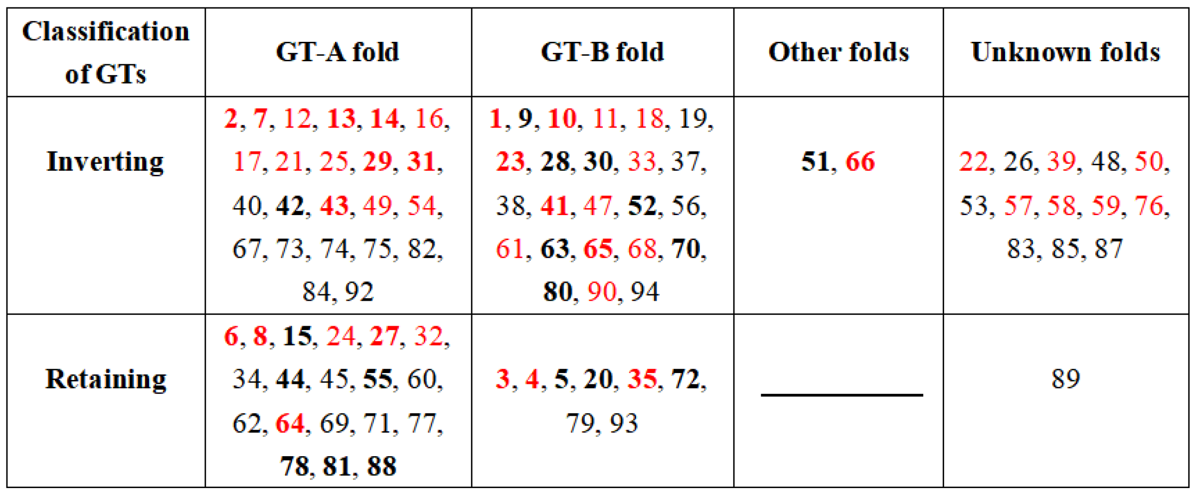
2.1. Donor Recognition and Specificity of UGTs

UGTs usually have a preferred UDP-sugar donor and specifically transfer the donor monosaccharides, such as glucose, xylose, mannose, galactose, fucose, N-acetylglucosamine (GlcNAc), N-acetylgalactosamine (GalNAc), or glucuronic acid (GLCA) linking to the specific receptors, allowing organisms to use the same monosaccharides to build a set of molecules or oligosaccharides.
The highly conserved C-terminal domains of UGTs decide the specific recognition of UDP-sugars. For example, Nair et al. constructed a 12 homology models containing the C-terminal domains of human UGT1, 2, 3, and 8 families using the X-ray crystal structure of UGT2B7CT (PDB ID: 206L) as a template and indicated Trp356 performing a key role in recognizing UDP-glucuronic acid (UDPGA) using the molecular dynamics simulations [32]. Miley et al. also found the yield of UDPGA- steroid products significantly decreased after mutating Trp356 in UGT2B7 to Ala/His, and further revealed that Gln359 and Glu382 formed hydrogen bonds with the ribose ring of UDP-sugar, and Thr373, His374, and Gly376 formed hydrogen bonds with the phosphate groups [33]. Chau et al. found the glucuronic acidification and glucosylation of morphine were completely inactivated after mutating Asp398/Gln399 to Ala or Leu [34].
In plants, UGTs can be categorized as 16 distinct groups (A-P) based on phylogeny [35]. UDP-sugars mainly interact with residues of the secondary metabolite glycosyltransferase (PSPG) motif, which is a highly conserved consensus sequence consisting of 44 amino acid residues [35]. Mutation of these key residues can alter the specificity to bind UDP-sugars. Noguchi et al. presented that substitution of Arg350 with Trp significantly decreased the 727-fold glucuronyl transfer activity of UGT88D7, while significantly increased 5.7-fold glucose transfer activity [36]. Similarly, replacement of Trps in sesame UGT88D6 and snapdragon UGT88D4 (corresponding position of Arg350 in UGT88D7) also resulted in a significant specificity change of UDP-sugars, which indicated the important roles of Arg350 for recognizing the specific UDP-sugars. Additionally, the specificity to UDP-GA can be changed to UGP-glu by replacing Trp to Arg while the mutation of Trp to Arg could not alter the specificity to UGP-Glu to UDP-Ga [37], which means that single amino acid residue might perform the decisive role, but UGTs require multiple residues to recognize sugar donors. Chen et al. also for the first time found that Lys360 in blackberry UGT78H2 expanded the selectivity of sugar donors, and conformation of UDP-sugar at the binding sites had significant changes by substituting Lys360 to Asn [38].
Complex donor specificities of TcCGT1 from the medicinal plant Trollius europaeus and GgCGT of a traditional Chinese medicine Glycyrrhiza uralensis were revealed to use UDP-glucose for glycosylating flavonoids and phenolic compounds, and utilize the UDP-xylose, UDP-galactose, and UDP-arabinose as the sugar donors even with lower catalytic efficiency [39]. A GTcp from the marine bacterium Candidatus Pelagibacter sp synthesized various glycoglycerolipids using UDP-glucose, UDP-galactose, or UDP-glucuronic acid as sugar donors [40]. These results mean that UGT might have multiple sugar donors and provide different sugar molecules on the same acceptor [22,41]. The existence of different receptors influenced the selectivity of UGT to donors. Meech et al. found that in comparison to UDP-glucose, UGT8A1 preferred UDP-galactose when using bile acids and bile acids as receptor substrates [42].
Until now, no functionally characterized fungal UGTs were found in the PDB database. Using the multiple sequence alignment, a UDP-glucosyltransferase GFUGT88A1 from mushroom G. frondosa was found to contain the typical UGT-defining sequence motif (WTPQQTILNHPATGWFVAHGGHNGVLEATTAGVPQIFWPFAADQ), which decided the selectivity of specific sugar donor [43]. Accordingly, the preference of GFUGT88A1 for UDP-glucose as sugar donor was confirmed since the amino acid residue glutamine (Q) existed at the position 44 in the motif (366 in GFUGT88A1) [44].

2.2. Broad Substrate Selection

UGTs can glycosylate many valuable molecules. Zhang et al. summarized the substrate patterns by phylogenetic analysis of 317 representative UGTs [5]. These UGTs are divided into three main groups A1, B1, and C1 according to their life domains. Over 97% UGTs in A1 group belong to bacteria, over 80% UGTs in A1 group belong to animals, and C1 group consists only of plant UGTs. Among them, UGTs in A1 group have broad substrate selectivity and accept all the listed compounds including high molecular compounds such as glycolipids-polypeptides and low molecular weight compounds, such as purines. Additionally, UGTs located at the close branches also can glycosylate different chemical skeletons indicating the bacterial UGTs have the complexity of their potential substrates. However, plant UGTs in C1 group prefer the benzophenones or flavonoids as substrates with relatively lower selectivity, while UGTs in B1 group trend to catalyze the glycosylation of sterols, bilirubin, or alkaloids. It should be noted that there is no strict catalytic boundary between the A1, B1, and C1 groups. UGTs originated from different sources can catalyze the same specific substrate. For example, all of UGTs in A1, B1, and C1 can catalyze sterols as natural substrates [45].
Bacterial UGTs showed the significant complexity to catalyze glycosylation of the substrates. For instance, a UDP-glycosyltransferase OleD from Streptomyces sp. showed an activity to alkaloids, flavonoids, macrolides, and sterols [46], and BsGT-1 from Bacillus subtilis also could glycosylate the compounds, such as phenyl ketones, flavonoids, terpenes, and macrolides [47]. Increasing evidence have been published that plant UGT could also use the various chemical skeletons as the acceptors. For example, TcCGT1 from Trollius europaeus catalyzed the 83 acceptors with different skeletons including lignan, hydroxynaphthalene, stilbene, curcumin, and anthraquinone [22]. GuGT14 and GuGT33 from Glycyrrhiza uralensis [48], and UGT73AE1 from Carthamus tinctorius L. are able to catalyze the glycosylation of phenylpropanoids (coumarins, flavonoids, isoflavones, etc.) and terpenes (glycyrrhetinic acid and licorice acid).
Fungal UGTs also showed the selectivity to their substrates. Liang et al. found that GFUGT88A1 from G. frondosa had highest specific activity of 132.56 ± 2.25 U and 131.06 ± 1.17 U when using vanillone and naringenin as acceptor substrates. Interestingly, GFUGT88A1 had an activity of transferring-glucose moiety to the tested oligosaccharides (DP = 2, 6, 9), and showed an extremely low activity when glucose was used as the acceptor. GFUGT88A1 had the highest specific activity of 84.52 pmol min−1 µg−1 in reaction with LAM9, which indicated that the high DP of the oligosaccharides was considerably beneficial for transferring the glucose moiety from donor UDP-glucose catalyzed by GFUGT88A1 [43].

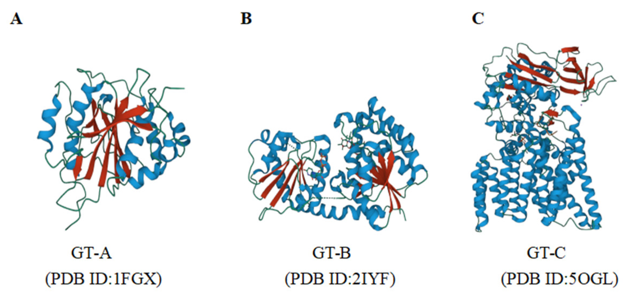
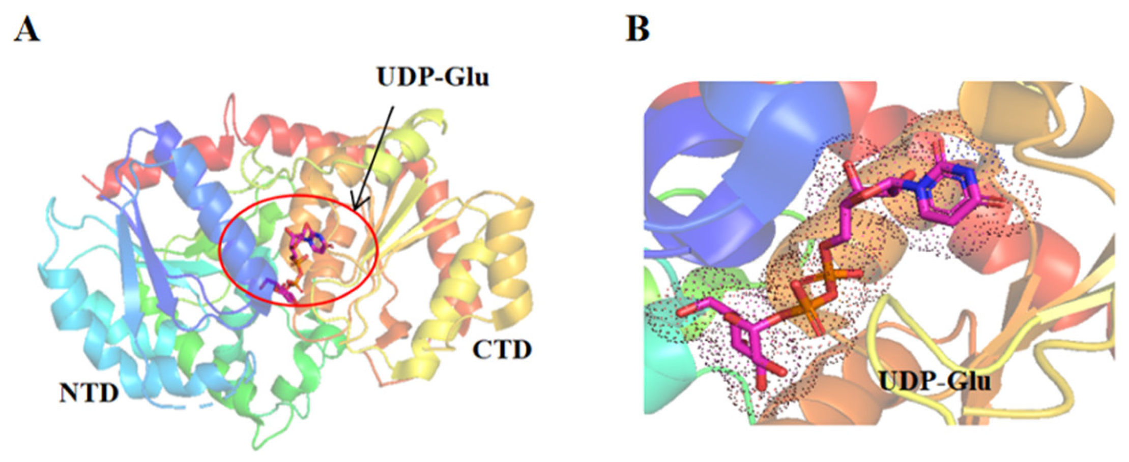
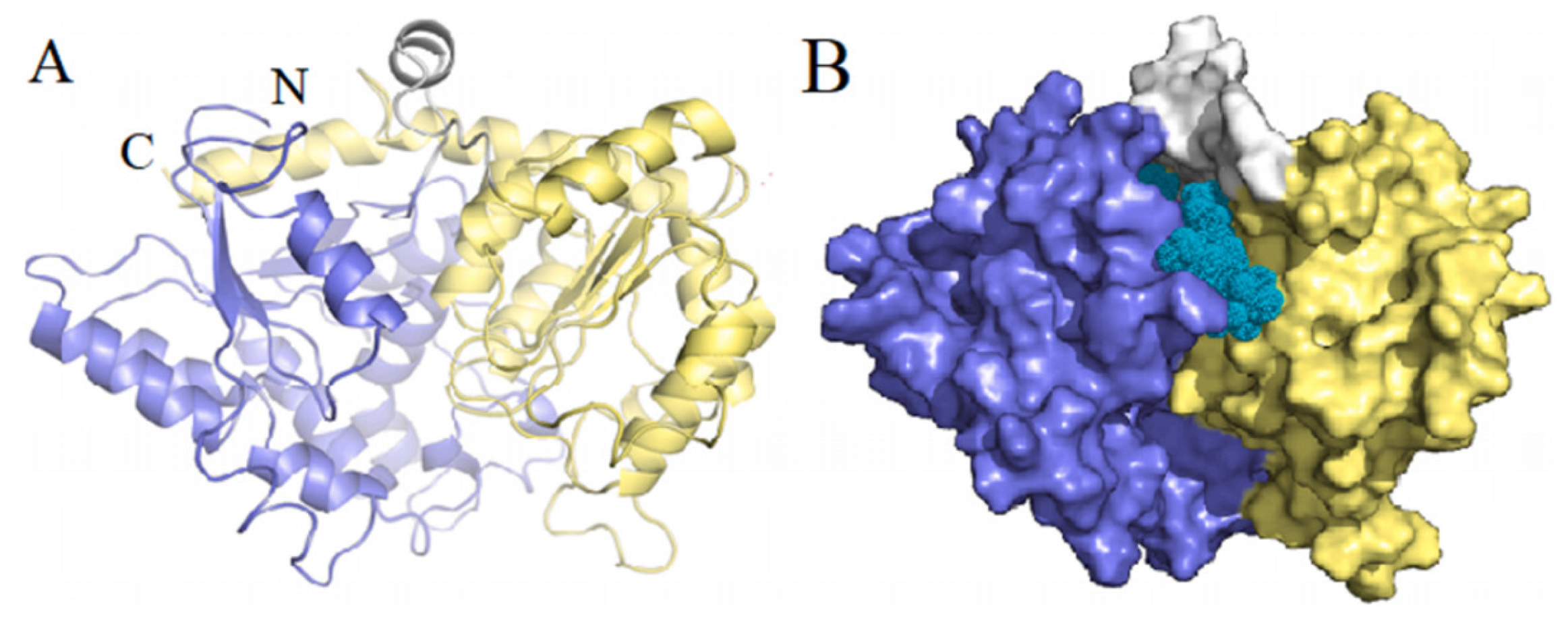
2.3. Structure and Function of UGTs

Although the protein sequences of UGTs from different species shared low homology, UGTs have high conserved GT-B structure [49]. As shown in Figure 3A, the GT-B structure consists of two independent β/α/β Rossman domains, namely the N-terminal domain and the C-terminal domain, joined by a connecting region and present a face-to-face state to form an interdomain cleft as the activity sites to bind the UDP-sugar donor and acceptor substrates (Figure 3B). The C-terminal and N-terminal domains are responsible for recognizing and binding the UDP-sugar donors, and the acceptors, respectively [50]. The C-terminal domain is more conserved since the kinds of UDP-sugar donors are limited [49].
Thus far, the crystal structures of many plant UGTs have been published in the PDB database, such as Medicago truncatula UGT78G1 (PDB ID: 3HBF) [51], Trollius chinensis TcCGT1 (PDB ID: 6JTD) [22], Arabidopsis thaliana UGT74F2 (PDB ID: 5U6M), and UGT89C1 (PDB ID: 6IJ7) [52,53], Clitoria ternatea UGT78K6 (PDB ID: 4REM) [54], and Oryza sativa Os79 (PDB ID: 5TME) [55]. The amino acid sequence identity of these UGTs is relatively low (usually 25–45%) while all of their 3D structures contain the typical GT-B folds. These plant UGTs also share a similarity of overall folding and core structure with bacterial UGTs, such as the GTc from Candidatus Pelagibacter sp. for glycolipid synthesis [40], and OleD and Olei from Streptomyces sp. for glycosylation of macrolides antibiotics [56].
Mammalian UGTs belong to the membrane proteins with a single transmembrane domain, and a signal peptide at the N-terminus involved in insertion into the endoplasmic reticulum and subsequent cleavage after maturation. Main UGT protein chain stays in the luminal of the endoplasmic reticulum (possibly interacting with the membrane) and leaves a short C-terminal region at the cytoplasmic side [57]. Due to the extreme difficulty to purify and crystallize the membrane-bound mammalian UGTs, a C-terminal domain (residues 285–451) of human UDP-glucuronyltransferase 2B7 (2B7CTD) was published for the first time until 2007 [33], which formed that the human UGT also belonged to the GT-B fold type and had a high similarity with the crystal structures of plant UGTs. In 2020, Zhang et al. published the second crystal structure of the C-terminal domain in human UGT2B15 binding to tartrate, which had a spherical appearance and the same fold type with UGT2B7. Up to now, no full X-ray crystal structure of any mammalian UGT is available [58].
The crystal structures of the fungal UGTs have not records in the PDB database. Liang et al. proposed a 3D model of GFUGT88A1 from G. frondosa using SWISS-MODEL homology modeling, with a sequence consistency of 22.43% using TcCGT1 (PDB ID: 6jtd) of Trollius chinensis as the template. Its predicted 3D structure was a typical GT-B type consisting of two independent and flexibly connected Rossmann domains (β/α/β), the N-terminal domain of GFUGT88A1 showing the core six β-sheets (Nβ1-Nβ6) organized in a canonical order, and the C-terminal domain showing four β-sheets (Cβ1-Cβ4) enclosed in the center (Figure 4) [43].
Currently, there are still many challenges in understanding the structures of UGTs [45,59]. For example, the multi-domain structures and considerable conformations of UGTs cannot meet the strict requirements of crystallization process. Recombinant UGTs cannot be expressed with high yield due to their integrative or membrane-bound forms in expression cells after post-translational modifications, such as N-glycosylation, disulfide bond formation, and folding with the assistance of chaperone. The binding and catalytic sites are also difficult to be identified due to the co-existence of donor and acceptor substrates. Although Cryo-electron microscopy (cryo-EM) is rapidly becoming an attractive method in the field of structural biology and has been applied as the first go-to method for protein structural analysis, few works have been conducted using cryo-EM to solve the UGT structures. The increased efficiency and accuracy of the machine learning strategies, including AlphaFold2 https://www.alphafold.ebi.ac.uk/ (accessed on 22 December 2022) [60] and RoseTTAFold [61], would guide the structure prediction, catalytic mechanism analysis, and rational design of UGTs. For example, Yan et al. constructed a 3D structure of a glycosyltransferase UGT71BD1 from desert herb plant Cistanche tubulosa using AlphaFold2, and found it consisting of two independent domains with similar Rossmann-type folds at the N and C terminal with docked UDP-Glc and acteoside inside [62].

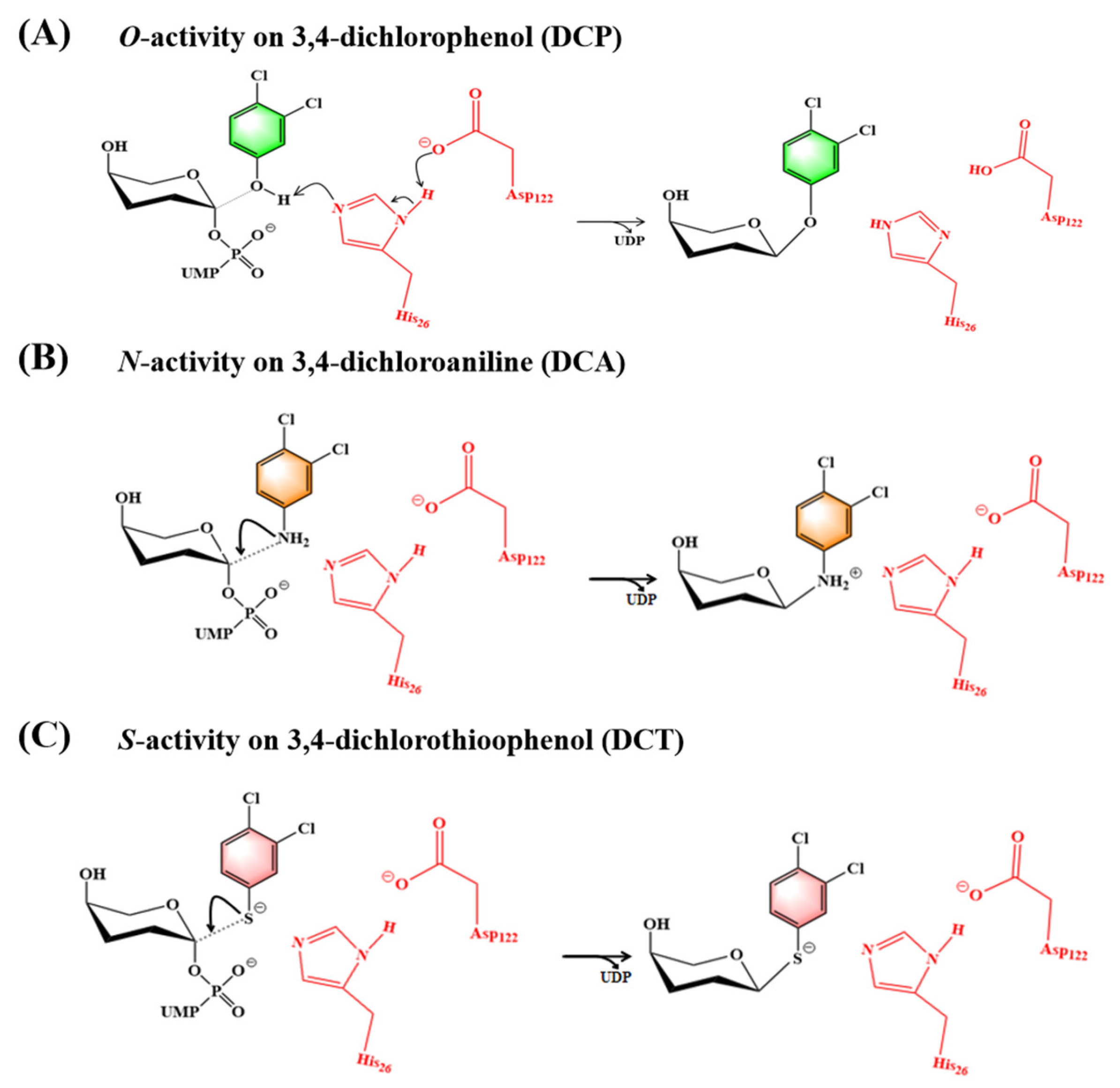
2.4. Catalytic Mechanism

All the UGTs should follow the “inverting” model to catalyze the glycosylation reaction, directly utilizing an SN2-like displacement reaction, in which the acceptor substrate replaces the phosphate position left in the sugar donor, resulting in a stereochemical inversion of the C-1 position in the glycosylation product [5]. According to the different glycosylation sites of receptor substrates, the glycosidic bonds formed by UGTs-catalyzed glycosylation products can be divided into C-O (-OH, -COOH), C-N (-NH2), C-S (-SH), or C-C (C-C) four different types [15,63].
O-glycosides are the most common glycosylation products. The specific manifestations of C-O glycosidic bond formation during glycosylation are as follows: under the action of UGT enzyme, an oxygen carbocation transition state is formed. A glycosidic bond is formed between the sugar donors and acceptor substrates and the configuration of C-1 is reversed. The transition state is usually formed with the help of the catalytic base provided by the active center side chains of UGTs, such as aspartic acid (Asp), glutamic acid (Glu), or histidine (His), as shown in Figure 5A, the catalytic base draws a proton from the OH group of the acceptor substrate, promoting the nucleophilic attack on the sugar donor C-1 [15], and the nucleophilic addition and glycosidic bond cleavage proceed almost simultaneously, accompanied by proton transfer. During the formation of the O-glycosidic bond, the C-1 atom of the UDP-sugar moves toward the nucleophilic oxygen with the rotation of the diphosphate group, which facilitates the glycosylation reaction [64]. Studies proved that His at near the position of 20 and conserved Asp residue perform a key role during the catalysis reaction. His responds to eliminate the resonance stabilization of the carboxylic acid group and activate the attacking oxyanion, and interaction of Asp and His stabilizes its charge and balances its charge by forming hydrogen bonds and the deprotonation of the acceptor [63]. Huang et al. found that the highly conserved His24 and Asp119 in UGT74AN2 from the medicinal plant Calotropis gigantea formed a catalytic dyad with the 3-OH of its receptor substrate digitoxin, through hydrogen bonding. Mutation of His24 and Asp119 to alanine lost its activity completely, confirming that the conserved catalytic dyad (His-Asp) is required for UGT74AN2 [65]. Li et al. also found that mutation of His18 and Asp111 completely inactivated the catalytic triterpene O-glycosylation of Siraitia grosvenorii UGT74AC1, indicating the irreplaceable catalytic role of these two residues [20]. However, when His18 and Asp111 residues are not conserved, possibly other amino acid residues deprotonate the acceptor for nucleophilic attack via the carboxylate oxygen [66]. The Ser15 in UGT74F1/ Thr15 in UGT74F2 from A. thaliana is the binding site for salicylic acid (SA). Their mutations significantly increased SA-glucoside (SAG) formation, suggesting that residue 15 is important for SAG formation in UGT74F1 and UGT74F2 [52].
The formation mechanism of C-N(-NH2) and C-S(-SH) glycosidic bond have proved that N-glycosylation and S-glycosylation occur with no requirement of proton transfer, and their glycosylation activity relies on the site of acceptors comparing to donors (Figure 5B,C). Teze et al. elucidated the molecular basis and mechanism of N-glycosylation of P. tinctorium UGT1 using mutation and QM/MM simulation, and found that the key factor affecting its N-glycosylation activity was the ability of the acceptor to approach the donor [15]. By constructing the binary complexes of active sites of P. tinctorium UGT1 with donor UDP-glucose (PDB ID: 6SU6) and N-acceptor 3,4-dichloroaniline (DCA) (PDB ID: 6SU7), respectively, and ternary complexes of active sites of UDP-glucose and DCA, it was found that the reaction could not start since DCA in the ternary structure was too far from the reaction center (DCA was 6.2 Å from UDP-Glc and 5.4 Å from His26). Molecular dynamics (MD) simulations results indicated that the N-acceptor quickly moves into a catalytically competent position (N1DCA located 4.7 Å from C1UDP-Glc, and H1DCA at 2.0 Å from Nε His26). In silico mutagenesis of His26 to Asp or Glu, the benefits the conformational change, while the His26Phe mutation led to a stacking of Phe26 and the glucose moiety, prevented DCA from approaching the reaction center. His19 in A.thaliana UGT72B1 also performed a function of directing and localizing the nucleophilic attack, not extracting protons from the DCA receptor [67]. S-glycosylation is not popular in nature, and few S-UGTs have been identified so far, such as tomato UGT74B1 involved in the metabolism of fluthiandin (FSF) in plants [68], SunS and Thus for S-glycopeptide bacteriocin biosynthesis [69], and YjiC from Bacillus licheniformis with thiol glycosylation activity [70]. Teze et al. performed His26Asp mutation on P. tinctorium UGT1 and found that Asp26 was not directly involved in catalysis, and its acceptor substrate 3,4-dichlorothiophenol (DCT) is completely ready in the absence of any catalytic residue, suggesting that the His26 mutation had a limited effect on S-glycosylation and both N- and S-glycosylation occurred without proton transfer of catalytic residues depending on the sites of the acceptors to the donors [15].
C-C (C-C) glycosidic bonds have better enzymatic or chemical hydrolytic stability than other types of glycosides (O-, N-, or S-glycosides) due to their rigid C-C bonds between sugar residues and aglycones [22]. A number of C-UGTs have been identified, including GtUF6CGT1 from Gentiana triflora utilizing flavonoids as substrates [71], PlUGT43 from Pueraria lobata showing activity with isoflavones, such as daidzein and genistein [72], TcCGT1 from Trollius chinensis recognizing 36 flavonoids and other flavonoids [22], UGT708C1 from Fagopyrum esculentum catalyzing phloretin and trihydroxyacetophenone [73]. Soe plant C-UGTs can catalyze disglycosylation, such as FcCGT and CuCGT from citrus plants [74], MiCGTb from mango, and GgCGT from licorice [75]. The catalytic mechanism for C-glycosylation shows that acceptor substrates are generally deprotonated with the assistance of residues His32 and Asp129 [73], which is consistent with O-glycosylation. The exceptional example is His24 in the docking model of TcCGT1/UDP-glucose was located far from the hydroxyl group of any sugar acceptor, and acceptor substrate was spontaneously deprotonated during its glycosylation [22]. The Glu396 was very close to the UDP-sugar forming hydrogen bonds with sugar hydroxyl groups. Mutating Glu396 to Ala396 significantly reduced or even eliminated the catalytic activity of TcCGT1 indicating Glu396 was the key residue to stabilize the donor substrate and position the sugar in the optimal direction for the glycosylation reaction. For UGTs with bifunctional C-glycosylation and O-glycosylation, it is important to judge their action roles as C-UGTs or O-UGTs depending on their binding sites to the substrate, i.e., C- or O- are closer to the C-1 position of the sugar. He et al. found that shrinking the receptor-substrate-binding pocket might block the canonical C-C-glycosylation of UGT receptor substrates, thereby exhibiting O-glycosylation activity [22]. For native O-UGT substrates, the reduction in the binding pocket caused by these mutations had no alteration of the O-glycosylation activity.
Until now, there are also many UGTs with multiple catalytic functions for different glycosides. For example, bifunctional maize UGT708A6 can generate C-glycosylated and O-glycosylated flavonoids [76]. Mango MiCGT [77], purple ginseng RyUGT3A, and RyUGT12 [78] are trifunctional UGTs capable of transferring various sugars to three nucleophilic groups (OH, NH2, and SH) in different compounds, resulting in O-, N-, and S-glycosides, and Glycyrrhiza glabra UGT708B4 and Nasturtium TcCGT1 [22] formed four types (C-C, C-O, C-N, and C-S)) of the glycosidic bond.

3. Recent Research Status of UDP-Glycosyltransferases

3.1. UDP-Glycosyltransferase in Plants

UDP-glycosyltransferases in plants can glycosylate almost all major classes of secondary metabolites, such as phenylpropionoids (flavonoids, coumarins, and lignans) [48], alkaloids (indole alkaloids, steroidal alkaloids, and cytokinins) [79], terpenes (monoterpenes, diterpenes, and triterpenes) [80,81], and polyketone compounds (phenol polyketones, and polycyclic aromatic polyketones) [82]. For example, Glycyrrhiza uralensis GuGT14, GuGT33 [48] and Carthamus tinctorius UGT73AE1 [83] have been shown to catalyze glycosylation of phenylpropines (coumarins, coumarins, flavonoids, and isoflavones) and terpenes (glycyrrhetinic acid and glycyrrhizic acid), while morning glory InGTase1 [84] could catalyze phenylpropane and alkaloids, and Phytolacca Americana PaGT2 [85] can catalyze phenylpropane and polyketones. The glycosylated products show a wide range of biological activities, such as triterpene glycosides have antimicrobial, insecticidal and antiparasitic activities, and cholesterol-lowering, anticancer, adjuvant and hemolytic activities, benefits for cardiovascular, and nervous and digestive systems [86]. The quercetin glycoside showed a chemoprotective activity in the oxidative stress, cancer, cardiovascular disease, diabetes, and allergic reactions [87], and anthocyanin glycosides inhibited the UVB-induced oxidative damage and inflammation, thereby reducing the risk of skin cancer [88].
Glycosylation significantly affects the physicochemical properties and biological activity of phytochemicals, thereby affecting solubility, cell localization, and biological activity. Various soluble cytosolic UGTs can transfer active glycans to a series of plant small molecule complex receptors, such as hormones, secondary metabolites, pathogen infections, plant internal, and external toxic substances, etc., forming glycosides with a wide range of biological activities, which perform a key role in plant growth and development, response to abiotic and biological stresses, and plant environmental defense [63].

3.2. UDP-Glycosyltransferases in Bacteria

UGTs from the genus Bacillus have catalytic activity for the glycosylation of flavonoids and ginsenosides [89]. B. subtilis UGTs, such as YjiC, YojK, and YdhE, have been shown to be effective for synthesis of glycosylated products including ginsenosides, flavonoids [89], oleanocarpine-type compounds [90], and toadadienolides [91]. Among them, YjiC can convert glycyrhetinic acid (GA) to glycyrrhizin (GL), GA-3-O-β-D-glucoside, and GA-30-O-β-D-glucoside, YojK convert saponin Rh1 to ginsenoside Rh1 and UGT109A1 has also been shown to glycosyl triterpene glycosides, such as proginsenodiol (PDD) and proginsetriol (PTT), resulting in unnatural ginsenosides [92]. Additionally, another glycosyltransferase YdhE, together with YjiC and YojK, is able to efficiently glycosylate 2-heptyl-1-hydroxyquinoline-4-one (HQNO), 2-heptyl-3-hydroxyquinoline, pyocyanin, 2,4-dihydroxyquinoline, vanillin, and quercetin [93].
Three bacterial UGTs BLYjiC, BLYdhE, and BLYojK were also identified from B. licheniformis [94]. Among them, BLYjiC can catalyze the formation of avermectin B1a (an anti-nematode agent) with various UDP sugars to produce four avermectin B1a glycosides, which significantly improves its water solubility (50 times) and anti-nematode activity (30 times) of abamectin B1a [95] and BLYjiC has also been shown to be able to add glucose to several tetracyclines [96]. On the other hand, BLYdhE and BLYojK have high complex recognition ability to donors and acceptors. Their glycosylation activity has been confirmed for O-glycosylation and transfer of glucose to suitable acceptors forming N-glycosides and S-glycosides, respectively [94]. A UDP-glycosyltransferase BC-GTA found from B. cereus WQ9-2 can glycosylate diterpenoids saffin, producing monosaccharylated saffronin-5, and disaccharide elated crocinoside-3 [39,97].

3.3. UDP-Glycosyltransferase in Yeasts

To date, many UGTs have been identified from yeasts. UGT51 in Saccharomyces cerevisiae could format the glycosides from natural substrates, such as ergosterol, sitosterol, cholesterol, and pregnenolone [98], and PPD and PPT are converted to ginsenoside glycosides [99]. Yeasts are the safe microorganisms with high tolerance to heterologous enzymes from plants and contain the MVA pathway to produces enough important precursors for ginsenoside synthesis (IPP and DMAPP) [100,101]. Hence, increasing literatures have use yeasts including S. cerevisiae, Yarrowia lipolytica, and Pichia pastoris as the hosts for synthesizing ginseng saponins [99]. There are still limited references available related to identification of fungal UGTs.

3.4. UDP-Glycosyltransferases in Edible Fungi

3.4.1. UDP-Glycosyltransferase in Cordyceps militaris

Cordyceps militaris is a highly valued edible/medicinal fungus and contains various polysaccharides with health benefits including antiproliferation of tumor cells, anti-inflammatory, and stimulation of immune system [102]. β-glucans are the most abundant types of glycans in fungal cell walls, mostly constructed from a backbone of β-1,3-linked glucans [103]. The synthesis process of microbial polysaccharides is extremely complex. For β-1,3-glucan biosynthesis, the key step is the polymerization and elongation of the glucan chain occurring near the plasma membrane (PM) by a membrane-bound β-1,3-glucan synthase complex constituted by a catalytic subunit, β-1,3-glucan synthase, and at least one regulatory subunit, Rho1 [104,105]. On this basis, β-1,3-glucan synthase, belonging to glycosyltransferase (GT) 48 family and GT-C type, was regarded as responsible for transferring UDP-glucose to polymerize the linear glucan as [β-1,3-D-glucosyl](n) + UDP-α-D-glucose = [β-1,3-D-glucosyl](n+1) +H+ + UDP [106]. A cmFKS1 in edible/medicinal fungus C. militaris was 5943bp encoding an integral membrane β-1,3-glucan synthase of 1981 amino acids with 16 potential transmembrane domains with a relatively large hydrophilic domain in the middle region [107]. However, β-1,3-glucan synthase multi-transmembrane domains (over 14 TMHs) and large molecular masses (over 100 kDa) significantly hamper understanding of their catalytic characteristics and mechanisms. Consequently, Fu et al. was partially purified with a specific activity of 87.72 pmol/min/μg, and a yield of 10.16% using a product-entrapment purification method. β-1,3-glucan synthase in C. militaris showed a strict specificity to UDP-glucose with a Km value of 84.28 μM at pH 7.0 and synthesized β-1,3-glucan with a maximum degree of polymerization (DP) of 70 [108].

3.4.2. UDP-Glycosyltransferase in Ganoderma lucidum

Ganoderma lucidum, commonly known as “Lingzhi” in Chinese, are well-known medicinal mushrooms with abundant polysaccharides (GLPs) and secondary metabolites, such as hydrocarbons, monoterpenes, and sesquiterpene. UDP-glycosyltransferases in G. lucidum are regarded to be responsible for their synthesis. Liu et al. identified six putative UGTs in G. lucidum genome transferring the active saccharides to the triterpenoid backbones and alter its physiological activity, forming glycosylation modification in the GAs biosynthesis [109]. Among these six putative UGTs, the gene (G_lucidum_10009504) was highly similar with UGT73K1, which glycosylated both triterpenoids and (iso)flavones in Medicago truncatula [110]. At least two 1,3-β-glucan synthases belonging to GT-C type also were identified to have key roles in the biosynthesis of 1,3-β-glucans in G. lucidum [111].

3.4.3. UDP-Glycosyltransferase in Grifola frondosa

Grifola frondosais is a basidiomycete fungus belonging to the order Aphyllophorales and family Polyporaceae and contains over 10 kinds of polysaccharides from fruiting bodies and fermented broths [112]. Previously, we optimized the cultivation conditions to maximally produce G. frondosa mycelia and mycelial/exopolysaccharide, elucidated the structures and biological functions of heteropolysaccharides and glucans [113], predicted the roles of UGP [114], and the glucan synthase complex in glucan synthesis and mycelial growth using constructed dual promoter RNAi and overexpression systems [115,116]. Notably, our group cloned and heterologously expressed a novel UGT, GFUGT88A1 from the edible mushroom G. frondosa for the first time. GFUGT88A1 had a molecular weight of 51.7 kDa with an optimum temperature of 37 °C and pH of 7.0. GFUGT88A1 showed preference for oligosaccharides with relatively higher polymerization degrees (≥6) as acceptors to extend its chain when using UDP-glucose as a donor. Molecular modeling and docking revealed the potential structure and catalytic binding features of GFUGT88A1 based on the spacious binding pocket containing several residues including Arg20, Trp323, Thr324, Gln327, Ser321, Glu349, and Ser321 [43]. The full sequence of two β-1,3-glucan synthase genes, GFGLS and GFGLS2 in G. frondosa were re-assembled with full sequences of 5927 bp and 5944 bp (GenBank: MK80801.9 and MN477285.1), respectively. GFGLS2p and GFGLSp were predicted as the 203.66 kDa and 195.2 kDa membrane-bound proteins with a high protein similarity of 72.56% and contained two large membrane domains of 6 transmembrane helices (TMHs) at the N-terminus and 9 TMHs at the C-terminus (Figure 3A) [115,117]. By using the constructed dual promoter RNAi system and PEG-Ca2+ transformation system, single-silencing, and co-silencing strains, the GFGLS and GFGLS2 perform major roles in the synthesis of the cell wall and the elongation of the hyphae by down-regulating the glucan synthesis [113,117].

3.4.4. UDP-Glycosyltransferase in Sparassis crispa

S. crispa is a polypore mushroom and produces unusually high amount of soluble β-1,3-glucan (β-glucan). Yang et al. isolated a β-glucan synthase gene from its genomic DNA with a full sequence of 5502 bp consisting of 10 exons separated by nine introns. Similar to other fungal β-glucan synthases, the β-glucan synthase protein (SCGLSp) is an integral membrane protein consisting of 1576 amino acid residues with the predicted structure consisting of 15 loops and 14 transmembrane α-helices. The catalytic loop in the S. crispa β-glucan synthase were proposed as the key domain for transferring UDP-glucose to polymerize the linear glucan and contributed to the high productivity of soluble β-glucan [118].

3.5. UDP-Glycosyltransferase in Other Fungi

UDP-glycosyltransferases are also widely found in other fungi with the functions of glycosylation of phenolics, flavonoids, synthesis of galactomannan, N-glycans, O-glycans, and glycosphingolipids. For example, Feng et al. identified a glycosyltransferase MhGT1 from Mucor hiemalis with the ability to catalyze the phenolic compounds [119]. The MhGT1 exhibited a significant glycosylation ability using UDP-glucose as a sugar donor to catalyze O-glycosylation of 72 acceptors with various structures. Xie et al. found a UGT58A1 from Absidia coerulea and a UGT59A1 from Rhizopus japonicas, which belonged to novel phenolic UDP-glycosyltransferases (P-UGTs) [120]. The recombinant rUGT58A1 and rUGT59A1 could regioselectively and stereoselectively glycosylate a variety of phenolic glycosides to produce corresponding glycosides. Additionally, a UDP-glycosyltransferases GT86 has been identified from the genome of Beauveria bassiana ARSEF 2, 860, which can effectively modify drug-like substrates including polyketones, anthraquinones, flavonoids, and naphthalene, and present a good substrate heterogeneity and regioselectivity [121]. Chen et al. identified a glycosyltransferase UGT1 from Aureobasidium melanogenum P16, which N-terminal part of the protein displayed a conserved sequence controlling both sugar donor and accepter for substrate specificity whereas its C-terminal part carried a DXD motif that coordinated donor sugar binding; The UGT1 which donors is UDP-glucose can catalyze the transfer of glucosyl from UDP-glucose to oligosaachardes, polysaccharides, lipids, proteins and other macromolecules in the yeast cells [122,123]. After deletion of the A. melanogenum P16 UGT1 gene, the disruptant 1, 152-3 produced much lower glucosyltransferase activity than its wild type of strain P16. Moreover, after overexpression of the gene UGT1, the transformant G63 could produce much higher glucosyltransferase activity than its wild type of strain P16. During the 10-Litter fermentation, the pullulan titer produced by the transformant G63 was 80.2 g/L within 132 h. The molecular weight (6.2 × 10⁵) of the produced pullulan by the transformant G63 was also higher than that (4.6 × 10⁵) of the produced pullulan by its wild type of strain P16. Therefore, the transformant G63 had highly potential application in biotechnology [124]. Engel et al. founded galactofuranose (Galf) from Aspergillus fumigatus having a five-membered antigenic ring performs an important role in the fungal biosynthesis of galactomannan, N-glycans, O-glycans, and glycosphingolipids [125]. The deletion of UGfT (glfB or ugtA) recognizing UDP-Galf in A. fumigatus had a significant effect on their cell wall structure, with a complete absence of Gal, which exposed the Man backbone and impaired cell growth [126,127].

4. Conclusions and Future Perspectives

UGTs format the glycosidic linkages in glycans, proteins, lipids, antibiotics, flavonols, and steroids to improve their chemical/biological features. The roles of UGTs in disease development and potential roles as therapeutic targets are also now being appreciated. Understanding the structures and molecular mechanisms of UGTs is important for controlling their function, boosting future bioengineering applications to synthesize novel glycosylated products. Hence, the recently updated literature has provided important structural and functional insights on the physiological and biochemical roles and reaction mechanism of UGTs from plants, bacteria, and fungi, which significantly accelerate the understanding of the structure-activity relationship of the UGT proteins and the molecular biosynthesis mechanism of glycosylated products. However, there are still new challenges to be resolved despite considerable progress in the understanding of their structure-function relationships. The studies of mining and engineering the UGTs from bacteria and fungi are extremely limited. More efforts should be stressed to identify the novel UGTs from bacterial and fungal sources. Their 3D structures, catalytic characteristics and mechanisms need to be comprehensively elucidated by recombination, protein crystallization or AlphaFold prediction, and site-directed mutation. On this basis, the further synthetic biology-driven metabolic engineering of desirable bacterial and fungal UGTs could be developed to produce interesting glycosides and oligosaccharides in industrial scale for application in functional foods, medicine, and cosmetics fields.

Funding

This work was supported by funding from Natural Science Foundation of China (32172187, 31771961) and Science and Technology Platform Construction Program of Jiangxi Province.

Conflicts of Interest

All authors declare that they have no conflict of interest.

References

  1. Mackenzie, P.I.; Owens, I.S.; Burchell, B.; Bock, K.W.; Bairoch, A.; Bélanger, A.; Fournel-Gigleux, S.; Green, M.; Hum, D.W.; Iyanagi, T.; et al. The UDP glycosyltransferase gene superfamily: Recommended nomenclature update based on evolutionary divergence. Pharmacogenetics 1997, 7, 255–269. [Google Scholar] [CrossRef] [PubMed]
  2. Bowles, D.; Isayenkova, J.; Lim, E.K.; Poppenberger, B. Glycosyltransferases: Managers of small molecules. Curr. Opin. Plant Biol. 2005, 8, 254–263. [Google Scholar] [CrossRef]
  3. Marschall, E.; Cryle, M.J.; Tailhades, J. Biological, chemical, and biochemical strategies for modifying glycopeptide antibiotics. J. Biol. Chem. 2019, 294, 18769–18783. [Google Scholar] [CrossRef] [PubMed]
  4. Lairson, L.L.; Henrissat, B.; Davies, G.J.; Withers, S.G. Glycosyltransferases: Structures, functions, and mechanisms. Annu. Rev. Biochem. 2008, 77, 521–555. [Google Scholar] [CrossRef] [PubMed]
  5. Zhang, P.; Zhang, Z.; Zhang, L.; Wang, J.; Wu, C. Glycosyltransferase GT1 family: Phylogenetic distribution, substrates coverage, and representative structural features. Comput. Struct. Biotechnol. J. 2020, 18, 1383–1390. [Google Scholar] [CrossRef] [PubMed]
  6. Chang, A.; Singh, S.; Phillips Jr, G.N.; Thorson, J.S. Glycosyltransferase structural biology and its role in the design of catalysts for glycosylation. Curr. Opin. Biotechnol. 2011, 22, 800–808. [Google Scholar] [CrossRef] [PubMed]
  7. Bohl, H.; Bai, L.; Li, H. Recent Progress in Structural Studies on the GT-C Superfamily of Protein Glycosyltransferases. In Macromolecular Protein Complexes III: Structure and Function; Springer: Cham, Switzerland, 2021; pp. 259–271. [Google Scholar]
  8. Gloster, T.M. Advances in understanding glycosyltransferases from a structural perspective. Curr. Opin. Struct. Biol. 2014, 28, 131–141. [Google Scholar] [CrossRef]
  9. Gantt, R.W.; Peltier-Pain, P.; Singh, S.; Zhou, M.; Thorson, J.S. Broadening the scope of glycosyltransferase-catalyzed sugar nucleotide synthesis. Proc. Natl. Acad. Sci. USA 2013, 110, 7648–7653. [Google Scholar] [CrossRef]
  10. Feng, Y.; Yao, M.; Wang, Y.; Ding, M.; Zha, J.; Xiao, W.; Yuan, Y. Advances in engineering UDP-sugar supply for recombinant biosynthesis of glycosides in microbes. Biotechnol. Adv. 2020, 41, 107538. [Google Scholar] [CrossRef]
  11. Liang, D.M.; Liu, J.H.; Wu, H.; Wang, B.B.; Zhu, H.J.; Qiao, J.J. Glycosyltransferases: Mechanisms and applications in natural product development. Chem. Soc. Rev. 2015, 44, 8350–8374. [Google Scholar] [CrossRef]
  12. Yu, H.; Takeuchi, M.; LeBarron, J.; Kantharia, J.; London, E.; Bakker, H.; Haltiwanger, R.; Li, H.; Takeuchi, H. Notch-modifying xylosyltransferase structures support an SNi-like retaining mechanism. Nat. Chem. Biol. 2015, 11, 847–854. [Google Scholar] [CrossRef] [PubMed]
  13. Breton, C.; Fournel-Gigleux, S.; Palcic, M.M. Recent structures, evolution and mechanisms of glycosyltransferases. Curr. Opin. Struct. Biol. 2012, 22, 540–549. [Google Scholar] [CrossRef] [PubMed]
  14. Paquette, S.; Møller, B.L.; Bak, S. On the origin of family 1 plant glycosyltransferases. Phytochemistry 2003, 62, 399–413. [Google Scholar] [CrossRef] [PubMed]
  15. Teze, D.; Coines, J.; Fredslund, F.; Dubey, K.D.; Bidart, G.N.; Adams, P.D.; Dueber, J.E.; Svensson, B.; Rovira, C.; Welner, D.H. O-/N-/S-Specificity in Glycosyltransferase Catalysis: From Mechanistic Understanding to Engineering. ACS Catal. 2021, 11, 1810–1815. [Google Scholar] [CrossRef]
  16. Yonekura-Sakakibara, K.; Hanada, K. An evolutionary view of functional diversity in family 1 glycosyltransferases. Plant J. 2011, 66, 182–193. [Google Scholar] [CrossRef]
  17. Campbell, J.A.; Davies, G.J.; Bulone, V.; Henrissat, B. A classification of nucleotide-diphospho-sugar glycosyltransferases based on amino acid sequence similarities. Biochem. J. 1997, 326 Pt 3, 929. [Google Scholar] [CrossRef]
  18. Qin, J.; Sun, C.; Zhang, M.; Wang, Y. Classification, function and evolution of plant UDP-glycosyltransferase. Genom. Appl. Biol. 2018, 37, 440–450. [Google Scholar]
  19. Wen, C.; Huang, W.; Zhu, X.L.; Li, X.S.; Zhang, F.; Jiang, R.W. UGT74AN1, a permissive glycosyltransferase from Asclepias curassavica for the regiospecific steroid 3-O-glycosylation. Org. Lett. 2018, 20, 534–537. [Google Scholar] [CrossRef]
  20. Li, J.; Yang, J.; Mu, S.; Shang, N.; Liu, C.; Zhu, Y.; Cai, Y.; Liu, P.; Lin, J.; Liu, W.; et al. Efficient O-glycosylation of triterpenes enabled by protein engineering of plant glycosyltransferase UGT74AC1. ACS Catal. 2020, 10, 3629–3639. [Google Scholar] [CrossRef]
  21. Takenaka, Y.; Kato, K.; Ogawa-Ohnishi, M.; Tsuruhama, K.; Kajiura, H.; Yagyu, K.; Takeda, A.; Takeda, Y.; Kunieda, T.; Hara-Nishimura, I.; et al. Pectin RG-I rhamnosyltransferases represent a novel plant-specific glycosyltransferase family. Nat. Plants 2018, 4, 669–676. [Google Scholar] [CrossRef]
  22. He, J.B.; Zhao, P.; Hu, Z.M.; Liu, S.; Kuang, Y.; Zhang, M.; Li, B.; Yun, C.H.; Qiao, X.; Ye, M. Molecular and Structural Characterization of a Promiscuous C-Glycosyltransferase from Trollius chinensis. Angew. Chem. 2019, 131, 11637–11644. [Google Scholar]
  23. Cai, X.; Taguchi, T.; Wang, H.; Yuki, M.; Tanaka, M.; Gong, K.; Xu, J.; Zhao, Y.; Ichinose, K.; Li, A. Identification of a C-Glycosyltransferase Involved in Medermycin Biosynthesis. ACS Chem. Biol. 2021, 16, 1059–1069. [Google Scholar] [CrossRef] [PubMed]
  24. Zhang, P.; Zhang, L.; Jiang, X.; Diao, X.T.; Li, S.; Li, D.D.; Zhang, Z.; Fang, J.Q.; Tang, Y.J.; Wu, D.L.; et al. Docking-guided rational engineering of a macrolide glycosyltransferase glycodiversifies epothilone B. Commun. Biol. 2022, 5, 100. [Google Scholar] [CrossRef] [PubMed]
  25. El Qaidi, S.; Zhu, C.; McDonald, P.; Roy, A.; Maity, P.K.; Rane, D.; Perera, C.; Hardwidge, P.R. High-throughput screening for bacterial glycosyltransferase inhibitors. Front. Cell. Infect. Microbiol. 2018, 8, 435. [Google Scholar] [CrossRef]
  26. Kattke, M.D.; Gosschalk, J.E.; Martinez, O.E.; Kumar, G.; Gale, R.T.; Cascio, D.; Sawaya, M.R.; Philips, M.; Brown, E.D.; Clubb, R.T. Structure and mechanism of TagA, a novel membrane-associated glycosyltransferase that produces wall teichoic acids in pathogenic bacteria. PLoS Pathog. 2019, 15, e1007723. [Google Scholar] [CrossRef]
  27. Asano, M. Various biological functions of carbohydrate chains learned from glycosyltransferase-deficient mice. Exp. Anim. 2020, 69, 261–268. [Google Scholar] [CrossRef]
  28. Pal, S.; Verma, J.; Mallick, S.; Rastogi, S.K.; Kumar, A.; Ghosh, A.S. Absence of the glycosyltransferase WcaJ in Klebsiella pneumoniae ATCC13883 affects biofilm formation, increases polymyxin resistance and reduces murine macrophage activation. Microbiology 2019, 165, 891–904. [Google Scholar] [CrossRef]
  29. Jacquin, L.; Reader, S.M.; Boniface, A.; Mateluna, J.; Patalas, I.; Pérez-Jvostov, F.; Hendry, A.P. Parallel and nonparallel behavioural evolution in response to parasitism and predation in Trinidadian guppies. J. Evol. Biol. 2016, 29, 1406–1422. [Google Scholar] [CrossRef]
  30. Yu, J.; Hu, F.; Dossa, K.; Wang, Z.; Ke, T. Genome-wide analysis of UDP-glycosyltransferase super family in Brassica rapa and Brassica oleracea reveals its evolutionary history and functional characterization. BMC Genom. 2017, 18, 474. [Google Scholar] [CrossRef]
  31. Li, Y.; Li, P.; Wang, Y.; Dong, R.; Yu, H.; Hou, B. Genome-wide identification and phylogenetic analysis of Family-1 UDP glycosyltransferases in maize (Zea mays). Planta 2014, 239, 1265–1279. [Google Scholar] [CrossRef]
  32. Nair, P.C.; Meech, R.; Mackenzie, P.I.; McKinnon, R.A.; Miners, J.O. Insights into the UDP-sugar selectivities of human UDP-glycosyltransferases (UGT): A molecular modeling perspective. Drug Metab. Rev. 2015, 47, 335–345. [Google Scholar] [PubMed]
  33. Miley, M.J.; Zielinska, A.K.; Keenan, J.E.; Bratton, S.M.; Radominska-Pandya, A.; Redinbo, M.R. Crystal structure of the cofactor-binding domain of the human phase II drug-metabolism enzyme UDP-glucuronosyltransferase 2B7. J. Mol. Biol. 2007, 369, 498–511. [Google Scholar] [CrossRef] [PubMed] [Green Version]
  34. Chau, N.; Elliot, D.J.; Lewis, B.C.; Burns, K.; Johnston, M.R.; Mackenzie, P.I.; Miners, J.O. Morphine glucuronidation and glucosidation represent complementary metabolic pathways that are both catalyzed by UDP-glucuronosyltransferase 2B7: Kinetic, inhibition, and molecular modeling studies. J. Pharmacol. Exp. Ther. 2014, 349, 126–137. [Google Scholar] [CrossRef] [PubMed]
  35. Rehman, H.M.; Nawaz, M.A.; Shah, Z.H.; Ludwig-Müller, J.; Chung, G.; Ahmad, M.Q.; Ynag, S.H.; Lee, S.I. Comparative genomic and transcriptomic analyses of Family-1 UDP glycosyltransferase in three Brassica species and Arabidopsis indicates stress-responsive regulation. Sci. Rep. 2018, 8, 1875. [Google Scholar] [CrossRef] [PubMed]
  36. Noguchi, A.; Horikawa, M.; Fukui, Y.; Fukuchi-Mizutani, M.; Iuchi-Okada, A.; Ishiguro, M.; Kiso, Y.; Nakayama, T.; Ono, E. Local differentiation of sugar donor specificity of flavonoid glycosyltransferase in Lamiales. Plant Cell 2009, 21, 1556–1572. [Google Scholar] [CrossRef] [PubMed]
  37. Osmani, S.A.; Bak, S.; Imberty, A.; Olsen, C.E.; Møller, B.L. Catalytic key amino acids and UDP-sugar donor specificity of a plant glucuronosyltransferase, UGT94B1: Molecular modeling substantiated by site-specific mutagenesis and biochemical analyses. Plant Physiol. 2008, 148, 1295–1308. [Google Scholar] [CrossRef] [PubMed]
  38. Chen, Q.; Liu, X.; Hu, Y.; Wang, Y.; Sun, B.; Chen, T.; Luo, Y.; Zhang, Y.; Li, M.; Liu, Z.; et al. Broaden the sugar donor selectivity of blackberry glycosyltransferase UGT78H2 through residual substitutions. Int. J. Biol. Macromol. 2021, 166, 277–287. [Google Scholar] [CrossRef]
  39. Na, L.; Li, R.; Chen, X. Recent progress in synthesis of carbohydrates with sugar nucleotide-dependent glycosyltransferases. Curr. Opin. Chem. Biol. 2021, 61, 81–95. [Google Scholar] [CrossRef]
  40. Wei, T.; Zhao, C.; Quareshy, M.; Wu, N.; Huang, S.; Zhao, Y.; Yang, P.; Mao, D.; Chen, Y. A Glycolipid Glycosyltransferase with Broad Substrate Specificity from the Marine BacteriumCandidatus Pelagibacter sp.” Strain HTCC7211. Appl. Environ. Microbiol. 2021, 87, e00326-21. [Google Scholar] [CrossRef]
  41. Wang, Z.; Wang, S.; Xu, Z.; Li, M.; Chen, K.; Zhang, Y.; Hu, Z.; Zhang, M.; Zhang, Z.; Qiao, X.; et al. Highly promiscuous flavonoid 3-O-glycosyltransferase from Scutellaria baicalensis. Org. Lett. 2019, 21, 2241–2245. [Google Scholar] [CrossRef]
  42. Meech, R.; Mubarokah, N.; Shivasami, A.; Rogers, A.; Nair, P.C.; Hu, D.G.; Mckinnon, R.A.; Mackenzie, P.I. A novel function for UDP glycosyltransferase 8: Galactosidation of bile acids. Mol. Pharmacol. 2015, 87, 442–450. [Google Scholar] [CrossRef]
  43. Liang, Y.Y.; Zan, X.Y.; Sun, L.; Fu, X.; Cui, F.J.; Tan, M.; Shao, Z.Y.; Sun, W.J. A uridine diphosphate-glycosyltransferase GFUGT88A1 derived from edible mushroom Grifola frondosa extends oligosaccharide chains. Process Biochem. 2022, 112, 80–91. [Google Scholar] [CrossRef]
  44. Xie, L.; Cao, Y.; Zhao, Z.; Ren, C.; Xing, M.; Wu, B.; Zhang, B.; Xu, C.; Chen, K.; Li, X. Involvement of MdUGT75B1 and MdUGT71B1 in flavonol galactoside/glucoside biosynthesis in apple fruit. Food Chem. 2020, 312, 126124. [Google Scholar] [CrossRef] [PubMed]
  45. Moremen, K.W.; Haltiwanger, R.S. Emerging structural insights into glycosyltransferase-mediated synthesis of glycans. Nat. Chem. Biol. 2019, 15, 853–864. [Google Scholar] [CrossRef] [PubMed]
  46. Zhou, M.; Hamza, A.; Zhan, C.G.; Thorson, J.S. Assessing the regioselectivity of OleD-catalyzed glycosylation with a diverse set of acceptors. J. Nat. Prod. 2013, 76, 279–286. [Google Scholar] [CrossRef] [PubMed]
  47. Zhang, P.; Zhang, Z.; Li, Z.F.; Chen, Q.; Li, Y.Y.; Gong, Y.; Yue, X.J.; Sheng, D.H.; Zhang, Y.M.; Wu, C.S.; et al. Phylogeny-guided characterization of glycosyltransferases for epothilone glycosylation. Microb. Biotechnol. 2019, 12, 763–774. [Google Scholar] [CrossRef]
  48. Chen, K.; Hu, Z.M.; Song, W.; Wang, Z.L.; He, J.B.; Shi, X.M.; Cui, Q.H.; Qiao, X.; Ye, M. Diversity of O-glycosyltransferases contributes to the biosynthesis of flavonoid and triterpenoid glycosides in Glycyrrhiza uralensis. ACS Synth. Biol. 2019, 8, 1858–1866. [Google Scholar] [CrossRef]
  49. Brandt, W.; Schulze, E.; Liberman-Aloni, R.; Bartelt, R.; Pienkny, S.; Carmeli-Weissberg, M.; Frydman, A.; Eyal, Y. Structural modeling of two plant UDP-dependent sugar-sugar glycosyltransferases reveals a conserved glutamic acid residue that is a hallmark for sugar acceptor recognition. J. Struct. Biol. 2021, 213, 107777. [Google Scholar] [CrossRef]
  50. Wilson, A.E.; Tian, L. Phylogenomic analysis of UDP-dependent glycosyltransferases provides insights into the evolutionary landscape of glycosylation in plant metabolism. Plant J. 2019, 100, 1273–1288. [Google Scholar] [CrossRef]
  51. Modolo, L.V.; Li, L.; Pan, H.; Blount, J.W.; Dixon, R.A.; Wang, X. Crystal structures of glycosyltransferase UGT78G1 reveal the molecular basis for glycosylation and deglycosylation of (iso) flavonoids. J. Mol. Biol. 2009, 392, 1292–1302. [Google Scholar] [CrossRef]
  52. George Thompson, A.M.; Iancu, C.V.; Neet, K.E.; Dean, J.V.; Choe, J.Y. Differences in salicylic acid glucose conjugations by UGT74F1 and UGT74F2 from Arabidopsis thaliana. Sci. Rep. 2017, 7, 46629. [Google Scholar] [CrossRef] [PubMed]
  53. Zong, G.; Fei, S.; Liu, X.; Li, J.; Gao, Y.; Yang, X.; Wang, X.; Shen, Y. Crystal structures of rhamnosyltransferase UGT89C1 from Arabidopsis thaliana reveal the molecular basis of sugar donor specificity for UDP-β-l-rhamnose and rhamnosylation mechanism. Plant J. 2019, 99, 257–269. [Google Scholar] [PubMed]
  54. Hiromoto, T.; Honjo, E.; Noda, N.; Tamada, T.; Kazuma, K.; Suzuki, M.; Blaber, M.; Kuroki, R. Structural basis for acceptor-substrate recognition of UDP-glucose: Anthocyanidin 3-O-glucosyltransferase from Clitoria ternatea. Protein Sci. 2015, 24, 395–407. [Google Scholar] [CrossRef]
  55. Wetterhorn, K.M.; Newmister, S.A.; Caniza, R.K.; Busman, M.; McCormick, S.P.; Berthiller, F.; Adam, G.; Rayment, I. Crystal structure of Os79 (Os04g0206600) from Oryza sativa: A UDP-glucosyltransferase involved in the detoxification of deoxynivalenol. Biochemistry 2016, 55, 6175–6186. [Google Scholar] [CrossRef]
  56. Bolam, D.N.; Roberts, S.; Proctor, M.R.; Turkenburg, J.P.; Dodson, E.J.; Martinez-Fleites, C.; Yang, M.; Davies, G.J.; Gilbert, H.J. The crystal structure of two macrolide glycosyltransferases provides a blueprint for host cell antibiotic immunity. Proc. Natl. Acad. Sci. USA 2007, 104, 5336–5341. [Google Scholar] [CrossRef]
  57. Smith, A.D.; Page, B.D.; Collier, A.C.; Coughtrie, M.W. Homology modeling of human uridine-5′-diphosphate-glucuronosyltransferase 1A6 reveals insights into factors influencing substrate and cosubstrate binding. ACS Omega 2020, 5, 6872–6887. [Google Scholar] [CrossRef]
  58. Zhang, L.; Zhu, L.; Qu, W.; Wu, F.; Hu, M.; Xie, W.; Liu, Z.; Wang, C. Insight into tartrate inhibition patterns in vitro and in vivo based on cocrystal structure with UDP-glucuronosyltransferase 2B15. Biochem. Pharmacol. 2020, 172, 113753. [Google Scholar] [CrossRef] [PubMed]
  59. Moremen, K.W.; Ramiah, A.; Stuart, M.; Steel, J.; Meng, L.; Forouhar, F.; Moniz, H.A.; Gahlay, G.; Gao, Z.; Chapla, D.; et al. Expression system for structural and functional studies of human glycosylation enzymes. Nat. Chem. Biol. 2018, 14, 156–162. [Google Scholar] [CrossRef]
  60. Tunyasuvunakool, K.; Adler, J.; Wu, Z.; Green, T.; Zielinski, M.; Žídek, A.; Bridgland, A.; Cowie, A.; Meyer, C.; Laydon, A.; et al. Highly accurate protein structure prediction for the human proteome. Nature 2021, 596, 590–596. [Google Scholar] [CrossRef]
  61. Pennisi, E. Protein structure prediction now easier, faster. Science 2021, 373, 262–263. [Google Scholar] [CrossRef]
  62. Yan, Y.; Mo, T.; Huang, W.; Xu, X.; Tian, W.; Wang, Y.; Song, Y.; Li, J.; Shi, S.; Liu, X.; et al. Glycosylation of Aromatic Glycosides by a Promiscuous Glycosyltransferase UGT71BD1 from Cistanche tubulosa. J. Nat. Prod. 2022, 85, 1826–1836. [Google Scholar] [CrossRef] [PubMed]
  63. Zhang, W.; Wang, S.; Yang, J.; Kang, C.; Huang, L.; Guo, L. Glycosylation of plant secondary metabolites: Regulating from chaos to harmony. Environ. Exp. Bot. 2022, 194, 104703. [Google Scholar] [CrossRef]
  64. Krupicka, M.; Tvaroska, I. Hybrid quantum mechanical/molecular mechanical investigation of the β-1, 4-galactosyltransferase-I mechanism. J. Phys. Chem. B 2009, 113, 11314–11319. [Google Scholar] [CrossRef] [PubMed]
  65. Huang, W.; He, Y.; Jiang, R.; Deng, Z.; Long, F. Functional and Structural Dissection of a Plant Steroid 3-O-Glycosyltransferase Facilitated the Engineering Enhancement of Sugar Donor Promiscuity. ACS Catal. 2022, 12, 2927–2937. [Google Scholar] [CrossRef]
  66. Wilson, A.E.; Feng, X.; Ono, N.N.; Holland, D.; Amir, R.; Tian, L. Characterization of a UGT84 family glycosyltransferase provides new insights into substrate binding and reactivity of galloylglucose ester-forming UGTs. Biochemistry 2017, 56, 6389–6400. [Google Scholar] [CrossRef]
  67. Brazier-Hicks, M.; Offen, W.A.; Gershater, M.C.; Revett, T.J.; Lim, E.K.; Bowles, D.J.; Davies, G.J.; Edwards, R. Characterization and engineering of the bifunctional N-and O-glucosyltransferase involved in xenobiotic metabolism in plants. Proc. Natl. Acad. Sci. USA 2007, 104, 20238–20243. [Google Scholar] [CrossRef]
  68. Zhang, J.J.; Cao, W.; Xi, P.; Li, L.; Qiao, S.; Luo, H.; Zhang, J.; Liu, X.; Du, N. S-Glycosylation of Fluensulfone in Tomatoes: An Important Way of Fluensulfone Metabolism. J. Agric. Food Chem. 2021, 69, 12974–12984. [Google Scholar] [CrossRef]
  69. Wang, H.; Oman, T.J.; Zhang, R.; Garcia De Gonzalo, C.V.; Zhang, Q.; Van Der Donk, W.A. The glycosyltransferase involved in thurandacin biosynthesis catalyzes both O-and S-glycosylation. J. Am. Chem. Soc. 2014, 136, 84–87. [Google Scholar] [CrossRef]
  70. Bashyal, P.; Thapa, S.B.; Kim, T.S.; Pandey, R.P.; Sohng, J.K. Exploring the nucleophilic N-and S-glycosylation capacity of Bacillus licheniformis YjiC enzyme. J. Microbiol. Biotechnol. 2020, 30, 1092–1096. [Google Scholar] [CrossRef]
  71. Sasaki, N.; Nishizaki, Y.; Yamada, E.; Tatsuzawa, F.; Nakatsuka, T.; Takahashi, H.; Nishihara, M. Identification of the glucosyltransferase that mediates direct flavone C-glucosylation in Gentiana triflora. FEBS Lett. 2015, 589, 182–187. [Google Scholar] [CrossRef]
  72. Wang, X.; Li, C.; Zhou, C.; Li, J.; Zhang, Y. Molecular characterization of the C-glucosylation for puerarin biosynthesis in Pueraria lobata. Plant J. 2017, 90, 535–546. [Google Scholar] [CrossRef]
  73. Liu, M.; Wang, D.; Li, Y.; Li, X.; Zong, G.; Fei, S.; Yang, X.; Lin, J.; Wang, X.; Shen, Y. Crystal structures of the C-glycosyltransferase UGT708C1 from buckwheat provide insights into the mechanism of C-glycosylation. Plant Cell 2020, 32, 2917–2931. [Google Scholar] [CrossRef]
  74. Ito, T.; Fujimoto, S.; Suito, F.; Shimosaka, M.; Taguchi, G. C-Glycosyltransferases catalyzing the formation of di-C-glucosyl flavonoids in citrus plants. Plant J. 2017, 91, 187–198. [Google Scholar] [CrossRef] [PubMed]
  75. Chen, D.; Fan, S.; Chen, R.; Xie, K.; Yin, S.; Sun, L.; Liu, J.; Yang, L.; Kong, J.; Yang, Z.; et al. Probing and engineering key residues for bis-C-glycosylation and promiscuity of a C-glycosyltransferase. ACS Catal. 2018, 8, 4917–4927. [Google Scholar] [CrossRef]
  76. Dai, L.; Hu, Y.; Chen, C.C.; Ma, L.; Guo, R.T. Flavonoid C-Glycosyltransferases: Function, Evolutionary Relationship, Catalytic Mechanism and Protein Engineering. ChemBioEng Rev. 2021, 8, 15–26. [Google Scholar] [CrossRef]
  77. Chen, D.; Chen, R.; Wang, R.; Li, J.; Xie, K.; Bian, C.; Sun, L.; Zhang, Z.; Liu, J.; Yang, L.; et al. Probing the catalytic promiscuity of a regio-and stereospecific C-glycosyltransferase from Mangifera indica. Angew. Chem. 2015, 127, 12869–12873. [Google Scholar] [CrossRef]
  78. Yi, S.; Kuang, T.; Miao, Y.; Xu, Y.; Wang, Z.; Dong, L.B.; Tan, N. Discovery and characterization of four glycosyltransferases involved in anthraquinone glycoside biosynthesis in Rubia yunnanensis. Org. Chem. Front. 2020, 7, 2442–2448. [Google Scholar] [CrossRef]
  79. Aoi, Y.; Hira, H.; Hayakawa, Y.; Liu, H.; Fukui, K.; Dai, X.; Tanaka, K.; Hayashi, K.; Zhao, Y.; Kasahara, H. UDP-glucosyltransferase UGT84B1 regulates the levels of indole-3-acetic acid and phenylacetic acid in Arabidopsis. Biochem. Biophys. Res. Commun. 2020, 532, 244–250. [Google Scholar] [CrossRef]
  80. Zhou, Y.; Li, W.; You, W.; Di, Z.; Wang, M.; Zhou, H.; Yuan, S.; Wong, N.; Xiao, Y. Discovery of Arabidopsis UGT73C1 as a steviol-catalyzing UDP-glycosyltransferase with chemical probes. Chem. Commun. 2018, 54, 7179–7182. [Google Scholar] [CrossRef]
  81. Šmehilová, M.; Dobrůšková, J.; Novák, O.; Takáč, T.; Galuszka, P. Cytokinin-specific glycosyltransferases possess different roles in cytokinin homeostasis maintenance. Front. Plant Sci. 2016, 7, 1264. [Google Scholar] [CrossRef]
  82. Pandey, R.P.; Parajuli, P.; Sohng, J.K. Metabolic engineering of glycosylated polyketide biosynthesis. Emerg. Top. Life Sci. 2018, 2, 389–403. [Google Scholar] [PubMed]
  83. Xie, K.; Chen, R.; Li, J.; Wang, R.; Chen, D.; Dou, X.; Dai, J. Exploring the catalytic promiscuity of a new glycosyltransferase from Carthamus tinctorius. Org. Lett. 2014, 16, 4874–4877. [Google Scholar] [CrossRef] [PubMed]
  84. Suzuki, H.; Hayase, H.; Nakayama, A.; Yamaguchi, I.; Asami, T.; Nakajima, M. Identification and characterization of an Ipomoea nil glucosyltransferase which metabolizes some phytohormones. Biochem. Biophys. Res. Commun. 2007, 361, 980–986. [Google Scholar] [CrossRef]
  85. Maharjan, R.; Fukuda, Y.; Shimomura, N.; Nakayama, T.; Okimoto, Y.; Kawakami, K.; Nakayama, T.; Hamada, H.; Inoue, T.; Ozaki, S. An Ambidextrous Polyphenol Glycosyltransferase Pa GT2 from Phytolacca americana. Biochemistry 2020, 59, 2551–2561. [Google Scholar] [CrossRef]
  86. Shao, H.; He, X.; Achnine, L.; Blount, J.; Dixon, R.; Wang, X. Crystal structures of a multifunctional triterpene/flavonoid glycosyltransferase from Medicago truncatula. Plant Cell 2005, 17, 3141–3154. [Google Scholar] [CrossRef] [PubMed]
  87. Wen, Z.; Zhang, Z.M.; Zhong, L.; Fan, J.; Li, M.; Ma, Y.; Zhou, Y.; Zhang, W.; Guo, B.; Chen, B. Directed Evolution of a Plant Glycosyltransferase for Chemo-and Regioselective Glycosylation of Pharmaceutically Significant Flavonoids. ACS Catal. 2021, 11, 14781–14790. [Google Scholar] [CrossRef]
  88. Pratheeshkumar, P.; Son, Y.O.; Wang, X.; Divya, S.P.; Joseph, B.; Hitron, J.A.; Wang, L.; Kim, D.; Yin, Y.; Roy, R.V.; et al. Cyanidin-3-glucoside inhibits UVB-induced oxidative damage and inflammation by regulating MAP kinase and NF-κB signaling pathways in SKH-1 hairless mice skin. Toxicol. Appl. Pharmacol. 2014, 280, 127–137. [Google Scholar] [CrossRef]
  89. Dai, L.; Li, J.; Yao, P.; Zhu, Y.; Men, Y.; Zeng, Y.; Yang, J.G.; Sun, Y. Exploiting the aglycon promiscuity of glycosyltransferase Bs-YjiC from Bacillus subtilis and its application in synthesis of glycosides. J. Biotechnol. 2017, 248, 69–76. [Google Scholar] [CrossRef]
  90. Chen, K.; He, J.; Hu, Z.; Song, W.; Yu, L.; Li, K.; Qiao, X.; Ye, M. Enzymatic glycosylation of oleanane-type triterpenoids. J. Asian Nat. Prod. Res. 2018, 20, 615–623. [Google Scholar] [CrossRef]
  91. Li, K.; Feng, J.; Kuang, Y.; Song, W.; Zhang, M.; Ji, S.; Qiao, X.; Ye, M. Enzymatic Synthesis of Bufadienolide O-Glycosides as Potent Antitumor Agents Using a Microbial Glycosyltransferase. Adv. Synth. Catal. 2017, 359, 3765–3772. [Google Scholar] [CrossRef]
  92. Liang, H.; Hu, Z.; Zhang, T.; Gong, T.; Chen, J.; Zhu, P.; Li, Y.; Yang, J. Production of a bioactive unnatural ginsenoside by metabolically engineered yeasts based on a new UDP-glycosyltransferase from Bacillus subtilis. Metab. Eng. 2017, 44, 60–69. [Google Scholar] [CrossRef] [PubMed]
  93. Thierbach, S.; Sartor, P.; Yücel, O.; Fetzner, S. Efficient modification of the Pseudomonas aeruginosa toxin 2-heptyl-1-hydroxyquinolin-4-one by three Bacillus glycosyltransferases with broad substrate ranges. J. Biotechnol. 2020, 308, 74–81. [Google Scholar] [CrossRef] [PubMed]
  94. Pandey, R.P.; Bashyal, P.; Parajuli, P.; Yamaguchi, T.; Sohng, J.K. Two trifunctional leloir glycosyltransferases as biocatalysts for natural products glycodiversification. Org. Lett. 2019, 21, 8058–8064. [Google Scholar] [CrossRef] [PubMed]
  95. Choi, H.Y.; Van Minh, N.; Choi, J.M.; Hwang, J.Y.; Seo, S.T.; Lee, S.K.; Kim, W.G. Enzymatic synthesis of avermectin B1a glycosides for the effective prevention of the pine wood nematode Bursaphelenchus xylophilus. Appl. Microbiol. Biotechnol. 2018, 102, 2155–2165. [Google Scholar] [CrossRef]
  96. Pandey, R.P.; Chu, L.L.; Kim, T.S.; Sohng, J.K. Bioconversion of tetracycline antibiotics to novel glucoside derivatives by single-vessel multienzymatic glycosylation. J. Microbiol. Biotechnol. 2018, 28, 298–304. [Google Scholar] [CrossRef]
  97. Pu, X.; He, C.; Yang, Y.; Wang, W.; Hu, K.; Xu, Z.; Song, J. In vivo production of five crocins in the engineered Escherichia coli. ACS Synth. Biol. 2020, 9, 1160–1168. [Google Scholar] [CrossRef]
  98. Chen, L.; Zhang, Y.; Feng, Y. Structural dissection of sterol glycosyltransferase UGT51 from Saccharomyces cerevisiae for substrate specificity. J. Struct. Biol. 2018, 204, 371–379. [Google Scholar] [CrossRef] [PubMed]
  99. Chu, L.L.; Montecillo, J.A.V.; Bae, H. Recent advances in the metabolic engineering of yeasts for ginsenoside biosynthesis. Front. Bioeng. Biotechnol. 2020, 8, 139. [Google Scholar] [CrossRef]
  100. Wang, C.; Liwei, M.; Park, J.B.; Jeong, S.H.; Wei, G.; Wang, Y.; Kim, S.W. Microbial platform for terpenoid production: Escherichia coli and yeast. Front. Microbiol. 2018, 9, 2460. [Google Scholar] [CrossRef]
  101. Wang, P.; Wei, W.; Ye, W.; Li, X.; Zhao, W.; Yang, C.; Li, C.; Yan, X.; Zhou, Z. Synthesizing ginsenoside Rh2 in Saccharomyces cerevisiae cell factory at high-efficiency. Cell Discov. 2019, 5, 5. [Google Scholar] [CrossRef]
  102. Yuan, Q.H.; Xie, F.; Tan, J.; Yuan, Y.; Mei, H.; Zheng, Y.R. Extraction, structure and pharmacological effects of the polysaccharides from Cordyceps sinensis: A review. J. Funct. Foods 2022, 89, 104909. [Google Scholar] [CrossRef]
  103. Felix, G.; Regenass, M.; Boller, T. Specific perception of subnanomolar concentrations of chitin fragments by tomato cells: Induction of extracellular alkalinization, changes in protein phosphorylation, and establishment of a refractory state. Plant J. Cell Mol. Biol. 2010, 4, 307–316. [Google Scholar] [CrossRef]
  104. Tomazett, P.K.; Félix, C.R.; Lenzi, H.L.; de Paula Faria, F.; de Almeida Soares, C.M.; Pereira, M. 1,3-β-D-Glucan synthase of Paracoccidioides brasiliensis: Recombinant protein, expression and cytolocalization in the yeast and mycelium phases. Fungal Biol. 2010, 114, 809–816. [Google Scholar] [CrossRef]
  105. Arellano, M.; Duran, A.; Perez, P. Rho 1 GTPase activates the (1, 3) beta-D-glucan synthase and is involved in Schizosaccharomyces pombe morphogenesis. EMBO J. 1996, 15, 4584–4591. [Google Scholar] [CrossRef]
  106. Okada, H.; Abe, M.; Asakawa-Minemura, M.; Hirata, A.; Qadota, H.; Morishita, K.; Ohnuki, S.; Nogami, S.; Ohya, Y. Multiple functional domains of the yeast l, 3-β-glucan synthase subunit Fks1p revealed by quantitative phenotypic analysis of temperature-sensitive mutants. Genetics 2010, 184, 1013–1024. [Google Scholar] [CrossRef] [PubMed]
  107. Ujita, M.; Katsuno, Y.; Suzuki, K.; Sugiyama, K.; Takeda, E.; Hara, A.; Yokoyama, E. Molecular cloning and sequence analysis of the β-1,3-glucan synthase catalytic subunit gene from a medicinal fungus, Cordyceps militaris. Mycoscience 2006, 47, 98–105. [Google Scholar] [CrossRef]
  108. Fu, X.; Zan, X.Y.; Sun, L.; Tan, M.; Cui, F.J.; Liang, Y.Y.; Meng, L.J.; Sun, W.J. Functional Characterization and Structural Basis of the β-1,3-Glucan Synthase CMGLS from Mushroom Cordyceps militaris. J. Agric. Food Chem. 2022, 70, 8725–8737. [Google Scholar] [CrossRef]
  109. Liu, D.; Gong, J.; Dai, W.; Kang, X.; Huang, Z.; Zhang, H.M.; Liu, W.; Liu, L.; Ma, J.; Xia, Z.; et al. The genome of Ganderma lucidum provide insights into triterpense biosynthesis and wood degradation. PLoS ONE 2012, 7, e36146. [Google Scholar]
  110. Achnine, L.; Huhman, D.V.; Farag, M.A.; Sumner, L.W.; Blount, J.W.; Dixon, R.A. Genomics-based selection and functional characterization of triterpene glycosyltransferases from the model legume Medicago truncatula. Plant J. 2005, 41, 875–887. [Google Scholar] [CrossRef]
  111. Chen, S.; Xu, J.; Liu, C.; Zhu, Y.; Nelson, D.R.; Zhou, S.; Li, C.; Wang, L.; Guo, X.; Sun, Y.; et al. Genome sequence of the model medicinal mushroom Ganoderma lucidum. Nat. Commun. 2012, 3, 913. [Google Scholar] [CrossRef]
  112. Borchers, A.; Stern, J.; Hackman, R.; Keen, C.; Gershwin, M. Mushrooms, tumors, and immunity. Proc. Soc. Exp. Biol. Med. 1999, 221, 281–293. [Google Scholar] [PubMed]
  113. Tao, T.L.; Cui, F.J.; Chen, X.X.; Sun, W.J.; Huang, D.M.; Zhang, J.S.; Yang, Y.; Wu, D.; Liu, W.M. Improved mycelia and polysaccharide production of Grifola frondosa by controlling morphology with microparticle Talc. Microb. Cell Fact. 2018, 17, 1. [Google Scholar] [CrossRef] [PubMed]
  114. Gow, N.A.R.; Latge, J.P.; Munro, C.A. The fungal cell wall: Structure, biosynthesis, and function. Microbiol. Spectr. 2017, 5. [Google Scholar] [CrossRef] [PubMed] [Green Version]
  115. Cui, F.; Wu, X.; Tao, T.; Zan, X.; Sun, W.; Mu, D.; Yang, Y.; Wu, D. Functions of a glucan synthase gene GFGLS in mycelial growth and polysaccharide production of Grifola frondosa. J. Agric. Food Chem. 2019, 67, 8875–8883. [Google Scholar] [CrossRef]
  116. Zan, X.; Wu, X.; Cui, F.; Zhu, H.; Sun, W.; Jiang, L.; Tao, T.; Zhao, X. UDP-glucose pyrophosphorylase gene affects mycelia growth and polysaccharide synthesis of Grifola frondosa. Int. J. Biol. Macromol. 2020, 20, 33580–33587. [Google Scholar] [CrossRef] [PubMed]
  117. Jiang, L.H.; Li, X.F.; Zan, X.Y.; Fu, X.; Cui, F.J.; Zhu, H.A.; Sun, W.J.; Tao, T.L. The β-1,3-glucan synthase gene GFGLS2 plays major roles in mycelial growth and polysaccharide synthesis in Grifola frondosa. Appl. Microbiol. Biotechnol. 2022, 106, 563–578. [Google Scholar] [CrossRef]
  118. Yang, Y.H.; Kang, H.W.; Ro, H.S. Cloning and molecular characterization of β-1, 3-glucan synthase from Sparassis crispa. Mycobiology 2014, 42, 167–173. [Google Scholar] [CrossRef]
  119. Feng, J.; Zhang, P.; Cui, Y.; Li, K.; Qiao, X.; Zhang, Y.T.; Li, S.M.; Cox, R.J.; Wu, B.; Ye, M.; et al. Regio- and stereospecificO-glycosylation of phenolic compounds catalyzed by a fungal glycosyltransferase from Mucor hiemalis. Adv. Synth. Catal. 2017, 359, 995–1006. [Google Scholar] [CrossRef]
  120. Xie, K.; Dou, X.; Chen, R.; Chen, D.; Fang, C.; Xiao, Z.; Dai, J. Two novel fungal phenolic UDP glycosyltransferases from Absidia coerulea and Rhizopus japonicus. Appl. Environ. Microbiol. 2017, 83, e03103-16. [Google Scholar] [CrossRef]
  121. Xie, L.N. Exploration and application of the glycosyltransferase-methyltransferase biosynthesis module of B. bassiana. Chin. Acad. Agric. Sci. 2021. [Google Scholar] [CrossRef]
  122. Schuma, B.; Alfaro, J.; Evans, S. Glycosyltransferase structure and function. Top. Curr. Chem. 2007, 271, 217–257. [Google Scholar]
  123. Wilson, I.B.; Breton, C.; Imberty, A.; Tvaroška, I. Molecular basis for the biosynthesis of oligo-and polysaccharides. Glycoscience 2008, 11, 2265–2323. [Google Scholar]
  124. Chen, X.; Wang, Q.Q.; Liu, N.N.; Liu, G.L.; Chi, Z.; Chi, Z.M. A glycosyltransferase gene responsible for pullulan biosynthesis in Aureobasidium melanogenum P16. Int. J. Biol. Macromol. 2017, 95, 539–549. [Google Scholar] [CrossRef] [PubMed]
  125. Engel, J.; Schmalhorst, P.S.; Dörk-Bousset, T.; Ferrieres, V.; Routier, F.H. A single UDP-galactofuranose transporter is required for galactofuranosylation in Aspergillus fumigatus. J. Biol. Chem. 2009, 284, 33859–33868. [Google Scholar] [CrossRef]
  126. Schmalhorst, P.S.; Krappmann, S.; Vervecken, W.; Rohde, M.; Muller, M.; Braus, G.H.; Contreras, R.; Braun, A.; Bakker, H.; Routier, F.H. Contribution of galactofuranose to the virulence of the opportunistic pathogen Aspergillus fumigatus. Eukaryot. Cell 2008, 7, 1268–1277. [Google Scholar] [CrossRef] [Green Version]
  127. Lee, D.J.; O’Donnell, H.; Routier, F.H.; Tiralongo, J.; Haselhorst, T. Glycobiology of Human Fungal Pathogens: New Avenues for Drug Development. Cells 2019, 8, 1348. [Google Scholar] [CrossRef] [Green Version]
Figure 1. Representative structures of glycosyltransferase folds. Note: (A): GT-A fold, glycosyltransferase from Bos taurus (PDB ID: 1FGX); (B): GT-B fold, glycosyltransferase from Streptomyces antibioticus (PDB ID: 2IYF); (C): GT-C fold, glycosyltransferase from Campylobacter lari (PDB ID: 5OGL).
Fermentation 09 00164 g001
Figure 2. Classification of glycosyltransferase families in structural superfamilies. Note: (a) GT families in bold characters have at least one member with a constructed 3D-structure. (b) GT families in red comprise human sequences.
Fermentation 09 00164 g002
Figure 3. The crystal structures of GgCGT/UDP-Glc. Note: (A): Molecular docking between GgCGT (from Glycyrrhiza glabra L.) and UDP-Glu; (B): UDP-Glu in dot by using Pymol.
Fermentation 09 00164 g003
Figure 4. The analysis of 3D structures of GFUGT88A1 [43]. Note: (A): Protein model of GFUGT88A1 constructed by I-TASSER server; (B): The surface diagram of the ligand-GFUGT88A1 protein interaction in active region.
Fermentation 09 00164 g004
Figure 5. Inverting UGTs catalytic mechanism. Note: (A): O-glycosylation; (B): N-glycosylation; (C): S-glycosylation. Red are amino acid residues of UGTs, black are sugar donors, and gray are acceptor substrates.
Fermentation 09 00164 g005
Disclaimer/Publisher’s Note: The statements, opinions and data contained in all publications are solely those of the individual author(s) and contributor(s) and not of MDPI and/or the editor(s). MDPI and/or the editor(s) disclaim responsibility for any injury to people or property resulting from any ideas, methods, instructions or products referred to in the content.

