Article

Isolation, Purification, and Antitumor Activity of a Novel Active Protein from Antrodia cinnamomea Liquid Fermentation Mycelia

State Key Laboratory of Agricultural Microbiology, College of Life Science and Technology, Huazhong Agricultural University, Wuhan 430070, China
*
Author to whom correspondence should be addressed.
These authors contributed equally to this work.
Fermentation 2023, 9(2), 185; https://doi.org/10.3390/fermentation9020185
Submission received: 18 January 2023 / Revised: 12 February 2023 / Accepted: 15 February 2023 / Published: 17 February 2023
(This article belongs to the Special Issue Bioactive Products from Edible and Medicinal Fungi by Fermentation)

Abstract

:
Antrodia cinnamomea, a rare medicinal fungus endemic to Taiwan, contains numerous active components and displays strong antitumor and anti-inflammatory effects. We isolated and purified a novel A. cinnamomea active protein (termed ACAP) from liquid fermentation mycelia and evaluated its antitumor activity. A homogeneous protein-eluted fraction was obtained by anion exchange chromatography and gel filtration chromatography, and ACAP was identified based on the antitumor activity screening of this fraction. An in vitro assay of three tumor cell lines (HeLa, Hep G2, and Hepa 1-6) revealed significant antiproliferative effects of ACAP at low concentrations, with IC50 values of 13.10, 10.70, and 18.69 µg/mL, respectively. Flow cytometric analysis showed that ACAP induced late apoptosis of Hep G2 cells. The apoptosis rate of 50 µg/mL ACAP-treated cells (60%) was significantly (p < 0.01) more than that of the control. A Western blotting assay of apoptotic pathway proteins showed that ACAP significantly upregulated p53 and downregulated caspase-3 expression levels. Our findings indicate that ACAP has strong antitumor activity and the potential for development as a therapeutic agent and/or functional food.

1. Introduction

The species Antrodia cinnamomea (AC) (division Basidiomycota, family Fomitopsidaceae) is a rare edible/medicinal fungus found on high-elevation mountains in Taiwan. AC is sometimes called “the king of medicine” and “the ruby of the forest” [1]. Wild AC grows primarily on brown heartwood of Cinnamomum kanehirae (Hayata) (small-flowered camphor tree; family Lauraceae), endemic to Taiwan, and the available supply is much smaller than the demand [2]. A few artificial culture techniques for AC (segmented wood cultivation, solid-state fermentation, and liquid fermentation) have been developed. AC has long been valued for its detoxifying and hepatoprotective effects, including the amelioration of alcohol-induced liver injury [3]. Numerous active components of AC have been identified, including terpenoids, polysaccharides, benzoquinone derivatives, ubiquinone, maleic acid derivatives, etc., [4,5,6] and have shown to have antitumor [7,8,9], anti-inflammatory [10], immunomodulatory [11], and anti-angiogenic effects [12].
Cancers are a leading cause of mortality in all human populations, and the incidence and death rates remain high every year. In contrast to the abundant research on polysaccharides and triterpenoids extracted from medicinal fungi, few studies have focused on fungal proteins that display antitumor activities. Fungal proteins have been categorized as immunomodulatory proteins, glycoproteins, lectins, ribosome-inactivating proteins, and unclassified proteins [13,14,15,16,17]. Recent improvements in isolation and identification techniques have led to increased purification efficiency and also allowed the preservation of protein activities, greatly facilitating detailed mechanistic studies of active proteins [18,19,20,21].
Several antitumor-active proteins from edible/medicinal fungi have been characterized during the past decade. An antitumor-active protein from Boletus edulis (termed BEAP) was isolated, and the mechanism whereby it induces apoptosis in human non-small-cell lung carcinoma A549 cells was investigated [19,22]. Apoptosis in this case resulted from the upregulation of MAPK signaling pathway proteins (p38, p-ERK, and p-JNK) and pro-apoptotic proteins (Bid, Bax) in the mitochondrial signaling pathway, and the downregulation of mitochondrial signaling pathway proteins (caspase-9 and anti-apoptotic protein Bcl-2) and death receptor signaling pathway proteins (PARP and caspase-8). J.L. Ko’s group isolated an active protein (FIP-fve) from Flammulina velutipes and showed that it inhibited the expression of RacGAP1, downregulated the expression of the Rho family protein Rac1/Cdc42, and suppressed the formation of A549 pseudopods and tumor cell migration and invasion at both gene and protein levels [23]. The findings to date suggest that antitumor effects of active proteins from edible/medicinal fungi are based primarily on apoptosis induction, the inhibition of tumor cell migration and invasion, and the disruption of tumor cell metabolism [24,25,26]. Antitumor-active proteins from AC have not been previously investigated.
In this study, we (i) isolated and purified a novel AC antitumor active protein (termed ACAP) from liquid fermentation mycelia via solvent extraction and chromatography and (ii) investigated the in vitro antiproliferative activity of ACAP against various tumor cell lines and the mechanism underlying such activity.

2. Materials and Methods

2.1. Materials and Reagents

A. cinnamomea FJ-01 was maintained on a potato-dextrose-agar (PDA) slant. First, 1-cm2 pieces were cut from the conserved slant and inoculated onto PDA plates at 28 °C for 25 d. Four 1-cm2 pieces were taken from the plates, inoculated in a liquid seed medium (47.8 g/L of corn flour, 31.9 g/L of YM medium, and an initial pH of 5.5), and incubated at 28 °C for 7 d. The resulting seed was inoculated in a liquid fermentation medium (80 g/L of bran, 15 g/L of yeast extract, an initial pH of 6.0, and a 10% inoculum size) and incubated at 28 °C for 8 d. HeLa (human cervical cancer cell line), Hep G2 (hepatoblastoma-derived cell line), and Hepa 1-6 (mouse hepatocarcinoma cell line) were from the American Type Culture Collection (Manassas, VA, USA). DMEM, RPMI-1640, trypsin, and penicillin-streptomycin (pen-strep) were from Gibco (New York, NY, USA). Cell Counting Kit-8 (CCK-8 Kit), the Bradford Protein Concentration Assay Kit, the BCA Protein Concentration Determination Kit, and the Annexin V-FITC Apoptosis Detection Kit were from Beyotime Institute of Biotechnology (Shanghai). 5-Fluorouracil (5-Fu) was from Sigma-Aldrich (St. Louis, MO, USA). Dimethyl sulfoxide (DMSO) was from Aladdin Technology Co. (Guangzhou). Anti-p53 and anti-caspase-3 antibodies were from Wuhan Sanying Biotechnology Inc. (Wuhan). The anti-β-actin antibody was from Cell Signaling Technology (Boston, MA, USA). HiTrap Q HP 5-mL columns, HiTrap SP HP 5-mL columns, and the Superdex 75 GL gel filtration column (separation range 3–70 kDa) were from GE Healthcare (Marlborough, MA, USA). Other reagents were from Sinopharm Chemical Reagent Co. (Shanghai).

2.2. Extraction of Mycelial Proteins (MP)

AC mycelia were collected following liquid fermentation, ground, and lyophilized, and MP was extracted according to the method of Sheu et al. [21] with minor modifications. Values of factors affecting the extraction yield (temperature, extractant, extraction time, and material/liquid ratio) were optimized based on single-factor tests. Mycelial powders were extracted with water, PBS, 10% NaCl, and Tris-HCl (pH 8.0) at 4, 25, 37, and 50 °C, with material/liquid ratios of 1:5, 1:10, 1:12, and 1:15, for 2, 4, 6, and 8 h.
The protein standard curve equation determined using the Bradford kit is shown in formula (1):
Y = 0.8411X + 0.6249, R2 = 0.9986
where Y = absorbance of protein solution at 595 nm and X = protein concentration.
The MP yield was calculated by formula (2):
Yield (%) = [(A − 0.6249)/0.8411] × B × 100
where A = absorbance of MP solution at 595 nm and B = mycelial weight for each extraction factor.
The optimization of the MP extraction condition based on single-factor tests was performed using an orthogonal test with four factors at three levels. The details of the orthogonal design are shown in Table 1.

2.3. Isolation and Purification of AC Antitumor Active Protein (ACAP)

A crude protein solution was added with ammonium sulfate powder to saturation at 0 °C, distilled to produce gradient saturation ranging from 0 to 80%, and kept at 4 °C overnight. The resulting precipitate was centrifuged at 10,000× g for 30 min, redissolved in PBS buffer, and dialyzed. The resulting protein solution (termed as ACPs) was loaded on HiTrap Q HP 5-mL columns and eluted with 50 mM Tris-HCl buffer containing 1 M NaCl at a flow rate of 3.5 mL/min. A single fraction was collected, concentrated by ultrafiltration (MWCO 30 kDa), loaded onto the Superdex 75 GL gel filtration column, and eluted with 20 mM PBS buffer at a flow rate of 0.3 mL/min. Purified ACAP was collected, concentrated, and stored at −20 °C.
ACAP was analyzed by SDS-PAGE using a 10% resolving gel and blue bromophenol as the tracking dye. The molecular weight was determined via a comparison of electrophoretic mobility with those of standard markers.

2.4. In Vitro Antitumor Activity Assay

ACAP antitumor activity was evaluated by measuring the proliferation of HeLa, Hep G2, and Hepa 1-6 cells with the CCK-8 kit. Cells (100 µL/well; initial concentration of 6 × 10⁴ cells/mL) were placed in 96-well plates and incubated in a saturated humidity incubator (Heracell VIOS 160i CO2 Incubator with Cell Locker; Thermo Fisher; Shanghai, China) in 5% CO2 atmosphere at 37 °C. Hep G2 and Hepa 1-6 cells were cultured in DMEM high sugar medium + 10% FBS + 1% pen-strep. HeLa cells were cultured in RPMI 1640 medium + 10% FBS + 1% pen-strep. 5-Fu 50 µg/mL was used as the positive control. Six replicate wells were set for each group. Cells were incubated for 12 h until wall attachment, added to ACAP and 5-Fu, incubated for 24 h, added to the CCK-8 solution (10 µL/well), and reacted for 2 h. Absorbance values at 450 nm were measured using an enzyme marker.
The inhibition rate was calculated as:
Inhibition rate (%) = 1 − [OD450 (sample)/OD450 (control)] × 100

2.5. Flow Cytometric Analysis of Apoptosis

Hep G2 cells (1 mL/well; 2 × 10⁵ cells/mL) were placed in 12-well plates and incubated at 37 °C for 24 h. ACAP solutions (10, 25, and 50 µg/mL) were added, and incubation continued (37 °C, 24 h). Apoptosis was measured by flow cytometry (CytoFLEX-LX; Becton-Dickinson; Franklin Lakes, NJ, USA) with data analysis by the FlowJo software program V. 10.8.1 (Becton-Dickinson).

2.6. Western Blotting Analysis

Hep G2 cells were treated with ACAP for 24 h, collected, washed with cold PBS, and placed in a lysis buffer. The total protein concentration was measured with the BCA kit (0, 10, 25, 50 µg/mL), and p53 and caspase-3 expression levels were analyzed by Western blotting. Protein samples were separated by 12% SDS-PAGE, transferred to the PVDF membrane, blocked for 2 h with 5% skim milk in TBST (25 mM Tris-HCl [pH 7.5], 125 mM NaCl, 0.05% Tween 20), and incubated with the primary antibody overnight at 4 °C. Membranes were washed with TBST for 15 min, incubated with the HRP-labeled secondary antibody for 2 h at room temperature, and visualized by an enhanced chemiluminescence (ECL) system (GBCBIO Technologies; Guangzhou).

2.7. Statistical Analysis

Experimental data were expressed as mean ± SEM, and differences between means were analyzed by a one-way ANOVA using the software program GraphPad Prism V. 8.0 (San Diego, CA, USA). Differences with p < 0.05 or p < 0.01 were considered significant or highly significant, respectively.

3. Results

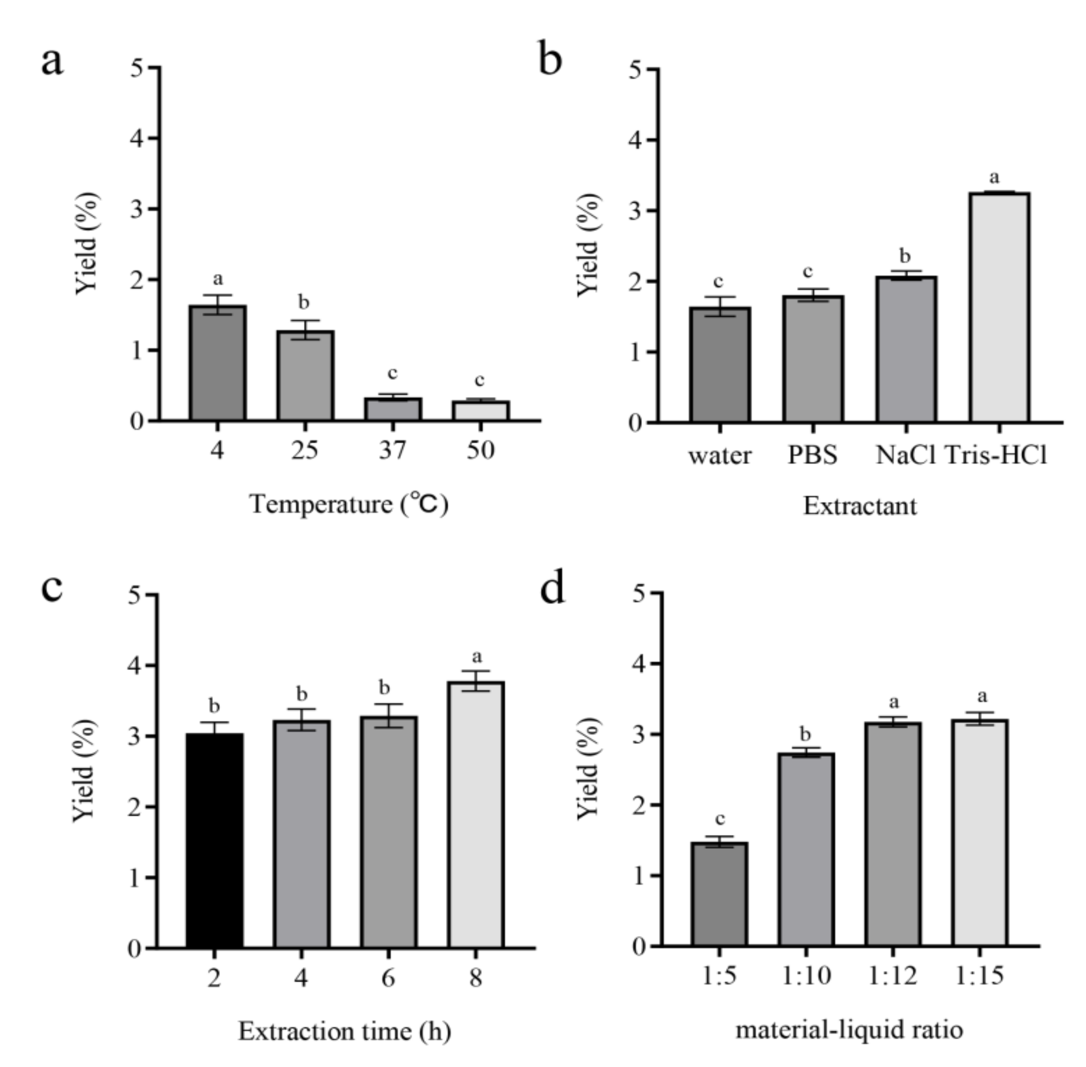
3.1. Effects of Various Factors on MP Extraction Yield

The effects of the temperature, extractant, extraction time, and material/liquid ratio on the MP extraction yield were evaluated. The yield at 4 °C was significantly (p < 0.05 or <0.01) higher than those at 25, 37, and 50 °C (Figure 1a), indicating that a lower temperature improves the protein yield. The effects of four extractants, (water, PBS, 10% NaCl, and Tris-HCl [pH 8.0]) were compared, and the yield for Tris-HCl was much greater (p < 0.01) than for the others (Figure 1b). The yield increased gradually as extraction time increased and reached the maximal value (3.82%) at 8 h (Figure 1c). The yield was inversely correlated with the material/liquid ratio and reached the maximal value (3.22%) for a ratio of 1:15 (Figure 1d).

3.2. Orthogonal Test for MP Extraction

A four-factor, three-level L9 (34) orthogonal test was performed (design shown in Table 1; results in Table 2). Of the nine experimental groups, No. 8 (4 °C, Tris-HCl, 1:20, 8 h) had the highest MP extraction yield (4.39%). Comparative analysis of average values for each factor indicated that the MP yield was highest for condition A3B2C1D3 (4 °C; Tris-HCl as the extractant; a material/liquid ratio of 1:20; extraction time of 8 h), i.e., No. 8, which was concluded to be the best extraction protocol. Extreme differences for factors A, B, C, and D were 1.967, 1.070, 1.654, and 1.043, respectively, indicating the magnitude order of A > C > B > D. Thus, the extractant was the factor with the greatest effect on the extraction yield, followed successively by temperature, extraction time, and the material/liquid ratio.

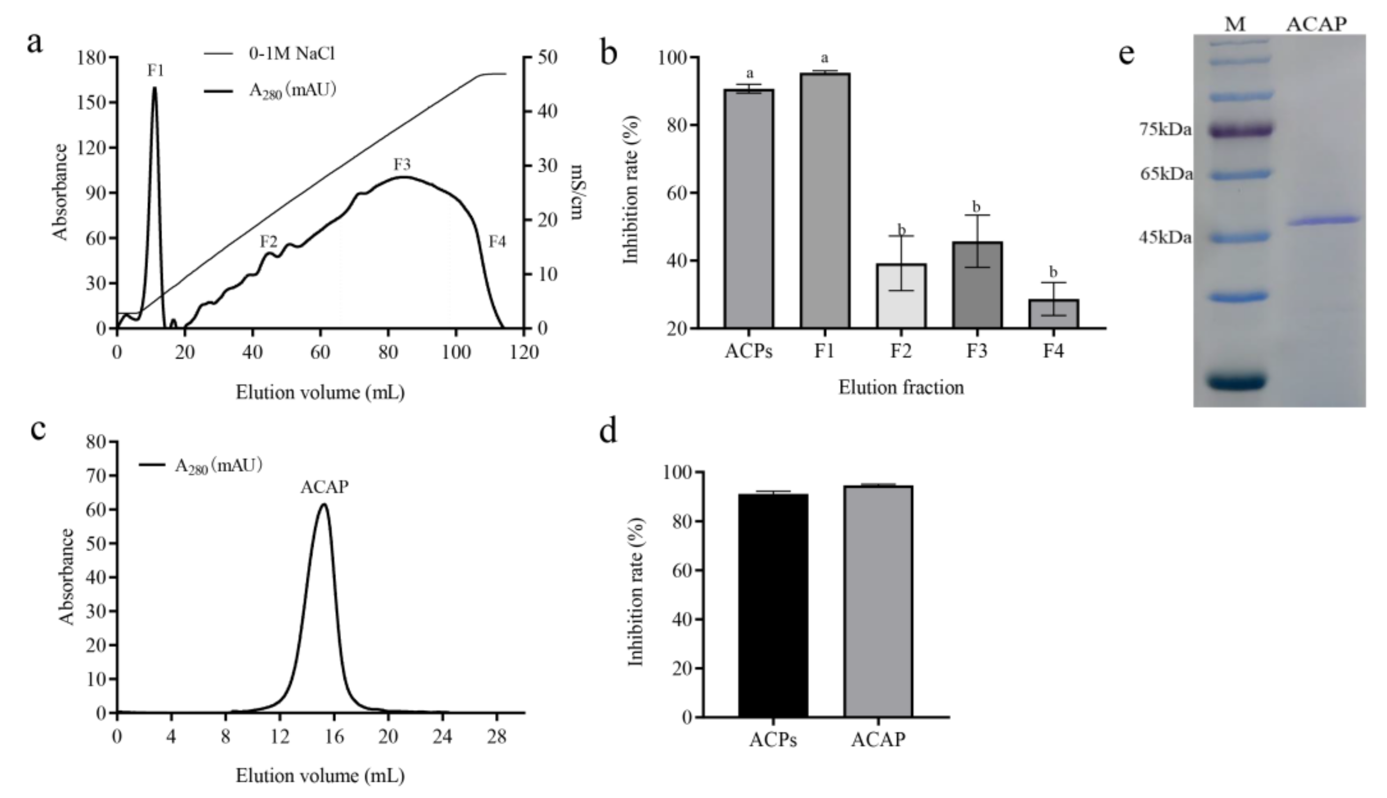
3.3. ACAP Purification by Ion Exchange Chromatography and Gel Filtration Chromatography

AC proteins (ACPs) were obtained by ammonium sulfate precipitation (to remove impurities) followed by ion exchange chromatography with the HiTrap Q anion column. Sequentially labeled absorption peaks in the elution curve were designated as F1 to F4 (Figure 2a). These four fractions were collected, and their antiproliferative effects on HeLa cells were evaluated. The inhibition rate of F1 (95.45%) was higher than those of ACPs and much higher (p < 0.01) than those of F2–F4 (Figure 2b). We concluded that F1 contained the target antitumor active protein (ACAP).
F1 was further purified by gel filtration chromatography with the Superdex 75 column, yielding a homogeneous protein elution fraction (ACAP) (Figure 2c). An in vitro antiproliferative effect assay with HeLa cells showed the inhibition rate for ACAP (94.60%) significantly higher than the control value. The inhibitory effect of ACAP did not differ significantly from those of ACPs. SDS-PAGE analysis of ACAP revealed a single 48 kDa band, according to the calculation of its mobility (Figure 2e).

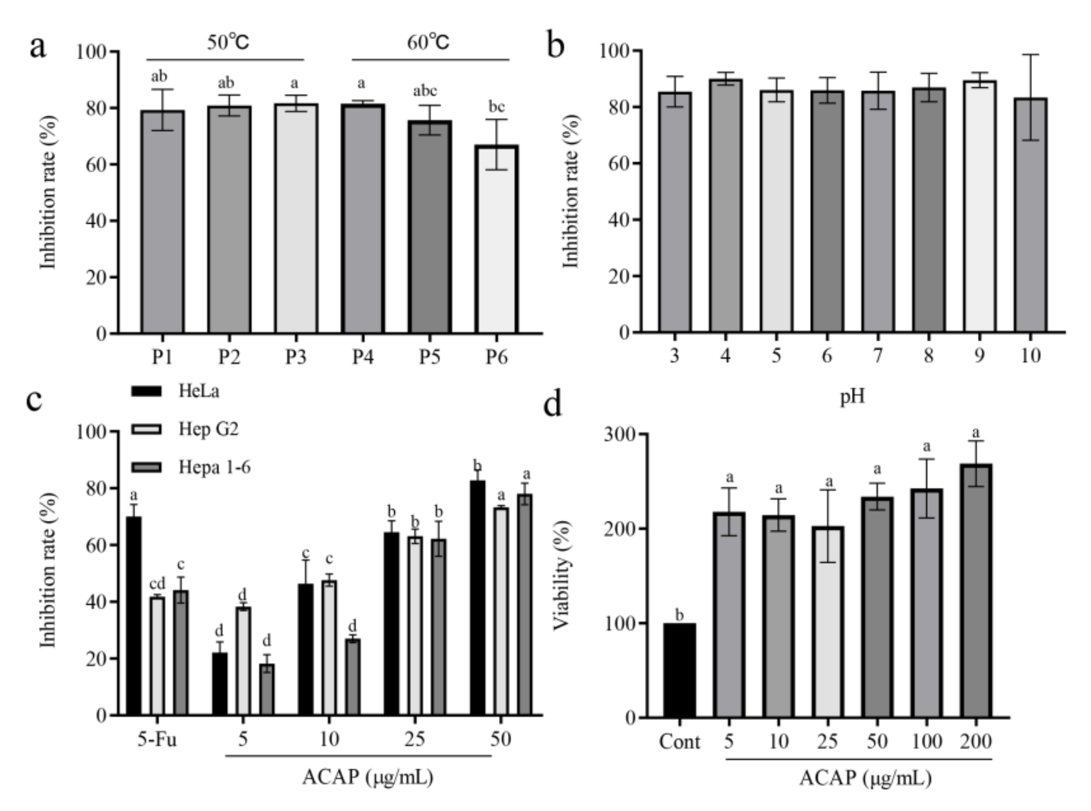
3.4. Effects of ACAP on Tumor Cell Proliferation

Antiproliferative properties of ACAP were investigated under various temperature and pH conditions. Under treatment at 50 °C for 30–90 min, ACAP showed an inhibition rate of ~80% for HeLa cells (Figure 3a). Under the 60 °C treatment for 30–90 min, ACAP retained substantial antitumor activity, indicating strong thermostability. ACAP maintained essentially constant antitumor activity at pH values ranging from 3 to 10 (Figure 3b), reflecting strong stability under varying acid and alkaline conditions.
Antiproliferative activities of ACAP on HeLa, Hep G2, and Hepa 1-6 at concentrations ranging from 5–50 µg/mL were investigated, with 5-Fu (50 µg/mL) as a positive control. ACAP displayed a strong, dose-dependent inhibitory effect on all three tumor cell lines (Figure 3c). At a 50 µg/mL concentration, inhibition rates of ACAP against HeLa, Hep G2, and Hepa 1-6 were 82.8%, 73.3%, and 78.0%, respectively, and IC50 values were 13.10, 10.70, and 18.69 µg/mL. ACAP had stronger antiproliferative effects on HeLa and Hep G2 than on Hepa 1-6. In contrast, ACAP had a dose-dependent enhancing effect on the proliferation of macrophage-derived RAW264.7 cells, indicating the lack of toxic effects on normal cells (Figure 3d).

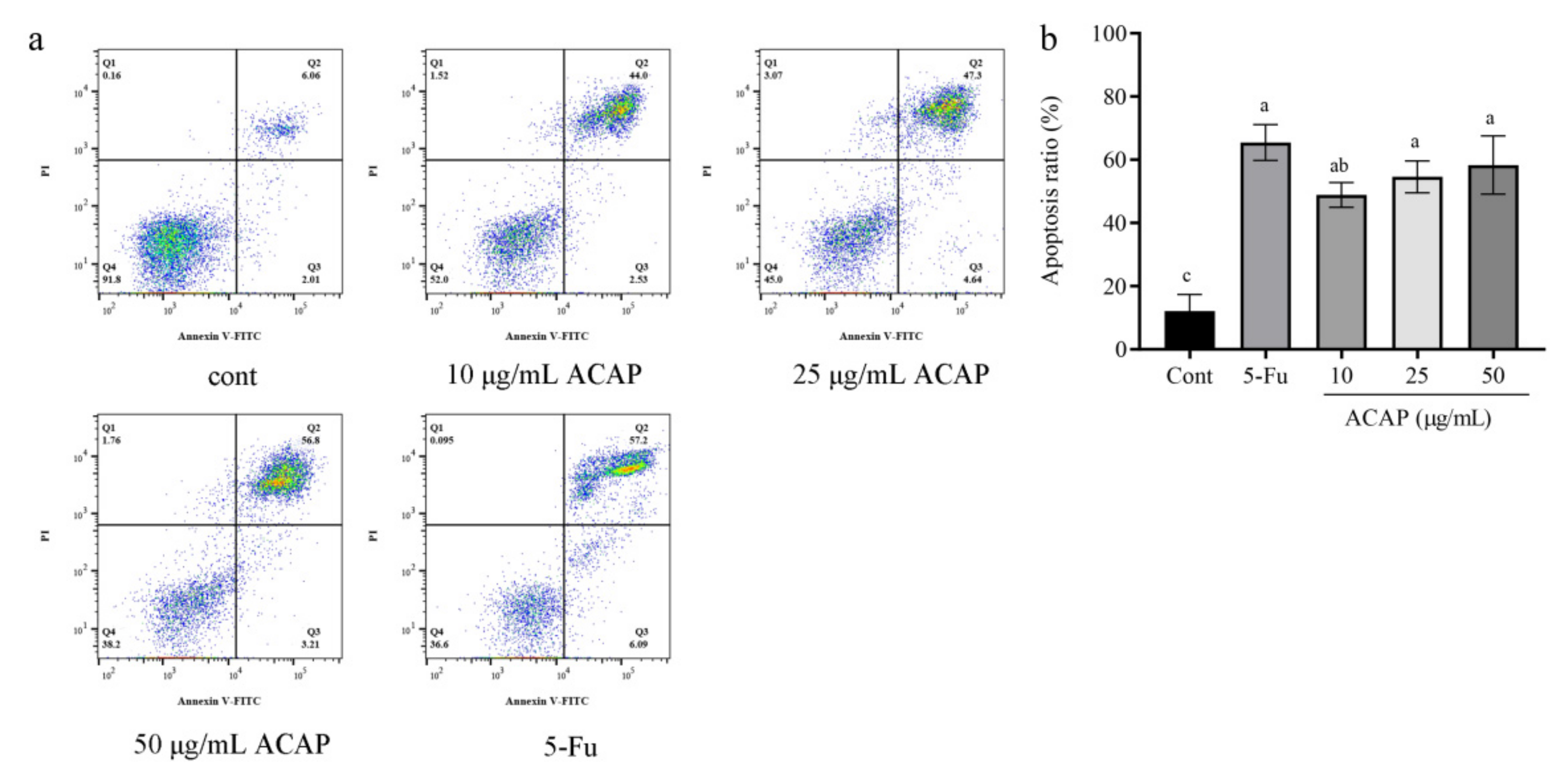
3.5. ACAP Induced Apoptosis of Hep G2 Cells

The induction of Hep G2 cell apoptosis by ACAP at concentrations of 10, 25, and 50 µg/mL was analyzed by flow cytometry with Annexin V-FITC/PI double staining. ACAP showed a significant (p < 0.01) dose-dependent apoptosis-inducing effect relative to the control (Figure 4a). Most of the induced cells were in the late-apoptotic stage; the proportion of early-apoptotic cells (lower right quadrant Q3) was low. The apoptosis rate for 50 µg/mL ACAP was 60.00% (Figure 4b), slightly lower than that of 5-Fu (63.29%). These findings indicate a strong pro-apoptotic effect of ACAP on tumor cells, comparable to the effect of the positive drug 5-Fu.

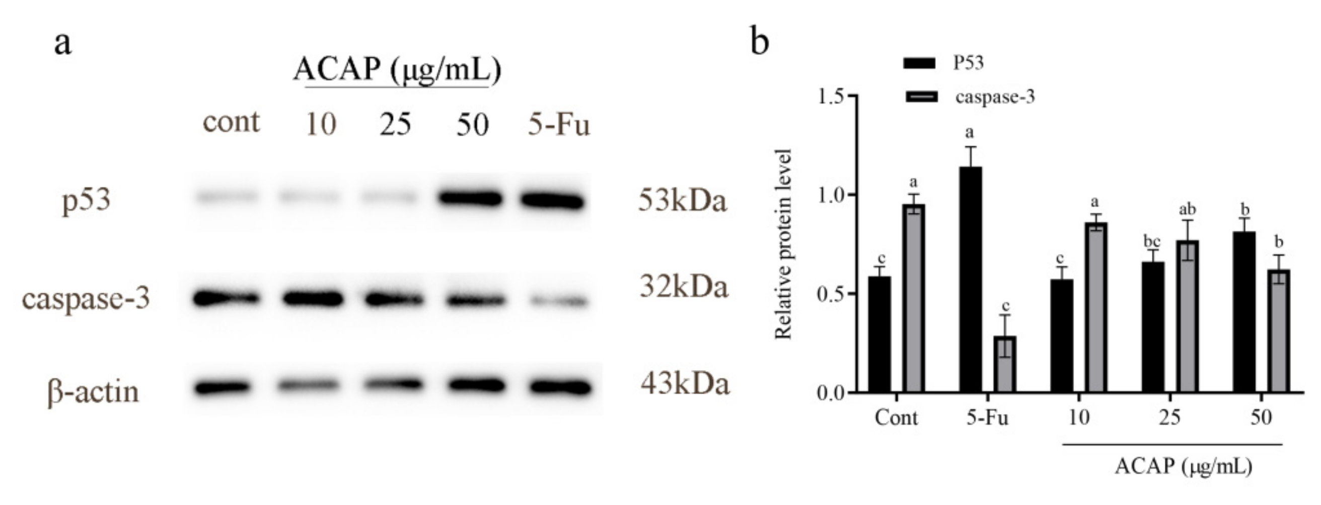
3.6. ACAP Affected Expression Levels of Apoptotic Pathway Proteins

In view of the significant pro-apoptotic effect of ACAP on Hep G2 cells, we investigated its effect on the expression levels of related apoptotic pathway proteins. p53 and caspase-3 expression levels were analyzed by Western blotting (Figure 5a). p53 levels were upregulated in a dose-dependent manner by ACAP relative to the control (Figure 5b). The p53 level of the 50 µg/mL ACAP-treated group was significantly (p < 0.05) higher than the control value, but not as high as that of the 5-Fu group. Caspase-3 levels of 10 and 25 µg/mL ACAP groups were lower than the control value, but the difference was not significant. The Caspase-3 level of the 50 µg/mL ACAP group was significantly (p < 0.05) lower than the control value but higher than that of the 5-Fu group. In summary, ACAP at the highest tested concentration induced strong upregulation of p53 expression and downregulation of caspase-3 expression, resulting in Hep G2 cell apoptosis.

4. Discussion

Cancer, in its various forms, is the leading cause of human mortality worldwide. Medical advances during recent decades have greatly improved anticancer therapeutic techniques. However, commonly used chemotherapeutic drugs frequently cause cytotoxic effects and other adverse reactions that impact patients’ quality of life. It is therefore highly desirable to identify and develop natural anticancer drugs [24]. Traditional Chinese medicine has been practiced and refined over the course of thousands of years, using a wide variety of source materials, particularly plants and large fungi. Modern scientific and technological advances have clearly revealed the beneficial effects of many Chinese medicines; however, the application of these medicines remains limited because their active ingredients and mechanisms of action are unclear. Edible/medicinal fungi are utilized as both food and medicine and comprise an important aspect of traditional Chinese medicine. Increasing numbers of research studies have focused on enhancing the production and utilization of these natural resources.
Fungal active proteins are promising natural resources for the development of antitumor agents. To research their antitumor activity and action mechanisms, the process of isolation and purification should be designed and optimized. Salting out has become a more common method for protein purification because of its simplicity and efficiency. Ammonium sulphate, a commonly used salt, was chosen in this study to remove the impurities and obtain ACPs. In order to obtain a pure target protein, chromatographic techniques are usually selected according to protein charge, molecular weight, hydrophobicity, and specific groups. Methods for the purification of antitumor-active proteins commonly involve anion/cation exchange chromatography followed by gel chromatography. For isolation and purification of the active protein of the medicinal fungus Hypsizygus tessulatus (beech mushroom; previously termed H. marmoreus), for example, the crude protein was obtained by 10% NaCl extraction followed by ammonium sulfate chromatography, DEAE-Sepharose was selected as the initial purification method based on the protein charge, and the single-protein fraction HM-3A was purified using the Sephacryl S-300 gel column [25]. In the present study, the crude protein was obtained by Tris-HCl (pH 8.0) extraction followed by ammonium sulfate chromatography, and single-component ACAP was purified using the HiTrap Q anion column and the Superdex 75 gel column.
Antitumor and other beneficial activities have been demonstrated for numerous medicinal fungi and their extracts. Terpenoids and polysaccharides are the active substances that have received the most research attention to date; however, fungal proteins often have notable medicinal functions as well. In edible/medicinal fungi, protein accounts for a large proportion of the dry weight and is one of the main bioactive components. In recent years, more and more scholars have focused on active protein components and have isolated many proteins with different biological activities, among which antitumor activity is becoming one of the hot issues. To date, a number of proteins extracted from a variety of fungi display antitumor activities. Y. Dong’s group isolated and purified a glycoprotein (GFG-3a) from Grifola frondosa mycelia, which showed high antitumor activity against S180 and Bel-7402 cells [27]. An antitumor active peptide extracted from Lentinus squarrosulus by C. Chaotham’s group had IC50 values of 26.84 ± 2.84, 2.80 ± 2.14, and 18.84 ± 0.30 µg/mL against lung cancer H460, H292, and H23 cells [28]. The results of the present study showed that the natural active protein ACAP from rare medicinal fungus AC had a significant dose-dependent in vitro inhibitory effect on the proliferation of HeLa, Hep G2, and Hepa 1-6 cells. At low concentrations, such an inhibitory effect was notably stronger for ACAP than for the positive drug 5-Fu.
Active proteins from edible/medicinal fungi generally exert antitumor effects by inducing apoptosis, inhibiting cell migration, and suppressing neovascularization. Cell death can be generally classified into three types: Apoptosis, cell autophagy, and cell necrosis, the first two of which can be classified as active programmed death under genetic control while necrosis is a passive process caused by external factors. Apoptosis can be classified as endogenous and exogenous apoptosis, which is influenced by a variety of proteins inside and outside the cell [24,25,26,29]. We used flow cytometric and Western blotting assays to confirm the apoptotic effects of ACAP on tumor cells. ACAP had pro-apoptotic effects on Hep G2, and the degree of apoptosis was correlated with the ACAP concentration. ACAP-induced apoptosis occurred primarily in the late stage, whereas 5-Fu-induced apoptosis occurred to similar degrees in the early and late stages.
p53, the most widely studied tumor suppressor protein, responds to various types of cellular stress and mediates a variety of antiproliferative processes. Enhanced p53 transcription levels affect numerous essential cellular physiological activities, including cell death, cell cycle arrest, senescence, DNA repair, angiogenesis, and cell migration [30,31]. We analyzed p53 expression in Hep G2 by Western blotting and observed significant upregulation in the ACAP-treated group. Our findings indicate that ACAP activated the tumor cell apoptotic pathway and induced a series of cascade reactions, including the conversion of the downstream caspase-3 protein precursor into the activated form of cleaved caspase-3, which mediates apoptosis. H. Xiao’s group, in a study on colon cancer cell lines HCT116 (human) and MC38 (mouse), similarly observed that the Pleurotus eryngii active protein (PEP) significantly upregulated c-PARP and c-caspase-3 expression in the apoptotic pathway, inducing early and late apoptosis [32]. S. Li’s group identified a fungal immunomodulatory protein (termed FIP-nha) from Fusarium solani (also known as Nectria haematococca), and RT-PCR analysis demonstrated that FIP-nha induced apoptosis by upregulating p53 expression [33]. Our findings described here indicate that ACAP has strong antitumor activity and the potential for development as a therapeutic agent and/or functional food. Studies on its molecular targets and detailed action mechanisms are in progress.

5. Conclusions

We isolated, via liquid fermentation, a novel protein with antitumor activity (termed ACAP) from mycelia of the medicinal fungus Antrodia cinnamomea and determined the optimal extraction and purification conditions. The antitumor activity of ACAP against several tumor cell lines was studied in vitro, and the underlying action mechanism was investigated. ACAP strongly inhibited tumor cell proliferation and induced late-stage apoptosis by upregulating p53 and downregulating caspase-3 levels. Our findings provide a basis for the development of AC-active proteins as novel therapeutic antitumor agents.

Author Contributions

Y.L. (Yingying Li): Investigation. J.G.: Writing—original draft. Y.L. (Yanbin Li): Formal analysis. S.Z.: Software. Y.L. (Yangyang Liu): Software. Y.L. (Yunxiang Liang): Supervision. Y.M.: Writing—review and editing, supervision, funding acquisition. All authors have read and agreed to the published version of the manuscript.

Funding

This study was supported by the National Natural Science Foundation of China (Grant No. 42271056) and the Natural Science Foundation of Hubei Province, China (Grant No. 2020CFB527).

Institutional Review Board Statement

Not applicable.

Informed Consent Statement

Not applicable.

Data Availability Statement

All data generated or analyzed during this study are included in this article.

Conflicts of Interest

The authors declare that they have no competing financial interests or personal relationships that could potentially influence the studies or findings described in this paper.

References

  1. Lu, M.; El-Shazly, M.; Wu, T.; Du, Y.; Chang, T.; Chen, C.; Hsu, Y.M.; Lai, K.; Chiu, C.P.; Chang, F.; et al. Recent research and development of Antrodia cinnamomea. Pharmacol. Ther. 2013, 139, 124–156. [Google Scholar] [CrossRef] [PubMed]
  2. Ao, Z.; Xu, Z.; Lu, Z.; Xu, H.; Zhang, X.; Dou, W. Niuchangchih (Antrodia camphorata) and its potential in treating liver diseases. J. Ethnopharmacol. 2009, 121, 194–212. [Google Scholar] [CrossRef] [PubMed]
  3. Geethangili, M.; Tzeng, Y.M. Review of pharmacological effects of Antrodia camphorata and its bioactive compounds. Evid.-based Complement Altern. 2011, 2011, 212641. [Google Scholar]
  4. Zhang, B.; Hu, P.; Huang, J.; Hu, Y.; Chen, L.; Xu, G. Current advances on the structure, bioactivity, synthesis, and metabolic regulation of novel ubiquinone derivatives in the edible and medicinal mushroom Antrodia cinnamomea. J. Agric. Food Chem. 2017, 65, 10395–10405. [Google Scholar] [CrossRef]
  5. Zhang, B.; Guan, Y.; Hu, P.; Chen, L.; Xu, G.; Liu, L.; Cheung, P.C.K. Production of bioactive metabolites by submerged fermentation of the medicinal mushroom Antrodia cinnamomea: Recent advances and future development. Crit. Rev. Biotechnol. 2019, 39, 541–554. [Google Scholar] [CrossRef]
  6. Zhang, Y.; Wang, Z.; Li, D.; Zang, W.; Zhu, H.; Wu, P.; Mei, Y.; Liang, Y. A polysaccharide from Antrodia cinnamomea mycelia exerts antitumor activity through blocking of TOP1/TDP1-mediated DNA repair pathway. Int. J. Biol. 2018, 120, 1551–1560. [Google Scholar] [CrossRef]
  7. Lee, Y.P.; Tsai, W.C.; Ko, C.J.; Rao, Y.; Yang, C.; Chen, D.; Yang, M.; Yang, C.; Tzeng, Y.M. Anticancer effects of eleven triterpenoids derived from Antrodia camphorata. Anticancer Res. 2012, 32, 2727–2734. [Google Scholar]
  8. Wu, T.; Huang, T.; Martel, J.; Liau, J.C.; Chiu, C.Y.; Leu, Y.L.; Jian, W.T.; Chang, I.; Lu, C.; Ojcius, D.M.; et al. Pinicolol B from Antrodia cinnamomea induces apoptosis of nasopharyngeal carcinoma cells. J. Ethnopharmacol. 2017, 201, 117–122. [Google Scholar] [CrossRef]
  9. Yeh, C.T.; Rao, Y.; Yao, C.; Yeh, C.F.; Li, C.H.; Chuang, S.; Luong, J.H.T.; Lai, G.; Tzeng, Y.M. Cytotoxic triterpenes from Antrodia camphorata and their mode of action in HT-29 human colon cancer cells. Cancer Lett. 2009, 285, 73–79. [Google Scholar] [CrossRef] [Green Version]
  10. Yen, I.C.; Shi, L.; Chung, M.; Ahmetaj-Shala, B.; Chang, T.; Lee, S.Y. Antrolone, a novel benzoid derived from Antrodia cinnamomea, inhibits the LPS-induced inflammatory response in RAW264.7 macrophage cells by balancing the NF-kappa B and Nrf2 pathways. Am. J. Chin. Med. 2018, 46, 1297–1313. [Google Scholar] [CrossRef]
  11. Liu, Y.; Yang, A.; Qu, Y.; Wang, Z.; Zhang, Y.; Liu, Y.; Wang, N.; Teng, L.; Wang, D. Ameliorative effects of Antrodia cinnamomea polysaccharides against cyclophosphamide-induced immunosuppression related to Nrf2/HO-1 signaling in BALB/c mice. Int. J. Biol. Macromol. 2018, 116, 8–15. [Google Scholar] [CrossRef]
  12. Huang, T.; Lan, Y.; Ko, Y.; Chen, C.; Lai, H.; Ojcius, D.M.; Martel, J.; Young, J.D.; Chong, K. Antrodia cinnamomea produces anti-angiogenic effects by inhibiting the VEGFR2 signaling pathway. J. Ethnopharmacol. 2018, 220, 239–249. [Google Scholar] [CrossRef]
  13. Sun, H.; Zhao, C.; Tong, X.; Qi, Y. A lectin with mycelia differentiation and antiphytovirus activities from the edible mushroom Agrocybe aegerita. J. Biochem. Mol. Biol. 2003, 36, 214–222. [Google Scholar] [CrossRef] [Green Version]
  14. Lam, S.K.; Ng, T.B. First simultaneous isolation of a ribosome inactivating protein and an antifungal protein from a mushroom (Lyophyllum shimeji) together with evidence for synergism of their antifungal effects. Arch. Biochem. Biophys. 2001, 393, 271–280. [Google Scholar] [CrossRef]
  15. El-Maradny, Y.A.; El-Fakharany, E.M.; Abu-Serie, M.M.; Hashish, M.H.; Selim, H.S. Lectins purified from medicinal and edible mushrooms: Insights into their antiviral activity against pathogenic viruses. Int. J. Biol. Macromol. 2021, 179, 239–258. [Google Scholar] [CrossRef]
  16. Cui, F.; Li, Y.; Zan, X.; Yang, Y.; Sun, W.; Qian, J.; Zhou, Q.; Yu, S. Purification and partial characterization of a novel hemagglutinating glycoprotein from the cultured mycelia of Hericium erinaceus. Process Biochem. 2014, 49, 1362–1369. [Google Scholar] [CrossRef]
  17. Kino, K.; Yamashita, A.; Yamaoka, K.; Watanabe, J.; Tanaka, S.; Ko, K.; Shimizu, K.; Tsunoo, H. Isolation and characterization of a new immunomodulatory protein, ling zhi-8 (LZ-8), from Ganoderma-lucidium. J. Biol. Chem. 1989, 264, 472–478. [Google Scholar] [CrossRef]
  18. Dai, R.; Liu, M.; Nabil, W.N.N.; Xi, Z.; Xu, H. Mycomedicine: A unique class of natural products with potent anti-tumour bioactivities. Molecules 2021, 26, 1113. [Google Scholar] [CrossRef]
  19. Zhang, Y.; Zhou, R.; Liu, F.; Ng, T.B. Purification and characterization of a novel protein with activity against non-small-cell lung cancer in vitro and in vivo from the edible mushroom Boletus edulis. Int. J. Biol. Macromol. 2021, 174, 77–88. [Google Scholar] [CrossRef]
  20. Zhang, Y.; Zhang, Y.; Gao, W.; Zhou, R.; Liu, F.; Ng, T.B. A novel antitumor protein from the mushroom Pholiota nameko induces apoptosis of human breast adenocarcinoma MCF-7 cells in vivo and modulates cytokine secretion in mice bearing MCF-7 xenografts. J. Biol. Macromol. 2020, 164, 3171–3178. [Google Scholar] [CrossRef]
  21. Sheu, F.; Chien, P.J.; Hsieh, K.Y.; Chin, K.L.; Huang, W.; Tsao, C.Y.; Chen, Y.; Cheng, H.; Chang, H. Purification, cloning, and functional characterization of a novel immunomodulatory protein from Antrodia camphorata (Bitter Mushroom) that exhibits TLR2-dependent NF-kappa B activation and m1 polarization within murine macrophages. J. Agric. Food Chem. 2009, 57, 4130–4141. [Google Scholar] [CrossRef] [PubMed]
  22. Zhang, Y.; Liu, F.; Ng, T.B. Interrelationship among paraptosis, apoptosis and autophagy in lung cancer A549 cells induced by BEAP, an antitumor protein isolated from the edible porcini mushroom Boletus edulis. Int. J. Biol. 2021, 188, 313–322. [Google Scholar] [CrossRef] [PubMed]
  23. Chang, Y.; Hsiao, Y.M.; Wu, M.; Ou, C.; Lin, Y.; Lue, K.; Ko, J.L. Interruption of lung cancer cell migration and proliferation by fungal immunomodulatory protein FIP-fve from Flammulina velutipes. J. Agric. Food Chem. 2013, 61, 12044–12052. [Google Scholar] [CrossRef] [PubMed]
  24. Russo, G.; Mischi, M.; Scheepens, W.; De la Rosette, J.J.; Wijkstra, H. Angiogenesis in prostate cancer: Onset, progression and imaging. BJU Int. 2012, 110, E794–E808. [Google Scholar] [CrossRef]
  25. Tsai, P.F.; Ma, C.; Wu, J.S.B. A novel glycoprotein from mushroom Hypsizygus marmoreus (Peck) Bigelow with growth inhibitory effect against human leukaemic U937 cells. Food Chem. 2013, 141, 1252–1258. [Google Scholar] [CrossRef]
  26. Lin, C.; Sheu, G.T.; Lin, Y.; Yeh, C.S.; Huang, Y.; Lai, Y.; Chang, J.; Ko, J.L. A new immunomodulatory protein from Ganoderma microsporum inhibits epidermal growth factor mediated migration and invasion in A549 lung cancer cells. Process Biochem. 2010, 45, 1537–1542. [Google Scholar] [CrossRef]
  27. Cui, F.; Zan, X.; Li, Y.; Yang, Y.; Sun, W.; Zhou, Q.; Yu, S.; Dong, Y. Purification and partial characterization of a novel anti-tumor glycoprotein from cultured mycelia of Grifola frondosa. Int. J. Biol. Macromol. 2013, 62, 684–690. [Google Scholar] [CrossRef]
  28. Prateep, A.; Sumkhemthong, S.; Suksomtip, M.; Chanvorachote, P.; Chaotham, C. Peptides extracted from edible mushroom: Lentinus squarrosulus induces apoptosis in human lung cancer cells. Pharm. Biol. 2017, 55, 1792–1799. [Google Scholar] [CrossRef] [Green Version]
  29. D’Arcy, M.S. Cell death: A review of the major forms of apoptosis, necrosis and autophagy. Cell Biol. Int. 2019, 43, 582–592. [Google Scholar] [CrossRef]
  30. Wachter, F.; Grunert, M.; Blaj, C.; Weinstock, D.M.; Jeremias, I.; Ehrhardt, H. Impact of the p53 status of tumor cells on extrinsic and intrinsic apoptosis signaling. Cell Commun. Signal. 2013, 11, 27. [Google Scholar] [CrossRef] [Green Version]
  31. Fridman, J.S.; Lowe, S.W. Control of apoptosis by p53. Oncogene 2003, 22, 9030–9040. [Google Scholar] [CrossRef] [Green Version]
  32. Yuan, B.; Ma, N.; Zhao, L.; Zhao, E.; Gao, Z.; Wang, W.; Song, M.; Zhang, G.; Hu, Q.; Xiao, H. In vitro and in vivo inhibitory effects of a Pleurotus eryngii protein on colon cancer cells. Food Funct. 2017, 8, 3553–3562. [Google Scholar] [CrossRef]
  33. Wang, J.; Wang, Y.; Hou, L.; Xin, F.; Fan, B.; Lu, C.; Zhang, L.; Wang, F.; Li, S. Immunomodulatory protein from Nectria haematococca induces apoptosis in lung cancer cells via the p53 pathway. Int. J. Mol. Sci. 2019, 20, 5348. [Google Scholar] [CrossRef] [Green Version]
Figure 1. Effect of various factors on extraction of A. cinnamomea proteins (ACPs): (a) Temperature; (b) extractant; (c) extraction time; (d) material/liquid ratio. Differing letters above bars indicate significant (p < 0.05) differences according to Tukey’s multiple comparisons test.
Fermentation 09 00185 g001
Figure 2. Purification and in vitro antitumor activities of ACPs: (a) Anion exchange chromatography; (b) in vitro antitumor activities of various fractions (F1–F4: fractions 1–4); (c) gel filtration chromatography; (d) in vitro antitumor activities of ACPs and ACAP; (e) SDS-PAGE assay of ACAP (M: Standard markers). The concentration of each fraction in (b,d) was 50 µg/mL. Differing letters above bars indicate significant (p < 0.05) differences according to Tukey’s multiple comparisons test.
Fermentation 09 00185 g002
Figure 3. In vitro activity assays of ACAP: (a) In vitro antitumor activity assay at the concentration of 50 µg/mL at 50 and 60 °C; (b) in vitro antitumor activities of ACAP (50 µg/mL) at pH 3–10; (c) inhibitory effects of ACAP on three tumor cell lines at indicated concentrations. 5-Fu: Positive control; (d) antiproliferative effect of ACAP on RAW264.7 cells. P1–P3: Protein fractions treated at 50 °C for 30, 60, 120 min; P4–P6: Protein fractions treated at 60 °C for 30, 60, 120 min. Cont: Normal control; ACAP: Treatment with ACAP at concentrations 5, 10, 25, and 50 µg/mL. 5-Fu: Positive control (50 µg/mL). Differing letters above bars indicate significant (p < 0.05) differences according to Tukey’s multiple comparisons test.
Fermentation 09 00185 g003
Figure 4. Effects of ACAP at three concentrations, and of controls, on Hep G2 apoptosis. (a) Analyzed by flow cytometry with Annexin V-FITC/PI double staining; (b) apoptosis ratio. Notations as in Figure 3. Differing letters above bars indicate significant (p < 0.05) differences according to Tukey’s multiple comparisons test.
Fermentation 09 00185 g004
Figure 5. Effects of ACAP on p53 and caspase-3 expression levels in Hep G2 cells: (a) Western blotting analysis; (b) data from panel (a) expressed as bar graph. Notations as in Figure 3. Differing letters above bars indicate significant (p < 0.05) differences according to Tukey’s multiple comparisons test.
Fermentation 09 00185 g005
Table 1. Orthogonal test schema.
LevelFactors
A 1B 2C 3D 4
110% NaCl641:10
2Distilled water8251:15
3Tris-HCl10371:20
1 Extractant, 2 Extraction time (h), 3 Temperature (°C), 4 Material–liquid ratio (w/v).
Table 2. Orthogonal test results.
Experimental NumberABCDProtein Yield (%)
111110.609
212220.461
313330.221
421230.574
522310.165
623120.972
731320.624
832134.390
933212.177
K1 11.2911.8075.9712.951
K2 11.7115.0163.2122.057
K3 17.1913.3701.0105.185
K1 20.4300.6021.9900.984
K2 20.5701.6721.0710.686
K3 22.3971.1230.3371.728
Range1.9671.0701.6541.043
1 the sum of yield for each factor at each level, 2 the average of the sum of yield for each factor at each level.
Disclaimer/Publisher’s Note: The statements, opinions and data contained in all publications are solely those of the individual author(s) and contributor(s) and not of MDPI and/or the editor(s). MDPI and/or the editor(s) disclaim responsibility for any injury to people or property resulting from any ideas, methods, instructions or products referred to in the content.

