Article

Okara Waste as a Substrate for the Microalgae Phaeodactylum tricornutum Enhances the Production of Algal Biomass, Fucoxanthin, and Polyunsaturated Fatty Acids

1
School of Chemistry, Chemical Engineering and Biotechnology, College of Engineering, Nanyang Technological University, 62 Nanyang Drive, Singapore 637459, Singapore
2
Interdisciplinary Graduate Programme, Nanyang Environmental and Water Research Institute, Nanyang Technological University, 62 Nanyang Drive, Singapore 637459, Singapore
*
Author to whom correspondence should be addressed.
These authors equally contributed to the work.
Fermentation 2023, 9(1), 31; https://doi.org/10.3390/fermentation9010031
Submission received: 2 November 2022 / Revised: 27 December 2022 / Accepted: 28 December 2022 / Published: 30 December 2022

Abstract

:
Despite the rich nutritional content of okara, the majority remains underutilized and discarded as food waste. In this study, solid-state fermentation of okara with food-grade fungi was performed to extract and solubilize any remnant nutrients locked within the lignocellulosic matrix to produce a nutrient-rich okara fermentate. Fermented okara media (FOM) was used as the sole nutrient source for growing marine diatom, Phaeodactylum tricornutum. Results have shown a two-fold increase in biomass production when grown on FOM (0.52 g L−1) as compared with conventional Guillard’s F/2 media (0.25 g L−1). Furthermore, cellular fucoxanthin content was enhanced significantly by two-fold to reach a final concentration of 15.3 mg g−1 compared to 7.3 mg g−1. Additionally, a significantly higher amount of polyunsaturated fatty acid (PUFA) was produced, particularly eicosapentaenoic acid (EPA) which yield has increased by nearly three-fold. Metabolomics analysis of intracellular contents in fermented okara culture revealed a significantly enhanced accumulation of nitrogenous metabolites, alongside the decrease in sugar metabolites as compared to F/2 culture, thus indicating metabolic flux towards pathways involved in cellular growth. This study demonstrated an innovative and low-cost strategy of using fermented okara as a nutritious substrate for achieving a sustainable media replacement for high density algal growth with a simultaneous enhancement of production in highly valued nutraceuticals, including fucoxanthin and EPA.

1. Introduction

Okara is a food waste by-product generated from the production of soymilk and tofu. High demand for soybean-derived products, particularly in the Asian countries, has led to huge biomass production of okara annually across the world. The annual generation of okara in China, Japan and Korea measured 2,800,000 tons, 800,000 tons, and 310,000 tons, respectively [1]. Okara contains a high nutritional value, which is comprised of: 55% fiber, 26% protein, 10% lipid, 4% carbohydrate and abundant minerals and vitamins [2]. However, the nutrients in okara are largely under-utilized at present, the majority of okara is discarded as waste, and only a minor portion is used as an animal feed, leading to economic loss [1]. The fact remains that okara is underutilized as an alternative food product, the bulk of okara waste is still treated as a non-salvageable waste, which sheer size of production cannot be sufficiently absorbed into secondary product stream and is eventually discarded. Instead, the valorization of okara to produce value-added bioproducts would be ideal. In line with the principles of circular economy design, okara can be bio transformed into a culture media, suitable for microalgae cultivation, and the subsequent production and extraction of bioactive compounds could then reenter the food chain as a food or feed ingredient [3].
Much research has been conducted to utilize the rich nutrient content in okara [1,2,4]. Yet, successful applications using okara are limited due to its insolubility, attributed to its high fiber content. Okara is a lignocellulosic plant biomass covered by rigid cell walls consisting of cellulose, hemicellulose, lignin, and pectin, which hinders accessibility to its nutritive factors, particularly proteins entrapped within the lignocellulosic matrix. To hydrolyze and fully recover the valuable nutrients entrapped in okara, various treatment methodologies have been explored [5,6]. For example, a study combining thermal treatment under autoclave conditions with enzymatic treatments such as cellulases and two pectinases to catabolize okara, resulted in 83–85% okara solubilization [5]. However, the use of commercial enzymes is rather costly. Thus, solid-state fermentation has also been explored as a low-cost strategy [6]. Fungi are attractive microorganisms which can secrete abundant enzymes including proteases, lipases and cellulolytic enzymes that can catabolize complex biomasses such as okara. Filamentous fungi secrete polysaccharides such as exoglucanases, endoglucanases and β-glucosidases which can break down fibers and release fermentable sugars including glucose and galactose [7,8]. The high diversity of fungal enzymes and high quantity of fermentable metabolites produced is the prime motivation for selecting fungi for okara fermentation, thus enabling further investigation of the change in nutritional composition before and after fermentation. Increased amounts of simpler nutritional compounds such as amino acids, sugars, and short-chain fatty acids were found in fermented okara, and the fermentation revealed a more effective recovery of nutrients than in bacteria culture (Lactobacillus plantarum) [9]. The observation was highly indicative that the valuable nutrients entrapped in the lignocellulosic matrix of okara can be more effectively recovered through fungal fermentation.
Microalgae have attracted significant attention as a green and renewable source of high-value bioactive components such as polyunsaturated fatty acids (PUFAs) and carotenoids [10]. However, the high production cost has limited the high throughput commercialization of microalgae for specialized food-grade biochemicals and micronutrients [11]. To enhance the competitiveness of microalgae as an alternative source of bioactive components, extensive studies have been conducted to increase the product yield [12,13,14,15,16]. For instance, algal cells were cultivated mixotrophically under both photosynthetic (through supplementation of light) and non-photosynthetic (through supplementation of organic substrates, such as glucose and glycerol) influence. Mixotrophic cultivation facilitates cell growth based on the synergistic effect of respiration and photosynthesis to uptake inorganic and organic substrates, respectively [12], leading to increased biomass and lipid yield. However, provision of additional organic substrates increases production cost, which is a major drawback for industrial application. As an alternative, heterogenous food waste has been explored as a low-cost organic nutrient source [17,18]. Canteen wastes were hydrolyzed by fungal fermentation to recover entrapped nutrients, including glucose, free amino nitrogen (FAN) and phosphate. Microalgae cells cultured in the obtained food waste hydrolysate had not only shown high growth rate, but also contained significantly higher protein and lipid amounts compared to that grown in conventional media, demonstrating high economic potential [18]. However, culture media derived from heterogenous food waste may be limited by batch-to-batch variation, as any change in the composition of the mixture of food waste will lead to different nutritional profile of the produced media. As a result, the media will be subjected to high variability in nutritional quality and quantity and toxicity exposure to metallic elements and surfactants, which may cause unpredictable and inconsistent cellular growth and biochemical characteristics. Alternatively, we considered the use of homogenous food side-stream, okara, which is more uniform and easier to characterize. We hypothesized that the use of fermented okara would serve as an excellent sole nutrient source for microalgae growth.
Among numerous microalgae species, the marine microalgae Phaeodactylum tricornutum (P. tricornutum) have garnered much interest as a sustainable source of high-value nutraceuticals. It is rich in omega-3 fatty acids, particularly EPA, which provides important health benefits such as lowering blood cholesterol, inflammation, preventing cardiovascular and coronary heart diseases [19,20]. Despite attempts to make EPA production more sustainable, marine fish oil is still the major source of EPA. Due to trending consumer’s interest in health products, there has been an increasing demand for EPA as a dietary supplement. However, any further increase in EPA production would put a massive strain on the already overfished marine stocks, further exacerbating the depletion of fish stocks. In fact, EPA is not intrinsically produced by marine fish, but accumulated through dietary intake of phytoplankton. As one of the planktonic origins of EPA, P. tricornutum is a promising alternative as a more direct and sustainable source of EPA.
In addition, P. tricornutum cells contain high fucoxanthin content, a major carotenoid in diatoms. Fucoxanthin displays extensive health benefits including anti-obesity, anti-diabetic, and anti-cancer properties [21,22]. For instance, supplementing fucoxanthin to mice undergoing high-fat diet significantly reduced plasma triglyceride and increased fecal lipids while reducing glucose level and insulin concentration [23]. Another study showed that addition of purified fucoxanthin extracted from P. tricornutum exerted antiproliferative effects on carcinoma cell lines and dose-dependent antioxidant activity in leukocytes [24]. To date, industrial production of fucoxanthin has been focused on using macroalgae. However, the obtainable fucoxanthin yield from macroalgae is generally smaller than 1 mg g−1 [25,26]. In contrast, it has recently been shown that microalgae contains fucoxanthin in an order of magnitude higher than that of macroalgae, emerging as a new promising source of the carotenoid [27].
Given that P. tricornutum contains both high fucoxanthin and EPA content, it has substantial commercial potential as a sustainable source for co-production of the bioactive compounds. Besides, P. tricornutum is one of the few unique microalgae species which are able to grow mixotrophically, in which supplementing organic substrates in the cultivation media enhances biomass, EPA and pigment contents [28]. Thus, in this study we selected P. tricornutum as our target algae to be cultivated in fermented okara substrate.
The overarching theme of this experiment was to demonstrate that sustainable production of highly valued nutraceuticals (EPA and fucoxanthin) could be attained by incorporating low-cost okara side-stream into the mixotrophic algae cultivation process. The growth parameters and biochemical contents (PUFA, EPA and fucoxanthin) of the FOM-cultured cells were compared to that grown in conventional marine algae media (Guillard’s F/2) to evaluate the functionality of fermented okara media in algal cultivation. Metabolomics analysis on the distribution and accumulation of intracellular metabolites in P. tricornutum using different culture media, aims to further elucidate how products of okara fermentation trigger specific metabolic pathways and encourage accumulation of highly prized endometabolites, specifically EPA and fucoxanthin. To the best of knowledge, there has been no paper discussing the use of fermented okara media in algae cultivation, in conjunction with metabolomics studies.

2. Materials and Methods

2.1. Algal Culture

Phaeodactylum tricornutum (P. tricornutum) UTEX 646 was obtained from the Culture Collection of Algae at The University of Texas at Austin (UTEX), USA. P. tricornutum in exponential growth phase (initial OD540 = 0.1) was seeded into 100 mL of Guillard’s F/2 media and fermented okara media (FOM), respectively. The cultures were maintained under continuous white fluorescence light at an irradiance of 50 μmol photons m−2 s−1 at 20 ± 1 ℃ with shaking at 140 rpm using an orbital shaker (Supplementary Figure S1). Prior to this, P. tricornutum was acclimatized in FOM for 3 months to ensure a consistent growth and biochemical state. P. tricornutum was maintained by sub-culturing every 10 days.

2.2. Determination of Algal Growth

The specific growth rate µ (day−1) of the linear growth phase was calculated from the equation:
µ = 1 / t l n ( X f / X O )
in which Xo and Xf are the initial and final concentrations of cells (g L−1) at each batch run, respectively, and t (days) is the duration of the run.
P. tricornutum biomass concentration was obtained by correlating the absorbance of the culture at 540 nm to dry-weight measurement using the standard curve equation (graph not shown):
D r i e d   c e l l   w e i g h t   ( g · L 1 ) = 0.1794 x
where x is the optical density measured at 540 nm. The equation was constructed by determining the dry weight of P. tricornutum. To do this, a certain volume of culture broth was centrifuged at 10,000× g and washed twice with Milli-Q water, followed by a 48 h lyophilization to obtain dried cells before gravimetric measurement.

2.3. Preparation of Growth Media

Guillard’s F/2 media was prepared using artificial seawater (ASW) as the base solution. To prepare ASW, sea salt (Sigma, St. Louis, MO, USA) was solubilized with deionized water to reach a stable salinity of 30 ppt and filter-sterilized with membranes of 0.22 μm pore size. The filtered ASW was enriched with concentrated F/2 without silicon (G0154, Sigma, USA) to constitute the F/2 algal culture media [29] and stored in 20 °C until further usage.
Fresh okara was provided by Vitasoy International Singapore Pte Ltd. (Singapore) and stored in −20 °C until used. Solid-state fermentation of okara and extraction of the nutrients to form FOM were performed according to [30]. Briefly, the prepared FOM was filter-sterilized and stored in −20 °C until further usage.

2.4. Nutritional Analysis of Fermented Okara Media

Nutritional analysis was carried out by an accredited analytical laboratory of ALS Technichem Pte Ltd. (Singapore). Nitrogen content was analyzed by the Kjeldahl method [31] and the carbohydrate content and phosphorus content were measured according to the Kirk and Sawyer methods [32]. Carbon was analyzed using elemental analysis (Vario EL III CHNS Elemental Analyser).

2.5. Quantification of Fucoxanthin and Chlorophyll a Production from P. tricornutum

Quantification of fucoxanthin and chlorophyll a was carried out on day 7 samples using an Agilent 1100 high performance liquid chromatography (HPLC), which was equipped with a photodiode array detector and a Phenomenex C6-phenyl reversed phase column (100 mm × 4.6 mm × 5 μm). Methanol and 1 M ammonium acetate (70:30) were used as the mobile phase with a flow rate of 1 mL/min. In a gradient condition, methanol proportion was increased from 5% to 95% over 80 min. Detection of fucoxanthin-chlorophyll a complex was recorded at 445 nm. Fucoxanthin standard (Sigma, USA) and chlorophyll a standard (Sigma, USA) was used to produce the standard curve.

2.6. Lipid Extraction and Yield Measurement

Biomass was harvested on day 7 and was lyophilized and mixed with 3 mL of chloroform: methanol in the ratio 2:1 (v/v). To this mixture, 10 μL of tripentadecanoin (20 mg mL−1) (T4257, Sigma) were added as an internal standard. Samples were then homogenized using a bead grinder (Fast Prep, MP Biomedicals, USA) at 6.5 ms−1 for 40 s for 8 cycles, with 60 s of cooling in between each cycle to minimize heating. Subsequently, 1 mL of 0.9% NaCl was added to the mixture and centrifuged at 10,000× g for 10 min. The bottom chloroform phase was then collected and evaporated by spurting N2 gas steam over the solvent until complete dryness. The dried residue obtained was the total lipid content of P. tricornutum extracted by organic solvent and was gravimetrically measured.

2.7. Fatty Acid Analysis

To characterize the individual fatty acid composition, fatty acids were trans-esterified into fatty acid methyl ester (FAME) by adding 500 μL of boron-trifluoride to the extracted total lipids and incubated at 95 °C for 20 min. After cooling the samples to room temperature, 300 μL of hexane and 300 μL of saturated NaCl were added and centrifuged at 10,000× g for 10 min. The upper hexane phase was then transferred into a glass vial for analysis using gas chromatography-mass spectrophotometry (GCMS) 7890A-5975C (Agilent Technologies, Santa Clara, CA, USA) equipped with HP-5MS UI column (30 m × 250 μm × 0.25 μm). The ion source temperature and injector temperature were set at 230 °C and 250 °C, respectively. The oven temperature was kept at 80 °C for 1 min and then raised to 250 °C at the rate of 7 °C min−1 and held for 8 min. Injection volume of 1 μL was used. Data were scanned from 35–600 m/z. NIST08 mass spectral library was used to identify the fatty acids and only those matching with 80% similarity index and above were selected. Fatty acid standard (CRM47885, USA) was used to produce standard curve.

2.8. Intracellular Metabolite Analysis

P. tricornutum cells harvested on day 2 of fermentation was lyophilized and mixed with 1.5 mL of chloroform: water: methanol 2:2:1 (v/v). A measure of 10 µL of ribitol (0.2 mg mL−1) was also added as an internal standard. Samples were then homogenized using a bead grinder (Fast Prep, MP Biomedicals, USA) at 6.5 ms−1 for 40 s for 8 cycles, with 60 s of cooling in between each cycle to minimize oxidation. The solution was centrifuged at 2500× g for 15 min and the upper phase was extracted and evaporated with N2 spurting until complete dryness. A measure of 40 µL of methoxyamine hydrochloride (20 mg mL−1) was added into the dried sample and incubated at 37 °C for 2 h. Subsequently, 70 µL of N-methyl-N-(trimethylsilyl)-trifluoroacetamide (MSTFA) was then added and incubated at 37 °C for 30 min. The sample was centrifuged at 10,000× g for 10 min and the supernatant was transferred into a glass vial. Untargeted metabolome analysis of the samples was carried out using GC-MS system (Agilent Technologies 5973N) equipped with HP-5MS column (30 m × 250 μm × 0.25 μm). Sample volume of 1 µL was injected into the system in splitless mode. The injector temperature and ion source temperature were set at 250 and 230 °C, respectively. The oven temperature was kept at 80 °C for 1 min and raised to 250 °C by 7 °C/min and held for 8 min. Data were recorded from 35 to 600 m/z with a scan time of 0.2 s [33].

2.9. Statistical Analysis

All the biochemical analysis of the microalgae cells was performed in triplicates and the values were reported as means ± standard deviation. Statistical analysis was performed using the Student’s 2-tailed t test, where equal variance of p < 0.05 was considered as statistically significant.

3. Results and Discussion

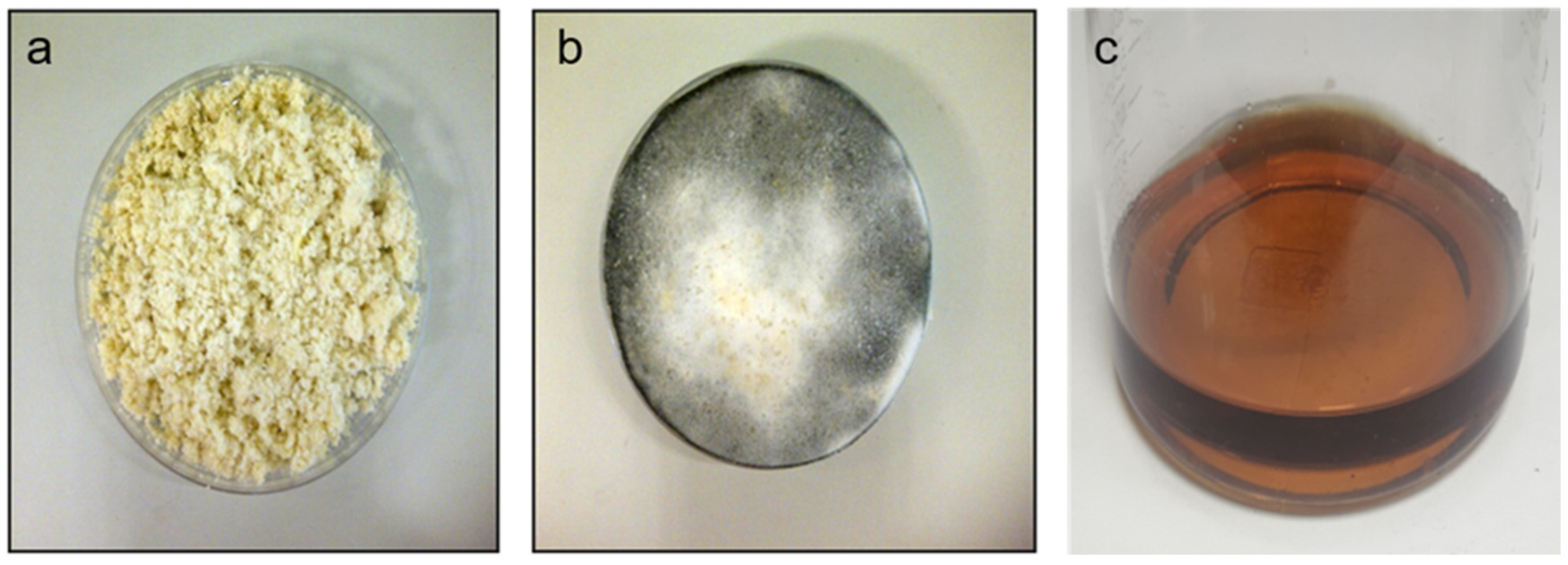
3.1. Okara Fermentation and Media Preparation

Solid-state fermentation of okara was performed (Figure 1) using spores of fungi and incubating the mixture at 30 °C. After 24 h of incubation, white mycelial filaments were observed on okara (Figure 1a), which later turned grey after longer incubation (Figure 1b). This observation indicated successful solid-state fermentation as there was obvious fungi growth (white mycelial filaments) and sporulation (greying) was triggered after slowing of growth rate after nutrient depletion. Aqueous extraction of nutrients from the fermented okara was then carried out in ASW, which was membrane-sterilized (0.22 µm) to produce a clear, brown-colored filtrate of FOM (Figure 1c). The obtained FOM was then used for P. tricornutum cultivation.

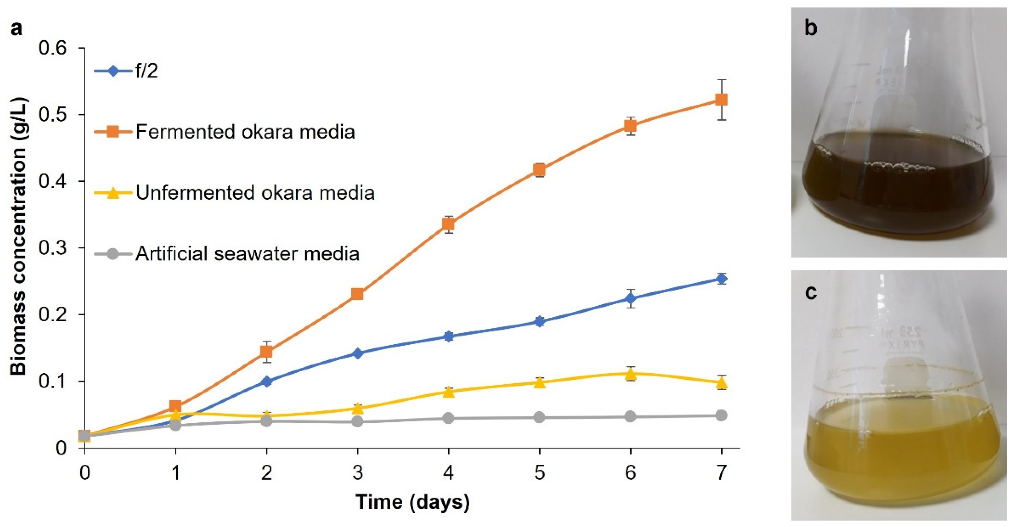
3.2. Growth of P. tricornutum in Fermented Okara Media

To investigate the functionality of the fermented okara media (FOM), we cultured microalgae and compared the growth to that in the conventional Guillard’s F/2 media. Our results have shown a significantly higher cell growth of P. tricornutum when grown on FOM as compared to that on F/2 media throughout the 7 days of fermentation (Figure 2). As shown in Figure 2a, exponential growth phase of P. tricornutum in F/2 culture lasted until day 3 post-incubation, followed by a stationary growth phase. On the other hand, P. tricornutum in fermented okara culture sustained an exponential growth phase until day 5 of incubation, followed by a stationary growth phase. Effectively, the exponential phase was lengthened by 2 days using FOM over F/2 media, thus enabling longer periods of biomass accumulation. The maximum growth rate of P. tricornutum when grown on fermented okara culture (0.48 day−1) was higher compared to F/2 culture (0.38 day−1). Comparing these maximal growth rates to that of several literature values (Supplementary Table S3, References cited in Supplementary Material), it shows that both photoautotrophic growth in F/2 media and mixotrophic growth with FOM outperforms other small-scale P. tricornutum cultivation, even during the absence of CO2 supplementation. This observation is likely the result of sufficient aeration provided by the orbital shaker mixing on a small-scale shake flask culture with high surface area ratio. Based on this comparison, the amount of algal growth gathered from this culture setup is thus sufficient for the purpose of this ‘proof-of-concept’ experiment. At the end of the algal cultivation (day 7), the maximum algal biomass obtained from fermented okara culture (0.52 g L−1) was approximately 2-fold higher compared to that from F/2 culture (0.25 g L−1). The difference in biomass density was visually noticeable, as observed by the darker brown color of the fermented okara culture as (Figure 2b) compared with F/2 culture (Figure 2c). Our result clearly demonstrated that in terms of biomass density, FOM has performed better than traditional F/2 media for the cultivation of P. tricornutum.
The substantially higher amounts of P. tricornutum biomass obtained from fermented okara media as compared to that from the unfermented okara media highlights the crucial role of fermentation at transforming okara into a highly functional media for culturing of P. tricornutum. In fact, our previous study has shown that the solid-state fermentation of okara increased amino acid content by approximately 2-fold, from 3.04 mg g−1 to 5.41 mg g−1, compared to unfermented okara [34]. Similarly, fermentation of brewers’ spent grain (BSG), a protein and fiber rich side-stream produced from the beer manufacturing industry, increased the amino acid content compared to unfermented BSG [35]. As a result, fermented BSG media supported the growth of nitrogen-consuming yeast Rhodosporidium toruloides, whereas no obvious growth was seen when the yeast was grown on the unfermented BSG media [36], consistent to our results. Thus, the high growth of P. tricornutum when grown on FOM is likely due to the effect of fermentation, by hydrolyzing macromolecules of okara into small molecules and enabling the microalgae to effectively assimilate the simple nutrients. Collectively, our result demonstrated that solid-state fermentation is an effective method of improving the nutritional content of food by-products, to produce a novel nutrient-rich media.

3.3. Nutrient Composition in Fermented Okara Media

To identify and understand the underlying reason behind the improved cellular growth of P. tricornutum when grown on fermented okara media (FOM), key microalgae nutrients, carbohydrate, nitrogen, and phosphorus were measured and compared to their levels in F/2 media, as shown in Table 1. Our results showed considerable amount of carbohydrate (3 g L−1) contained in FOM, while no carbohydrate was present in F/2 media. As expected, carbon content was markedly higher by 14-fold in FOM than in F/2 media. In addition, remarkable amounts of nitrogen (1500 mg L−1) and phosphorus (140 mg L−1) were found in the FOM, each of them 12-fold higher as compared to their levels in F/2 media, respectively. The high concentration of the key nutrients found in the FOM had likely promoted P. tricornutum growth, leading to the higher biomass produced.
Specifically, the presence of carbohydrate in the FOM could serve as an organic carbon source for microalgae growth. This allows the microalgae to utilize respiration for carbon fixation in addition to photosynthesis, and synergistically carry out the two different metabolic processes. Such mixotrophic growth mode have been reported to result in higher cell growth rate and biomass production than a photoautotrophic growth mode, consistent with our results [15].
Nitrogen and phosphorus are crucial nutrients required for microalgae growth, in which the former is used in the synthesis of nucleic acids, amino acids, proteins, pigments and carbon accumulation [37], while the latter plays a major role in cellular division [38]. One study varied the P. tricornutum culture conditions and reported the highest biomass yield in 10-times concentrated F/2 media which contains both high nitrogen and phosphorus content, followed by 10-times nitrate supplemented F/2 media and lastly F/2 media without any nutrient addition [39]. Similarly, the markedly higher concentration of nitrogen and phosphorus in the fermented okara media as compared to F/2 media may have contributed to the increased biomass production.

3.4. Fucoxanthin Production from P. tricornutum Cells in Fermented Okara Culture

To understand the biochemical response of the P. tricornutum cells cultured in fermented okara media (FOM), we analyzed the pigment contents of chlorophyll a and fucoxanthin in fermented okara culture and F/2 culture (Table 2). Results revealed that cells cultured in FOM had extraordinarily higher chlorophyll-a content of 76.5 mg g−1 as compared to 15.5 mg g−1 in F/2 media. Additionally, notably higher fucoxanthin content of 15.3 mg g−1 was measured in the fermented okara culture compared with 7.3 mg g−1 in the F/2 culture. Overall, the total fucoxanthin production was greatly increased by 5-fold, reaching 8.20 mg L−1 in the fermented okara culture.
Nitrogen concentration in the cultivation media and light intensity have been reported as the major factors influencing pigment production of microalgae [39,40,41,42]. A recent study observed a prominent increase in fucoxanthin production from 23.2 mg g−1 to 59.2 mg g−1 in P. tricornutum cells when it was grown on F/2 media enriched with 10-times additional nitrate [39]. Additionally, a separate study reported a 3-times increase in fucoxanthin concentration from 6.71 to 18.14 mg g−1 in O. aurita after nitrate supplementation [40]. As such, the higher production of fucoxanthin by P. tricornutum observed in the fermented okara culture might be attributed to the high nitrogen availability in the media.
The reduced light intensity reaching the cells in fermented okara culture may also have promoted fucoxanthin production. Fucoxanthin is an accessory pigment that plays an important role in harvesting light [43]. Increased cell density results in mutual shading of cells, which would reduce the average level of light intensity received by cells. As light progressively becomes the limiting factor, cells in the fermented okara may have stimulated fucoxanthin production in compensation to increase light harvesting efficiency. The effect of reduced light intensity on fucoxanthin production has been previously reported in which 4-fold increase in fucoxanthin concentration (42.8 mg g−1) of P. tricornutum grown under low light intensity (100 μmol photons m−2 s−1) was observed compared to that under high light intensity (210 μmol photons m−2 s−1, 9.9 mg g−1) [39]. Considering these reports, increased fucoxanthin production found in fermented okara culture is likely the result of favorable nutrient concentration coupled with reduced light intensity.

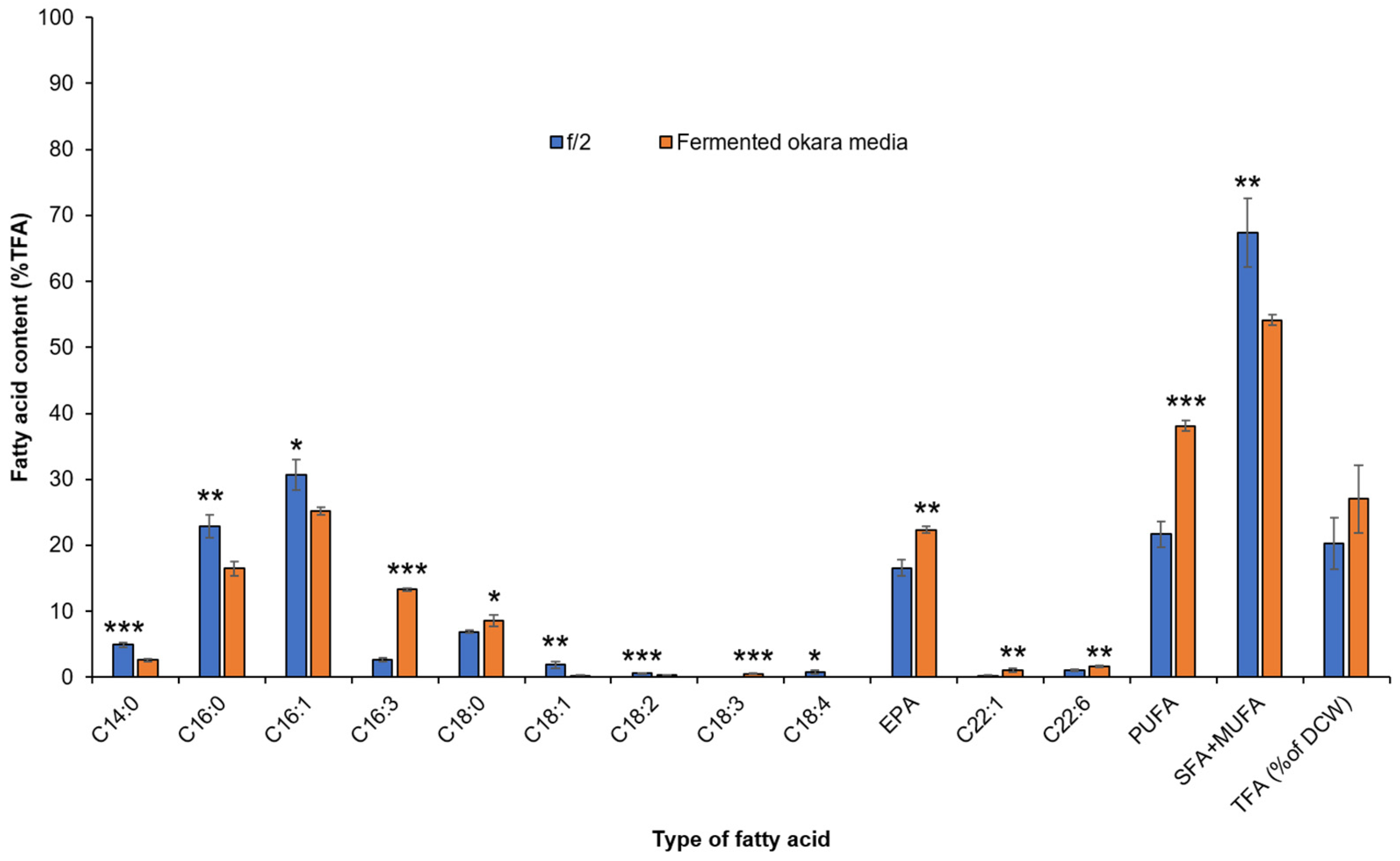
3.5. Fatty Acid Production from P. tricornutum Cells in Fermented Okara Culture

The fatty acid composition of P. tricornutum cells was determined (Figure 3). The major fatty acids produced were similar under both culture conditions, namely C16:0, C16:1, C16:3, C18:0 and C20:5. However, the relative degree of fatty acid saturation differed. Saturated fatty acids (SFAs) C14:0, C16:0, and mono-unsaturated fatty acid (MUFA) C16:1 comprised a greater proportion of the fatty acids in cells from the F/2 culture, whereas production of the polyunsaturated fatty acids (PUFAs) C16:3 and C20:5 (EPA) was significantly higher in fermented okara culture.
PUFAs are principal components of polar lipids that form the structural component of the cell, including the photosynthetic membrane of the chloroplast [44] while SFAs and MUFAs are mainly found in neutral lipids in the form of triacylglycerides, that are responsible for energy storage [45]. Under nutrient replete condition, cells increase PUFA proportion to support cellular proliferation, rather than energy storage [46,47]. Given the higher carbohydrate, nitrogen, and phosphorus concentration, FOM is a more nutrient-rich cultivation media as compared to F/2. Thus, high cell growth rate and pigment content in fermented okara culture leads to a high demand for PUFAs to form the membrane lipids in newly synthesized cells.
Notably, observations of high total fatty acid content (TFAC)and PUFA content in cells from fermented okara culture was likely due to the presence of carbohydrates in the media that allows greater availability of carbon for the cells to utilize and channel additional metabolic energy towards lipid biosynthesis. In agreement with our result, the supplementation of glycerol as carbon substrate in a mixotrophic culture of P. tricornutum has increased lipid content as compared to non-supplemented cultures [48]. It is of great significance to achieve both high TFAC and EPA content, boosting the total EPA yield by nearly 3-fold in fermented okara culture compared to that in F/2 culture (Table 3). This underscores the commercial potential of using low-cost okara as a sole nutrient source for effectively producing high-value EPA.

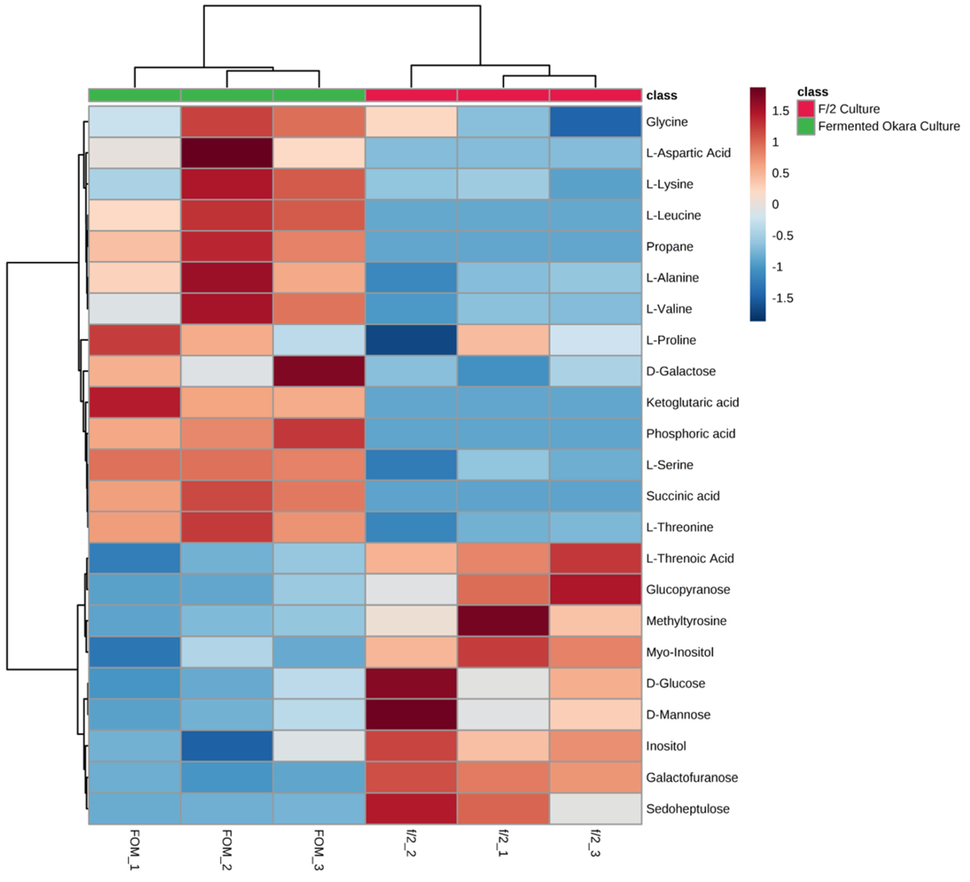
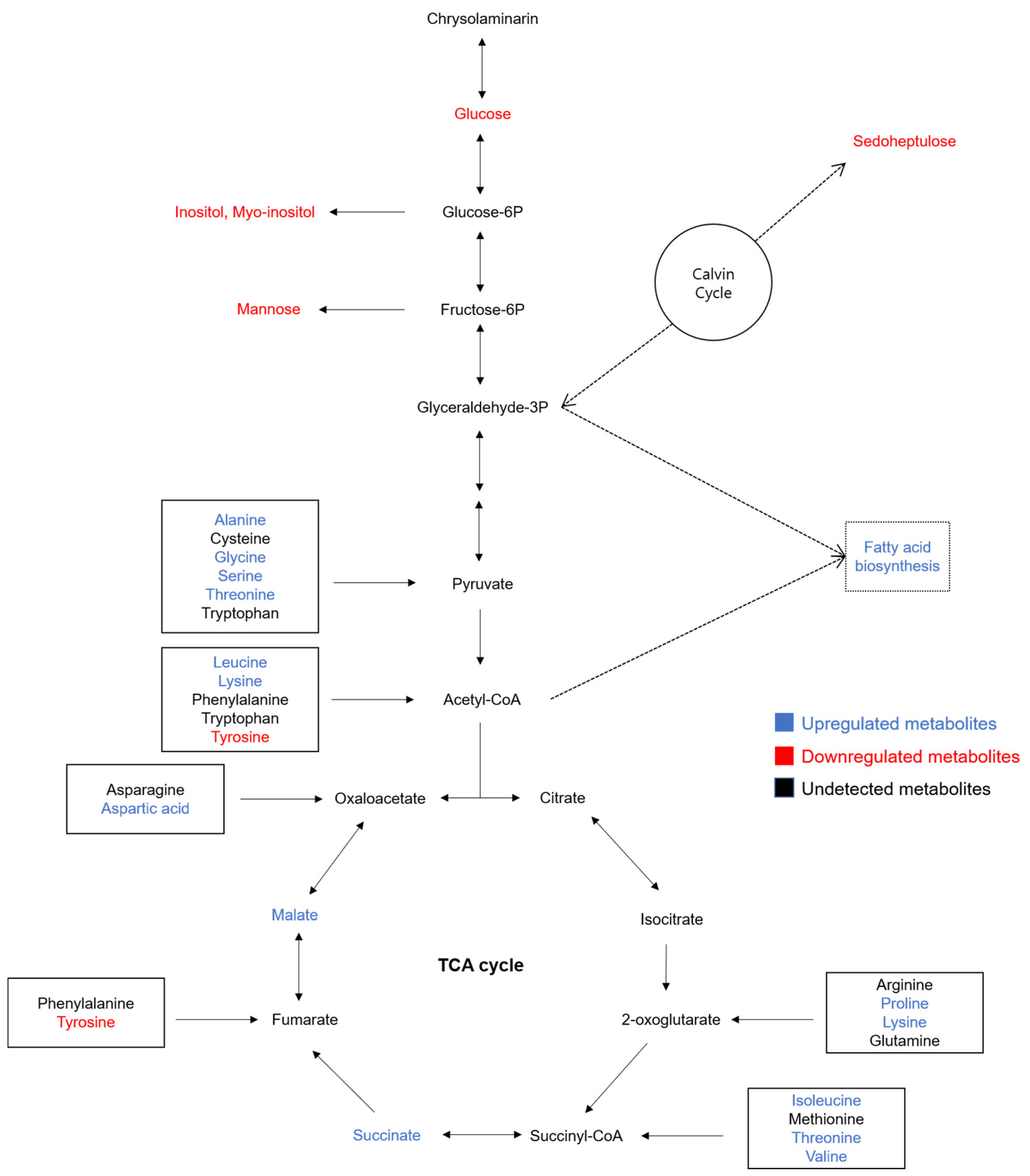
3.6. Metabolomic Profiling of the P. tricornutum Cells in Fermented Okara Culture

To gain more insights into the underlying biochemical state of cells in the fermented okara culture, we conducted untargeted metabolomic profiling and heatmap analysis. Heatmap analysis visualizes data by showing the relative magnitude of the parameter (metabolite) between groups as colors, where relatively larger metabolite concentration is represented as red and lower concentration is represented as blue. Thus, heatmap analysis was conducted to obtain an overview of the metabolite accumulation pattern in fermented okara and F/2 culture. As shown in Figure 4, clear differences in metabolite concentration were observed between the two cultures, where cells in fermented okara culture (indicated as FOM) accumulated substantially higher amount of amino acids (indicated by red colored blocks) and less simple sugar and sugar acids (indicated by blue colored blocks) as compared to F/2 culture. Marked differential metabolite accumulation between cultures was supported by partial least square discriminant analysis (Supplementary Figure S3). The metabolomic analyses were conducted on Day 2 harvest to capture the metabolic flux during exponential phase, as opposed to Day 7 where algae growth has stagnated and moved into stationary phase. The implication of the distinct metabolite concentration will be discussed in subsequent sections.

3.6.1. Remodeling of N Metabolism

Cells in fermented okara culture contained higher overall amino acid levels when compared with cells in F/2 culture (Supplementary Table S2). Reconciling this observation with the high content of chlorophyll a and fucoxanthin observed in Table 2, our result indicates a larger accumulation of nitrogen-containing compounds. Nitrogen is a crucial nutrient for microalgae to synthesize nitrogenous biomolecules such as nucleic acids, amino acids/proteins and pigments that are required for DNA replication and growth. Studies have shown a coordinated regulation of chlorophyll a and fucoxanthin levels in response to nitrogen availability, which corresponds to the photosynthetic capacity of cells for amino acid biosynthesis and cell division for growth [47]. In accordance with the study, the remodeling of metabolism towards amino acid and pigment biosynthesis in fermented okara culture favored proliferation and is likely the underlying mechanism behind the superior cell growth over F/2 culture (Figure 5).

3.6.2. Reprogramming of Carbon Allocation

Sugar compounds such as D-glucose and myo-inositol were significantly reduced in fermented okara culture as compared to that in the F/2 culture (Supplementary Table S2). It was suggested that P. tricornutum may utilize organic carbon differentially based on the physiological state of the cells. Sugars could be partially allocated to support normal metabolism of the cells while others could be utilized for carbon storage as triacylglycerides [49]. Cells in fermented okara culture grow under mixotrophic mode, utilizing both exogenous sugars in the media and CO2 in the air, unlike cells in F/2 culture, which solely depend on CO2 for carbon fixation. The high carbon availability in the FOM may have directed the cells to prioritize immediate sugar metabolism rather than sugar accumulation and storage, in order to support rapid cellular growth by producing energy in the form of ATP and NADPH. In contrast, lower carbon availability experienced in the F/2 culture may have influenced preferential storage of sugars. The accumulated sugar could subsequently be metabolized to provide carbon-intermediates for energy storage in the form of polysaccharides or neutral lipids.
Relative chrysolaminarin synthesis and degradation may also have contributed to the differential concentration of sugar metabolites between the cultures. Under favorable culture condition, diatoms accumulate carbon as chrysolaminarin, a known storage polysaccharide [50]. However, under nutritionally demanding condition, diatom shifts the carbon storage into lipids by degrading chrysolaminarin into glucose and further downstream metabolism [47]. Nutritionally enriched fermented okara culture may have thus metabolized sugars for chrysolaminarin synthesis, resulting in lower concentration of sugar compounds.
Among the detected carbon compounds, sedoheptulose is a particularly interesting metabolite. While limited information is available about sedoheptulose, lower sedoheptulose production in P. tricornutum as compared to cells under nutrient-stress condition had been reported. It was hypothesized that sedoheptulose may serve as an intermediate carbon sink that can quickly enter central carbon metabolism [51]. Similarly, we observed significantly decreased sedoheptulose content in cells grown in nutrient-rich FOM by 0.04-fold, as compared to that in F/2 media (Supplementary Table S2). Based on our metabolomic profiling, we suggest that the monosaccharide sedoheptulose may play a similar role to known sugar compounds in microalgae, which may be accumulated under nutritionally demanding condition and be metabolized to support fatty acid biosynthesis. We can conclude that reduced sugar metabolite found in cells from the fermented okara culture may be the consequence of sugar metabolism to support proliferation (Figure 5).
P. tricornutum is one of the most widely studied diatom in which the whole genome was sequenced, and large-scale transcriptome analysis has been conducted [52,53]. The availability of genetic information has garnered interests to genetically manipulate P. tricornutum cells for enhancement of both biomass and precious nutraceuticals, including fucoxanthin. For instance, by overexpressing a rate-limiting enzyme in the carotenoid synthesis pathway, a phytoene synthase (psy) encoding gene, a 1.8-fold increase in fucoxanthin content in P. tricornutum cells was demonstrated [54]. Insertion of glucose transporter encoding genes, glut 1 or hup 1, into P. tricornutum has shown that the engineered cells adopted a new metabolism which allowed more efficient glucose uptake than in wild-type cells, leading to enhanced biomass growth [55]. Similarly, by growing P. tricornutum in FOM, the relative abundance of certain nitrogenous and sugar-based nutrients within the media might trigger a biochemical upregulation of inserted genes to facilitate selective uptake of organic substrates, enabling maximal utility of the media. Based on the underlying metabolic mechanism of elevated fucoxanthin and EPA production in fermented okara culture that we have revealed in this study, further extensions into transcriptomic analyses to investigate the upregulated/downregulated genes would shed light on potential targets for metabolic engineering and pave the way for achieving commercial production.

4. Conclusions

Our work demonstrates that a low-cost food by-product can be transformed into a nutrient-rich microalgae culture media by employing solid-state fermentation to liberate entrapped nutrients. Moreover, we showed that the media solely derived from homogenous okara side-stream outperforms conventional media in the production of both biomass and high-value nutraceuticals, including EPA and fucoxanthin. Our analysis on the nutrient composition of the media and metabolomics of the cells suggested high concentration of key nutrients in the fermented okara media, including nitrogen and carbon, which triggers P. tricornutum cells to channel metabolic flux towards growth proliferation. Overall, our study provides insights on valorizing agro-industrial side-streams and contributing to the circular economy.

Supplementary Materials

The following supporting information can be downloaded at: https://www.mdpi.com/article/10.3390/fermentation9010031/s1, Figure S1: Microalgae culture setup in a chilling incubator with white fluorescence light and an orbital shaker; Figure S2: P. tricornutum grown in artificial seawater and unfermented okara media.; Figure S3: Partial least square discriminate analysis of intracellular metabolites from fermented okara culture and F/2; Table S1: Components of F/2 media (G0154, Sigma, USA); Table S2: Metabolite quantitation of cells from fermented okara culture and F/2; Table S3: Maximal growth rate and method of aeration from P. tricornutum in this study compared to previous literatures. References [56,57,58,59] are cited in the Supplementary Materials.

Author Contributions

Conceptualization, J.K., J.L. and A.Y.H.V.; Investigation, J.K., J.L., Y.X.T., W.K.M., A.Z.L. and A.Y.H.V.; Writing—Original draft, J.K., J.L. and A.Y.H.V.; Supervision, W.N.C.; Writing—Review and editing, W.N.C. All authors have read and agreed to the published version of the manuscript.

Funding

This research received no external funding.

Institutional Review Board Statement

Not Applicable.

Informed Consent Statement

Not Applicable.

Data Availability Statement

Not Applicable.

Acknowledgments

The authors would like to thank Raymond Lau and Daryl Lee from Nanyang Technological University for their technical assistance. We also thank Sven Tan, Sean Yam, and Geraldine Tan from Raffles Institution for their assistance. The research was supported by the School of Chemistry, Chemical Engineering and Biotechnology at Nanyang Technological University. The authors also acknowledge the kind sponsorship of scholarship by Interdisciplinary Graduate Programme, Nanyang Environmental and Water Research Institute, Nanyang Technological University which has enabled this work.

Conflicts of Interest

The authors declare no conflict of interest.

References

  1. Li, B.; Qiao, M.; Lu, F. Composition, Nutrition, and Utilization of Okara (Soybean Residue). Food Rev. Int. 2012, 28, 231–252. [Google Scholar] [CrossRef]
  2. van der Riet, W.B.; Wight, A.W.; Cilliers, J.J.L.; Datel, J.M. Food Chemical Investigation of Tofu and Its Byproduct Okara. Food Chem. 1989, 34, 193–202. [Google Scholar] [CrossRef]
  3. Colletti, A.; Attrovio, A.; Boffa, L.; Mantegna, S.; Cravotto, G. Valorisation of By-Products from Soybean (Glycine max (L.) Merr.) Processing. Molecules 2020, 25, 2129. [Google Scholar] [CrossRef]
  4. O’Toole, D.K. Characteristics and Use of Okara, the Soybean Residue from Soy Milk Production—A Review. J. Agric. Food Chem. 1999, 47, 363–371. [Google Scholar] [CrossRef] [PubMed]
  5. Kasai, N.; Murata, A.; Inui, H.; Sakamoto, T.; Kahn, R.I. Enzymatic High Digestion of Soybean Milk Residue (Okara). J. Agric. Food Chem. 2004, 52, 5709–5716. [Google Scholar] [CrossRef]
  6. Yogo, T. Influence of Dried Okara-Tempeh on the Composition and Metabolites of Fecal Microbiota in Dogs. Int. J. Appl. Res. Vet. Med. 2011, 9, 176. [Google Scholar]
  7. Enari, T.-M.; Markkanen, P. Production of Cellulolytic Enzymes by Fungi. In Advances in Biochemical Engineering; Springer: Berlin/Heidelberg, Germany, 2005; Volume 5, pp. 1–24. [Google Scholar] [CrossRef]
  8. Lee, J.J.L.; Cooray, S.T.; Mark, R.; Chen, W.N. Effect of Sequential Twin Screw Extrusion and Fungal Pretreatment to Release Soluble Nutrients from Soybean Residue for Carotenoid Production. J. Sci. Food Agric. 2019, 99, 2646–2650. [Google Scholar] [CrossRef]
  9. Gupta, S.; Lee, J.J.L.; Chen, W.N. Analysis of Improved Nutritional Composition of Potential Functional Food (Okara) after Probiotic Solid-State Fermentation. J. Agric. Food Chem. 2018, 66, 5373–5381. [Google Scholar] [CrossRef]
  10. Chu, W. Biotechnological Applications of Microalgae. In Biotechnological Applications of Microalgae; CRC Press: Boca Raton, FL, USA, 2013; pp. 24–37. [Google Scholar] [CrossRef] [Green Version]
  11. Norsker, N.H.; Barbosa, M.J.; Vermuë, M.H.; Wijffels, R.H. Microalgal Production—A Close Look at the Economics. Biotechnol. Adv. 2011, 29, 24–27. [Google Scholar] [CrossRef]
  12. Bhatnagar, A.; Chinnasamy, S.; Singh, M.; Das, K.C. Renewable Biomass Production by Mixotrophic Algae in the Presence of Various Carbon Sources and Wastewaters. Appl. Energy 2011, 88, 3425–3431. [Google Scholar] [CrossRef]
  13. Liang, Y.; Sarkany, N.; Cui, Y. Biomass and Lipid Productivities of Chlorella Vulgaris under Autotrophic, Heterotrophic and Mixotrophic Growth Conditions. Biotechnol. Lett. 2009, 31, 1043–1049. [Google Scholar] [CrossRef] [PubMed]
  14. Li, Y.; Zhou, W.; Hu, B.; Min, M.; Chen, P.; Ruan, R.R. Effect of Light Intensity on Algal Biomass Accumulation and Biodiesel Production for Mixotrophic Strains Chlorella Kessleri and Chlorella Protothecoide Cultivated in Highly Concentrated Municipal Wastewater. Biotechnol. Bioeng. 2012, 109, 2222–2229. [Google Scholar] [CrossRef] [PubMed]
  15. Cerón Garcí, M.C.; Fernández Sevilla, J.M.; Acién Fernández, F.G.; Molina Grima, E.; García Camacho, F. Mixotrophic Growth of Phaeodactylum tricornutum on Glycerol: Growth Rate and Fatty Acid Profile. J. Appl. Phycol. 2000, 12, 239–248. [Google Scholar] [CrossRef]
  16. Cerón García, M.C.; Sánchez Mirón, A.; Fernández Sevilla, J.M.; Molina Grima, E.; García Camacho, F. Mixotrophic Growth of the Microalga Phaeodactylum tricornutum: Influence of Different Nitrogen and Organic Carbon Sources on Productivity and Biomass Composition. Process. Biochem. 2005, 40, 297–305. [Google Scholar] [CrossRef]
  17. Wang, X.; Balamurugan, S.; Liu, S.F.; Zhang, M.M.; Yang, W.D.; Liu, J.S.; Li, H.Y.; Lin, C.S.K. Enhanced Polyunsaturated Fatty Acid Production Using Food Wastes and Biofuels Byproducts by an Evolved Strain of Phaeodactylum tricornutum. Bioresour. Technol. 2020, 296, 122351. [Google Scholar] [CrossRef]
  18. Pleissner, D.; Lam, W.C.; Sun, Z.; Lin, C.S.K. Food Waste as Nutrient Source in Heterotrophic Microalgae Cultivation. Bioresour. Technol. 2013, 137, 139–146. [Google Scholar] [CrossRef]
  19. Lopez-Huertas, E. Health Effects of Oleic Acid and Long Chain Omega-3 Fatty Acids (EPA and DHA) Enriched Milks. A Review of Intervention Studies. Pharmacol. Res. 2010, 61, 200–207. [Google Scholar] [CrossRef]
  20. Swanson, D.; Block, R.; Mousa, S.A. Omega-3 Fatty Acids EPA and DHA: Health Benefits throughout Life. Adv. Nutr. 2012, 3, 1–7. [Google Scholar] [CrossRef] [Green Version]
  21. Sachindra, N.M.; Sato, E.; Maeda, H.; Hosokawa, M.; Niwano, Y.; Kohno, M.; Miyashita, K. Radical Scavenging and Singlet Oxygen Quenching Activity of Marine Carotenoid Fucoxanthin and Its Metabolites. J. Agric. Food Chem. 2007, 55, 8516–8522. [Google Scholar] [CrossRef]
  22. Maeda, H.; Hosokawa, M.; Sashima, T.; Funayama, K.; Miyashita, K. Fucoxanthin from Edible Seaweed, Undaria Pinnatifida, Shows Antiobesity Effect through UCP1 Expression in White Adipose Tissues. Biochem. Biophys. Res. Commun. 2005, 332, 392–397. [Google Scholar] [CrossRef]
  23. Woo, M.N.; Jeon, S.M.; Kim, H.J.; Lee, M.K.; Shin, S.K.; Shin, Y.C.; Park, Y.B.; Choi, M.S. Fucoxanthin Supplementation Improves Plasma and Hepatic Lipid Metabolism and Blood Glucose Concentration in High-Fat Fed C57BL/6N Mice. Chem. Biol. Interact. 2010, 186, 316–322. [Google Scholar] [CrossRef] [PubMed]
  24. Neumann, U.; Derwenskus, F.; Flister, V.F.; Schmid-Staiger, U.; Hirth, T.; Bischoff, S.C. Fucoxanthin, a Carotenoid Derived from Phaeodactylum tricornutum Exerts Antiproliferative and Antioxidant Activities in Vitro. Antioxidants 2019, 8, 183. [Google Scholar] [CrossRef] [PubMed] [Green Version]
  25. Kanda, H.; Kamo, Y.; Machmudah, S.; Wahyudiono; Goto, M. Extraction of Fucoxanthin from Raw Macroalgae Excluding Drying and Cell Wall Disruption by Liquefied Dimethyl Ether. Mar. Drugs 2014, 12, 2383–2396. [Google Scholar] [CrossRef] [PubMed]
  26. Xiao, X.; Si, X.; Yuan, Z.; Xu, X.; Li, G. Isolation of Fucoxanthin from Edible Brown Algae by Microwave-Assisted Extraction Coupled with High-Speed Countercurrent Chromatography. J. Sep. Sci. 2012, 35, 2313–2317. [Google Scholar] [CrossRef]
  27. Kim, S.M.; Jung, Y.J.; Kwon, O.N.; Cha, K.H.; Um, B.H.; Chung, D.; Pan, C.H. A Potential Commercial Source of Fucoxanthin Extracted from the Microalga Phaeodactylum tricornutum. Appl. Biochem. Biotechnol. 2012, 166, 1843–1855. [Google Scholar] [CrossRef]
  28. Cerón García, M.C.; García Camacho, F.; Sánchez Mirón, A.; Fernández Sevilla, J.M.; Chisti, Y.; Molina Grima, E. Mixotrophic Production of Marine Microalga Phaeodactylum tricornutum on Various Carbon Sources. J. Microbiol. Biotechnol. 2006, 16, 689–694. [Google Scholar]
  29. Guillard Robert, R.L. Culture of Marine Invertebrates; Smith, W.L., Chanley, M.H., Eds.; Springer: New York, NY, USA, 1975. [Google Scholar]
  30. Uçkun Kiran, E.; Salakkam, A.; Trzcinski, A.P.; Bakir, U.; Webb, C. Enhancing the Value of Nitrogen from Rapeseed Meal for Microbial Oil Production. Enzym. Microb. Technol. 2012, 50, 337–342. [Google Scholar] [CrossRef] [Green Version]
  31. Kirk, P.L. Kjeldahl Method for Total Nitrogen. Anal. Chem. 1950, 22, 354–358. [Google Scholar] [CrossRef]
  32. Kirk, S.R.; Ronald, S. Pearson’s Composition and Analysis of Foods; Longman Group Ltd.: Harlow, UK, 1991. [Google Scholar]
  33. Kim, J.; Lyu, X.M.; Lee, J.J.L.; Zhao, G.; Chin, S.F.; Yang, L.; Chen, W.N. Metabolomics Analysis of Pseudomonas Chlororaphis JK12 Algicidal Activity under Aerobic and Micro-Aerobic Culture Condition. AMB Express 2018, 8, 131. [Google Scholar] [CrossRef] [Green Version]
  34. Mok, W.K.; Tan, Y.X.; Lee, J.; Kim, J.; Chen, W.N. A Metabolomic Approach to Understand the Solid-State Fermentation of Okara Using Bacillus Subtilis WX-17 for Enhanced Nutritional Profile. AMB Express 2019, 9, 60. [Google Scholar] [CrossRef]
  35. Cooray, S.T.; Chen, W.N. Valorization of Brewer’s Spent Grain Using Fungi Solid-State Fermentation to Enhance Nutritional Value. J. Funct. Foods 2018, 42, 85–94. [Google Scholar] [CrossRef]
  36. Cooray, S.T.; Lee, J.J.L.; Chen, W.N. Evaluation of Brewers’ Spent Grain as a Novel Media for Yeast Growth. AMB Express 2017, 7, 117. [Google Scholar] [CrossRef] [PubMed] [Green Version]
  37. Yodsuwan, N.; Sawayama, S.; Sirisansaneeyakul, S. Effect of Nitrogen Concentration on Growth, Lipid Production and Fatty Acid Profiles of the Marine Diatom Phaeodactylum tricornutum. Agric. Nat. Resour. 2017, 51, 190–197. [Google Scholar] [CrossRef]
  38. Chauton, M.S.; Olsen, Y.; Vadstein, O. Biomass Production from the Microalga Phaeodactylum tricornutum: Nutrient Stress and Chemical Composition in Exponential Fed-Batch Cultures. Biomass Bioenergy 2013, 58, 87–94. [Google Scholar] [CrossRef]
  39. McClure, D.D.; Luiz, A.; Gerber, B.; Barton, G.W.; Kavanagh, J.M. An Investigation into the Effect of Culture Conditions on Fucoxanthin Production Using the Marine Microalgae Phaeodactylum tricornutum. Algal Res. 2018, 29, 41–48. [Google Scholar] [CrossRef]
  40. Xia, S.; Wang, K.; Wan, L.; Li, A.; Hu, Q.; Zhang, C. Production, Characterization, and Antioxidant Activity of Fucoxanthin from the Marine Diatom Odontella Aurita. Mar. Drugs 2013, 11, 2667–2681. [Google Scholar] [CrossRef]
  41. Guo, B.; Liu, B.; Yang, B.; Sun, P.; Lu, X.; Liu, J.; Chen, F. Screening of Diatom Strains and Characterization of Cyclotella Cryptica as a Potential Fucoxanthin Producer. Mar. Drugs 2016, 14, 125. [Google Scholar] [CrossRef] [Green Version]
  42. Gómez-Loredo, A.; Benavides, J.; Rito-Palomares, M. Growth Kinetics and Fucoxanthin Production of Phaeodactylum Tricornutum and Isochrysis Galbana Cultures at Different Light and Agitation Conditions. J. Appl. Phycol. 2016, 28, 849–860. [Google Scholar] [CrossRef]
  43. Owens, T.G. Light-Harvesting Function in the Diatom Phaeodactylum tricornutum. Plant Physiol. 1986, 80, 739–746. [Google Scholar] [CrossRef] [Green Version]
  44. Molina Grima, E.; Sánchez Pérez, J.A.; García Camacho, F.; Fernández Sevilla, J.M.; Acién Fernández, F.G. Effect of Growth Rate on the Eicosapentaenoic Acid and Docosahexaenoic Acid Content of Isochrysis Galbana in Chemostat Culture. Appl. Microbiol. Biotechnol. 1994, 41, 23–27. [Google Scholar] [CrossRef]
  45. Hodgson, P.A.; Henderson, R.J.; Sargent, J.R.; Leftley, J.W. Patterns of Variation in the Lipid Class and Fatty Acid Composition of Nannochloropsis Oculata (Eustigmatophyceae) during Batch Culture—I. The Growth Cycle. J. Appl. Phycol. 1991, 3, 169–181. [Google Scholar] [CrossRef]
  46. Qiao, H.; Cong, C.; Sun, C.; Li, B.; Wang, J.; Zhang, L. Effect of Culture Conditions on Growth, Fatty Acid Composition and DHA/EPA Ratio of Phaeodactylum tricornutum. Aquaculture 2016, 452, 311–317. [Google Scholar] [CrossRef]
  47. Alipanah, L.; Rohloff, J.; Winge, P.; Bones, A.M.; Brembu, T. Whole-Cell Response to Nitrogen Deprivation in the Diatom Phaeodactylum tricornutum. J. Exp. Bot. 2015, 66, 6281–6296. [Google Scholar] [CrossRef] [PubMed] [Green Version]
  48. Villanova, V.; Fortunato, A.E.; Singh, D.; Bo, D.D.; Conte, M.; Obata, T.; Jouhet, J.; Fernie, A.R.; Marechal, E.; Falciatore, A.; et al. Investigating Mixotrophic Metabolism in the Model Diatom Phaeodactylum tricornutum. Philos. Trans. R. Soc. B Biol. Sci. 2017, 372, 20160404. [Google Scholar] [CrossRef] [Green Version]
  49. Remmers, I.M.; D’Adamo, S.; Martens, D.E.; de Vos, R.C.H.; Mumm, R.; America, A.H.P.; Cordewener, J.H.G.; Bakker, L.V.; Peters, S.A.; Wijffels, R.H.; et al. Orchestration of Transcriptome, Proteome and Metabolome in the Diatom Phaeodactylum tricornutum during Nitrogen Limitation. Algal Res. 2018, 35, 33–49. [Google Scholar] [CrossRef]
  50. Granum, E.; Myklestad, S.M. A Simple Combined Method for Determination of β-1,3-Glucan and Cell Wall Polysaccharides in Diatoms. Hydrobiologia 2002, 477, 155–161. [Google Scholar] [CrossRef]
  51. Popko, J.; Herrfurth, C.; Feussner, K.; Ischebeck, T.; Iven, T.; Haslam, R.; Hamilton, M.; Sayanova, O.; Napier, J.; Khozin-Goldberg, I.; et al. Metabolome Analysis Reveals Betaine Lipids as Major Source for Triglyceride Formation, and the Accumulation of Sedoheptulose during Nitrogen-Starvation of Phaeodactylum tricornutum. PLoS ONE 2016, 11, e0164673. [Google Scholar] [CrossRef] [Green Version]
  52. Bowler, C.; Allen, A.E.; Badger, J.H.; Grimwood, J.; Jabbari, K.; Kuo, A.; Maheswari, U.; Martens, C.; Maumus, F.; Otillar, R.P.; et al. The Phaeodactylum Genome Reveals the Evolutionary History of Diatom Genomes. Nature 2008, 456, 239–244. [Google Scholar] [CrossRef] [Green Version]
  53. Rastogi, A.; Maheswari, U.; Dorrell, R.G.; Vieira, F.R.J.; Maumus, F.; Kustka, A.; McCarthy, J.; Allen, A.E.; Kersey, P.; Bowler, C.; et al. Integrative Analysis of Large Scale Transcriptome Data Draws a Comprehensive Landscape of Phaeodactylum tricornutum Genome and Evolutionary Origin of Diatoms. Sci. Rep. 2018, 8, 4834. [Google Scholar] [CrossRef] [Green Version]
  54. Eilers, U.; Bikoulis, A.; Breitenbach, J.; Büchel, C.; Sandmann, G. Limitations in the Biosynthesis of Fucoxanthin as Targets for Genetic Engineering in Phaeodactylum tricornutum. J. Appl. Phycol. 2016, 28, 123–129. [Google Scholar] [CrossRef]
  55. Zaslavskaia, L.A.; Lippmeier, J.C.; Shih, C.; Ehrhardt, D.; Grossman, A.R.; Apt, K.E. Trophic Conversion of an Obligate Photoautotrophic Organism through Metabolic Engineering. Science 2001, 292, 2073–2075. [Google Scholar] [CrossRef] [PubMed]
  56. Wang, H.; Zhang, Y.; Chen, L.; Cheng, W.; Liu, T. Combined Production of Fucoxanthin and EPA from Two Diatom Strains Phaeodactylum Tricornutum and Cylindrotheca Fusiformis Cultures. Bioprocess Biosyst. Eng. 2018, 41, 1061–1071. [Google Scholar] [CrossRef] [PubMed]
  57. Gao, B.; Chen, A.; Zhang, W.; Li, A.; Zhang, C. Co-Production of Lipids, Eicosapentaenoic Acid, Fucoxanthin, and Chrysolaminarin by Phaeodactylum Tricornutum Cultured in a Flat-Plate Photobioreactor under Varying Nitrogen Conditions. J. Ocean. Univ. China 2017, 16, 916–924. [Google Scholar] [CrossRef]
  58. Yang, R.; Wei, D. Improving Fucoxanthin Production in Mixotrophic Culture of Marine Diatom Phaeodactylum Tricornutum by LED Light Shift and Nitrogen Supplementation. Front. Bioeng. Biotechnol. 2020, 8. [Google Scholar] [CrossRef] [PubMed]
  59. Wu, H.; Li, T.; Wang, G.; Dai, S.; He, H.; Xiang, W. A Comparative Analysis of Fatty Acid Composition and Fucoxanthin Content in Six Phaeodactylum Tricornutum Strains from Diff Erent Origins. Chin. J. Oceanol. Limnol. 2016, 34, 391–398. [Google Scholar] [CrossRef]
Figure 1. Preparation of FOM: (a) unfermented and (b) solid-state fermented okara and (c) FOM.
Fermentation 09 00031 g001
Figure 2. P. tricornutum growth in conventional media (F/2), fermented okara media, artificial seawater and unfermented okara media in terms of biomass concentration. (a) growth curve of P. tricornutum in different culture media; Images of algae grown in (b) fermented okara media; (c) F/2 media.
Fermentation 09 00031 g002
Figure 3. Fatty acid composition of P. tricornutum cells from F/2 and fermented okara culture. Statistical analysis was performed by Student’s 2-tailed t test. * p < 0.05, ** p < 0.01, *** p < 0.001.
Fermentation 09 00031 g003
Figure 4. Heatmap analysis of the intracellular metabolites from F/2 culture and fermented okara culture.
Fermentation 09 00031 g004
Figure 5. Metabolic changes in the P. tricornutum cells mapped onto metabolic pathways when grown on fermented okara culture compared to F/2 culture.
Fermentation 09 00031 g005
Table 1. Key nutrient composition of fermented okara media and F/2 media.
ParametersFermented Okara MediaF/2 aFold Change
Carbohydrates (mg L−1)3000N.A.
Nitrogen (mg L−1)15012.712
Phosphorus (mg L−1)141.112
Carbon (wt.%)2.090.1514
a F/2 nutritional composition of F/2 media (G0154, Sigma) was obtained from the supplier (Supplementary Table S1).
Table 2. Chlorophyll and fucoxanthin quantification in P. tricornutum cells from fermented okara media and F/2 media.
ParametersFermented Okara MediaF/2 MediaFold Change
Chlorophyll a content (mg g−1)76.5 ± 5.623.2 ± 6.23
Fucoxanthin content (mg g−1)15.3 ± 0.67.3 ± 1.82
Fucoxanthin yield (mg L−1)8.2 ± 0.31.7 ± 0.35
Table 3. Fatty acid quantification in P. tricornutum cells from fermented okara media and F/2 media.
Fatty Acid Yield (mg L−1)Growth Media
Fermented Okara MediaF/2 MediaFold Change
C14:03.66 ± 0.463.32 ± 0.191.1
C16:023.32 ± 5.7815.62 ± 2.241.5
C16:135.41 ± 6.2720.93 ± 3.241.7
C16:318.67 ± 3.321.83 ± 0.1110.2
C18:012.19 ± 3.404.69 ± 0.372.6
C18:10.28 ± 0.241.28 ± 0.440.2
C18:20.45 ± 0.050.39 ± 0.041.2
C18:30.70 ± 0.05N.D.-
C18:4N.D.0.53 ± 0.25-
C20:531.36 ± 5.3811.26 ± 0.882.8
C22:11.45 ± 0.100.12 ± 0.1212.1
C22:62.30 ± 0.490.70 ± 0.053.3
SFA39.17 ± 9.6423.63 ± 2.761.7
MUFA37.14 ± 6.1122.33 ± 3.781.7
PUFA52.78 ± 9.1914.71 ± 1.053.6
SFA+MUFA76.31 ± 15.5945.96 ± 6.551.7
TFAC140.69 ± 27.0068.09 ± 6.712.1
Disclaimer/Publisher’s Note: The statements, opinions and data contained in all publications are solely those of the individual author(s) and contributor(s) and not of MDPI and/or the editor(s). MDPI and/or the editor(s) disclaim responsibility for any injury to people or property resulting from any ideas, methods, instructions or products referred to in the content.

