Article

Testing of Diamond Electrodes as Biosensor for Antibody-Based Detection of Immunoglobulin Protein with Electrochemical Impedance Spectroscopy

1
Fraunhofer Institute for Surface Engineering and Thin Films IST, Bienroder Weg 54e, 38108 Braunschweig, Germany
2
Department of Chemistry and Molecular Biology, University of Gothenburg, Kemigården 4, 421 96 Gothenburg, Sweden
3
naconLabs a Technology Transfer Center of iTUBS mbH, Wilhelmsgarten 3, 38100 Braunschweig, Germany
4
Department of Biotechnology, Institute for Biochemistry, Biotechnology and Bioinformatics, Technische Universität Braunschweig, Spielmannstrasse 7, 38106 Braunschweig, Germany
*
Author to whom correspondence should be addressed.
Submission received: 20 September 2022 / Revised: 27 November 2022 / Accepted: 1 December 2022 / Published: 6 December 2022
(This article belongs to the Special Issue Carbon Nanohybrids for Biomedical Applications)

Abstract

:
To control the increasing virus pandemics, virus detection methods are essential. Today’s standard virus detections methods are fast (immune assays) or precise (PCR). A method that is both fast and precise would enable more efficient mitigation measures and better life comfort. According to recent papers, electrochemical impedance spectroscopy (EIS) has proven to detect viruses fast and precise. Boron-doped diamond (BDD) was used as a high-performance electrode material in these works. The aim of this work was to perform an initial test of BDD-based EIS for biosensing. As an easily available standard biomaterial, human immunoglobulin G (IgG) was used as analyte. Niobium plates were coated via hot-filament activated chemical vapor deposition with polycrystalline diamond, and doped with boron for electrical conductivity. An anti-human IgG antibody was immobilised on the BDD electrodes as a biosensing component. Four different analyte concentrations up to 1.1 µg per litre were tested. During EIS measurements, both impedance over frequency curves and Nyquist plot demonstrated no clear sign of a change of the charge transfer resistance. Thus, no positive statement about a successful biosensing could be made so far. It is assumed that these issues need to be investigated and improved, including the relation of BDD electrode size to electrolyte volume, termination of the BDD electrodes (H, O) for a successful functionalisation and EIS frequency range. The work will be continued concerning these improvement issues in order to finally use virus materials as analyte.

Graphical Abstract

1. Introduction

We are facing increasing worldwide virus pandemics with extreme impact on health and economy [1]. Besides vaccination, a fast detection of a virus load is of vital importance to implement mitigation measures such as access restrictions and quarantine. Two requirements are essential for the virus detection methods: The detection must deliver reliable results to achieve an acceptable efficiency of the measures, and it should be as fast as possible to enable efficient mitigation actions while keeping as much normal life as possible during lock-down or quarantine periods. The two detection methods that are used today each offer only one of these requirements: The lateral flow immune assays for use at home or in testing centres delivers results in a few minutes, but with a limited reliability [2]. The PCR method as gold standard is precise but takes hours or days, in general [3].
This gives a strong demand for detection methods that are both fast and precise. Several methods are under development:
  • Loop-mediated isothermal amplification, LAMP [3];
  • Surface molecular imprinting technology (SM-MIT) [3];
  • Electrochemical methods [4].
Amongst the electrochemical methods, the electrochemical impedance spectroscopy (EIS), in particular, has been proven to detect viruses. Two methods for EIS immunosensing are reported: capacitive and impedimetric EIS [5]. Nidzworski et al. [6], Matsubara et al. [7,8], Siuzdak et al. [9], Witt et al. [10] and Białobrzeska et al. [11] have both used impedimetric EIS with boron-doped diamond electrodes (BDD) to detect influenza, HIV and SARS-CoV-2 viruses. Lasserre et al. [12], Liustrovaite [13] and Drobysh et al. [14] were successful in detecting SARS-CoV-2 using gold electrodes. The electrodes in these works were coated with virus-specific proteins, enabling an adaption of the detection method according to the virus type. Incubation times were short: five minutes [6] and 15–45 min [8].
BDD electrodes have several advantages compared to gold electrodes: a wider potential window, faster charge-transfer kinetics, weaker molecule adsorption and lower background current [15]. Today, BDD electrodes are produced industrially in large-scale chemical vapour deposition (CVD) reactors [16,17]. For a virus detection kit, a small electrode size of only a few square millimetres would be sufficient. This would enable low manufacturing costs, which is an important pre-condition for quick tests with disposable electrodes. Thus, EIS with BDD electrodes has the potential to become a detection method that is both precise and fast for viruses, filling the gap that exists today between lateral-flow immune assay and PCR.
The initial testing of virus detection with EIS consists of seven steps: 1. The analyte from sputum or blood, containing virus material, is provided in the electrolyte. 2. The working electrode is functionalised with virus-specific antibody. 3. The working electrode is brought into contact with the virus-containing electrolyte. 4. A binding of the virus particle takes place through the surface of the protein structure of the antibody. 5. An alternating current with different frequencies is applied to the working electrode and counter electrode. 6. The charge transfer resistance of the electrode surface increases due to the binding of virus particles. 7. This changes the impedance of the electrochemical system and can be accessed via electronic procedures and mathematical operations. These results are a measure of the virus presence and quantity.
Antibodies are a general tool to detect pathogens easily and very specifically [18]. Nowadays, more and more antibodies are developed animal-free with in vitro methods such as phage display [19]. This development has the advantage that the sequence of the antibodies is completely known and therefore the antibody can be engineered for different applications. For easy immobilisation of the antibody onto the electrode, an antibody is used, which carries a free cysteine in the fragment crystallisable (Fc) part of the antibody. This allows for a simpler and gentler method of immobilisation to surfaces.
To achieve this final goal of a fast and precise virus detection method, preliminary investigations are necessary to gain the necessary knowledge about electrode preparation as well as the specific biochemical and electrochemical processes. As the work with virus material is a potential health hazard and therefore costly regarding safety measures, the investigations started using easier-to-handle biomaterials as the analyte. Thus, the aim of the presented work was the general evaluation of BDD-based EIS for biosensing using the standard protein immunoglobuline G (IgG) as an analyte. The focus of this work was to establish a way to prepare the BDD biosensing electrodes with the antibody corresponding to the antigen, complemented with very first EIS analyses of IgG in different concentrations. After finishing this first step successfully, the work will be continued with virus-specific antibodies and virus material as an analyte.

2. Materials and Methods

2.1. Methodology

To achieve a fast and precise virus detection method via BDD-based EIS, several technological issues must be solved: The manufacturing technology for BDD electrodes is existing, but the electrodes’ surfaces must be prepared by chemical functionalisation or coating with interlayers to obtain the necessary adhesion of the antibody. Biochemical processes must be developed to add the antibody onto the prepared electrode. Quality analysis methods must be selected and approved to assure the reliability of these different functionalisation and coating steps. A suitable EIS cell design must be selected for this detection purpose, such as cell volume, working electrode arrangement, selection and arrangement of counter electrode, selection and arrangement of reference electrode. Finally, the right analytical EIS method and data processing must be identified.
This work describes the results of a first step towards the final goal, using an easily available standard biomaterial as an analyte to evaluate EIS with BDD electrodes for biosensing in principle. Human IgG was used as the antigen.
Further work will compare BDD electrodes with other electrode materials concerning their performance for EIS-based biosensing. As the final step, virus-specific antibody and virus material will be used to develop the virus detection method.

2.2. Electrolyte and Protein Analyte
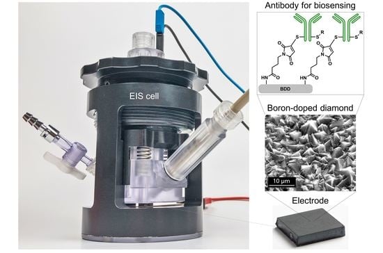
The electrolyte for the EIS measurements was 1 mM potassium ferricyanide K3[Fe(CN)6] in 0.1 M phosphate-buffered saline (PBS), mixed in deionized (DI) water.
The analyte was human IgG in concentrations of 1.1 pg/mL, 1.1 ng/mL and 1.1 µg/mL.

2.3. Electrodes

2.3.1. Electrode Materials and Manufacturing

For the BDD electrodes, Niobium plates with dimensions of 50 mm for both width and length and two millimetres of thickness were coated via hot-filament activated chemical vapor deposition (CVD), using a self-designed large-scale CVD reactor with a coating area of 0.5 m2 and a maximum electrical power of 80 kW to heat the 0.5 mm diameter filaments [10]. The substrate plates were pre-treated before coating by sandblasting, cleaning in DI water and seeding by dipping five minutes in a suspension of nanometre-sized diamond particles in water. The carbon source was methane in a concentration of 1.83 vol-% in Hydrogen. Gas pressure was 20 mbar. To achieve the necessary electrical conductivity, the diamond layer was doped during the deposition by adding 0.49 vol-% Trimethylborane. The electrodes were coated twice for each side, and the coating process duration for one side was 40 h. Substrate temperature was around 950 °C. The diamond layer was polycrystalline with a film thickness of six to eight µm. The plates were laser-cut to the required electrode size of 10 mm for the width and length.

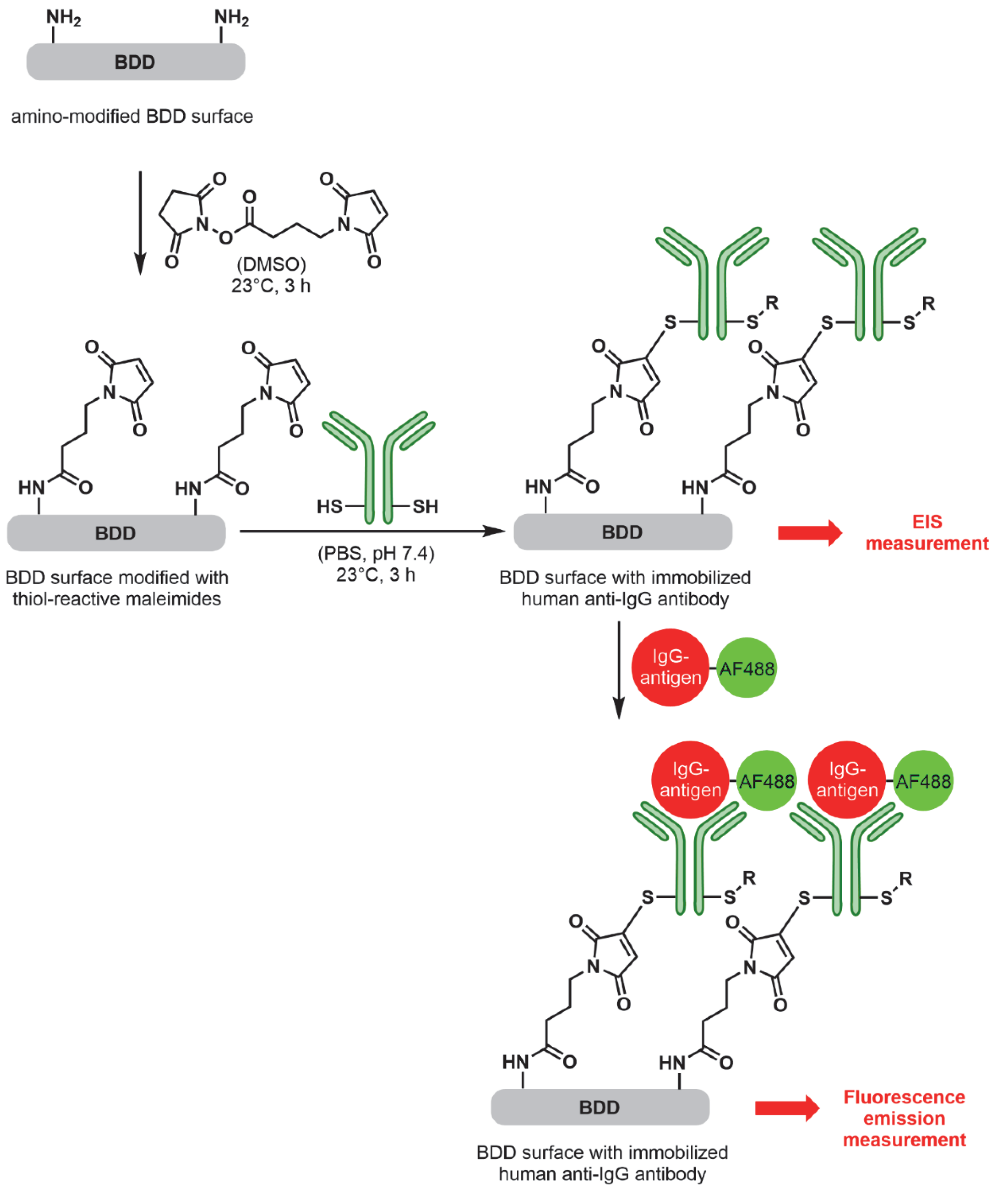
2.3.2. Electrode Functionalisation

To achieve the required adhesion of the biomarker antibody, the working electrodes were functionalised with amino groups via atmospheric-pressure plasma treatment, using a commercially available plasma source (Nadir SRL; Padova, Italy). Plasma was ignited using a combination of a high-frequency and a radio frequency generator, operating with the following parameters: (1) high-voltage generator with 10 kV, a current of 1 A and a frequency of 15.332 kHz, and (2) a radio-frequency generator of 27,120 kHz with a power of 15 W. The process gas was introduced by gas-flow controllers regulating the mass flow for the process gas (2 L min−1 STP argon + precursor), carrier gas (7 L min−1 STP argon) and nitrogen flushing gas. For homogeneous coating, the plasma source was placed on a moving stage. The electrodes were cleaned before functionalisation by rinsing in isopropyl alcohol and drying in nitrogen gas. The precursor was 3-aminopropyl-trimethoxysilane (APTMS, Sigma-Aldrich, Schnelldorf, Germany). Argon was bubbled through APTMS to bring it into the gas phase. This gas mixture was ionised via a high-frequency generator and radio-frequency generator, producing a plasma stream via a gas nozzle that was directed onto the electrode surface. The electrode was moved laterally with a speed of 1 millimetre per second. The functionalised electrodes were stored in a nitrogen atmosphere afterwards to prevent reaction with oxygen from the air.
To check if the amino functionalisation was successful, a fluorescence analysis with fluorescein isothiocyanate (FITC) marker was performed. The electrodes were dipped in a marker solution with 0.01 mM FITC in an aqueous carbonate buffer of pH 8: sodium bicarbonate in a concentration of 49 mM and Sodium carbonate decahydrate in a concentration of 0.874 mM. The electrodes were incubated in the marker solution for one hour under light exclusion. Afterwards, they were washed twice with an aqueous carbonate buffer of pH 8, next with 0.5% aqueous sodium dodecyl sulfate solution, and finally with DI water and then dried under nitrogen stream. A fluorescence emission reader was used with a dual bandpass filter of 469 nm and 525 nm centre wavelengths (Cytation, BioTek, Friedrichshall, Germany). Three sample types were compared: 1. BDD electrodes with amino functionalisation and fluorescence marker, 2. BDD electrodes without amino functionalisation without the fluorescence marker, and 3. BDD electrodes with amino functionalisation without fluorescence marker.

2.3.3. Immobilisation of Antibody on Electrodes

As a biosensing antibody, a recombinant anti-human IgG (Fc specific) antibody with modified Fc-part (C-terminal free Cysteine) was used (ABK23-E03-M-Cys, Abcalis GmbH, Germany), which was immobilized on the surface of the amino-modified BDD electrodes installing first an amino-reactive linker bearing a thiol-reactive maleimide-moiety and finally linking the cysteine-containing antibody via 1,4-conjugate addition reaction to the surface of the electrode (Figure 1). This strategy was envisaged to provide a well defined attachment of the antibodies on the electrode surface through the cysteine engineered Fc part of the antibody. Alternative lysine reactive bioconjugation is known to lead also to attachment via lysine residues in the fab part, potentially blinding the antibodies from the recognition of their antigens and furthermore, the conjugation via cysteine residue after partial reducing of disulfide bridges between the light and heavy chains of the antibody are known to negatively impact the overall stability of antibodies. Therefore, the amino-modified electrodes were placed into glass snap lid jars filled with 300 µL of a 5 mM solution of N-Succinimidyl 4-maleimidobutyrate in dry DMSO (Acros Organic, Geel, Belgium, AcroSeal™ over molsieve) and shaken gently for 3 h at 23 °C on a centripetal shaker, before the electrodes were flipped to the other side and shaken for an additional 1.5 h at 23 °C.
The electrodes were washed by placing them consecutively into three glass beakers filled with 30 mL of DMSO (HPLC quality, >99.7% Merck) for 1 min each and afterwards into three glass beakers filled with 30 mL of dist. water (HPLC quality, VWR) for 1 min each. Then, the electrodes were placed into glass snap lid jars, again covered with a solution of 300 µL Cys-Antibody (ABK23-E03-M-Cys, anti-human IgG Fc, c = 0.31 mg/mL, Lot: YABC71-1) in PBS (10 mM, pH = 7.4), giving a final concentration of 0.1 mg/mL. The electrodes were incubated for 3 h at 23 °C with gentle shaking and flipping the electrodes after 1.5 h.
Again, the electrodes were washed by placing them consecutively into three glass beakers filled with 30 mL of DMSO (HPLC quality, >99.7% Merck) for 1 min each and afterwards into three glass beakers filled with 30 mL of dist. water (HPLC quality, VWR) for 1 min each. Finally, the blocking of the unoccupied surface was conducted to avoid unspecific interactions between the analyte and the electrode surface [5]. Therefore, the antibody-modified electrodes were placed into glass snap lid jars and covered with a solution of 500 µL skim milk powder 2 %w in 10 mM PBS buffer (pH 7.4) and incubated for 1 h at 23 °C, with gentle shaking and flipping of the electrodes after 30 min. Then, the blocked electrodes were washed, placing them consecutively into three glass beakers filled with 30 mL of dist. water (HPLC quality, VWR). The finally modified BDD electrodes were stored at 4 °C covered with 10 mM PBS buffer (pH 7.4) solution until their use. Appropriate controls were prepared accordingly but leaving out respective functionalisation steps as indicated below.
Again, a fluorescence analysis was performed to check whether the biosensing antibody immobilisation was successful. Therefore, 300 µL of a 5 µg/mL solution of the fluorescently labelled human IgG-AF488 antigen (AF = AlexaFluoro) was added on top of the modified BDD electrode surface and incubated for 1 h at 23 °C, flipping the electrodes after 30 min. Afterwards, the electrodes were washed, placing them consecutively into three glass beakers filled with 30 mL of dist. water (HPLC quality, VWR). The binding of fluorescently labelled antigen was then analysed using an ELISA plate reader at the excitation wavelength of 485 nm and detection wave length of 535 nm. Four NH2-functionalised sample types were compared: 1. after antibody immobilisation, blocked and incubated with fluorescently labelled antigen, 2. after antibody immobilisation, blocked and without fluorescently labelled antigen (reference 1), 3. no antibody immobilisation, blocked and with fluorescently labelled antigen (reference 2), 4. only blocked (reference 3) and each conducted as triplates (n = 3).

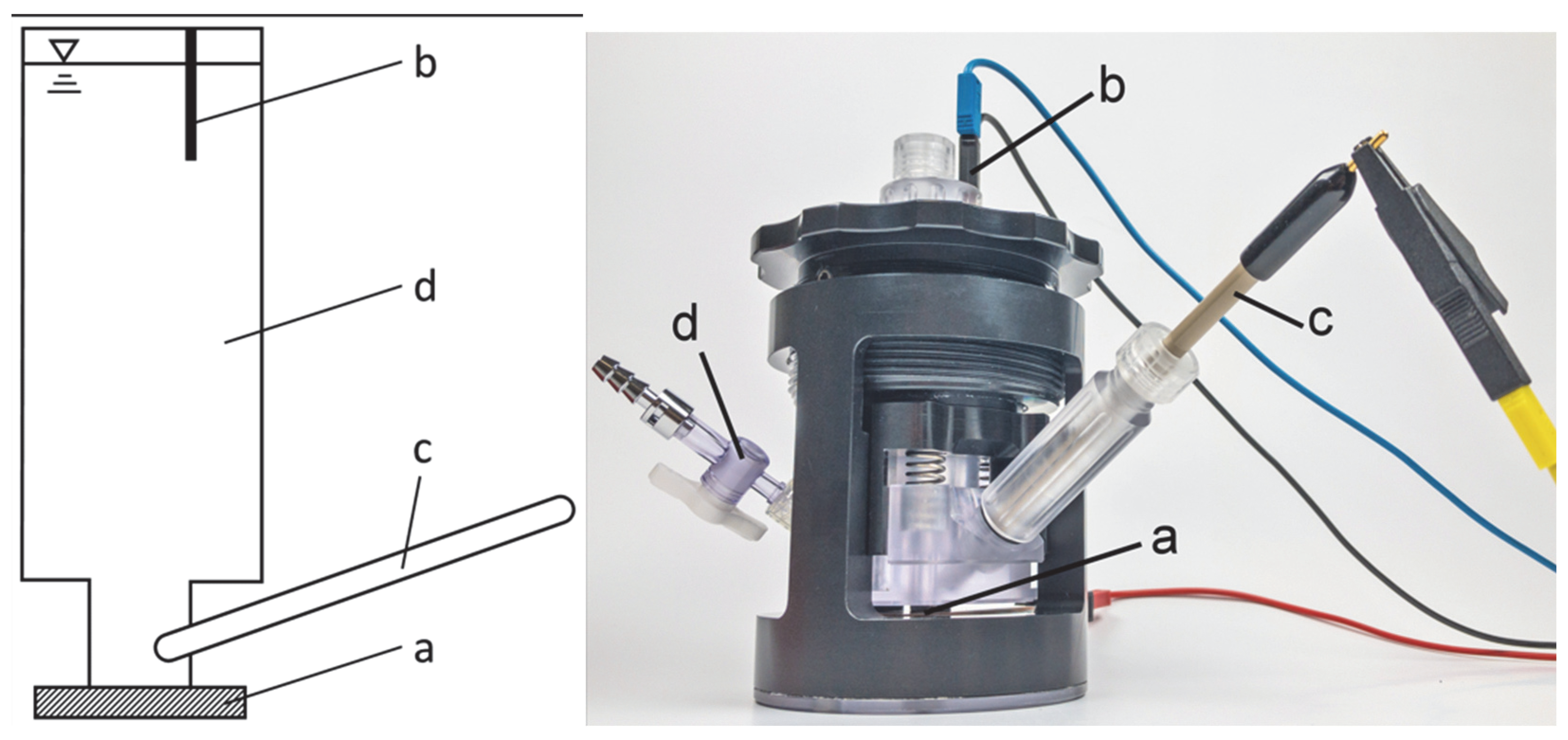
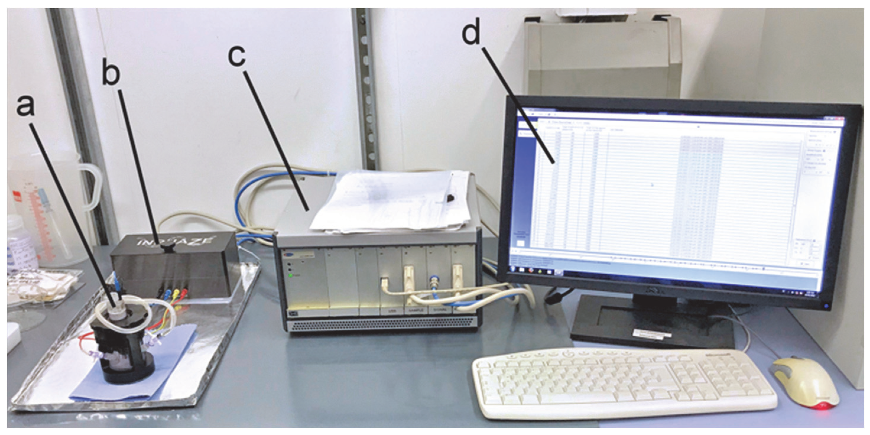
2.4. EIS Cell and Spectrometer

For the electrochemical impedance spectrometry, a 3-terminal Chamber (INPHAZE UG, Horhausen, Germany) was used (see Figure 2). The electrolyte volume was 12 mL. The working electrodes consisted of amino-functionalised electrodes with an immobilised antibody. The active working electrode area was 95 mm2. The counter electrode was a gold-plated wire. The reference electrode was an Ag/AgCl electrode.
The spectrometer (see Figure 3) was an INPHAZE Sys impedance spectrometer unit, model IMSP with a corresponding INPHAZE Sys amplifier unit, model AMP and an adaptable reference circuit (INPHAZE UG, Horhausen, Germany, and eDAQ Pty Ltd., Denistone East, Australia). To control the spectrometer, the INPHAZE Control Panel software version 4.11.303 was used (INPHAZE UG). The data evaluation was conducted with the INPHAZE Impedance Analyzer software, version 4.11.207 (INPHAZE UG). To avoid the influence of external electromagnetic fields, a cage of Aluminium sheet was put around the cell and amplifier unit (base sheet can be observed in Figure 3).
Before measuring, the system was calibrated and a spot test was performed to find the best-fitting reference circuit.

2.5. Spectroscopy Parameters and Procedure

Table 1 shows the measurement parameters:
To test the general ability of antibody detection, the antibody concentration in the electrolyte was varied in a very large range of six magnitudes (0, pg, ng, µg). If specific bonding between human IgG antigen in electrolyte and anti-human IgG antibody on electrodes takes place, the charge transfer resistance should increase. To detect this change, two data evaluation methods were applied: First, the impedance for different analyte concentrations of human IgG antigen was plotted vs. frequency; if the charge transfer resistance increases, a rise of the impedance should be measured. Secondly, a Nyquist plot of the impedance was created; if the charge transfer resistance increases, a rise of the semicircle radius should be observed.

3. Results

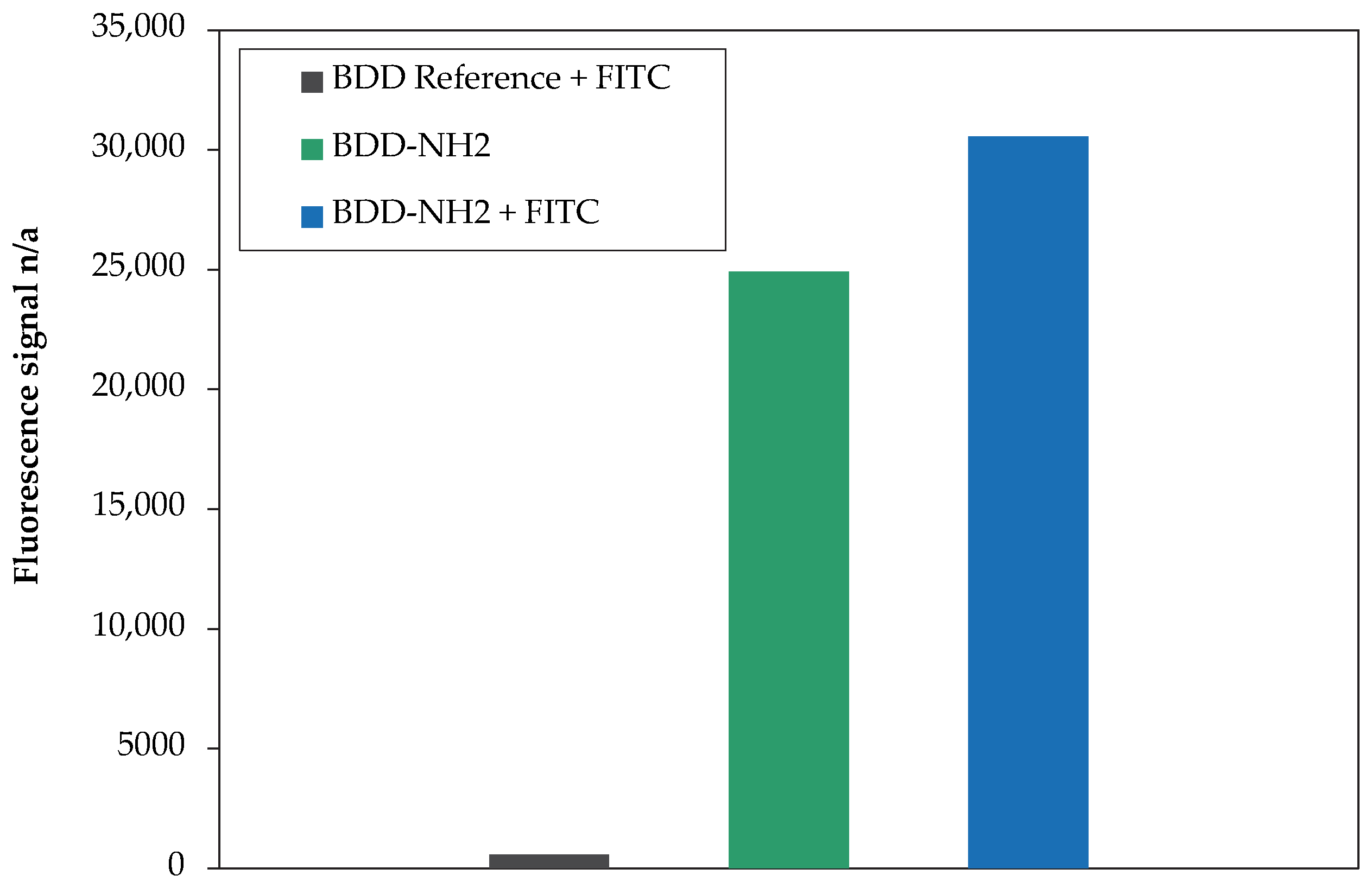
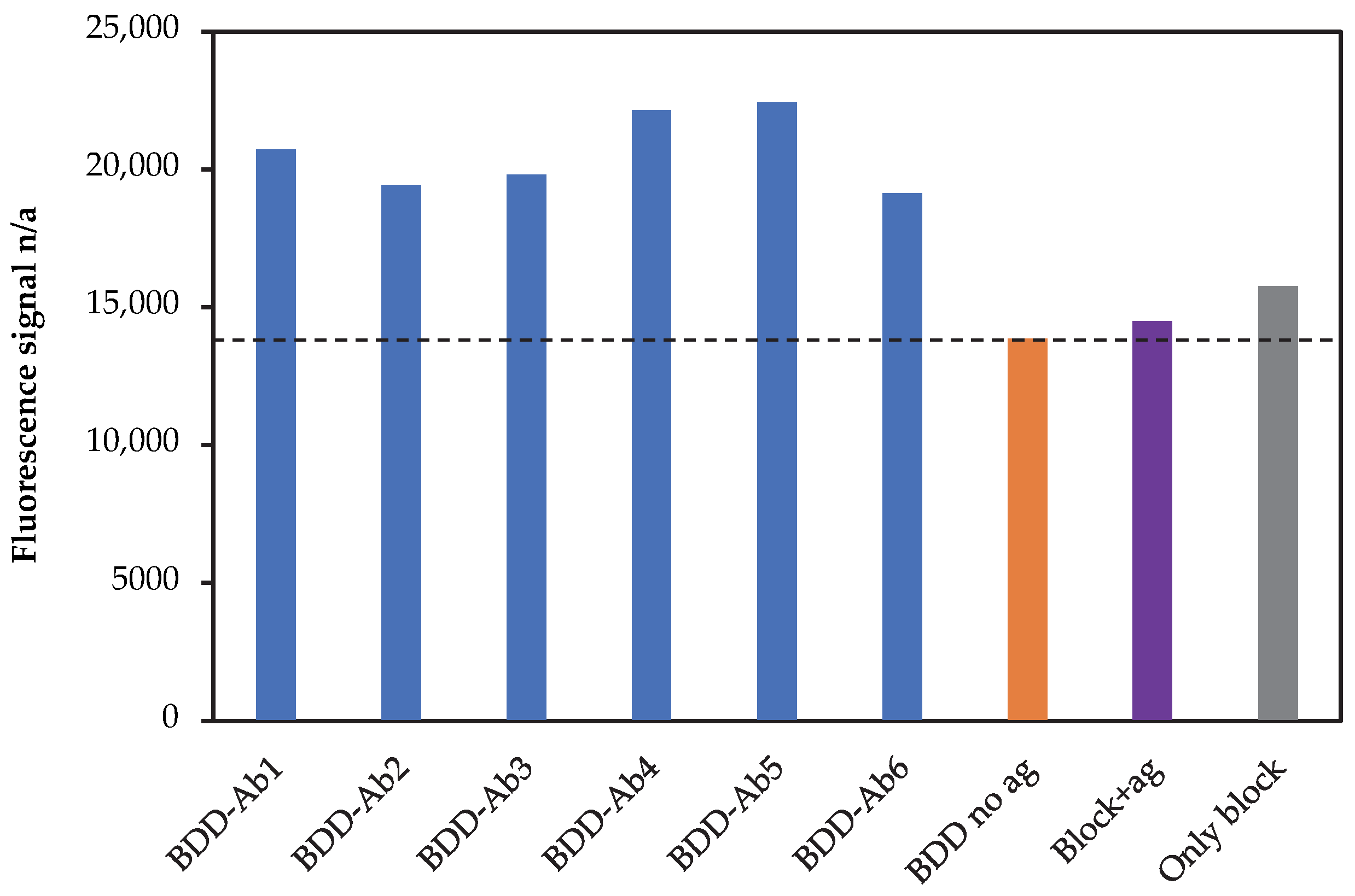
3.1. Fluorescence Analysis of Electrode Functionalisation Steps

Figure 4 shows the results of the fluorescence analysis to check the amino functionalisation by plasma treatment.
It can be observed that the plasma treated BDD (blue bar) shows a much higher fluorescence signal compared to BDD electrodes without plasma treatment (black bar). This demonstrates a clear evidence of successfully added NH2 groups onto the BDD electrodes.
Furthermore, it can be observed that the plasma-treated BDD electrodes without fluorescence marker (green bar) shows nearly the same fluorescence signal as with fluorescence marker (blue bar). The reason for this unexpected observation is probably an auto-fluorescence effect of the amino groups or an interfering wavelength. Nevertheless, the amino functionalisation can be regarded as successful.
Figure 5 shows the fluorescence signals of the BDD electrodes after amino functionalisation, subsequent immobilisation of Fc-modified anti-human IgG antibody and incubation with the fluorescently labelled human IgG-AF488 antigen.
It can be observed that all BDD electrodes with antibody immobilisation (blue) show a higher fluorescence signal compare to the controls, indicating an antibody immobilisation. However, all reference samples (without fluorescently labelled antigen, without antibody immobilisation but with fluorescently labelled antigen and neither antibody nor fluorescently labelled antigen) show a relatively strong fluorescence signal. Most presumably, the reason is that the auto-fluorescence effect through the amino functionalisation (see Figure 4) is persistent throughout the antibody immobilisation.

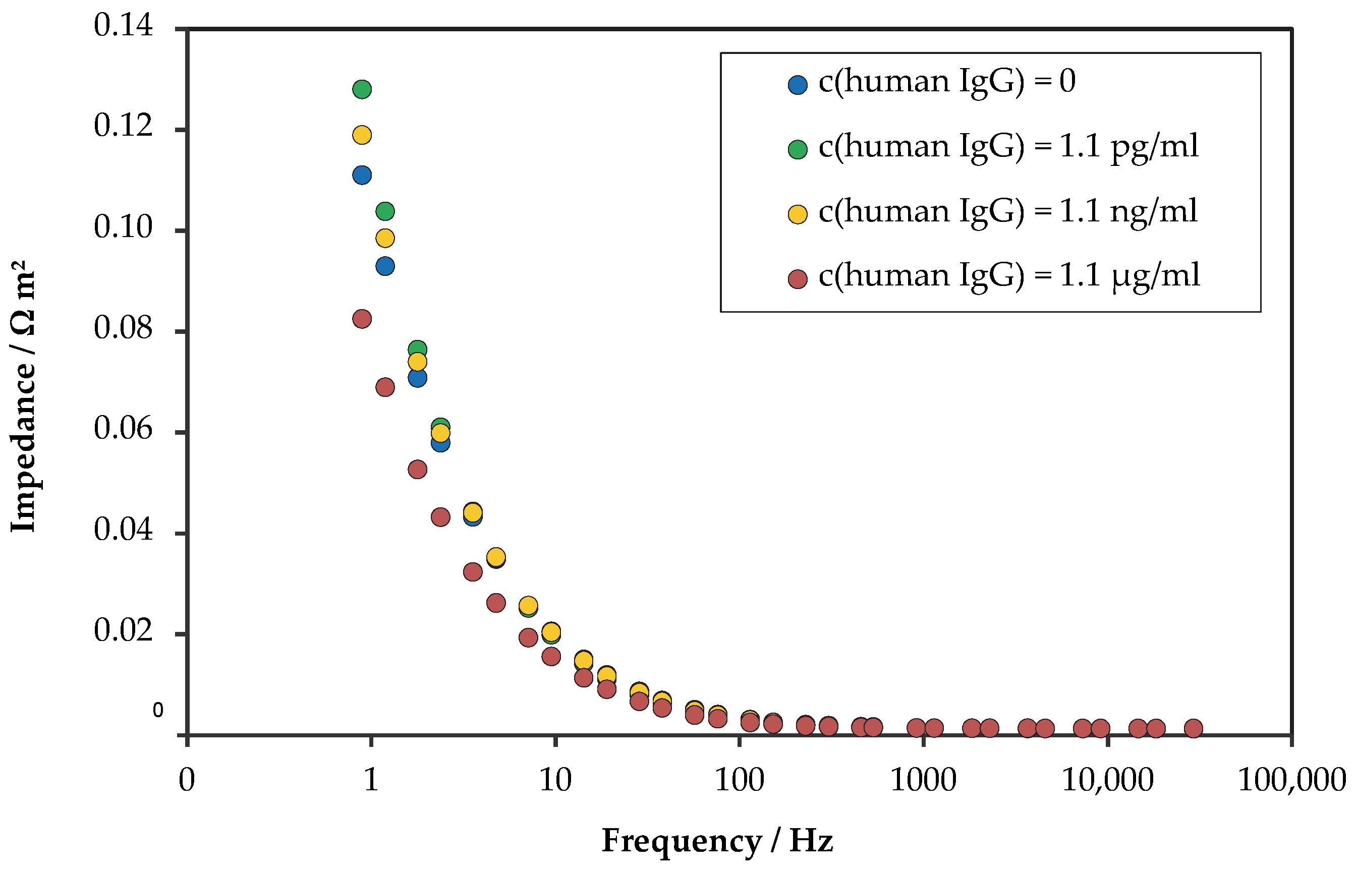
3.2. Electrochemical Impedance Spectroscopy Results

EIS was performed with the BDD electrodes after amino-functionalisation and biomarker antibody immobilisation. Figure 6 shows impedance over frequency as a measure for the change of the charge transfer resistance by the bonding of the analyte (human IgG antigen) to the biomarker on the electrodes (anti-human IgG antibody).
All antibody concentrations show an exponential decrease in the impedance with increasing frequency. This usual curve progression indicates the basic stability of the measurement.
However, no significant increase in the impedance can be observed for increasing antibody concentration. Only slight differences can be observed at lower frequencies; but no clear trend is visible. The reason might be uncertainties during measurements or minor inconsistencies during cell preparation. Thus, no positive statement about a successful antibody detection can be made. It seems that no interaction between the biosensing surface (anti-human IgG) and the protein analyte (human IgG) is indicated.
As a second evaluation of the charge transfer resistance change, a Nyquist plot was drawn from real and imaginary parts of the impedance, as shown in Figure 7.
As a first observation, no formation of more than one semicircle is visible; thus, there is no evidence that several electrochemical processes occurred.
The semicircle diameter would be a measure of the analyte concentration. However, no clear formation of a full semicircle can be observed, so that a conclusion about the analyte concentration cannot be made in this way.
Furthermore, a relatively high scattering is observed for the analyte concentration of 1.1 pg/mL. There is no clear trend visible between antigen concentration and curve progression; the differences between the curves are small compared to the amount of scattering. A possible reason might be inconsistencies during measurement or during cell preparation.
Finally, no positive statement about a successful human-IgG antigen detection can be made from the Nyquist plot.

4. Discussion

With the presented work results, the aim of realising a biosensor using EIS and antibody-equipped BDD electrodes was not achieved. However, Nidzworski et al. successfully showed the EIS-based detection of viruses with functionalized BDD electrodes [6]. This gives the motivation to continue this work towards the final goal of achieving a fast and reliable virus test method that can help to control future pandemics.
The outlook of this work includes the following issues: The first step will be to find a more conclusive quality assurance of the functionalisation process with amino groups and the immobilisation of the biosensing antibodies, e.g., by conducting the fluorescence analysis at a non-interfering wavelength or identifying an alternative method. Furthermore, the chemical termination of the BDD working electrodes will be varied between H and O according to Suffredini et al. [20] to investigate the effect on the functionalisation. The relation of BDD electrode size to electrolyte volume plays an important role for successful EIS and will be considered for variation. The EIS experiments will be continued with an enlarged frequency range, especially with lower frequencies, which are important to separate the analyte concentration. Moreover, investigations of the stability of electrolyte, functionalisation and antibody immobilisation over time will be performed, eventually resulting in adapted experimental procedures. To find the limit of detection, even higher antigen concentrations will be used.
If still not successful, EIS measurements will be conducted with other antibodies or biomolecules as the analyte. To demonstrate the performance and necessity of boron-doped diamond as electrode material, gold and other electrode materials, such as glassy carbon, will be used for comparison. A DC bias during the EIS measurements will be considered, which might improve the results.

Supplementary Materials

The following supporting information can be downloaded at: https://www.mdpi.com/article/10.3390/c8040074/s1, Spreadsheet S1: Measured fluorescence intensity after incubation of antibody functionalized BDD electrodes with fluorescently labelled antigen (xslx), Spreadsheet S2: Measured impedance values over frequency (xslx), Spreadsheet S3: Impedance imaginary and real parts (xslx).

Author Contributions

Conceptualisation: M.M., C.S.G.G., V.S., K.L., E.V.W., G.R., P.K. and J.G.; methodology: M.M. and E.M.; validation: M.M.; investigation: M.M., C.S.G.G. and M.R.; resources: C.S.G.G. and E.V.W.; writing—original draft preparation: J.G.; writing—review and editing: M.M., V.S., K.L., E.V.W. and P.K.; project administration: J.G. All authors have read and agreed to the published version of the manuscript.

Funding

This research received no external funding.

Data Availability Statement

Not applicable.

Acknowledgments

The authors acknowledge the contribution of Torsten Hochsattel, Fraunhofer IST, for support in EIS cell handling, Stephan Ulrich, Fraunhofer IST, for discussion of EIS results, and Veysel Zeren, Fraunhofer IST, for performing the plasma treatments.

Conflicts of Interest

Competing interests: GR and EVW are co-founders and shareholders of Abcalis GmbH, a company producing antibodies for diagnostics.

References

  1. El Keshky, M.E.S.; Basyouni, S.S.; Al Sabban, A.M. Getting Through COVID-19: The Pandemic’s Impact on the Psychology of Sustainability, Quality of Life, and the Global Economy—A Systematic Review. Front. Psychol. 2020, 11, 585897. [Google Scholar] [CrossRef] [PubMed]
  2. Mistry, D.A.; Wang, J.Y.; Moeser, M.-E.; Starkey, T.; Lee, L.Y.W. A systematic review of the sensitivity and specificity of lateral flow devices in the detection of SARS-CoV-2. BMC Infect. Dis. 2021, 21, 828. [Google Scholar] [CrossRef] [PubMed]
  3. Bu, J.; Deng, Z.; Liu, H.; Li, J.; Wang, D.; Yang, Y.; Zhong, S. Current methods and prospects of coronavirus detection. Talanta 2021, 225, 121977. [Google Scholar] [CrossRef] [PubMed]
  4. Khan, M.Z.H.; Hasan, M.R.; Hossain, S.I.; Ahommed, M.S.; Daizy, M. Ultrasensitive detection of pathogenic viruses with electrochemical biosensor: State of the art. Biosens. Bioelectron. 2020, 166, 112431. [Google Scholar] [CrossRef] [PubMed]
  5. Prodromidis, M.I. Impedimetric immunosensors—A review. Electrochim. Acta 2010, 55, 4227–4233. [Google Scholar] [CrossRef]
  6. Nidzworski, D.; Siuzdak, K.; Niedziałkowski, P.; Bogdanowicz, R.; Sobaszek, M.; Ryl, J.; Weiher, P.; Sawczak, M.; Wnuk, E.; Goddard, W.A.; et al. A rapid-response ultrasensitive biosensor for influenza virus detection using antibody modified boron-doped diamond. Sci. Rep. 2017, 7, 15707. [Google Scholar] [CrossRef] [PubMed] [Green Version]
  7. Matsubara, T.; Ujie, M.; Yamamoto, T.; Akahori, M.; EINAGA, Y.; Sato, T. Highly sensitive detection of influenza virus by boron-doped diamond electrode terminated with sialic acid-mimic peptide. Proc. Natl. Acad. Sci. USA 2016, 113, 8981–8984. [Google Scholar] [CrossRef] [PubMed] [Green Version]
  8. Matsubara, T.; Ujie, M.; Yamamoto, T.; Einaga, Y.; Daidoji, T.; Nakaya, T.; Sato, T. Avian Influenza Virus Detection by Optimized Peptide Termination on a Boron-Doped Diamond Electrode. ACS Sens. 2020, 5, 431–439. [Google Scholar] [CrossRef] [PubMed]
  9. Siuzdak, K.; Niedziałkowski, P.; Sobaszek, M.; Łęga, T.; Sawczak, M.; Czaczyk, E.; Dziąbowska, K.; Ossowski, T.; Nidzworski, D.; Bogdanowicz, R. Biomolecular influenza virus detection based on the electrochemical impedance spectroscopy using the nanocrystalline boron-doped diamond electrodes with covalently bound antibodies. Sens. Actuators B Chem. 2019, 280, 263–271. [Google Scholar] [CrossRef]
  10. Witt, S.; Rogien, A.; Werner, D.; Siegenthaler, J.; Lesiyon, R.; Kurien, N.; Rechenberg, R.; Baule, N.; Hardy, A.; Becker, M. Boron doped diamond thin films for the electrochemical detection of SARS-CoV-2 S1 protein. Diam. Relat. Mater. 2021, 118, 108542. [Google Scholar] [CrossRef] [PubMed]
  11. Białobrzeska, W.; Ficek, M.; Dec, B.; Osella, S.; Trzaskowski, B.; Jaramillo-Botero, A.; Pierpaoli, M.; Rycewicz, M.; Dashkevich, Y.; Łęga, T.; et al. Performance of electrochemical immunoassays for clinical diagnostics of SARS-CoV-2 based on selective nucleocapsid N protein detection: Boron-doped diamond, gold and glassy carbon evaluation. Biosens. Bioelectron. 2022, 209, 114222. [Google Scholar] [CrossRef] [PubMed]
  12. Lasserre, P.; Balansethupathy, B.; Vezza, V.J.; Butterworth, A.; Macdonald, A.; Blair, E.O.; McAteer, L.; Hannah, S.; Ward, A.C.; Hoskisson, P.A.; et al. SARS-CoV-2 Aptasensors Based on Electrochemical Impedance Spectroscopy and Low-Cost Gold Electrode Substrates. Anal. Chem. 2022, 94, 2126–2133. [Google Scholar] [CrossRef] [PubMed]
  13. Liustrovaite, V.; Drobysh, M.; Rucinskiene, A.; Baradoke, A.; Ramanaviciene, A.; Plikusiene, I.; Samukaite-Bubniene, U.; Viter, R.; Chen, C.-F.; Ramanavicius, A. Towards an Electrochemical Immunosensor for the Detection of Antibodies against SARS-CoV-2 Spike Protein. J. Electrochem. Soc. 2022, 169, 37523. [Google Scholar] [CrossRef]
  14. Drobysh, M.; Liustrovaite, V.; Baradoke, A.; Rucinskiene, A.; Ramanaviciene, A.; Ratautaite, V.; Viter, R.; Chen, C.-F.; Plikusiene, I.; Samukaite-Bubniene, U.; et al. Electrochemical Determination of Interaction between SARS-CoV-2 Spike Protein and Specific Antibodies. Int. J. Mol. Sci. 2022, 23, 6768. [Google Scholar] [CrossRef] [PubMed]
  15. Lourencao, B.C.; Brocenschi, R.F.; Medeiros, R.A.; Fatibello-Filho, O.; Rocha-Filho, R.C. Analytical Applications of Electrochemically Pretreated Boron-Doped Diamond Electrodes. ChemElectroChem 2020, 7, 1291–1311. [Google Scholar] [CrossRef]
  16. Fryda, M.; Matthée, T.; Mulcahy, S.; Hampel, A.; Schäfer, L.; Tröster, I. Fabrication and application of Diachem electrodes. Diam. Relat. Mater. 2003, 12, 1950–1956. [Google Scholar] [CrossRef]
  17. Schäfer, L.; Höfer, M.; Kröger, R. The versatility of hot-filament activated chemical vapor deposition. Thin Solid Film. 2006, 515, 1017–1024. [Google Scholar] [CrossRef]
  18. Bertoglio, F.; Meier, D.; Langreder, N.; Steinke, S.; Rand, U.; Simonelli, L.; Heine, P.A.; Ballmann, R.; Schneider, K.-T.; Roth, K.D.R.; et al. SARS-CoV-2 neutralizing human recombinant antibodies selected from pre-pandemic healthy donors binding at RBD-ACE2 interface. Nat. Commun. 2021, 12, 1577. [Google Scholar] [CrossRef] [PubMed]
  19. Roth, K.D.R.; Wenzel, E.V.; Ruschig, M.; Steinke, S.; Langreder, N.; Heine, P.A.; Schneider, K.-T.; Ballmann, R.; Fühner, V.; Kuhn, P.; et al. Developing Recombinant Antibodies by Phage Display against Infectious Diseases and Toxins for Diagnostics and Therapy. Front. Cell. Infect. Microbiol. 2021, 11, 697876. [Google Scholar] [CrossRef] [PubMed]
  20. Suffredini, H.B.; Pedrosa, V.A.; Codognoto, L.; Machado, S.A.; Rocha-Filho, R.C.; Avaca, L.A. Enhanced electrochemical response of boron-doped diamond electrodes brought on by a cathodic surface pre-treatment. Electrochim. Acta 2004, 49, 4021–4026. [Google Scholar] [CrossRef]
Figure 1. Overview about immobilisation of antibody on the surface of the working electrodes.
Carbon 08 00074 g001
Figure 2. EIS cell; (left): schematic; (right): photo of the cell; (a) working electrode, (b) counter electrode, (c) reference electrode and (d) electrolyte inlet.
Carbon 08 00074 g002
Figure 3. Electrochemical impedance spectrometry set-up; (a) EIS cell, (b) amplifier unit with reference circuit, (c) impedance spectrometer unit and (d) PC for analyser and measurement control software.
Carbon 08 00074 g003
Figure 4. Fluorescence signal of the electrodes after amino functionalisation. BDD Reference + FITC: BDD-coated electrode with fluorescence marker. BDD-NH2: BDD-coated electrode with amino functionalisation. BDD-NH2 + FITC: BDD-coated electrode with amino functionalisation and with fluorescence marker.
Carbon 08 00074 g004
Figure 5. Fluorescence signal of the BDD electrodes after amino functionalisation, antibody immobilisation and incubation with fluorescently labelled human IgG-AF488 antigen; BDD-Ab1-6: six samples after antibody immobilisation, blocked and incubated with fluorescently labelled antigen. BDD no ag: after antibody immobilisation, blocked, without fluorescently labelled antigen (reference 1). Block + ag: no antibody immobilisation, blocked and incubated with fluorescently labelled antigen (reference 2). Only block: only blocked (reference 3). Dotted line represents background level due to auto-fluorescence of NH2-modified BDD electrodes. For full data including standard deviations, see Spreadsheet S1.
Carbon 08 00074 g005
Figure 6. EIS impedance over frequency for the four different concentrations of human IgG antigen in the electrolyte, using BDD with biosensing surface as working electrode; arithmetical mean values of three measurements. For full data including standard deviations, see Spreadsheet S2.
Carbon 08 00074 g006
Figure 7. EIS impedance imaginary part over real part (Nyquist plot) for the four different concentrations of human IgG antigen in the electrolyte, using BDD with biosensing surface as working electrode; arithmetical mean values of three measurements each. For full data, see Spreadsheet S3.
Carbon 08 00074 g007
Table 1. EIS measurement parameters.
ParameterValue
Electrolyte volume10 mL
Frequency range0.9 Hz–29 kHz *
Number of frequencies within the range31
Measurements per frequency3
AC amplitude10 mV +/− 10%
Spectra delay1 min
DC bias0 V
Electrolyte temperature22 °C (+/−4 °C)
Electrolyte volume10 mL
* The exact frequencies can be observed in the supplementary data.
Publisher’s Note: MDPI stays neutral with regard to jurisdictional claims in published maps and institutional affiliations.

