Article

Effects of Controlled Ozone Exposure on Circulating microRNAs and Vascular and Coagulation Biomarkers: A Mediation Analysis

1
Oak Ridge Institute for Science and Education, Oak Ridge, TN 37830, USA
2
Curriculum in Toxicology and Environmental Medicine, University of North Carolina at Chapel Hill, Chapel Hill, NC 27599, USA
3
Public Health and Integrated Toxicology Division, Center for Public Health and Environmental Assessment, U.S. Environmental Protection Agency, 104 Mason Farm Rd, Chapel Hill, NC 27514, USA
*
Author to whom correspondence should be addressed.
These authors contributed equally to this work.
Current address: Department of Occupational and Environmental Health, School of Public Health, Guangxi Medical University, 22 Shuangyong Rd, Nanning 530021, China.
Non-Coding RNA 2023, 9(4), 43; https://doi.org/10.3390/ncrna9040043
Submission received: 1 February 2023 / Revised: 6 July 2023 / Accepted: 28 July 2023 / Published: 1 August 2023
(This article belongs to the Special Issue Non-coding RNA in the USA: Latest Advances and Perspectives)

Abstract

:
Exposure to ozone (O3) is associated with adverse respiratory and cardiovascular outcomes. Alterations in circulating microRNAs (miRNAs) may contribute to the adverse vascular effects of O3 exposure through inter-cellular communication resulting in post-transcriptional regulation of messenger RNAs by miRNAs. In this study, we investigated whether O3 exposure induces alterations in circulating miRNAs that can mediate effects on downstream vascular and coagulation biomarkers. Twenty-three healthy male adults were exposed on successive days to filtered air and 300 ppb O3 for 2 h. Circulating miRNA and protein biomarkers were quantified after each exposure session. The data were subjected to mixed-effects model and mediation analyses for the statistical analyses. The results showed that the expression level of multiple circulating miRNAs (e.g., miR-19a-3p, miR-34a-5p) was significantly associated with O3 exposure. Pathway analysis showed that these miRNAs were predictive of changing levels of downstream biomarkers [e.g., D-dimer, C-reactive protein, tumor necrosis factor α (TNFα)]. Mediation analysis showed that miR-19a-3p may be a significant mediator of O3-exposure-induced changes in blood TNFα levels [0.08 (0.01, 0.15), p = 0.02]. In conclusion, this preliminary study showed that O3 exposure of healthy male adults resulted in changes in circulating miRNAs, some of which may mediate vascular effects of O3 exposure.

1. Introduction

Tropospheric ozone (O3) is a ubiquitous, highly reactive oxidant pollutant produced by photochemical reactions between nitrogen oxides and volatile organic compounds in the presence of ultraviolet light [1]. Ambient O3 levels are regulated by the U.S. Environmental Protection Agency under the Clean Air Act, with the current National Ambient Air Quality Standard (NAAQS) set at 0.07 ppm [1]. Despite significant air quality improvement in recent decades in the U.S., more than 120 million Americans currently live in regions that exceed the NAAQS for O3 [2]. Human exposure to O3 is associated with respiratory and cardiovascular health impacts [3]. Acute inhalational exposure to O3 in clinical studies induces dose-dependent decrements in spirometric parameters, including forced vital capacity (FVC) and forced expiratory volume in the first second (FEV1) [4,5]. Controlled O3 exposure also induces a transient neutrophilic influx in the airways, elevations in markers of vascular inflammation, as well as changes in markers of fibrinolysis and autonomic control of heart rate and cardiac repolarization [6]. Furthermore, epidemiological studies have revealed positive associations between ambient O3 exposure and cardiovascular morbidity and mortality [7,8].
Mechanistically, O3 is believed to induce health effects through stimulation of intraepithelial nociceptive nerves, oxidative stress, and inflammation [9]. Inhaled O3 quickly reacts with airway surface lining fluid to produce lipid hydroperoxides and oxidizes cellular membranes, the products of which may contribute to dynamic respiratory and cardiovascular responses [10,11]. One such response is transient polymorphonuclear neutrophilic infiltration of the airways and the release of cytokines and chemokines including interleukin 1 (IL-1), IL-6, and tumor necrosis factor alpha (TNFα) into the lung tissue [4,6,12]. However, the mechanisms underlying O3-induced cardiovascular effects remain unclear.
While the adverse cardiovascular effects of O3 inhalation may be explained by systemic inflammation and oxidative stress regulated at a transcriptional level, it may also be controlled by epigenetic regulatory mechanisms, such as those involving microRNAs (miRNA) [13,14]. miRNAs are short (about 22 nucleotides in length), single-stranded, noncoding RNAs that are vital in the post-transcriptional regulation of gene expression in cells [13,15]. After synthesis, miRNAs are secreted into the circulation, often contained in extracellular vesicles (EV) that have a role in intercellular communication between respiratory cells exposed to air pollutants such as O3 and those located in cardiovascular or neurological systems [13,14,16]. miRNAs can regulate post-transcriptional gene expression by inhibiting or destabilizing the translation of mRNA into protein, potentially leading to pathophysiological changes [13]. Thus, it is possible that acute exposure to O3 causes adverse changes in vascular and coagulation biomarkers through altered profiles of circulating miRNAs.
Exposure to ambient air pollutants, including fine particulate matter (PM2.5), coarse particulate matter (PM10), O3, and nitrogen oxides (NOx), has been previously associated with altered expression of circulating miRNAs, including miR-146a-5p, miR-150-5p, miR-155-5p, miR-21-5p, and miR-25-3p [17,18,19,20]. While several studies have investigated miRNAs’ mediational role in the cardiovascular pathophysiology induced by air pollution exposure [19,21,22,23], most of these studies have focused on PM, and only a few have specifically investigated effects of O3 exposure. In fact, we recently reported that human exposure to ambient O3 induces changes in plasma C-reactive protein (CRP) and total cholesterol levels, possibly through altered expression of circulating miR-26a-5p [24]. Thus, more human exposure studies are needed to establish whether circulating miRNAs constitute a viable mechanistic intermediate for O3-induced cardiovascular effects.
Mediation analysis is a statistical technique that attempts to quantitatively assess the relative impact of individual pathways and mechanisms through which an exposure affects an outcome [25]. By employing this method, we set out to determine whether certain miRNAs play a significant role in the etiology of the cardiovascular effect of O3 exposure. Mono- and poly-unsaturated fatty acids have anti-oxidant and anti-inflammatory properties. We recently reported that the cardiopulmonary effects of acute ozone exposure are modulated by dietary supplementation of fish oil or olive oil in a cohort of healthy volunteers [26]. We further found that acute exposure to 300 ppb O3-induced decrements of pulmonary function and elevation in systolic blood pressure were attenuated by dietary supplementation with these beneficial oils [26]. In the present study, we analyzed human plasma samples from this randomized chamber exposure study to specifically investigate whether changes in specific circulating miRNAs are significantly associated with acute human exposure to O3 and further examined their possible mediational role in the induction of vascular markers of inflammation. We additionally investigated whether fish oil or olive oil supplementation modulates the mediational pathways of O3–miRNA–mRNA/protein biomarkers. The results of this study may offer insights into the molecular mechanisms through which O3 exposure induces cardiovascular effects and identify miRNAs that may have utility as biomarkers of the response to O3 inhalation.

2. Methods

2.1. Study Participants

This study is a part of a randomized controlled trial “OMEGOZ” (ClinicalTrials.gov, NCT03395119). Detailed information of study participants and design has been described previously [26]. Briefly, healthy participants having no history of cardiovascular disease, pulmonary disease, cancer, or other diseases, not taking dietary supplements or medications such as β-adrenergic receptor blockers or anti-inflammatory drugs, were enrolled. Written informed consent was given by all participants prior to enrollment. This protocol was reviewed and approved by the Institutional Review Board of the University of North Carolina at Chapel Hill and the U.S. Environmental Protection Agency Human Subjects Review Office.

2.2. Study Design

As described previously in the main study [26], eligible participants were randomly assigned into three groups and received no supplements (control, CTL), 3 g of fish oil (FO) daily (Pharmavite, LLC, San Fernando, CA, USA), or 3 g of olive oil (OO) (Arista Industries, Inc., Wilton, CT, USA) daily for 4 weeks. Dietary and medication restrictions were applied to all participants during the study. At the end of the dietary supplementation, participants were exposed for 2 h to filtered air on the first day and to O3 (mean concentration 300 ± 30 ppb) on the second day while exercising intermittently to a targeted ventilation (VE) rate at 20 L/min/m2 on an ergometer every other 15 min. While the O3 concentration of 300 ppb employed in the present study was higher than that of the NAAQS (70 ppb), it is attained episodically in the U.S. and China [27,28]. This concentration of O3 also falls in the range that has been previously shown to effectively induce acute pulmonary responses in human chamber studies [4,5,6].
To conduct an exploratory analysis of miRNAs’ mediation role and limit the impacts of sex, this study only assessed O3 effects on miRNA and protein biomarkers of 23 male participants. Venous blood samples collected approximately 1 h post filtered air and O3 exposure were included in the analysis.

2.3. Biomarker Measurement

2.3.1. miRNA Profiling

Sodium citrate plasma samples were stored at −80 °C until assayed. The FirePlex® cardiology panel (ABCAM, Cambridge, MA, USA) was employed to measure the plasma levels of 65 miRNAs which have been associated with cardiovascular health. The method has been described previously [24]. Briefly, 40 μL plasma sample was mixed with Digest Buffer and Protease Mix to a final volume of 80 μL and then incubated at 60 °C for 45 min. A reaction mixture (25 μL of the prepared sample + 35 μL FirePlex Particles + 25 μL of hybridization buffer) was incubated at 37 °C for 60 min in a 96-well plate. After rinsing to remove unbound RNA, 75 μL of labeling buffer was added to each well and incubated for 60 min at room temperature. Adapted-modified miRNAs were eluted using 95 °C water and collected for PCR amplification using a PCR master mix. PCR product was then hybridized with hybridization buffer at 37 °C for 30 min. Particles were then rinsed and scanned on an EMD Millipore Guava 8HT flow cytometer. Raw output was background subtracted, normalized using the geometric mean of three normalizer miRNAs (miR-15b-5p, miR-17-5p, and miR-93-5p).

2.3.2. Protein Marker Measurement

Commercially available ELISA kits were used to quantify plasma levels of interleukin 1β (IL-1β), IL-6, IL-8, tumor necrosis factor alpha (TNFα), CRP, soluble vascular cell adhesion molecule 1 (sVCAM1), soluble intercellular adhesion molecule 1 (sICAM1), serum amyloid A (SAA), E-selectin, D-dimer, and von Willebrand factor (vWF) (MesoScale Diagnostics, Gaithersburg, MD, USA). All assays were carried out according to manufacturer’s instructions.

2.4. Data Analysis

2.4.1. Identifying Differentially Expressed Circulating miRNA and Protein Biomarkers

All miRNA and protein data were log2 transformed to approximate normal distribution of residuals. To assess changes in biological endpoints between O3 exposure and dietary supplementation, we used a mixed-effects model with a participant-specific random intercept and the Tukey’s tests adjusted for pair-wise comparisons. SAS 9.4 software (Cary, NC, USA) was used for statistical analysis. A preliminary analysis suggested that there was limited modifying effects of dietary supplementation on miRNAs expression in response to O3 exposure (Supplemental Table S1). Due to the small sample size in each group, we did not differentiate the dietary groups when conducting the mediation analysis in this study. Rather, we pooled data from all three dietary groups to increase the sample size and identify O3-exposure associated miRNAs while including dietary supplementation as a covariate. We used the statistical results of the Type III Tests of Fixed Effects to estimate the overall effects of O3 on the miRNAs [29]. Statistical results of p < 0.05 were considered significant.

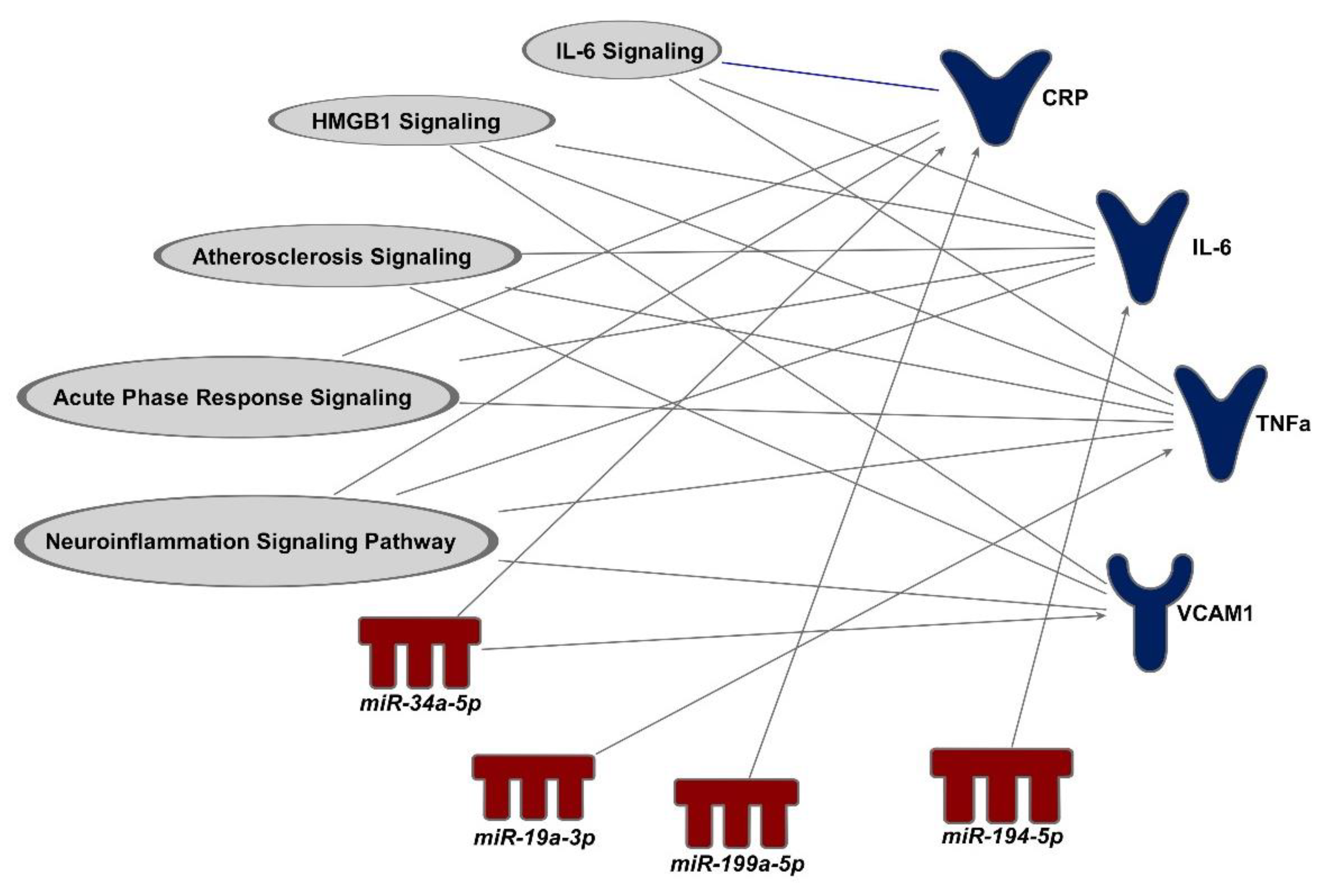
2.4.2. Functional Annotation between miRNA and Protein Biomarkers

We conducted an in silico analysis using the Ingenuity Pathway Analysis (IPA, Ingenuity Systems®, Redwood City, CA, USA) to identify mRNA targets of the statistically significant miRNAs; the miRNA–mRNA relationships were based on experimental results indexed in the established database. Because this was a preliminary study, we did not seek to validate the links between miRNA and mRNA. The analyzed miRNAs were those significantly associated with O3 exposure identified in the previous step, and mRNAs were for those protein biomarkers that have been measured in this study.

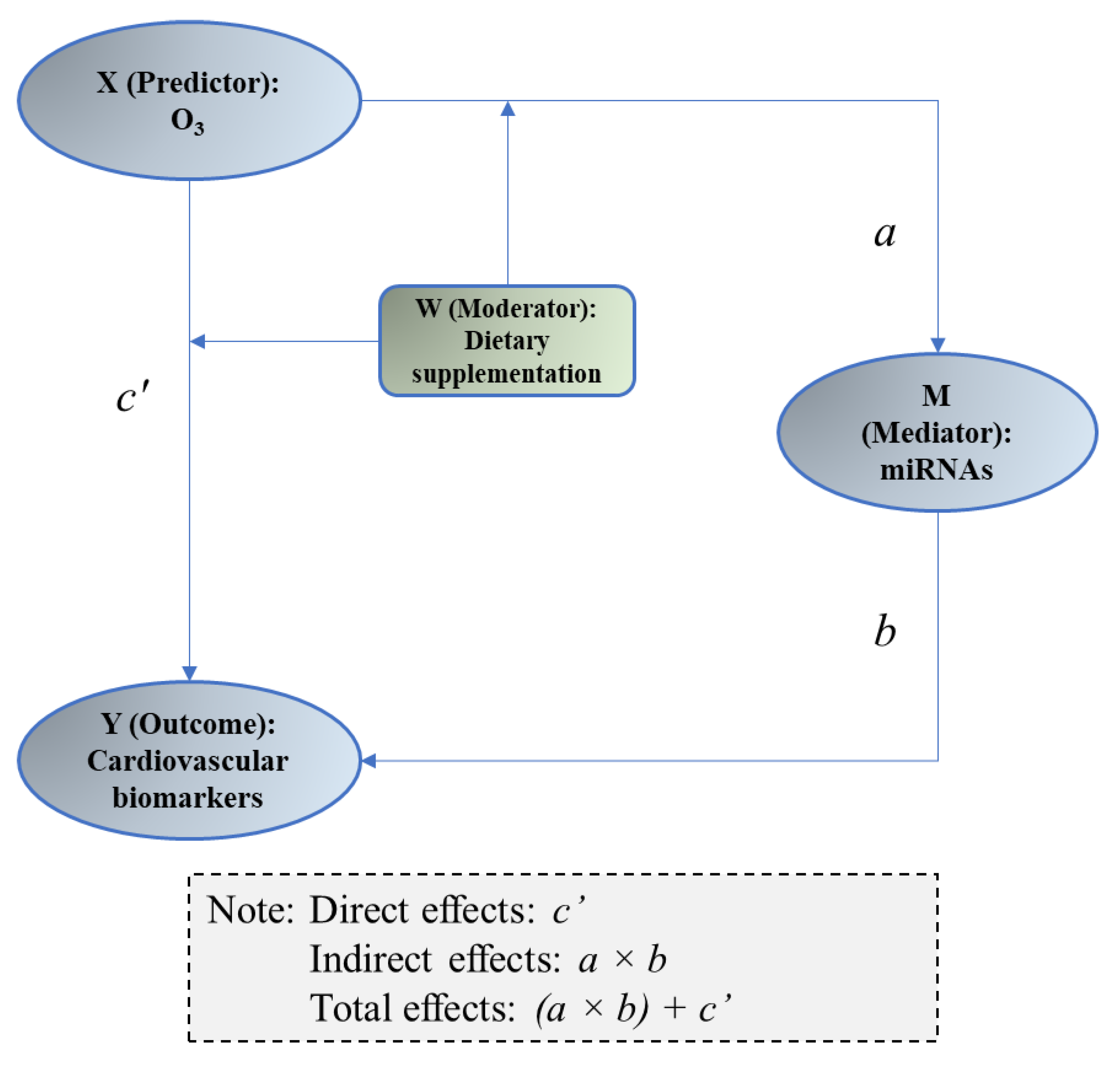
2.4.3. Mediation Analysis

Mediation analysis was employed to assess the statistical significance of the O3–miRNA–protein biomarker matches. The methods have been detailed previously [24]. In brief, we estimated the statistical significance of the indirect effects of miRNAs on the association between exposure to O3 and protein biomarkers. This approach decomposes the total estimated effects of air pollutants on the biomarker expression into an estimate of the direct effect and an estimate of the indirect effect of air pollutants on the biomarker expression mediated through miRNA. To allow for a comparable analysis, we standardized the data of exposure (O3 exposure), mediator (miRNAs), and outcomes (protein biomarkers) by subtracting the mean and dividing by the standard deviation. Two models have been fitted using the generalized linear mixed model (Figure 1): (1) model “X → M” was to assess the association between O3 exposure (X) and miRNAs (M) and acquire coefficient of X as “a”; (2) model “X + M → Y” was to assess the link between miRNAs and protein biomarkers (Y) while also considering O3 exposure (X), acquiring coefficient of M as “b”. Sobel test was conducted to evaluate if the indirect effect (a × b) was statistically significant. The results of p < 0.05 are considered as “statistically significant”.

3. Results

3.1. Participant Characteristics

Venous blood samples obtained from 23 male participants (6, 7, and 10 in CTL, FO, and OO groups, respectively) were included in the analysis (Table 1). The average age of the participants was 26 years old, and the BMI was 25.3. There was no statistical difference in the mean age and BMI among the three groups. There was a statistically significant difference in the number of participants who self-identified as being members of a specific race/ethnicity between the three groups (p = 0.04).

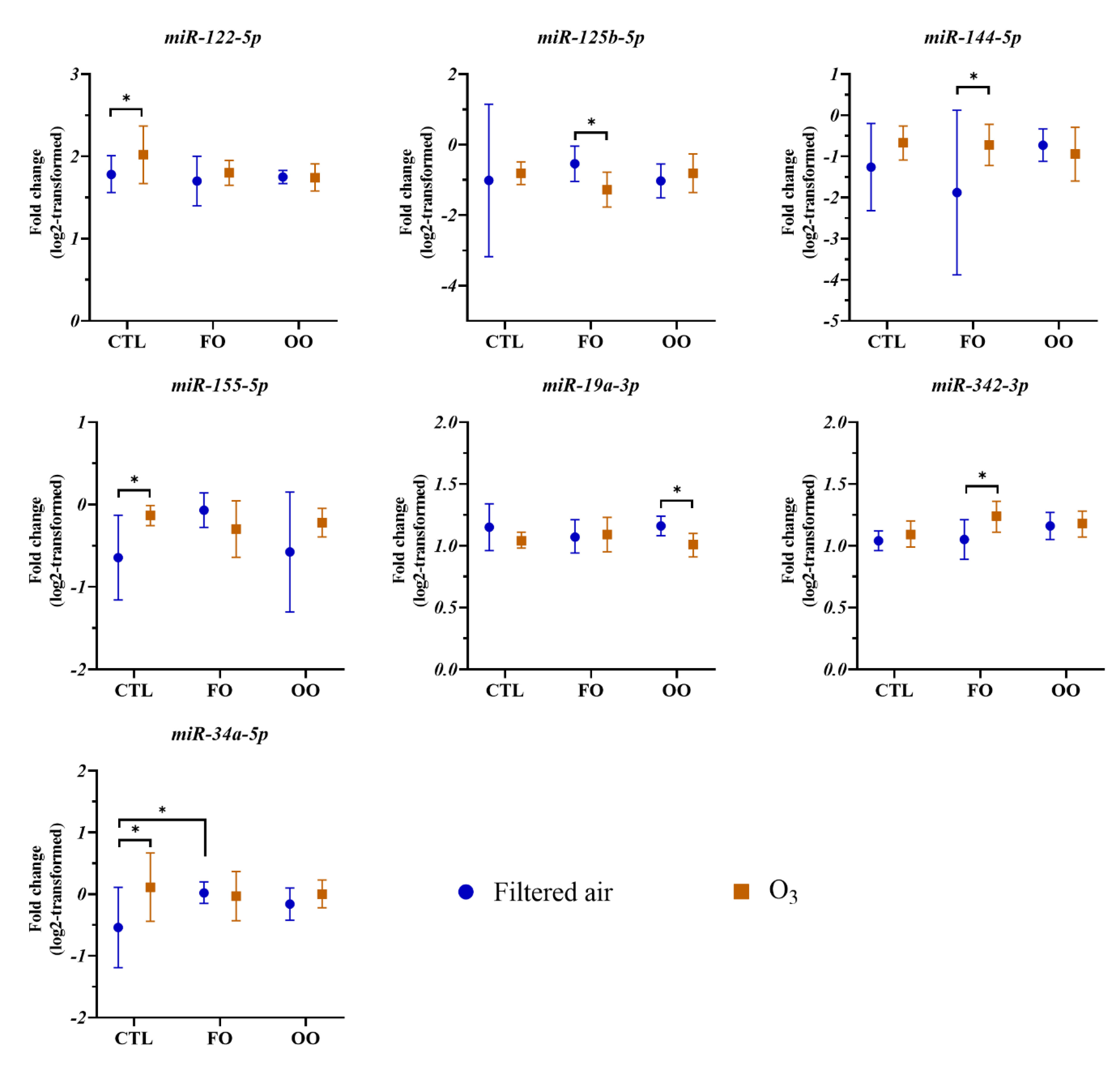
3.2. Effects of Dietary Supplementation and O3 Exposure on Circulating miRNAs

Descriptive statistics for the 65 miRNAs that were analyzed in the blood samples are presented in Supplemental Table S1. Among these miRNAs, dietary supplementation with FO or OO did not significantly alter the expression of the assessed miRNAs following filtered air or O3 exposure with the exception of the level of miR-34a-5p expression which was significantly elevated by FO supplementation compared with that of the CTL after exposure to filtered air. However, we found that the expression levels for seven miRNAs were significantly altered by O3 exposure in at least one of the dietary groups. Figure 2 presents log-transformed expression data of miRNAs significantly affected by O3 exposure. For example, the expression of circulating miR-122-5p was significantly elevated after O3 exposure compared with that of filtered air exposure [1.78 (1.56, 2.01) vs. 2.02 (1.67, 2.37), p = 0.007] in the CTL diet group. Compared with those of filtered air exposure, changes in circulating miRNA levels after O3 exposure were significantly altered for miR-125b-5p [−0.54 (−1.04, −0.04) vs. −1.27 (−1.77, −0.78), p = 0.04] and miR-144-5p [−1.88 (−3.88, 0.12) vs. −0.72 (−1.22, −0.22), p = 0.03] in the FO group, miR-155-5p in the CTL group [−0.64 (−1.15, −0.13) vs. −0.13 (−0.25, −0.01), p = 0.047], miR-19a-3p in the OO group [1.16 (1.08, 1.24) vs. 1.01 (0.91, 1.10), p = 0.008], miR-342-3p in the FO group [1.05 (0.89, 1.21) vs. 1.24 (1.11, 1.36), p = 0.02], and miR-34a-5p in the CTL group [−0.54 (−1.19, 0.11) vs. 0.11 (−0.44, 0.67), p = 0.01].

3.3. miRNAs Associated with O3 Exposure

As shown above, dietary supplementation did not have a significant effect on miRNA expression after filtered air exposure. O3 exposure was associated with changes in the expression of several miRNAs in at least one of the dietary groups. However, the sample size was relatively small in each dietary group and insufficient to conduct a meaningful mediation analysis. In order to examine miRNAs’ role in mediating the effects between O3 exposure and vascular biomarkers, we considered the overall effects of O3 on specific miRNA levels by pooling data from all three dietary groups together, while controlling dietary supplementation status as a covariate. We employed type III statistics in SAS, which examined for overall fixed effects of parameters for O3 and dietary supplementation (Supplemental Table S2). Because the effect of dietary supplementation on miRNA parameters were relatively small, the role of dietary supplementation is not further considered in this study. Among the 65 miRNAs, type III statistics showed that O3 exposure was associated with the expression of circulating miR-122-5p (F = 7.09, p = 0.015), miR-144-5p (F = 6.39, p = 0.045), miR-192-5p (F = 4.37, p = 0.0496), miR-194-5p (F = 7.83, p = 0.011), miR-199a-5p (F = 7.14, p = 0.015), miR-19a-3p (F = 5.47, p = 0.030), and miR-34a-5p (F = 4.77, p = 0.044) (Table 2).

3.4. Proteins That Are Predicted to Be Downstream Biomarkers of miRNAs

After identifying miRNAs associated with O3 exposure, we conducted a pathway analysis to search for potential matches between protein targets measured in the study and specific miRNAs. In this analysis, we examined the circulating levels of proteins associated with inflammation, coagulation, and vascular reactivity, including CRP, D-dimer, E-selectin, IL-6, IL-8, IL-1β, SAA, TNFα, sICAM1, sVCAM1, and vWF. The comparisons on theses biomarkers among different dietary groups and O3 exposures were reported previously [26]. We also observed elevated blood levels of IL-6 and decreased E-selectin levels post O3 exposure in at least one of the dietary groups among the male participants (Supplemental Table S3). Although not all biomarkers were significantly affected by O3 exposure, we presumed that mediational effects of miRNAs could still occur even if the independent variable (i.e., O3 exposure) was not significantly associated with a dependent variable (i.e., protein biomarkers) [30]. Based on this assumption, we further identified possible “O3–miRNA–protein biomarker” matches using the IPA application. As shown in Table 2, four out of the seven O3 exposure-associated miRNAs were predicted to regulate downstream mRNA/protein targets. Specifically, expression change in miR-194-5p was predicted to regulate the changes in circulating levels of D-dimer. Expression changes in circulating miR-199a-5p and miR-19a-3p were predicted to regulate the changes in circulating levels of CRP and TNFα, respectively, following O3 exposure. In addition, miR-34a-5p expression levels were predicted to regulate circulating CRP and sVCAM1 (Figure 3).

3.5. Mediation of miRNAs between O3 Exposure and Changes in Vascular and Coagulation Biomarkers

Next, we conducted a mediation analysis to determine whether the predicted “O3–miRNA–protein” matches were statistically significant, potentially validating the mediational role of miRNAs in the effects of O3 exposure on their downstream biomarkers. This analysis investigated the indirect effects of M (miRNAs) between X (O3 exposure) and Y (protein biomarkers) (Figure 1). Among the five predicted “O3–miRNA–protein” matches, “O3miR-19a-3p–TNFα” showed a statistically significant indirect effect of O3 exposure on TNFα levels through changes in circulating miR-19a-3p (Table 3). Specifically, the expression changes in circulating miR-19a-3p significantly down-regulated approximately 67.1% of O3 exposure-induced effects on TNFα [indirect effects, 0.08 (0.01, 0.15), p = 0.02]. We did not observe any other significant indirect effects of miRNAs between O3 exposure and protein biomarkers. In addition, we investigated possible moderating effects of dietary supplementation on the significant mediational model (O3miR-19a-3p–TNFα) as suggested in Figure 1. However, we did not find a link to suggest that dietary supplementation with FO or OO significantly alters the mediational pathways: X → M [interaction between X (O3) and W (dietary supplementation): F = 1.91, p = 0.17] or X + M → Y [interaction between X (O3) exposure and W (dietary supplementation): F = 1.22, p = 0.30) (Supplemental Table S4).

4. Discussion

Exposure to tropospheric O3 has been associated with increased morbidity and mortality; however, the mechanisms underlying such health impacts remain to be elucidated. Circulating miRNAs could serve as a possible mediator from O3-exposed pulmonary cells to those in the extrapulmonary system. In this human study, we investigated whether exposure to O3 altered blood levels of coagulation and vascular inflammation biomarkers through changes in circulating miRNAs among healthy male participants.
It has been shown previously that exposure to O3 is associated with altered expression of certain miRNAs in humans. For instance, exposure to ambient O3 among healthy adults was significantly associated with changes in circulating miRNAs, including let-7e-5p, miR-125-5p, and miR-26a-5p [24]. Another study also demonstrated significant changes in blood exosomal miR-150-5p and miR-155-5p in coronary artery disease patients exposed to short-term ambient O3 [17]. In a controlled chamber exposure study, exposure to 400 ppb O3 for 2 h induced altered expression of several miRNAs (e.g., miR-143, miR-145, miR-199a, miR-222, and miR-25) in induced sputum samples of healthy volunteers, suggesting the involvement of innate immune responses [31]. In the present study, among the 65 circulating miRNAs we investigated, 7 were associated with O3 exposure, including miR-122-5p, miR-125b-5p, miR-144-5p, miR-155-5p, miR-19a-3p, miR-342-3p, and miR-34a-5p. Therefore, our findings are consistent with previously published studies showing that O3 exposure leads to alterations in the expression of specific miRNAs.
The function of miRNA is to regulate the expression of target genes/proteins. The RNA-inducing silencing complex (RISC), which is a cytoplasmic multi-protein complex formed with single stranded RNA fragments including miRNAs, is able to regulate the translation of target mRNA through a post-initiation step [32]. miRNAs released into the extracellular space can reach different tissues and organs, thereby relaying biological information through regulation of mRNA expression to promote cell-to-cell communication that is essential in the cardiovascular effects of lung exposure to air pollutants [16]. O3 exposure—associated miRNAs identified in the present study—have been linked to cardiovascular pathophysiology. let-7e-5p expression is correlated with MAP kinase activation that involves CASP3 and TGFBR1, serving as a biomarker for ischemic stroke [33]. miR-122-5p is a key regulator of the SIRT6-elabela-ACE2 signaling pathway that is involved in angiotensin II-mediated apoptosis and oxidative stress in vascular endothelial cells [34]. Circulating miR-150-5p, miR-342-3p, and miR-34a-5p are also being investigated as biomarkers of acute heart failure [35] and vascular inflammation associated with vascular diseases [36,37,38]. Therefore, these altered expression levels of miRNAs may implicate multiple molecular mechanisms involved in cardiovascular disease induced by O3 exposure.
The altered circulating miRNAs in this study are believed to be involved in biological signaling pathways including IL-6 signaling, atherosclerosis signaling, and acute phase response signaling, implicating vascular inflammation and injury. Furthermore, the mediation results suggested that circulating miR-19a-3p may significantly relay the effects of exposure to O3 on the downstream inflammation marker TNFα. Elevated expression of miR-19a-3p has been reported in blood samples in patients with cardiovascular diseases such as acute heart failure [39]. In vitro studies have shown that overexpression of miR-19a-3p may promote vascular inflammation [40] and neuroinflammation [41]. It is possible that acute O3 exposure may stimulate a defense mechanism that downregulated expression of pro-inflammatory miRNA miR-19a-3p, which could inhibit pro-inflammatory cytokines such as TNFα in circulation.
Dietary supplementation with unsaturated fatty acids from fish oil or olive oil has been associated with cardiopulmonary protection against exposure to ambient air pollution [8,26,42,43]. Fish oil is rich in omega-3 polyunsaturated fatty acids such as eicosapentaenoic acid (EPA) and docosahexaenoic acid (DHA) that may mitigate adverse effects of air pollution exposure through antioxidant and anti-inflammatory properties [44]. Olive oil is rich in oleic acid, a monounsaturated fatty acid that has also shown health benefits [45]. In the present study, we did not focus on the impacts of dietary supplementation on miRNAs and their derived mediation analysis due to the small sample size in each dietary group. However, functional studies have suggested that omega-3 PUFA may modify the biological signaling pathways of several miRNAs. For example, consistent with our finding that FO treatment was associated with elevated expression of circulating miR-34a-5p, EPA and DHA may modulate the activation of p53/miR-34a-5p/Bcl-12 axis in myeloma cells [46]. A previous study reported that dietary intake of omega-3 PUFA may significantly modify both direct and indirect effects between air pollutant exposure and cardiovascular biomarkers through miR-26a-5p [24]. Future studies with larger sample size are warranted to examine the role of dietary interventions on the miRNA-mediated air pollution health effects.
The results of this study suggest a possible mediational role of miRNAs in O3-exposure-induced vascular inflammation and that certain miRNAs may be biological biomarkers in the mechanistic investigation of O3 induced health effects. However, there are a few limitations of this study. First, this study was a preliminary investigation with a relatively small sample size and was limited to young healthy male participants. Using a small sample size may be prone to type II statistical errors and the study may not be generalizable to a broader population. Second, because we only assessed blood biomarkers at one timepoint, it is possible that a more sensitive timepoint in which to observe potentially larger effects of O3 exposure on miRNAs was missed. Third, only protein levels of inflammation, coagulation, and vascular injury biomarkers were assessed, thus limiting our interpretation of the mRNA levels of these markers, which are direct regulating targets of miRNAs. In addition, besides miRNAs, we did not consider other possible mediational pathways through systemic inflammation and oxidative stress. We also cannot assume causal relationships of O3–miRNA–protein pathways, especially when miRNA and protein markers were assessed at the same time, raising the need for future functional studies of the specific pathway. Finally, statistical results were not adjusted for multiple testing, increasing the chance of both type I and type II errors. Despite these limitations, this exploratory study suggests that miRNAs may be able to mediate the acute effects of O3 exposure on vascular inflammation.

5. Conclusions

To our knowledge, this is the first study investigating whether circulating miRNAs may mediate the effects of inhalational exposure to O3 on coagulation and vascular inflammation biomarkers among healthy participants in a controlled chamber exposure trial. We have found that several circulating miRNAs (e.g., miR-122-5p, miR-144-5p, miR-19a-3p, miR-34a-5p) were significantly altered by O3 exposure. Among those, miR-19a-3p is a possible intermediator between O3 exposure and change in blood TNFα levels, consistent with miRNA–mRNA matches identified in an existing database. These findings offer preliminary evidence of the role of circulating miRNAs as a biological mechanism of vascular inflammation induced by O3 exposure.

Supplementary Materials

The following supporting information can be downloaded at: https://www.mdpi.com/article/10.3390/ncrna9040043/s1, Table S1: Descriptive statistics of all miRNA parameters; Table S2: Parameters of type III statistics of changes in circulating miRNAs affected by experimental conditions; Table S3: Descriptive statistics of blood protein parameters; Table S4: Parameters of type III statistics of the interaction term of O3 exposure and dietary supplementation.

Author Contributions

Conceptualization, H.C., J.M.S. and H.T.; methodology, H.C., A.G.R. and H.T.; software, H.C. and S.M.; validation, H.C., S.M. and H.T.; formal analysis, H.C. and S.M.; investigation, H.C. and H.T.; resources, H.T., A.G.R., J.M.S. and D.D.-S.; data curation, H.C. and H.T.; writing—original draft preparation, H.C.; writing—review and editing, all authors; visualization, H.C. and S.M.; supervision, A.G.R. and D.D.-S.; project administration, H.T.; funding acquisition, H.T. All authors have read and agreed to the published version of the manuscript.

Funding

The U.S. Environmental Protection Agency Intramural Research Program supported this research.

Institutional Review Board Statement

The study was conducted in accordance with the Declaration of Helsinki, and approved by the Institutional Review Board of the University of North Carolina at Chapel Hill (protocol 15-2960, date: 28 June 2017) and the U.S. Environmental Protection Agency Human Subjects Review Office.

Informed Consent Statement

Informed consent was obtained from all subjects involved in the study.

Data Availability Statement

The data presented in the current study will be made available in ScienceHub (https://catalog.data.gov/dataset/epa-sciencehub).

Acknowledgments

We thank Tracey Montilla, Julie Wood, Martin Case, Lisa Dailey, Joleen Soukup, and Shirley Harder for their excellent technical support. We thank the recruitment service from MPF Federal. This project was supported in part by an appointment to the Research Participation Program at the U.S. Environmental Protection Agency, administered by the Oak Ridge Institute for Science and Education through an interagency agreement between the U.S. Department of Energy and EPA.

Conflicts of Interest

The research described in this article has been reviewed by the Center for Public Health and Environmental Assessment, EPA, and approved for publication. The contents of this article should not be construed to represent agency policy nor does mention of trade names or commercial products constitute endorsement or recommendation for use.

References

  1. EPA. Integrated Science Assessment (ISA) for Ozone and Related Photochemical Oxidants; Final Report, Feb 2013; U.S.E.P. Agency, Ed.; EPA: Washington, DC, USA, 2013.
  2. American Lung Association. The “State of the Air” 2021 Report. 2021. Available online: https://www.lung.org/getmedia/17c6cb6c-8a38-42a7-a3b0-6744011da370/sota-2021.pdf (accessed on 31 January 2023).
  3. Zhang, J.J.; Wei, Y.; Fang, Z. Ozone Pollution: A Major Health Hazard Worldwide. Front. Immunol. 2019, 10, 2518. [Google Scholar] [CrossRef] [PubMed] [Green Version]
  4. Arjomandi, M.; Balmes, J.R.; Frampton, M.W.; Bromberg, P.; Rich, D.Q.; Stark, P.; Alexis, N.E.; Costantini, M.; Hollenbeck-Pringle, D.; Dagincourt, N.; et al. Respiratory Responses to Ozone Exposure. MOSES (The Multicenter Ozone Study in Older Subjects). Am. J. Respir. Crit. Care Med. 2018, 197, 1319–1327. [Google Scholar] [CrossRef] [PubMed]
  5. Samet, J.M.; Hatch, G.E.; Horstman, D.; Steck-Scott, S.; Arab, L.; Bromberg, P.A.; Levine, M.; Mcdonnell, W.F.; Devlin, R.B. Effect of antioxidant supplementation on ozone-induced lung injury in human subjects. Am. J. Respir. Crit. Care Med. 2001, 164, 819–825. [Google Scholar] [CrossRef] [PubMed]
  6. Devlin, R.B.; Duncan, K.E.; Jardim, M.; Schmitt, M.T.; Rappold, A.G.; Diaz-Sanchez, D. Controlled exposure of healthy young volunteers to ozone causes cardiovascular effects. Circulation 2012, 126, 104–111. [Google Scholar] [CrossRef]
  7. Yin, P.; Chen, R.; Wang, L.; Meng, X.; Liu, C.; Niu, Y.; Lin, Z.; Liu, Y.; Liu, J.; Qi, J.; et al. Ambient Ozone Pollution and Daily Mortality: A Nationwide Study in 272 Chinese Cities. Environ. Health Perspect. 2017, 125, 117006. [Google Scholar] [CrossRef] [Green Version]
  8. Lim, C.C.; Hayes, R.B.; Ahn, J.; Shao, Y.; Silverman, D.T.; Jones, R.R.; Garcia, C.; Bell, M.L.; Thurston, G.D. Long-Term Exposure to Ozone and Cause-Specific Mortality Risk in the United States. Am. J. Respir. Crit. Care Med. 2019, 200, 1022–1031. [Google Scholar] [CrossRef]
  9. Bromberg, P.A. Mechanisms of the acute effects of inhaled ozone in humans. Biochim. Biophys. Acta 2016, 1860, 2771–2781. [Google Scholar] [CrossRef]
  10. Mudway, I.; Kelly, F. Ozone and the lung: A sensitive issue. Mol. Asp. Med. 2000, 21, 1–48. [Google Scholar] [CrossRef]
  11. Corteselli, E.M.; Gold, A.; Surratt, J.; Cui, T.; Bromberg, P.; Dailey, L.; Samet, J.M. Supplementation with omega-3 fatty acids potentiates oxidative stress in human airway epithelial cells exposed to ozone. Environ. Res. 2020, 187, 109627. [Google Scholar] [CrossRef]
  12. Devlin, R.B.; McDonnell, W.F.; Mann, R.; Becker, S.; House, D.E.; Schreinemachers, D.; Koren, H.S. Exposure of humans to ambient levels of ozone for 6.6 h causes cellular and biochemical changes in the lung. Am. J. Respir. Cell Mol. Biol. 1991, 4, 72–81. [Google Scholar] [CrossRef]
  13. Chen, H.; Smith, G.J.; Masood, S.; Tong, H. Extracellular MicroRNAs as Putative Biomarkers of Air Pollution Exposure, in Biomarkers in Toxicology; Patel, V.B., Preedy, V.R., Rajendram, R., Eds.; Springer International Publishing: Cham, Switzerland, 2022; pp. 1–24. [Google Scholar]
  14. Smith, G.J.; Tovar, A.; Kanke, M.; Wang, Y.; Deshane, J.S.; Sethupathy, P.; Kelada, S.N.P. Ozone-induced changes in the murine lung extracellular vesicle small RNA landscape. Physiol. Rep. 2021, 9, e15054. [Google Scholar] [CrossRef] [PubMed]
  15. MacFarlane, L.-A.; Murphy, P.R. MicroRNA: Biogenesis, Function and Role in Cancer. Curr. Genom. 2010, 11, 537–561. [Google Scholar] [CrossRef] [PubMed] [Green Version]
  16. Zhao, C.; Sun, X.; Li, L. Biogenesis and function of extracellular miRNAs. Exrna 2019, 1, 38. [Google Scholar] [CrossRef] [Green Version]
  17. Chen, H.; Xu, Y.; Rappold, A.; Diaz-Sanchez, D.; Tong, H. Effects of ambient ozone exposure on circulating extracellular vehicle microRNA levels in coronary artery disease patients. J. Toxicol. Environ. Health Part A 2020, 83, 351–362. [Google Scholar] [CrossRef] [PubMed]
  18. Krauskopf, J.; Caiment, F.; van Veldhoven, K.; Chadeau-Hyam, M.; Sinharay, R.; Chung, K.F.; Cullinan, P.; Collins, P.; Barratt, B.; Kelly, F.J.; et al. The human circulating miRNome reflects multiple organ disease risks in association with short-term exposure to traffic-related air pollution. Environ. Int. 2018, 113, 26–34. [Google Scholar] [CrossRef] [Green Version]
  19. Motta, V.; Favero, C.; Dioni, L.; Iodice, S.; Battaglia, C.; Angelici, L.; Vigna, L.; Pesatori, A.C.; Bollati, V. MicroRNAs are associated with blood-pressure effects of exposure to particulate matter: Results from a mediated moderation analysis. Environ. Res. 2016, 146, 274–281. [Google Scholar] [CrossRef] [Green Version]
  20. Rodosthenous, R.S.; Coull, B.A.; Lu, Q.; Vokonas, P.S.; Schwartz, J.D.; Baccarelli, A.A. Ambient particulate matter and microRNAs in extracellular vesicles: A pilot study of older individuals. Part. Fibre Toxicol. 2016, 13, 13. [Google Scholar] [CrossRef] [Green Version]
  21. Chen, R.; Li, H.; Cai, J.; Wang, C.; Lin, Z.; Liu, C.; Niu, Y.; Zhao, Z.; Li, W.; Kan, H. Fine Particulate Air Pollution and the Expression of microRNAs and Circulating Cytokines Relevant to Inflammation, Coagulation, and Vasoconstriction. Environ. Health Perspect. 2018, 126, 017007. [Google Scholar] [CrossRef] [Green Version]
  22. Pergoli, L.; Cantone, L.; Favero, C.; Angelici, L.; Iodice, S.; Pinatel, E.; Hoxha, M.; Dioni, L.; Letizia, M.; Albetti, B.; et al. Extracellular vesicle-packaged miRNA release after short-term exposure to particulate matter is associated with increased coagulation. Part. Fibre Toxicol. 2017, 14, 32. [Google Scholar] [CrossRef] [Green Version]
  23. Rodosthenous, R.S.; Kloog, I.; Colicino, E.; Zhong, J.; Herrera, L.A.; Vokonas, P.; Schwartz, J.; Baccarelli, A.A.; Prada, D. Extracellular vesicle-enriched microRNAs interact in the association between long-term particulate matter and blood pressure in elderly men. Environ. Res. 2018, 167, 640–649. [Google Scholar] [CrossRef]
  24. Chen, H.; Zhang, S.; Yu, B.; Xu, Y.; Rappold, A.G.; Diaz-Sanchez, D.; Samet, J.M.; Tong, H. Circulating microRNAs as putative mediators in the association between short-term exposure to ambient air pollution and cardiovascular biomarkers. Ecotoxicol. Environ. Saf. 2022, 239, 113604. [Google Scholar] [CrossRef] [PubMed]
  25. VanderWeele, T.J. Mediation Analysis: A Practitioner’s Guide. Annu. Rev. Public Health 2016, 37, 17–32. [Google Scholar] [CrossRef] [PubMed] [Green Version]
  26. Chen, H.; Tong, H.; Shen, W.; Montilla, T.S.; Case, M.W.; Almond, M.A.; Wells, H.B.; Alexis, N.E.; Peden, D.B.; Rappold, A.G.; et al. Fish oil blunts lung function decrements induced by acute exposure to ozone in young healthy adults: A randomized trial. Environ. Int. 2022, 167, 107407. [Google Scholar] [CrossRef] [PubMed]
  27. California Environmental Protection Agency. Environmental Protection Indicators for California, 2004 Update; California Environmental Protection Agency: Sacramento, CA, USA, 2005. [Google Scholar]
  28. Wang, T.; Ding, A.; Gao, J.; Wu, W.S. Strong ozone production in urban plumes from Beijing, China. Geophys. Res. Lett. 2006, 33. [Google Scholar] [CrossRef] [Green Version]
  29. Kenward, M.G.; Roger, J.H. Small sample inference for fixed effects from restricted maximum likelihood. Biometrics 1997, 53, 983–997. [Google Scholar] [CrossRef] [Green Version]
  30. MacKinnon, D.P.; Lockwood, C.M.; Hoffman, J.M.; West, S.G.; Sheets, V. A comparison of methods to test mediation and other intervening variable effects. Psychol. Methods 2002, 7, 83–104. [Google Scholar] [CrossRef]
  31. Fry, R.C.; Rager, J.E.; Bauer, R.; Sebastian, E.; Peden, D.B.; Jaspers, I.; Alexis, N.E. Air toxics and epigenetic effects: Ozone altered microRNAs in the sputum of human subjects. Am. J. Physiol. Cell. Mol. Physiol. 2014, 306, L1129–L1137. [Google Scholar] [CrossRef] [Green Version]
  32. Abdellatif, M. Differential expression of microRNAs in different disease states. Circ. Res. 2012, 110, 638–650. [Google Scholar] [CrossRef]
  33. Huang, S.; Lv, Z.; Guo, Y.; Li, L.; Zhang, Y.; Zhou, L.; Yang, B.; Wu, S.; Zhang, Y.; Xie, C.; et al. Identification of Blood Let-7e-5p as a Biomarker for Ischemic Stroke. PLoS ONE 2016, 11, e0163951. [Google Scholar] [CrossRef] [Green Version]
  34. Song, J.J.; Yang, M.; Liu, Y.; Song, J.W.; Wang, J.; Chi, H.J.; Liu, X.Y.; Zuo, K.; Yang, X.C.; Zhong, J.C. MicroRNA-122 aggravates angiotensin II-mediated apoptosis and autophagy imbalance in rat aortic adventitial fibroblasts via the modulation of SIRT6-elabela-ACE2 signaling. Eur. J. Pharmacol. 2020, 883, 173374. [Google Scholar] [CrossRef]
  35. Scrutinio, D.; Conserva, F.; Passantino, A.; Iacoviello, M.; Lagioia, R.; Gesualdo, L. Circulating microRNA-150-5p as a novel biomarker for advanced heart failure: A genome-wide prospective study. J. Heart Lung Transplant. 2017, 36, 616–624. [Google Scholar] [CrossRef] [PubMed] [Green Version]
  36. Zhao, L.; Zhang, Y. miR-342-3p affects hepatocellular carcinoma cell proliferation via regulating NF-kappaB pathway. Biochem. Biophys. Res. Commun. 2015, 457, 370–377. [Google Scholar] [CrossRef] [PubMed]
  37. Jiang, Q.; Li, Y.; Wu, Q.; Huang, L.; Xu, J.; Zeng, Q. Pathogenic role of microRNAs in atherosclerotic ischemic stroke: Implications for diagnosis and therapy. Genes Dis. 2022, 9, 682–696. [Google Scholar] [CrossRef] [PubMed]
  38. Raucci, A.; Macrì, F.; Castiglione, S.; Badi, I.; Vinci, M.C.; Zuccolo, E. MicroRNA-34a: The bad guy in age-related vascular diseases. Cell. Mol. Life Sci. 2021, 78, 7355–7378. [Google Scholar] [CrossRef]
  39. Su, Y.; Sun, Y.; Tang, Y.; Li, H.; Wang, X.; Pan, X.; Liu, W.; Zhang, X.; Zhang, F.; Xu, Y.; et al. Circulating miR-19b-3p as a Novel Prognostic Biomarker for Acute Heart Failure. J. Am. Heart Assoc. 2021, 10, e022304. [Google Scholar] [CrossRef]
  40. Chen, H.; Li, X.; Liu, S.; Gu, L.; Zhou, X. MircroRNA-19a promotes vascular inflammation and foam cell formation by targeting HBP-1 in atherogenesis. Sci. Rep. 2017, 7, 12089. [Google Scholar] [CrossRef]
  41. Kim, T.; Valera, E.; Desplats, P. Alterations in Striatal microRNA-mRNA Networks Contribute to Neuroinflammation in Multiple System Atrophy. Mol. Neurobiol. 2019, 56, 7003–7021. [Google Scholar] [CrossRef]
  42. Tong, H.; Zhang, S.; Shen, W.; Chen, H.; Salazar, C.; Schneider, A.; Rappold, A.G.; Diaz-Sanchez, D.; Devlin, R.B.; Samet, J.M. Lung Function and Short-Term Ambient Air Pollution Exposure: Differential Impacts of Omega-3 and Omega-6 Fatty Acids. Ann. Am. Thorac. Soc. 2022, 19, 583–593. [Google Scholar] [CrossRef]
  43. Chen, H.; Zhang, S.; Shen, W.; Salazar, C.; Schneider, A.; Wyatt, L.H.; Rappold, A.G.; Diaz-Sanchez, D.; Devlin, R.B.; Samet, J.M.; et al. Omega-3 fatty acids attenuate cardiovascular effects of short-term exposure to ambient air pollution. Part. Fibre Toxicol. 2022, 19, 12. [Google Scholar] [CrossRef]
  44. Albert, B.B.; Cameron-Smith, D.; Hofman, P.L.; Cutfield, W.S. Oxidation of Marine Omega-3 Supplements and Human Health. BioMed Res. Int. 2013, 2013, 464921. [Google Scholar] [CrossRef] [Green Version]
  45. Perona, J.; Cabellomoruno, R.; Ruizgutierrez, V. The role of virgin olive oil components in the modulation of endothelial function. J. Nutr. Biochem. 2006, 17, 429–445. [Google Scholar] [CrossRef] [PubMed] [Green Version]
  46. Dai, X.; Li, M.; Geng, F. Omega-3 Polyunsaturated Fatty Acids Eicosapentaenoic Acid and Docosahexaenoic Acid Enhance Dexamethasone Sensitivity in Multiple Myeloma Cells by the p53/miR-34a/Bcl-2 Axis. Biochemistry 2017, 82, 826–833. [Google Scholar] [CrossRef] [PubMed]
Figure 1. Schematic showing the mediation model employed in this study. Letter “a” indicates the coefficient of X derived from the statistical analysis for “X → M”, “b” indicates the coefficient of M in the model “M → Y”, and “c” indicates the coefficient of X in the model “X  +  M → Y”.
Ncrna 09 00043 g001
Figure 2. Changes in circulating miRNAs for participants in control (CTL), fish oil (FO), and olive oil (OO) groups immediately after exposure to filtered air or 300 ppb O3. Shown are log2-transformed fold change values in miR-122-5p, miR-125b-5p, miR-144-5p, miR-155-5p, miR-19a-3p, miR-342-3p, and miR-34a-5p. Bars with whiskers indicate means with their respective 95% confidence interval. * p < 0.05 as “significant” between two conditions.
Ncrna 09 00043 g002
Figure 3. Pathway analysis showing the link between O3 exposure-induced miRNAs and vascular proteins in the plasma and their related biological pathways. Four miRNAs (in red) that were associated with O3 exposure and their matched mRNA/protein targets (in dark blue) and their associated downstream inflammatory signaling pathways (in gray) were identified by the Ingenuity Pathway Analysis software.
Ncrna 09 00043 g003
Table 1. Participant demographics.
CharacteristicsCTL (n = 6)FO (n = 7)OO (n = 10)All (n = 23)
Age (years)23.5 (3.7)27.4 (4.7)26.5 (3.0)26.0 (3.9)
BMI (kg/m2)24.9 (3.6)25.8 (3.8)25.1 (2.7)25.3 (3.2)
Omega-3 index (%)4.0 (0.2)6.1 (1.2)4.0 (0.4)4.6 (1.2)
Race/ethnicity (no. of participants)
African-American 0202
Asian1102
Caucasian341017
Hispanic2002
BMI: body mass index; CTL: control; FO: fish oil; OO: olive oil.
Table 2. O3 exposure—associated miRNAs and their predicted downstream cardiovascular targets.
miRNAsF ValuepPredicted Targets
miR-122-5p7.090.015 *-
miR-144-5p6.390.0448 *-
miR-192-5p4.370.0496 *-
miR-194-5p7.830.0111 *D-dimer
miR-199a-5p7.140.0146 *CRP
miR-19a-3p5.470.0299 *TNFα
miR-34a-5p4.770.0441 *CRP, sVCAM1
O3 exposure associated miRNAs were identified using a mixed-effects model with subject as random effect and adjusting for covariates including dietary supplementation and the interaction product of O3 exposure and dietary supplementation. * p < 0.05 O3 exposure was a “significant” factor for changes in the circulating miRNA. Measured protein biomarkers were predicted and matched with the identified miRNAs using the Ingenuity Pathway Analysis. CRP: c-reactive protein, IL-6: interleukin 6, IL-8: interleukin 8, sVCAM1: soluble vascular cell adhesion molecule 1, TNFα: tumor necrosis factor α.
Table 3. Mediation effects of miRNAs on the association between exposure to ozone and biomarkers.
miRNAs (M)Biomarker (Y)X → M (a)M → Y (b)Direct Effects (c′)Indirect Effects (a × b)
Coefficient (95%CI)Proportion (%)p
miR-194-5pD-dimer0.45 (−0.2, 1.1)0.16 (0.09, 0.24)−0.17 (−0.29, −0.05)0.07 (−0.04, 0.19)−76.780.20
miR-199a-5pCRP−0.38 (−1.07, 0.31)0.02 (−0.05, 0.09)0.34 (0.22, 0.46)−0.01 (−0.04, 0.02)−2.170.64
miR-19a-3pTNFα−1.03 (−1.81, −0.25)−0.08 (−0.11, −0.05)−0.21 (−0.28, −0.13)0.08 (0.01, 0.15)−67.090.02 *
miR-34a-5pCRP0.32 (−0.53, 1.18)0.22 (0.16, 0.27)0.27 (0.16, 0.37)0.07 (−0.11, 0.25)20.790.46
miR-34a-5psVCAM10.32 (−0.53, 1.18)0.05 (0.02, 0.08)0.05 (−0.01, 0.11)0.02 (−0.03, 0.06)25.630.47
The letters X, M, and Y denote independent variable (X for O3 exposure), mediator (M for miRNAs), and dependent variable (Y for cardiovascular biomarkers). The letters a, b, and c′ denote coefficients of X, M, and X in their respective models shown in Figure 1. Data were presented as the coefficients with 95% confidence interval (95%CI). Bold font indicates that the coefficient was statistically significant (p < 0.05). The statistical significance of indirect effects (a × b) was assessed using the Sobel Test. * p < 0.05 indicates that the indirect effects were statistically significant. Proportion of indirect effects in the total effects (a × b + c′) was calculated. CRP: c-reactive protein, TNFα: tumor necrosis factor α, sVCAM1: soluble vascular cell adhesion molecule 1.
Disclaimer/Publisher’s Note: The statements, opinions and data contained in all publications are solely those of the individual author(s) and contributor(s) and not of MDPI and/or the editor(s). MDPI and/or the editor(s) disclaim responsibility for any injury to people or property resulting from any ideas, methods, instructions or products referred to in the content.

