Article

Potential Application of Ovalbumin Gel Nanoparticles Loaded with Carvacrol in the Preservation of Fresh Pork

1
State Key Laboratory of Food Science and Technology, Jiangnan University, Wuxi 214122, China
2
College of Food Science and Engineering, Yangzhou University, Yangzhou 225127, China
*
Author to whom correspondence should be addressed.
Gels 2023, 9(12), 941; https://doi.org/10.3390/gels9120941
Submission received: 6 November 2023 / Revised: 27 November 2023 / Accepted: 28 November 2023 / Published: 30 November 2023
(This article belongs to the Special Issue Advancements in Food Gelation: Exploring Mechanisms and Applications)

Abstract

:
Plant essential oil has attracted much attention in delaying pork spoilage due to its safety, but its low antibacterial efficiency needs to be solved by encapsulation. Our previous research had fabricated a type of ovalbumin gel nanoparticles loaded with carvacrol (OCGn-2) using the gel-embedding method, which had a high encapsulation rate and antibacterial activity. The main purpose of this study was to further evaluate the stability and slow-release characteristics of OCGn-2 and potential quality effects of the nanoparticles on the preservation of fresh pork pieces during 4 °C storage. The particle test showed that the nanoparticles had better heat stability below 85 °C and salt stability below 90 mM. The in vitro release study indicated that the carvacrol in OCGn-2 followed a Fickian release mechanism. The pork preservation test suggested that the OCGn-2 coating treatments could remarkably restrict the quality decay of pork slices compared to free carvacrol or a physical mixture of ovalbumin and carvacrol treatment. Nano-encapsulation of ovalbumin is beneficial to the sustained release, enhanced oxidation resistance, and improved antibacterial activity of carvacrol. The study suggested that ovalbumin gel nanoparticles embedded with carvacrol could be applied as an efficient bacterial active packaging to extend the storage life of pork.

Graphical Abstract

1. Introduction

Among the common meat products, pork is consumed in huge quantities throughout the world. However, a mass of polyunsaturated fatty acids in pork is harmful because biochemical reactions such as oxidation and microbial spoilage can bring about color changes and rancid taste [1]. The deterioration of meat during manufacturing and storage involves three mechanisms: microbial spoilage, lipid oxidation, and enzymatic autolysis. Red meat such as pork [2], beef [3], and mutton [4] are frequently related to outbursts mainly because of the existence of Salmonella spp., Staphylococcus spp., Clostridium spp., and Listeria spp. In recent years, Salmonella has been reported as the main reason for foodborne disease transmission leading to hospitalization [5]. Salmonella in the feces can contaminate the surface of the carcass during the slaughter process, and intestinal diseases and bacterial infections can probably be generated after accidental consumption [6]. Therefore, contamination with foodborne human pathogens, especially by Salmonella, is becoming an increasing concern for the pork industry. The spoilage factors in meat products sometimes represent a high risk for producers’ interest and poses a threat to consumer health. Therefore, preventing the impact that the storage process has on Salmonella prevalence is a critical step in developing strategies for pathogen reduction in meat and can guarantee economy and safety of the meat processing industry.
In meat preservation, it is common that the addition of different preservatives in various meat products could prevent lipid oxidation, delay rancidity, and improve color sharpness. Despite the excellent efficiency and stability of synthetic preservatives, in the past several years, researchers have taken increasing interest in replacing them with natural preservatives such as plant essential oil (PEO) on account of their potential impacts on toxicity, carcinogenesis, teratogenicity, and mutagenicity [7]. PEO, frequently used by meat producers, is extracted from specific plants, such as clove, thyme, and ginger [8,9,10]. Many of them are generally recognized as safe (GRAS) for use in the food industry, as declared by the United States Food and Drug Administration (FDA) [11]. Carvacrol (Car), purified from the secondary metabolites of oregano and thyme, has strong abilities against both microorganisms and oxidization [12]. Nonetheless, the application of Car in food processing is blocked because it is quickly volatile and hardly soluble in water.
Encapsulation, regarded to be a burgeoning and current method, can capture and improve the solubility of PEO, and likewise effectively prevent sustained release during processing and action, to manufacture a nano-delivery system in the form of nanoparticles [13]. The use of active food packaging, which utilizes PEO-loaded nanoparticles as active agents, has become gradually attractive due to their properties against pathogenic microorganisms and oxidation as well as stability by resisting varieties of environmental impacts [7]. Evidently, gel nanoparticles represent intricate, three-dimensional nanoscale biomacromolecular constructs that are intricately cross-linked, capable of accommodating substantial fluid volumes, and, consequently, a noteworthy quantity of active constituents [14]. Innovative hydrogel carriers, varying in matrix sizes from micrometers to nanometers, bestow heightened transport capabilities. Gel nanoparticles seamlessly amalgamate the advantages of hydrogels and nanoparticles, boasting traits such as water solubility, distinctive adhesive properties, a spacious specific surface area amenable to manipulation, an elevated loading capacity, meticulously regulated delayed release kinetics, and a superior degree of biocompatibility and biodegradability [15]. Protein (e.g., whey protein [16], gliadin [17], zein [18], and ovalbumin) is one of the most generally employed materials for the manufacture of esculent nanoparticles since it can be assembled by covalent or noncovalent bonds. Ovalbumin (OVA), the most plentiful protein component in egg white protein (EWP), comprises about 192 hydrophobic amino acids and about 128 of which are electropositive/electronegative; these demonstrate the feasibility of OVA as an efficient carrier for agent delivery [19].
Our previous studies have explored the physicochemical properties and antibacterial properties of ovalbumin–carvacrol (OVA-Car) nanocomposites prepared by the oil-in-water method [20] and gel-embedding method [21]. It was found that OVA-Car prepared by gel-embedding method had better properties than those of OVA-Car from the oil-in-water method. Moreover, among different pH conditions (2, 5, 7, and 9), which can affect gel microstructure, OCGn-2 (OVA gel nanoparticles prepared loaded with Car at pH 2) was selected because its gel structure was more dense and stable, and has higher encapsulation efficiency contributing to higher sterilization rate.
However, previous studies have only explored the physicochemical properties and antibacterial properties of nanoparticles while application tests on preserving fresh pork are lacking. Hence, the purpose of this study was to apply OCGn-2 to evaluate its influence on the quality characteristic of fresh pork contaminated with Salmonella during 4 °C storage. This is based on the investigation of the influence of environmental stresses on the stability of nanoparticles and the release profiles of Car from OCGn-2, which have the possibility to be exploited as an effective economic approach for meat preservation. In the long term, this study may offer new creative approach into the development of delivery systems with improved biological preservatives.

2. Results and Discussion
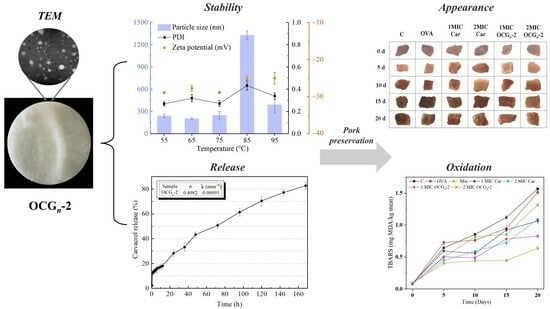
2.1. Form Analysis

The solid sample was a light-yellow powder with tiny and uniform particles (Figure 1a). It was easily soluble in water and appeared as a white liquid with high turbidity at high concentrations (Figure 1b). The nanoparticle image demonstrated spherical and regularly distributed particles, which had good homogeneity and stability without adverse reactions during the preparation process (Figure 1c,d). The results of TEM indicated that the size distribution of most nanoparticles was between 40 and 60 nm. The reason for the larger particle diameter might be that the protein layer outside the single particle swells and/or the single particle agglomerates when dispersed in the solution. OVA encapsulation is indicated to improve the delivery mode of antibacterial agents on pathogenic bacteria and provided a reference for future research.

2.2. Effects of Environmental Stresses on the Stability of OCGn-2

Figure 2a depicted the impact of thermal treatment in the temperature range of 55–95 °C for 15 min on the particle size, PDI and zeta potential of the solutions. Noticeable trends included an increase in particle size and a widening of the size distribution at 85 °C on account of the poor thermal stability of OCGn-2. At other temperatures, their average particle size remained below 350 nm, while the PDI fluctuated slightly around 0.3 because the composite particles tend to aggregate, resulting in larger particle sizes than expected. When the temperature fell below 85 °C, the zeta potential exhibited slight fluctuations between −27 mV and −29 mV. This indicated that OCGn-2 successfully withstood thermally induced aggregation at this stage. However, surpassing 85 °C diminished the net charge content, leading to particle aggregation and consequently an upsurge in the measured particle size. Together, the above results indicated that OCGn-2 became stable under low-temperature treatment.
As shown in Figure 2b, the effect of NaCl concentration on the stability of OCGn-2 was assessed by assessing the particle size, PDI and zeta potential of the solution at different concentrations of NaCl (10–170 mM). The particle size of OCGn-2 experienced a slight decrease within the range of 10 to 90 mM NaCl. This phenomenon can be attributed to the capacity of low NaCl concentrations to induce protein-based nanoparticle structures to contract, leading to a smaller particle size. A comparable trend was observed in composite condensed nanoparticles crosslinked with tannic acid [22]. Starting with the introduction of a 90 mM NaCl solution, the PDI of the solution exhibited a notable and rapid increase in response to an elevated ionic strength, surpassing the changes observed at the 0.3 level. Ultimately, the nanoparticles displayed the greatest particle size and PDI in 170 mM NaCl solution. The net charge of OCGn-2 exhibited a gradual decline as the NaCl concentration increased. Particularly noteworthy is that the net charge dropped below 20 mV upon surpassing 130 mM, signifying significant particle aggregation. These observations suggest that the electrostatic repulsion and steric hindrance between nanoparticles have been severely compromised due to the screening effects induced by high ionic strength [23]. As a result, instability ensues, leading to extensive nanoparticle aggregation. A similar phenomenon was also observed in the NaCl stability results of Camellia seed albumin nanoparticles [24]. In summary, OCGn-2 subjected to temperatures below 85 °C or treated with NaCl concentrations below 130 mM retained a uniform small particle size. This might make the contact area between the samples and pathogenic microorganisms large enough to facilitate the diffusion of the antibacterial active ingredients of OCGn-2 on the bacterial cell membrane.

2.3. In Vitro Release Studies

The in vitro release profiles of Car from the nanoparticles under ambient temperature are shown in Figure 3. The results showed that the release of Car from OCGn-2 was divided into two stages according to the rate of release. A burst release of Car was discovered during the preliminary 60 h of the initial phase. Car was released up to 12.13% in the buffer solution. The release at this stage was mainly due to the free Car attached to the surface of the nanoparticles [25]. The release speed of the second stage drops slightly, that is, between 60 h and 168 h, when the accumulated amount of Car released was 82.71% in the buffer solution.
For the spherical nanoparticles in this study, n = 0.4082, which is less than 0.45, indicating that the Fickian release mechanism was followed [26]. The linear relation of ln ( M t M 0 ) versus ln(t) was used to measure the value of n and k.

2.4. Meat Storage Test

2.4.1. pH Test

The pH level of fresh, unprocessed pork typically falls within the range of 5.18 to 6.17 [27]. As exhibited in Figure 4, the initial pH was 5.53–5.82 at 4 °C. At the beginning of the storage, we observed a slight decline in pH value, attributing to the proliferation of lactic acid in pork samples [28]. The pH reached the upper limit for fresh meat (pH = 6.8) in around 10 days. The pH rose sharply especially after the 15-day storage period, ranging from 7.96 to 8.54 except for two meat samples coated with OCGn-2. This could be ascribed to the accretion of ammonia and the dissolution of the amino acids for the reason that bacteria used and consumed amino acids [29]. It could be noticed that the spoilage degree of pork samples coated with OCGn-2 was less severe than free Car groups, indicating the ovalbumin nano-encapsulation enhanced the rot-fastness ability of Car. The pH of the meat sample treated with 2 MIC OCGn-2 did not change significantly in the first 15 days, indicating that 2 MIC OCGn-2 could restrain the chemical changes in the meat sample, accordingly achieving the purpose of preservation.

2.4.2. Weight Loss Test

Water loss can result in the reduction of meat weight, primarily attributable to the irreversible transformation of the protein colloid, thus preventing the moisture in the gel structure from being retained and flowing out of the tissue. As shown in Figure 5, the untreated group exhibited a notably greater decline in weight compared to the other treatment groups, and this trend intensified with the passage of time during storage. It was because, during the storage process of pork, the water-holding capacity of pork became worse because of microbial contamination, which exacerbated the water loss. According to the general trend, the degree of the weight loss of pork peaked in the initial 15-day storage period and then decreased slightly at 20 days. It was speculated that the free water in the pork samples had almost completely evaporated during the 15-day storage period of refrigeration, leaving the non-volatile bound water. It could be seen that the weight loss of samples coated with OCGn-2 after 10 days was remarkably lower than that of other treatment groups. In contrast to the free Car group, the weight loss of pork samples coated with 2 MIC OCGn-2 was reduced by up to 43%. The findings indicated that nanoparticles were proficient in significantly mitigating the rate of weight loss in pork, indicating that ovalbumin nano-encapsulation improved the action efficiency of free Car.

2.4.3. Color Measurement

The color change of the pork surface is an important indicator that reflects its visual quality, which influences consumer acceptability. L*, a*, and b* of the selected color parameters are generally used to estimate the visible quality of fresh meat [30]. The color parameters of different pork samples stored at 4 °C are exhibited in Table 1. With the extension of time, L* and b* values showed an upward trend, while the a* value showed a downward trend. On the micro level, as meat deteriorated, deoxymyoglobin (purple) or oxymyoglobin (red) was oxidized to a brown color [31]. L* values were barely influenced by an oxidation reaction, just revealing a slight growth with the storage period. The increase in protein oxidation over the storage period resulted in reduced water retention and hence greater dispersion of light [32]. The red color (a*) of meat is often regarded as an indicator of meat freshness [33]. At 4 °C, the initial range of the a* value in the study spanned from 10.81 to 15.23. These distinctions among treatments might be due to the dosage of reagents used that might impart color to the samples. As depicted in Figure 6, the visual aspects of the various pork samples stored at 4 °C substantiate the dependability of the parameters.

2.4.4. Lipid Oxidation

Lipid oxidation stands as the predominant factor contributing to the deterioration in meat quality due to the occurrence of rancidity, smelly substance, and other adverse reactions, leading to the decrease in shelf life and public acceptability [34]. The assessment of lipid oxidation in the pork samples was carried out through the measurement of the TBARS value. A higher TBARS value indicates a greater degree of lipid oxidation in the pork samples. As depicted in Figure 7, during the initial phase of storage at 4 °C, TBARS values exhibited fluctuations within the range of 0.075 to 0.093 mg MDA/kg. As anticipated, TBARS values in all seven treatments exhibited a significant increase (p < 0.05) over the storage duration, signifying the advancement of lipid oxidation. It was observed that the TBARS value of the free Car group showed severe lipid oxidation than samples coated with OCGn-2, indicating that the ovalbumin nano-encapsulation enhanced the exertion of inoxidizability of the free Car. Zhang [35] proposed that in contrast to free essential oils, nano-encapsulated essential oils demonstrate heightened antimicrobial and antioxidant potency. In addition, during a 15-day storage period, the TBARS values of 2 MIC OCGn-2-treated groups consistently remained below 0.5 mg MDA/kg, a threshold considered sensorially acceptable to prevent rancidity [36].

2.4.5. Protein Oxidation

The presence of thiol groups resulting from protein oxidation within the samples is depicted in Figure 8. Within a dose effect, OCGn-2 with added 2 MIC indicated the furthest restraint effect on the protein oxidation; the thiol contents decreased by 26.8%, lower than that of the free Car group. It could be judged that ovalbumin nano-encapsulation conduced free Car to decrease the loss of thiol contents. As described by Xiong [37], the controlled release mechanism of the nano-encapsulation system demonstrated the ability to enhance the antioxidant efficacy of essential oils in meat products. Additionally, it is common knowledge that phenolics are characterized by hydroxyl groups connected to the aromatic ring, which can provide electrons that can neutralize free-radical reactions, resulting in the inhibition of oxidizing reaction [38].

2.4.6. Microbiological Analysis

The change in TVCs of pork samples is revealed in Figure 9. The initial TVC exhibited a relatively narrow range, spanning from 2.87 to 4.12 log CFU/g. Over the 4 °C storage, the pork samples coated with 2MIC OCGn-2 had a TVC remarkably lower than that of the control samples. According to Huang et al. [39], a TVC value of 7 log CFU/g can be established as the threshold indicative of the freshness and quality of pork. The pork samples treated with OCGn-2 were far below this threshold value compared with free Car groups after 20 d during 4 °C storage, demonstrating that the fabricated nano-delivery system could effectively help free essential oil to persistently protect the pork from spoilage microorganisms within a certain edible period.

3. Conclusions

The present study investigated the application of OCGn-2 developed previously for fresh pork preservation. Under certain conditions (thermal treatment and ionic strength), OCGn-2 could remain relatively stable, while different durations greatly affected the controlled-release process of Car. Application of OCGn-2 reduced discoloration of fresh pork, successfully decreased lipid and protein oxidation, prevented weight loss and pH change, and inhibited microbial growth. Compared with free Car, OCGn-2 with a concentration of 0.32 mg/mL (i.e., 2 MIC) provided extended shelf life of fresh pork. To sum up, the results demonstrated broad application prospects of OCGn-2 in pork preservation processing in comparison with free essential oil. Nevertheless, it is necessary to further uncover the underlying mechanism and to evaluate the possible physical toxicity.

4. Materials and Methods

4.1. Materials and Chemicals

Fresh eggs were obtained from a local supermarket (Yangzhou, China). Car (99%) and 1,1,3,3-tetraethoxypropane were obtained from Sigma-Aldrich (St. Louis, MO, USA). The 5,5′-dithiobis-(2-nitrobenzoic acid) was purchased from Biological Technology Co., Ltd. (Shanghai, China). All other chemicals were of analytical grade. Fresh pork loins were obtained from a local market (Yangzhou, China).

4.2. Preparation of OCGn-2 Nanoparticles

The preparation method was referred to our previous study [21]. An equal volume of deionized water was mixed with the separated egg white, and then the pH was adjusted to 2.0 with HCl (1 M). After undergoing magnetic stirring for a duration of 3 h, the suspension was subsequently subjected to centrifugation at 8000× g for a period of 15 min. Egg white protein supernatant was collected, and 31 mg/mL Car was added. The mixture was agitated at 25 °C for a span of 3 h within a water bath maintained at 90 °C for 30 min, after which it was promptly cooled to 4 °C by immersing it in an ice bath. OVA-Car gel at pH 2 (OCG-2) formed after refrigeration overnight at 4 °C. The gels were disintegrated through stirring and homogenized with the aid of an ultrasonic processor to achieve uniform OVA-Car gel nanoparticles at a pH level of 2 (OCGn-2).

4.3. Effect of Environmental Stress on the Stability of OCGn-2

4.3.1. Effect of Thermal Treatment

The samples were dissolved in an equal volume of phosphate buffer at a pH of 2. The sample solutions were heated at different temperatures (55, 65, 75, 85, and 95 °C) for 15 min. The treated samples were then cooled in a water bath and diluted to 0.5 mg/mL to scale the physical stability. In addition, the particle size, analyzed through the procedure in Section 4.5, was used to denote the stability of the solution [40].

4.3.2. Effect of Ionic Strength

The samples were dissolved in an equal volume of phosphate buffer at a pH of 2. The sample solutions were diluted with different concentrations (10, 50, 90, 130, and 170 mmol/L) of NaCl solutions and stored (4 °C). The particle size, analyzed by the procedure in Section 4.5, was used to express the stability of the solution.

4.4. Transmission Electron Microscopy (TEM) Analysis

The morphology analysis of nanoparticles by TEM was conducted in OCGn-2 with a Tecnai 12 (Philips, Amsterdam, The Netherlands) operated at 100 kV. The sample previously diluted in ultrapure water was placed on carbon-coated copper grids and then observed under the microscope after drying.

4.5. Particle Size and Polydispersity Index (PDI) Determination

Dynamic light scattering was employed for the determination of particle size distribution utilizing the Zetasizer Nano Es90 instrument (Malvern Instruments, Worcestershire, UK). Approximately 1 mL of the test samples was placed in a Marvin measuring chamber, whose temperature was set to 25 °C.

4.6. In Vitro Release Studies

A certain amount of OCGn-2 was incubated in a test tube containing 3 mL of pH 2 phosphate buffer at 25 °C, and samples were taken for centrifugation at specific time points. The total cumulative amount of Car released was measured by a UV–Vis spectrophotometer scan (UV-2550, Shimadzu, Kyoto, Japan) at 275 nm, and the volume of the released medium (mL) was converted into the released amount (g) [41].
The mechanism of Car release was studied using Equation (1):
M t M 0 = kt n
where Mt is the content of Car released in a certain period, M0 is the content of Car encapsulated in the delivery system, t represents the release duration, k denotes the kinetic constant governing the system, and n is a diffusional exponent used to determine the release mechanism.

4.7. Application of Nanoparticles on Fresh Pork

4.7.1. Preparation of Fresh Pork Samples

The preparation of pork samples adhered to the methodology outlined by Wang et al. [9], albeit with certain tailored adjustments. Upon the removal of discernible fat and connective tissues, the pork loins were meticulously sectioned into diminutive segments of 2 cm × 2 cm × 2 cm (about 10 ± 1 g each). A total of 105 pork samples were randomly chosen and apportioned among seven distinct treatment cohorts. The formulation and final effective concentration of prepared coating solutions used in each group were listed in Table 2.
The pork samples were fully submerged in the meticulously prepared coating solutions for a 30 s. Subsequently, any surplus solution adhering to the surface of the pork samples was eliminated through a 10 min air-drying process on the countertop. After that, pork samples were, respectively, placed in the disposable sterile polystyrene tray and packed into individual polyethylene self-seal bags (10 cm × 15 cm), and then stored at 4 °C. The evaluation encompassed measurements of pH levels, weight loss, lipid oxidation, protein oxidation, and the overall microbial count on days 0, 5, 10, 15, and 20.

4.7.2. pH Measurement

After the surface liquid was removed, the pork samples were homogenized and centrifuged. The specimens were measured by an easy pH meter (PHS-3C, Shanghai, China). The change in pH value of differently treated samples was measured at a specific time interval (every 5 day) [42].

4.7.3. Weight Loss Test

To evaluate the availability of the nanoparticles in reducing myoserum loss, a weighing method was used. Liquid on the pork samples surface was eliminated before measurement. The weight loss of the pork samples can be calculated using Formula (2) as follows:
W e i g h t   l o s s   ( % ) = m 0 m n m 0 × 100 %
where m0 is the mass of the untreated fresh pork sample, and mn is the mass of the pork sample on day n during the storage period.

4.7.4. Color Measurement

The colors of the pork samples were determined by a color sensor (SC-80C Automatic Color Sensor; FTC, Stirling, VA, USA), following the method of [43]. The values were determined based on color parameters, including L* (representing lightness), a* (indicating redness), and b* (signifying yellowness).

4.7.5. Lipid Oxidation

Thiobarbituric acid reactive substances (TBARS) assay was used to determine the lipid oxidation of pork samples, designed by [44] with some modification. In brief, 10 g of the pork samples were blended with 20 mL of trichloroacetic acid (TCA, 7.5%, w/v) for 1 h, after which malonaldehyde (MDA) was extracted. A volume of five milliliters of the clarified supernatant was combined with an equivalent volume of 0.02 M 2-thiobarbituric acid (TBA) and subjected to a 30 min incubation within a vigorously boiling water bath. Subsequent to cooling in an ice-water bath, the absorbance of the samples was quantified at 530 nm using a UV–Vis spectrophotometer scan (UV-2550, Shimadzu, Japan). A standard curve for 1,1,3,3-tetraethoxypropane was established to ascertain the MDA content. The outcomes were presented in milligrams of MDA (equivalent) per kilogram of the sample.

4.7.6. Protein Oxidation

The assessment of protein oxidation was carried out employing the 5,5′-dithiobis-(2-nitrobenzoic acid) (DTNB) method, raised by Ellman (1959) and modified by [45]. In brief, the pork sample (2 g) was homogenized in 25 mL of 5% (w/v) sodium dodecylsulfate (SDS) in 0.1 M phosphate-buffered saline (pH 8.0), and then 0.5 mL filtered aliquot was mixed with 0.5 mL 10 mM DTNB in 0.1 M Tris buffer (pH 8.0). Following light-protected incubation at 25 °C for a duration of 30 min, the sample absorbance was quantified at 412 nm using a spectrophotometer, and the results were converted into the expression of nanomoles of thiol per milligram of protein.

4.7.7. Microbiological Analysis

The samples from different treatment groups (each 1 g) were completely homogenized with sterile normal saline (each 9 mL) and serially diluted. The total viable count (TVC) was ascertained through the plate counting technique, involving an incubation at 37 °C for a duration of 48 h. The data were presented as the logarithm of colony-forming units per gram (log cfu/g).

4.7.8. Statistical Analysis

Each experiment was performed in triplicate, and the results were provided as the mean ± standard deviation. In order to determine the significance of differences between the means, a thorough statistical analysis was conducted using SPSS 19.0 software. Statistical significance was deemed to be achieved at a threshold of p < 0.05.

Author Contributions

Data curation, R.Z.; Investigation, R.Z. and G.X.; Writing—original draft, R.Z.; Resources, G.X. and Y.S.; Visualization, G.X.; Validation, Y.S.; Conceptualization, S.R.; Methodology, S.R.; Project Administration, S.R.; Supervision, S.R. All authors have read and agreed to the published version of the manuscript.

Funding

This research was supported by the Open Project Program of State Key Laboratory of Food Science and Technology, Jiangnan University (No. SKLF-KF-202105), and the Foundation of China National Key Research & Development Program (2018YFD0400303).

Institutional Review Board Statement

Not applicable.

Informed Consent Statement

Not applicable.

Data Availability Statement

The data presented in this study are openly available in article.

Conflicts of Interest

The authors declare no conflict of interest.

References

  1. Liu, Y.; Yu, H.; Xie, Y.; Yang, F.; Yao, W. Screening of combination of essential oil components with synergistic inhibitory effect on quorum sensing with hexanal. J. Food Sci. Technol. 2022, 40, 72–81. [Google Scholar]
  2. Liu, J.; Fan, X.; Kong, B.; Wang, H. Effect of sodium alginate/crab shell powder bi-crosslinked water absorbent pad added with cinnamaldehyde on preservation of chilled meat. J. Food Sci. Technol. 2022, 40, 148–158. [Google Scholar]
  3. Imran, K.; Charles, N.T.; Deog-Hwan, O. Development and evaluation of chitosan and its derivative for the shelf life extension of beef meat under refrigeration storage. Int. J. Food Sci. Technol. 2017, 52, 1111–1121. [Google Scholar]
  4. Hussain, Z.; Li, X.; Zhang, D.; Hou, C.; Ijaz, M.; Bai, Y.; Xiao, X.; Zheng, X. Influence of adding cinnamon bark oil on meat quality of ground lamb during storage at 4 °C. Meat Sci. 2021, 171, 295–301. [Google Scholar] [CrossRef] [PubMed]
  5. Eastwood, L.C.; Taylor, T.M.; Savell, J.W.; Gehring, K.B.; Arnold, A.N. Efficacy of antimicrobial interventions in reducing Salmonella enterica, Shiga toxin-producing Escherichia coli, Campylobacter, and Escherichia coli biotype I surrogates on non-chilled and chilled, skin-on and skinless pork. Meat Sci. 2020, 172, 108309. [Google Scholar] [CrossRef] [PubMed]
  6. Lang, C.; Zhang, Y.; Mao, Y.; Yang, X.; Wang, X.; Luo, X.; Dong, P.; Zhu, L. Acid tolerance response of Salmonella during simulated chilled beef storage and its regulatory mechanism based on the PhoP/Q system. Food Microbiol. 2021, 95, 103716. [Google Scholar] [CrossRef] [PubMed]
  7. Rehman, A.; Jafari, S.M.; Aadil, R.M.; Assadpour, E.; Randhawa, M.A.; Mahmood, S. Development of active food packaging via incorporation of biopolymeric nanocarriers containing essential oils. Trends Food Sci. Technol. 2020, 101, 106–121. [Google Scholar] [CrossRef]
  8. Khaleque, M.A.; Keya, C.A.; Hasan, K.N.; Hoque, M.M.; Inatsu, Y.; Bari, M.L. Use of cloves and cinnamon essential oil to inactivate Listeria monocytogenes in ground beef at freezing and refrigeration temperatures. LWT-Food Sci. Technol. 2016, 74, 219–223. [Google Scholar] [CrossRef]
  9. Wang, L.; Liu, T.; Liu, L.; Liu, Y.; Wu, X. Impacts of chitosan nanoemulsions with thymol or thyme essential oil on volatile compounds and microbial diversity of refrigerated pork meat. Meat Sci. 2022, 185, 108706. [Google Scholar] [CrossRef]
  10. Tang, M.; Liu, F.; Wang, Q.; Wang, D.; Wang, D.; Zhu, Y.; Sun, Z.; Xu, W. Physicochemical characteristics of ginger essential oil nanoemulsion encapsulated by zein/NaCas and antimicrobial control on chilled chicken. Food Chem. 2022, 374, 131624. [Google Scholar] [CrossRef]
  11. Jiang, J.; Xiong, Y.L. Natural antioxidants as food and feed additives to promote health benefits and quality of meat products: A review. Meat Sci. 2016, 120, 107–117. [Google Scholar] [CrossRef] [PubMed]
  12. Baser, K.H.C. Biological and pharmacological activities of carvacrol and carvacrol bearing essential oils. Curr. Pharm. Des. 2008, 14, 3106–3199. [Google Scholar] [CrossRef] [PubMed]
  13. Rehman, A.; Ahmad, T.; Aadil, R.M.; Spotti, M.J.; Bakry, A.M.; Khan, I.M.; Zhao, L.; Riaz, T.; Tong, Q. Pectin polymers as wall materials for the nano-encapsulation of bioactive compounds. Trends Food Sci. Technol. 2019, 90, 35–46. [Google Scholar] [CrossRef]
  14. Abaee, A.; Mohammadian, M.; Jafari, S.M. Whey and soy protein-based hydrogels and nano-hydrogels as bioactive delivery systems. Trends Food Sci. Technol. 2017, 70, 69–81. [Google Scholar] [CrossRef]
  15. Hajebi, S.; Rabiee, N.; Bagherzadeh, M.; Ahmadi, S.; Rabiee, M.; Roghani-Mamaqani, H.; Tahriri, M.; Tayebi, L.; Hamblin, M.R. Stimulus-responsive polymeric nanogels as smart drug delivery systems. Acta Biomater. 2019, 92, 1–18. [Google Scholar] [CrossRef] [PubMed]
  16. Li, S.; Sun, J.; Yan, J.; Zhang, S.; Shi, C.; McClements, D.J.; Liu, X.; Liu, F. Development of antibacterial nanoemulsions incorporating thyme oil: Layer-by-layer self-assembly of whey protein isolate and chitosan hydrochloride. Food Chem. 2020, 339, 128016. [Google Scholar] [CrossRef] [PubMed]
  17. Joye, I.J.; Nelis, V.A.; McClements, D.J. Gliadin-based nanoparticles: Stabilization by post-production polysaccharide coating. Food Hydrocoll. 2015, 43, 236–242. [Google Scholar] [CrossRef]
  18. Veneranda, M.; Hu, Q.; Wang, T.; Luo, Y.; Castro, K.; Madariaga, J.M. Formation and characterization of zein-caseinate-pectin complex nanoparticles for encapsulation of eugenol. LWT-Food Sci. Technol. 2018, 89, 596–603. [Google Scholar] [CrossRef]
  19. Niu, F.G.; Pan, W.C.; Su, Y.J.; Yang, Y.J. Physical and antimicrobial properties of thyme oil emulsions stabilized by ovalbumin and gum arabic. Food Chem. 2016, 212, 138–145. [Google Scholar] [CrossRef]
  20. Rao, S.Q.; Xu, G.W.; Lu, X.N.; Zhang, R.; Gao, L.; Wang, Q.; Yang, Z.; Jiao, X. Characterization of ovalbumin-carvacrol inclusion complexes as delivery systems with antibacterial application. Food Hydrocoll. 2020, 105, 105753. [Google Scholar] [CrossRef]
  21. Rao, S.Q.; Xu, G.W.; Zeng, H.W.; Zheng, X.F.; Hu, Q.; Wang, Q.Y.; Yang, Z.Q.; Jiao, X.A. Physicochemical and antibacterial properties of fabricated ovalbumin-carvacrol gel nanoparticles. Food Funct. 2020, 11, 5133–5141. [Google Scholar] [CrossRef] [PubMed]
  22. Jia, C.S.; Cao, D.D.; Ji, S.P.; Zhang, X.M.; Muhoza, B. Tannic acid-assisted cross-linked nanoparticles as a delivery system of eugenol: The characterization, thermal degradation and antioxidant properties. Food Hydrocoll. 2020, 104, 105717. [Google Scholar] [CrossRef]
  23. Wu, W.; Kong, X.; Zhang, C.; Hua, Y.; Chen, Y. Improving the stability of wheat gliadin nanoparticles—Effect of gum arabic addition. Food Hydrocoll. 2018, 80, 78–87. [Google Scholar] [CrossRef]
  24. Yu, N.; Shao, S.; Huan, W.; Ye, Q.; Nie, X.; Lu, Y.; Meng, X. Preparation of novel self-assembled albumin nanoparticles from Camellia seed cake waste for lutein delivery. Food Chem. 2022, 389, 133032. [Google Scholar] [CrossRef] [PubMed]
  25. Woranuch, S.; Yoksan, R. Eugenol-loaded chitosan nanoparticles: I. Thermal stability improvement of eugenol through encapsulation. Carbohydr. Polym. 2013, 96, 578–585. [Google Scholar] [CrossRef]
  26. Keawchaoon, L.; Yoksan, R. Preparation, characterization and in vitro release study of carvacrol-loaded chitosan nanoparticles. Colloids Surf. B Biointerfaces 2011, 84, 163–171. [Google Scholar] [CrossRef]
  27. Choi, I.; Lee, J.Y.; Lacroix, M.; Han, J. Intelligent pH indicator film composed of agar/potato starch and anthocyanin extracts from purple sweet potato. Food Chem. 2017, 218, 122–128. [Google Scholar] [CrossRef]
  28. Pearce, K.L.; Rosenvold, K.; Andersen, H.J.; Hopkins, D.L. Water distribution and mobility in meat during the conversion of muscle to meat and ageing and the impacts on fresh meat quality attributes—A review. Meat Sci. 2011, 89, 111–124. [Google Scholar] [CrossRef]
  29. Krishnan, K.R.; Babuskin, S.; Babu, P.A.S.; Sasikala, M.; Sabina, K.; Archana, G.; Sivarajan, M.; Sukumar, M. Antimicrobial and antioxidant effects of spice extracts on the shelf life extension of raw chicken meat. Int. J. Food Microbiol. 2014, 171, 32–40. [Google Scholar] [CrossRef]
  30. Pathare, P.B.; Opara, U.L.; Al-Said, F.A.-J. Colour measurement and analysis in fresh and processed foods: A review. Food Bioprocess Technol. 2013, 6, 36–60. [Google Scholar] [CrossRef]
  31. Fang, Z.; Lin, D.; Warner, R.D.; Ha, M. Effect of gallic acid/chitosan coating on fresh pork quality in modified atmosphere packaging. Food Chem. 2018, 260, 90–96. [Google Scholar] [CrossRef] [PubMed]
  32. Fernandes, R.D.P.P.; Freire, M.T.D.A.; Paula, E.S.M.D.; Kanashiro, A.L.S.; Catunda, F.A.P.; Rosa, A.F.; Balieiro, J.C.D.C.; Trindade, M.A. Stability of lamb loin stored under refrigeration and packed in different modified atmosphere packaging systems. Meat Sci. 2014, 96, 554–561. [Google Scholar] [CrossRef] [PubMed]
  33. Jung, D.-C.; Lee, S.-Y.; Yoon, J.-H.; Hong, K.-P.; Kang, Y.-S.; Park, S.-R.; Park, S.K.; Ha, S.-D.; Kim, G.-H.; Bae, D.-H. Inhibition of pork and fish oxidation by a novel plastic film coated with horseradish extract. LWT-Food Sci. Technol. 2008, 42, 856–861. [Google Scholar] [CrossRef]
  34. Huang, L.; Kong, B.H.; Zhao, J.Y.; Liu, Q.; Diao, X.P. Contributions of fat content and oxidation to the changes in physicochemical and sensory attributes of pork dumpling filler during frozen storage. J. Agric. Food Chem. 2014, 62, 6390–6399. [Google Scholar] [CrossRef] [PubMed]
  35. Zhang, H.; Liang, Y.; Li, X.; Kang, H. Effect of chitosan-gelatin coating containing nano-encapsulated tarragon essential oil on the preservation of pork slices. Meat Sci. 2020, 166, 108137. [Google Scholar] [CrossRef] [PubMed]
  36. Jin, S.-K.; Choi, J.S.; Yang, H.-S.; Park, T.-S.; Yim, D.-G. Natural curing agents as nitrite alternatives and their effects on the physicochemical, microbiological properties and sensory evaluation of sausages during storage. Meat Sci. 2018, 146, 34–40. [Google Scholar] [CrossRef] [PubMed]
  37. Xiong, Y.; Li, S.; Warner, R.D.; Fang, Z. Effect of oregano essential oil and resveratrol nanoemulsion loaded pectin edible coating on the preservation of pork loin in modified atmosphere packaging. Food Control 2020, 114, 107226. [Google Scholar] [CrossRef]
  38. Pateiro, M.; Vargas, F.C.; Chincha, A.A.I.A.; Sant, A.S.; Strozzi, I.; Rocchetti, G.; Barba, F.J.; Domínguez, R.; Lucini, L.; Sobral, P.J.D.A.; et al. Guarana seed extracts as a useful strategy to extend the shelf life of pork patties: UHPLC-ESI/QTOF phenolic profile and impact on microbial inactivation, lipid and protein oxidation and antioxidant capacity. Food Res. Int. 2018, 114, 55–63. [Google Scholar] [CrossRef]
  39. Huang, X.W.; Zou, X.B.; Shi, J.Y.; Guo, Y.N.; Zhao, J.W.; Zhang, J.C.; Hao, L.M. Determination of pork spoilage by colorimetric gas sensor array based on natural pigments. Food Chem. 2014, 145, 549–554. [Google Scholar] [CrossRef]
  40. Jiang, B.; Wang, X.; Wang, L.; Wu, S.; Li, D.; Liu, C.; Feng, Z. Fabrication and characterization of a microemulsion stabilized by integrated phosvitin and gallic acid. J. Agric. Food Chem. 2020, 68, 5437–5447. [Google Scholar] [CrossRef]
  41. Hosseini, S.F.; Zandi, M.; Rezaei, M.; Farahmandghavi, F. Two-step method for encapsulation of oregano essential oil in chitosan nanoparticles: Preparation, characterization and in vitro release study. Carbohydr. Polym. 2013, 95, 50–56. [Google Scholar] [CrossRef] [PubMed]
  42. Cao, Y.; Warner, R.D.; Fang, Z. Effect of chitosan/nisin/gallic acid coating on preservation of pork loin in high oxygen modified atmosphere packaging. Food Control 2019, 101, 9–16. [Google Scholar] [CrossRef]
  43. Holman, B.W.B.; Collins, D.; Kilgannon, A.K.; Hopkins, D.L. The effect of technical replicate (repeats) on Nix Pro Color Sensor™ measurement precision for meat: A case-study on aged beef colour stability. Meat Sci. 2018, 135, 42–45. [Google Scholar] [CrossRef] [PubMed]
  44. Sorensen, G.; Jorgensen, S.S. A critical examination of some experimental variables in the 2-thiobarbituric acid (TBA) test for lipid oxidation in meat products. Z. Für Lebensm. Unters. Und-Forsch. 1996, 202, 205–210. [Google Scholar] [CrossRef]
  45. Bao, Y.; Ertbjerg, P. Relationship between oxygen concentration, shear force and protein oxidation in modified atmosphere packaged pork. Meat Sci. 2015, 110, 174–179. [Google Scholar] [CrossRef]
Figure 1. (a,b) Sample morphology of OCGn-2. (c,d) Transmission electron microscope images of OCGn-2.
Gels 09 00941 g001
Figure 2. Effect of environmental factors of temperature (a) and NaCl concentration (b) on the particle size, polydispersity index (PDI) and zeta potential of OCGn-2. Values are means ± standard deviations, indicated by error bars.
Gels 09 00941 g002
Figure 3. The n and k values for the Car release and in vitro release profiles of Car from OCGn-2 nanoparticles. Values are means ± standard deviations, indicated by error bars.
Gels 09 00941 g003
Figure 4. Changes in pH value of pork samples with different treatments during 4 °C storage. Values are means ± standard deviations, indicated by error bars. Values with different letters of each sample indicate significant difference at p < 0.05.
Gels 09 00941 g004
Figure 5. Changes in weight loss of pork samples with different treatments during 4 °C storage. Values are means ± standard deviations, indicated by error bars. Values with different letters of each sample indicate significant difference at p < 0.05.
Gels 09 00941 g005
Figure 6. The appearance of different pork samples stored at 4 °C for 20 days.
Gels 09 00941 g006
Figure 7. Changes in lipid oxidation of pork samples with different treatments during 4 °C storage. Values are means ± standard deviations, indicated by error bars.
Gels 09 00941 g007
Figure 8. Changes in protein oxidation of pork samples with different treatments during 4 °C storage. Values are means ± standard deviations, indicated by error bars.
Gels 09 00941 g008
Figure 9. Changes in total viable counts (TVC) of pork samples with different treatments during 4 °C storage. Values are means ± standard deviations, indicated by error bars. Values with different letters in each row indicate significances different at p < 0.05.
Gels 09 00941 g009
Table 1. Changes in color characteristics of pork samples stored at 4 °C for 20 days. Values are mean ± standard deviation. Values with different letters in each row indicate significant difference at p < 0.05.
AttributesTreatment GroupsStorage Days
05101520
L* C36.38 ± 1.31 b35.29 ± 1.87 b36.74 ± 0.94 b39.44 ± 1.61 b46.53 ± 0.92 a
OVA40.00 ± 1.47 b40.43 ± 1.33 b42.45 ± 0.73 b42.98 ± 0.62 b45.44 ± 0.81 a
Mix37.23 ± 2.12 b39.11 ± 1.03 b39.98 ± 0.75 b45.57 ± 0.78 a44.12 ± 1.51 a
1MIC Car33.73 ± 1.12 b36.86 ± 1.69 ab40.41 ± 1.38 a40.87 ± 0.82 a42.46 ± 1.15 a
2MIC Car40.66 ± 1.17 b41.96 ± 0.95 b40.31 ± 2.12 b49.07 ± 0.76 a48.50 ± 1.73 a
1MIC OCGn-245.09 ± 0.83 b43.21 ± 0.98 b44.16 ± 2.31 b37.08 ± 2.27 c50.23 ± 1.93 a
2MIC OCGn-251.20 ± 1.12 a44.89 ± 1.83 b48.97 ± 0.95 a49.05 ± 1.83 a48.45 ± 0.77 a
a* C11.90 ± 0.61 a10.47 ± 0.77 b9.01 ± 0.22 c4.25 ± 0.34 d3.21 ± 0.37 e
OVA10.81 ± 0.38 a7.26 ± 0.37 c9.14 ± 0.16 b6.04 ± 0.33 d4.15 ± 0.12 e
Mix11.24 ± 0.38 a9.73 ± 0.22 b7.35 ± 0.28 c5.85 ± 0.87 d3.29 ± 0.11 e
1MIC Car11.75 ± 0.46 a10.64 ± 0.25 b7.23 ± 0.60 c5.73 ± 0.38 d3.77 ± 0.16 e
2MIC Car10.83 ± 0.13 a9.46 ± 0.76 b8.96 ± 0.39 b7.18 ± 0.19 c4.06 ± 0.15 d
1MIC OCGn-214.23 ± 0.33 a13.44 ± 0.44 a9.66 ± 0.17 b5.84 ± 0.17 c3.24 ± 0.11 d
2MIC OCGn-215.23 ± 0.41 a14.89 ± 0.61 a14.05 ± 0.44 a9.38 ± 0.28 b8.91 ± 0.12 b
b* C7.67 ± 0.46 c7.24 ± 0.51 c7.79 ± 0.11 c9.12 ± 0.49 b11.72 ± 0.72 a
OVA8.88 ± 0.52 b8.83 ± 0.32 b9.60 ± 0.37 b10.70 ± 0.74 ab11.80 ± 0.49 a
Mix9.12 ± 0.42 b9.89 ± 0.42 b10.27 ± 0.49 b11.29 ± 0.53 ab12.10 ± 0.22 a
1MIC Car9.46 ± 0.67 b9.72 ± 0.14 b10.02 ± 0.23 b11.70 ± 0.75 a12.60 ± 0.77 a
2MIC Car8.23 ± 0.45 b8.41 ± 0.28 b8.39 ± 0.18 b9.84 ± 0.36 a10.43 ± 0.41 a
1MIC OCGn-212.38 ± 0.49 b13.30 ± 0.57 b13.53 ± 0.63 b14.2 ± 0.82 ab15.44 ± 0.43 a
2MIC OCGn-28.02 ± 0.13 c8.93 ± 0.31 b8.84 ± 0.18 b9.07 ± 0.31 b10.22 ± 0.27 a
Table 2. The formulation and final effective concentration of different coating solutions.
TreatmentAbbreviationNanoparticle (mg/mL)OVA (mg/mL)Car (mg/mL)
untreated samplesC000
samples coated with OVAOVA00.240
samples coated with a physical mixture of OVA and CarMix00.240.16
samples coated with 1 MIC free Car solution1MIC Car000.39
samples coated with 2 MIC free Car solution2MIC Car000.78
samples coated with 1 MIC OCGn-2 solution1MIC OCGn-20.400.240.16
samples coated with 2 MIC OCGn-2 solution2MIC OCGn-20.800.480.32
Disclaimer/Publisher’s Note: The statements, opinions and data contained in all publications are solely those of the individual author(s) and contributor(s) and not of MDPI and/or the editor(s). MDPI and/or the editor(s) disclaim responsibility for any injury to people or property resulting from any ideas, methods, instructions or products referred to in the content.

