Article

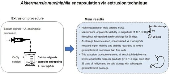
Akkermansia muciniphila Encapsulated in Calcium-Alginate Hydrogelated Matrix: Viability and Stability over Aerobic Storage and Simulated Gastrointestinal Conditions

Universidade Católica Portuguesa, CBQF—Centro de Biotecnologia e Química Fina—Laboratório Associado, Escola Superior de Biotecnologia, Rua Diogo Botelho 1327, 4169-005 Porto, Portugal
*
Author to whom correspondence should be addressed.
These authors contributed equally to this work.
Gels 2023, 9(11), 869; https://doi.org/10.3390/gels9110869
Submission received: 27 September 2023 / Revised: 23 October 2023 / Accepted: 26 October 2023 / Published: 1 November 2023
(This article belongs to the Special Issue Hydrogelated Matrices: Structural, Functional and Applicative Aspects)

Abstract

:
Akkermansia muciniphila is considered a next-generation probiotic to be incorporated in new food and pharmaceutical formulations. Effective delivery systems are required to ensure high probiotic viability and stability during product manufacture, shelf-life, and post-consumption, namely, throughout digestion. Hydrogelated matrices have demonstrated promising potential in this dominion. Hence, this work aimed to evaluate the effect of a calcium-alginate hydrogelated matrix on A. muciniphila viability during 28-days refrigerated aerobic storage and when exposed to simulated gastrointestinal conditions, in comparison with that of free cells. Akkermansia muciniphila was successfully encapsulated in the calcium-alginate matrix via extrusion (60% encapsulation yield). Furthermore, encapsulated A. muciniphila exhibited high stability (a loss in viability lower than 0.2 log-cycle) after 28-days of refrigerated aerobic storage, maintaining its viability around 108 CFU/g. Prominently, as the storage time increased, encapsulated A. muciniphila revealed higher viability and stability regarding in vitro gastrointestinal conditions than free cells. This suggests that this encapsulation method may attenuate the detrimental effects of prolonged aerobic storage with a subsequent gastrointestinal passage. In conclusion, encapsulation via extrusion using a calcium-alginate hydrogelated matrix seems to be a promising and adequate strategy for safeguarding A. muciniphila from adverse conditions encountered during refrigerated aerobic storage and when exposed to the gastrointestinal passage.

Graphical Abstract

1. Introduction

Probiotics are widely known as live microorganisms whose adequate consumption confers health-promoting effects on the host [1]. Traditionally, the microorganisms belonging to Bifidobacterium and former Lactobacillus genera (recently reclassified into 25 genera [2]) are recognized as well-studied and the most commercialized probiotics [3]. However, the growing trend and expansion of the probiotic market have led to a continuous search for the diversification of the available products. Taking this into account, several studies aiming at the selection of novel strains with different and specific functional properties have been conducted [4]. In this alignment, various bacterial species isolated from human gut microbiota, such as Akkermansia muciniphila and Faecalibacterium species, have been considered as novel probiotic candidates, also known as next-generation probiotics [3].
In the microbiology field, A. muciniphila is typically described as an oval-shaped, anaerobic, Gram-negative, and mucin-degrading bacterium that accounts for approximately 1 to 3% of the total fecal microbiota of healthy adults [5,6]. Recent evidence demonstrated that A. muciniphila plays a key role in the preservation of gut barrier integrity, modulation of the host immune response, and improvement of several metabolic pathways, making it a promising therapeutic tool in several metabolic, cardiovascular, neurological, and oncological disorders [7]. For A. muciniphila to be applied as a probiotic, effective delivery systems must be developed to guarantee the survival of this novel probiotic during manufacturing, the distribution chain, and shelf-life/storage. Additionally, these strategies must ensure protection throughout the gastrointestinal passage to guarantee that probiotic bacteria reach the intestine (target site) in adequate amounts to exert the desired health benefits [3].
Encapsulation techniques have been proposed as effective strategies to protect probiotic microorganisms against adverse conditions, as these methods may facilitate the controlled release and successful delivery of probiotics to the site of action [8]. Typically, it is defined as an entrapment process of substances (active ingredients) within another material (encapsulant). Several polymers may be used for probiotic encapsulation, including gelatin, chitosan, and alginate, the latter being one of the most commonly applied biomaterials. Alginate is a polyanionic polysaccharide, composed of (1–4)-linked β-D-mannuronate and C-5 epimer α-L-guluronate (G), which exhibits interesting properties in terms of a non-toxic nature, biocompatibility, high hydrophilicity, biodegradability, easy handling, being low in cost, a capability to form a strong gel structure through ionic crosslinking with calcium ions, and pH responsiveness (i.e., it is stable at lower pH levels and unstable at higher pH conditions which is advantageous in tailoring release profiles) [8,9]. Among the various encapsulation techniques, extrusion is recognized as the oldest and most popular methodology to encapsulate probiotics given its attractive characteristics, namely, simplicity, straightforwardness, employment of gentle conditions (without the involvement of organic/harmful solvents and extreme temperatures or pH values), low operational costs, and high probiotic viability [8]. In the extrusion process, probiotics are added and mixed into a biopolymer solution. Subsequently, the suspension is placed in an extruder (pilot scale) or a syringe needle (laboratory scale), which drips off into a hardening solution (most frequently calcium chloride) under gentle stirring [8]. Indeed, previous studies demonstrated that the entrapment of probiotic bacteria in calcium-alginate capsules offered protection against harsh conditions encountered during storage or when subjected to in vitro gastrointestinal conditions [10,11]. Based on this rationale, the present study aimed to evaluate the viability and stability of A. muciniphila DSM 22959 entrapped in calcium-alginate capsules produced via extrusion during refrigerated aerobic storage and when exposed to an in vitro gastrointestinal passage.

2. Results and Discussion

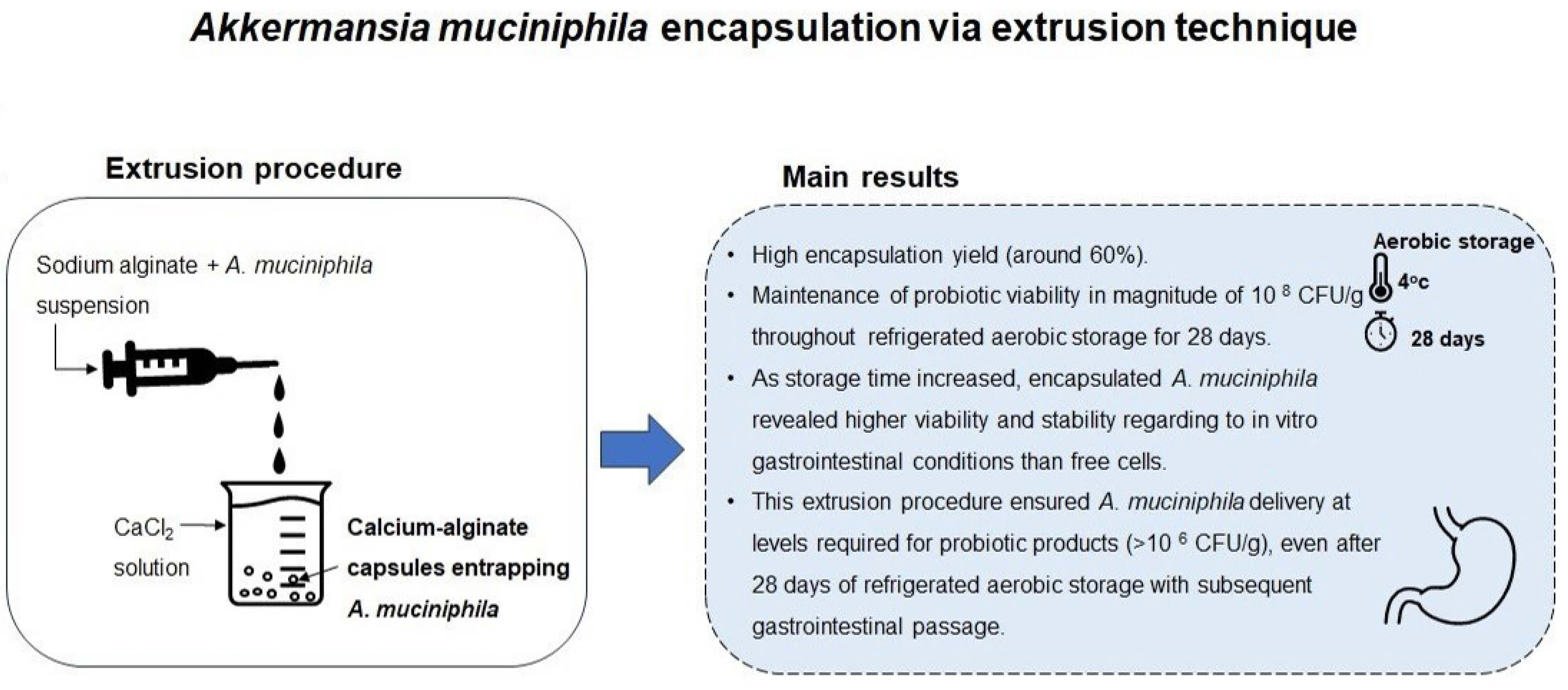
2.1. Capsules Morphology and Encapsulation Yield of Calcium-Alginate Capsules Entrapping Akkermansia muciniphila

Calcium-alginate hydrogelated capsules were selected as a strategy to study the viability and stability enhancement of A. muciniphila because, in general, these have good thermal stability and mechanical strength to withstand eventual food processing (mixing) and physiological digestion (passage through gastric conditions to finally release their content in the intestine).
Figure 1 shows the morphology of the A. muciniphila-loaded calcium-alginate hydrogelated capsules analyzed macroscopically under the naked eye (Figure 1a), under optical microscopy (Figure 1b,c), and scanning electron microscopy (Figure 1d). Morphologically, A. muciniphila capsules were presented mostly as spherical structures, with a continuous surface (Figure 1d), estimating a size of approximately 4 mm (Figure 1c).
The encapsulation yield (EY) was 60% (±18%), calculated considering the initial A. muciniphila colony-forming units (CFU) number (suspension used for extrusion; 2.10 × 10¹⁰ CFU) and the final CFU number (calcium-alginate capsules; 1.26 × 10¹⁰ CFU). This value indicates that this encapsulation technique is suitable for A. muciniphila entrapment since the order of magnitude of the cell density (1010 CFU) was maintained. After comparing the obtained EY with other studies using similar encapsulation techniques but different probiotic strains, it falls within the reported range. For example, a study conducted by Amine et al. achieved EY values ranging from 31% to 65% for encapsulating Bifidobacterium longum ATCC 15708 using an extrusion procedure with sodium alginate and/or O-palmitoylated alginate at various concentrations [12]. Additionally, Frakolaki and coworkers implemented encapsulation via extrusion for Bifidobacterium animalis subsp. lactis BB-12® using alginate alone or in combination with other encapsulating agents, obtaining EY values ranging from 58.6% to 100%, depending on the employed encapsulating matrix [13].
Notably, van der Ark et al. were the pioneers in A. muciniphila encapsulation when these researchers reported an EY of 97.5% using a water-in-oil-in-water double emulsion technique. However, the encapsulated A. muciniphila suffered sharp viability reduction after 3 days of storage at 4 °C either under aerobic or anaerobic conditions [14]. Afterward, Marcial-Coba and colleagues encapsulated A. muciniphila DSM 22959 in a xanthan and gellan gum matrix, via the extrusion method, with a subsequent freeze-drying step, reporting an encapsulation efficiency ranging from 12.1% to 76.2%, depending on the employed cryoprotective agents [15]. Recently, Almeida and collaborators immobilized A. muciniphila DSM 22959, with a 64.4% entrapment efficacy, in a dual hydrocolloid matrix of alginate and denatured whey protein isolate by the emulsification/internal gelation method [16]. While the wide variety of encapsulating materials and techniques may explain the distinct values obtained in this work and in those described in the literature, our results suggest that the extrusion technique in the calcium-alginate matrix seems to be an efficient technological strategy for the entrapment of A. muciniphila.

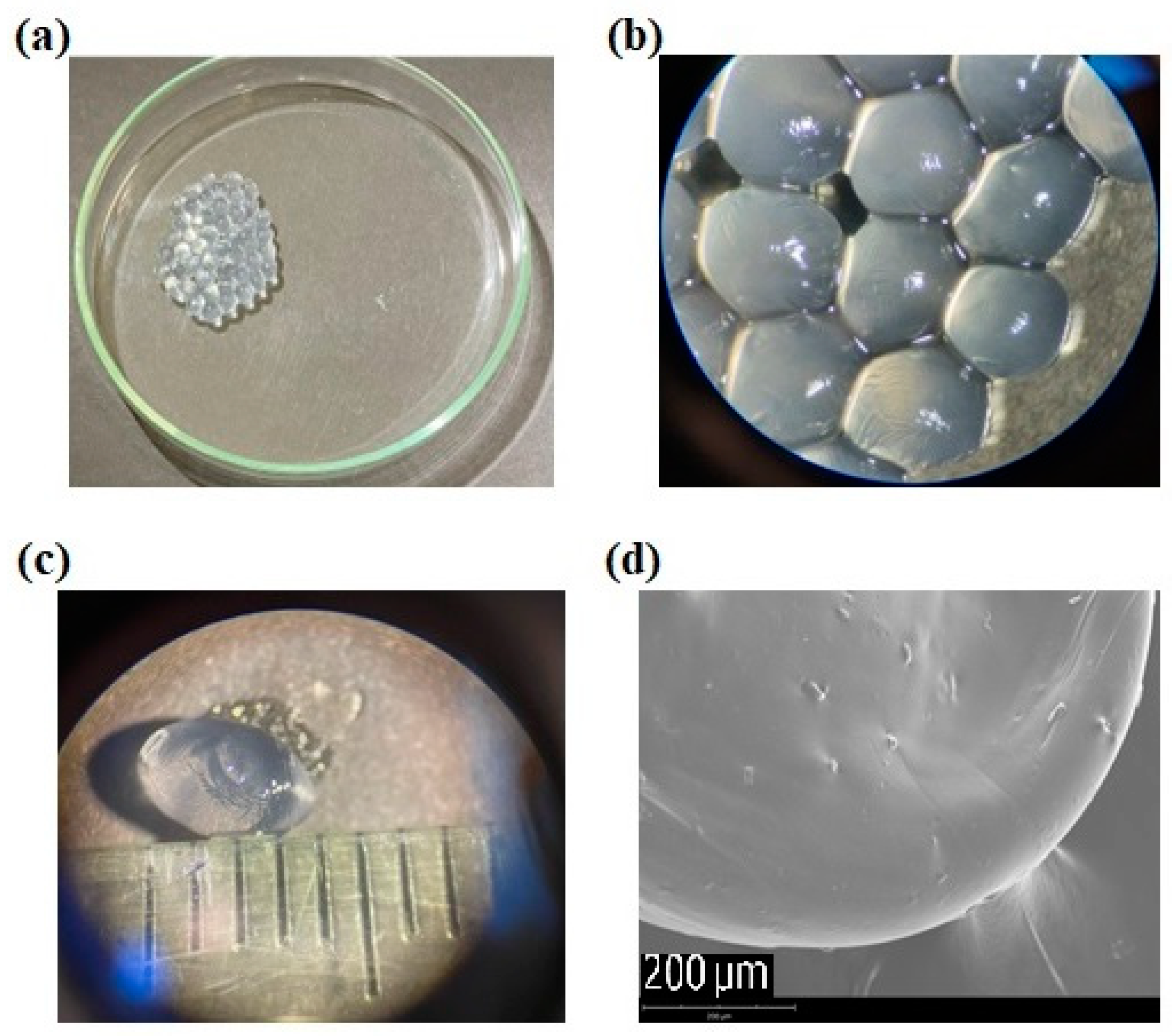
2.2. Viability of Akkermansia muciniphila in Encapsulated and Free Forms during Refrigerated Aerobic Storage

Although there is no consensus regarding the minimum effective probiotic dose, it is usually accepted that probiotic products should have a minimum concentration of 106 CFU/mL or per gram and that a total of 108–109 probiotic microorganisms should be consumed daily to elicit health benefits [17]. For this reason, one of the major aspects when developing probiotic formulations refers to the ability of these to ensure the maintenance of probiotic viability throughout the manufacturing procedure and chain distribution until it reaches the consumer [3]. Additionally, in the literature, a refrigeration temperature of 4 °C has been related with a high viability level of A. muciniphila, either in free or encapsulated forms [15,16,18]. Taking this into account, free and encapsulated A. muciniphila were stored at 4 °C under aerobic conditions and their viability was assayed, at specific timepoints, for 28 days. As it can be observed in Figure 2a,b, encapsulated A. muciniphila exhibited a high stability in viability (loss lower than 0.2 log-cycle that corresponds to a normalized survival percentage of around 65%, see Figure 2b) after 28 days of refrigerated aerobic storage, maintaining its viability within the magnitude of 108 CFU/g. In contrast, free cell numbers decreased by approximately 1 log-cycle (corresponding to a normalized survival percentage of around 15%, see Figure 2b) within the same period. Notably, the present results concerning encapsulated A. muciniphila viability and stability throughout refrigerated aerobic storage contrast positively with those reported in the literature up to the present moment. In fact, van der Ark et al. reported a sharp viability reduction in A. muciniphila encapsulated in a water-in-oil-in-water double emulsion when stored at 4 °C for 72 h in both atmospheric conditions: anaerobiosis and aerobiosis [14]. Later, Marcial-Coba and colleagues evaluated the viability of A. muciniphila encapsulated via extrusion in a xanthan and gellan gum matrix with subsequent freeze-drying, throughout 30 days, in both aerobic and anaerobic storage conditions at temperatures of 4 °C and 25 °C. These researchers reported a significant decrease (of at least 0.5 log-cycle) in the viability of the freeze-dried microencapsulated A. muciniphila after 30 days of storage under both anaerobic and aerobic conditions, at both 4 °C and 25 °C, when compared with the initial concentration [15]. Chang and colleagues encapsulated A. muciniphila in a matrix of succinate-grafted alginate doped with epigallocatechin-3-gallate via spray-drying. The authors observed a protective effect of the matrix on the bacterial viability when comparing with the free cells; however, these observations are only verified for 12 days of anaerobic and refrigerated (4 °C) storage [19]. Another investigation conducted by Barbosa and coworkers explored the spray-drying encapsulation technique with different dairy-based matrices to enhance the viability of A. muciniphila over aerobic storage. The results indicated that the viability remained around 107 CFU/g up to 28 days at 4 °C under aerobic conditions, using a 10% skim milk matrix [18]. More recently, Almeida and colleagues encapsulated A. muciniphila in a dual hydrocolloid matrix containing alginate and denatured whey protein isolate by emulsification/internal gelation and they assessed the viability of the encapsulated bacteria and free counterpart over 95 days of refrigerated storage under aerobic and anaerobic conditions. These researchers reported that during the initial 30 days of refrigerated storage in both atmospheres, there was a similar reduction in the viability of both free and encapsulated cells. However, after 95 days of storage, the viability of the encapsulated A. muciniphila experienced a sharper decrease in both anaerobic and aerobic conditions [16]. Thus, our results suggest that extrusion in the calcium-alginate matrix ensures the maintenance of the viability of encapsulated A. muciniphila cells at levels around 108 CFU/g during 28 days under feasible household storage conditions, namely aerobic storage at 4 °C. However, the size of the capsules produced via extrusion is relatively high. For instance, the capsules’ size produced herein was estimated at approximately 4 mm (Figure 1c). In the literature, the high dimension/size of the capsules obtained via extrusion is pointed to as one of the main drawbacks of this encapsulation technique [8]; nevertheless, such a drawback may be overcome by adapting their nature to the type of delivery food matrix.

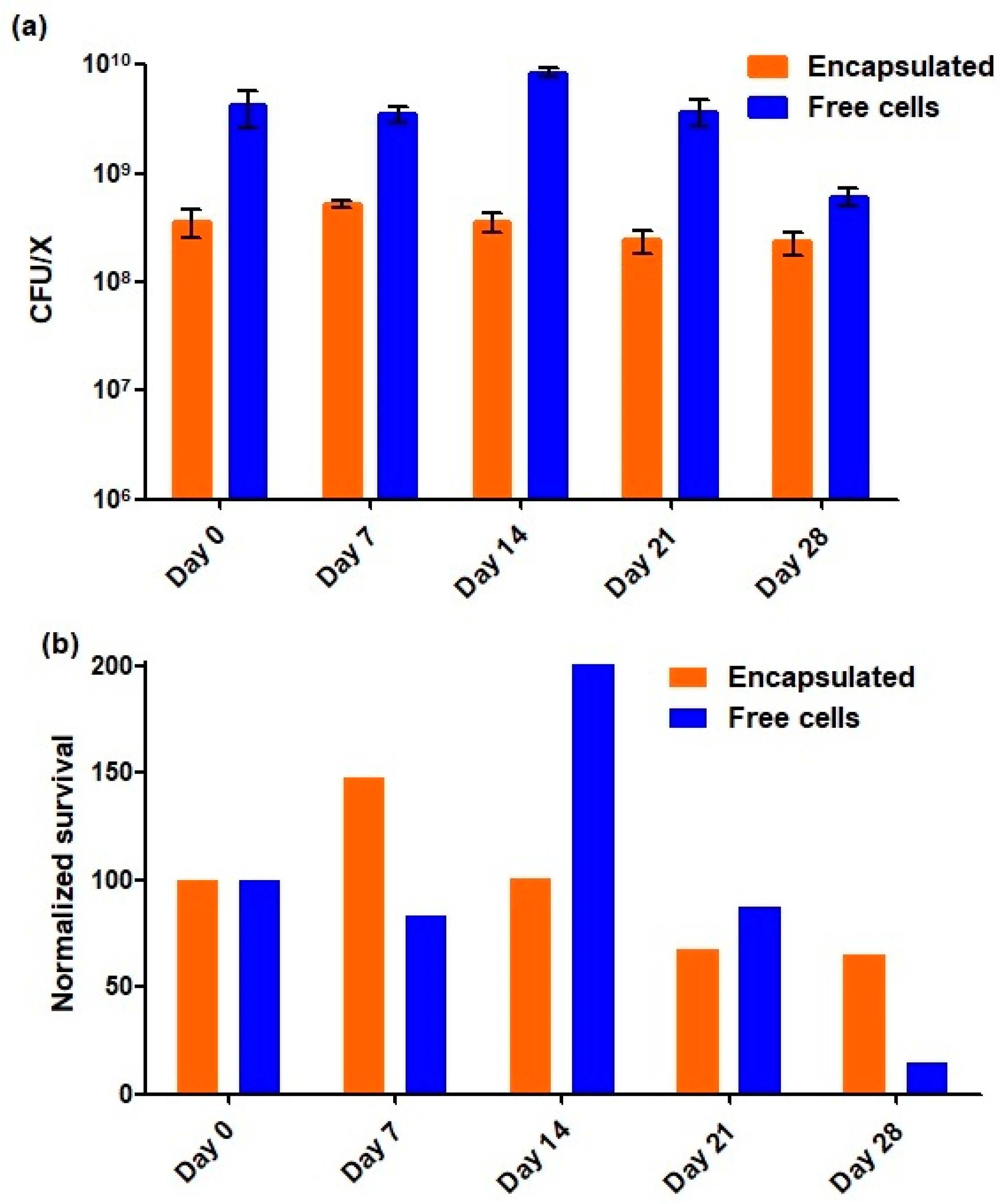
2.3. Survival of Akkermansia muciniphila in Encapsulated and Free Forms When Exposed to In Vitro Simulated Gastrointestinal Conditions

It has been postulated that to elicit its beneficial effect, a probiotic microorganism must reach the target site in adequate viable numbers [1]. Therefore, it is important to ensure that delivery systems enclosing probiotic strains are resistant to the adverse gastrointestinal conditions, thus allowing the delivery of the probiotic strain in the optimal required conditions to the target site to trigger the expected benefits. In the present study, the survival of encapsulated and free A. muciniphila cells was assessed under simulated gastrointestinal conditions in the following timepoints: 1 and 28 days of refrigerated aerobic storage.
As presented in Table 1, on day 1 of storage, both encapsulated and free A. muciniphila maintained viability throughout the in vitro gastrointestinal passage, observing the maintenance of the order of magnitude with a viability of 108 CFU/g and 109 CFU/mL, respectively. In fact, in the literature it has been reported that A. muciniphila in its free form exhibits a certain resilience when exposed to gastrointestinal conditions [16,20]. This natural resilience observed in the early stage (at day 1) may be related with the reduced activity of bile salts against Gram-negative bacteria [21] and the presence of an acid resistance system on A. muciniphila cells [22].
Regarding the 28 days of storage, when exposed to the in vitro digestion protocol, the encapsulated bacteria reduced the number of viable cells by around 1 log-cycle (achieving a viability level in 107 CFU/g magnitude), while the free cells recorded a viability reduction of higher than 2 log-cycles (viability lower than 8 × 10⁵ CFU/mL). These results showed a higher stability of encapsulated bacteria throughout the gastrointestinal passage compared to its free counterpart at the timepoint of 28 days of storage. In this alignment, a study conducted by Almeida et al. demonstrated that as the storage time increased, A. muciniphila encapsulated in an alginate of a denatured whey protein isolate matrix via emulsification/internal gelation showed higher stability when exposed to the gastrointestinal passage than its free counterpart. Specifically, these researchers recorded viability reductions in the free and encapsulated A. muciniphila of ca. 2 and lower than 1 log-cycle, respectively, at the timepoint of 30 days of refrigerated aerobic storage [16]. Still, in this context, Barbosa and coworkers showed that the encapsulation of A. muciniphila via spray-drying in 10% skim milk using inlet/outlet temperatures of 150/65 °C mitigated the detrimental effects of extended refrigerated aerobic storage for up 60 days, with the subsequent gastrointestinal passage allowing probiotic survival at levels of at least 10 7 CFU/g [18]. Thus, our results suggest that the calcium-alginate capsules produced via extrusion seem to be a promising and adequate strategy to deliver A. muciniphila in an intestinal environment at recommended viability levels for probiotic products (above 106 CFU/g). Although the parameters related to A. muciniphila release from the capsules were not assayed, a possible mechanism of releasing these probiotic cells may be hypothesized. In the literature, it is described that alginate confers gastric-acid-protective effects on encapsulated probiotics [23,24]. In fact, the alginate capsules tend to shrink at a low pH (such as in a gastric environment), which prevents the release of encapsulated probiotic cells [8,25]. Once it passes to the higher pH of the intestinal environment, the alginate becomes a soluble alginic acid layer, allowing the release of probiotic cells [8].

3. Conclusions

This study showed that this extrusion technique, using a calcium-alginate encapsulating matrix, is an interesting and adequate strategy for A. muciniphila delivery. Indeed, this encapsulation technique guaranteed the efficient production of high-loaded capsules that maintained probiotic viability in a magnitude of 108 CFU/g throughout refrigerated aerobic storage for 28 days. Additionally, as the storage time increased, encapsulated A. muciniphila revealed higher viability and stability regarding in vitro gastrointestinal conditions than free cells. This finding suggests that this encapsulation procedure may attenuate the detrimental effects of prolonged aerobic storage with a subsequent gastrointestinal passage. At the same time, this extrusion method ensured A. muciniphila delivery at levels required for probiotic products (above 106 CFU/g), even after 28 days of refrigerated aerobic storage with a subsequent in vitro gastrointestinal passage. To enable the consolidation of the potential application of these probiotic calcium-alginate capsules in suitable food and pharmaceutical products, further studies on their size and morphology are required. Also, additional work assessing the viability and stability of encapsulated A. muciniphila under longer aerobic storage periods should be performed, to simulate the storage conditions of commercial probiotic products more realistically. Likewise, the rate and time of A. muciniphila’s release from capsules should be determined to evaluate if a controlled release of A. muciniphila occurs and, consequently, its efficient delivery to the target site (intestine).

4. Materials and Methods

4.1. Bacterial Strain and Culture Conditions

Freeze-dried A. muciniphila DSM 22959 strain obtained from DSMZ collection (Leibniz Institute DSMZ—German Collection of Microorganisms and Cell Cultures, Braunschweig, Germany) was used in present study. For long-term storage, this strain was maintained frozen at −80 °C in PYG broth supplemented with 0.1% (m/v) mucin [PYGM; media composition as recommended by DSMZ [26] except that no resazurin was added], with 20% (v/v) glycerol (Fisher Scientific, Loughborough, UK). For each experiment, a glycerol stock of A. muciniphila DSM 22959 was thawed and grown in PYGM broth at 37 °C for 20–24 h under anaerobic conditions (85% N2, 5% H2, and 10% CO2) achieved in an anaerobic incubator (Whitley A35 HEPA anaerobic workstation, Bingley, UK). The bacterial cultures were then propagated, at least two subsequent culturing steps, by inoculating fresh medium (PYGM broth) at 10% (v/v) and incubating under same growth conditions. The resulting cultures were centrifuged at 12,000× g for 30 min at 4 °C (Sorvall LYNX 4000 Superspeed Centrifuge, Thermo Fisher Scientific, Waltham, MA, USA) and washed once with the same volume of physiological saline solution (NaCl at 0.85% m/v). After centrifugation in the same previous conditions, the pelleted biomass was resuspended in physiological saline solution to reach a final cell concentration around 109 CFU/mL. This resulting bacterial suspension was either used directly as free cells control or for encapsulation procedure via extrusion (5 mL).

4.2. Extrusion Procedure

Encapsulation via the extrusion technique was based on Sousa and colleagues’ protocol where optimum conditions were already adapted for different commercial probiotic strains [11]; herein, the protocol underwent some modifications. Briefly, A. muciniphila saline suspension (with a concentration of around 109 CFU/mL) was added at 10% (v/v) to 2% (m/v) sodium alginate with molecular weight ranging from 12,000–40,000 Daltons (Sigma-Aldrich, St. Louis, MO, USA). Afterwards, the alginate–culture mixtures (in a proportion of 45 mL alginate:5 mL bacterial suspension) were loaded in a 60 mL syringe (Enfa, Jiangsu Kanghua Medical Equipment Co., Ltd., Changzhou, China) coupled with a syringe needle (21 G × 1.5″, ICOplus 3, KD Medical GmbH Hospital Products, Berlin, Germany). This mixture (50 mL) was then extruded into 200 mL of 4% (m/v) CaCl2 solution and stirred at 200 rpm (Heidolph MR 3001 magnetic stirrer; Heidolph Instruments GmbH and Co. KG, Schwabach, Germany). The extrusion rate was 4.0 mL/min. The flow rate was controlled using a syringe pump (Braintree Scientific BS-300 syringe pump, Braintree Scientific, Braintree, MA, USA). Afterward, the resulting capsules were left in contact with the CaCl2 solution for 30 min at room temperature to ensure complete solidification. The CaCl2 solution was subsequently removed by decanting and the capsules were suspended in physiological saline solution. Lastly, the capsules were recovered using a fine mesh strainer and they were stored according to conditions described in Section 4.6.

4.3. Enumeration of Free and Encapsulated Akkermansia muciniphila Cells

For the enumeration of free A. muciniphila cells, decimal dilutions were performed in PBS and spotted, in triplicate, on PYGM agar plates [PYGM broth supplemented with 1.5% (m/v) agar (BIOKAR Diagnostics, Beauvais, France)]. Plates were incubated for 5–7 days at 37 °C under anaerobic conditions, and results were expressed in CFU/mL. Concerning the encapsulated bacteria, the capsules were suspended in a tri-sodium citrate dihydrate (Merck KGaA, Darmstadt, Germany) solution at 2.28% (m/v) in a 1:9 (g/mL) ratio and subjected to the mechanical action of sterile pellet pestle (Sigma-Aldrich, St. Louis, MO, USA) assisted with vortexing for 5 min to allow the complete release of A. muciniphila cells from capsules. The resulting suspension was then serially diluted, as described for the free cells, and the results were expressed as CFU/g.

4.4. Encapsulation Yield Calculation

Encapsulation yield (EY) is a combined measurement of the efficacy of entrapment and survival of viable cells during the encapsulation procedure, and it was calculated according to the formula proposed by Martin and coworkers [27]:
EY (%) = (N/N0) × 100,
where N is the number of CFU released from capsules and N0 is the number of CFU present in bacterial suspension added to the alginate solution during the encapsulation procedure.

4.5. Capsules Morphology

The appearance of A. muciniphila-loaded capsules was analyzed under optical microscopy using an Olympus Stereo Microscope SZ60 (Olympus, Tokyo, Japan). Furthermore, capsules morphology was assessed through scanning electron microscopy. Initially, alginate capsules were fixed with a 2.5% (v/v) glutaraldehyde solution for 1 h. The samples were then washed with water, and dehydrated through immersion in a graded ethanol series (30, 50, 70, 80, 90, and 100% (v/v) for a minimum of 10 min each). Afterwards, capsules were swiftly dried by dripping hexamethyldisilazane on top and immediately evaporating under a gentle stream of nitrogen. The capsules were placed on observation pins, covered with double-sided adhesive carbon tape (NEM tape; Nisshin, Tokyo, Japan), sputter-coated with gold/palladium, and visualized using a Phenom XL G2 (Thermo Fischer Scientific, Waltham, MA, USA) scanning electron microscope. The micrographs were obtained at an accelerating voltage of 15 kV, utilizing the secondary electron detector.

4.6. Viability of Free and Encapsulated Akkermansia muciniphila Cells during Refrigerated Aerobic Storage

The effect of encapsulation via extrusion on A. muciniphila viability was assayed, in comparison to free cells, throughout refrigerated aerobic storage for 28 days. Free cells were stored as follows: equal portions (1 mL) of cell suspension were stored at 4 °C under aerobic conditions into 10 sterile microtubes, corresponding to two replicas for each sampling timepoint. Concerning the encapsulated bacteria, capsules were weighed into 50 mL sterile centrifuge tubes and suspended in physiological saline solution in a 1:9 (g/mL) ratio; these preparations were performed in duplicates for each sampling timepoint, and subsequently stored at 4 °C under aerobic conditions. The viability of free and encapsulated A. muciniphila cells was evaluated on the day of encapsulation and after 7, 14, 21, and 28 days of refrigerated aerobic storage, following the protocol previously described in Section 4.3.

4.7. Survival of Free and Encapsulated Akkermansia muciniphila Cells in In Vitro Simulated Gastrointestinal Passage

The viability of free and encapsulated A. muciniphila cells when exposed to in vitro simulated gastrointestinal conditions was determined 1 and 28 days after the extrusion procedure using a standardized digestion method [28] with minor changes. Briefly, either 0.5 mL of free cells in 0.85% (m/v) NaCl or 0.5 g of calcium-alginate capsules were distributed into independent tubes (two replicates per timepoint and condition). To simulate the temperature and peristaltic movements of the human digestion, an orbital shaker incubator (Wiggen Hauser, Berlin, Germany) was used at 37 °C and 200 rpm. For each assay, all enzyme solutions were freshly prepared. For the gastric phase, samples were exposed to 2 mL of simulated gastric fluid (pH 3) containing pepsin (2000 U/mL—from porcine gastric mucosa; Sigma Aldrich, St. Louis, MO, USA) for 2 h. Afterward, intestinal conditions were simulated for 3 h at pH 7 by adding 4 mL of simulated intestinal fluid containing pancreatin (based on the trypsin activity at 100 U/mL in the final mixture; Sigma Aldrich, St. Louis, MO, USA) and bile salts (Sigma Aldrich, St. Louis, MO, USA). To evaluate the effect of gastric and intestinal conditions on A. muciniphila viability (free and encapsulated forms), samples were collected at the end of each phase (gastric and intestinal) and cell enumeration was performed according to the procedure previously described in Section 4.3. Note that in vitro digestion protocol was performed under an aerobic atmosphere, while the PYGM agar plates were incubated under anaerobic conditions.

Author Contributions

Conceptualization, D.M., J.C.B. and A.M.G.; methodology, D.M. and A.M.G.; formal analysis, D.M., M.F., R.V. and S.S.; investigation, D.M., M.F., R.V. and S.S.; writing—original draft preparation, D.M. and M.F.; writing—review and editing, D.M., M.F., R.V., J.C.B. and A.M.G.; supervision, D.M., J.C.B. and A.M.G.; project administration, D.M. and A.M.G.; funding acquisition, D.M. and A.M.G. All authors have read and agreed to the published version of the manuscript.

Funding

This work was supported by the PROBIOCARE project (EXPL/BIA-MIC/0258/2021), financed by national funds through the Foundation for Science and Technology, I.P. (FCT). We would like to thank the scientific collaboration of CBQF (UIDB/50016/2020) and the Scientific Employment Stimulus—Individual Call (CEEC Individual—CEECIND/00520/2017/CP1404/CT0001).

Institutional Review Board Statement

Not applicable.

Informed Consent Statement

Not applicable.

Data Availability Statement

The data presented in this study are available in the article.

Conflicts of Interest

The authors declare no conflict of interest.

References

  1. Hill, C.; Guarner, F.; Reid, G.; Gibson, G.R.; Merenstein, D.J.; Pot, B.; Morelli, L.; Canani, R.B.; Flint, H.J.; Salminen, S.; et al. Expert Consensus Document: The International Scientific Association for Probiotics and Prebiotics Consensus Statement on the Scope and Appropriate Use of the Term Probiotic. Nat. Rev. Gastroenterol. Hepatol. 2014, 11, 506–514. [Google Scholar] [CrossRef] [PubMed]
  2. Zheng, J.; Wittouck, S.; Salvetti, E.; Franz, C.M.A.P.; Harris, H.M.B.; Mattarelli, P.; O’toole, P.W.; Pot, B.; Vandamme, P.; Walter, J.; et al. A Taxonomic Note on the Genus Lactobacillus: Description of 23 Novel Genera, Emended Description of the Genus Lactobacillus beijerinck 1901, and Union of Lactobacillaceae and Leuconostocaceae. Int. J. Syst. Evol. Microbiol. 2020, 70, 2782–2858. [Google Scholar] [CrossRef] [PubMed]
  3. El Hage, R.; Hernandez-Sanabria, E.; Van de Wiele, T. Emerging Trends in “Smart Probiotics”: Functional Consideration for the Development of Novel Health and Industrial Applications. Front. Microbiol. 2017, 8, 1889. [Google Scholar] [CrossRef] [PubMed]
  4. de Melo Pereira, G.V.; de Oliveira Coelho, B.; Magalhães Júnior, A.I.; Thomaz-Soccol, V.; Soccol, C.R. How to Select a Probiotic? A Review and Update of Methods and Criteria. Biotechnol. Adv. 2018, 36, 2060–2076. [Google Scholar] [CrossRef]
  5. Derrien, M.; Vaughan, E.E.; Plugge, C.M.; de Vos, W.M. Akkermansia Municiphila Gen. Nov., Sp. Nov., a Human Intestinal Mucin-Degrading Bacterium. Int. J. Syst. Evol. Microbiol. 2004, 54, 1469–1476. [Google Scholar] [CrossRef] [PubMed]
  6. Derrien, M.; Collado, M.C.; Ben-Amor, K.; Salminen, S.; De Vos, W.M. The Mucin Degrader Akkermansia muciniphila Is an Abundant Resident of the Human Intestinal Tract. Appl. Environ. Microbiol. 2008, 74, 1646–1648. [Google Scholar] [CrossRef]
  7. Pellegrino, A.; Coppola, G.; Santopaolo, F.; Gasbarrini, A.; Ponziani, F.R. Role of Akkermansia in Human Diseases: From Causation to Therapeutic Properties. Nutrients 2023, 15, 1815. [Google Scholar] [CrossRef]
  8. Koh, W.Y.; Lim, X.X.; Tan, T.-C.; Kobun, R.; Rasti, B. Encapsulated Probiotics: Potential Techniques and Coating Materials for Non-Dairy Food Applications. Appl. Sci. 2022, 12, 10005. [Google Scholar] [CrossRef]
  9. Lee, K.Y.; Mooney, D.J. Alginate: Properties and Biomedical Applications. Prog. Polym. Sci. 2012, 37, 106–126. [Google Scholar] [CrossRef]
  10. Chandramouli, V.; Kailasapathy, K.; Peiris, P.; Jones, M. An Improved Method of Microencapsulation and Its Evaluation to Protect Lactobacillus Spp. in Simulated Gastric Conditions. J. Microbiol. Methods 2004, 56, 27–35. [Google Scholar] [CrossRef]
  11. Sousa, S.; Gomes, A.M.; Pintado, M.M.; Malcata, F.X.; Silva, J.P.; Sousa, J.M.; Costa, P.; Amaral, M.H.; Rodrigues, D.; Rocha-santos, T.A.P.; et al. Encapsulation of Probiotic Strains in Plain or Cysteine-Supplemented Alginate Improves Viability at Storage below Freezing Temperatures. Eng. Life Sci. 2012, 12, 457–465. [Google Scholar] [CrossRef]
  12. Amine, K.M.; Champagne, C.P.; Salmieri, S.; Britten, M.; St-Gelais, D.; Fustier, P.; Lacroix, M. Effect of Palmitoylated Alginate Microencapsulation on Viability of Bifidobacterium Longum during Freeze-Drying. LWT Food Sci. Technol. 2014, 56, 111–117. [Google Scholar] [CrossRef]
  13. Frakolaki, G.; Giannou, V.; Topakas, E.; Tzia, C. Effect of Various Encapsulating Agents on the Beads’ Morphology and the Viability of Cells during BB-12 Encapsulation through Extrusion. J. Food Eng. 2021, 294, 110423. [Google Scholar] [CrossRef]
  14. Van der Ark, K.C.H.; Nugroho, A.D.W.; Berton-Carabin, C.; Wang, C.; Belzer, C.; de Vos, W.M.; Schroen, K. Encapsulation of the Therapeutic Microbe Akkermansia muciniphila in a Double Emulsion Enhances Survival in Simulated Gastric Conditions. Food Res. Int. 2017, 102, 372–379. [Google Scholar] [CrossRef]
  15. Marcial-Coba, M.S.; Cieplak, T.; Cahú, T.B.; Blennow, A.; Knøchel, S.; Nielsen, D.S. Viability of Microencapsulated Akkermansia muciniphila and Lactobacillus plantarum during Freeze-Drying, Storage and in Vitro Simulated Upper Gastrointestinal Tract Passage. Food Funct. 2018, 9, 5868–5879. [Google Scholar] [CrossRef]
  16. Almeida, D.; Machado, D.; Sousa, S.; Seabra, C.L.; Barbosa, J.C.; Andrade, J.C.; Gomes, A.M.; Freitas, A.C. Effect of Emulsification/Internal Gelation-Based Microencapsulation on the Viability of Akkermansia muciniphila upon Prolonged Storage and Simulated Gastrointestinal Passage. Food Hydrocoll. Health 2022, 2, 100084. [Google Scholar] [CrossRef]
  17. Kechagia, M.; Basoulis, D.; Konstantopoulou, S.; Dimitriadi, D.; Gyftopoulou, K.; Skarmoutsou, N.; Fakiri, E.M. Health Benefits of Probiotics: A Review. ISRN Nutr. 2013, 2013, 481651. [Google Scholar] [CrossRef]
  18. Barbosa, J.; Almeida, D.; Machado, D.; Sousa, S.; Freitas, A.; Andrade, J.; Gomes, A. Spray-Drying Encapsulation of the Live Biotherapeutic Candidate Akkermansia muciniphila DSM 22959 to Survive Aerobic Storage. Pharmaceuticals 2022, 15, 628. [Google Scholar] [CrossRef]
  19. Chang, Y.; Yang, Y.; Xu, N.; Mu, H.; Zhang, H.; Duan, J. Improved Viability of Akkermansia muciniphila by Encapsulation in Spray Dried Succinate-Grafted Alginate Doped with Epigallocatechin-3-Gallate. Int. J. Biol. Macromol. 2020, 159, 373–382. [Google Scholar] [CrossRef]
  20. Machado, D.; Almeida, D.; Seabra, C.L.; Andrade, J.C.; Gomes, A.M.; Freitas, A.C. Uncovering Akkermansia muciniphila Resilience or Susceptibility to Different Temperatures, Atmospheres and Gastrointestinal Conditions. Anaerobe 2020, 61, 102135. [Google Scholar] [CrossRef]
  21. Begley, M.; Gahan, C.G.M.; Hill, C. The Interaction between Bacteria and Bile. FEMS Microbiol. Rev. 2005, 29, 625–651. [Google Scholar] [CrossRef] [PubMed]
  22. Ottman, N.A. Host Immunostimulation and Substrate Utilization of the Gut Symbiont Akkermansia muciniphila; Wageningen University: Wageningen, The Netherlands, 2015. [Google Scholar]
  23. Tan, L.L.; Ang, K.L.; Loo, S.C.J. Alginate Encapsulation Improves Probiotics Survival in Carbonated Sodas and Beers. PLoS ONE 2023, 18, e0283745. [Google Scholar] [CrossRef] [PubMed]
  24. Zhang, Z.; Gu, M.; You, X.; Sela, D.A.; Xiao, H.; McClements, D.J. Encapsulation of Bifidobacterium in Alginate Microgels Improves Viability and Targeted Gut Release. Food Hydrocoll. 2021, 116, 106634. [Google Scholar] [CrossRef]
  25. Chen, S.-C.; Wu, Y.-C.; Mi, F.-L.; Lin, Y.-H.; Yu, L.-C.; Sung, H.-W. A Novel PH-Sensitive Hydrogel Composed of N,O-Carboxymethyl Chitosan and Alginate Cross-Linked by Genipin for Protein Drug Delivery. J. Control. Release 2004, 96, 285–300. [Google Scholar] [CrossRef] [PubMed]
  26. DSMZ Akkermansia muciniphila DSM 22959. Available online: https://www.dsmz.de/collection/catalogue/details/culture/DSM-22959 (accessed on 22 May 2023).
  27. Martin, M.J.; Lara-Villoslada, F.; Ruiz, M.A.; Morales, M.E. Effect of Unmodified Starch on Viability of Alginate-Encapsulated Lactobacillus fermentum CECT5716. LWT Food Sci. Technol. 2013, 53, 480–486. [Google Scholar] [CrossRef]
  28. Minekus, M.; Alminger, M.; Alvito, P.; Ballance, S.; Bohn, T.; Bourlieu, C.; Carrière, F.; Boutrou, R.; Corredig, M.; Dupont, D.; et al. A Standardised Static in Vitro Digestion Method Suitable for Food—An International Consensus. Food Funct. 2014, 5, 1113–1124. [Google Scholar] [CrossRef]
Figure 1. Akkermansia muciniphila-loaded capsules observed (a) under naked eye; (b) grouped under optical microscopy (magnification 20×); (c) individualized under optical microscopy (magnification 20×); (d) under scanning electron microscopy (magnification: 700×).
Gels 09 00869 g001
Figure 2. Viability of encapsulated (orange bars) and free cells (blue bars) of A. muciniphila DSM 22959 throughout 28 days of refrigerated aerobic storage (a) expressed in CFU/X in which X correspond to grams (g) and milliliters (mL) for encapsulated and free cells, respectively, and (b) expressed in terms of normalized survival in which the values of starting point (Day 0) were normalized to 100, and the subsequent time points were represented as ratios relative to this starting point.
Gels 09 00869 g002
Table 1. Evolution of viable cell numbers of A. muciniphila DSM 22959 in encapsulated (CFU/g) and free (CFU/mL) forms during in vitro gastrointestinal passage at 1 and 28 days of refrigerated aerobic storage. Data are shown as the mean ± standard deviation.
ConditionDay 1Day 28
EncapsulatedFreeEncapsulatedFree
Initial Concentration(3.82 ± 1.26) × 108(3.40 ± 0.39) × 109(2.32 ± 0.55) × 108(6.12 ± 1.11) × 108
After gastric phase(2.74 ± 1.17) × 108(2.57 ± 0.31) × 1010(1.15 ± 0.12) × 108(7.46 ± 6.91) × 106
After intestinal phase(1.57 ± 0.33) × 108(4.83 ± 0.36) × 109(2.93 ± 1.09) × 107<LOD 1
1 LOD means limit of detection, corresponding in CFU plating technique to 8 × 10⁵ CFU/mL or /g.
Disclaimer/Publisher’s Note: The statements, opinions and data contained in all publications are solely those of the individual author(s) and contributor(s) and not of MDPI and/or the editor(s). MDPI and/or the editor(s) disclaim responsibility for any injury to people or property resulting from any ideas, methods, instructions or products referred to in the content.

