
Current Biomedical Applications of 3D-Printed Hydrogels
 ,
,
Abstract
:1. Brief History and Terminologies in 3D Printing
2. Hydrogels and 3D Printing
2.1. Emergence of Hydrogels as a 3D Printing Feedstock Material
2.2. Techniques for 3D Printing Hydrogels
2.3. Hydrogel Compositions Used in 3D Printing
2.3.1. Nature-Derived Polymers
2.3.2. Synthetic Polymers
2.3.3. Inorganic Materials
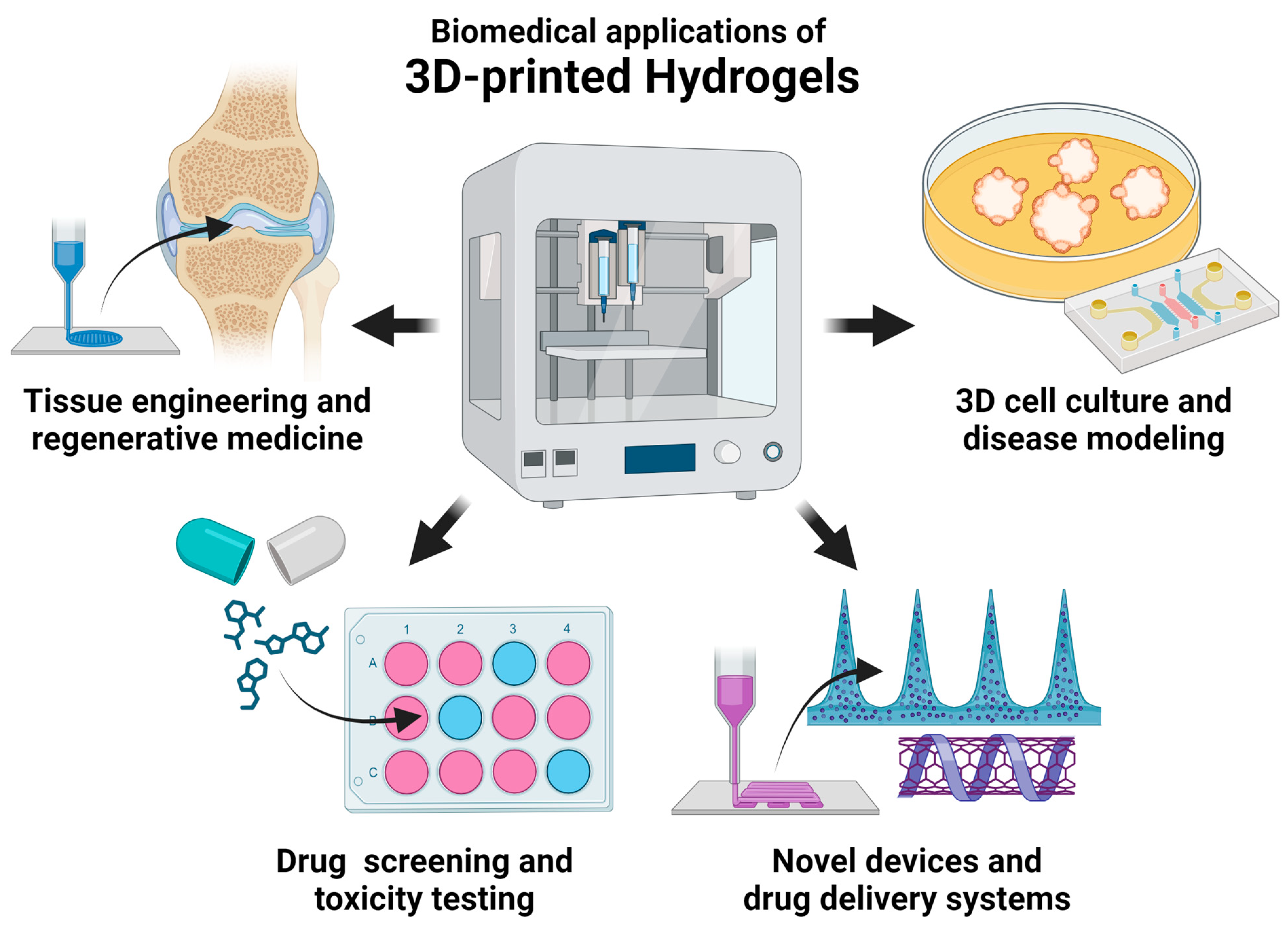
3. Current Biomedical Applications
3.1. Tissue Engineering and Regenerative Medicine
3.1.1. Bone Tissue Engineering
3.1.2. Cardiovascular Tissue Engineering
3.1.3. Cartilage Tissue Engineering
3.1.4. Genitourinary Tissue Engineering
3.1.5. Hepatic and Pancreatic Tissue Engineering
3.1.6. Muscle and Tendon Tissue Engineering
3.1.7. Nervous Tissue Engineering
3.1.8. Respiratory Tissue Engineering
3.1.9. Skin Tissue Engineering and Wound Healing
3.2. Three-dimensional Cell Culture and Disease Modeling
3.3. Drug Screening and Toxicity Testing
3.4. Novel Devices and Drug Delivery Systems
4. Current Challenges and Future Directions
4.1. Improving Resolution and Structural Complexity
4.2. Optimizing Cell Viability and Function
4.3. Improving Cost Efficiency and Accessibility
4.4. Addressing Ethical and Regulatory Concerns toward Clinical Translation
Author Contributions
Funding
Institutional Review Board Statement
Informed Consent Statement
Data Availability Statement
Conflicts of Interest
References
- ISO/ASTM52900-21; Additive Manufacturing—General Principles—Fundamentals and Vocabulary. International Organization for Standardization: Geneva, Switzerland; American Society for Testing and Materials: West Conshohocken, PA, USA, 2022.
- Narayan, R. Additive Manufacturing and 3D Printing; Springer International Publishing AG: Cham, Switzerland, 2020; pp. 621–652. [Google Scholar]
- Hull, C.W. Apparatus for Production of Three-Dimensional Objects by Stereolithography; 3D Systems Inc.: Valencia, CA, USA, 1986. [Google Scholar]
- Deckard, C.R. Method and Apparatus for Producing Parts by Selective Sintering; University of Texas System: Austin, TX, USA, 1989. [Google Scholar]
- Crump, S.S. Apparatus and Method for Creating Three-Dimensional Objects; Stratasys Inc.: Valencia, CA, USA, 1992. [Google Scholar]
- Sachs, E.M.; Haggerty, J.S.; Cima, M.J.; Williams, P.A. Three-Dimensional Printing Techniques; Massachusetts Institute of Technology: Cambridge, MA, USA, 1993. [Google Scholar]
- Alexander, A.E.; Wake, N.; Chepelev, L.; Brantner, P.; Ryan, J.; Wang, K.C. A guideline for 3D printing terminology in biomedical research utilizing ISO/ASTM standards. 3D Print. Med. 2021, 7, 8. [Google Scholar] [CrossRef] [PubMed]
- Wang, K.C. Standard Lexicons, Coding Systems and Ontologies for Interoperability and Semantic Computation in Imaging. J. Digit. Imaging 2018, 31, 353–360. [Google Scholar] [CrossRef] [PubMed]
- Kaliaraj, G.S.; Shanmugam, D.K.; Dasan, A.; Mosas, K.K.A. Hydrogels—A Promising Materials for 3D Printing Technology. Gels 2023, 9, 260. [Google Scholar] [CrossRef] [PubMed]
- Thomas, S. Sustainable Hydrogels: Synthesis, Properties, and Applications; Elsevier: Amsterdam, The Netherlands, 2023. [Google Scholar]
- Wichterle, O.; LÍM, D. Hydrophilic Gels for Biological Use. Nature 1960, 185, 117–118. [Google Scholar] [CrossRef]
- Zare, M.; Bigham, A.; Zare, M.; Luo, H.; Rezvani Ghomi, E.; Ramakrishna, S. pHEMA: An Overview for Biomedical Applications. Int. J. Mol. Sci. 2021, 22, 6376. [Google Scholar] [CrossRef] [PubMed]
- Ahmed, E.M. Hydrogel: Preparation, characterization, and applications: A review. J. Adv. Res. 2015, 6, 105–121. [Google Scholar] [CrossRef]
- Li, J.; Wu, C.; Chu, P.K.; Gelinsky, M. 3D printing of hydrogels: Rational design strategies and emerging biomedical applications. Mater. Sci. Eng. R Rep. 2020, 140, 100543. [Google Scholar] [CrossRef]
- Nele, V.; Wojciechowski, J.P.; Armstrong, J.P.K.; Stevens, M.M. Tailoring Gelation Mechanisms for Advanced Hydrogel Applications. Adv. Funct. Mater. 2020, 30, 2002759. [Google Scholar] [CrossRef]
- Morozova, S.M.; Gevorkian, A.; Kumacheva, E. Design, characterization and applications of nanocolloidal hydrogels. Chem. Soc. Rev. 2023, 52, 5317–5339. [Google Scholar] [CrossRef]
- Advincula, R.C.; Dizon, J.R.C.; Caldona, E.B.; Viers, R.A.; Siacor, F.D.C.; Maalihan, R.D.; Espera, A.H. On the progress of 3D-printed hydrogels for tissue engineering. MRS Commun. 2021, 11, 539–553. [Google Scholar] [CrossRef]
- Xu, C.; Dai, G.; Hong, Y. Recent advances in high-strength and elastic hydrogels for 3D printing in biomedical applications. Acta Biomater. 2019, 95, 50–59. [Google Scholar] [CrossRef] [PubMed]
- Pradeep, P.V.; Paul, L. Review on novel biomaterials and innovative 3D printing techniques in biomedical applications. Mater. Today Proc. 2022, 58, 96–103. [Google Scholar] [CrossRef]
- Yang, X.; Li, X.; Wu, Z.; Cao, L. Photocrosslinked methacrylated natural macromolecular hydrogels for tissue engineering: A review. Int. J. Biol. Macromol. 2023, 246, 125570. [Google Scholar] [CrossRef] [PubMed]
- Hinchliffe, J.D.; Parassini Madappura, A.; Syed Mohamed, S.M.D.; Roy, I. Biomedical Applications of Bacteria-Derived Polymers. Polymers 2021, 13, 1081. [Google Scholar] [CrossRef] [PubMed]
- Barja, F. Bacterial nanocellulose production and biomedical applications. J. Biomed. Res. 2021, 35, 310–317. [Google Scholar] [CrossRef] [PubMed]
- Li, M.; Sun, L.; Liu, Z.; Shen, Z.; Cao, Y.; Han, L.; Sang, S.; Wang, J. 3D bioprinting of heterogeneous tissue-engineered skin containing human dermal fibroblasts and keratinocytes. Biomater. Sci. 2023, 11, 2461–2477. [Google Scholar] [CrossRef]
- Tao, J.; Zhu, S.; Liao, X.; Wang, Y.; Zhou, N.; Li, Z.; Wan, H.; Tang, Y.; Yang, S.; Du, T.; et al. DLP-based bioprinting of void-forming hydrogels for enhanced stem-cell-mediated bone regeneration. Mater. Today Bio 2022, 17, 100487. [Google Scholar] [CrossRef]
- Ouyang, L.; Armstrong, J.P.K.; Lin, Y.; Wojciechowski, J.P.; Lee-Reeves, C.; Hachim, D.; Zhou, K.; Burdick, J.A.; Stevens, M.M. Expanding and optimizing 3D bioprinting capabilities using complementary network bioinks. Sci. Adv. 2020, 6, abc5529. [Google Scholar] [CrossRef]
- Li, Z.; Li, S.; Yang, J.; Ha, Y.; Zhang, Q.; Zhou, X.; He, C. 3D bioprinted gelatin/gellan gum-based scaffold with double-crosslinking network for vascularized bone regeneration. Carbohydr. Polym. 2022, 290, 119469. [Google Scholar] [CrossRef]
- Bordoni, M.; Karabulut, E.; Kuzmenko, V.; Fantini, V.; Pansarasa, O.; Cereda, C.; Gatenholm, P. 3D Printed Conductive Nanocellulose Scaffolds for the Differentiation of Human Neuroblastoma Cells. Cells 2020, 9, 682. [Google Scholar] [CrossRef]
- Zhuang, P.; Greenberg, Z.; He, M. Biologically Enhanced Starch Bio-Ink for Promoting 3D Cell Growth. Adv. Mater. Technol. 2021, 6, 2100551. [Google Scholar] [CrossRef] [PubMed]
- Liu, J.; Tian, B.; Liu, Y.; Wan, J.B. Cyclodextrin-Containing Hydrogels: A Review of Preparation Method, Drug Delivery, and Degradation Behavior. Int. J. Mol. Sci. 2021, 22, 13516. [Google Scholar] [CrossRef] [PubMed]
- Kabir, S.M.F.; Sikdar, P.P.; Haque, B.; Bhuiyan, M.A.R.; Ali, A.; Islam, M.N. Cellulose-based hydrogel materials: Chemistry, properties and their prospective applications. Prog. Biomater. 2018, 7, 153–174. [Google Scholar] [CrossRef]
- Krakos, A.; Cieślak, A.; Hartel, E.; Łabowska, M.B.; Kulbacka, J.; Detyna, J. 3D bio-printed hydrogel inks promoting lung cancer cell growth in a lab-on-chip culturing platform. Mikrochim. Acta 2023, 190, 349. [Google Scholar] [CrossRef] [PubMed]
- Qu, G.; Huang, J.; Li, Z.; Jiang, Y.; Liu, Y.; Chen, K.; Xu, Z.; Zhao, Y.; Gu, G.; Wu, X.; et al. 4D-printed bilayer hydrogel with adjustable bending degree for enteroatmospheric fistula closure. Mater. Today. Bio 2022, 16, 100363. [Google Scholar] [CrossRef] [PubMed]
- Liu, X.; Song, S.; Chen, Z.; Gao, C.; Li, Y.; Luo, Y.; Huang, J.; Zhang, Z. Release of O-GlcNAc transferase inhibitor promotes neuronal differentiation of neural stem cells in 3D bioprinted supramolecular hydrogel scaffold for spinal cord injury repair. Acta Biomater. 2022, 151, 148–162. [Google Scholar] [CrossRef] [PubMed]
- Zhao, X.; Lu, X.; Li, K.; Song, S.; Luo, Z.; Zheng, C.; Yang, C.; Wang, X.; Wang, L.; Tang, Y.; et al. Double crosslinked biomimetic composite hydrogels containing topographical cues and WAY-316606 induce neural tissue regeneration and functional recovery after spinal cord injury. Bioact. Mater. 2022, 24, 331–345. [Google Scholar] [CrossRef]
- Shariatinia, Z.; Jalali, A.M. Chitosan-based hydrogels: Preparation, properties and applications. Int. J. Biol. Macromol. 2018, 115, 194–220. [Google Scholar] [CrossRef]
- Teoh, J.H.; Mozhi, A.; Sunil, V.; Tay, S.M.; Fuh, J.; Wang, C.H. 3D Printing Personalized, Photocrosslinkable Hydrogel Wound Dressings for the Treatment of Thermal Burns. Adv. Funct. Mater. 2021, 31, 2105932. [Google Scholar] [CrossRef]
- Loi, G.; Stucchi, G.; Scocozza, F.; Cansolino, L.; Cadamuro, F.; Delgrosso, E.; Riva, F.; Ferrari, C.; Russo, L.; Conti, M. Characterization of a Bioink Combining Extracellular Matrix-like Hydrogel with Osteosarcoma Cells: Preliminary Results. Gels 2023, 9, 129. [Google Scholar] [CrossRef]
- Abasalizadeh, F.; Moghaddam, S.V.; Alizadeh, E.; Akbari, E.; Kashani, E.; Fazljou, S.M.B.; Torbati, M.; Akbarzadeh, A. Alginate-based hydrogels as drug delivery vehicles in cancer treatment and their applications in wound dressing and 3D bioprinting. J. Biol. Eng. 2020, 14, 8. [Google Scholar] [CrossRef]
- Zucca, P.; Fernandez-Lafuente, R.; Sanjust, E. Agarose and Its Derivatives as Supports for Enzyme Immobilization. Molecules 2016, 21, 1577. [Google Scholar] [CrossRef] [PubMed]
- Lopez, J.; Ruiz-Toranzo, M.; Antich, C.; Chocarro-Wrona, C.; López-Ruiz, E.; Jiménez, G.; Marchal, J.A. Biofabrication of a Tri-layered 3D-Bioprinted CSC-based Malignant Melanoma Model for Personalized Cancer Treatment. Biofabrication 2022, 15, 035016. [Google Scholar] [CrossRef]
- Amaral, A.J.R.; Gaspar, V.M.; Lavrador, P.; Mano, J.F. Double network laminarin-boronic/alginate dynamic bioink for 3D bioprinting cell-laden constructs. Biofabrication 2021, 13, 035045. [Google Scholar] [CrossRef] [PubMed]
- Antoine, E.E.; Vlachos, P.P.; Rylander, M.N. Review of collagen I hydrogels for bioengineered tissue microenvironments: Characterization of mechanics, structure, and transport. Tissue Eng. Part B Rev. 2014, 20, 683–696. [Google Scholar] [CrossRef] [PubMed]
- Tong, L.; Pu, X.; Liu, Q.; Li, X.; Chen, M.; Wang, P.; Zou, Y.; Lu, G.; Liang, J.; Fan, Y.; et al. Nanostructured 3D-Printed Hybrid Scaffold Accelerates Bone Regeneration by Photointegrating Nanohydroxyapatite. Adv. Sci. 2023, 10, 2300038. [Google Scholar] [CrossRef] [PubMed]
- Levin, A.A.; Karalkin, P.A.; Koudan, E.V.; Senatov, F.S.; Parfenov, V.A.; Lvov, V.A.; Petrov, S.V.; Pereira, F.D.A.S.; Kovalev, A.V.; Osidak, E.O.; et al. Commercial articulated collaborative in situ 3D bioprinter for skin wound healing. Int. J. Bioprint. 2023, 9, 380–393. [Google Scholar] [CrossRef]
- Shi, W.; Mirza, S.; Kuss, M.; Liu, B.; Hartin, A.; Wan, S.; Kong, Y.; Mohapatra, B.; Krishnan, M.; Band, H.; et al. Embedded Bioprinting of Breast Tumor Cells and Organoids Using Low-Concentration Collagen-Based Bioinks. Adv. Healthc. Mater. 2023, 12, 2300905. [Google Scholar] [CrossRef]
- Clark, C.C.; Yoo, K.M.; Sivakumar, H.; Strumpf, K.; Laxton, A.W.; Tatter, S.B.; Strowd, R.E.; Skardal, A. Immersion bioprinting of hyaluronan and collagen bioink-supported 3D patient-derived brain tumor organoids. Biomed. Mater. 2022, 18, 015014. [Google Scholar] [CrossRef]
- Maloney, E.; Clark, C.; Sivakumar, H.; Yoo, K.; Aleman, J.; Rajan, S.A.P.; Forsythe, S.; Mazzocchi, A.; Laxton, A.W.; Tatter, S.B.; et al. Immersion Bioprinting of Tumor Organoids in Multi-Well Plates for Increasing Chemotherapy Screening Throughput. Micromachines 2020, 11, 208. [Google Scholar] [CrossRef]
- Berg, J.; Weber, Z.; Fechler-Bitteti, M.; Hocke, A.C.; Hippenstiel, S.; Elomaa, L.; Weinhart, M.; Kurreck, J. Bioprinted multi-cell type lung model for the study of viral inhibitors. Viruses 2021, 13, 1590. [Google Scholar] [CrossRef] [PubMed]
- Salahuddin, B.; Wang, S.; Sangian, D.; Aziz, S.; Gu, Q. Hybrid Gelatin Hydrogels in Nanomedicine Applications. ACS Appl. Bio Mater. 2021, 4, 2886–2906. [Google Scholar] [CrossRef] [PubMed]
- Sbrana, F.V.; Pinos, R.; Barbaglio, F.; Ribezzi, D.; Scagnoli, F.; Scarfò, L.; Redwan, I.N.; Martinez, H.; Farè, S.; Ghia, P.; et al. 3D Bioprinting Allows the Establishment of Long-Term 3D Culture Model for Chronic Lymphocytic Leukemia Cells. Front. Immunol. 2021, 12, 639572. [Google Scholar] [CrossRef] [PubMed]
- Nulty, J.; Freeman, F.E.; Browe, D.C.; Burdis, R.; Ahern, D.P.; Pitacco, P.; Lee, Y.B.; Alsberg, E.; Kelly, D.J. 3D bioprinting of prevascularised implants for the repair of critically-sized bone defects. Acta Biomater. 2021, 126, 154–169. [Google Scholar] [CrossRef] [PubMed]
- Sun, Y.; Wu, Q.; Zhang, Y.; Dai, K.; Wei, Y. 3D-bioprinted gradient-structured scaffold generates anisotropic cartilage with vascularization by pore-size-dependent activation of HIF1α/FAK signaling axis. Nanomed. Nanotechnol. Biol. Med. 2021, 37, 102426. [Google Scholar] [CrossRef] [PubMed]
- Li, T.; Hou, J.; Wang, L.; Zeng, G.; Wang, Z.; Yu, L.; Yang, Q.; Yin, J.; Long, M.; Chen, L.; et al. Bioprinted anisotropic scaffolds with fast stress relaxation bioink for engineering 3D skeletal muscle and repairing volumetric muscle loss. Acta Biomater. 2023, 156, 21–36. [Google Scholar] [CrossRef]
- Zhang, G.; Wang, Z.; Han, F.; Jin, G.; Xu, L.; Xu, H.; Su, H.; Wang, H.; Le, Y.; Fu, Y.; et al. Mechano-regulation of vascular network formation without branches in 3D bioprinted cell-laden hydrogel constructs. Biotechnol. Bioeng. 2021, 118, 3787–3798. [Google Scholar] [CrossRef]
- Neves, M.I.; Araújo, M.; Moroni, L.; da Silva, R.M.P.; Barrias, C.C. Glycosaminoglycan-Inspired Biomaterials for the Development of Bioactive Hydrogel Networks. Molecules 2020, 25, 978. [Google Scholar] [CrossRef]
- Zhang, W.; Du, A.; Liu, S.; Lv, M.; Chen, S. Research progress in decellularized extracellular matrix-derived hydrogels. Regen. Ther. 2021, 18, 88–96. [Google Scholar] [CrossRef]
- Meng, X.; Zhou, Z.; Chen, X.; Ren, F.; Zhu, W.; Zhu, S.; Liu, H. A sturgeon cartilage extracellular matrix-derived bioactive bioink for tissue engineering applications. Int. J. Bioprint. 2023, 9, 768. [Google Scholar] [CrossRef]
- Xie, X.; Wu, S.; Mou, S.; Guo, N.; Wang, Z.; Sun, J. Microtissue-Based Bioink as a Chondrocyte Microshelter for DLP Bioprinting. Adv. Healthc. Mater. 2022, 11, 2201877. [Google Scholar] [CrossRef] [PubMed]
- Wu, J.; Fu, L.; Yan, Z.; Yang, Y.; Yin, H.; Li, P.; Yuan, X.; Ding, Z.; Kang, T.; Tian, Z.; et al. Hierarchical porous ECM scaffolds incorporating GDF-5 fabricated by cryogenic 3D printing to promote articular cartilage regeneration. Biomater. Res. 2023, 27, 7. [Google Scholar] [CrossRef] [PubMed]
- Li, C.; Zhang, W.; Nie, Y.; Jiang, D.; Jia, J.; Zhang, W.; Li, L.; Yao, Z.; Qin, L.; Lai, Y. Integrated and Bifunctional Bilayer 3D Printing Scaffold for Osteochondral Defect Repair. Adv. Funct. Mater. 2023, 33, 2214158. [Google Scholar] [CrossRef]
- Zheng, J.; Liu, Y.; Hou, C.; Li, Z.; Yang, S.; Liang, X.; Zhou, L.; Guo, J.; Zhang, J.; Huang, X. Ovary-derived Decellularized Extracellular Matrix-based Bioink for Fabricating 3D Primary Ovarian Cells-laden Structures for Mouse Ovarian Failure Correction. Int. J. Bioprint. 2022, 8, 269–282. [Google Scholar] [CrossRef] [PubMed]
- Bashiri, Z.; Amiri, I.; Gholipourmalekabadi, M.; Falak, R.; Asgari, H.; Maki, C.B.; Moghaddaszadeh, A.; Koruji, M. Artificial testis: A testicular tissue extracellular matrix as a potential bio-ink for 3D printing. Biomater. Sci. 2021, 9, 3465–3484. [Google Scholar] [CrossRef] [PubMed]
- Wang, D.; Guo, Y.; Zhu, J.; Liu, F.; Xue, Y.; Huang, Y.; Zhu, B.; Wu, D.; Pan, H.; Gong, T.; et al. Hyaluronic acid methacrylate/pancreatic extracellular matrix as a potential 3D printing bioink for constructing islet organoids. Acta Biomater. 2023, 165, 86–101. [Google Scholar] [CrossRef]
- Chen, S.; Luo, J.; Shen, L.; Liu, X.; Wang, W.; Xu, J.; Ren, Y.; Ye, Y.; Shi, G.; Cheng, F.; et al. 3D Printing Mini-Capsule Device for Islet Delivery to Treat Type 1 Diabetes. ACS Appl. Mater. Interfaces 2022, 14, 23139–23151. [Google Scholar] [CrossRef]
- Jin, R.; Cui, Y.; Chen, H.; Zhang, Z.; Weng, T.; Xia, S.; Yu, M.; Zhang, W.; Shao, J.; Yang, M.; et al. Three-dimensional bioprinting of a full-thickness functional skin model using acellular dermal matrix and gelatin methacrylamide bioink. Acta Biomater. 2021, 131, 248–261. [Google Scholar] [CrossRef]
- Fu, H.; Zhang, D.; Zeng, J.; Fu, Q.; Chen, Z.; Sun, X.; Yang, Y.; Li, S.; Chen, M. Application of 3D-printed tissue-engineered skin substitute using innovative biomaterial loaded with human adipose-derived stem cells in wound healing. Int. J. Bioprint. 2023, 9, 394–406. [Google Scholar] [CrossRef]
- Hu, Y.; Wu, B.; Xiong, Y.; Tao, R.; Panayi, A.C.; Chen, L.; Tian, W.; Xue, H.; Shi, L.; Zhang, X.; et al. Cryogenic 3D printed hydrogel scaffolds loading exosomes accelerate diabetic wound healing. Chem. Eng. J. 2021, 426, 130634. [Google Scholar] [CrossRef]
- Yan, J.; Liu, X.; Liu, J.; Zhang, X.; Zheng, Q.; Nan, J.; Lin, M.; Pan, H.; Wang, Y.; Cai, X.; et al. A dual-layer cell-laden tubular scaffold for bile duct regeneration. Mater. Des. 2021, 212, 110229. [Google Scholar] [CrossRef]
- Li, L.; Shi, J.; Ma, K.; Jin, J.; Wang, P.; Liang, H.; Cao, Y.; Wang, X.; Jiang, Q. Robotic in situ 3D bio-printing technology for repairing large segmental bone defects. J. Adv. Res. 2021, 30, 75–84. [Google Scholar] [CrossRef] [PubMed]
- Ma, K.; Zhao, T.; Yang, L.; Wang, P.; Jin, J.; Teng, H.; Xia, D.; Zhu, L.; Li, L.; Jiang, Q.; et al. Application of robotic-assisted in situ 3D printing in cartilage regeneration with HAMA hydrogel: An in vivo study. J. Adv. Res. 2020, 23, 123–132. [Google Scholar] [CrossRef] [PubMed]
- Yu, Y.; Fischenich, K.M.; Schoonraad, S.A.; Weatherford, S.; Uzcategui, A.C.; Eckstein, K.; Muralidharan, A.; Crespo-Cuevas, V.; Rodriguez-Fontan, F.; Killgore, J.P.; et al. A 3D printed mimetic composite for the treatment of growth plate injuries in a rabbit model. Npj Regen. Med. 2022, 7, 60. [Google Scholar] [CrossRef] [PubMed]
- Tao, J.; Liu, H.; Wu, W.; Zhang, J.; Liu, S.; Zhang, J.; Huang, Y.; Xu, X.; He, H.; Yang, S.; et al. 3D-Printed Nerve Conduits with Live Platelets for Effective Peripheral Nerve Repair. Adv. Funct. Mater. 2020, 30, 2004272. [Google Scholar] [CrossRef]
- Song, S.; Zhou, J.; Wan, J.; Zhao, X.; Li, K.; Yang, C.; Zheng, C.; Wang, L.; Tang, Y.; Wang, C.; et al. Three-dimensional printing of microfiberreinforced hydrogel loaded with oxymatrine for treating spinal cord injury. Int. J. Bioprint. 2023, 9, 692. [Google Scholar] [CrossRef] [PubMed]
- Song, S.; Li, Y.; Huang, J.; Cheng, S.; Zhang, Z. Inhibited astrocytic differentiation in neural stem cell-laden 3D bioprinted conductive composite hydrogel scaffolds for repair of spinal cord injury. Biomater. Adv. 2023, 148, 213385. [Google Scholar] [CrossRef] [PubMed]
- Galván, N.T.N.; Paulsen, S.J.; Kinstlinger, I.S.; Marini, J.C.; Didelija, I.C.; Yoeli, D.; Grigoryan, B.; Miller, J.S. Blood Flow Within Bioengineered 3D Printed Vascular Constructs Using the Porcine Model. Front. Cardiovasc. Med. 2021, 8, 629313. [Google Scholar] [CrossRef]
- Sang, S.; Wang, X.; Duan, J.; Cao, Y.; Shen, Z.; Sun, L.; Duan, Q.; Liu, Z. 3D printing to construct in vitro multicellular models of melanoma. Biotechnol. Bioeng. 2023, 120, 2853–2864. [Google Scholar] [CrossRef]
- Utama, R.H.; Tan, V.T.G.; Tjandra, K.C.; Sexton, A.; Nguyen, D.H.T.; O’Mahony, A.P.; Du, E.Y.; Tian, P.; Ribeiro, J.C.C.; Kavallaris, M.; et al. A Covalently Crosslinked Ink for Multimaterials Drop-on-Demand 3D Bioprinting of 3D Cell Cultures. Macromol. Biosci. 2021, 21, 2100125. [Google Scholar] [CrossRef]
- Li, Y.; Dong, D.; Qu, Y.; Li, J.; Chen, S.; Zhao, H.; Zhang, Q.; Jiao, Y.; Fan, L.; Sun, D. A Multidrug Delivery Microrobot for the Synergistic Treatment of Cancer. Small 2023, 19, 2301889. [Google Scholar] [CrossRef] [PubMed]
- Huang, D.; Cheng, Y.; Chen, G.; Zhao, Y. 3D-Printed Janus Piezoelectric Patches for Sonodynamic Bacteria Elimination and Wound Healing. Research 2023, 6, 0022. [Google Scholar] [CrossRef] [PubMed]
- El-Habashy, S.E.; El-Kamel, A.H.; Essawy, M.M.; Abdelfattah, E.Z.A.; Eltaher, H.M. Engineering 3D-printed core–shell hydrogel scaffolds reinforced with hybrid hydroxyapatite/polycaprolactone nanoparticles for in vivo bone regeneration. Biomater. Sci. 2021, 9, 4019–4039. [Google Scholar] [CrossRef] [PubMed]
- Shi, W.; Fang, F.; Kong, Y.; Greer, S.E.; Kuss, M.; Liu, B.; Xue, W.; Jiang, X.; Lovell, P.; Mohs, A.M.; et al. Dynamic hyaluronic acid hydrogel with covalent linked gelatin as an anti-oxidative bioink for cartilage tissue engineering. Biofabrication 2021, 14, 014107. [Google Scholar] [CrossRef] [PubMed]
- Hori, T.; Ito, Y.; Raut, B.; Ostrovidov, S.; Nashimoto, Y.; Nagai, N.; Abe, T.; Kaji, H. Three-dimensional-printed Refillable Drug Delivery Device for Long-term Sustained Drug Delivery to Retina. Sens. Mater. 2023, 35, 1301–1313. [Google Scholar] [CrossRef]
- Dong, C.; Wei, H.; Zhang, X.; Li, Y.; Huang, L.; Wa, Q.; Luo, Y. 3D printed hydrogel/wesselsite-PCL composite scaffold with structural change from core/shell fibers to microchannels for enhanced bone regeneration. Compos. Part B Eng. 2022, 246, 110264. [Google Scholar] [CrossRef]
- Huan, Y.; Zhou, D.; Wu, X.; He, X.; Chen, H.; Li, S.; Jia, B.; Dou, Y.; Fei, X.; Wu, S.; et al. 3D bioprinted autologous bone particle scaffolds for cranioplasty promote bone regeneration with both implanted and native BMSCs. Biofabrication 2023, 15, 025016. [Google Scholar] [CrossRef]
- You, D.; Chen, G.; Liu, C.; Ye, X.; Wang, S.; Dong, M.; Sun, M.; He, J.; Yu, X.; Ye, G.; et al. 4D Printing of Multi-Responsive Membrane for Accelerated In Vivo Bone Healing Via Remote Regulation of Stem Cell Fate. Adv. Funct. Mater. 2021, 31, 2103920. [Google Scholar] [CrossRef]
- Cao, Y.; Cheng, P.; Sang, S.; Xiang, C.; An, Y.; Wei, X.; Yan, Y.; Li, P. 3D printed PCL/GelMA biphasic scaffold boosts cartilage regeneration using co-culture of mesenchymal stem cells and chondrocytes: In vivo study. Mater. Des. 2021, 210, 110065. [Google Scholar] [CrossRef]
- Jiang, X.; Kong, Y.; Kuss, M.; Weisenburger, J.; Haider, H.; Harms, R.; Shi, W.; Liu, B.; Xue, W.; Dong, J.; et al. 3D bioprinting of multilayered scaffolds with spatially differentiated ADMSCs for rotator cuff tendon-to-bone interface regeneration. Appl. Mater. Today 2022, 27, 101510. [Google Scholar] [CrossRef]
- Lee, H.S.; Jeon, E.Y.; Nam, J.J.; Park, J.H.; Choi, I.C.; Kim, S.H.; Chung, J.J.; Lee, K.; Park, J.W.; Jung, Y. Development of a regenerative porous PLCL nerve guidance conduit with swellable hydrogel-based microgrooved surface pattern via 3D printing. Acta Biomater. 2022, 141, 219–232. [Google Scholar] [CrossRef] [PubMed]
- Félix Lanao, R.P.; Jonker, A.M.; Wolke, J.G.; Jansen, J.A.; van Hest, J.C.; Leeuwenburgh, S.C. Physicochemical properties and applications of poly(lactic-co-glycolic acid) for use in bone regeneration. Tissue Eng. Part B Rev. 2013, 19, 380–390. [Google Scholar] [CrossRef] [PubMed]
- Sun, Y.; You, Y.; Jiang, W.; Wu, Q.; Wang, B.; Dai, K. Generating ready-to-implant anisotropic menisci by 3D-bioprinting protein-releasing cell-laden hydrogel-polymer composite scaffold. Appl. Mater. Today 2020, 18, 100469. [Google Scholar] [CrossRef]
- Ward, M.A.; Georgiou, T.K. Thermoresponsive Polymers for Biomedical Applications. Polymers 2011, 3, 1215–1242. [Google Scholar] [CrossRef]
- Zu, S.; Wang, Z.; Zhang, S.; Guo, Y.; Chen, C.; Zhang, Q.; Liu, T.; Liu, Q.; Zhang, Z. A bioinspired 4D printed hydrogel capsule for smart controlled drug release. Mater. Today Chem. 2022, 24, 100789. [Google Scholar] [CrossRef]
- Es Sayed, J.; Khoonkari, M.; Oggioni, M.; Perrin, P.; Sanson, N.; Kamperman, M.; Włodarczyk-Biegun, M.K. Multi-Responsive Jammed Micro-Gels Ink: Toward Control over the Resolution and the Stability of 3D Printed Scaffolds. Adv. Funct. Mater. 2022, 32, 2207816. [Google Scholar] [CrossRef]
- Belyaeva, A.A.; Tretyakov, I.V.; Kireynov, A.V.; Nashchekina, Y.A.; Solodilov, V.I.; Korzhikova-Vlakh, E.G.; Morozova, S.M. Fibrillar biocompatible colloidal gels based on cellulose nanocrystals and poly(N-isopropylacrylamide) for direct ink writing. J. Colloid. Interface Sci. 2023, 635, 348–357. [Google Scholar] [CrossRef]
- Wang, X.; Yu, Y.; Yang, C.; Shang, L.; Zhao, Y.; Shen, X. Dynamically Responsive Scaffolds from Microfluidic 3D Printing for Skin Flap Regeneration. Adv. Sci. 2022, 9, 2201155. [Google Scholar] [CrossRef]
- Zhang, J.; Xin, W.; Qin, Y.; Hong, Y.; Xiahou, Z.; Zhang, K.; Fu, P.; Yin, J. “All-in-one” zwitterionic granular hydrogel bioink for stem cell spheroids production and 3D bioprinting. Chem. Eng. J. 2022, 430, 132713. [Google Scholar] [CrossRef]
- Xu, Z.; Zhang, Q.; Fan, C.; Xiao, M.; Yang, R.; Yao, Y.; Wu, Y.; Nie, X.; Wang, H.; Liu, W. A gel microparticle-based self-thickening strategy for 3D printing high-modulus hydrogels skeleton cushioned with PNAGA hydrogel mimicking anisotropic mechanics of meniscus. Bioact. Mater. 2023, 26, 64–76. [Google Scholar] [CrossRef]
- Zhang, Y.; Wang, Z.; Hu, Q.; Luo, H.; Lu, B.; Gao, Y.; Qiao, Z.; Zhou, Y.; Fang, Y.; Gu, J.; et al. 3D Bioprinted GelMA-Nanoclay Hydrogels Induce Colorectal Cancer Stem Cells Through Activating Wnt/β-Catenin Signaling. Small 2022, 18, 2200364. [Google Scholar] [CrossRef] [PubMed]
- Serafin, A.; Rubio, M.C.; Carsi, M.; Ortiz-Serna, P.; Sanchis, M.J.; Garg, A.K.; Oliveira, J.M.; Koffler, J.; Collins, M.N. Electroconductive PEDOT nanoparticle integrated scaffolds for spinal cord tissue repair. Biomater. Res. 2022, 26, 63. [Google Scholar] [CrossRef] [PubMed]
- Ohtsuki, C.; Kamitakahara, M.; Miyazaki, T. Bioactive ceramic-based materials with designed reactivity for bone tissue regeneration. J. R. Soc. Interface 2009, 6 (Suppl. S3), S349–S360. [Google Scholar] [CrossRef] [PubMed]
- Dubey, N.; Ferreira, J.A.; Malda, J.; Bhaduri, S.B.; Bottino, M.C. Extracellular Matrix/Amorphous Magnesium Phosphate Bioink for 3D Bioprinting of Craniomaxillofacial Bone Tissue. ACS Appl. Mater. Interfaces 2020, 12, 23752–23763. [Google Scholar] [CrossRef] [PubMed]
- Jeong, J.E.; Park, S.Y.; Shin, J.Y.; Seok, J.M.; Byun, J.H.; Oh, S.H.; Kim, W.D.; Lee, J.H.; Park, W.H.; Park, S.A. 3D Printing of Bone-Mimetic Scaffold Composed of Gelatin/β-Tri-Calcium Phosphate for Bone Tissue Engineering. Macromol. Biosci. 2020, 20, 2000256. [Google Scholar] [CrossRef]
- Hao, X.; Miao, S.; Li, Z.; Wang, T.; Xue, B.; Chen, J.; Xian, C.; Bi, L. 3D printed structured porous hydrogel promotes osteogenic differentiation of BMSCs. Mater. Des. 2023, 227, 111729. [Google Scholar] [CrossRef]
- Liu, B.; Li, J.; Lei, X.; Cheng, P.; Song, Y.; Gao, Y.; Hu, J.; Wang, C.; Zhang, S.; Li, D.; et al. 3D-bioprinted functional and biomimetic hydrogel scaffolds incorporated with nanosilicates to promote bone healing in rat calvarial defect model. Mater. Sci. Eng. C 2020, 112, 110905. [Google Scholar] [CrossRef]
- Cao, B.; Lin, J.; Tan, J.; Li, J.; Ran, Z.; Deng, L.; Hao, Y. 3D-printed vascularized biofunctional scaffold for bone regeneration. Int. J. Bioprint. 2023, 9, 702. [Google Scholar] [CrossRef]
- Liu, C.; Qin, W.; Wang, Y.; Ma, J.; Liu, J.; Wu, S.; Zhao, H. 3D Printed Gelatin/Sodium Alginate Hydrogel Scaffolds Doped with Nano-Attapulgite for Bone Tissue Repair. Int. J. Nanomed. 2021, 16, S339500. [Google Scholar] [CrossRef]
- Lowe, B.; Hardy, J.G.; Walsh, L.J. Optimizing Nanohydroxyapatite Nanocomposites for Bone Tissue Engineering. ACS Omega 2020, 5, 1–9. [Google Scholar] [CrossRef]
- Rajula, M.P.B.; Narayanan, V.; Venkatasubbu, G.D.; Mani, R.C.; Sujana, A. Nano-hydroxyapatite: A Driving Force for Bone Tissue Engineering. J. Pharm. Bioallied Sci. 2021, 13, S11–S14. [Google Scholar] [CrossRef] [PubMed]
- Giester, G.; Rieck, B. Wesselsite, SrCu[Si4O10], a further new gillespite-group mineral from the Kalahari Manganese Field, South Africa. Mineral. Mag. 1996, 60, 795–798. [Google Scholar] [CrossRef]
- Dang, W.; Chen, W.C.; Ju, E.; Xu, Y.; Li, K.; Wang, H.; Wang, K.; Lv, S.; Shao, D.; Tao, Y.; et al. 3D printed hydrogel scaffolds combining glutathione depletion-induced ferroptosis and photothermia-augmented chemodynamic therapy for efficiently inhibiting postoperative tumor recurrence. J. Nanobiotechnol. 2022, 20, 266. [Google Scholar] [CrossRef] [PubMed]
- Colak, B.; Cihan, M.C.; Ertas, Y.N. 3D-Printed, Implantable Alginate/CuS Nanoparticle Scaffolds for Local Tumor Treatment via Synergistic Photothermal, Photodynamic, and Chemodynamic Therapy. ACS Appl. Nano Mater. 2023, 6, 16076–16085. [Google Scholar] [CrossRef]
- Pétrot, R.; Devillers, T.; Stéphan, O.; Cugat, O.; Tomba, C. Multi-Material 3D Microprinting of Magnetically Deformable Biocompatible Structures. Adv. Funct. Mater. 2023, 33, 2304445. [Google Scholar] [CrossRef]
- Jang, S.; Park, S. 4D printed untethered milli-gripper fabricated using a biodegradable and biocompatible electro- and magneto-active hydrogel. Sens. Actuators B Chem. 2023, 384, 133654. [Google Scholar] [CrossRef]
- Siebert, L.; Luna-Cerón, E.; García-Rivera, L.E.; Oh, J.; Jang, J.H.; Rosas-Gómez, D.A.; Pérez-Gómez, M.D.; Maschkowitz, G.; Fickenscher, H.; Oceguera-Cuevas, D.; et al. Light-Controlled Growth Factors Release on Tetrapodal ZnO-Incorporated 3D-Printed Hydrogels for Developing Smart Wound Scaffold. Adv. Funct. Mater. 2021, 31, 2007555. [Google Scholar] [CrossRef]
- Kushioka, J.; Chow, S.K.H.; Toya, M.; Tsubosaka, M.; Shen, H.; Gao, Q.; Li, X.; Zhang, N.; Goodman, S.B. Bone regeneration in inflammation with aging and cell-based immunomodulatory therapy. Inflamm. Regen. 2023, 43, 29. [Google Scholar] [CrossRef]
- Ku, J.K.; Lee, K.G.; Ghim, M.S.; Kim, Y.K.; Park, S.H.; Park, Y.; Cho, Y.S.; Lee, B.K. Onlay-graft of 3D printed Kagome-structure PCL scaffold incorporated with rhBMP-2 based on hyaluronic acid hydrogel. Biomed. Mater. 2021, 16, 055004. [Google Scholar] [CrossRef]
- Seo Lee, J.; Nah, H.; Lee, D.; An, S.H.; Ko, W.K.; Jin Lee, S.; Yeon Lee, S.; Min Park, K.; Bok Lee, J.; Yi, H.j.; et al. Immediately implantable extracellular matrix-enriched osteoinductive hydrogel-laden 3D-printed scaffold for promoting vascularized bone regeneration in vivo. Mater. Des. 2022, 219, 110801. [Google Scholar] [CrossRef]
- Yun, S.; Choi, D.; Choi, D.-J.; Jin, S.; Yun, W.-S.; Huh, J.-B.; Shim, J.-H. Bone Fracture-Treatment Method: Fixing 3D-Printed Polycaprolactone Scaffolds with Hydrogel Type Bone-Derived Extracellular Matrix and β-Tricalcium Phosphate as an Osteogenic Promoter. Int. J. Mol. Sci. 2021, 22, 9084. [Google Scholar] [CrossRef] [PubMed]
- Xue, X.; Hu, Y.; Deng, Y.; Su, J. Recent Advances in Design of Functional Biocompatible Hydrogels for Bone Tissue Engineering. Adv. Funct. Mater. 2021, 31, 2009432. [Google Scholar] [CrossRef]
- Liu, C.; Lou, Y.; Sun, Z.; Ma, H.; Sun, M.; Li, S.; You, D.; Wu, J.; Ying, B.; Ding, W.; et al. 4D Printing of Personalized-Tunable Biomimetic Periosteum with Anisotropic Microstructure for Accelerated Vascularization and Bone Healing. Adv. Healthc. Mater. 2023, 12, 2202868. [Google Scholar] [CrossRef] [PubMed]
- Celikkin, N.; Mastrogiacomo, S.; Dou, W.; Heerschap, A.; Oosterwijk, E.; Walboomers, X.F.; Święszkowski, W. In vitro and in vivo assessment of a 3D printable gelatin methacrylate hydrogel for bone regeneration applications. J. Biomed. Mater. Res.—Part B Appl. Biomater. 2022, 110, 2133–2145. [Google Scholar] [CrossRef]
- Kim, K.S.; Joo, H.J.; Choi, S.C.; Kim, J.H.; Park, C.Y.; Song, M.H.; Noh, J.M.; Cha, J.J.; Hong, S.J.; Ahn, T.H.; et al. Transplantation of 3D bio-printed cardiac mesh improves cardiac function and vessel formation via ANGPT1/Tie2 pathway in rats with acute myocardial infarction. Biofabrication 2021, 13, 045014. [Google Scholar] [CrossRef] [PubMed]
- Han, X.; Chang, S.; Zhang, M.; Bian, X.; Li, C.; Li, D. Advances of Hydrogel-Based Bioprinting for Cartilage Tissue Engineering. Front. Bioeng. Biotechnol. 2021, 9, 746564. [Google Scholar] [CrossRef] [PubMed]
- Yang, X.; Li, S.; Ren, Y.; Qiang, L.; Liu, Y.; Wang, J.; Dai, K. 3D printed hydrogel for articular cartilage regeneration. Compos. Part B Eng. 2022, 237, 109863. [Google Scholar] [CrossRef]
- Zhang, H.; Huang, H.; Hao, G.; Zhang, Y.; Ding, H.; Fan, Z.; Sun, L. 3D Printing Hydrogel Scaffolds with Nanohydroxyapatite Gradient to Effectively Repair Osteochondral Defects in Rats. Adv. Funct. Mater. 2021, 31, 2006697. [Google Scholar] [CrossRef]
- Sang, S.; Mao, X.; Cao, Y.; Liu, Z.; Shen, Z.; Li, M.; Jia, W.; Guo, Z.; Wang, Z.; Xiang, C.; et al. 3D Bioprinting Using Synovium-Derived MSC-Laden Photo-Cross-Linked ECM Bioink for Cartilage Regeneration. ACS Appl. Mater. Interfaces 2023, 15, 8895–8913. [Google Scholar] [CrossRef]
- Lee, J.S.; Park, H.S.; Jung, H.; Lee, H.; Hong, H.; Lee, Y.J.; Suh, Y.J.; Lee, O.J.; Kim, S.H.; Park, C.H. 3D-printable photocurable bioink for cartilage regeneration of tonsil-derived mesenchymal stem cells. Addit. Manuf. 2020, 33, 101136. [Google Scholar] [CrossRef]
- Pei, Z.; Gao, M.; Xing, J.; Wang, C.; Zhao, P.; Zhang, H.; Qu, J. Experimental study on repair of cartilage defects in the rabbits with GelMA-MSCs scaffold prepared by three-dimensional bioprinting. Int. J. Bioprint. 2022, 9, 662. [Google Scholar] [CrossRef]
- Jiang, G.; Li, S.; Yu, K.; He, B.; Hong, J.; Xu, T.; Meng, J.; Ye, C.; Chen, Y.; Shi, Z.; et al. A 3D-printed PRP-GelMA hydrogel promotes osteochondral regeneration through M2 macrophage polarization in a rabbit model. Acta Biomater. 2021, 128, 150–162. [Google Scholar] [CrossRef] [PubMed]
- Zhao, X.; Hua, Y.; Wang, T.; Ci, Z.; Zhang, Y.; Wang, X.; Lin, Q.; Zhu, L.; Zhou, G. In vitro Cartilage Regeneration Regulated by a Hydrostatic Pressure Bioreactor Based on Hybrid Photocrosslinkable Hydrogels. Front. Bioeng. Biotechnol. 2022, 10, 916146. [Google Scholar] [CrossRef]
- Hong, H.; Seo, Y.B.; Kim, D.Y.; Lee, J.S.; Lee, Y.J.; Lee, H.; Ajiteru, O.; Sultan, M.T.; Lee, O.J.; Kim, S.H.; et al. Digital light processing 3D printed silk fibroin hydrogel for cartilage tissue engineering. Biomaterials 2020, 232, 119679. [Google Scholar] [CrossRef] [PubMed]
- Fu, Z.; Chu, Y.; Geng, X.; Ma, Y.; Chi, K.; Song, C.; Liao, S.; Hong, Q.; Wu, D.; Wang, Y. Artificial Kidney Capsule Packed with Mesenchymal Stem Cell-Laden Hydrogel for the Treatment of Rhabdomyolysis-Induced Acute Kidney Injury. ACS Biomater. Sci. Eng. 2022, 8, 1726–1734. [Google Scholar] [CrossRef] [PubMed]
- Ji, W.; Hou, B.; Lin, W.; Wang, L.; Zheng, W.; Li, W.; Zheng, J.; Wen, X.; He, P. 3D Bioprinting a human iPSC-derived MSC-loaded scaffold for repair of the uterine endometrium. Acta Biomater. 2020, 116, 268–284. [Google Scholar] [CrossRef]
- Zhao, W.; Hu, C.; Xu, T.; Lin, S.; Wang, Z.; Zhu, Y. Subaqueous Bioprinting: A Novel Strategy for Fetal Membrane Repair with 7-Axis Robot-Assisted Minimally Invasive Surgery. Adv. Funct. Mater. 2022, 32, 2207496. [Google Scholar] [CrossRef]
- Yang, Y.; Yu, Z.; Lu, X.; Dai, J.; Zhou, C.; Yan, J.; Wang, L.; Wang, Z.; Zang, J. Minimally invasive bioprinting for in situ liver regeneration. Bioact. Mater. 2023, 26, 465–477. [Google Scholar] [CrossRef]
- Hwang, D.G.; Jo, Y.; Kim, M.; Yong, U.; Cho, S.; Choi, Y.M.; Kim, J.; Jang, J. A 3D bioprinted hybrid encapsulation system for delivery of human pluripotent stem cell-derived pancreatic islet-like aggregates. Biofabrication 2021, 14, 014101. [Google Scholar] [CrossRef]
- Lee, H.; Kim, S.H.; Lee, J.S.; Lee, Y.J.; Lee, O.J.; Ajiteru, O.; Sultan, M.T.; Lee, S.W.; Park, C.H. Functional Skeletal Muscle Regeneration Using Muscle Mimetic Tissue Fabricated by Microvalve-Assisted Coaxial 3D Bioprinting. Adv. Healthc. Mater. 2023, 12, 2202664. [Google Scholar] [CrossRef]
- Jo, S.; Lee, J.; Lee, H.; Ryu, D.; Kim, G. The one-step fabrication of porous hASC-laden GelMa constructs using a handheld printing system. NPJ Regen. Med. 2023, 8, 30. [Google Scholar] [CrossRef]
- Quint, J.P.; Mostafavi, A.; Endo, Y.; Panayi, A.; Russell, C.S.; Nourmahnad, A.; Wiseman, C.; Abbasi, L.; Samandari, M.; Sheikhi, A.; et al. In Vivo Printing of Nanoenabled Scaffolds for the Treatment of Skeletal Muscle Injuries. Adv. Healthc. Mater. 2021, 10, 2002152. [Google Scholar] [CrossRef] [PubMed]
- Li, S.; Sun, Y.; Chen, Y.; Lu, J.; Jiang, G.; Yu, K.; Wu, Y.; Mao, Y.; Jin, H.; Luo, J.; et al. Sandwich Biomimetic Scaffold Based Tendon Stem/Progenitor Cell Alignment in a 3D Microenvironment for Functional Tendon Regeneration. ACS Appl. Mater. Interfaces 2023, 15, 4652–4667. [Google Scholar] [CrossRef] [PubMed]
- Li, C.; Wang, J.; Yang, W.; Yu, K.; Hong, J.; Ji, X.; Yao, M.; Li, S.; Lu, J.; Chen, Y.; et al. 3D-printed hydrogel particles containing PRP laden with TDSCs promote tendon repair in a rat model of tendinopathy. J. Nanobiotechnol. 2023, 21, 177. [Google Scholar] [CrossRef] [PubMed]
- Liu, X.; Zhang, G.; Wei, P.; Hao, L.; Zhong, L.; Zhong, K.; Liu, C.; Liu, P.; Feng, Q.; Wang, S.; et al. 3D-printed collagen/silk fibroin/secretome derived from bFGF-pretreated HUCMSCs scaffolds enhanced therapeutic ability in canines traumatic brain injury model. Front. Bioeng. Biotechnol. 2022, 10, 995099. [Google Scholar] [CrossRef] [PubMed]
- Hwang, S.H.; Kim, J.; Heo, C.; Yoon, J.; Kim, H.; Lee, S.H.; Park, H.W.; Heo, M.S.; Moon, H.E.; Kim, C.; et al. 3D printed multi-growth factor delivery patches fabricated using dual-crosslinked decellularized extracellular matrix-based hybrid inks to promote cerebral angiogenesis. Acta Biomater. 2023, 157, 137–148. [Google Scholar] [CrossRef] [PubMed]
- Yang, J.; Yang, K.; Man, W.; Zheng, J.; Cao, Z.; Yang, C.Y.; Kim, K.; Yang, S.; Hou, Z.; Wang, G.; et al. 3D bio-printed living nerve-like fibers refine the ecological niche for long-distance spinal cord injury regeneration. Bioact. Mater. 2023, 25, 160–175. [Google Scholar] [CrossRef]
- Park, H.S.; Lee, J.S.; Kim, C.B.; Lee, K.H.; Hong, I.S.; Jung, H.; Lee, H.; Lee, Y.J.; Ajiteru, O.; Sultan, M.T.; et al. Fluidic integrated 3D bioprinting system to sustain cell viability towards larynx fabrication. Bioeng. Transl. Med. 2023, 8, e10423. [Google Scholar] [CrossRef]
- Barthes, J.; Lagarrigue, P.; Riabov, V.; Lutzweiler, G.; Kirsch, J.; Muller, C.; Courtial, E.J.; Marquette, C.; Projetti, F.; Kzhyskowska, J.; et al. Biofunctionalization of 3D-printed silicone implants with immunomodulatory hydrogels for controlling the innate immune response: An in vivo model of tracheal defect repair. Biomaterials 2021, 268, 120549. [Google Scholar] [CrossRef]
- Sierra-Sánchez, Á.; Kim, K.H.; Blasco-Morente, G.; Arias-Santiago, S. Cellular human tissue-engineered skin substitutes investigated for deep and difficult to heal injuries. NPJ Regen. Med. 2021, 6, 35. [Google Scholar] [CrossRef]
- Liao, W.; Duan, X.; Xie, F.; Zheng, D.; Yang, P.; Wang, X.; Hu, Z. 3D-bioprinted double-crosslinked angiogenic alginate/chondroitin sulfate patch for diabetic wound healing. Int. J. Biol. Macromol. 2023, 236, 123952. [Google Scholar] [CrossRef]
- Wu, Y.; Liang, T.; Hu, Y.; Jiang, S.; Luo, Y.; Liu, C.; Wang, G.; Zhang, J.; Xu, T.; Zhu, L. 3D bioprinting of integral ADSCs-NO hydrogel scaffolds to promote severe burn wound healing. Regen. Biomater. 2021, 8, rbab014. [Google Scholar] [CrossRef] [PubMed]
- Jang, M.J.; Bae, S.K.; Jung, Y.S.; Kim, J.C.; Kim, J.S.; Park, S.K.; Suh, J.S.; Yi, S.J.; Ahn, S.H.; Lim, J.O. Enhanced wound healing using a 3D printed VEGF-mimicking peptide incorporated hydrogel patch in a pig model. Biomed. Mater. 2021, 16, 045013. [Google Scholar] [CrossRef] [PubMed]
- Fayyazbakhsh, F.; Khayat, M.J.; Leu, M.C. 3D-Printed Gelatin-Alginate Hydrogel Dressings for Burn Wound Healing: A Comprehensive Study. Int. J. Bioprint. 2022, 8, 274–291. [Google Scholar] [CrossRef] [PubMed]
- Ma, Y.; Wang, Y.; Chen, D.; Su, T.; Chang, Q.; Huang, W.; Lu, F. 3D bioprinting of a gradient stiffened gelatin–alginate hydrogel with adipose-derived stem cells for full-thickness skin regeneration. J. Mater. Chem. B 2023, 11, 2989–3000. [Google Scholar] [CrossRef] [PubMed]
- Zhao, M.; Wang, J.; Zhang, J.; Huang, J.; Luo, L.; Yang, Y.; Shen, K.; Jiao, T.; Jia, Y.; Lian, W.; et al. Functionalizing multi-component bioink with platelet-rich plasma for customized in-situ bilayer bioprinting for wound healing. Mater. Today Bio 2022, 16, 100334. [Google Scholar] [CrossRef] [PubMed]
- Guo, L.; Niu, X.; Chen, X.; Lu, F.; Gao, J.; Chang, Q. 3D direct writing egg white hydrogel promotes diabetic chronic wound healing via self-relied bioactive property. Biomaterials 2022, 282, 121406. [Google Scholar] [CrossRef] [PubMed]
- Wu, S.D.; Dai, N.T.; Liao, C.Y.; Kang, L.Y.; Tseng, Y.W.; Hsu, S.h. Planar-/Curvilinear-Bioprinted Tri-Cell-Laden Hydrogel for Healing Irregular Chronic Wounds. Adv. Healthc. Mater. 2022, 11, 2201021. [Google Scholar] [CrossRef] [PubMed]
- Xu, L.; Zhang, Z.; Jorgensen, A.M.; Yang, Y.; Jin, Q.; Zhang, G.; Cao, G.; Fu, Y.; Zhao, W.; Ju, J.; et al. Bioprinting a skin patch with dual-crosslinked gelatin (GelMA) and silk fibroin (SilMA): An approach to accelerating cutaneous wound healing. Mater. Today Bio 2023, 18, 100550. [Google Scholar] [CrossRef]
- Hao, L.; Tao, X.; Feng, M.; Zhou, K.; He, Y.; Yang, J.; Mao, H.; Gu, Z. Stepwise Multi-Cross-Linking Bioink for 3D Embedded Bioprinting to Promote Full-Thickness Wound Healing. ACS Appl. Mater. Interfaces 2023, 15, 24034–24046. [Google Scholar] [CrossRef]
- Chen, H.; Zhang, Y.; Zhou, D.; Ma, X.; Yang, S.; Xu, T. Mechanical engineering of hair follicle regeneration by in situ bioprinting. Biomater. Adv. 2022, 142, 213127. [Google Scholar] [CrossRef]
- Noroozi, R.; Shamekhi, M.A.; Mahmoudi, R.; Zolfagharian, A.; Asgari, F.; Mousavizadeh, A.; Bodaghi, M.; Hadi, A.; Haghighipour, N. In vitrostatic and dynamic cell culture study of novel bone scaffolds based on 3D-printed PLA and cell-laden alginate hydrogel. Biomed. Mater. 2022, 17, 045024. [Google Scholar] [CrossRef] [PubMed]
- Jensen, C.; Teng, Y. Is It Time to Start Transitioning from 2D to 3D Cell Culture? Front. Mol. Biosci. 2020, 7, 00033. [Google Scholar] [CrossRef] [PubMed]
- Bin, Y.; Dongzhen, Z.; Xiaoli, C.; Jirigala, E.; Wei, S.; Zhao, L.; Tian, H.; Ping, Z.; Jianjun, L.; Yuzhen, W.; et al. Modeling human hypertrophic scars with 3D preformed cellular aggregates bioprinting. Bioact. Mater. 2022, 10, 247–254. [Google Scholar] [CrossRef] [PubMed]
- Wei, X.; Huang, B.; Chen, K.; Fan, Z.; Wang, L.; Xu, M. Dot extrusion bioprinting of spatially controlled heterogenous tumor models. Mater. Des. 2022, 223, 111152. [Google Scholar] [CrossRef]
- Bui, H.-T.D.; Cho, W.; Park, J.K.; Lee, M.S.; Kim, H.K.; Yoo, H.S. Korean Amberjack Skin-Inspired Hyaluronic Acid Bioink for Reconstruction of Human Skin. ACS Omega 2023, 8, 22752–22761. [Google Scholar] [CrossRef]
- Li, Q.; Qi, G.; Liu, X.; Bai, J.; Zhao, J.; Tang, G.; Zhang, Y.S.; Chen-Tsai, R.; Zhang, M.; Wang, D.; et al. Universal Peptide Hydrogel for Scalable Physiological Formation and Bioprinting of 3D Spheroids from Human Induced Pluripotent Stem Cells. Adv. Funct. Mater. 2021, 31, 2104046. [Google Scholar] [CrossRef]
- Gori, M.; Giannitelli, S.M.; Torre, M.; Mozetic, P.; Abbruzzese, F.; Trombetta, M.; Traversa, E.; Moroni, L.; Rainer, A. Biofabrication of Hepatic Constructs by 3D Bioprinting of a Cell-Laden Thermogel: An Effective Tool to Assess Drug-Induced Hepatotoxic Response. Adv. Healthc. Mater. 2020, 9, 2001163. [Google Scholar] [CrossRef]
- Gu, Z.; Xie, M.; Lv, S.; Liu, N.; He, J.; Li, Y.; Zhu, Y.; Fu, J.; Lin, H.; Xie, C.; et al. Perfusable Vessel-on-a-Chip for Antiangiogenic Drug Screening with Coaxial Bioprinting. Int. J. Bioprint. 2022, 8, 292–306. [Google Scholar] [CrossRef]
- Sabetta, S.; Vecchiotti, D.; Clementi, L.; Di Vito Nolfi, M.; Zazzeroni, F.; Angelucci, A. Comparative Analysis of Dasatinib Effect between 2D and 3D Tumor Cell Cultures. Pharmaceutics 2023, 15, 372. [Google Scholar] [CrossRef]
- Xie, M.; Gao, Q.; Fu, J.; Chen, Z.; He, Y. Bioprinting of novel 3D tumor array chip for drug screening. Bio-Des. Manuf. 2020, 3, 175–188. [Google Scholar] [CrossRef]
- Hong, S.; Song, J.M. 3D bioprinted drug-resistant breast cancer spheroids for quantitative in situ evaluation of drug resistance. Acta Biomater. 2022, 138, 228–239. [Google Scholar] [CrossRef]
- Fan, Z.; Wei, X.; Chen, K.; Wang, L.; Xu, M. 3D Bioprinting of an Endothelialized Liver Lobule-like Construct as a Tumor-Scale Drug Screening Platform. Micromachines 2023, 14, 878. [Google Scholar] [CrossRef] [PubMed]
- Gebeyehu, A.; Surapaneni, S.K.; Huang, J.; Mondal, A.; Wang, V.Z.; Haruna, N.F.; Bagde, A.; Arthur, P.; Kutlehria, S.; Patel, N.; et al. Polysaccharide hydrogel based 3D printed tumor models for chemotherapeutic drug screening. Sci. Rep. 2021, 11, 372. [Google Scholar] [CrossRef] [PubMed]
- Hiendlmeier, L.; Zurita, F.; Vogel, J.; Duca, F.D.; Al Boustani, G.; Peng, H.; Kopic, I.; Nikić, M.; Teshima, T.F.; Wolfrum, B.; et al. 4D-Printed Soft and Stretchable Self-Folding Cuff Electrodes for Small-Nerve Interfacing. Adv. Mater. 2023, 35, 2210206. [Google Scholar] [CrossRef] [PubMed]
- Veerubhotla, K.; Lee, Y.; Lee, C.H. Parametric Optimization of 3D Printed Hydrogel-Based Cardiovascular Stent. Pharm. Res. 2021, 38, 885–900. [Google Scholar] [CrossRef]
- Ha, D.H.; Chae, S.; Lee, J.Y.; Kim, J.Y.; Yoon, J.; Sen, T.; Lee, S.W.; Kim, H.J.; Cho, J.H.; Cho, D.W. Therapeutic effect of decellularized extracellular matrix-based hydrogel for radiation esophagitis by 3D printed esophageal stent. Biomaterials 2021, 266, 120477. [Google Scholar] [CrossRef]
- Li, C.; Li, C.; Ma, Z.; Chen, H.; Ruan, H.; Deng, L.; Wang, J.; Cui, W. Regulated macrophage immune microenvironment in 3D printed scaffolds for bone tumor postoperative treatment. Bioact. Mater. 2022, 19, 474–485. [Google Scholar] [CrossRef]
- Khvorostina, M.; Mironov, A.; Nedorubova, I.; Bukharova, T.; Vasilyev, A.; Goldshtein, D.; Komlev, V.; Popov, V. Osteogenesis Enhancement with 3D Printed Gene-Activated Sodium Alginate Scaffolds. Gels 2023, 9, 315. [Google Scholar] [CrossRef]
- Tan, Y.; Fan, S.; Wu, X.; Liu, M.; Dai, T.; Liu, C.; Ni, S.; Wang, J.; Yuan, X.; Zhao, H.; et al. Fabrication of a three-dimensional printed gelatin/sodium alginate/nano-attapulgite composite polymer scaffold loaded with leonurine hydrochloride and its effects on osteogenesis and vascularization. Int. J. Biol. Macromol. 2023, 249, 126028. [Google Scholar] [CrossRef]
- Li, Z.; Wang, H.; Zhang, K.; Yang, B.; Xie, X.; Yang, Z.; Kong, L.; Shi, P.; Zhang, Y.; Ho, Y.P.; et al. Bisphosphonate-based hydrogel mediates biomimetic negative feedback regulation of osteoclastic activity to promote bone regeneration. Bioact. Mater. 2022, 13, 9–22. [Google Scholar] [CrossRef]
- Wang, L.; Shen, M.; Hou, Q.; Wu, Z.; Xu, J.; Wang, L. 3D printing of reduced glutathione grafted gelatine methacrylate hydrogel scaffold promotes diabetic bone regeneration by activating PI3K/Akt signaling pathway. Int. J. Biol. Macromol. 2022, 222, 1175–1191. [Google Scholar] [CrossRef]
- Luo, Y.; Liu, Y.; Wang, B.; Tu, X. CHIR99021-Treated Osteocytes with Wnt Activation in 3D-Printed Module Form an Osteogenic Microenvironment for Enhanced Osteogenesis and Vasculogenesis. Int. J. Mol. Sci. 2023, 24, 6008. [Google Scholar] [CrossRef] [PubMed]
- Hao, L.; Tianyuan, Z.; Zhen, Y.; Fuyang, C.; Jiang, W.; Zineng, Y.; Zhengang, D.; Shuyun, L.; Chunxiang, H.; Zhiguo, Y.; et al. Biofabrication of cell-free dual drug-releasing biomimetic scaffolds for meniscal regeneration. Biofabrication 2021, 14, 015001. [Google Scholar] [CrossRef] [PubMed]
- Talebian, S.; Shim, I.K.; Foroughi, J.; Orive, G.; Vine, K.L.; Kim, S.C.; Wallace, G.G. 3D-printed coaxial hydrogel patches with mussel-inspired elements for prolonged release of gemcitabine. Polymers 2021, 13, 4367. [Google Scholar] [CrossRef] [PubMed]
- Cao, W.; Peng, S.; Yao, Y.; Xie, J.; Li, S.; Tu, C.; Gao, C. A nanofibrous membrane loaded with doxycycline and printed with conductive hydrogel strips promotes diabetic wound healing in vivo. Acta Biomater. 2022, 152, 60–73. [Google Scholar] [CrossRef] [PubMed]
- Xia, S.; Weng, T.; Jin, R.; Yang, M.; Yu, M.; Zhang, W.; Wang, X.; Han, C. Curcumin-incorporated 3D bioprinting gelatin methacryloyl hydrogel reduces reactive oxygen species-induced adipose-derived stem cell apoptosis and improves implanting survival in diabetic wounds. Burn. Trauma. 2022, 10, tkac001. [Google Scholar] [CrossRef]
- Zhang, X.; Wei, H.; Dong, C.; Wang, J.; Zhang, T.; Huang, L.; Ni, D.; Luo, Y. 3D printed hydrogel/bioceramics core/shell scaffold with NIR-II triggered drug release for chemo-photothermal therapy of bone tumors and enhanced bone repair. Chem. Eng. J. 2023, 461, 141855. [Google Scholar] [CrossRef]
- Wang, H.; Guo, Y.; Hu, Y.; Zhou, Y.; Chen, Y.; Huang, X.; Chen, J.; Deng, Q.; Cao, S.; Hu, B.; et al. Ultrasound-controlled nano oxygen carriers enhancing cell viability in 3D GelMA hydrogel for the treatment of myocardial infarction. Int. J. Biol. Macromol. 2023, 244, 125139. [Google Scholar] [CrossRef]
- Yang, M.; Zhang, Y.; Fang, C.; Song, L.; Wang, Y.; Lu, L.; Yang, R.; Bu, Z.; Liang, X.; Zhang, K.; et al. Urine-Microenvironment-Initiated Composite Hydrogel Patch Reconfiguration Propels Scarless Memory Repair and Reinvigoration of the Urethra. Adv. Mater. 2022, 34, 2109522. [Google Scholar] [CrossRef]
- Li, Z.; Zheng, A.; Mao, Z.; Li, F.; Su, T.; Cao, L.; Wang, W.; Liu, Y.; Wang, C. Silk fibroin–gelatin photo-crosslinked 3D-bioprinted hydrogel with MOF-methylene blue nanoparticles for infected wound healing. IJB 2023, 9, 773. [Google Scholar] [CrossRef]
- Boot, W.; Schmid, T.; D’Este, M.; Guillaume, O.; Foster, A.; Decosterd, L.; Richards, R.G.; Eglin, D.; Zeiter, S.; Moriarty, T.F. A hyaluronic acid hydrogel loaded with gentamicin and vancomycin successfully eradicates chronic methicillin-resistant staphylococcus aureus orthopedic infection in a sheep model. Antimicrob. Agents Chemother. 2021, 65, e01840-20. [Google Scholar] [CrossRef]
- Zandrini, T.; Florczak, S.; Levato, R.; Ovsianikov, A. Breaking the resolution limits of 3D bioprinting: Future opportunities and present challenges. Trends Biotechnol. 2023, 41, 604–614. [Google Scholar] [CrossRef] [PubMed]
- Loewner, S.; Heene, S.; Baroth, T.; Heymann, H.; Cholewa, F.; Blume, H.; Blume, C. Recent advances in melt electro writing for tissue engineering for 3D printing of microporous scaffolds for tissue engineering. Front. Bioeng. Biotechnol. 2022, 10, 896719. [Google Scholar] [CrossRef] [PubMed]
- Castilho, M.; Levato, R.; Bernal, P.N.; de Ruijter, M.; Sheng, C.Y.; van Duijn, J.; Piluso, S.; Ito, K.; Malda, J. Hydrogel-Based Bioinks for Cell Electrowriting of Well-Organized Living Structures with Micrometer-Scale Resolution. Biomacromolecules 2021, 22, 855–866. [Google Scholar] [CrossRef] [PubMed]
- Dobos, A.; Gantner, F.; Markovic, M.; Van Hoorick, J.; Tytgat, L.; Van Vlierberghe, S.; Ovsianikov, A. On-chip high-definition bioprinting of microvascular structures. Biofabrication 2021, 13, 015016. [Google Scholar] [CrossRef] [PubMed]
- Xiong, Z.; Li, H.; Kunwar, P.; Zhu, Y.; Ramos, R.; McLoughlin, S.; Winston, T.; Ma, Z.; Soman, P. Femtosecond laser induced densification within cell-laden hydrogels results in cellular alignment. Biofabrication 2019, 11, 035005. [Google Scholar] [CrossRef] [PubMed]
- Xu, H.-Q.; Liu, J.-C.; Zhang, Z.-Y.; Xu, C.-X. A review on cell damage, viability, and functionality during 3D bioprinting. Mil. Med. Res. 2022, 9, 70. [Google Scholar] [CrossRef]
- Barreiro Carpio, M.; Dabaghi, M.; Ungureanu, J.; Kolb, M.R.; Hirota, J.A.; Moran-Mirabal, J.M. 3D Bioprinting Strategies, Challenges, and Opportunities to Model the Lung Tissue Microenvironment and Its Function. Front. Bioeng. Biotechnol. 2021, 9, 773511. [Google Scholar] [CrossRef]
- Tashman, J.W.; Shiwarski, D.J.; Feinberg, A.W. Development of a high-performance open-source 3D bioprinter. Sci. Rep. 2022, 12, 22652. [Google Scholar] [CrossRef]
- Kahl, M.; Gertig, M.; Hoyer, P.; Friedrich, O.; Gilbert, D.F. Ultra-Low-Cost 3D Bioprinting: Modification and Application of an Off-the-Shelf Desktop 3D-Printer for Biofabrication. Front. Bioeng. Biotechnol. 2019, 7, 184. [Google Scholar] [CrossRef]
- Lo, B.; Parham, L. Ethical issues in stem cell research. Endocr. Rev. 2009, 30, 204–213. [Google Scholar] [CrossRef]


| Generalized Standard Term | Commercial and Other Term Examples | Description | RadLex Identifier |
|---|---|---|---|
| Binder Jetting (BJT) |
| Liquid materials are selectively dropped onto powder media. Subsequent infiltration or heating may be needed. | RID50562 |
| Directed Energy Deposition (DED) |
| Focused application of energy and material selectively melted and fused on a build platform or part. | RID50563 |
| Material Extrusion (MEX) |
| Material is dispensed onto a build platform, typically through a heated nozzle. | RID50564 |
| Material Jetting (MJT) |
| A print head dispenses droplets of material onto a build platform where each layer is solidified. | RID50565 |
| Powder Bed Fusion (PBF) |
| Powder media is deposited on a build platform and subsequently bonded together through a heating process. | RID50566 |
| Sheet Lamination (SL) |
| Discrete layers of material are fused together to form a product. | RID50567 |
| Vat Photopolymerization (VP) |
| Liquid photopolymer is selectively exposed to a light source to facilitate layer-by-layer solidification. | RID50568 |
| Hydrogel Composition | Technique | Model | Physicochemical Properties | In Vitro Efficacy | In Vivo Efficacy | Ref. |
|---|---|---|---|---|---|---|
| Alginate, gelatin, laponite | Extrusion | Rat | Pore size: 400 μm Swelling ratio: ~10 Compressive modulus: ~65 kPa | The scaffold enhanced the proliferation and osteogenic differentiation of MSCs. | After 8 weeks, the ectopically implanted porous hydrogel improved in vivo mineralization and osteogenesis (Bone volume/tissue volume (BV/TV): 15%). | [103] |
| Alginate, gelatin, nano-attapulgite | Extrusion | Rabbit | Pore size: ~500 μm Swelling ratio: 3–4 Compressive strength: ~25 MPa | The scaffold supported the proliferation and enhanced the osteogenic differentiation of MSCs. | Histological analysis of tibia bone defects after 12 weeks demonstrated that the composite hydrogels effectively promoted bone regeneration. | [106] |
| Alginate, gelatin, laponite, MSCs | Extrusion | Rat | Pore size: ~500 μm Swelling ratio: 9.6 Compressive modulus: ~100 kPa | The scaffold supported MSC growth and enhanced osteogenic differentiation and mineralization. | Compared to scaffolds without laponite or cells, the nanocomposite scaffolds significantly accelerated bone regeneration in rat calvarial defects over 12 weeks (BV/TV: 29.82%). | [104] |
| Alginate, gelatin, PCL, wesselsite, SDF-1α | Extrusion | Rat | Porosity: ~65% Compressive strength: 1.07 MPa Release: 70–90% of loaded SDF-1α after 14 days | Wesselsite improved the proliferation and osteogenic differentiation of MSCs. SDF-1α improved the migration and tube formation of human umbilical vein endothelial cells (HUVEC). | After 12 weeks, the prepared scaffolds demonstrated enhanced bone repair capacity (BV/TV: 29.82%) with profuse new bone formation and blood vessel ingrowth in the region of cranial defect. | [83] |
| Alginate, PEGDA, GelMA | Photopolymerization | Pig | Compressive modulus: 78.1 kPa | The scaffold enhanced the osteogenic differentiation of MC3T3-E1 preosteoblast cells. | Through robotic in situ 3D printing, long segmental defects on the right tibia of pigs were repaired with significantly decreased operative time. After 3 months, the printed scaffold produced thick cortical bone tissues (BV/TV: 74.8%) with a smooth surface. | [69] |
| Alginate, gelatin, autologous bone, PCL, MSCs | Extrusion | Dog | Pore size: ~500 μm | The scaffold supported the survival and enhanced the osteogenic differentiation of MSCs. | The scaffold was implanted in the cranial defects of beagle dogs for up to 9 months, and it enhanced the formation of new bone (BV/TV: 17%) through the in situ differentiation of transplanted MSCs and the recruitment of native MSCs. | [84] |
| Alginate methacrylate, GelMA, PRP, laponite | Photopolymerization | Rat | Porosity: ~80% Swelling ratio: 0.16 Compressive modulus: 180.55 kPa Release: 90% release of growth factors over 14 days. | The addition of PRP and laponite enhanced the proliferation and osteogenic differentiation of MSCs. PRP and laponite improved in vitro HUVEC proliferation and tubule generation. | The scaffolds promoted vascular inward growth and enhanced bone regeneration after 4 weeks (BV/TV: ~27%). | [105] |
| Collagen, GelMA, hyaluronic acid, vinyl-modified nHA | Photopolymerization | Rabbit | Pore size: ~700 μm Swelling ratio: 3.5 Compressive strength: 20 MPa | The scaffold enhanced MSC proliferation and osteogenic differentiation. | The scaffold achieved significant bone reconstruction in the rabbit cranial defect model, obtaining 61.3% breaking load strength and 73.1% bone volume fractions in comparison to natural cranium. | [43] |
| Dextran, GelMA, MSCs | Photopolymerization | Rat | Pore size: 10–50 μm Compressive modulus: 0.5 kPa | The void-forming scaffold promoted the migration, proliferation, cell spreading, and osteogenic differentiation of encapsulated MSCs. | In vivo evaluations revealed that the void-forming hydrogel has the potential to deliver MSCs and can substantially promote bone regeneration in cranial defects (BV/TV: ~65%). | [24] |
| Fibrinogen, gelatin, HUVECs, MSCs | Extrusion | Rat | Spreading ratio: 1.76 | The scaffold supported the proliferation of MSCs and enhanced the in vitro formation of a stable primitive vascular network. | Establishing microvessels within bioprinted tissues in vitro prior to their implantation resulted in improved vascularization and bone formation in femoral defects (BV/TV: ~10%) after 12 weeks. | [51] |
| Gelatin, β-TCP | Extrusion | Rat | Pore size: 500 μm Compressive strength: 11.45 MPa | The scaffold enhanced the proliferation and osteogenic differentiation of MC3T3-E1 cells. | The scaffold induced bone formation in calvarial defects (BV/TV: ~55%) without any inflammatory responses | [102] |
| Gelatin, hyaluronic acid, hydroxyapatite/PCL nanoparticles, PVA | Extrusion | Rabbit | Pore size: 71.5–116.6 μm Compressive strength: 80.1–147 kPa | Not examined | In a rabbit tibial model, the scaffold enabled osteoconduction and bone healing by serving as a template for new bone formation over 6 weeks (BV/TV: ~100%). | [80] |
| GelMA, gellan gum methacrylate, deferoxamine-loaded ethosomes | Photopolymerization | Rat | Swelling ratio: 3–5 Compressive strength: 282.71 kPa Release: 60% of deferoxamine over ~500 h | Deferoxamine enhanced the migration of HUVECs and osteogenic differentiation of MSCs. | By activating the hypoxia-inducible factor 1-alpha signaling pathway, the composite scaffold stimulated angiogenesis and bone regeneration in cranial defects after 12 weeks (BV/TV: 42.32%). | [26] |
| GelMA, MSCs | Photopolymerization | Mouse | Aligned microstructure | The aligned microstructure promoted the migration and angiogenesis of co-cultured cells and promoted the osteogenic differentiation of MSCs. | Experiments in vivo reveal that the aligned biomimetic periosteum can actively promote local angiogenesis and osteogenesis in cranial defects after 12 weeks (BV/TV: ~20%). | [120] |
| GelMA, MSCs | Photopolymerization | Rat | Pore size: ~500 μm | The scaffold promoted the proliferation and osteogenic differentiation of encapsulated MSCs. | In vivo implantation in a rat condyle defect model for 8 weeks revealed tissue integration and no indications of fibrotic encapsulation or bone formation inhibition. | [121] |
| Octapeptide hydrogel, AMP | Extrusion | Rat | Pore size: 500–1000 μm | The scaffold enhanced the osteogenic differentiation of dental pulp stem cells. | The presence of AMP in the bioink significantly increased bone formation after 8 weeks (BV/TV: ~15%). | [101] |
| Hydrogel Composition | Technique | Model | Physicochemical Properties | In Vitro Efficacy | In Vivo Efficacy | Ref. |
|---|---|---|---|---|---|---|
| Acrylamide, alginate, nHA, MSCs | Photopolymerization | Rat | Pore size: 500–1000 μm Swelling ratio: 6 Compressive strength: 900 kPa | The scaffold supported the proliferation of goat temporomandibular joint disc cells. | After 12 weeks, the MSC-loaded gradient scaffolds exhibited superior coverage of knee cartilage defect (International Cartilage Repair Society [ICRS] score: 10.67) compared to other scaffolds. | [125] |
| Chondroitin sulfate methacrylate, GelMA, hyaluronic acid methacrylate, TGF-β1 | Photopolymerization | Rat | Swelling ratio: 8.4 Compressive strength: 82.3 kPa Release: 70% of loaded TGF-β1 after 21 days | TGF-β1 enhanced the proliferation and chondrogenic differentiation of MSCs. | After 12 weeks, the scaffold effectively promoted knee cartilage regeneration (ICRS: ~10) and functional recovery of injured joints. | [126] |
| dECM methacrylate | Photopolymerization | Mouse | Pore size: 50–100 μm Swelling ratio: 4 Compressive strength: ~70 kPa | The scaffold supported the proliferation of chondrocytes. | After 4 weeks, the subcutaneously implanted scaffold demonstrated cartilage regeneration and maturation. | [57] |
| dECM, GelMA | Photopolymerization | Mouse | Swelling ratio: 8 Compressive modulus: ~350 kPa | The addition of dECM enhanced chondrocyte viability and ECM secretion. | After 12 weeks, the subcutaneously implanted scaffold enhanced cartilage regeneration. | [58] |
| dECM, GDF-5 | Extrusion | Rabbit | Pore size: 16.2 μm Porosity: 73.8% Compressive modulus: 97 kPa Release: 85% of loaded GDF-5 after 1 month | GDF-5 enhanced the migration and chondrogenic differentiation of MSCs. | The scaffolds recruited MSCs and provided an ideal regenerative microenvironment for them. After 12 weeks, the scaffolds significantly enhanced in situ knee cartilage repair (ICRS: ~10). | [59] |
| dECM, PCL, magnesium oxide nanoparticles coated with polydopamine | Extrusion | Rat | Compressive strength: 0.43–0.58 MPa Release: 30 mM of Mg2+ over 12 weeks | The bilayer scaffold promoted the proliferation and enhanced the chondrogenic/osteogenic differentiation of MSCs. | After implantation into a rat’s osteochondral defect, the integrated bilayer scaffold demonstrated simultaneous regeneration of knee cartilage (ICRS: 11) and subchondral bone. | [60] |
| Fibrinogen, gelatin, hyaluronic acid, PCL, MSCs | Extrusion | Rabbit | Pore size: 150–750 μm | Pore size-dependent chondrogenic differentiation and ECM formation were demonstrated in vitro. | The cartilage scaffold with a gradient structure demonstrated a superior knee cartilage repair effect. Blood vessel ingrowth and cartilage tissue maturation were mediated by a pore-size-dependent mechanism. | [52] |
| GelMA, PCL, chondrocytes, MSCs, TGF-β3 | Photopolymerization | Rat | Pore size: 308.7 μm Porosity: 61.6% Compressive modulus: 7.24 MPa | TGF-β3 enhanced the proliferation and ECM deposition of MSCs and chondrocytes. | When implanted for 12 weeks in a rat model of knee osteochondral defect, the pre-cultured scaffolds demonstrated excellent cartilage regenerative capability (ICRS: ~11). The scaffold also resulted in less pain and normalization of gait. | [86] |
| GelMA, glycidyl-methacrylated hyaluronic acid, MSCs | Photopolymerization | Mouse | Compressive strength: ~0.7 MPa | The scaffold enhanced the proliferation and chondrogenic differentiation of MSCs. | The histological findings demonstrated that the construct’s cells survived in the subcutaneously implanted scaffold until the third week after transplantation and that cartilage-like tissues developed over time. | [127] |
| GelMA, MSCs | Photopolymerization | Rabbit | Pore size: 176 μm Compressive modulus: ~4 kPa | The scaffold promoted the proliferation and chondrogenic/osteogenic differentiation of MSCs. | After 12 weeks, the scaffold stimulated the regeneration of cartilage in a model of rabbit knee cartilage injury. | [128] |
| GelMA, PRP | Photopolymerization | Rabbit | Pore size: 127 μm Porosity: 75% Swelling ratio: 8.9 Compressive modulus: 174 kPa | PRP enhanced the proliferation, migration, and chondrogenic/osteogenic differentiation of MSCs. It also promoted M2 macrophage polarization. | After 18 weeks, the 3D-printed composite scaffold promoted osteochondral repair of knee defects (ICRS: ~11) by regulating the immune system via M2 polarization. | [129] |
| Gelatin, hyaluronic acid methacrylate, norbornene-grafted hyaluronic acid | Photopolymerization | Mouse | Compressive strength: ~0.21 MPa | The scaffold enhanced the proliferation and ECM deposition of chondrocytes. | After implantation for 8 weeks, in vitro-regenerated cartilage formed homogenous and mature cartilage similar to the native cartilage after subcutaneous implantation. | [130] |
| Glycidyl-methacrylated silk fibroin | Photopolymerization | Rabbit | Not evaluated | The scaffold supported the proliferation and ECM deposition of chondrocytes. | Experiments on a rabbit model with a partial defect in the trachea demonstrated the presence of cartilage-like tissue and epithelium surrounding the transplanted hydrogel after 6 weeks. | [131] |
| Hyaluronic acid methacrylate, PEG | Photopolymerization | Rabbit | Compressive modulus: 6.91 GPa | Not evaluated | The knee osteochondral defect could be repaired in approximately sixty seconds, and the regenerated cartilage in the hydrogel implantation and in situ 3D printing groups exhibited identical biomechanical and biochemical performance after 12 weeks (ICRS: ~9). | [70] |
| Phenylboronic acid grafted-hyaluronic acid, PVA | Extrusion | Mouse | Pore size: ~50 μm Swelling ratio: 0.5 Compressive strength: 15.5 kPa | The scaffold supported the growth and chondrogenic differentiation of MSCs. | Injecting the hydrogel intra-articularly into mice revealed stability and biocompatibility in vivo after 3 weeks. | [81] |
| PEGDA | Photopolymerization | Rabbit | Pore size: 250–1000 μm Compressive modulus: 1.09 MPa | The scaffold supported the growth and chondrogenic differentiation of MSCs. | After 8 weeks, the printed growth plate resulted in greater tibial lengthening than the control group but did not demonstrate cartilage regeneration in vivo. | [71] |
| PLGA, MSCs, CTGF, TGF-β3 | Extrusion | Goat | Tensile strength: ~23 MPa | The scaffold supported the growth and enhanced the chondrogenic differentiation of MSCs. | After 24 weeks, the meniscus construct exhibited similarity to the native meniscus and conferred better mobility in daily movement. | [90] |
| PNAGA, poly(N-acryloylsemicarbazide) | Photopolymerization | Rabbit | Compressive modulus: 2.11 MPa | Not evaluated | A biomimetic meniscus substitute was fabricated that exhibited anisotropic mechanics comparable to those of the native tissue. At 12 weeks, the scaffold alleviated the wear of articular cartilage. | [97] |
| PNIPAM, PSBMA, MSCs | Photopolymerization | Rabbit | Pore size: 200 μm Porosity: 85% | The scaffold supported the growth of MSCs. | The granular hydrogel allowed the formation of numerous stem cell spheroids within the scaffold. Histological analysis revealed that the cartilage defect filled with the hydrogel was replaced with cartilage-like neotissue. | [96] |
| Hydrogel Composition | Technique | Model | Physicochemical Properties | In Vitro Efficacy | In Vivo Efficacy | Ref. |
|---|---|---|---|---|---|---|
| Alginate, chondroitin sulfate methacrylate, VEGF | Photopolymerization | Mouse | Pore size: ~200 μm Release: ~90% of VEGF over 9 days. | The scaffold supported the survival of dermal fibroblasts and enhanced the migration and tube formation of HUVECs. | The scaffold produced the largest vascular area compared with the other groups, and it improved wound healing in mice with type 1 diabetes after 9 days (Wound closure [WC]: ~95%). | [148] |
| Alginate, gelatin, fibroblasts | Extrusion | Rat | Pore size: ~5 mm Swelling ratio: 0.8 Tensile modulus: 0.5 MPa | The scaffold supported the proliferation of dermal fibroblasts. | The scaffold enhanced the healing of deep partial-thickness burns in rats after 28 days (WC: ~95%). | [151] |
| Alginate, gelatin, MSCs | Extrusion | Mouse | Pore size: 32.6–103.8 μm Swelling ratio: ~2.5 Tensile modulus: 264 kPa Compressive modulus: 187 kPa | The scaffold supported the proliferation and enhanced the paracrine secretion of MSCs. | Enhanced paracrine secretion of adipose-derived stem cells enhanced angiogenesis and healing of full-thickness wounds after 14 days (WC: ~95%). | [152] |
| Alginate, gelatin, MSCs, nitric oxide | Extrusion | Mouse | Pore size: ~1 mm Release: ~80% of nitric oxide over 5 days | The scaffold enhanced the migration and angiogenesis of HUVECs. | The scaffold accelerated the healing of burn wounds (Wound closure: ~90%) by increasing neovascularization, epithelialization, and collagen deposition after 14 days. | [149] |
| Alginate, gelatin, PRP | Extrusion | Rat | Tensile strength: ~47 kPa Release: 50% burst release of PRP within 4 h | The scaffold enhanced the proliferation, migration, and function of dermal fibroblasts. | After 21 days, the integration of PRP accelerated the closure of a full-thickness defect (WC: ~95%), modulated inflammation, and initiated angiogenesis. | [153] |
| Chitosan methacrylate | Photopolymerization | Rat | Pore size: 1–2 mm Swelling ratio: 1–2 | The scaffold supported the survival of NIH/3T3 mouse fibroblast cells. | After 21 days, the scaffolds promoted wound healing (WC: ~95%) and did not cause any adverse effects. | [36] |
| Collagen, platelet lysate, fibroblasts | Extrusion | Rat and pig | Pore size: 600 μm | The addition of platelet lysate improved the migration of HUVEC spheroids | In all animals, the defects completely healed within 4 weeks. | [44] |
| dECM, GelMA, fibroblasts, HUVECs, and keratinocytes | Photopolymerization | Mouse | Pore size: 61–196 μm Swelling ratio: 1–6 Young’s modulus: 0.61–160 kPa | The scaffold supported the proliferation of fibroblasts, HUVECs, and keratinocytes. | The multi-layered scaffold could maintain cell viability for at least one week in vivo. It stimulated dermal ECM secretion, angiogenesis, and wound healing after 14 days (WC: ~100%). | [65] |
| dECM, GelMA, hyaluronic acid methacrylate, MSCs | Photopolymerization | Mouse | Pore size: 73 μm Porosity: 65% | The scaffold supported the growth of MSCs. | The 3D-printed skin substitutes accelerated the healing of full-thickness wounds over 14 days (WC: ~90%). | [66] |
| Decellularized small intestinal submucosa, mesoporous bioactive glass, exosomes | Extrusion | Rat | Pore size: 50–500 μm Release: ~80% of exosomes over 14 days. | The scaffold enhanced the proliferation and angiogenesis of HUVECs. | The scaffolds increased wound blood flow and stimulated angiogenesis, thereby accelerating wound healing in rats with diabetes over 14 days (WC: ~90%). | [67] |
| Egg white | Extrusion | Mouse | Pore size: 83 μm Tensile modulus: 17.7 kPa | The scaffold promoted the proliferation and migration of fibroblasts. | In the absence of exogenous growth factors, the scaffold enhanced angiogenesis, collagen deposition, and healing of normal (WC: ~100% after 14 days) and diabetic wounds (WC: ~100% after 18 days). | [154] |
| Gelatin, fibrin, PCL, fibroblasts, HUVECs | Extrusion | Mouse | Displacement: 38.6 μm | The scaffold supported the proliferation of fibroblasts and HUVECs. | Perfusion was observed within the dermally implanted scaffold after 14 days. The mature vessels expanded in their original orientation with few branches. | [54] |
| Gelatin, polyurethane, endothelial progenitor cells, fibroblasts, keratinocytes | Extrusion | Rat | Swelling ratio: 2.91 | The scaffold supported the proliferation of fibroblasts and keratinocytes. | The large and irregular rat skin wounds treated with the hydrogel demonstrated full repair after 28 days (WC: 100%). | [155] |
| GelMA, nanocellulose, fibroblasts, keratinocytes | Photopolymerization | Mouse | Pore size: 30–80 μm Swelling ratio: 5–9 Compressive modulus: 20–70 kPa | The scaffold supported the proliferation of fibroblasts and keratinocytes. | After 14 days, the scaffold enhanced full-thickness wound healing (WC: ~95%). The scaffold generated hair follicles and early-stage rete ridge structures, which resembled normal skin in vivo. | [23] |
| GelMA, silk fibroin | Photopolymerization | Mouse | Pore size: 100.54 μm Swelling ratio: 10.96 | The scaffold supported the migration and proliferation of fibroblasts. | The scaffold accelerated wound healing in mice over 12 days (WC: 100%). | [156] |
| GelMA, VEGF | Photopolymerization | Pig | Pore size: 1 mm Swelling ratio: 2 Tensile strength: 175 kPa Release: ~85% of VEGF over 6 days | The scaffold enhanced NIH/3T3 proliferation and HUVEC tubule formation. | After 28 days, the patch accelerated wound healing by promoting collagen deposition and angiogenesis (WC: ~95%). | [150] |
| Hyaluronic acid methacrylate, pluronic F127, MSCs | Photopolymerization | Mouse | Pore size: 140.11 μm Swelling ratio: 7.48 Compressive modulus: 24.05 kPa | The scaffold enhanced the proliferation of MSCs. | By modulating inflammation and accelerating collagen deposition and angiogenesis, the scaffold promoted the healing of full-thickness wounds after 14 days (WC: ~100%). | [157] |
| Matrigel, epidermal stem cells, skin-derived precursors | Extrusion | Mouse | Not evaluated | Not evaluated | Four weeks after in situ bioprinting, the scaffolds showed successful regeneration of hair follicles and other cutaneous appendages. | [158] |
| Cells | Hydrogel Composition | Technique | Cell Density | In Vitro Results | Ref. |
|---|---|---|---|---|---|
| Astrocytes | Gelatin, GelMA | Photopolymerization | 2 × 10⁶ cells/mL | The scaffolds supported the 3D culture of primary rat astrocytes for 7 days. The astrocytes were homogeneously distributed within the construct and exhibited a characteristic stellar morphology. | [25] |
| Breast cancer cells, fibroblasts | GelMA | Photopolymerization | 2 × 10⁶–1 × 10⁷ cells/mL | The scaffold supported the proliferation of MCF-7 human breast cancer cells for 14 days. Printed structures include homogeneous multi-spheroid beads and heterogeneous tumor models containing MCF-7 cells and normal human dermal fibroblasts. | [162] |
| Breast cancer cells, endothelial cells, fibroblasts | Collagen, hyaluronic acid, PNIPAM | Extrusion | 1 × 10⁵–4 × 10⁶ cells/mL | The scaffold supported the proliferation of breast tumors for 7 days. The scaffold enhanced the formation of acinar colonies by 21PT human breast cancer cells. C3(1)-tag tumor organoids within the scaffold replicated the morphology of in vivo tumors. The co-culture of 21PT cells, H16NF fibroblasts, and HUVECs allowed the modeling of the effect of hypoxia on tumor vascularization, epithelial–mesenchymal transition, and tumor invasion. | [45] |
| Breast cancer cells, fibroblasts, osteoblast precursors | Alginate, boronic acid-functionalized laminarin | Extrusion | 1 × 10⁶–5 × 10⁶ cells/mL | The bioprinted hydrogels allowed homogenous cell distribution and supported the proliferation of MC3T3-E1, MDA-MB-231 breast adenocarcinoma cells, and L929 fibroblasts for 14 days. | [41] |
| Chronic lymphocytic leukemia cells | Laminin, arginine-glycine-aspartic acid (RGD) | Extrusion | 1 × 10⁸ cells/mL | The scaffold supported the proliferation of MEC1 chronic B-cell leukemia cells, and the cells showed a different gene expression profile than 2D-cultured cells. The scaffold also supported the proliferation of primary chronic lymphocytic leukemia cells for 28 days. | [50] |
| Colorectal cancer stem cells | GelMA, laponite | Photopolymerization | 1 × 10⁶ cells/mL | The scaffolds enhanced the proliferation and sphere formation of SW480 human colon adenocarcinoma cells and hCC001 human primary cancer cells. The scaffolds also enhanced the stemness and in vivo tumorigenic potential of SW480 cells. | [98] |
| Fibroblasts | Collagen, gelatin, starch | Extrusion | 1 × 10⁶ cells/mL | The nanocomposite starch hydrogel scaffold enhanced the proliferation rate of NIH/3T3 fibroblasts for 7 days. | [28] |
| Fibroblasts, keratinocytes | dECM, hyaluronic acid | Extrusion | ~1 × 10⁵ cells/mL | The scaffold enhanced the proliferation of NIH/3T3 fibroblasts for 7 days. The scaffold also facilitated the co-culture of human dermal fibroblasts and HaCat human epidermal keratinocytes. An artificial skin was created through the implantation of human dermal fibroblasts and keratinocytes into 3D-printed hydrogels. | [163] |
| Fibroblasts, melanoma cells | GelMA, PEGDA | Photopolymerization | 2 × 10⁶ cells/mL | The multicellular 3D model composed of A375 human melanoma cells and human fibroblasts displayed higher proliferation than the A375 model over 7 days. The multicellular culture model also had higher MMP-2, higher MMP-9, lower E-cadherin, higher VEGF expression, and higher resistance to luteolin. | [76] |
| iPSCs | h9e peptide | Extrusion | 2 × 10⁵ cells/mL | The scaffolds supported the proliferation and spheroid formation of iPSCs. The spheroids in the peptide hydrogel exhibited superior pluripotency and differentiation potential based on multiple biomarkers. | [164] |
| Lung cancer cells | Agar, alginate, chitosan, gelatin, methylcellulose | Extrusion | Not reported | The bioinks supported the proliferation of H69AR epithelial lung cancer cells for 48 h. The best results were obtained for the hydrogel composed of 3% alginate, 7% gelatin, and 90% NaCl (0.9%). | [31] |
| Neuroblastoma cells | Alginate, carbon nanotubes, cellulose nanofibrils | Extrusion | Not reported | Electrical conductivity promoted the differentiation of SH-SY5Y human neuroblastoma cells into mature neural cells. | [27] |
| Osteosarcoma cells | Chitosan, gelatin | Extrusion | 6 × 10⁶ cells/mL | The hydrogel supported growth the growth of UMR-106 rat osteosarcoma cells, but fragmentation of the constructs appeared after 14 days. | [37] |
| Pancreatic cancer cells, fibroblasts | PEG | Extrusion | 1 × 10⁷ cells/mL | The scaffold’s modularity was demonstrated for pancreatic ductal adenocarcinoma and human dermal fibroblast cells. | [77] |
| Disease | Hydrogel Composition | Technique | Cells | Tested Substances | In Vitro Results | Ref. |
|---|---|---|---|---|---|---|
| Angiogenesis | GelMA | Photopolymerization | HUVECs, 3 × 10⁶ cells/mL | Bevacizumab | A drug-screening chip composed of HUVECs that sprout in response to VEGF was established. Bevacizumab, an anti-VEGF antibody, was shown to inhibit the sprouting of HUVECs after 3 days of perfusion. | [166] |
| Brain cancer | Methacrylated collagen, thiolated hyaluronic acid | Photopolymerization | Patient-derived glioblastoma (GBM) cells, 8 × 10⁶ cells/mL | NSC59984, temozolomide | The printed organoids supported the growth of patient-derived GBM cells. A dose-dependent response to NSC59984, an experimental p53 activator compound, and temozolomide, the most frequently prescribed drug for patients with brain tumors, was demonstrated. | [46] |
| Brain cancer, prostate cancer | Alginate, gelatin | Extrusion | DU145 prostate cancer cells, U87 GBM cells, 5 × 10⁴ cells/mL | Dasatinib | The scaffold supported the growth of DU145 and U87 cells. The cells were significantly more resistant to dasatinib, a tyrosine kinase inhibitor, than corresponding monolayer-cultured cells. | [167] |
| Brain cancer, soft tissue cancer | Collagen methacrylate, hyaluronic acid | Photopolymerization | Patient-derived GMB and sarcoma cells | Dacomitinib, doxorubicin, imatinib, NSC59984 | The scaffold supported the growth of patient-derived GBM and sarcoma cells. It also allowed the proof-of-concept screening of drugs against GBM (i.e., dacomitinib, NSC59984) and sarcoma cells (i.e., doxorubicin, imatinib). | [47] |
| Breast cancer | GelMA | Photopolymerization | MDA-MB-231 cells, 2–8 × 10⁶ cells/mL | Epirubicin, paclitaxel | The scaffold maintained the viability and ability of MDA-MB-231 cells to spread. The cells in the hydrogel had a higher viability against epirubicin and paclitaxel. | [168] |
| Breast cancer | Alginate, gelatin | Extrusion | MCF-7 cells, 3 × 10⁶ cells/mL | Camptothecin, paclitaxel | The scaffolds were used to compare the resistance of MCF-7 and a CD44-positive subset against camptothecin and paclitaxel. | [169] |
| Liver cancer | GelMA | Photopolymerization | HepG2/C3A cells, HUVECs, 2–4 × 10⁶ cells/mL | Sorafenib | The endothelialized liver lobule-like constructs were used for sorafenib screening, and stronger drug resistance was obtained when compared to hepatocyte spheroids alone. | [170] |
| Liver toxicity | Alginate, pluronic F127 | Extrusion | HepG2/C3A cells, 2–2 × 10⁶ cells/mL | Acetaminophen | The cells demonstrated high viability and liver-specific metabolic activity in the scaffolds. Compared to 2D cultures, the cells in 3D constructs exhibited an increased sensitivity to a well-known hepatotoxic drug, acetaminophen. | [165] |
| Lung cancer | Vitrogel | Extrusion | HCC827 cells, MDA-MB-231 cells, and on-small cell lung cancer cells (NSCLC), 5 × 10⁴ cells/scaffold | Docetaxel, doxorubicin, erlotinib | The 3D scaffolds supported the rapid growth of spheroids, and IC50 values demonstrated higher resistance of 3D-cultured HCC827 lung adenocarcinoma cells, MDA-MB-231 cells, and patient-derived non-small cell lung cancer cells (NSCLC) to docetaxel, doxorubicin, and erlotinib compared to 2D monolayers. | [171] |
| Respiratory infection | Alginate, collagen, gelatin, alveolar epithelial cells | Extrusion | A549 cells, fibroblasts, and THP-1 cells, 3.5 × 10⁶–2.5 × 10⁷ cells/scaffold | Oseltamivir | The printed lung model was composed of A549 human lung adenocarcinoma cells, normal human fibroblasts, and macrophage-like THP-1 cells. The administration of neuraminidase inhibitor oseltamivir inhibited the growth of influenza A virus. | [48] |
| Disease | Hydrogel Composition | Technique | Model | Physicochemical Properties | In Vitro Efficacy | In Vivo Efficacy | Ref. |
|---|---|---|---|---|---|---|---|
| Bone cancer | Calcium phosphate, chitosan, chondroitin sulfate, methacrylated hyaluronic acid, colony-stimulating factor 1 receptor inhibitor (GW2580) | Photopolymerization | Mouse | Pore size: ~500 μm Release: ~90% of GW2580 over 120 h | The scaffold inhibited the proliferation and osteoclastogenesis of bone-marrow derived monocytes and reduced M2 macrophage polarization. | The scaffold reduced the growth of subcutaneously implanted 4T1 tumors after 9 days. It also reduced the polarization of macrophages to an M2 phenotype. | [175] |
| Bone defects | Alginate, BMP2 plasmid | Extrusion | Rat | Pore size: 200–400 μm | The gene-activated scaffold increased the BMP-2 secretion and osteogenic differentiation of MSCs. | After 8 weeks, the scaffold significantly increased new bone volume formation in the cranial defects (BV/TV: ~46%). | [176] |
| Bone defects | Alginate, gelatin, nano-attapulgite, leonurine hydrochloride | Extrusion | Rat | Pore size: 10–500 μm Swelling ratio: ~5 Release: ~15% of leonurine hydrochloride over 800 h | The scaffold enhanced the osteogenic differentiation of MSCs and the vascularization of HUVECs. | The drug-loaded scaffold enhanced both angiogenesis and bone formation in rat skull defects (BV/TV: ~17%). | [177] |
| Bone defects | Hyaluronic acid methacrylate, bisphosphonates | Photopolymerization | Rat | Compressive modulus: 50 kPa Release: 10–13% release of bisphosphonates over 7 days | The hydrogel can release bisphosphonates in response to acidic pH. It inhibited the osteoclastic differentiation of macrophages and promoted the apoptosis of osteoclasts. | After 8 weeks, the bisphosphonate-loaded scaffold enhanced in situ bone regeneration in calvarial defects (BV/TV: ~14%). | [178] |
| Bone defects | GelMA, reduced glutathione | Photopolymerization | Mouse | Pore size: ~500 μm Swelling ratio: ~3 Compressive modulus: 3.5 kPa Release: ~400 μM of reduced glutathione over 15 days | The scaffold enhanced the proliferation, migration, and osteogenic differentiation of MC3T3-E1 cells. | After 8 weeks, the implantation of the scaffold into the calvarial defects of diabetic rodents resulted in enhanced bone regeneration (BV/TV: ~45%). | [179] |
| Bone defects | GelMA, PCL, osteoblasts, Wnt agonist (CHIR99021) | Photopolymerization | In vitro | Not evaluated | The scaffold promoted the osteogenic differentiation of ST2 cells and tube formation of HUVECs. | Not evaluated | [180] |
| Cartilage defects | dECM, GelMA, hyaluronic acid methacrylate, PCL, PLGA, platelet-derived growth factor-BB (PDGF-BB), kartogenin | Photopolymerization | Rabbit | Pore size: 80–1000 μm Compressive modulus: 12.99 MPa Release: ~70% and ~60% of PDGF-BB and kartogenin over 40 days, respectively | The scaffold enhanced the proliferation and chondrogenic differentiation of MSCs. | After 6 months, the drug-loaded scaffolds substantially boosted in vivo neo-meniscal regeneration in knee cartilage defects. | [181] |
| Pancreatic cancer | Methacrylated alginate, gemcitabine | Photopolymerization | Mouse | Pore size: ~3 mm Tensile modulus: 0.17 MPa Release: 20–40% of gemcitabine over 10–150 h. | The scaffolds inhibited the growth of MIA-PaCa-2 human pancreatic cancer cells. | After 4 weeks, the patches containing gemcitabine inhibited tumor growth without causing severe toxicity. | [182] |
| Retinal disease | PVA, fluorescein isothiocyanate conjugate-albumin (FITC-albumin) | Extrusion | Rabbit | Release: ~100% of FITC-albumin release over 200 days. | Not evaluated | In vivo implantation of the scaffold into the sclera of rabbits for 2 weeks revealed that FITC-albumin reached the target tissues of the retina and choroid. | [82] |
| Spinal cord injury | β-cyclodextrin, GelMA, neural stem cells, O-GlcNAc transferase inhibitor (OSMI-4) | Photopolymerization | Rat | Pore size: 100–150 μm Swelling ratio: 11 Release: 80% of OSMI-4 after 72 h | The scaffold supported the differentiation of neural stem cells into mature neurons. | After 8 weeks, the scaffold stimulated neuronal regeneration and axonal growth, resulting in a significant recovery of locomotor function in spinal cord injury models (Basso–Beattie–Bresnahan (BBB): 7.5). | [33] |
| Spinal cord injury | β-cyclodextrin, gelatin, spinal cord dECM, PCL, PEGDA, secreted frizzled-related protein 1 inhibitor (WAY-316606) | Photopolymerization | Rat | Pore size: 10–20 μm | The scaffold supported the differentiation of neural stem cells into mature neurons | After 8 weeks, the composite hydrogel considerably improved rat motor function after induction of spinal cord injury (BBB: 18.4). | [34] |
| Wounds | [2-(acryloyloxy) ethyl] Trimethylammonium chloride, GelMA, polyurethane, doxycycline | Extrusion | Rat | Pore size: 2 mm Compressive modulus: ~45 kPa Release: ~90% of doxycycline over 5 h | The scaffold promoted the migration of HUVECs and the M2 polarization of macrophages. | After 8 weeks, the drug-loaded scaffold promoted wound healing (WC: ~100%) by decreasing ROS and inflammation. | [183] |
| Wounds | GelMA, curcumin | Photopolymerization | Mouse | Pore size: ~200 μm Porosity: ~65% Swelling ratio: ~2 Young’s modulus: ~110 kPa | The scaffold reduced ROS generation and apoptosis in MSCs. | After 21 days, the scaffold containing curcumin promoted cell survival and accelerated in vivo diabetic wound healing (WC: ~95%). | [184] |
| Stimulus | Hydrogel Composition | Technique | Model | Physicochemical Properties | In Vitro and In Vivo Efficacy | Ref. |
|---|---|---|---|---|---|---|
| Calcium concentration | Alginate, nHA, silicon quantum dots | Extrusion | Rabbit | Pore size: 200–400 μm Swelling ratio: 1–1.2 | The Ca2+-rich urine reactivated the crosslinking of the scaffolds. Higher Ca2+ concentration promoted stiffness elevation, which contributed to the repair of the urethra in vivo after 8 weeks. | [187] |
| Irradiation | Alginate, BSA, copper sulfide nanoparticles | Extrusion | Mouse | Pore size: ~100 μm | Under 808 nm irradiation, the scaffolds demonstrated efficient photothermal, photodynamic, and chemodynamic effects against 4T1 tumors in vitro and in vivo. | [111] |
| Irradiation | Alginate, gelatin, copper oxide nanoparticles | Extrusion | Mouse | Pore size: ~50 μm Swelling ratio: 17.4 Release: ~60 μg of Cu2+ over 24 h | The copper oxide nanoparticles released ions that generated intracellular ROS and acted as photothermal agents. After 10 days, the recurrence of H22 murine hepatocellular carcinoma after primary resection was suppressed. | [110] |
| Irradiation | Alginate, MXenes, PNIPAM, VEGF | Photopolymerization | Mouse | Pore size: ~500 μm Release: ~600 pg of VEGF over 5 days | The scaffolds demonstrated an NIR-responsive shrinkage/swelling behavior, which facilitated the controlled release of VEGF. After 9 days, the scaffolds enhanced skin flap survival by promoting angiogenesis, reducing inflammation, and inhibiting apoptosis. | [95] |
| Irradiation | Gelatin, β-TCP, wesselsite, doxorubicin | Extrusion | Rat | Pore size: 0.5–1 mm Porosity: 60–70% Release: 60% of doxorubicin over 80 min | The scaffold generated hyperthermia that could induce the gel-sol transition of the gelatin, which triggered on-demand doxorubicin release. The scaffold inhibited the proliferation of MG-63 osteosarcoma cells. | [185] |
| Irradiation | Gelatin, silk fibroin, methylene blue-loaded UiO-66 nanoparticles | Extrusion | Mice | Pore size: 20–500 μm Compressive strength: 20–125 kPa | The scaffold demonstrated antibacterial properties from the photodynamic therapy effect of the nanoparticles and promoted the healing of infected wounds after 14 days (WC: ~95%). | [188] |
| Irradiation | GelMA, zinc oxide microparticles, VEGF | Photopolymerization | Mouse | Pore size: ~100 μm Compressive strength: 80–150 kPa | The patches, which encapsulated VEGF and antibacterial tetrapodal zinc oxide, can be activated through ultraviolet/visible light exposure. The scaffolds showed less immunogenicity and enhanced wound healing in mice after 14 days (WC: ~95%). | [114] |
| Magnetic field | Chitosan, citric acid-coated superparamagnetic iron oxide nanoparticles | Extrusion | In vitro | Swelling ratio: 2.29 | The untethered milli-gripper could successfully grasp a cargo, transfer it to the desired position, and then release it under the influence of an applied magnetic field. | [113] |
| Magnetic field | GelMA, neodymium-iron-boron magnetic beads | Photopolymerization | In vitro | Swelling ratio: 1.6 Young’s modulus: ~100 kPa | A magnetically deformable scaffold was developed so that cells can be examined in an environment that replicates the dynamic, curved, and flexible characteristics of living tissues. | [112] |
| Magnetic field | GelMA, PEGDA, iron oxide nanoparticles | Photopolymerization | Mouse | Release: 1.4 mM of acetylsalycilic acid over 60 h, 70 μg/mL of doxorubicin over 30 h | The scaffold can respond to magnetic fields for actuation and drug delivery. In vivo studies indicate that the microrobots carrying acetylsalycilic acid and doxorubicin inhibited the growth of HeLa cells after 14 days. | [78] |
| Temperature | PNIPAM | Photopolymerization | In vitro | Pore size: ~2 mm | The thermosensitivity of the hydrogels endowed the scaffold with reversible enhancement of resolution, while the supramolecular crosslinking provided the benefit of on-demand disintegration. | [93] |
| Temperature | Hyaluronic acid, PNIPAM, gentamicin, vancomycin | Photopolymerization | Sheep | Release: 100% of gentamicin after 336 h, 100% of vancomycin after 336 h | The thermoresponsive scaffold laden with gentamicin and vancomycin outperformed current clinical practice in eradicating chronic methicillin-resistant Staphylococcus aureus orthopedic infection in sheep. | [189] |
| Ultrasound | GelMA, perfluorohexane nanodroplets with hemoglobin | Photopolymerization | Rat | Pore size: ~20 μm Release: ~6 mg/L of dissolved oxygen at 200 s | Ultrasound-controlled oxygen release within the nanodroplets improved the oxygen supply in the cardiac patch, which increased cell viability within the patch and enhanced the left ventricular ejection fraction in mice after 14 days. | [186] |
| Ultrasound | GelMA, PEGDA, gold-nanoparticle-decorated tetragonal barium titanate, VEGF | Photopolymerization | Rat | Pore size: ~1 mm Swelling ratio: 1.5–2 | Under the influence of ultrasound, the piezoelectric hydrogel patch exerted significant infection elimination activity. Its sustained release of growth factors enhanced wound healing after 10 days (WC: ~95%). | [79] |
Disclaimer/Publisher’s Note: The statements, opinions and data contained in all publications are solely those of the individual author(s) and contributor(s) and not of MDPI and/or the editor(s). MDPI and/or the editor(s) disclaim responsibility for any injury to people or property resulting from any ideas, methods, instructions or products referred to in the content. |
© 2023 by the authors. Licensee MDPI, Basel, Switzerland. This article is an open access article distributed under the terms and conditions of the Creative Commons Attribution (CC BY) license (https://creativecommons.org/licenses/by/4.0/).
Share and Cite
Barcena, A.J.R.; Dhal, K.; Patel, P.; Ravi, P.; Kundu, S.; Tappa, K. Current Biomedical Applications of 3D-Printed Hydrogels. Gels 2024, 10, 8. https://doi.org/10.3390/gels10010008
Barcena AJR, Dhal K, Patel P, Ravi P, Kundu S, Tappa K. Current Biomedical Applications of 3D-Printed Hydrogels. Gels. 2024; 10(1):8. https://doi.org/10.3390/gels10010008
Chicago/Turabian StyleBarcena, Allan John R., Kashish Dhal, Parimal Patel, Prashanth Ravi, Suprateek Kundu, and Karthik Tappa. 2024. "Current Biomedical Applications of 3D-Printed Hydrogels" Gels 10, no. 1: 8. https://doi.org/10.3390/gels10010008