Article

The Frq–Frh Complex Light-Dependently Delays Sfl1-Induced Microsclerotia Formation in Verticillium dahliae

Department of Molecular Microbiology and Genetics, Institute of Microbiology and Genetics and Göttingen Center for Molecular Biosciences (GZMB), University of Göttingen, Grisebachstr. 8, D-37077 Göttingen, Germany
*
Authors to whom correspondence should be addressed.
J. Fungi 2023, 9(7), 725; https://doi.org/10.3390/jof9070725
Submission received: 10 May 2023 / Revised: 19 June 2023 / Accepted: 28 June 2023 / Published: 4 July 2023
(This article belongs to the Special Issue Plant and Fungal Interactions, 2nd Edition)

Abstract

:
The vascular plant pathogenic fungus Verticillium dahliae has to adapt to environmental changes outside and inside its host. V. dahliae harbors homologs of Neurospora crassa clock genes. The molecular functions and interactions of Frequency (Frq) and Frq-interacting RNA helicase (Frh) in controlling conidia or microsclerotia development were investigated in V. dahliae JR2. Fungal mutant strains carrying clock gene deletions, an FRH point mutation, or GFP gene fusions were analyzed on transcript, protein, and phenotypic levels as well as in pathogenicity assays on tomato plants. Our results support that the Frq–Frh complex is formed and that it promotes conidiation, but also that it suppresses and therefore delays V. dahliae microsclerotia formation in response to light. We investigated a possible link between the negative element Frq and positive regulator Suppressor of flocculation 1 (Sfl1) in microsclerotia formation to elucidate the regulatory molecular mechanism. Both Frq and Sfl1 are mainly present during the onset of microsclerotia formation with decreasing protein levels during further development. Induction of microsclerotia formation requires Sfl1 and can be delayed at early time points in the light through the Frq–Frh complex. Gaining further molecular knowledge on V. dahliae development will improve control of fungal growth and Verticillium wilt disease.

1. Introduction

Fungi can sense and adapt to changes in their environment [1,2]. A circadian clock allows for the anticipation and better preparation of rhythmic changes such as changing light and temperature conditions. This circadian clock system has been extensively studied using Neurospora crassa as reference organism [3]. In this fungus, the transcription factors White collar 1 (Wc1) and White collar 2 (Wc2) form the White collar complex (WCC), which is required for blue-light sensing and regulates expression of many light- and clock-regulated genes, including the circadian clock gene FREQUENCY (FRQ) [2,4,5,6,7]. The WCC induces FRQ transcription and subsequently alternative splicing leads to the production of two different Frq proteins, which are important for the fine-tuning of the clock [8,9,10,11,12]. Frq is an intrinsically disordered protein, meaning that it does not have a defined structure on its own and therefore requires interactions with other molecules. Homodimers of Frq associate with the highly structured Frq-interacting RNA helicase (Frh) to form the Frq–Frh complex [13,14,15,16]. Through this complex formation, Frh stabilizes Frq and regulates its cellular localization and interaction with the WCC [13,15,17,18]. In a negative feedback loop, the Frq–Frh complex promotes phosphorylation of the WCC, thereby inhibiting WCC activity and repressing FRQ transcription [19,20,21]. Additionally, the Frq–Frh complex mediates exosomal degradation of FRQ mRNA [22]. Frq itself becomes extensively phosphorylated and is eventually ubiquitinated. The latter is mediated by the F-box/WD-40 repeat-containing protein-1 (Fwd1) and results in degradation through the 26S proteasome [8,23,24,25,26,27,28]. This negative feedback loop allows for rhythmic expression of clock-regulated genes such as FRQ itself. A mutation in the essential FRH gene resulted in an amino acid (aa) exchange of Frh, namely, FrhR806H [29]. This significantly weakened but still allowed Frq–Frh complex formation, whereas interaction of the Frq–Frh complex with the WCC was impaired and thus the negative feedback abolished [29,30].
Putative homologous genes encoding Wc1, Wc2, Frq, and Frh are widely distributed among the fungal clades and can be found in Ascomycota, Basidiomycota, and Mucoromycota [31,32]. Recently, the roles of circadian clock proteins in the insect-pathogenic ascomycetes Beauveria bassiana and Metarhizium robertsii have been investigated [33,34,35,36,37]. Notably, the FRH gene was not essential for cell viability in either of the entomopathogenic fungi. The respective clock gene homologs were found to have overlapping but also distinct or dispensable functions in important processes such as conidiation or virulence toward larvae of the wax moth Galleria mellonella [33,34,35,36,37]. Moreover, circadian clocks or clock gene homologs are not only relevant for plant immunity and defense responses against plant pathogens [38,39] but also for the development and virulence of phytopathogenic fungi [40,41,42,43,44,45,46]. MgWc-1 and MoFrq, as well as MoFwd1, which, as Fwd1 in N. crassa, modulates the degradation of MoFrq, control developmental processes and the virulence or disease severity of the rice blast fungus Magnaporthe oryzae (formerly M. grisea, anamorph: Pyricularia oryzae) [42,44,45]. In contrast, the Wc1 proteins of the soil-borne pathogens Fusarium graminearum and Fusarium oxysporum are dispensable for virulence on plants, although F. oxysporum Wc1 is required for full virulence on mammals [46,47]. Most fungi contain only one or two FRQ homologous gene copies, whereas even three genes were found in the F. oxysporum genome [48]. However, to date, the role of FRQ in Fusarium species has not been investigated [49]. In the necrotrophic plant pathogenic fungus Botrytis cinerea, the functions of BcWcl1 and BcFrq1, encoded by WC1 and FRQ homologs, have been analyzed. BcWcl1 forms the WCC with BcWcl2 and mediates some transcriptional changes in response to light [41,50]. Further, BcWcl1 is involved in the response to oxidative stress, required for full virulence in light, and for suppression of conidiation [40,41]. The FRQ homolog of B. cinerea encodes BcFrq1, a functional circadian oscillator that modulates fungal virulence during A. thaliana infection. BcFrq1 was further found to be medium-dependently involved in the production of macroconidia and the repression of sclerotia formation in the light [40]. Altogether, this underlines the similar and yet distinct functions of core clock gene homologs in different fungi.
The fungus Verticillium dahliae is a broad-range plant pathogen in regions with temperate to subtropical temperatures that causes disease (“Verticillium wilt”) in diverse crops grown in these regions, such as olive trees, cotton, or tomato plants [51,52]. V. dahliae microsclerotia, which are its melanized resting structures, persist in the soil and ensure survival of the fungus for years until the next host plant can be infected [51,53,54]. Upon recognition of a nearby host, microsclerotia germinate and fungal hyphae can use the roots as entry points into the plant [51,55]. Once V. dahliae reaches the vasculature, it forms conidiospores, which serve to distribute the fungus within the xylem vessels of the host plant [56]. The fungus has to adapt to growth in xylem sap, which is poor and unbalanced in nutrients [57]. This adaptation has been studied using transcriptomics [58,59] and secretomics [60]. If conidia become stuck in different places in the plant xylem system, they germinate and proliferate in adjacent tissue. This is accompanied by the onset of disease symptoms [51,52]. Eventually, as the plant tissue becomes necrotic or senescent, V. dahliae forms microsclerotia as resting structures, which are released back into the soil [51]. This underlines the importance of both conidiospores for distribution through the xylem sap and microsclerotia for the ex planta survival of the fungus. Different transcription factors control the melanization, formation of microsclerotia, or conidiation in V. dahliae [58,61,62,63,64,65,66,67,68,69,70,71,72,73,74]. These include, among others, the Som1 and Vta3 transcription factors with shared and distinct regulatory networks. Downstream transcription factors of these regulatory networks include Vta1, Vta2, Aba1, Mtf1, and Suppressor of flocculation 1 (Sfl1) [58,61,67,68], of which Sfl1 was shown to be required for the formation of microsclerotia [61].
V. dahliae, like many other fungi, harbors clock gene homologs [31,48]. However, so far, no evidence for an endogenous circadian regulation in V. dahliae has been described [48]. FRQ plays a nutrient-dependent role in the vegetative growth of two different V. dahliae isolates from strawberry when external cues such as light or temperature are applied. However, FRQ was dispensable for rhythmic conidiation and microsclerotia formation. RNA-seq analyses suggested that V. dahliae FRQ controls the transcript levels of transcription factor-encoding genes as well as the transcript levels of genes involved in redox processes, transport, and metabolism, including several candidates for the biosynthesis of secondary metabolites. FRQ enhances infectivity and disease of a moderately but not of a highly virulent V. dahliae isolate [48].
The question of whether FRQ affects the amounts of conidia or microsclerotia produced by V. dahliae remains unresolved. Moreover, it is unknown whether the presence of a weakly conserved Frq–Frh interaction domain of V. dahliae Frq (4 out of 10 aa [48]) permits the formation of the Frq–Frh complex and whether Frh might affect protein amounts or localization of Frq. We addressed these questions genetically by comparing wild-type with mutant strains producing GFP fusion proteins or the FrhR806H protein, FRQ deletion mutant strains, and for a more complete picture, WC1 deletion strains. The tomato isolate V. dahliae JR2 [75] as wild-type background and the mutant strains were examined on transcript, protein, and phenotypic levels. Moreover, plant pathogenicity assays were conducted with these strains to explore whether there are connections to disease induction. This genetic comparative analysis revealed that the main function of the Frq–Frh complex in the analyzed V. dahliae strains is to slow down the Sfl1-mediated formation of microsclerotia as the resting structure during illumination.

2. Materials and Methods

2.1. Bioinformatic Methods

V. dahliae gene annotations were obtained from the Ensembl Fungi database [76], and protein sequences of other fungi used for comparisons were retrieved from FungiDB [77] or NCBI [78]. The InterPro website [79] was used for analysis of protein sequences, and alignments were performed using Clustal Omega [80,81]. Subcellular localization of proteins was predicted using DeepLoc 2.0 [82]. Statistical significances were calculated with independent two-sample t-tests using the iCalcu website (https://www.icalcu.com/stat/two-sample-t-test-calculator.html, accessed on 9 May 2023). Welch’s t-tests were used in case of unequal variance, and student’s t-tests were used on samples with equal variance as previously described [58]. In case of plant pathogenicity assay data, two-tailed Mann–Whitney U tests [83] were used (n.s.: not significant, *: p < 0.05, **: p < 0.01, ***: p < 0.001).

2.2. Cultivation of Microorganisms

Escherichia coli and Agrobacterium tumefaciens strains were cultivated in liquid or on solid lysogeny broth (LB) medium [84] according to previous descriptions [69,70]. V. dahliae cultures were incubated at 25 °C as previously described [60,68,70]. Conidia were harvested and concentrations determined as described [69] or using a Thoma counting chamber (Paul Marienfeld GmbH & Co. KG, Lauda-Königshofen, Germany).

2.3. Verification of Gene Annotation

The gene annotations of V. dahliae JR2 FRQ (VDAG_JR2_Chr1g01960a), FRH (VDAG_JR2_Chr4g00070aa), WC1 (VDAG_JR2_Chr2g01990a), and SFL1 (VDAG_JR2_Chr4g02790a), available at Ensembl Fungi database [76], were verified via the amplification and sequencing of the wild-type cDNA. Oligonucleotides ML60/ML61 were used for cDNA amplification of FRQ, AN67/AN64 for FRH cDNA, AN76/AN77 for amplification of WC1 cDNA, and SFL1 cDNA was amplified using RH664/RH665. Fragments were ligated into the pJET1.2/blunt cloning vector using the CloneJET PCR Cloning Kit (Thermo Fisher Scientific, Waltham, MA, USA) and sequenced (Microsynth Seqlab, Göttingen, Germany).

2.4. Plasmid and Verticillium Strain Construction

The general steps conducted for cloning and strain construction were previously described [59]. All primer oligonucleotides used in this study are listed in Table S1 and plasmids are listed in Table S2. Newly constructed plasmids were transformed into chemically competent E. coli DH5α (Invitrogen, Thermo Fisher Scientific) using the heat shock method [85,86]. Confirmed plasmids were transformed into A. tumefaciens AGL1 [87] as described [88]. V. dahliae mutant strains were generated via A. tumefaciens-mediated transformation (ATMT) based on the method described [89]. Plasmid and strain constructions are described in detail in Methods S1. All bacterial and fungal strains that were used or generated in this study are listed in Table S3.

2.5. Genomic DNA Extraction and Southern Hybridization

Constructed strains were verified through Southern hybridizations as depicted in Figures S1–S5. Mycelium for the extraction of genomic DNA (gDNA) was obtained according to the previous description [59]. The gDNA extraction protocol was modified from [90] according to [60]. Southern hybridization was performed as previously described [60,61] using either Amersham Hybond-N membrane (GE Healthcare) or Whatman Nytran N nylon membrane (Cytiva).

2.6. Cultivation of V. dahliae for Time Point Experiments

Fungal strains were inoculated either into 50 mL liquid simulated xylem medium (SXM) using 1 × 10⁶ spores or onto 30 mL SXM agar covered with a nylon membrane (Cytiva) using 1 × 10⁶ or 4 × 10⁶ spores. Cultures were incubated at 25 °C for two, four, and six days, respectively. Mycelia from SXM agar plates were scraped off the membrane, transferred to Miracloth filters, dried between paper towels, and frozen in liquid nitrogen. Mycelia from liquid cultures were collected in Miracloth filters and washed with 0.96% (w/v) NaCl before drying and freezing. Material of one to three cultures was combined for one biological replicate (N = 1). The experiment was performed three times (N = 3). Representative pictures of cultures are presented in Figure S6.

2.7. Western Experiments and Quantification of GFP Fusion Protein Levels

Proteins were extracted from ground mycelium. The strains were cultivated as described above for quantitative western experiments, or for three days in potato dextrose medium (PDM). B* buffer was used for extraction, and protein concentrations were determined as previously described [69]. The extracts were subjected to western experiments according to [60]. A total of 50 to 100 µg protein extracts were separated in non-gradient 8–10% SDS gels. Signals were visualized as described [69]. Detected signals were quantified, and Ponceau S staining was used for normalization as previously described [91]. Three independent biological replicates were conducted (N = 3). Images were processed afterwards using GNU Image Manipulation Program version 2.8.2 (https://www.gimp.org/).

2.8. RNA Extraction, cDNA Synthesis and Transcript Level Quantification

RNAs were extracted from ground V. dahliae mycelia using the TRIzol/chloroform protocol according to [70] with the following modifications: The mycelium was mixed with 1.3 mL TRIzol for 10 min and the RNA-containing aqueous phase that was obtained after addition of chloroform was mixed with 300 µL isopropanol and 300 µL high-salt buffer. Extracted RNAs were dried at 65 °C and then dissolved in 60–80 µL RNase-free water. Synthesis of cDNA was performed as described [59] and gDNA contamination was excluded via PCR with primer pair SZ19/SZ20 [68]. Quantitative reverse transcription PCR (qRT-PCR) was conducted using the CFX Connect Real Time PCR Detection System (Bio-Rad Laboratories) with MESA GREEN qPCR MasterMix Plus for SYBR Assay (Eurogentec) and primers listed in Table S4. Three independent experiments, each consisting of one biological replicate (N = 3), were conducted for transcript level analysis, with the exception of CMR1 and VTA1 transcript level analysis after two-day cultivation, for which four experiments with a total of six biological replicates (N = 6) were conducted. The qRT-PCRs were run for 30 cycles, or in the case of VTA3, for 35 cycles. Transcript levels were quantified relative to the reference genes histone H2A and EIF2B using the 2 C T method [92]. Wild-type transcript levels after two days of incubation were set to one.

2.9. Phenotypical Analysis

Freshly harvested spores of the V. dahliae JR2 wild-type and respective mutant strains were point-inoculated onto Czapek-Dox medium (CDM), SXM, and PDM agar as previously described [68,70] and grown either in light or darkness for 10 to 14 days. For the analysis of colony melanization over time, 50,000 spores were point-inoculated onto CDM plates and incubated in the light for 24 days. The bottom view of the colonies was documented after 10, 14, 17, 21, and 24 days.

2.10. Quantification of Microsclerotia and Conidiospore Formation

Melanization was quantified as indication for microsclerotia formation as previously described [70]. Colony melanization was quantified after 19 to 14 days of incubation at 25 °C either in light or in darkness. Conidiospore formation was quantified after five days of incubation according to previous descriptions [69,70]. Two to four technical replicates were conducted per biological replicate (N = 1) for all experiments.

2.11. Pathogenicity Assay on Tomato Plants

Pathogenicity assays were conducted using Solanum lycopersicum (‘Moneymaker’, Kiepenkerl Bruno Nebelung). Surface sterilization of the seeds was performed as previously described [59,60]. Ten-day-old seedlings were infected via root dipping in 50 mL of a 1 × 10⁷ spores per ml suspension, grown for 21 days under long-day conditions and evaluated according to the protocol described previously [60,68]. Briefly, plant height, weight, and longest leaf length were measured and compared with the means of water-inoculated (mock) plants, each set as 100%. Each parameter was characterized as either healthy (≥80%; (1)) or as a mild (60–79%; (2)), strong (40–59%; (3)) or very strong symptom (≤39%; (4)). The mean disease level of the three parameters determined the disease score of the individual plant. Plants with a mean disease level of 1–1.99, 2–2.99, 3–3.99 or 4 were rated as healthy, as having weak symptoms, as having strong symptoms, or as having very strong symptoms, respectively. Stacking diagrams show the relative number of plants assigned to the respective symptom category. Hypocotyl discoloration was observed by binocular microscopy (SZX12-ILLB2-200, illuminated with KL1500 LCD, Olympus).

2.12. Confocal Microscopy

Subcellular localizations of GFP fusion proteins were examined via fluorescence microscopy as described [69]. Briefly, approximately 1 × 10⁴ to 1 × 10⁶ spores were inoculated into 300 µL PDM and grown overnight at 25 °C in the light. Either a 100×/1.4 oil objective (Plan-Apochromat) or 63×/0.75 air objective (Plan-Neofluar) were used with the Axio Observer Z1 system (Zeiss) with Laser Lunch System (Model 3iL32, Intelligent Imaging Innovations), QuantEM:512SC camera (Photometrics), and Slide Book 6.0 imaging software (Intelligent Imaging Innovations).
Nuclei were visualized either through ectopically expressed RFP–H2B fusion constructs or staining with 4′,6-diamidino-2-phenylindole (DAPI, Carl Roth GmbH + Co. KG, Karlsruhe, Germany).

2.13. In Vitro Protein Pull-Down and LC/MS Analyses

As many as 5 × 10⁹ freshly harvested spores of an FRH–GFP-expressing strain, FRHR806H–GFP-expressing strain, ectopically GFP-overexpressing strain, or V. dahliae JR2 wild-type strain were used to inoculate 500 mL SXM. After two days of incubation in light with shaking at 25 °C, mycelia from one to three cultures each were combined (N = 1) and ground in liquid nitrogen. Ground mycelia of V. dahliae wild-type and the GFP-overexpressing strain VGB45 were mixed to obtain approximately the same level of free GFP in the wild-type control compared with Frh–GFP fusion protein (Figure S7; 59/60 wild-type, 1/60 VGB45). Protein extraction with B*-buffer, the in vitro protein pull-down, subsequent chloroform–methanol extraction, protein digestion with trypsin in the presence of RapiGest SF (Waters), and subsequent TFA treatment were conducted for three biological replicates (N = 3) as previously described [70] with the following modification: the three eluates of the protein pull-down were mixed. Peptides were purified with StageTips [93,94] according to the described protocol [58]. Purified peptides were solved and liquid chromatography/mass spectrometry (LC/MS) analysis was conducted as previously described [70]. Raw MS data were analyzed and processed using MaxQuant 1.6.10.43 [95] and Perseus 1.6.0.7 [96] according to the described protocol [70]. Rows were filtered for a minimum of three valid values in samples of either the FRH–GFP or the FRHR806H–GFP-expressing strain. The replacement of missing values from normal distribution was independently conducted four times. The fusion protein-expressing strain (FRH–GFP or FRHR806H–GFP) was used as the first group in the volcano plot and the control as the second group. The mass spectrometry proteomics data have been deposited to the ProteomeXchange Consortium via the PRIDE [97] partner repository with the dataset identifier PXD041716.

3. Results

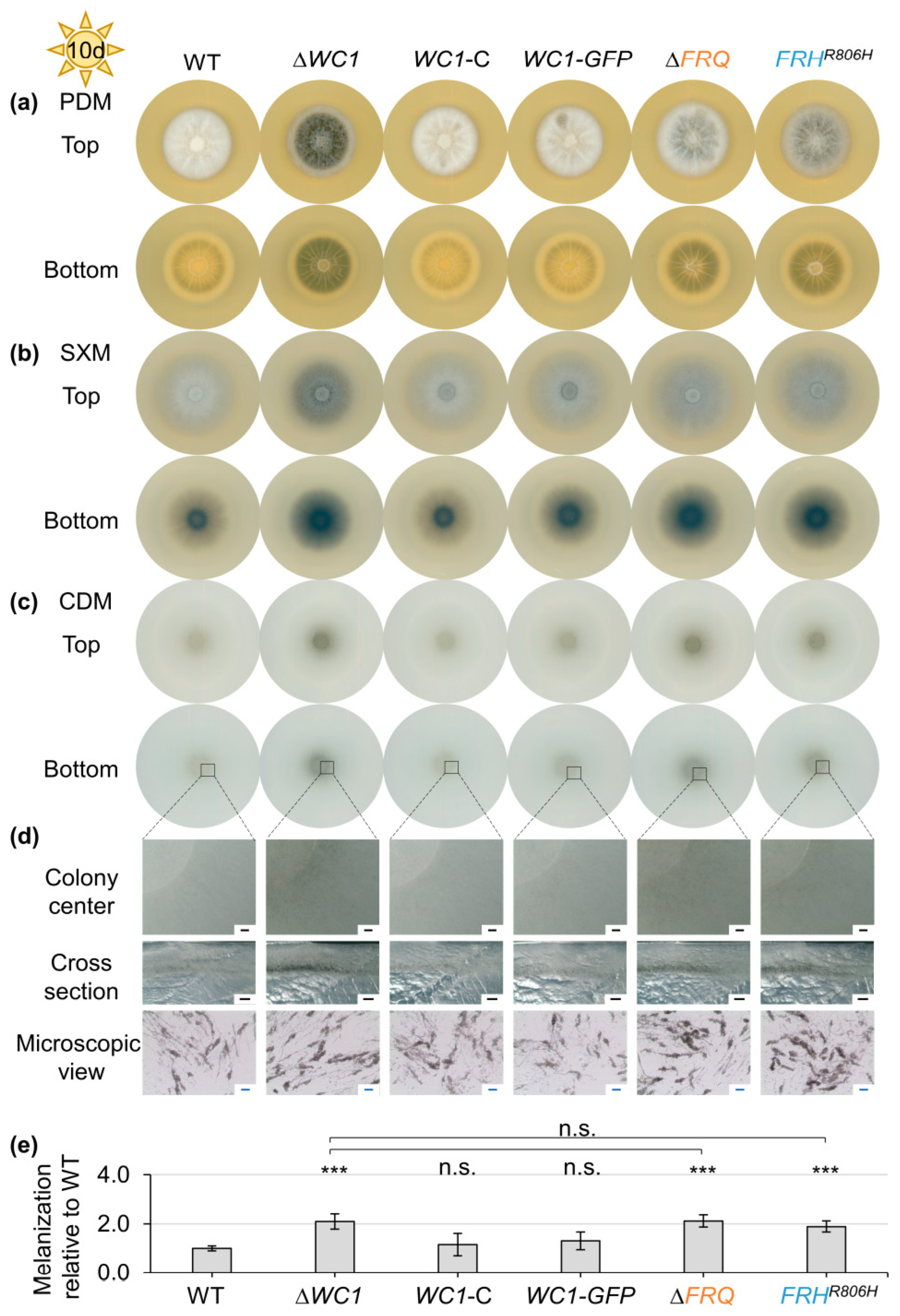
3.1. Light-Dependent Repression of Microsclerotia Formation and Induction of Aerial Hyphae Formation in V. dahliae Require FRQ, FRH, and WC1

Homologs of the key circadian clock genes known from Neurospora crassa, including FRQ, FRH, WC1, and WC2, are present in V. dahliae [48], but no circadian rhythmicity in V. dahliae has yet been described. The V. dahliae genomic loci and deduced protein structures of the corresponding clock gene homologs that were used for this study (FRQ: VDAG_JR2_Chr1g01960a, FRH: VDAG_JR2_Chr4g00070aa, WC1: VDAG_JR2_Chr2g01990a) are depicted in Figure S8. The 1175 aa protein sequence of V. dahliae JR2 Wc1 is 63 aa longer than the previously depicted and analyzed Wc1 sequence [48]. V. dahliae JR2 Wc1 shares 58.16% identity with N. crassa Wc-1 (NCU02356). An alignment of both protein sequences is depicted in Figure S9.
V. dahliae FRQ is dispensable for rhythmic conidiation or microsclerotia formation under oscillating light or temperature conditions, but a corresponding deletion affects metabolic and redox processes [48]. The impact of a FRQ deletion on V. dahliae development itself including the efficiency to produce conidia or microsclerotia is yet elusive and was addressed for FRQ as well as the clock gene homologs FRH and WC1 of the tomato isolate V. dahliae JR2. As resting structures, melanized microsclerotia are the start and the end point of the plant infection cycle of V. dahliae [51]. Therefore, we examined whether clock gene homologs are required for microsclerotia formation. Strains carrying a WC1 deletion (∆WC1), FRQ deletion (∆FRQ), or constructs for Frq–GFP fusion protein production were constructed (Figures S1–S3). The V. dahliae FRH gene was modified by a single point mutation (Figure S4) because it exhibits not only clock but also additional essential functions in N. crassa [15]. A point mutation from guanine to adenine at position 2481 leads to an arginine to histidine codon exchange (FRHR806H) and is known to permit these essential functions; however, it interrupts clock functions of Frh, leading to increased FRQ transcript levels [29].
Spores of the wild-type, FRQ, and FRH mutants, as well as complementation strains, were point-inoculated onto a glucose-rich medium (PDM), a pectin-rich medium (SXM), and a minimal medium (CDM). The ex planta phenotypes of the colonies were analyzed after ten days (Figure 1). FRQ deletion and FRHR806H point mutation strains formed darker colonies on all media tested (Figure 1b–d). This was best visible on the bottom of the colony due to the production of aerial hyphae at the agar surface. Colony centers of CDM cultures were further investigated through binocular and light microscopy (Figure 1e). The observed intensification in colony pigmentation resulted from increased production of melanized microsclerotia compared with wild-type. FRQ deletion and FRHR806H point mutation resulted in an earlier onset of microsclerotia development as colonies of the mutant strains continuously melanized faster than those of the wild-type (Figure S10). Reintroduction of FRQ or introduction of the FRQ–GFP construct into the FRQ deletion strain (Figure S2) and reintroduction of wild-type FRH into the FRHR806H mutant strain (Figure S4) complemented the deletion or mutation phenotype, respectively. Notably, not only the expression of the FRQ–GFP construct under control of the endogenous promoter (FRQ–GFP) but also the gpdA promoter-controlled expression of FRQ–GFP (FRQ–GFP OE) allowed for growth similar to wild-type (Figure 1). Quantification of FRQ–GFP transcript levels in the presence of either wild-type or point-mutated FRH revealed that gpdA promoter-mediated expression did not result in elevated transcript levels. The Frq–GFP protein level was even reduced after two-day cultivation on SXM agar (Figure S11). Melanization of colonies grown on CDM was quantified as indication for microsclerotia formation (Figure 1f). Colony melanization was significantly increased through deletion of FRQ (~300% increased) and FRHR806H point mutation in either wild-type, FRQ–GFP, or FRQ–GFP OE background (~200% increased) compared with wild-type. The increase in microsclerotia formation due to FRHR806H point mutation does not significantly differ from the FRQ deletion effect. The melanization level of complementation and FRQ–GFP-expressing strains with intact FRH was similar to the wild-type. However, when incubated in the dark, colonies of the wild-type and strains with either FRQ deletion or FrhR806H aa substitution were melanized to a similar extent (Figure S12). These results demonstrate that FRQ and FRH are both required for repression of microsclerotia formation specifically when grown in the light. In addition to this repressive effect of Frq and Frh on microsclerotia formation, both proteins also positively affect aerial hyphae formation. This is especially visible when the fungus is grown on PDM plates, but it is also observable for colonies grown on SXM plates (Figure 1b,c). There, FRQ deletion and FRHR806H mutation result in reduced aerial hyphae growth.
FRQ expression in N. crassa is mostly dependent on the presence of an intact WC1 gene, but also WC-independent expression of FRQ was found [98,99,100,101,102,103]. Analysis of FRQ transcript levels in a WC1 deletion mutant under different conditions reveals that also in V. dahliae, FRQ expression partially depends on WC1 (Figure S11). The ex planta phenotype of the WC1 deletion strain was analyzed in comparison with wild-type, FRQ deletion, and FRHR806H mutation as well as complementation strains. In the latter strains, either the WC1 gene or a WC1–GFP fusion construct replaced the WC1 deletion (Figures S3 and S12). Spores of these strains were point-inoculated onto different media and the colony appearance was analyzed after cultivation for ten days as described before (Figure 2). WC1 deletion, similar to FRQ deletion and FRHR806H mutation, led to enhanced colony melanization on all media tested when cultivated in the light (Figure 2a–d), which was quantified for colonies grown on CDM (~100% increase compared with wild-type, Figure 2e). In contrast, deletion of WC1 resulted in slightly but significantly reduced melanization (~9.6% reduced) when cultivated in darkness (Figure S12). Complementation through reintegration of WC1 or WC1–GFP into the WC1 deletion strain allowed for growth similar to wild-type. Like FRQ and FRH, WC1 is required for wild-type-like growth of aerial hyphae. However, the role of Wc1 in positive control of aerial hyphae formation appeared stronger than that of Frq and Frh (Figure 2a,b). The ex planta phenotypes of ∆WC1/∆FRQ and ∆WC1/FRHR806H double mutant strains were indistinguishable from that of the WC1 single-deletion mutant strain (Figure S13). Taken together, aerial hyphae formation in the light depends stronger on the presence of WC1 than FRQ or FRH, but all three are required for light-dependent repression of microsclerotia formation. WC1 is also partially required for induction of microsclerotia formation in darkness.

3.2. V. dahliae FRQ and FRH Positively Affect Conidiation

Since WC1, FRQ, and FRH are required for microsclerotia formation, potential additional contributions to the developmental process of conidiation were investigated. Once V. dahliae reaches the xylem vessels, it forms conidiospores for fast proliferation in the vasculature [51,52,56]. Thus, conidiospore formation is important for effective fungal plant colonization. Spores of the previously described FRQ, FRH, and WC1 mutant strains were inoculated into liquid SXM, a pectin-rich medium favoring V. dahliae conidiation. After five days of incubation, conidia were quantified relative to wild-type control set as one.
FRQ deletion or the FrhR806H aa substitution in all different backgrounds led to significantly decreased conidia production (~25–29% reduced) compared with wild-type (Figure 3a). Reintegration of FRQ or FRQ–GFP under endogenous or gpdA-promoter control, as well as FRH reintegration, allowed for conidiation similar to wild-type. Deletion of WC1 resulted in a small but significant decrease in produced conidia (~7% reduced). However, the positive effect of Wc1 on conidiation was significantly lower than the FRQ- and FRH-mediated induction (~33–39%) of conidia production (Figure 3b). Thus, Frq and Frh are required for induction of conidiation, whereas Wc1 plays only a minor role in this developmental process.

3.3. Symptom Induction in V. dahliae Infected Tomato Plants Is Independent of FRQ, FRH, and WC1

In V. dahliae strawberry isolates, the presence of FRQ is important for disease induction. This effect is isolate dependent and was not found for disease induction of highly virulent isolates [48]. We previously observed a role of the clock gene homologs in microsclerotia formation and conidiation, with the latter being important for host plant colonization. Thus, we analyzed if these genes are also important for disease induction in the tomato isolate V. dahliae JR2. Tomato seedlings were infected with spores of wild-type, clock gene mutant strains, and their respective complementation strains or inoculated with water (mock). Plant height, leaf length, and plant fresh weight were measured after 21 days to characterize plants as being healthy, or showing mild, strong, or very strong symptoms compared with mock plants [60,68]. The percentage of plants belonging to the individual categories is displayed in stack diagrams (Figure 4). The spore-treated plants were stunted compared with mock-treated plants and had brownish discolored hypocotyls. We found no significant difference between disease symptoms induced by strains with FRQ or WC1 deletion, FrhR806H aa exchange, or respective complementation strains compared with wild-type. Thus, V. dahliae JR2 FRQ, FRHR806, and WC1 were dispensable for disease induction in its host plant tomato.

3.4. The Frq–Frh Complex Formation in V. dahliae Depends on Frh Amino Acid Residue Arginine 806

Previous research in N. crassa suggested that the FRHR806H mutation breaks the negative feedback loop and therefore causes FRQ overexpression [29]. In V. dahliae, however, codon exchange for the respective residue (FrhR806H) leads to the same developmental phenotypes as the FRQ deletion (Figure 1 and Figure 3). Even gpdA promoter-mediated FRQ expression in the presence of the mutated FRH did not result in elevated Frq levels, but cultivation on SXM agar for two days even led to significantly reduced Frq–GFP levels (Figure S11). Thus, we further investigated how the FRHR806H point mutation affects endogenously expressed FRQ transcript and protein levels. RNAs and proteins were extracted from cultures grown under conditions favoring either conidiation (in liquid SXM) or microsclerotia formation (on SXM agar). Samples were harvested after two, four, and six days (Figure S6). Relative normalized transcript levels of FRQ, WC1, and WC2 are depicted in Figure 5a,b. Wild-type expression after two days was set as one. No FRQ transcript was detected in the FRQ deletion strain; therefore, in this case no significance of differences were calculated. Neither expression of WC1 nor WC2 differed significantly between wild-type, FRQ deletion strain, or the FRQ–GFP-expressing strain with or without aa substitution in Frh during cultivation under conidiation favoring conditions (Figure 5a). The transcript level of FRQ was not significantly affected by FRHR806H point mutation in either of the two cultivation conditions (Figure 5a,b). When the strains were cultivated under microsclerotia favoring conditions, WC1 expression was slightly elevated in the FRQ–GFP-expressing strain (~12% increased) compared with wild-type after four days of cultivation (Figure 5b). The transcript level of WC2 was only significantly decreased upon FRH point mutation (~46% decreased) after two days of cultivation on SXM agar. Taken together, the FrhR806H aa exchange did not affect FRQ transcript levels and only led to a reduction in WC2 transcript levels after two days.
As the transcript level does not necessarily reflect the protein level, we next investigated if the FrhR806H aa substitution influences the protein abundance of Frq. Frq–GFP protein levels were quantified via western experiments using a GFP antibody. Proteins were extracted after two, four, and six days of incubation (Figure S6). The level of Frq–GFP was compared between the FRQ–GFP-expressing strains in wild-type FRH and FRHR806H background. Frq–GFP has a predicted molecular weight of ~133 kDa but fusion protein signals slightly above 180 kDa were also detected. This difference likely originates from post-translational modification of Frq, which is known to be extensively phosphorylated in N. crassa [8], with more than 100 identified phosphorylation sites [25,26,27,28]. Of these phosphorylation sites, 51 seem to be conserved in V. dahliae Frq (Figure S9). For quantification of Frq–GFP levels, signals ≥ 133 kDa were measured. In general, the Frq–GFP protein level decreased after two days of cultivation, and free GFP (~27 kDa) was detected as a degradation product after four and six days of cultivation (Figure 5c,d). A direct comparison of the Frq–GFP levels after two days of cultivation in liquid SXM or on SXM agar revealed that the growth conditions and presumably the developmental program which is mainly induced by the respective environment influenced Frq protein levels (Figure S14). When cultured under microsclerotia-promoting conditions (on SXM agar), Frq–GFP levels were significantly reduced in comparison with cultivation in an environment promoting conidiation (in liquid SXM). Frq–GFP was also present when cultures were grown in the dark, and the protein level did not significantly differ from light-incubated cultures (Figure S14). These results indicate that the protein level of V. dahliae Frq does not oscillate in a circadian manner but depends on nutrition and fungal development. The FRHR806H point mutation could not even affect Frq–GFP protein levels of natively expressed FRQ–GFP under any of the tested cultivation conditions (Figure 5c,d).
As neither the endogenous FRQ transcript nor Frq protein levels were affected in the FRHR806H strain, another possible explanation for the deletion-like phenotype would be an altered localization of Frq due to the FRH point mutation. V. dahliae Frh was predicted to localize in nuclei, whereas Frq was predicted to be localized both nuclear and cytoplasmic. Nuclear localization of N. crassa Frq is required for its clock-related functions, but still most Frq protein was detected to be cytoplasmic [15,17,104]. The C-terminal part of N. crassa Frq, including the Frq–Frh interaction domain, is required for the regulation of Frq distribution between the nucleus and cytoplasm [17]. A portion of the mainly nuclear localized Frh was also detected in cytoplasmic extracts, and reduction of Frh amounts led to increased nuclear localization of Frq [15,17]. Therefore, localizations of the V. dahliae GFP-fused proteins Frq–GFP and Frh–GFP were investigated through fluorescence microscopy. Strains expressing either FRH–GFP or the point mutated FRHR806H–GFP construct were obtained (Figure S4). Both Frq–GFP and Frh–GFP were predominantly localized in nuclei. This localization was unaffected by FRHR806H point mutation under the tested condition (Figure 6). Thus, neither protein abundance nor localization seem to be affected by the point mutation. These observations support that the aa exchange in Frh hinders Frq from performing its functions.
N. crassa Frq and Frh form a heteromeric Frq–Frh complex [15]. Co-immunoprecipitation experiments using the protein variant with aa exchange (FrhR806H) in N. crassa suggested that the Frq–Frh complex can still be formed [29] although interaction of Frq and FrhR806H seemed significantly weakened [30]. An in vitro protein pull-down was conducted to investigate whether the Frq–Frh complex is also formed in V. dahliae and whether complex formation is affected by the FRHR806H aa exchange. Strains expressing FRH–GFP or FRHR806H–GFP fusion gene constructs at the native locus were examined. Expression of the Frh–GFP fusion protein allowed for development similar to the wild-type, whereas the FRHR806H–GFP-expressing strain had the same phenotype of increased microsclerotia formation as the FRHR806H strain (Figure 7a). C-terminal GFP fusion only causes a slight reduction of fungal growth, as corresponding colonies of the FRH–GFP and FRHR806H–GFP strains are smaller than wild-type or FRHR806H colonies. Before the in vitro protein pull-down was conducted, production of the fusion proteins was verified via western experiments. The cultures were incubated for two, four, and six days before proteins were extracted (Figure S6). Both Frh–GFP and FrhR806H–GFP have a predicted molecular weight of ~152 kDa. The largest detected molecular weight of the fusion proteins in western experiments, however, was slightly above 180 kDa, which indicates additional potential post-translational modifications. Signals ≥ 152 kDa were used for quantification of Frh–GFP and FrhR806H–GFP protein amounts. Levels of both fusion proteins did not significantly differ from each other when cultivated under the same conditions (Figure 7b,c).
Extracts of FRH–GFP and FRHR806H–GFP strains from two-day cultivation in liquid SXM were used for in vitro protein pull-downs, when both Frh–GFP and FrhR806H–GFP fusion proteins were sufficiently and comparably detected in western experiments. V. dahliae wild-type protein extracts with levels of free GFP comparable to the fusion protein levels were used as control (Figure S7). Significantly enriched proteins are depicted in the top right part of the volcano plots (Figure 7d,e).
Frh–GFP interacted with Frq and five other proteins (Table S5). Predicted domains or protein family types of the identified interacting proteins are listed in Table S6. With FrhR806H–GFP as bait, no other proteins were significantly enriched (Table S7). This suggests that Frq–Frh interaction depends on the arginine at position 806 in the Frh protein and is abolished or at least significantly weakened by the aa exchange. These results indicate that the function of Frq in controlling V. dahliae development depends on Frq–Frh complex formation.

3.5. Sfl1 Positively Affects Microsclerotia Formation Prior to Frq and Contributes to V. dahliae Spore Formation

The role of Frq and the importance of the Frq–Frh complex in the development of V. dahliae have to be connected to corresponding pathways. The Som1- and Vta3-regulatory networks are involved in initial plant infection and conidia formation, as well as microsclerotia formation of V. dahliae [61]. The transcription factor Vta3 controls the expression of other transcription factors involved in the regulation of microsclerotia formation and conidiation. Expression of the Vta3-encoding gene is, in turn, induced by Som1, which also stimulates microsclerotia formation and conidiation by a Vta3-independent pathway [61]. We investigated if point mutation of FRH or deletion of WC1 or FRQ affects the transcript levels of transcription factors that are part of these regulatory networks. Neither SOM1 nor VTA3 transcript levels did in any case significantly differ from wild-type control (Figure S15). Under conidiation-favoring conditions, transcript levels of ABA1 were not affected by FRQ deletion or FRH point mutation. The transcript levels of VTA2 did significantly differ from wild-type levels in the FRHR806H mutant strain after two days of incubation (liquid SXM, ~15% increased). FRH mutation, however, resulted in reduced conidiation (Figure 3), and Vta2 is a positive regulator of this developmental process [67]. Moreover, VTA2 levels were reduced in the FRQ–GFP strain after cultivation in liquid SXM for six days (~46% reduced). Vta2 is also involved in repression of microsclerotia formation, but its transcript levels did not significantly change upon WC1 deletion during growth on SXM agar (Figure S15). In contrast, deletion of WC1 resulted in significantly elevated transcript levels of CMR1 (~250% increased) and VTA1 (~150% increased) after two days of cultivation under microsclerotia-favoring conditions (on SXM agar, Figure S15). Both are transcription factors that positively affect melanization in V. dahliae [68,71]. Thus, the increased demand of melanization upon enhanced microsclerotia formation in the WC1 deletion strain seems to be mediated by elevated transcript levels of VTA1 and CMR1. This supports that Wc1 and presumably also the Frq–Frh complex are involved in reducing VTA1- and CMR1-mediated melanization. The question of by which factor or factors the light-dependent microsclerotia repression is regulated remains elusive.
Sfl1 is another Vta3-regulated transcription factor that positively affects microsclerotia formation and is a potential target of Frq–Frh-mediated light-dependent microsclerotia repression [61]. The reduction of SFL1 transcript levels by Frq could be a possible explanation for Frq’s negative effect on microsclerotia development. Quantification of SFL1 transcript levels in the WC1 deletion strain, however, indicated no significant influence of Wc1 and therefore presumably no influence of Frq on SFL1 transcript levels (Figure S15). As previously described (Figure 1), deletion of FRQ leads to increased melanization in the light, which is due to enhanced microsclerotia formation. In contrast, deletion of SFL1 resulted in reduced microsclerotia production in the light [61]. Sfl1 might be required for the onset of microsclerotia formation. A strain that produces a GFP–Sfl1 fusion protein was constructed to further investigate the role of Sfl1 in microsclerotia formation (Figure S5). GFP–Sfl1 was present at the initial phase of microsclerotia development, whereas the protein amounts decreased after microsclerotia were formed (Figure S16). Thus, both the inducing Sfl1 and the reducing Frq protein seem to be required early in the control of microsclerotia development.
Based on the opposing roles of Frq and Sfl1 in the development of microsclerotia, we tested the effect of a strong and constitutive expression of Sfl1 on the phenotype of a FRQ deletion strain. A GFP–SFL1 overexpression construct was ectopically integrated into a FRQ deletion as well as a SFL1 deletion strain (Figure S5). Strains that harbored the GFP–SFL1 construct at the endogenous locus grew similar to wild-type, whereas melanization of strains with the ectopically integrated GFP–SFL1 overexpression construct was slightly reduced in comparison with wild-type (Figure S17). The presence of the fusion protein was confirmed through fluorescence microscopy, in which the protein localized in the nuclei. Increased production of the GFP–Sfl1 fusion protein in the strain harboring the overexpression construct was verified through comparison with fusion protein amounts resulting from endogenously expressed GFP–SFL1 in a western experiment (Figure S17). Introduction of the GFP–SFL1 overexpression construct into a FRQ deletion strain was confirmed through fluorescence microscopy (Figure 8a) and western experiments (Figure S17). Deletion of FRQ resulted in increased melanization in the light, and ectopic reintroduction of the gene allowed for growth similar to wild-type (Figure S18). Ectopic overexpression of GFP–SFL1 in the FRQ deletion strain background also led to increased colony melanization compared with the wild-type (~67% increased) during cultivation in the light (Figure 8b,c). The melanization of the FRQ deletion strain was similar to wild-type when cultured in the dark, whereas melanization of the FRQ deletion strain containing the GFP–SFL1 overexpression construct was significantly reduced compared with wild-type as well as the FRQ deletion strain (~14–17% reduced; Figure 8b,c).
The role of Frq and Sfl1 in the control of microsclerotia formation was further investigated through the construction (Figure S1) and analysis of double mutants. The FRQ and SFL1 double-deletion strain grew like the SFL1 single-deletion strain, with reduced microsclerotia formation independent of light-conditions and increased aerial hyphae formation in the dark after ten days (Figure 9a). The melanization of 14-day-old light-incubated CDM cultures was quantified (Figure 9b), and it was confirmed that the FRQ/SFL1 double-deletion strain colonies were significantly less melanized compared with wild-type (~55% reduced) and the FRQ deletion strain (~69% reduced). The double-deletion strain was still significantly more melanized than the SFL1 single-deletion strain (~80% reduced compared with wild-type). The respective ectopic complementation strains melanized similar to wild-type. The fact that the FRQ/SFL1 double-deletion growth phenotype was more similar to the SFL1 single-deletion strain than to the FRQ single-deletion strain indicates that Sfl1 is required earlier in the induction of microsclerotia formation than the repressive activity of Frq.
The spore production of the SFL1 single and FRQ/SFL1 double-deletion strains was quantified in comparison with wild-type and the FRQ deletion strain to investigate if SFL1 also affects conidiation (Figure 10a). Deletion of FRQ results in reduced conidia production compared with wild-type (~36% reduced). SFL1 deletion strains also produced significantly less conidia (~14% reduced). This reduction, however, was significantly smaller than the FRQ-dependent effect. The mean conidia production of the FRQ/SFL1 double-deletion strain was slightly lower than in the FRQ single-deletion strain (47% reduced compared with wild-type). The reduction in the FRQ single- and FRQ/SFL1 double-deletion strains, however, did not significantly differ from each other. Altogether, SFL1 positively affects V. dahliae conidiation to a lesser extent than Frq. SFL1 is dispensable for initial colonization of A. thaliana roots [61]; therefore, a potential role for SFL1 in the later stages of plant colonization was analyzed (Figure 10b). Tomato plants were inoculated with water (mock), spores of the V. dahliae wild-type, the SFL1 single-, or the FRQ/SFL1 double-deletion strains. The treatments with the FRQ single-deletion strain and respective complementation strains served as controls. Symptom severity was determined after 21 days [60,68]. Plants that were challenged with fungal spores had brownish discolored hypocotyls and were stunted compared with mock-treated plants. Deletion of SFL1 alone, like the FRQ single-deletion, did not significantly affect the symptom induction. Only the plants treated with spores of the FRQ/SFL1 double-deletion strain were slightly, but significantly less diseased than wild-type infected plants.
Taken together, Sfl1 plays a major role in microsclerotia formation and an additional minor role in conidiation. Through combined actions with Frq, it also contributes to disease development in tomato plants. Our data further suggest that Sfl1 is only required for the onset of microsclerotia formation but is dispensable thereafter, and that the Sfl1-induced microsclerotia formation can be light-dependently delayed by the heteromeric Frq–Frh complex.

4. Discussion

The V. dahliae JR2 clock proteins Frq and Frh form the Frq–Frh complex, which is important for Frq function. Circadian clocks generally help organisms to adapt to rhythmic changes in the environment. The V. dahliae Frq–Frh heteromer enhances conidiation and, during illumination, delays the Sfl1-induced microsclerotia formation for survival in the soil. Cellular Frq amounts or localization are not affected. Figure 11 summarizes these findings for V. dahliae Frq and Frh and their interplay with Wc1 and Sfl1 in the fungal life, plant infection, and resting structure formation cycle.
Domains of the clock proteins are well conserved [48] and there are additional similarities between V. dahliae Frh and Frq and corresponding N. crassa counterparts. N. crassa and V. dahliae Frq are both largely intrinsically disordered proteins [13,14]. N. crassa Frq associates with Frh, which presumably serves as nanny protein by conferring stability and structure to Frq [13,105]. Similarly, both Frh proteins carry a predicted disordered region in their N-termini [14,30]. This disordered region seems to be present only in fungi carrying FRQ orthologs [14]. The interaction between Frq and Frh in N. crassa depends on a part of the Frh N-terminus that partially overlaps with the disordered region and on a ten-aa region in the C-terminus of Frq [13,18]. We found evidence for Frq–Frh complex formation (Figure 7d and Table S5), although only four of these ten aa are conserved in V. dahliae Frq [18,48]. The formation of the Frq–Frh complex was abolished or at least substantially weakened upon point mutation of FRH, as Frq was not detected as the interaction partner of the FrhR806H–GFP variant (Figure 7e and Table S7).
Both Frq and Frh were localized predominantly in V. dahliae JR2 nuclei of hyphal cells under the tested condition (Figure 6). This is similar to N. crassa, B. bassiana, and M. robertsii, where Frh is also primarily a nuclear protein, except a N. crassa Frh subpopulation that is localized in cytoplasm [15,33,37]. Cytoplasmic Frh is presumably in complex with Frq homodimers in N. crassa [15]. In this fungus, Frq is localized in nuclei as well as the cytoplasm but is mainly found in the latter [15,17]. However, the Frq regulatory function requires nuclear localization [104]. The two Frq proteins of B. bassiana exhibit opposing rhythms of nuclear localization, which ensure approximately constant levels of nuclear Frq for promoting continuous, non-rhythmic conidiation. Incubation in light results in nuclear localization of Frq1, whereas Frq2 is localized in nuclei upon cultivation in the dark [33]. B. bassiana Frh mediates these localization dynamics by ensuring stable accumulation of Frq proteins. Frq–GFP fusion proteins of M. robertsii are mainly in nuclei, and the ratio of nuclear and cytoplasmic localization is partially mediated to a similar extent by Frh, Wc1, and Wc2 [37]. Impairment of the Frq–Frh interaction or hairpin RNA-mediated FRH silencing increases relative nuclear localization of N. crassa Frq [17]. This is different in V. dahliae, where nuclear localization of Frq is independent of the Frh interaction, which is impaired in a genetic background with the Frh R806H aa substitution.
All attempts to generate an FRH deletion strain have been unsuccessful, suggesting a potential essential function which can still be fulfilled by the FrhR806H variant. Essential Frh functions have been also described for Saccharomyces cerevisiae Mtr4 (54.94% shared aa identity) or N. crassa Frh (70.05% shared aa identity) [15,106,107]. In contrast, the homologous Frh-encoding genes of ascomycete entomopathogenic fungi B. bassiana (72.82% shared aa identity) or M. robertsii (73.39% shared aa identity) are not essential [33,37].
The FRHR806H mutation had originally been described in N. crassa, where it disturbs Frh clock function and still allows a weakened Frq–Frh interaction [29,30]. The N. crassa FRH point mutation resulted in elevated Frq protein levels in combination with reduced Wc1 stability [29]. The V. dahliae FrhR806H variant neither significantly affected Frq protein nor FRQ or WC1 transcript levels compared with wild-type (Figure 5). However, FRQ deletion or FRH point mutation resulted in changes in fungal development. Conidiation was reduced and microsclerotia formation was increased in presence of light. A WC1 deletion also resulted in increased microsclerotia formation in light but in a weaker defect in conidiation and in a reduced microsclerotia formation in the dark in comparison with the FRQ deletion or FRH codon exchange mutation (Figure 3 and Figure S12). Therefore, a functional Frq–Frh complex is required for controlling conidiation or for delaying microsclerotia formation in presence of light.
The fact that V. dahliae Wc1 was less important for conidiation than Frq and Frh (Figure 3) may appear unexpected, since similar phenotypes were observed regarding the repression of microsclerotia formation. A possible explanation is that Wc1 not only controls transcript levels of genes encoding proteins that promote conidiation, such as Frq, but also of those that are involved in suppression of conidiation. Likewise, the V. dahliae transcription factor Vta3 promotes the expression of SFL1 and VTA1, which encode two transcription factors that induce microsclerotia formation and melanin production, respectively [61,68]. However, Vta3 also enhances the expression of VTA2, which encodes a transcription factor involved in the repression of microsclerotia formation [61,67]. This might reflect a fine-tuned regulatory process. Conidiation was also enhanced by the Frq-, Wc1-, and Wc2-encoding homologs of B. bassiana and M. robertsii, FgWc1 and FgWc2 of F. graminearum, as well as MoFrq, MoFwd1, and to a seemingly lesser extent, MgWc-1 of M. oryzae [33,35,36,37,42,45,46]. In B. cinerea, BcFrq1 is relevant for macroconidiation [40]. In contrast, the WCC of B. cinerea and ZtWco-1 of Zymoseptoria tritici suppress production of conidia and micropycnidiospores, respectively [41,43]. The B. cinerea WCC-mediated suppression of conidia formation is accompanied by enhanced aerial hyphae formation [41]. Similarly, Wc1 is presumably involved in aerial hyphae development in V. dahliae (Figure 2 and Figure S13) and in F. oxysporum [47]. In contrast to our observation that both FRQ deletion and FRH point mutation resulted in similar reductions of V. dahliae conidiation, the Frq and Frh proteins of B. bassiana play different roles in controlling conidiation. B. bassiana Frq is required and Frh is dispensable for conidiation [33]. Frq and Frh of M. robertsii both enhance conidiation under certain light conditions, with Frh playing a more prominent role than Frq [37]. These comparisons support a broad range and high variability in the regulatory functions for these two proteins between different fungi.
Conidiation is essential for propagation within the plant, whereas formation of microsclerotia is necessary for survival in soil and the next plant infection in the V. dahliae life cycle [51,54]. Wc1 is required for light-dependent repression of microsclerotia formation, presumably through transcriptional activation of FRQ expression and the resulting Frq–Frh complex (Figure 1, Figure 2 and Figure S11). The Frq–Frh complex is probably dispensable for the control of microsclerotia formation in the dark, when Wc1 contributes to microsclerotia formation. Similarly, Z. tritici ZtWco-1 positively regulates melanization and B. cinerea BcFrq1 represses the formation of sclerotia in a medium- and light-dependent manner [40,43]. V. dahliae JR2 Wc1, and presumably also the Frq–Frh complex reduce VTA1 and CMR1 transcript levels to suppress melanin production. In contrast, V. dahliae 12253 Frq does not control transcript levels of either VTA1, CMR1, or their downstream regulated target gene, PKS1 [48,68,71]. This underlines that the regulatory functions of clock proteins vary even within different isolates of the same fungal species.
The transcription factor Sfl1 induces microsclerotia formation in V. dahliae [61]. However, overexpression of GFP–SFL1 did not enhance microsclerotia formation when compared with wild-type. Elevated levels of Sfl1 might be inefficient in transcriptional activation as not enough cofactors can be recruited [108]. The Frq–Frh complex represses this Sfl1-mediated induction at early time points in light. Furthermore, the Frq–Frh complex also represses Sfl1-independently induced microsclerotia formation, as the FRQ/SFL1 double-deletion strain melanized more than the SFL1 single-deletion strain (Figure 9). Thus, the Frq–Frh complex might interfere with the downstream signaling of Sfl1 or its regulated targets, as well as other so far unknown inducer(s) of microsclerotia formation. Further work will be required to shed light on the exact regulatory molecular mechanism.
Only the combined actions of Frq and Sfl1 can significantly enhance symptom induction in V. dahliae JR2-infected tomato plants (Figure 10). The reduced conidial production of the double-deletion strain presumably results in a slowed colonization of host plants. The individual clock proteins did not significantly affect symptom induction. Similarly, individual clock proteins are not required for disease induction by other fungi. B. bassiana Wc1 and Wc2 are dispensable for the infection cycle and M. robertsii Frq does not affect fungal virulence [35,37]. Likewise, F. oxysporum and F. graminearum Wc1 proteins are dispensable for disease induction in plants [46,47]. However, clock proteins can also enhance virulence of pathogenic fungi. M. robertsii Wc1 and Wc2 increase virulence of the insect pathogen during cuticle infection [36]. The B. bassiana Frq proteins are important for full virulence, the secretion of cuticle-degrading Pr1 proteases, and the formation of blastospores, which are required for proliferation in the wax moth G. mellonella [34]. Deletion of Z. tritici ZtWCO-1 delays disease progression on wheat, and M. oryzae MoFwd1 and MoFrq1 enhance lesion development on leaves of rice or barley [42,43]. B. cinerea BcWcl1 increases lesion formation in a light-dependent manner and BcFrq1 mediates the production of macroconidia required for host infection in a medium-dependent manner [40,41]. B. cinerea causes a more severe infection on A. thaliana if the first contact between fungus and plant occurs at dusk compared with dawn. This time-of-day-dependent infection severity is mediated by BcFrq1. Deletion of BcFRQ1 results in a strong infection, independent of the time of day, whereas overexpression of BcFRQ1 leads to reduced lesion size and spread on A. thaliana leaves [40]. Thus, BcFrq1 and the circadian clock regulation of B. cinerea reduce lesion formation at dawn. Likewise, M. oryzae MgWc-1 light-dependently suppresses disease [44]. This indicates a broad range of functions for clock proteins, ranging between promoting, repressing, and being dispensable for disease induction, respectively. V. dahliae Frq can even play different roles in the disease induction of two different strawberry-infecting strains. Frq is required for the full symptom induction of a moderately virulent strain, whereas it is dispensable for disease induction by a highly virulent isolate [48]. Thus, in some fungi, the function of an individual clock protein is already required for disease progression, whereas Frq is only important for symptom induction of V. dahliae JR2 in combination with Sfl1.
Altogether, we have shed light on the function of clock gene homologs in the development of V. dahliae JR2. The Frq–Frh complex is required for enhanced production of conidiospores and controlled formation of Sfl1-induced microsclerotia. These are both important structures in the plant-pathogenic lifestyle of V. dahliae. Gaining further knowledge on the regulation of these developmental programs and the role of the clock proteins therein may one day help to find new ways to combat Verticillium wilt disease.

Supplementary Materials

The following supporting information can be downloaded at: https://www.mdpi.com/article/10.3390/jof9070725/s1, Methods S1 [25,26,28,60,61,67,68,69,70,71,75,76,77,79,81,97,109,110]; Figure S1: Verification of V. dahliae FRQ deletion, ectopic complementation, and FRQ/SFL1 double-deletion strains; Figure S2: Verification of V. dahliae FRQ in locus complementation and FRQ–GFP-expressing strains; Figure S3: Verification of WC1 deletion, complementation, and WC1–GFP-expressing strains; Figure S4: Verification of FRH point mutation, complementation, and FRHGFP-expressing strains; Figure S5: Verification of the V. dahliae strains with GFP–SFL1 in locus or ectopically overexpressed GFP–SFL1 in either SFL1 or FRQ (NATR) deletion strains; Figure S6: V. dahliae FRQ and WC1 deletion as well as FRH point mutation strains melanize stonger than wild-type if cultivated in liquid or on solid SXM; Figure S7: Amount of free GFP in the wild-type control is similar to Frh–GFP and FrhR806H–GFP protein levels; Figure S8: V. dahliae FRQ, FRH, and WC1 genes with deduced protein structures; Figure S9: Alignments of Wc1 and Frq protein sequences from N. crassa and V. dahliae JR2; Figure S10: V. dahliae FRQ, FRH, and WC1 mutant strains continuously melanize faster than wild-type; Figure S11: FRQ–GFP overexpression is prevented independent of Frh amino acid residue arginine 806 and the transcript level of FRQ depends partially, but not solely on Wc1; Figure S12: Wc1 slightly enhances microsclerotia formation in the dark and the WC1–GFP strain produces the Wc1–GFP fusion protein; Figure S13: Ex planta phenotypes of the WC1/FRQ double-deletion or WC1/FRHR806H double mutant strains equal the WC1 single-deletion strain phenotype; Figure S14: The Frq–GFP protein level depends on the cultivation conditions, which favor different developmental processes; Figure S15: Wc1-, Frq-, and Frh-mediated control of V. dahliae development seems independent of the Som1- and Vta3-regulatory networks; Figure S16: Sfl1 is required in the initial phase of microsclerotia production; Figure S17: The GFP–Sfl1 fusion protein is present, functional, and predominantly localized in nuclei; Figure S18: Ectopic FRQ complementation in the ∆FRQ (NATR) background allows for wild-type-like growth; Table S1: Primers used in this study; Table S2: Plasmids used in this study; Table S3: List of bacterial and fungal strains used in this work; Table S4: Oligonucleotides for qRT-PCR; Table S5: Significantly enriched proteins with LFQ intensities, MS/MS counts, sequence coverage, and unique peptides in all three replicates of Frh–GFP in comparison with the wild-type control; Table S6: Predicted protein domains or family types of proteins significantly enriched with Frh–GFP; Table S7: Significantly enriched protein with LFQ intensities, MS/MS counts, sequence coverage, and unique peptides in all three replicates of FrhR806H–GFP in comparison with the wild-type control; Data S1. and Images S1.

Author Contributions

Conceptualization, A.N., M.L., R.H. and G.H.B.; validation, A.N., M.L., I.M., J.S. and R.H.; formal analysis, A.N., M.L., R.H. and G.H.B.; investigation, A.N., M.L., I.M., J.S., K.S., O.V. and R.H.; resources, G.H.B.; data curation, A.N., K.S., O.V. and G.H.B.; writing—original draft preparation, A.N., R.H. and G.H.B.; writing—review and editing, A.N., M.L., I.M., J.S., K.S., O.V., R.H. and G.H.B.; visualization, A.N. and M.L.; supervision, R.H. and G.H.B.; project administration, A.N. and M.L.; funding acquisition, G.H.B. All authors have read and agreed to the published version of the manuscript.

Funding

This research was funded by the German Research Foundation (DFG, GRK 2172 to G.H.B.). A.N., M.L., I.M. and J.S. were supported by the IRTG 2172: PRoTECT program (GRK 2172, project number 273134146) of the Göttingen Graduate School GGNB. R.H. was supported by the German Research Foundation (BR1502/15-2 to G.H.B.). LC/MS analysis was performed by the Service Unit LCMS Protein Analytics of the Göttingen Center for Molecular Biosciences (GZMB) at the University of Göttingen (Grant DFGGZ: INST 186/1230-1 FUGG). We acknowledge support by the Open Access Publication Funds of the Göttingen University. The funders had no role in study design, data collection and analysis, decision to publish, or preparation of the manuscript.

Institutional Review Board Statement

Not applicable.

Informed Consent Statement

Not applicable.

Data Availability Statement

The mass spectrometry proteomics data have been deposited to the ProteomeXchange Consortium via the PRIDE partner repository with the dataset identifier PXD041716. Other data supporting reported results can be found in the Supplementary Material (Data S1 and Images S1).

Acknowledgments

The authors thank C. H. Haney, V. Lipka, K. Heimel, and J. W. Kronstad for fruitful discussions, N. Scheiter for excellent technical assistance, and A. Höfer, A. K. Sieven, K. A. Vaupel, M. Süß, M. Bromm, Y.-Y. Chen, and C. Kunz for support.

Conflicts of Interest

The authors declare no conflict of interest. The funders had no role in the design of the study; in the collection, analyses, or interpretation of data; in the writing of the manuscript; or in the decision to publish the results.

References

  1. Braunsdorf, C.; Mailänder-Sánchez, D.; Schaller, M. Fungal Sensing of Host Environment. Cell Microbiol. 2016, 18, 1188–1200. [Google Scholar] [CrossRef] [PubMed] [Green Version]
  2. Yu, Z.; Fischer, R. Light Sensing and Responses in Fungi. Nat. Rev. Microbiol. 2019, 17, 25–36. [Google Scholar] [CrossRef] [PubMed]
  3. Diernfellner, A.C.R.; Brunner, M. Phosphorylation Timers in the Neurospora crassa Circadian Clock. J. Mol. Biol. 2020, 432, 3449–3465. [Google Scholar] [CrossRef] [PubMed]
  4. Froehlich, A.C.; Liu, Y.; Loros, J.J.; Dunlap, J.C. White Collar-1, a Circadian Blue Light Photoreceptor, Binding to the frequency Promoter. Science 2002, 297, 815–819. [Google Scholar] [CrossRef]
  5. He, Q.; Cheng, P.; Yang, Y.; Wang, L.; Gardner, K.H.; Liu, Y. White Collar-1, a DNA Binding Transcription Factor and a Light Sensor. Science 2002, 297, 840–843. [Google Scholar] [CrossRef]
  6. Talora, C.; Franchi, L.; Linden, H.; Ballario, P.; Macino, G. Role of a White Collar-1-White Collar-2 Complex in Blue-Light Signal Transduction. EMBO J. 1999, 18, 4961–4968. [Google Scholar] [CrossRef]
  7. Hurley, J.M.; Dasgupta, A.; Emerson, J.M.; Zhou, X.; Ringelberg, C.S.; Knabe, N.; Lipzen, A.M.; Lindquist, E.A.; Daum, C.G.; Barry, K.W.; et al. Analysis of Clock-Regulated Genes in Neurospora Reveals Widespread Posttranscriptional Control of Metabolic Potential. Proc. Natl. Acad. Sci. USA 2014, 111, 16995–17002. [Google Scholar] [CrossRef]
  8. Garceau, N.Y.; Liu, Y.; Loros, J.J.; Dunlap, J.C. Alternative Initiation of Translation and Time-Specific Phosphorylation Yield Multiple Forms of the Essential Clock Protein FREQUENCY. Cell 1997, 89, 469–476. [Google Scholar] [CrossRef] [Green Version]
  9. Diernfellner, A.C.R.; Schafmeier, T.; Merrow, M.W.; Brunner, M. Molecular Mechanism of Temperature Sensing by the Circadian Clock of Neurospora crassa. Genes Dev. 2005, 19, 1968–1973. [Google Scholar] [CrossRef] [Green Version]
  10. Colot, H.V.; Loros, J.J.; Dunlap, J.C. Temperature-Modulated Alternative Splicing and Promoter Use in the Circadian Clock Gene frequency. Mol. Biol. Cell 2005, 16, 5563–5571. [Google Scholar] [CrossRef] [Green Version]
  11. Liu, Y.; Garceau, N.Y.; Loros, J.J.; Dunlap, J.C. Thermally Regulated Translational Control of FRQ Mediates Aspects of Temperature Responses in the Neurospora Circadian Clock. Cell 1997, 89, 477–486. [Google Scholar] [CrossRef] [PubMed] [Green Version]
  12. Diernfellner, A.; Colot, H.V.; Dintsis, O.; Loros, J.J.; Dunlap, J.C.; Brunner, M. Long and Short Isoforms of Neurospora Clock Protein FRQ Support Temperature Compensated Circadian Rhythms. FEBS Lett. 2007, 581, 5759–5764. [Google Scholar] [CrossRef] [PubMed] [Green Version]
  13. Hurley, J.M.; Larrondo, L.F.; Loros, J.J.; Dunlap, J.C. Conserved RNA Helicase FRH Acts Nonenzymatically to Support the Intrinsically Disordered Neurospora Clock Protein FRQ. Mol. Cell 2013, 52, 832–843. [Google Scholar] [CrossRef] [Green Version]
  14. Pelham, J.F.; Dunlap, J.C.; Hurley, J.M. Intrinsic Disorder Is an Essential Characteristic of Components in the Conserved Circadian Circuit. Cell Commun. Signal. 2020, 18, 181. [Google Scholar] [CrossRef] [PubMed]
  15. Cheng, P.; He, Q.; He, Q.; Wang, L.; Liu, Y. Regulation of the Neurospora Circadian Clock by an RNA Helicase. Genes Dev. 2005, 19, 234–241. [Google Scholar] [CrossRef] [Green Version]
  16. Cheng, P.; Yang, Y.; Heintzen, C.; Liu, Y. Coiled-Coil Domain-Mediated FRQ-FRQ Interaction Is Essential for Its Circadian Clock Function in Neurospora. EMBO J. 2001, 20, 101–108. [Google Scholar] [CrossRef] [Green Version]
  17. Cha, J.; Yuan, H.; Liu, Y. Regulation of the Activity and Cellular Localization of the Circadian Clock Protein FRQ. J. Biol. Chem. 2011, 286, 11469–11478. [Google Scholar] [CrossRef] [Green Version]
  18. Guo, J.; Cheng, P.; Liu, Y. Functional Significance of FRH in Regulating the Phosphorylation and Stability of Neurospora Circadian Clock Protein FRQ. J. Biol. Chem. 2010, 285, 11508–11515. [Google Scholar] [CrossRef] [Green Version]
  19. Schafmeier, T.; Haase, A.; Káldi, K.; Scholz, J.; Fuchs, M.; Brunner, M. Transcriptional Feedback of Neurospora Circadian Clock Gene by Phosphorylation-Dependent Inactivation of Its Transcription Factor. Cell 2005, 122, 235–246. [Google Scholar] [CrossRef] [Green Version]
  20. He, Q.; Cha, J.; He, Q.; Lee, H.-C.; Yang, Y.; Liu, Y. CKI and CKII Mediate the FREQUENCY-Dependent Phosphorylation of the WHITE COLLAR Complex to Close the Neurospora Circadian Negative Feedback Loop. Genes Dev. 2006, 20, 2552–2565. [Google Scholar] [CrossRef] [Green Version]
  21. Wang, B.; Kettenbach, A.N.; Zhou, X.; Loros, J.J.; Dunlap, J.C. The Phospho-Code Determining Circadian Feedback Loop Closure and Output in Neurospora. Mol. Cell 2019, 74, 771–784. [Google Scholar] [CrossRef] [PubMed]
  22. Guo, J.; Cheng, P.; Yuan, H.; Liu, Y. The Exosome Regulates Circadian Gene Expression in a Posttranscriptional Negative Feedback Loop. Cell 2009, 138, 1236–1246. [Google Scholar] [CrossRef] [PubMed] [Green Version]
  23. He, Q.; Cheng, P.; Yang, Y.; He, Q.; Yu, H.; Liu, Y. FWD1-Mediated Degradation of FREQUENCY in Neurospora Establishes a Conserved Mechanism for Circadian Clock Regulation. EMBO J. 2003, 22, 4421–4430. [Google Scholar] [CrossRef] [PubMed] [Green Version]
  24. He, Q.; Liu, Y. Degradation of the Neurospora Circadian Clock Protein FREQUENCY through the Ubiquitin-Proteasome Pathway. Biochem. Soc. Trans. 2005, 33, 953–956. [Google Scholar] [CrossRef] [PubMed]
  25. Baker, C.L.; Kettenbach, A.N.; Loros, J.J.; Gerber, S.A.; Dunlap, J.C. Quantitative Proteomics Reveals a Dynamic Interactome and Phase-Specific Phosphorylation in the Neurospora Circadian Clock. Mol. Cell 2009, 34, 354–363. [Google Scholar] [CrossRef] [Green Version]
  26. Tang, C.-T.; Li, S.; Long, C.; Cha, J.; Huang, G.; Li, L.; Chen, S.; Liu, Y. Setting the Pace of the Neurospora Circadian Clock by Multiple Independent FRQ Phosphorylation Events. Proc. Natl. Acad. Sci. USA 2009, 106, 10722–10727. [Google Scholar] [CrossRef]
  27. Wang, B.; Stevenson, E.-L.; Dunlap, J.C. Functional Analysis of 110 Phosphorylation Sites on the Circadian Clock Protein FRQ Identifies Clusters Determining Period Length and Temperature Compensation. G3 Genes Genomes Genet. 2023, 13, jkac334. [Google Scholar] [CrossRef]
  28. Chen, X.; Liu, X.; Gan, X.; Li, S.; Ma, H.; Zhang, L.; Wang, P.; Li, Y.; Huang, T.; Yang, X.; et al. Differential Regulation of Phosphorylation, Structure and Stability of Circadian Clock Protein FRQ Isoforms. J. Biol. Chem. 2023, 299, 104597. [Google Scholar] [CrossRef]
  29. Shi, M.; Collett, M.; Loros, J.J.; Dunlap, J.C. FRQ-Interacting RNA Helicase Mediates Negative and Positive Feedback in the Neurospora Circadian Clock. Genetics 2010, 184, 351–361. [Google Scholar] [CrossRef] [Green Version]
  30. Conrad, K.S.; Hurley, J.M.; Widom, J.; Ringelberg, C.S.; Loros, J.J.; Dunlap, J.C.; Crane, B.R. Structure of the Frequency-interacting RNA Helicase: A Protein Interaction Hub for the Circadian Clock. EMBO J. 2016, 35, 1707–1719. [Google Scholar] [CrossRef] [Green Version]
  31. Salichos, L.; Rokas, A. The Diversity and Evolution of Circadian Clock Proteins in Fungi. Mycologia 2010, 102, 269–278. [Google Scholar] [CrossRef] [Green Version]
  32. Valim, H.F.; Dal Grande, F.; Otte, J.; Singh, G.; Merges, D.; Schmitt, I. Identification and Expression of Functionally Conserved Circadian Clock Genes in Lichen-Forming Fungi. Sci. Rep. 2022, 12, 15884. [Google Scholar] [CrossRef]
  33. Tong, S.-M.; Wang, D.-Y.; Cai, Q.; Ying, S.-H.; Feng, M.-G. Opposite Nuclear Dynamics of Two FRH-Dominated Frequency Proteins Orchestrate Non-Rhythmic Conidiation in Beauveria bassiana. Cells 2020, 9, 626. [Google Scholar] [CrossRef] [PubMed] [Green Version]
  34. Tong, S.-M.; Gao, B.-J.; Peng, H.; Feng, M.-G. Essential Roles of Two FRQ Proteins (Frq1 and Frq2) in Beauveria bassiana’s Virulence, Infection Cycle, and Calcofluor-Specific Signaling. Appl. Environ. Microbiol. 2021, 87, e02545-20. [Google Scholar] [CrossRef] [PubMed]
  35. Xu, S.-Y.; Yu, L.; Luo, X.-C.; Ying, S.-H.; Feng, M.-G. Co-Regulatory Roles of WC1 and WC2 in Asexual Development and Photoreactivation of Beauveria bassiana. J. Fungi 2023, 9, 290. [Google Scholar] [CrossRef] [PubMed]
  36. Peng, H.; Guo, C.-T.; Tong, S.-M.; Ying, S.-H.; Feng, M.-G. Two White Collar Proteins Protect Fungal Cells from Solar UV Damage by Their Interactions with Two Photolyases in Metarhizium robertsii. Environ. Microbiol. 2021, 23, 4925–4938. [Google Scholar] [CrossRef] [PubMed]
  37. Peng, H.; Zhang, Y.-L.; Ying, S.-H.; Feng, M.-G. The Essential and the Nonessential Roles of Four Clock Elements in the Circadian Rhythm of Metarhizium robertsii. J. Biol. Chem. 2022, 8, 558. [Google Scholar] [CrossRef]
  38. de Leone, M.J.; Hernando, C.E.; Mora-García, S.; Yanovsky, M.J. It’s a Matter of Time: The Role of Transcriptional Regulation in the Circadian Clock-Pathogen Crosstalk in Plants. Transcription 2020, 11, 100–116. [Google Scholar] [CrossRef]
  39. Butt, G.R.; Qayyum, Z.A.; Jones, M.A. Plant Defence Mechanisms Are Modulated by the Circadian System. Biology 2020, 9, 454. [Google Scholar] [CrossRef]
  40. Hevia, M.A.; Canessa, P.; Müller-Esparza, H.; Larrondo, L.F. A Circadian Oscillator in the Fungus Botrytis cinerea Regulates Virulence When Infecting Arabidopsis thaliana. Proc. Natl. Acad. Sci. USA 2015, 112, 8744–8749. [Google Scholar] [CrossRef]
  41. Canessa, P.; Schumacher, J.; Hevia, M.A.; Tudzynski, P.; Larrondo, L.F. Assessing the Effects of Light on Differentiation and Virulence of the Plant Pathogen Botrytis cinerea: Characterization of the White Collar Complex. PLoS ONE 2013, 8, e84223. [Google Scholar] [CrossRef] [Green Version]
  42. Shi, H.-B.; Chen, N.; Zhu, X.-M.; Liang, S.; Li, L.; Wang, J.-Y.; Lu, J.-P.; Lin, F.-C.; Liu, X.-H. F-Box Proteins MoFwd1, MoCdc4 and MoFbx15 Regulate Development and Pathogenicity in the Rice Blast Fungus Magnaporthe oryzae. Environ. Microbiol. 2019, 21, 3027–3045. [Google Scholar] [CrossRef]
  43. Tiley, A.M.M.; Lawless, C.; Pilo, P.; Karki, S.J.; Lu, J.; Long, Z.; Gibriel, H.; Bailey, A.M.; Feechan, A. The Zymoseptoria tritici white collar-1 Gene, ZtWco-1, Is Required for Development and Virulence on Wheat. Fungal Genet. Biol. 2022, 161, 103715. [Google Scholar] [CrossRef] [PubMed]
  44. Kim, S.; Singh, P.; Park, J.; Park, S.; Friedman, A.; Zheng, T.; Lee, Y.-H.; Lee, K. Genetic and Molecular Characterization of a Blue Light Photoreceptor MGWC-1 in Magnaporth oryzae. Fungal Genet. Biol. 2011, 48, 400–407. [Google Scholar] [CrossRef]
  45. Lee, K.; Singh, P.; Chung, W.-C.; Ash, J.; Kim, T.S.; Hang, L.; Park, S. Light Regulation of Asexual Development in the Rice Blast Fungus, Magnaporthe oryzae. Fungal Genet. Biol. 2006, 43, 694–706. [Google Scholar] [CrossRef] [PubMed]
  46. Kim, H.; Kim, H.-K.; Lee, S.; Yun, S.-H. The White Collar Complex Is Involved in Sexual Development of Fusarium graminearum. PLoS ONE 2015, 10, e0120293. [Google Scholar] [CrossRef]
  47. Ruiz-Roldán, M.C.; Garre, V.; Guarro, J.; Mariné, M.; Roncero, M.I.G. Role of the White Collar 1 Photoreceptor in Carotenogenesis, UV Resistance, Hydrophobicity, and Virulence of Fusarium oxysporum. Eukaryot. Cell 2008, 7, 1227–1230. [Google Scholar] [CrossRef] [Green Version]
  48. Cascant-Lopez, E.; Crosthwaite, S.K.; Johnson, L.J.; Harrison, R.J. No Evidence That Homologs of Key Circadian Clock Genes Direct Circadian Programs of Development or mRNA Abundance in Verticillium dahliae. Front. Microbiol. 2020, 11, 1977. [Google Scholar] [CrossRef]
  49. Pardo-Medina, J.; Limón, M.C.; Avalos, J. Fusarium Photoreceptors. J. Fungi 2023, 9, 319. [Google Scholar] [CrossRef] [PubMed]
  50. Schumacher, J. Tools for Botrytis cinerea: New Expression Vectors Make the Gray Mold Fungus More Accessible to Cell Biology Approaches. Fungal Genet. Biol. 2012, 49, 483–497. [Google Scholar] [CrossRef]
  51. Fradin, E.F.; Thomma, B.P.H.J. Physiology and Molecular Aspects of Verticillium Wilt Diseases Caused by V. dahliae and V. albo-atrum. Mol. Plant Pathol. 2006, 7, 71–86. [Google Scholar] [CrossRef] [PubMed]
  52. Klosterman, S.J.; Atallah, Z.K.; Vallad, G.E.; Subbarao, K.V. Diversity, Pathogenicity, and Management of Verticillium Species. Annu. Rev. Phytopathol. 2009, 47, 39–62. [Google Scholar] [CrossRef] [Green Version]
  53. Pegg, G.F.; Brady, B.L. Verticillium Wilts; CABI Publishing: Wallingford, UK, 2002; ISBN 978-1-84593-322-7. [Google Scholar]
  54. Carroll, C.L.; Carter, C.A.; Goodhue, R.E.; Lawell, C.-Y.C.L.; Subbarao, K.V. A Review of Control Options and Externalities for Verticillium Wilts. Phytopathology 2018, 108, 160–171. [Google Scholar] [CrossRef] [PubMed] [Green Version]
  55. Eynck, C.; Koopmann, B.; Grunewaldt-Stoecker, G.; Karlovsky, P.; von Tiedemann, A. Differential Interactions of Verticillium longisporum and V. dahliae with Brassica napus Detected with Molecular and Histological Techniques. Eur. J. Plant Pathol. 2007, 118, 259–274. [Google Scholar] [CrossRef]
  56. Tian, J.; Kong, Z. Live-Cell Imaging Elaborating Epidermal Invasion and Vascular Proliferation/Colonization Strategy of Verticillium dahliae in Host Plants. Mol. Plant Pathol. 2022, 23, 895–900. [Google Scholar] [CrossRef]
  57. Singh, S.; Braus-Stromeyer, S.A.; Timpner, C.; Tran, V.-T.; Lohaus, G.; Reusche, M.; Knüfer, J.; Teichmann, T.; von Tiedemann, A.; Braus, G.H. Silencing of Vlaro2 for Chorismate Synthase Revealed That the Phytopathogen Verticillium longisporum Induces the Cross-Pathway Control in the Xylem. Appl. Microbiol. Biotechnol. 2010, 85, 1961–1976. [Google Scholar] [CrossRef] [Green Version]
  58. Maurus, I.; Harting, R.; Herrfurth, C.; Starke, J.; Nagel, A.; Mohnike, L.; Chen, Y.-Y.; Schmitt, K.; Bastakis, E.; Süß, M.T.; et al. Verticillium dahliae Vta3 Promotes ELV1 Virulence Factor Gene Expression in Xylem Sap, but Tames Mtf1-Mediated Late Stages of Fungus-Plant Interactions and Microsclerotia Formation. PLoS Pathog. 2023, 19, e1011100. [Google Scholar] [CrossRef]
  59. Maurus, I.; Leonard, M.; Nagel, A.; Starke, J.; Kronstad, J.W.; Harting, R.; Braus, G.H. Tomato Xylem Sap Hydrophobins Vdh4 and Vdh5 Are Important for Late Stages of Verticillium dahliae Plant Infection. J. Fungi 2022, 8, 1252. [Google Scholar] [CrossRef]
  60. Leonard, M.; Kühn, A.; Harting, R.; Maurus, I.; Nagel, A.; Starke, J.; Kusch, H.; Valerius, O.; Feussner, K.; Feussner, I.; et al. Verticillium longisporum Elicits Media-Dependent Secretome Responses with Capacity to Distinguish between Plant-Related Environments. Front. Microbiol. 2020, 11, 1876. [Google Scholar] [CrossRef]
  61. Bui, T.-T.; Harting, R.; Braus-Stromeyer, S.A.; Tran, V.-T.; Leonard, M.; Höfer, A.; Abelmann, A.; Bakti, F.; Valerius, O.; Schlüter, R.; et al. Verticillium dahliae Transcription Factors Som1 and Vta3 Control Microsclerotia Formation and Sequential Steps of Plant Root Penetration and Colonisation to Induce Disease. New Phytol. 2019, 221, 2138–2159. [Google Scholar] [CrossRef]
  62. Tian, L.; Yu, J.; Wang, Y.; Tian, C. The C2H2 Transcription Factor VdMsn2 Controls Hyphal Growth, Microsclerotia Formation, and Virulence of Verticillium dahliae. Fungal Biol. 2017, 121, 1001–1010. [Google Scholar] [CrossRef]
  63. Lai, M.; Cheng, Z.; Xiao, L.; Klosterman, S.J.; Wang, Y. The bZip Transcription Factor VdMRTF1 Is a Negative Regulator of Melanin Biosynthesis and Virulence in Verticillium dahliae. Microbiol. Spectr. 2022, 10, e0258121. [Google Scholar] [CrossRef] [PubMed]
  64. Luo, X.; Mao, H.; Wei, Y.; Cai, J.; Xie, C.; Sui, A.; Yang, X.; Dong, J. The Fungal-Specific Transcription Factor Vdpf Influences Conidia Production, Melanized Microsclerotia Formation and Pathogenicity in Verticillium dahliae. Mol. Plant Pathol. 2016, 17, 1364–1381. [Google Scholar] [CrossRef] [PubMed] [Green Version]
  65. Liu, T.; Qin, J.; Cao, Y.; Subbarao, K.V.; Chen, J.; Mandal, M.K.; Xu, X.; Shang, W.; Hu, X. Transcription Factor VdCf2 Regulates Growth, Pathogenicity, and the Expression of a Putative Secondary Metabolism Gene Cluster in Verticillium dahliae. Appl. Environ. Microbiol. 2022, 88, e0138522. [Google Scholar] [CrossRef] [PubMed]
  66. Tang, C.; Jin, X.; Klosterman, S.J.; Wang, Y. Convergent and Distinctive Functions of Transcription Factors VdYap1, VdAtf1, and VdSkn7 in the Regulation of Nitrosative Stress Resistance, Microsclerotia Formation, and Virulence in Verticillium dahliae. Mol. Plant Pathol. 2020, 21, 1451–1466. [Google Scholar] [CrossRef] [PubMed]
  67. Tran, V.-T.; Braus-Stromeyer, S.A.; Kusch, H.; Reusche, M.; Kaever, A.; Kühn, A.; Valerius, O.; Landesfeind, M.; Aßhauer, K.; Tech, M.; et al. Verticillium Transcription Activator of Adhesion Vta2 Suppresses Microsclerotia Formation and Is Required for Systemic Infection of Plant Roots. New Phytol. 2014, 202, 565–581. [Google Scholar] [CrossRef]
  68. Harting, R.; Höfer, A.; Tran, V.-T.; Weinhold, L.-M.; Barghahn, S.; Schlüter, R.; Braus, G.H. The Vta1 Transcriptional Regulator Is Required for Microsclerotia Melanization in Verticillium dahliae. Fungal Biol. 2020, 124, 490–500. [Google Scholar] [CrossRef]
  69. Starke, J.; Harting, R.; Maurus, I.; Leonard, M.; Bremenkamp, R.; Heimel, K.; Kronstad, J.W.; Braus, G.H. Unfolded Protein Response and Scaffold Independent Pheromone MAP Kinase Signaling Control Verticillium dahliae Growth, Development, and Plant Pathogenesis. J. Fungi 2021, 7, 305. [Google Scholar] [CrossRef]
  70. Höfer, A.M.; Harting, R.; Aßmann, N.F.; Gerke, J.; Schmitt, K.; Starke, J.; Bayram, Ö.; Tran, V.-T.; Valerius, O.; Braus-Stromeyer, S.A.; et al. The Velvet Protein Vel1 Controls Initial Plant Root Colonization and Conidia Formation for Xylem Distribution in Verticillium Wilt. PLoS Genet. 2021, 17, e1009434. [Google Scholar] [CrossRef]
  71. Wang, Y.; Hu, X.; Fang, Y.; Anchieta, A.; Goldman, P.H.; Hernandez, G.; Klosterman, S.J. Transcription Factor VdCmr1 Is Required for Pigment Production, Protection from UV Irradiation, and Regulates Expression of Melanin Biosynthetic Genes in Verticillium dahliae. Microbiology 2018, 164, 685–696. [Google Scholar] [CrossRef]
  72. Xiong, D.; Wang, Y.; Tian, L.; Tian, C. MADS-Box Transcription Factor VdMcm1 Regulates Conidiation, Microsclerotia Formation, Pathogenicity, and Secondary Metabolism of Verticillium dahliae. Front. Microbiol. 2016, 7, 1192. [Google Scholar] [CrossRef] [Green Version]
  73. Sarmiento-Villamil, J.L.; Prieto, P.; Klosterman, S.J.; García-Pedrajas, M.D. Characterization of Two Homeodomain Transcription Factors with Critical but Distinct Roles in Virulence in the Vascular Pathogen Verticillium dahliae. Mol. Plant Pathol. 2018, 19, 986–1004. [Google Scholar] [CrossRef] [PubMed] [Green Version]
  74. Sarmiento-Villamil, J.L.; García-Pedrajas, N.E.; Baeza-Montañez, L.; García-Pedrajas, M.D. The APSES Transcription Factor Vst1 Is a Key Regulator of Development in Microsclerotium- and Resting Mycelium-Producing Verticillium Species. Mol. Plant Pathol. 2018, 19, 59–76. [Google Scholar] [CrossRef] [Green Version]
  75. Fradin, E.F.; Zhang, Z.; Juarez Ayala, J.C.; Castroverde, C.D.M.; Nazar, R.N.; Robb, J.; Liu, C.-M.; Thomma, B.P.H.J. Genetic Dissection of Verticillium Wilt Resistance Mediated by Tomato Ve1. Plant Physiol. 2009, 150, 320–332. [Google Scholar] [CrossRef] [Green Version]
  76. Yates, A.D.; Allen, J.; Amode, R.M.; Azov, A.G.; Barba, M.; Becerra, A.; Bhai, J.; Campbell, L.I.; Carbajo Martinez, M.; Chakiachvili, M.; et al. Ensembl Genomes 2022: An Expanding Genome Resource for Non-Vertebrates. Nucleic Acids Res. 2022, 50, D996–D1003. [Google Scholar] [CrossRef] [PubMed]
  77. Basenko, E.Y.; Pulman, J.A.; Shanmugasundram, A.; Harb, O.S.; Crouch, K.; Starns, D.; Warrenfeltz, S.; Aurrecoechea, C.; Stoeckert, C.J., Jr.; Kissinger, J.C.; et al. FungiDB: An Integrated Bioinformatic Resource for Fungi and Oomycetes. J. Fungi 2018, 4, 39. [Google Scholar] [CrossRef] [Green Version]
  78. Sayers, E.W.; Bolton, E.E.; Brister, J.R.; Canese, K.; Chan, J.; Comeau, D.C.; Connor, R.; Funk, K.; Kelly, C.; Kim, S.; et al. Database Resources of the National Center for Biotechnology Information. Nucleic Acids Res. 2022, 50, D20–D26. [Google Scholar] [CrossRef] [PubMed]
  79. Paysan-Lafosse, T.; Blum, M.; Chuguransky, S.; Grego, T.; Pinto, B.L.; Salazar, G.A.; Bileschi, M.L.; Bork, P.; Bridge, A.; Colwell, L.; et al. InterPro in 2022. Nucleic Acids Res. 2023, 51, D418–D427. [Google Scholar] [CrossRef]
  80. Madeira, F.; Pearce, M.; Tivey, A.R.N.; Basutkar, P.; Lee, J.; Edbali, O.; Madhusoodanan, N.; Kolesnikov, A.; Lopez, R. Search and Sequence Analysis Tools Services from EMBL-EBI in 2022. Nucleic Acids Res. 2022, 50, W276–W279. [Google Scholar] [CrossRef]
  81. Sievers, F.; Wilm, A.; Dineen, D.; Gibson, T.J.; Karplus, K.; Li, W.; Lopez, R.; McWilliam, H.; Remmert, M.; Söding, J.; et al. Fast, Scalable Generation of High-Quality Protein Multiple Sequence Alignments Using Clustal Omega. Mol. Syst. Biol. 2011, 7, 539. [Google Scholar] [CrossRef]
  82. Thumuluri, V.; Almagro Armenteros, J.J.; Rosenberg Johansen, A.; Nielsen, H.; Winther, O. DeepLoc 2.0: Multi-Label Subcellular Localization Prediction Using Protein Language Models. Nucleic Acids Res. 2022, 50, W228–W234. [Google Scholar] [CrossRef] [PubMed]
  83. Marx, A.; Backes, C.; Meese, E.; Lenhof, H.-P.; Keller, A. EDISON-WMW: Exact Dynamic Programing Solution of the Wilcoxon-Mann-Whitney Test. Genom. Proteom. Bioinform. 2016, 14, 55–61. [Google Scholar] [CrossRef] [PubMed] [Green Version]
  84. Bertani, G. Studies on Lysogenesis. I. The Mode of Phage Liberation by Lysogenic Escherichia coli. J. Bacteriol. 1951, 62, 293–300. [Google Scholar] [CrossRef] [Green Version]
  85. Hanahan, D.; Jessee, J.; Bloom, F.R. Plasmid Transformation of Escherichia coli and Other Bacteria. Meth. Enzymol. 1991, 204, 63–113. [Google Scholar] [CrossRef]
  86. Inoue, H.; Nojima, H.; Okayama, H. High Efficiency Transformation of Escherichia coli with Plasmids. Gene 1990, 96, 23–28. [Google Scholar] [CrossRef] [PubMed]
  87. Lazo, G.R.; Stein, P.A.; Ludwig, R.A. A DNA Transformation-Competent Arabidopsis Genomic Library in Agrobacterium. Nat. Biotechnol. 1991, 9, 963–967. [Google Scholar] [CrossRef] [PubMed]
  88. Jyothishwaran, G.; Kotresha, D.; Selvaraj, T.; Srideshikan, S.M.; Rajvanshi, P.K.; Jayabaskaran, C. A Modified Freeze-Thaw Method for Efficient Transformation of Agrobacterium tumefaciens. Curr. Sci. 2007, 93, 770–772. [Google Scholar]
  89. Mullins, E.D.; Chen, X.; Romaine, P.; Raina, R.; Geiser, D.M.; Kang, S. Agrobacterium-Mediated Transformation of Fusarium oxysporum: An Efficient Tool for Insertional Mutagenesis and Gene Transfer. Phytopathology 2001, 91, 173–180. [Google Scholar] [CrossRef] [Green Version]
  90. Kolar, M.; Punt, P.J.; van den Hondel, C.A.M.J.J.; Schwab, H. Transformation of Penicillium chrysogenum Using Dominant Selection Markers and Expression of an Escherichia coli LacZ Fusion Gene. Gene 1988, 62, 127–134. [Google Scholar] [CrossRef] [PubMed]
  91. Meister, C.; Thieme, K.G.; Thieme, S.; Köhler, A.M.; Schmitt, K.; Valerius, O.; Braus, G.H. COP9 Signalosome Interaction with Uspa/Usp15 Deubiquitinase Controls VeA-Mediated Fungal Multicellular Development. Biomolecules 2019, 9, 238. [Google Scholar] [CrossRef] [PubMed] [Green Version]
  92. Livak, K.J.; Schmittgen, T.D. Analysis of Relative Gene Expression Data Using Real-Time Quantitative PCR and the 2−ΔΔCT Method. Methods 2001, 25, 402–408. [Google Scholar] [CrossRef]
  93. Rappsilber, J.; Mann, M.; Ishihama, Y. Protocol for Micro-Purification, Enrichment, Pre-Fractionation and Storage of Peptides for Proteomics Using StageTips. Nat. Protoc. 2007, 2, 1896–1906. [Google Scholar] [CrossRef]
  94. Rappsilber, J.; Ishihama, Y.; Mann, M. Stop and Go Extraction Tips for Matrix-Assisted Laser Desorption/Ionization, Nanoelectrospray, and LC/MS Sample Pretreatment in Proteomics. Anal. Chem. 2003, 75, 663–670. [Google Scholar] [CrossRef]
  95. Cox, J.; Mann, M. MaxQuant Enables High Peptide Identification Rates, Individualized p.p.b.-Range Mass Accuracies and Proteome-Wide Protein Quantification. Nat. Biotechnol. 2008, 26, 1367–1372. [Google Scholar] [CrossRef]
  96. Tyanova, S.; Temu, T.; Sinitcyn, P.; Carlson, A.; Hein, M.Y.; Geiger, T.; Mann, M.; Cox, J. The Perseus Computational Platform for Comprehensive Analysis of (Prote)Omics Data. Nat. Methods 2016, 13, 731–740. [Google Scholar] [CrossRef] [PubMed]
  97. Perez-Riverol, Y.; Bai, J.; Bandla, C.; García-Seisdedos, D.; Hewapathirana, S.; Kamatchinathan, S.; Kundu, D.J.; Prakash, A.; Frericks-Zipper, A.; Eisenacher, M.; et al. The PRIDE Database Resources in 2022: A Hub for Mass Spectrometry-Based Proteomics Evidences. Nucleic Acids Res. 2022, 50, D543–D552. [Google Scholar] [CrossRef] [PubMed]
  98. Cao, X.; Liu, X.; Li, H.; Fan, Y.; Duan, J.; Liu, Y.; He, Q. Transcription Factor CBF-1 Is Critical for Circadian Gene Expression by Modulating WHITE COLLAR Complex Recruitment to the frq Locus. PLoS Genet. 2018, 14, e1007570. [Google Scholar] [CrossRef] [PubMed]
  99. Crosthwaite, S.K.; Dunlap, J.C.; Loros, J.J. Neurospora wc-1 and wc-2: Transcription, Photoresponses, and the Origins of Circadian Rhythmicity. Science 1997, 276, 763–769. [Google Scholar] [CrossRef]
  100. Gai, K.; Cao, X.; Dong, Q.; Ding, Z.; Wei, Y.; Liu, Y.; Liu, X.; He, Q. Transcriptional Repression of frequency by the IEC-1-INO80 Complex Is Required for Normal Neurospora Circadian Clock Function. PLoS Genet. 2017, 13, e1006732. [Google Scholar] [CrossRef] [Green Version]
  101. Liu, X.; Li, H.; Liu, Q.; Niu, Y.; Hu, Q.; Deng, H.; Cha, J.; Wang, Y.; Liu, Y.; He, Q. Role for Protein Kinase A in the Neurospora Circadian Clock by Regulating White Collar-Independent frequency Transcription through Phosphorylation of RCM-1. Mol. Cell. Biol. 2015, 35, 2088–2102. [Google Scholar] [CrossRef] [Green Version]
  102. Sun, G.; Zhou, Z.; Liu, X.; Gai, K.; Liu, Q.; Cha, J.; Kaleri, F.N.; Wang, Y.; He, Q. Suppression of WHITE COLLAR-Independent frequency Transcription by Histone H3 Lysine 36 Methyltransferase SET-2 Is Necessary for Clock Function in Neurospora. J. Biol. Chem. 2016, 291, 11055–11063. [Google Scholar] [CrossRef] [Green Version]
  103. Zhou, Z.; Liu, X.; Hu, Q.; Zhang, N.; Sun, G.; Cha, J.; Wang, Y.; Liu, Y.; He, Q. Suppression of WC-Independent frequency Transcription by RCO-1 Is Essential for Neurospora Circadian Clock. Proc. Natl. Acad. Sci. USA 2013, 110, E4867–E4874. [Google Scholar] [CrossRef]
  104. Luo, C.; Loros, J.J.; Dunlap, J.C. Nuclear Localization Is Required for Function of the Essential Clock Protein FRQ. EMBO J. 1998, 17, 1228–1235. [Google Scholar] [CrossRef] [Green Version]
  105. Tsvetkov, P.; Reuven, N.; Shaul, Y. The Nanny Model for IDPs. Nat. Chem. Biol. 2009, 5, 778–781. [Google Scholar] [CrossRef]
  106. Liang, S.; Hitomi, M.; Hu, Y.-H.; Liu, Y.; Tartakoff, A.M. A DEAD-Box-Family Protein Is Required for Nucleocytoplasmic Transport of Yeast mRNA. Mol. Cell. Biol. 1996, 16, 5139–5146. [Google Scholar] [CrossRef] [PubMed] [Green Version]
  107. de la Cruz, J.; Kressler, D.; Tollervey, D.; Linder, P. Dob1p (Mtr4p) Is a Putative ATP-Dependent RNA Helicase Required for the 3′ End Formation of 5.8S rRNA in Saccharomyces cerevisiae. EMBO J. 1998, 17, 1128–1140. [Google Scholar] [CrossRef] [Green Version]
  108. Reiter, F.; Wienerroither, S.; Stark, A. Combinatorial Function of Transcription Factors and Cofactors. Curr. Opin. Genet. Dev. 2017, 43, 73–81. [Google Scholar] [CrossRef] [PubMed]
  109. Covert, S.F.; Kapoor, P.; Lee, M.; Briley, A.; Nairn, C.J. Agrobacterium tumefaciens-Mediated Transformation of Fusarium circinatum. Mycol. Res. 2001, 105, 259–264. [Google Scholar] [CrossRef]
  110. Zhou, L.; Zhao, J.; Guo, W.; Zhang, T. Functional Analysis of Autophagy Genes via Agrobacterium-Mediated Transformation in the Vascular Wilt Fungus Verticillium dahliae. J. Genet. Genomics 2013, 40, 421–431. [Google Scholar] [CrossRef] [PubMed]
Figure 1. V. dahliae Frequency (Frq) and Frq-interacting RNA helicase (Frh) repress microsclerotia formation and induce aerial hyphae production. (a) Schematic depiction of Frq and Frh proteins present in respective strains. (bf) The ex planta phenotype was analyzed of V. dahliae wild-type (WT), a FRQ deletion strain (∆FRQ), strains expressing FRQ–GFP under native- (FRQ–GFP) or gpdA-promoter control (FRQ–GFP OE), strains with a FRH point mutation in wild-type (FRHR806H) or mutant strain background (FRQ–GFP/FRHR806H, FRQ–GFP OE/FRHR806H), as well as respective complementation strains (FRQ-C, FRH-C). 50,000 spores were point-inoculated onto (b) potato dextrose medium (PDM), (c) simulated xylem medium (SXM), and (df) Czapek-Dox medium (CDM) agar and incubated at 25 °C in the light for ten days. Deletion of FRQ and point mutation of FRH resulted in darker, stronger melanized colonies, suggesting enhanced microsclerotia formation compared with wild-type. (e) Representative pictures of the colony center after removal of aerial hyphae, cross-sections, and microscopic images of microsclerotia of CDM cultures are depicted. Black scale bar: 500 µm; blue scale bar: 50 µm. (f) Colony melanization was quantified on CDM after ten days. The experiment was performed three times with two independent transformants of each mutant strain and one to three independent WT cultures (N = 6). Depicted is the mean of six biological replicates with standard deviation. WT melanization was set as one. Statistical differences were calculated using t-tests (n.s.: not significant, **: p < 0.01, ***: p < 0.001). Differences compared with WT are indicated on top of the bars and non-significant differences between FRQ deletion and FRH point mutation strains are shown by the connecting lines. Deletion of FRQ and FRH point mutation in all different backgrounds led to significantly increased colony melanization.
Jof 09 00725 g001
Figure 2. V. dahliae White collar 1 (Wc1) is required for repression of microsclerotia formation and induction of aerial hyphae production. A total of 50,000 spores of the V. dahliae wild-type (WT), WC1 deletion (∆WC1), complementation (WC1-C), and WC1–GFP fusion protein producing (WC1–GFP) strains, as well as the FRQ deletion (∆FRQ) and FRH point mutation (FRHR806H) strains, were point-inoculated onto (a) PDM, (b) SXM, and (ce) CDM. The phenotype was investigated after incubation in the light for ten days. Similar to the FRQ deletion and FRHR806H point mutation, WC1 deletion resulted in increased colony melanization. (d) Depicted are representative pictures of the colony center after removal of aerial hyphae, cross-sections through the colony center, and microscopic images of microsclerotia of CDM-grown colonies. Black scale bar: 500 µm, blue scale bar: 50 µm. (e) The melanization of the colony center was quantified relative to WT (set as one) after incubation on CDM in the light for ten days. The experiment was performed three times with two independent mutant strain transformants or independent WT cultures (N = 6). Only one WC1–GFP-expressing transformant was analyzed (N = 3). Depicted is the mean of biological replicates with standard deviation. Statistical differences were calculated using t-tests (n.s.: not significant, ***: p < 0.001). Differences compared with WT are indicated above the bars and the result from statistical comparison between WC1 deletion and FRQ deletion or FRH point mutation strains are shown by the connecting lines. Similar to FRQ deletion and FRHR806H strain colonies, the ∆WC1 strain colony was stronger melanized than the WT colony. FRQ, FRH, and WC1 are similarly involved in repression of microsclerotia formation.
Jof 09 00725 g002
Figure 3. V. dahliae Frq and wild-type Frh positively affect spore production. A total of 200,000 spores of the V. dahliae wild-type (WT), FRQ deletion (∆FRQ), Frq–GFP fusion protein producing (FRQ–GFP; FRQ–GFP OE), FRHR806H point mutation (FRHR806H, FRQ–GFP/FRHR806H, FRQ–GFP OE/FRHR806H), WC1 deletion (∆WC1), WC1–GFP-expressing (WC1–GFP), and respective complementation strains (FRQ-C, FRH-C, WC1-C) were inoculated into 50 mL liquid SXM and incubated under agitation at 25 °C in the light. The number of produced spores was quantified after five days. The experiment was performed three times with two transformants of the mutant strains and independent wild-type cultures (N = 6), except for WC1–GFP, of which only one transformant was used (N = 3). Bars represent the mean of biological replicates with standard deviation. WT spore production was set as one. Statistical differences were calculated using t-tests (n.s.: not significant, *: p < 0.05, **: p < 0.01, ***: p < 0.001). Results of statistical analyses compared with the WT are displayed above the bars, and other comparisons are indicated with the connecting lines. (a) Significantly fewer conidia were produced by FRQ deletion and FRH point mutation strains compared with WT. (b) WC1 deletion also resulted in significantly reduced conidiation compared with WT. However, the positive effect on conidiation mediated by WC1 was significantly smaller than that mediated by FRQ and FRH.
Jof 09 00725 g003
Figure 4. Clock gene homologs, FRQ, Frh amino acid residue arginine 806, and WC1 are dispensable for V. dahliae-mediated symptom induction in tomato plants. Ten-day-old tomato plant seedlings were wounded and infected with spores of V. dahliae wild-type (WT), (a) FRQ deletion (∆FRQ), FRHR806H point mutation (FRHR806H), or their respective complementation strains (FRQ-C, FRH-C), as well as (b) WC1 deletion (∆WC1) or complementation (WC1-C) strains. Two independent transformants of mutant strains served as biological replicates. Control plants were inoculated with water (mock). After incubation under long-day-conditions for 21 days, plants were classified as being healthy (mean disease level = 1–1.99; green), or showing mild (mean disease level = 2–2.99; yellow), strong (mean disease level = 3–3.99; orange), or very strong (mean disease level = 4; red) symptoms based on the height of the plant, the length of the longest leaf, and the fresh weight. Depicted are results from (a) three or (b) two independent experiments with 11 to 15 plants per biological replicate. The number of total plants (N) is indicated above the bars. Statistical significances compared with WT infection were calculated with Mann–Whitney U tests (n.s: not significant, ***: p < 0.001). Pictures of plant trays, representative individual plants, and hypocotyl cross sections are depicted below the diagram. V. dahliae FRQ, Frh amino acid residue arginine 806, and WC1 were dispensable for wild-type-like symptom development in tomato plants.
Jof 09 00725 g004
Figure 5. V. dahliae Frh amino acid residue arginine 806 is largely dispensable for FRQ, WC1, and WHITE COLLAR 2 (WC2) transcript and Frq protein levels. As many as 1 × 10⁶ spores of the V. dahliae wild-type (WT), FRQ deletion strain (∆FRQ) as well as FRQ–GFP-expressing strains with either wild-type FRH (FRQ–GFP) or point mutated FRH (FRQ–GFP/FRHR806H) were grown in (a,c) liquid SXM or (b,d) on SXM agar covered with a nylon membrane. Cultures were incubated at 25 °C in the light for two (2d), four (4d), and six days (6d) before (a,b) RNAs or (c,d) proteins were extracted. (a,b) Transcript levels of FRQ, WC1, and WC2 were investigated via quantitative reverse transcription PCR for indicated strains and time points. Depicted is the mean of three biological replicates with standard deviation (N = 3). Reference genes H2A and EIF2B were used for normalization and the two-day WT transcript levels were set as one. Significant differences to WT samples of respective time points are labeled above the bar. Non-significant differences are indicated by connecting lines (calculated with t-tests, n.s.: not significant, *: p < 0.05). Differences in FRQ transcript levels between WT and the ∆FRQ strain could not be statistically analyzed as no FRQ transcript was detected in the latter strain. FRQ transcript levels were not significantly changed upon FrhR806H amino acid exchange. The WC2 transcript level was only significantly decreased upon FRH mutation during cultivation on SXM agar for two days. (c,d) The protein amount of Frq–GFP was quantified using a GFP antibody in western experiments. Images of representative replicates are depicted below the graphs. The pixel density of signals was normalized to the Ponceau S staining. The protein signal intensity of the FRQ–GFP strain after two days of growth was set as one. The mean of three biological replicates with standard deviation is depicted (N = 3). Statistical significances were determined with t-tests (n.s.: not significant, *: p < 0.05, **: p < 0.01). Differences to FRQ–GFP after two days are indicated above the bars. As shown by the connecting lines, no significant difference was observed between four-day or six-day Frq–GFP protein levels. The amino acid exchange in the Frh protein did not significantly affect the Frq–GFP protein levels.
Jof 09 00725 g005
Figure 6. Nuclear localization of Frq–GFP and Frh–GFP is unaffected by Frh amino acid substitution. For the analysis of subcellular localization of Frq–GFP, Frh–GFP, or FrhR806H–GFP fusion proteins, spores of the indicated strains were inoculated into PDM and incubated overnight in the light. (a) Fluorescence signals of V. dahliae WT with RFP-labeled histones (WT/RFP–H2B), as well as a strain expressing FRQ–GFP under control of the native promoter either without (FRQ–GFP) or with expression of the RFP–H2B fusion construct (FRQ–GFP/RFP–H2B), were compared. Frq–GFP and RFP–H2b co-localized in the nuclei. Scale bar: 10 µm. (b,c) The GFP signal localization of strains producing (b) Frq–GFP with wild-type FRH (FRQ–GFP) or point-mutated FRH (FRQ–GFP/FRHR806H) or (c) Frh–GFP (FRH–GFP) or FrhR806H–GFP fusion proteins (FRHR806H–GFP) was analyzed. V. dahliae wild-type ectopically overexpressing GFP (WT/GFP OE) served as control. Localization of Frq–GFP and Frh–GFP was unaffected by FrhR806H amino acid substitution. Scale bar: 10 µm.
Jof 09 00725 g006
Figure 7. V. dahliae Frh arginine at position 806 is dispensable for Frh–GFP protein levels but is required for Frh-Frq interaction. (a) A total of 50,000 spores of V. dahliae wild-type (WT), an FRH point mutation strain (FRHR806H), and strains expressing either FRH–GFP or FRHR806H–GFP (FRH–GFP, FRHR806H–GFP) under control of the native promoter were point-inoculated onto PDM, SXM, and CDM. The phenotype was investigated after incubation at 25 °C in the light for ten days. The strain producing the Frh–GFP fusion protein grew similar to the WT. (b,c) Spores of V. dahliae strains expressing FRH–GFP and FRHR806H–GFP were inoculated (b) into liquid SXM (1 × 10⁶ spores) or (c) spread on solid SXM covered with a nylon membrane (4 × 10⁶ spores). Cultures were grown at 25 °C for two (2d), four (4d), and six days (6d). Proteins were extracted and the protein amount of Frh–GFP and FrhR806H–GFP (≥152 kDa) was quantified via western experiments using a GFP antibody. The pixel density of detected signals was quantified and normalized to the respective Ponceau S staining. Images of one replicate are depicted below the graphs. The fusion protein amount of the FRH–GFP strain after two days of cultivation was set as one. Depicted is the mean of three biological replicates with standard deviation (N = 3). Statistical significances were determined with t-tests (n.s.: not significant, *: p < 0.05, **: p < 0.01). Significances compared with Frh–GFP 2d fusion protein amount are indicated on top of the bars. Comparisons between the 4d or 6d time points are shown by connecting lines. The amino acid exchange does not significantly affect the fusion protein levels. (d,e) As many as 5 × 10⁹ spores of V. dahliae WT, V. dahliae WT with ectopically overexpressed GFP, FRH–GFP, and FRHR806H–GFP strains were inoculated into 500 mL SXM and grown at 25 °C for two days. One to three cultures were combined per sample (N = 1). As control, mycelium of wild-type strains with and without GFP overexpression were mixed approx. 1/60 (with GFP/without GFP). GFP trap pull-down was performed with total protein extracts. Peptides obtained through trypsin digestion were analyzed by LC/MS. The experiment was performed in triplicates (N = 3). During subsequent analysis, missing values were replaced by imputation four times. Interaction candidates found to be significant in all four imputation repetitions are colored in the volcano plots—Frh (bait): blue; Frq: orange; other significant interaction partners: brown. GFP is colored green. Proteins in the upper right part are significant interactors. (d) Frh interacted with six proteins, including Frq. (e) FrhR806H did not significantly interact with any other protein.
Jof 09 00725 g007
Figure 8. High abundance of the Suppressor of flocculation 1 (Sfl1)–GFP fusion protein in the FRQ deletion mutant does not lead to enhanced microsclerotia formation. GFP–Sfl1 protein localization and microsclerotia production of a FRQ deletion strain ectopically overexpressing GFP–SFL1 (∆FRQ (NATR)/GFP–SFL1 OE) were analyzed. The FRQ deletion strain with nourseothricin resistance cassette (∆FRQ (NATR)) was used as background strain. (a) Spores were inoculated into PDM and grown at 25 °C overnight. Subcellular localization of GFP–Sfl1 was analyzed by fluorescence microscopy. Wild-type ectopically overexpressing GFP (WT/GFP OE) served as control. GFP–Sfl1 is predominantly localized in nuclei. Scale bars = 10 µm. (b,c) A total of 50,000 spores of the V. dahliae wild-type (WT) and ∆FRQ (NATR), as well as ∆FRQ (NATR)/GFP–SFL1 OE strains, were point-inoculated onto minimal medium (CDM). Plates were incubated at 25 °C in the light (left) or in the dark (right) for ten days. (b) Colonies and cross sections of the colony center are depicted. Scale bar = 500 µm. (c) Melanization of the colonies was quantified. The experiment was performed three times with one to two independent transformants or cultures as biological replicates (N = 4–6). Depicted is the mean of biological replicates with standard deviation. WT melanization was set as one. Significant differences were calculated with t-tests (n.s.: not significant, *: p < 0.05). Significant differences compared with WT are depicted above the bars. Comparisons between other strains are indicated by connecting lines. Light-incubated cultures of ∆FRQ (NATR) and ∆FRQ (NATR)/GFP–SFL1 OE strains were significantly more melanized than the WT. When grown in the dark, FRQ deletion did not affect colony melanization, but GFP–SFL1 overexpression led to significantly reduced colony melanization compared with WT.
Jof 09 00725 g008
Figure 9. SFL1 acts epistatically on FRQ in the regulatory pathway of microsclerotia formation. Microsclerotia production of V. dahliae wild-type (WT), FRQ and SFL1 single-deletion strains (∆FRQ,SFL1), ectopic complementation strains (FRQ-eC, SFL1-eC), and double-deletion strain (∆FRQ/∆SFL1) was analyzed. (a) A total of 50,000 spores of the strains were point-inoculated onto SXM or CDM and incubated at 25 °C in the light (left panel) or in the dark (right panel) for ten days. Top-view pictures of the colonies are shown. Of CDM cultures, the colony center without aerial hyphae, cross sections of colony centers, and microscopic views are depicted. The FRQ and SFL1 double-deletion strain grew similar to the SFL1 single-deletion strain. Black scale bars = 1 mm, blue scale bars = 100 µm. (b) A total of 50,000 spores were point-inoculated onto CDM and incubated at 25 °C for 14 days. Melanization of the colonies was quantified and WT was set to one. Two independent transformants (N = 2) of the deletion strains were tested, and one transformant (N = 1) was used for each of the complementation strains and wild-type. Mean values with standard deviation are depicted (N = 3–6). Significant differences compared with WT were calculated with t-tests and are indicated above the bars (n.s.: not significant, *: p < 0.05, ***: p < 0.001). The SFL1 single-deletion and FRQ/SFL1 double-deletion strains were significantly less melanized compared with wild-type.
Jof 09 00725 g009
Figure 10. Sfl1 is, to a lesser extent than Frq, involved in induction of V. dahliae conidiospore production, and the combined actions of Frq and Sfl1 slightly enhance disease development. The spore production and disease induction of V. dahliae wild-type (WT), FRQ and SFL1 single-deletion strains (∆FRQ, ∆SFL1), ectopic complementation strains (FRQ-eC, SFL1-eC), and the FRQ and SFL1 double-deletion strain (∆FRQ/∆SFL1) were analyzed. (a) A total of 200,000 spores of the respective strains were inoculated into SXM and incubated at 25 °C for five days under agitation. Conidiospore production was quantified. One WT culture, one transformant of each complementation strain, and two independent transformants of the single- and double-deletion strains were tested. WT conidia concentration was set to one. The experiment was performed three times with all strains (complementation strains: N = 3) and a fourth time without the complementation strains (WT: N = 4, deletion strains: N = 8). Bars represent the mean relative concentration with standard deviation. Significant differences compared with WT were calculated with t-tests and are indicated above the bars (n.s.: not significant, **: p < 0.01, ***: p < 0.001). Conidiation was significantly reduced in the SFL1 deletion strain and deletion of FRQ in presence or absence of SFL1 resulted in a greater decrease in conidiation compared with WT. (b) Tomato plants were treated with spores of indicated strains and grown for 21 days under long-day-conditions. Two independent deletion or complementation transformants served as biological replicates. Water-inoculated plants (mock) served as control. Plant height, length of the longest leaf, and fresh weight were measured and compared with respective mock plant values to calculate the mean disease level of each plant. Thereby, plants were classified as being healthy (mean disease level = 1–1.99, green), or showing mild (mean disease level = 2–2.99, yellow), strong (mean disease level = 3–3.99, orange), or very strong (mean disease level = 4, red) symptoms. Depicted are the results of two (∆FRQ, FRQ-eC, ∆FRQ/∆SFL1) to four independent experiments (mock, WT, ∆SFL1, SFL1-eC) with 13 to 15 plants per biological replicate. The total number of plants (N) is indicated above the bars. Statistical significances compared with WT infection were calculated with Mann–Whitney U tests (n.s: not significant, *: p < 0.05). Representative pictures of plant trays, individual plants, and hypocotyl cross sections are depicted below the diagram. Only plants treated with the FRQ/SFL1 double-deletion strains were slightly, but significantly less diseased.
Jof 09 00725 g010
Figure 11. Schematic representation of Frq-, Frh-, Sfl1-, and Wc1-mediated control of V. dahliae development. The scheme summarizes the interplay of the investigated proteins and their functions in controlling the development of V. dahliae. Inducing functions are indicated by green arrows and a plus (+), whereas repressive functions are indicated by red arrows and a minus (−). Smaller effects are represented by dashed lines. The Frq–Frh complex is involved in enhanced conidiation. This developmental process is, to a lesser extent, also enhanced by Sfl1 and Wc1. Frq, Frh, Wc1, or Sfl1, individually, did not affect the symptom development in tomato plants, but the combined effects of Sfl1 and Frq slightly enhanced symptom induction in planta. This is potentially due to Sfl1- and Frq-mediated increased conidiation and thus enhanced colonization of the host plant. The control of microsclerotia formation was more complex. Sfl1 is required for enhanced microsclerotia formation regardless of the light conditions. In the light, the Frq–Frh complex mainly represses the Sfl1-dependently induced microsclerotia formation, but also the Sfl1-independently enhanced development of microsclerotia. Wc1 enhances transcription of FRQ and presumably thereby also represses microsclerotia formation in the light. In darkness, however, Wc1 enhances microsclerotia formation.
Jof 09 00725 g011
Disclaimer/Publisher’s Note: The statements, opinions and data contained in all publications are solely those of the individual author(s) and contributor(s) and not of MDPI and/or the editor(s). MDPI and/or the editor(s) disclaim responsibility for any injury to people or property resulting from any ideas, methods, instructions or products referred to in the content.

