Article

Transcription Factor VM1G_06867: A Requirement for Growth, Pathogenicity, Development, and Maintenance of Cell Wall Integrity in Valsa mali

1
Shandong Research Center for Forestry Harmful Biological Control Engineering and Technology, College of Plant Protection, Shandong Agricultural University, Tai’an 271018, China
2
Mountain Tai Forest Ecosystem Research Station of State Forestry Administration, Forestry College, Shandong Agricultural University, Tai’an 271018, China
*
Authors to whom correspondence should be addressed.
J. Fungi 2023, 9(6), 692; https://doi.org/10.3390/jof9060692
Submission received: 22 May 2023 / Revised: 16 June 2023 / Accepted: 19 June 2023 / Published: 20 June 2023
(This article belongs to the Special Issue Molecular Biology of Fungal Plant Pathogens)

Abstract

:
Apple canker disease, caused by Valsa mali, is one of the most serious apple tree diseases in China. VmSom1 is an important transcription factor that acts on the cyclic adenosine signaling pathway (cAMP/PKA), regulating the growth, development, morphological differentiation, and pathogenic forces of the pathogen. We perform transcriptome analysis of the VmSom1 deletion mutant and the wild-type strain 11-175 and identify a significantly differentially expressed gene, VM1G_06867, a zinc finger motif transcription factor in V. mali. In this study, we obtain the VM1G_06867 gene using the single deletion mutant via homologous recombination. To determine the relationship between VmSom1 and VM1G_06867, we also obtain a double deletion mutant ΔVmSom1/06867. Compared to the wild-type strain 11-175, the single deletion mutant VM1G_06867 shows a drastic reduction in growth rate and forms more pycnidia on the PDA medium. Additionally, the growth of the mutant is inhibited by SDS, Congo red, and fluorescent brighteners. In comparison to the single deletion mutant VmSom1, the double deletion mutant ΔVmSom1/06867 shows no significant change in growth or conidiation and is unable to produce conidia. The growth rate is significantly increased in Congo red, NaCl, and Sorbitol mediums. These results demonstrate that VM1G_06867 plays important roles in growth, pathogenicity, asexual development, and maintenance of cell wall integrity. VM1G_06867 can recover osmotic stress and cell wall integrity defects caused by the deletion of VmSom1, as well as restore the loss of pathogenicity caused by the deletion of the VmSom1 gene, but not completely.

1. Introduction

Apple canker disease, caused by Valsa mali, is a serious apple tree disease in China, leading to tree death and orchard destruction. The disease mainly occurs in apple-producing areas of East Asia [1,2].
A transcription factor is a special structure protein that regulates the initiation of gene transcription by binding to the upstream promoter sequence of the target gene [3]. Transcription factors interact with elements that regulate the gene’s promoter region, playing a crucial role in physiological function and cell death [4,5]. Class Zn2Cys6 transcription factors, also known as C6, Zn2C6, zinc bi-nuclear cluster, and zinc cluster, are unique to fungi [6]. The study of Alternaria brasicola reveals that Zn2Cys6-type transcription factors affected fungal pathogenicity, and knocking down transcription factors containing two Zn2Cys6 domains and one transmembrane domain resulted in the loss of pathogenicity [7]. The Zn2Cys6 transcription factor EBR1 is also a fungal-unique transcription factor, and its knockout in Fusarium graminearum PH-1 leads to decreased polar growth and lower pathogenicity [8]. Through the study of 104 fungal-unique Zn2Cys6 transcription factors, it is found that 61 of them are necessary for the growth and development of the fungus. Furthermore, these transcription factors function in multiple stages of growth and development and also regulate the pathogenicity of Magnaporthe grisea [9]. In Sordaria macrospola, the Zn2Cys6 class transcription factor Pro1 plays a crucial role in conidial maturation, and the knockout of ProA leads to sexual reproduction [10]. Additionally, fungal-unique Zn2cys6 transcription factors play an important role in nutrient utilization during pathogen infection and secondary metabolism. For example, the specific transcription factor hmgR of Penicillium marneffei regulates the expression of genes related to tyrosine synthesis, thereby affecting the yield of melanin precursors [11]. All the studies mentioned above demonstrate that unique fungal transcription factors regulate the morphology and development of the pathogen and play a significant role in the pathogen’s infection process. However, there have been few studies on transcription factors in the apple canker pathogen to date.
Som1 (cAMP-dependent protein kinase pathway protein) is an important transcription factor located downstream of the cAMP-PKA signaling pathway [12]. In Verticillium dahliae, Som1 controls adhesion, the oxidative stress response, and the developmental genetic networks required for conidia, microsclerotia formation, and pathogenicity [13]. In Magnaporthe oryzae, Som1 is essential for spore and attachment production and plays a role in cell wall differentiation, regulation of melanin deposition, and surface hydrophobicity [14]. In Aspergillus fumigatus, the absence of Som1 leads to slow growth of mycelia and blocked asexual development and only undifferentiated aerial mycelia can be formed [15]. The lack of Som1 in Metarhizium acridum lowers conidial production, delays conidial germination, and weakens heat and UV-B tolerance [16]. Previously, Som1 had not been studied in apple putrefaction.
Previous studies conducting transcriptome analyses found an upregulated and downregulated series of differentially expressed transcription factors in V. mali (unpublished). In this study, we perform double-join PCR and gap-repair techniques to generate the deletion mutant ΔVM1G_06867 and the complemented strain ΔVM1G_06867ct. In addition to affecting growth, VM1G_06867 is involved in conidiation, osmotic stress response, and maintenance of cell wall integrity in V. mali.

2. Materials and Methods

2.1. Strains and Culture Conditions

The wild-type strain sdau11-175 is provided by the Research Laboratory of Forest Pathogen and Host Molecular Interactions at the College of Shandong Agricultural University. All strains in this study are cultured in potato dextrose agar (PDA) medium (per 1000 mL medium, 200 g potato extract, 20 g dextrose, and 1.5 g/mL agar).
The growth rate of the wild-type strain, deletion mutant, and supplementation strains on PDA media is assayed by measuring colony diameter three days post-inoculation (dpi) at 25 °C. TB3 media containing hygromycin B (per 1000 mL, 3 g yeast extract, 3 g casamino acids, 200 g sucrose, and 1.5 g/mL agar) is used to select resistant transformants in the process of gene deletion or complementation. NaCl (0.5 M), sorbitol (0.5 M, 1M), Congo red (CR, 300 μg/mL), fluorescent whitening agent (CFW, 200 μg/mL), and sodium lauryl sulfate (SDS, 0.005%, 0.01%) are added to PDA for stress response assays. The carbon and nitrogen sources are analyzed with Czapek’s medium, and the required nutrient elements are determined by changing different carbon and nitrogen sources.

2.2. Bioinformatic Analysis

The VM1G_06867 protein was downloaded from the whole genomic sequence of Valsa mali [17]. Homologous sequences of other fungi were downloaded from the GenBank database (http://www.ncbi.nlm.nih.gov, accessed on 10 April 2023). The VM1G_06867 protein sequences were analyzed using the ClustalW2 tool, and the phylogenetic tree was constructed using MEGA 7.0 [18]. The conserved domains are predicted using the Conserved Domain Search Service (NCBI).

2.3. Identification, VM1G_06867 Gene Replacement, and Complementation

The VM1G_06867 deletion mutant is constructed by replacing the open reading frame (ORF) of VM1G_06867 gene with the hygromycin b phosphotransferase gene (hph) gene [19]. Primer pairs 06867-SY-F/R and 06867-XY-F/R are used to amplify the 1 kb upstream and 1 kb downstream flanking sequences, respectively, while primer pairs HPH-F and HPH-R are used to amplify hph. Meanwhile, the replacement fragment is constructed using double-joint PCR, and the polyethylene glycol (PEG) method is used to transform the VM1G_06867 replacement fragment into 11-175 protoplasts [20]. The confirmation of the VM1G_06867 deletion mutant ΔVM1G_06867 is performed via PCR with four primer pairs (Table S1) as described.
The complementation of the deletion mutant is generated using the gap repair approach. VM1G_06867 fragments with their promoters (about 2.5 kb) are generated using primer pairs 06867-HB-F/R. The XhoI-digested plasmid pFL2 and the amplified fragment are co-transformed into the yeast strain XK1-25. The resulting constructs are then transformed into the protoplasts of the VM1G_06867 deletion mutant [21]. The geneticin-resistant transformants ΔVM1G_06867-C are confirmed using PCR with primer pair 06867-HB-F/R (Table S1).

2.4. Double Deletion Genes VM1G 06867 and VmSom1

The double deletion method is the same as the single deletion, with some modifications. The PEG method is used to transform the VM1G_06867 replacement fragment into protoplasts of the VmSom1 deletion mutant. The geneticin-resistant transformants are confirmed via PCR. The double deletion mutant of VM1G_06867 and VmSom1 was obtained and named ΔVmSom1/06867 (Table S1).

2.5. Growth Rate Determination

To evaluate the roles of VM1G_06867 in V. mali, we measure the wild type, single deletion mutant, and double deletion mutant mycelial growth on PDA medium. After three days of culture, we measure the colony diameter. For each strain, the experiment is repeated ten times, and the experiment itself is repeated three times.

2.6. Conidial Production and Germination

To determine whether VM1G_06867 is involved in conidiation, we count the number of pycnidia in the unit colony area of different strains. To further assess whether VM1G_06867 affects conidia germination, we coat the conidia on PDA media. We repeat the experiment ten times for each strain, and the experiment itself is repeated three times.

2.7. Stress Sensitivity Assay

We add the osmotic stress factors of NaCl (0.5 M) and sorbitol (0.5 M) to the PDA medium to assay whether the gene regulates the osmotic stress of the pathogen. We add cell wall interferer factors of Congo red (300 µg/mL) and fluorescent brightener (200 µg/mL) to the PDA medium to assay the sensitivity of the mutant to cell wall interferers. Moreover, we add the cell-membrane-damaging agent SDS (0.01%) to the PDA medium to assay the sensitivity of the mutant to cell membrane interferers. We determine the colony after three days of inoculation. Each treatment is repeated ten times, and the experiment is repeated three times.

2.8. Analysis of the Effect of Different Carbon-Nitrogen on the Growth of Gene VM1G_06867

To make media with different carbon sources, we replace sucrose with lactose, trehalose, fructose, maltose, and glucose of equal quality. We prepare media from different nitrogen sources with equal mass: peptone, ammonium chloride, urea, sodium nitrate, potassium nitrate, and ammonium sulfate. We determine the colony size after three days. Each treatment is repeated ten times, and the experiment is repeated three times.

2.9. Infection Assays on Apple Fruit and Twigs

For assaying virulence on apple fruit and twigs, we culture the wild type, single deletion mutant, and double deletion mutant on PDA for three days. We take 5 mm agar plugs from the edge of a colony and inoculate armature and scald wounds on fruit and twigs of Malus domestica borkh. ‘Fuji’ inoculated tissues are incubated at 25 °C for nine days, and the length of the lesions is recorded. Each treatment is repeated ten times, and the experiment is repeated three times.

2.10. Statistical Analysis

We use Fisher’s least significant difference (LSD) in the SPSS software package for statistical analysis (p < 0.05).

3. Results

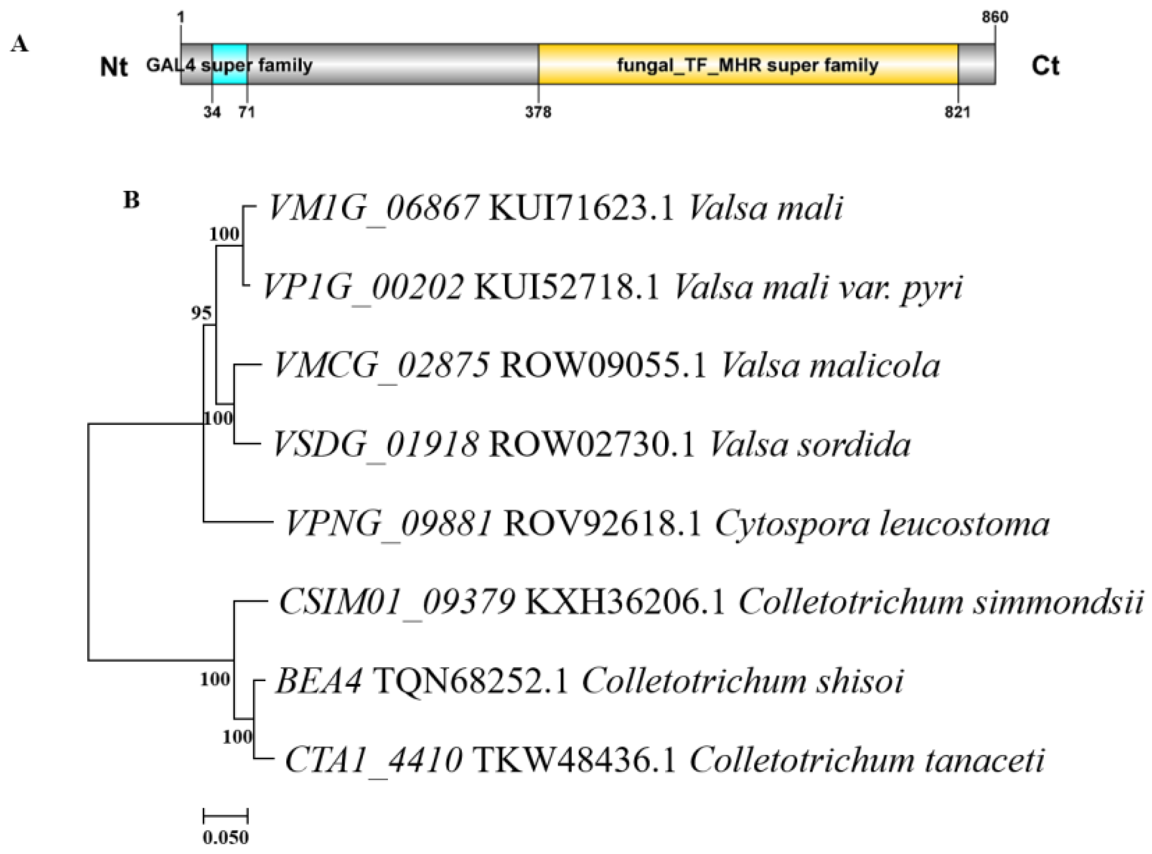
3.1. Bioinformatical Characterizations of VM1G_06867 in V. mali

We identify the single copy VM1G_06867 gene (Accession number KUI71623.1) in the genome of V. mali. The gene sequence is 3228 bp and encodes an 860 amino acid (AA) protein. The VM1G_06867 protein contains a GAL4 super family domain at 34–71AA. Additionally, a fungal_TF_MHR super family domain is predicted at amino acids 373–821 of VM1G_06867 protein. (Figure 1A). A phylogenetic tree was constructed revealing VM1G_06867 showed high similarity with orthologous genes of other fungi. VP1G_00202 showed 95.58% similarity, VMCG_02875 showed 90.82% similarity, VSDG_01918 had 90.01% similarity, VPNG_09881 has 86.86% similarity, CSIMO1_09379 showed 65.43% similarity, the BEA4 similarity was 65.97%, and the CTA1_4410 similarity was 65.29%. The VM1G_06867 had a 100% identity with its homolog in the industrially important Valsa mali var. Pyri (Figure 1B).

3.2. Role of VM1G_06867 in Growth

To investigate the function of VM1G_06867 in V. mali, we use the hygromycin b phosphotransferase gene (hph) to replace the entire ORF of VM1G_06867 to generate the VM1G_06867 deletion mutant. The detection of VM1G_06867 shows that an expected band is found in the deletion mutants. The detection of complementation in the VM1G_06867 deletion mutant shows that an expected band is found in the complemented mutants. Double deletion genes are obtained in the same way (Figure S1).
To assay the roles of VM1G_06867 in V. mali, we inoculate the wild type and deletion mutants on a PDA medium. The radial growth rate of the VM1G_06867 mutant is reduced by approximately 13% compared to the wild type. The growth rate of the complemented mutant is restored to that of the wild type. The double deletion mutant ΔVmSom1/06867 shows the same characteristics as the single deletion mutant VmSom1, and there is no significant difference in the growth rate between them. Compared with the wild type, the aerial mycelia are reduced, the colony color is white, and the growth rate of mycelia is significantly reduced (Figure 2). These results show that VM1G_06867 plays an important role in the growth of V. mali.

3.3. Negative Regulation of Conidiation and Impact on Conidial Germination by VM1G_06867

To evaluate whether VM1G_06867 is involved in conidiation, we measure the number of pycnidia in the unit colony area of wild-type and deletion mutant strains. The deletion mutant strain produces more pycnidia than the wild type on PDA at 20 dpi. The previously observed phenomenon is reversed when the VM1G_06867 gene is reintroduced into the deletion mutant strain (Figure 3A). This result indicates that VM1G_06867 is a negative regulator of conidiation. To further evaluate whether VM1G_06867 affects conidial germination, we collect conidia and observe the spore germination rate using the suspension drop method. After 18 h post-inoculation (hpi), the conidia swell readily and become ellipsoidal or globular in shape. At 24 hpi, conidia typically germinate and produce one to two germ tubes from both ends of a conidium in the wild type, and the hyphae of the wild type are longer than those of the VM1G_06867 deletion mutant. However, the conidia of the deletion mutant are able to develop into mature hyphae (Figure 3B). This result indicates that the deletion of VM1G_06867 slows conidial germination.

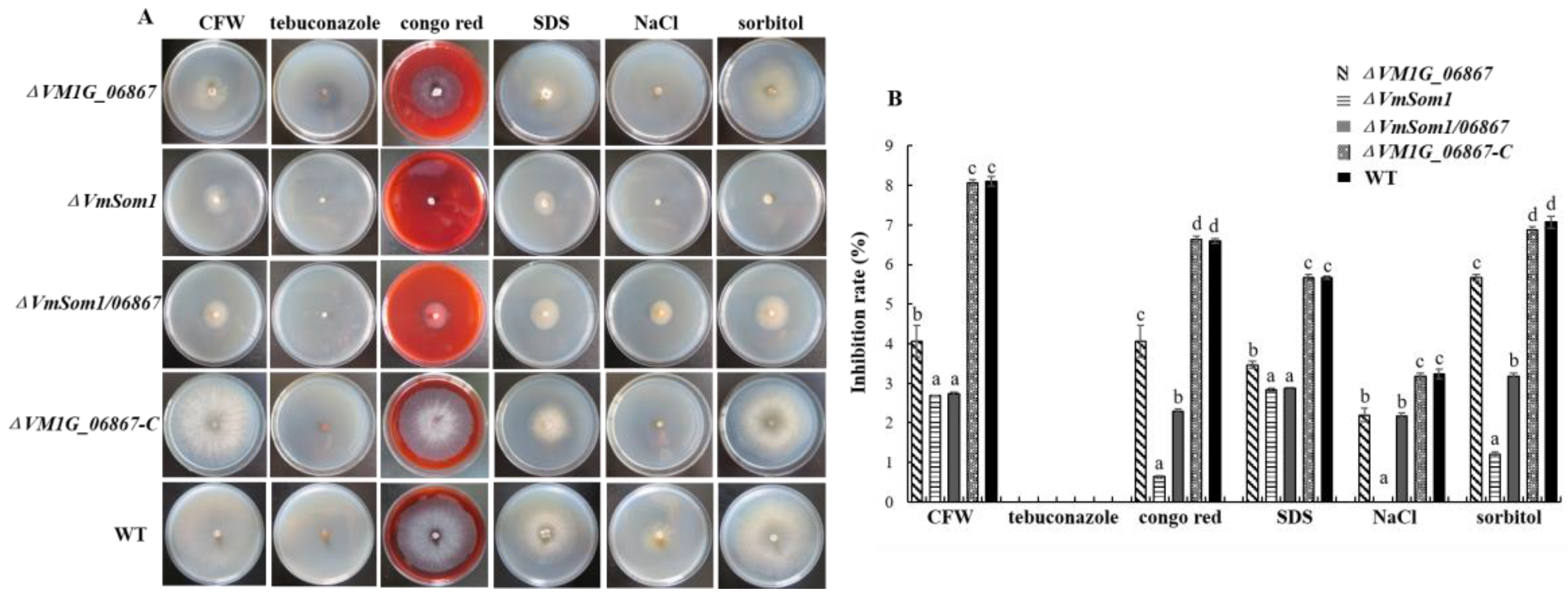
3.4. Stress Influence of VM1G_06867 on Response to Osmotic Stress and Cell-Wall Integrity Inhibitor

To test whether VM1G_06867 plays a role in the response to abiotic stress, we inoculate the wild type, VM1G_06867 single deletion mutant, complemented strain, and double deletion mutant on PDA supplemented with NaCl, sorbitol (osmotic stress), Congo red, fluorescent brightener (cell wall inhibitor), tebuconazole (germicide), and SDS (cell membrane damaging agent). Results show that the growth inhibitions of fluorescent brightener, NaCl, SDS, sorbitol, and Congo red on the VM1G_06867 deletion mutant are higher than those on the wild-type and the complemented mutant strains. However, the inhibition rate of the VM1G_06867 deletion mutant is not significantly different from the wild-type strain on PDA supplemented with tebuconazole (Figure 4). These results show that VM1G_06867 regulates V. mali’s response to osmotic stress and maintenance of cell wall integrity.
Subsequently, the gene double deletion mutant ΔVmSom1/06867 is cultured in a medium containing NaCl (0.5 M), Sorbitol (0.5 M), Congo red (400 µg/mL), fluorescent brightener (400 µg/mL), and SDS (0.01%). The growth rate of mycelium is significantly lower than that of the wild-type strain. Then, the double deletion mutant ΔVmSom1/06867 is compared with the VmSom1 single deletion mutant. On a medium containing Congo red, NaCl, and Sorbitol, the growth rate increases significantly (Figure 4). The results show that the deletion of gene VM1G_06867 can compensate for the deficiency of gene VmSom1 in the medium containing Congo red, NaCl, and Sorbitol. But it fails to completely remedy this defect.

3.5. Impact of VM1G_06867 on Utilization of Carbon and Nitrogen Sources

We incubate all strains on Czapek’s medium containing different carbon sources to assay the effect of gene VM1G_06867 on the utilization of various carbon sources. Lactose, trehalose, fructose, maltose, and glucose of equal quality, respectively, are used in place of sucrose. The results indicate that the mycelial growth rate of the VM1G_06867 single deletion mutant is significantly higher than that of the wild-type strain in sucrose and fructose and significantly less than that of the wild-type strain in lactose and glucose. There is no difference in the growth rate of mycelium in other carbon sources. The analysis of carbon source utilization in the gene double deletion mutants reveals that the hyphal growth rate of ΔVmSom1/06867 is significantly lower than that of the VmSom1 single deletion mutant in the medium containing lactose and fructose. The growth rate of hyphae in a sucrose medium is significantly higher than that of the VmSom1 single deletion mutant (Figure 5).
Moreover, to assay the effect of the VM1G_06867 gene on the utilization of different nitrogen sources, we incubate all strains on Czapek’s medium containing different nitrogen sources. NaNO3 is replaced with peptone, urea, KNO3, and (NH4)2SO4 of equal quality, respectively. The results of nitrogen source utilization analysis show that the mycelial growth rate of the VM1G_06867 single deletion mutant is significantly higher than that of the wild-type strain in peptone, KNO3, and (NH4)2SO4. When using multicomponent complex nitrogen sources such as organic nitrogen peptone, the VM1G_06867 single deletion mutant has the fastest mycelium growth rate. When using single-component nitrogen sources such as urea, the growth rate of mycelium in the VM1G_06867 single deletion mutant is slower compared to sdau11-175. This indicates that the gene VM1G_06867 has an effect on the utilization of nitrogen sources. Analysis of nitrogen source utilization in the gene double deletion mutants reveals that the growth rate of ΔVmSom1/06867 in the medium containing peptone is significantly higher than that of wild-type strain 11-175 and the VmSom1 single deletion mutant (Figure 5).

3.6. Absence of VM1G_06867 in Pathogenicity

We perform pathogenicity tests to determine whether VM1G_06867 is involved in disease transmission. We inoculate mycelial plugs of various strains onto apple twigs and fruit. After inoculating for 9 days, it was observed that the lengths of the lesions on the apple fruit with VM1G_06867 deletion mutant inoculations had decreased by approximately 10.8% compared to those with the WT (Figure 6B). It was also found that the lengths of the lesions on the apple twigs were reduced by approximately 12.6% (Figure 6C). These results indicate that gene VM1G_06867 is involved in the pathogenic process of V. mali. Compared with 11-175, the double deletion mutant ΔVmSom1/06867 reduces the diameter of apple fruit spots by 79.9%, whereas the VmSom1 single deletion mutant increases the diameter of apple fruit spots by 75.9% (Figure 6). The above results show that the deletion of gene VM1G_06867 can compensate for the deficiency of reduced pathogenicity caused by the deletion of VmSom1 but cannot completely compensate.

4. Discussion

VM1G_06867 is a Zn2Cys6-type zinc finger transcription factor. The zinc finger transcription factor family consists of transcription factors widely found in eukaryotes [22,23]. Zn2Cys6-type transcription factors are not highly conserved, and their function varies greatly, being involved in the regulation of different functions in different fungi [24]. These transcription factors have important regulatory effects on the mycelial growth rate, type, and morphology, spore morphology and quantity, metabolite formation, stress response, and pathogenicity. Currently, more in-depth studies have been conducted on the homologous gene flo8 (S. cerevisiae) of the Som1 gene, including studies in filamentous fungi such as A. nidulans, M. oryzae, V. dahliae, A. fumigatus, and M. acridum [13,14,15,16].
In this study, we identify VM1G_06867 in V. mali. The results show that VM1G_06867 performs crucial roles in growth, pathogenicity, asexual development, and the maintenance of cell wall integrity. VM1G_06867 restores osmotic stress and cell wall integrity defects caused by the deletion of VmSom1, and it also restores the loss of pathogenicity caused by the deletion of the VmSom1 gene but not completely. VM1G_06867 is a C6-type zinc finger protein transcription factor of V. mali that affects the growth of V. mali hyphae. The results are consistent with those of Magnaporthe grisea [9,25].
It is well established that conidial reproduction is important for fungi to survive in nature, especially as V. mali mainly infects the host bark via conidia. In this study, conidiation capacity increases in the VM1G_06867 deletion mutant, which shows that VM1G_06867 negatively regulates conidiation, similar to the findings in P. marneffei [26]. In addition, in A. parasiticus and A. flavus, the msnA proteins act as negative regulators of conidiation [27]. Although the deletion of VM1G_06867 slows conidial germination, aberrant conidia are not observed. In A. brassicicola, Abvf19 is independent in conidial germination [28].
Furthermore, VM1G_06867 is involved in the pathogenic process of apple putrefaction in this study. However, in Fusarium graminearum, EBR1(enhanced branching 1) affects the polar growth and virulence of the pathogen. Detailed analysis shows that the reduced radial growth might be due to reduced apical dominance of the hyphal tip, leading to increased hyphal branching, and preventing the hyphal tip from puncturing the host cell [8]. In addition, Chambers et al. found that the Zn (II) 2Cys6 -type transcription factor ptf1 of Leptosphaeria maculan weakened the pathogenicity via base pair substitution during in vitro passage [29]. In Valsa mali, the deletion of VM1G_06867 slows conidial germination, which may be one of the reasons for the reduced pathogenicity.
VM1G_06867 affects the cell wall integrity of the apple rot fungus, resulting in a significant decrease in the tolerance of mutants to cell wall growth inhibitors such as SDS, CFW, and Congo red. Studies have shown that Congo Red and CFW exert significant inhibitory effects on deletion mutants ΔmidA and Δrom2 in A. fumigatus [30]. Similarly, the tolerance of mutants to cell wall synthetic inhibitors decreases significantly after MAPK slt2 gene knockout in Candida glabrata [31]. These changes are related to the cell wall integrity pathway (CWI). However, the cell wall growth inhibitor also has a significant inhibitory effect on the knockout mutant of the VM1G_06867 gene. Further investigation is needed to understand the role of the VM1G_06867 gene in the CWI pathway in the future.
In general, the regulation of growth and development by transcription factors is multilevel and multifaceted, and changes in a single phenotype may be controlled by multiple transcription factors, while a single transcription factor may also affect multifaceted phenotypic changes. Each TF gene regulates the expression of many downstream genes, and each gene expression is regulated by many TF genes so that each mutant phenotype is controlled via the expression changes of several genes [9].

5. Conclusions

In summary, we constructed VM1G_06867 single deletion mutants and double deletion mutants. VM1G_06867 plays important roles in growth, asexual development, maintenance of cell wall integrity, and carbon source utilization. VM1G_06867 can recover osmotic stress and cell wall integrity defects caused by the deletion of VmSom1, as well as restore the loss of pathogenicity caused by the deletion of the VmSom1 gene, but not completely. Our research provides clear evidence that testifies to the molecular pathogenic mechanism of VM1G_06867 in V. mali.

Supplementary Materials

The following supporting information can be downloaded at: https://www.mdpi.com/article/10.3390/jof9060692/s1, Table S1. Primers used in this study. Figure S1. VM1G_06867 gene replacement and complementarity. (A) Amplification of the upstream and downstream fragments. (B) Amplification of the hygromycin b phosphotransferase gene (hph) fragment using primer pairs HPH-F and HPH-R. (C) Generation of the replacement fragment by double-join PCR. (D) PCR Screening and confirmation of the VM1G_06867 single deletion mutant using the four primer pairs as described. (E) PCR screening and confirmation of the VM1G_06867 double deletion mutant using the four primer pairs as described. (F) Generation of VM1G_06867 fragments with their promoters using primer pairs 06867-HB-F/R.

Author Contributions

Conceptualization, Y.D. and J.J.; methodology, X.X.; software, C.Y.; validation, Y.T. and J.J.; formal analysis, J.J.; investigation, X.X.; resources, Y.D.; data curation, Y.D., X.X. and J.J.; writing—original draft preparation, Y.D.; writing—review and editing, H.L.; visualization, H.L.; supervision, D.L. and H.L.; project administration, H.L. and C.Y.; funding acquisition, H.L. All authors have read and agreed to the published version of the manuscript.

Funding

This research was funded by the National Natural Science Foundation of China (grant number 31770684) and the Shandong Province of China (Innovation Project of Forestry Science and Technology) Project (2019LY003-4).

Institutional Review Board Statement

Not applicable.

Informed Consent Statement

Not applicable.

Data Availability Statement

All the data that support the findings of this study are available in the paper and its Supplementary Material published online.

Conflicts of Interest

The authors declare no conflict of interest.

References

  1. Wang, C.X.; Li, C.; Li, B.H.; Li, G.F.; Dong, X.L.; Wang, G.P.; Zhang, Q.M. Toxins produced by Valsa mali var. mali and their relationship with Pathogenicity. Toxins 2014, 6, 1139–1154. [Google Scholar] [PubMed] [Green Version]
  2. Li, B.H.; Wang, C.X.; Dong, X.L. Research progress in apple diseases and problems in the diseases management in China. Plant Prot. 2013, 39, 46–54. [Google Scholar]
  3. Cui, X.Q.; Ding, Z.; Wu, L.; Che, Q.; Li, D.H.; Gu, Q.Q.; Zhu, T.J. Research progress on the pathway-specific transcription factors and their applocation in activating cryptic fungal secondary metabolites. Chin. J. Mar. Drugs 2017, 36, 75–82. [Google Scholar]
  4. Wu, Y.X.; Xu, L.S.; Yin, Z.Y.; Hao, F.; Huang, L.L. Transcription factor VmSeb1 is required for the growth, development, and virulence in Valsa mali. Microb. Pathog. 2018, 123, 132–138. [Google Scholar] [CrossRef]
  5. Shelest, E. Transcription factors in fungi. FEMS Microbiol. Lett. 2008, 286, 145–151. [Google Scholar] [CrossRef] [Green Version]
  6. Benjamin, P.K.; Nancy, P.K. Key players in the regulation of fungal secondary metabolism. Fungal Biol. 2015, 2, 13–28. [Google Scholar]
  7. Chung, H.; Choi, J.; Park, S.Y.; Jeon, J.; Lee, Y.H. Two conidiation-related Zn2Cys6 transcription factor genes in the rice blast fungus. Fungal Genet. Biol. 2013, 61, 133–141. [Google Scholar] [CrossRef] [Green Version]
  8. Zhao, C.Z.; Waalwijk, C.; Wit, P.J.G.M.D.; Lee, T.V.D.; Tang, D.Z. EBR1, a novel Zn(2)Cys(6) transcription factor, affects virulence and apical dominance of the hyphal tip in Fusarium graminearum. Mol. Plant-Mol. Interact. MPMI 2011, 24, 1407–1418. [Google Scholar] [CrossRef] [Green Version]
  9. Lu, J.P.; Cao, H.J.; Zhang, L.L.; Huang, P.Y.; Lin, F.C. Systematic analysis of Zn2Cys6 transcription factors required for development and pathogenicity by high-throughput gene knockout in the rice blast fungus. PLoS Pathog. 2014, 10, el004432. [Google Scholar] [CrossRef]
  10. Tanaka, A.; Cartwright, G.M.; Saikia, S.; Kayano, Y.; Takemoto, D.; Kato, M.; Tsuge, T.; Scott, B. ProA, a transcriptional regulator of fungal fruiting body development, regulates leaf hyphal network development in the Epichloe festucae-Lolium perenne symbiosis. Mol. Microbiol. 2013, 90, 551–568. [Google Scholar] [CrossRef]
  11. Boyce, K.J.; McLauchlan, A.; Schreider, L.; Andrianopoulos, A. Intracellular growth is dependent on tyrosine catabolism in the dimorphic fungal pathogen Penicillium marneffei. PLoS Pathog. 2015, 11, e1004790. [Google Scholar] [CrossRef] [PubMed] [Green Version]
  12. Rispail, N.; Soanes, D.M.; Ant, C.; Czajkowski, R.; Grünler, A.; Huguet, R.; Perez-Nadales, E.; Poli, A.; Sartorel, E.; Valiante, V.; et al. Comparative genomics of MAP kinase and calcium-calcineurin signaling components in plant and human pathogenic fungi. Fungal Genet. Biol. 2009, 46, 287–298. [Google Scholar] [CrossRef] [PubMed]
  13. Tri-Thuc, B.; Harting, R.; Braus-Stromeyer, S.A.; Van-Tuan, T.; Leonard, M.; Hoefer, A.; Abelmann, A.; Bakti, F.; Valerius, O.; Schlueter, R.; et al. Verticillium dahliae transcription factors Som1 and Vta3 control microsclerotia formation and sequential steps of plant root penetration and colonisation to induce disease. New Phytol. 2018, 221, 2138–2159. [Google Scholar]
  14. Yan, X.; Li, Y.; Yue, X.F.; Wang, C.C.; Que, Y.W.; Kong, D.D.; Ma, Z.H.; Talbot, N.J.; Wang, Z.Y. Two novel transcriptional regulators are essential for infection-related morphogenesis and pathogenicity of the rice blast fungus Magnaporthe oryzae. PLoS Pathog. 2011, 7, e1002385. [Google Scholar] [CrossRef] [PubMed] [Green Version]
  15. Lin, C.J.; Sasse, C.; Gerke, J.; Valerius, O.; Irmer, H.; Frauendorf, H.; Heinekamp, T.; Strβburger, M.; Tran, V.T.; Herzog, B.; et al. Transcription factor SomA is required for adhesion, development and virulence of the human pathogen Aspergillus fumigatus. PLoS Pathog. 2015, 11, e1005205. [Google Scholar] [CrossRef] [PubMed] [Green Version]
  16. Du, Y.; Jin, K.; Xia, Y. Involvement of MaSom1, a downstream transcriptional factor of cAMP/PKA pathway, in conidial yield, stress tolerances, and virulence in Metarhizium acridum. Appl. Microbiol. Biotechnol. 2018, 102, 5611–5623. [Google Scholar] [CrossRef]
  17. Yin, Z.Y.; Liu, H.Q.; Li, Z.P.; Ke, X.W.; Dou, D.L.; Gao, X.N.; Song, N.; Dai, Q.Q.; Wu, Y.X.; Xu, J.R.; et al. Genome sequence of Valsa canker pathogens uncovers a potential adaptation of colonization of woody bark. New Phytol. 2015, 208, 1202–1216. [Google Scholar] [CrossRef]
  18. Tamura, K.; Stecher, G.; Peterson, D.; Alan, F.; Sudhir, K. MEGA6: Molecular Evolutionary Genetics Analysis version 6.0. Mol. Biol. Evol. 2013, 30, 2725–2729. [Google Scholar] [CrossRef] [Green Version]
  19. Yu, J.H.; Hamari, Z.; Han, K.H.; Seo, J.A.; Reyes-Domínguez, Y.; Scazzocchio, C. Double-joint PCR: A PCR-based molecular tool for gene manipulations in filamentous fungi. Fungal Genet. Biol. 2004, 41, 973–981. [Google Scholar] [CrossRef]
  20. Chen, L.; Lun, Y.Y.; Sun, G.W.; Zhao, Y.T.; He, B.L.; Wang, H.K.; Liu, H.X. Preparation of protoplasm and optimization of regeneration conditions of Valsa mali. Sci. Agric. Sin. 2014, 46, 109–112. [Google Scholar]
  21. Zhou, X.Y.; Zhang, H.F.; Li, G.T.; Shaw, B.; Xu, J.R. The Cyclase-associated protein Cap1 is important for proper regulation of infection-related morphogenesis in Magnaporthe oryzae. PLoS Pathog. 2012, 8, 503–512. [Google Scholar] [CrossRef] [PubMed] [Green Version]
  22. Akhtar, A.; Becker, P.B. The histone H4 acetyltransferase MOF uses a C2HC zinc finger for substrate recognition. EMBO Rep. 2001, 2, 113–118. [Google Scholar] [CrossRef] [PubMed] [Green Version]
  23. Cass, D.; Hotchko, R.; Barber, P.; Jones, K.; Gates, D.P.; Berglund, J.A. The four Zn fingers of MBNL1 provide a flexible platform for recognition of its RNA binding elements. BMC Mol. Biol. 2011, 12, 20. [Google Scholar] [CrossRef] [Green Version]
  24. Son, H.; Sco, Y.S.; Min, K.; Park, A.R.; Lcc, J.; Jin, J.M.; Lin, Y.; Cao, P.; Hong, S.Y.; Kim, E.K.; et al. Agmhenome-based functiona analysis of transcription factors in the cereal head blight fungus, Fusarium gaminearum. PLoS Pathog. 2011, 7, e100231. [Google Scholar] [CrossRef]
  25. Cao, H.J. Analysis of Transcription Factor Function of Zn2Cys6 and bHLH Family in Magnaporthe oryzae; Zhejiang University: Hangzhou, China, 2015. [Google Scholar]
  26. Bugeja, H.E.; Hynes, M.J.; Andrianopoulos, A. HgrA is necessary and suffiffifficient to drive hyphal growth in the dimorphic pathogen Penicillium marneffffei. Mol. Microbiol. 2013, 88, 998–1014. [Google Scholar] [CrossRef]
  27. Chang, P.K.; Scharfenstein, L.L.; Luo, M.; Mahoney, N.; Molyneux, R.J.; Yu, J.J.; Brown, R.L.; Campbell, B.C. Loss of msnA, a putative stress regulatory gene, in Aspergillus parasiticus and Aspergillus flflavus increased production of conidia, aflflatoxins and kojic acid. Toxins 2011, 3, 82–104. [Google Scholar] [CrossRef] [Green Version]
  28. Srivastava, A.; Ohm, R.A.; Oxiles, L.; Brooks, F.; Lawrence, C.B.; Grigoriev, I.V.; Cho, Y. A zinc-fifinger-family transcription factor, AbVf19, is required for the induction of a gene subset important for virulence in Alternaria brassicicola. Mol. Plant Microb. Interact. 2012, 25, 443–452. [Google Scholar] [CrossRef] [PubMed] [Green Version]
  29. Chambers, K.R.; Wouw, A.P.V.D.; Gardiner, D.M.; Elliott, C.E.; Idnurm, A. A conserved Zn2Cys6 transcription factor, identified in a spontaneous mutant from in vitro passaging, is involved in pathogenicity of the blackleg fungus Leptosphaeria maculans. Fungal Biol. 2021, 125, 541–550. [Google Scholar] [CrossRef]
  30. Samantaray, S.; Neubauer, M.; Helmschrott, C.; Wagener, J. Role of the Guanine Nucleotide Exchange Factor Rom2 in Cell Wall Integrity Maintenance of Aspergillus fumigatus. Eukaryot. Cell 2013, 12, 288–298. [Google Scholar] [CrossRef] [PubMed] [Green Version]
  31. Miyazaki, T.; Inamine, T.; Yamauchi, S.; Nagayoshi, Y.; Saijo, T.; Izumikawa, K.; Seki, M.; Kakeya, H.; Yamamoto, Y.; Yanajihara, K.; et al. Role of the Slt2 mitogen-activated protein kinase pathway in cell wall integrity and virulence in Candida glabrata. FEMS Yeast Res. 2010, 10, 343–352. [Google Scholar] [CrossRef] [Green Version]
Figure 1. Domain structures and phylogenetic analysis of VM1G_06867. (A) The gene sequence is 3228-bp and encodes a protein of 860 amino acids (AA). The VM1G_06867 protein contains a GAL4 super family domain at 34–71AA. Additionally, a fungal_TF_MHR super family domain is predicted at amino acids 373–821 of VM1G_06867 protein. (B) Phylogenetic analysis of VM1G_06867 of Valsa mali and its homologs in other fungi. The VM1G_06867 proteins are analyzed using MEGA 7 and neighbor-joining analysis with 1000 bootstrap replicates. The numbers on the branches represent the percentage of replicates supporting each branch. The bar represents a divergence of 20% in the sequence.
Jof 09 00692 g001
Figure 2. Effects of deletion of VM1G_06867 on mycelial growth in Valsa mali. (A) Colony morphologies on PDA after growth for 3 days. (B) Measurement of colony diameter after 3 days. The mean and standard deviation, which are calculated using data from three biological replicates. The statistical analysis is performed using Fisher’s least significant difference (LSD) in the SPSS software package. Different letters indicate statistically significant differences (Duncan’s new multiple range test, p < 0.05).
Jof 09 00692 g002
Figure 3. Effects of deletion of VM1G_06867 on conidiation and conidia germination in Valsa mali. (A) Growth of pycnidia in wild type, VM1G_06867 single deletion mutant, complemented mutant, and double deletion mutant on PDA for 20 days PDA. (B) Quantification of produced pycnidia produced as described in Section 2. Data from three replicates are analyzed using the protected Fisher’s least significant difference (LSD) test. Different letters indicate statistically significant differences (Duncan’s new multiple range test, p < 0.05). (C) Coating of spore suspensions from different strains onto PDA media at 25 °C for 30 h. Bars = 10 μm.
Jof 09 00692 g003
Figure 4. Effects of deletion of VM1G_06867 on stress responses in Valsa mali. (A) The wild type, VM1G_06867 single deletion mutant, complemented mutant, and double deletion mutant are inoculated on PDA supplemented with NaCl, sorbitol (osmotic stress), Congo red, fluorescent brightener (cell wall inhibitor), tebuconazole (germicide), and SDS. Images are taken after 3 days on different stress media. (B) The mean and standard deviation, which are calculated using data from three biological replicates. The Fisher’s least significant difference (LSD) in the SPSS software package is used for statistical analysis. Different letters indicate statistically significant differences (Duncan’s new multiple range test, p < 0.05). Bars indicate standard deviations of the mean of the three replicates.
Jof 09 00692 g004
Figure 5. Effect of carbon and nitrogen sources on hyphal growth of gene VM1G_06867. (A) All strains are incubated on Czapek’s medium containing different carbon sources, respectively. Sucrose is replaced with lactose, fructose, maltose, and glucose of equal quality, respectively. (B) The mean and standard deviation, which are calculated using data from three biological replicates. The Fisher’s least significant difference (LSD) in the SPSS software package is used for statistical analysis. Different letters indicate statistically significant differences (Duncan’s new multiple range test, p < 0.05). Bars indicate standard deviations of the mean of the three replicates. (C) All strains are incubated on Czapek’s medium containing different nitrogen sources, respectively. NaNO3 is replaced with peptone, urea, KNO3, and (NH4)2SO4 of equal quality, respectively. (D) The mean and standard deviation, which are calculated using data from three biological replicates. The Fisher’s least significant difference (LSD) in the SPSS software package is used for statistical analysis. Different letters indicate statistically significant differences (Duncan’s new multiple range test, p < 0.05). Bars denote standard deviations of the mean of the three replicates.
Jof 09 00692 g005
Figure 6. Effects of VM1G_06867 on pathogenicity in Valsa mali. (A) Apple fruit is scalded and inoculated with mycelium agar plugs from the wild type, VM1G_06867 single deletion mutant, complemented mutant, and double deletion mutant. (B) The lesion length that is measured at 7 days post inoculation (dpi). The experiments are repeated three times. Different letters indicate statistically significant differences (Duncan’s new multiple range test, p < 0.05). (C) Apple twigs are scalded and inoculated with mycelium agar plugs from the wild type, VM1G_06867 single deletion mutant, complemented mutant, and double deletion mutant. Images are taken at 7 dpi. (D) The lesion length that is measured at 7 dpi. The experiments are repeated three times. Different letters indicate statistically significant differences (Duncan’s new multiple range test, p < 0.05).
Jof 09 00692 g006
Disclaimer/Publisher’s Note: The statements, opinions and data contained in all publications are solely those of the individual author(s) and contributor(s) and not of MDPI and/or the editor(s). MDPI and/or the editor(s) disclaim responsibility for any injury to people or property resulting from any ideas, methods, instructions or products referred to in the content.

