Article

Phenotypic and Genotypic Characterization and Antifungal Susceptibility of Sporothrix schenckii sensu stricto Isolated from a Feline Sporotrichosis Outbreak in Bangkok, Thailand

by
Kanokporn Yingchanakiat
1,
Orawan Limsivilai
1,
Supita Sunpongsri
2,
Waree Niyomtham
3,
Kittitat Lugsomya
4 and
Chompoonek Yurayart
1,*
1
Department of Microbiology and Immunology, Faculty of Veterinary Medicine, Kasetsart University, 50 Ngamwongwan Road, Bangkok 10900, Thailand
2
Veterinary Teaching Hospital, Faculty of Veterinary Medicine, Kasetsart University, 50 Ngamwongwan Road, Bangkok 10900, Thailand
3
Department of Veterinary Microbiology, Faculty of Veterinary Science, Chulalongkorn University, Henri-Dunant Road, Pathumwan, Bangkok 10330, Thailand
4
Antimicrobial Resistance and Infectious Diseases Laboratory, Harry Butler Institute, Murdoch University, 90, South Street, Murdoch, Perth 6150, Australia
*
Author to whom correspondence should be addressed.
J. Fungi 2023, 9(5), 590; https://doi.org/10.3390/jof9050590
Submission received: 12 April 2023 / Revised: 15 May 2023 / Accepted: 16 May 2023 / Published: 18 May 2023
(This article belongs to the Special Issue New Perspectives on Sporothrix and Sporotrichosis)

Abstract

:
Sporotrichosis, an invasive fungal infection caused by Sporothrix schenckii, has emerged in Southeast Asia, affecting cats and posing a potential zoonotic risk to humans. We evaluated 38 feline sporotrichosis cases in and around Bangkok, Thailand, from 2017 to 2021. The isolates were phenotypically and genotypically characterized. The cats infected with sporotrichosis were mainly young adults, males, and domestic short hairs with uncontrolled outdoor access, and they lived in Bangkok. All isolates showed low thermotolerance and converted to the yeast phase at 35 °C. Based on the internal transcribed spacer region of rDNA sequences, our strains belonged to S. schenckii sensu stricto and clustered with clinical clade D. Based on the concatenated tree of calmodulin and beta-tubulin genes, five groups of S. schenckii were generated, and the monophyletic clade, Group II, of Thai strains was recognized. In vitro antifungal susceptibility testing demonstrated that the MIC50 of our isolates to amphotericin B, itraconazole, and posaconazole were within the limit of the species-specific epidemiological cutoff values, suggesting that the organisms were the wild type. Addressing the outbreak of feline sporotrichosis in Thailand by providing guidelines for diagnosis and effective treatment may help control the spread of disease and reduce the risk of cat-transmitted sporotrichosis to humans.

1. Introduction

Sporotrichosis is a subcutaneous and systemic mycotic infection that affects both humans and animals. It is caused by pathogenic dimorphic fungi of the genus Sporothrix, which is plentiful in soil and plant debris. Sporadic infection through skin trauma during activities involving contact with vegetation and soil, such as gardening, farming, or mining among humans, or digging and clawing among cats, has been reported globally [1]. In endemic areas of human and feline sporotrichosis, such as Latin America (Brazil, Colombia, Peru, Argentina, and Venezuela) and Southeast Asia (Malaysia), sporotrichosis has become an emerging health problem from epizootic (cat–cat or cat–dog) and zoonotic (cat–human) infections [2].
Cats with sporotrichosis may exhibit a wide range of clinical signs, from no symptoms to fetal disseminated systemic disease. The most common lesions observed are skin ulcers, granulomatous nodules, and crusts at various sites on the body, particularly on the head, nose, ears, distal limbs, and tail base. Lesions in the nasal passages may make the infection difficult to treat and can lead to extracutaneous signs, such as sneezing, rhinorrhea, and dyspnea, which can lead to therapeutic failure and death [3].
Early diagnosis of sporotrichosis usually relies on direct microscopy of budding yeasts with cigar-shaped bodies from suspected lesions. However, misdiagnosis often occurs, especially among other organisms with yeast-like morphology, such as Histoplasma capsulatum and Cryptococcus neoformans. The gold standard for sporotrichosis diagnosis is fungal isolation and identification; this approach can inform proper treatment and provide useful data for reducing clinical loss and the risk of sporotrichosis outbreaks in humans and animals [4].
S. schenckii and related species are the most prevalent agents causing worldwide invasive zoonotic and epizootic infections in humans and animals [5,6]. Genotypes of Sporothrix spp. are characterized by various genetic markers; the internal transcribed spacer (ITS) region of rDNA, calmodulin (CAL), beta-tubulin (b-TUB), and translation elongation factor-1 alpha (TEF1a) inform the epidemiological distribution associated with geographic areas [7]. S. brasiliensis is emerging in Brazil, S. schenckii sensu stricto (S. schenckii s. str.) and S. globosa are distributed in many continents, and S. luriei is found in Africa, Italy, and India [8,9,10]. Currently, the presence of feline-related sporotrichosis is increasing in Asia. A study of the outbreak in Malaysia revealed a single clonal strain of clinical clade D, S. schenckii s. str., as the predominant trait in the environment causing human and feline sporotrichosis there [11].
The first feline sporotrichosis case in Thailand was described in 2017 and was followed by a few Thai cases of sporotrichosis in humans with no history of previous trauma or contact with an affected animal. Therefore, promoting knowledge about feline sporotrichosis in Thailand is urgent. In this study, we collected and summarized clinical data on feline sporotrichosis; characterized the phenotype and genotype of S. schenckii isolated from a feline sporotrichosis outbreak in Bangkok, Thailand, and the surrounding regions during 2017–2021; and assessed the susceptibility of the organisms to commonly used antifungal drugs.

2. Materials and Methods

2.1. Fungal Isolates and Data Collection

Forty-two isolates from cats (n = 38) and a cat owner (n = 1) with skin lesions suspected of sporotrichosis were obtained from the Veterinary Teaching Hospital at Kasetsart University, Bangkok, Thailand, and isolates from untraceable source data (n = 3) were obtained from a private veterinary diagnostic laboratory in Bangkok. All isolates were recovered between December 2017 and December 2021.
Informed consent was obtained from the cat owners before veterinarians performed direct examination by impression smears and sample collection by wound swabbing. Samples were sent for fungal culture and identification to Microbiology Testing Services in the Department of Microbiology and Immunology, Faculty of Veterinary Medicine, Kasetsart University (registered project number: 00751/63). Samples were then inoculated on Sabouraud dextrose agar (SDA) and brain heart infusion agar (BHIA) containing chloramphenicol (100 mg/L) and amoxicillin (50 mg/L) and incubated at 30 and 35 °C for 5–14 days. The causative agent was initially identified based on dimorphic characteristics of macroscopic and microscopic features of Sporothrix spp. All isolates were stored in sterile water as fungal culture collections for further study.
Demographic and clinical data on the 38 feline sporotrichosis cases were recorded, including geographic origin, age, sex, breed, castration, cat rearing, immunodeficiency disorder, possible route of transmission, number of lesions, type of lesion, treatment, and outcome.

2.2. Phenotypic Characterization

We observed the growth of 26 isolates of S. schenckii on various media (SDA, potato dextrose agar [PDA], and BHIA) and at various temperatures (30, 35, 37, and 40 °C). The growth and transitions to the mycelial and yeast phases, influenced by the culture medium and temperature, were closely monitored under a stereomicroscope (SZ2-ILST, Olympus) for 14 days. The colony diameter was measured by calibrating the ocular micrometer on the imaging software (cellSens V3.2 software, Olympus). The microscopic features of the mold form were evaluated using the slide culture technique on PDA, incubated at 30 °C for 14 days. Morphologies of the yeast-like and yeast forms were obtained from culturing on BHIA at 30 and 35 °C for 14 days. The slides were examined under a microscope (BX43, Olympus), and the cells were measured by a calibrated ocular micrometer on the imaging software (cellSens V3.2, Olympus). The percent growth inhibition (% GI) was calculated using the following formula: [(average colony diameter at low temperature-average colony diameter at high temperature)/average colony diameter at low temperature] × 100. Isolates exhibiting more than 50% GI were classified as having low thermotolerance, whereas those showing less than 50% GI were classified as having high thermotolerance [12].

2.3. Genotypic Characterization

The genomic DNA of 26 isolates was extracted by grinding with a pestle in liquid nitrogen, followed by the Ferrer et al. [13] extraction method. The ITS region was amplified with primers SR6R (5’-AAGTATAAGTCGTAACAAGG-3’) and ITS4 (5’-TCCTCCGCTTATTGATATGC-3’) [14], the CAL gene was amplified with primers CL1 (5’-GA(GA)T(AT)CAAGGAGGCCTTCTC-3’) and CL2A (5’-TTTTTGCATCATGAGTTGGAC-3’) [15], and the b-TUB gene was amplified with primers BT2-F (5’-GGTAACCAAATCGGTGCTGCTTTC-3’) and BT2-R (5’-ACCCTCAGTGTAGTGACCCTTGGC-3’) [16]. The amplicons were sequenced using the capillary electrophoresis sequencing method, using ABI Bigdye Terminator v. 3.1 (Macrogen Inc., Seoul, South Korea). The sequences generated in both forward and reverse directions were assembled and manually corrected for consensus sequences using BioEdit software v. 7.0.5.3. Species of all isolates were predicted using the BLASTn tool of the National Center for Biotechnology Information (https://blast.ncbi.nlm.nih.gov/Blast.cgi, accessed on 3 September 2020 for isolates recovered during 2017–2020, and on 15 August 2021 for isolates recovered in 2021).
The ITS phylogenetic analysis was performed using MEGA11 software for Sporothrix species identification [17]. The phylogenetic trees were constructed using the maximum likelihood (ML) method, the reliability of each node was assessed using a bootstrap analysis of 1000 replicates, and the evolutionary distances were computed using the Kamura 2-parameter method [18,19]. The concatenated CAL and b-TUB genes were aligned by MEGA11, and the phylogenetic tree was generated using the ML method with the GTR+Γ substitution model and 1000 bootstrap replicates in the IQ-TREE 2 [20]. The tree was visualized with iTOL online [21]. All sequences in this study were analyzed against the reference sequences from the previous studies presented in Table 1. In brief, 165 sequences were retrieved from nucleotide sequence databases (GenBank): 135 sequences of S. schenckii (49 human, 70 cat, 3 environment, and 13 undefined origin), 20 sequences of S. globosa (11 human, 9 undefined origin), 1 sequence of S. luriei (human origin), 7 sequences of S. brasiliensis (4 human and 3 undefined origin), 1 sequence of S. pallida (environment origin), and 1 sequence of S. chilensis (human origin) [22,23,24,25,26,27].

2.4. Antifungal Susceptibility Test

Twenty-six isolates of S. schenckii were tested for their susceptibility to eight antifungal agents: amphotericin B (AMB), itraconazole (ITR), posaconazole (POS), ketoconazole (KET), fluconazole (FLU), miconazole (MCZ), terbinafine (TERB), and griseofulvin (GRI). The minimum inhibitory concentration (MIC) and minimum fungicidal concentration (MFC) were determined using the broth microdilution (BMD) method according to the Clinical and Laboratory Standards Institute (CLSI M38-A2) [28]. Antifungal agents were dissolved according to CLSI recommendations and diluted in the RPMI 1640 (Invitrogen, Waltham, MA, USA) buffered to a pH of 7.0 with 0.65 M of MOPS buffer (Sigma, St. Louis, MO, USA) with L-glutamine and without bicarbonate to obtain 2X concentrations. They were loaded to 96-well U-shaped microplates at a final concentration of 0.06 to 32 µg/mL for AMB, ITR, POS, and GRI; 0.0156 to 8 µg/mL for KET, MCZ, and TERB; and 0.25 to 256 µg/mL for FLU (all were purchased from Sigma, St. Louis, MO, USA). Antifungal susceptibilities of both the mold and yeast phases were tested by harvesting the cells from 3-day-old cultures growing on PDA at 30 °C and BHIA at 35 °C, respectively. The conidia and yeast cells were scraped from the colony surface using a sterile cotton swab and stirred into a sterile saline solution. The inoculum suspension was vortexed and allowed to settle for 5 min, and the upper homogeneous layer was transferred, measured, and adjusted to an optical density range of 0.09 to 0.11 at 530 nm. The suspensions were diluted at 1:50 in an RPMI 1640 culture medium to obtain the final concentration of 0.4 × 10⁴ to 5 × 10⁴ CFU/mL.
The inoculated microplates were incubated at 35 °C for 72 h before observing results with the naked eye. The MICs of AMB, ITR, and POS were defined as the lowest concentration that inhibited 100% growth, when compared with an inoculum-free medium control. For the MICs of KET, FLU, MCZ, TERB, and GRI, MICs were defined as the lowest concentration that inhibited 50% growth when compared with a drug-free growth control. The MFCs of all drugs were determined by transferring 200 µL of suspensions of the optically clear well and the higher concentrations (2 to 4 higher concentrations) into the fresh SDB medium in a microtube and incubated at 30 °C for 7 days. The MFCs were detected as the lowest concentration of antifungal drugs that resulted in an optically clear tube by killing all fungal cells. A quality control strain of Candida parapsilosis ATCC 22019 was included in the study.

3. Results

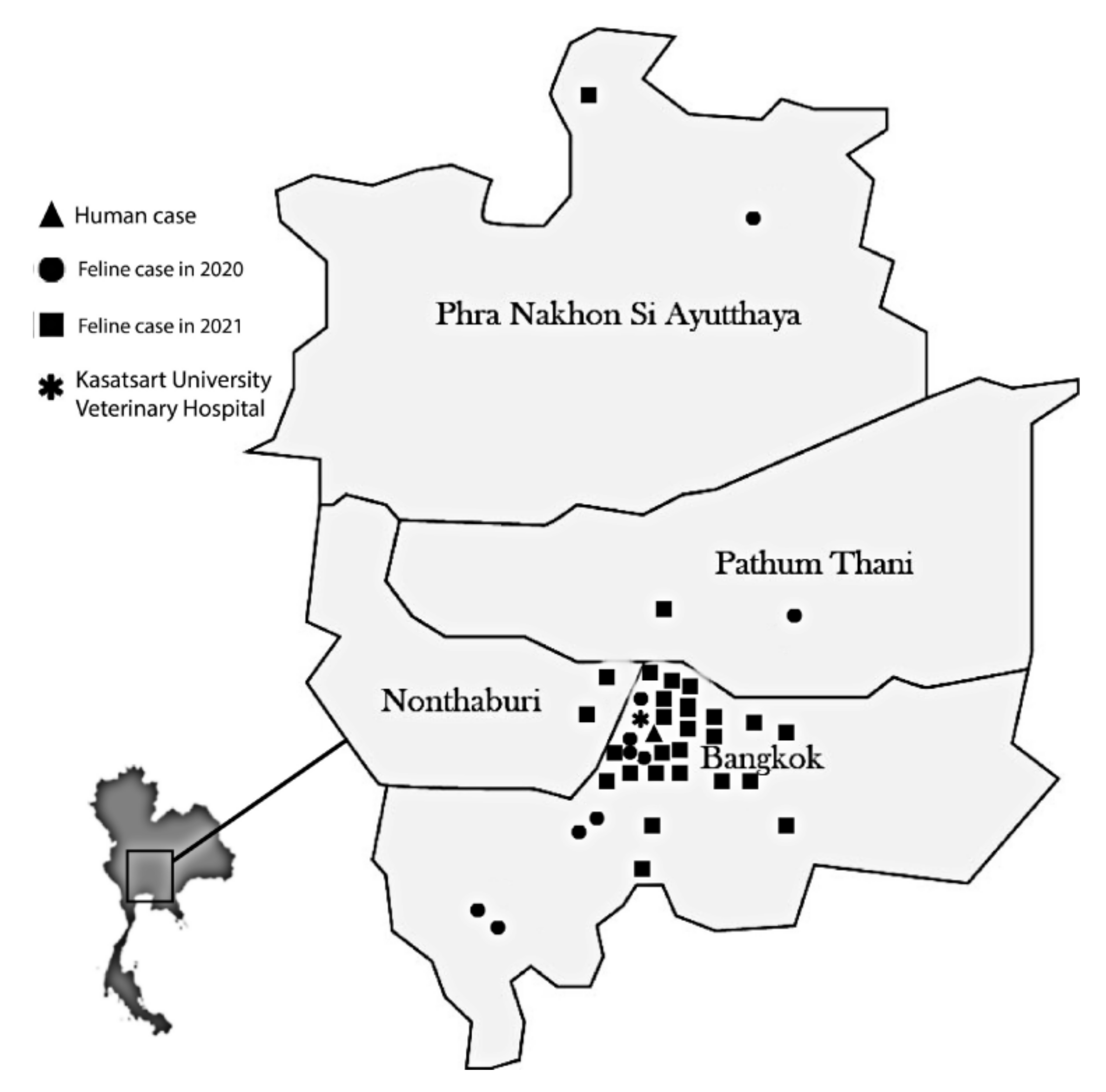
3.1. Geographic Distribution, Demographic Characteristics, and Clinical Data

A total of 42 isolates of S. schenckii were phenotypically identified, and 38 cases of feline sporotrichosis were diagnosed in Bangkok and the Bangkok metropolitan region of Thailand. The number of cases, year of diagnosis, and geographic distribution of the sporotrichosis cases are shown in Figure 1. Most cases occurred in 2021 (n = 27) and were predominantly found in the Bangkok area (85%). The other cases were scattered around the surrounding provinces (Nonthaburi: 5%, Pathum Thani: 5%, Ayutthaya: 5%). One zoonotic case was confirmed in 2020 in a cat owner with a history of scratches from her infected cat prior to the occurrence of a skin lesion.
Table 2 summarizes the demographic and clinical characteristics of feline sporotrichosis cases among the Thai cats in this study, including age (mean ± standard deviation [SD]: 3.96 ± 3.93 years), sex (male: 61%, female: 39%), breed (domestic short hair: 95%, purebred: 5%), castration (yes: 58%, no: 42%), cat rearing (outdoor access: 76%, indoor only: 24%), and feline leukemia virus (FeLV)/feline immunodeficiency virus (FIV) infection (no: 55%, yes: 5%, no data: 40%). History of an associated penetrating wound by fighting with other cats was mentioned in 50% of cases. Clinical presentation included skin lesions at different sites (skin lesions at 1 site: 24%, 2 sites: 29%, ≥3: 47%). The lesions mostly appeared as ulcers (92%) and were occasionally found as granulomatous nodules (8%). To treat sporotrichosis, 82% of cases used only antifungal drugs, while the rest involved surgery for wound debridement combined with antifungal drugs. For treatment outcomes, we found that 53% of cases recovered and 37% died; treatment was still ongoing for the other 10% of cases.

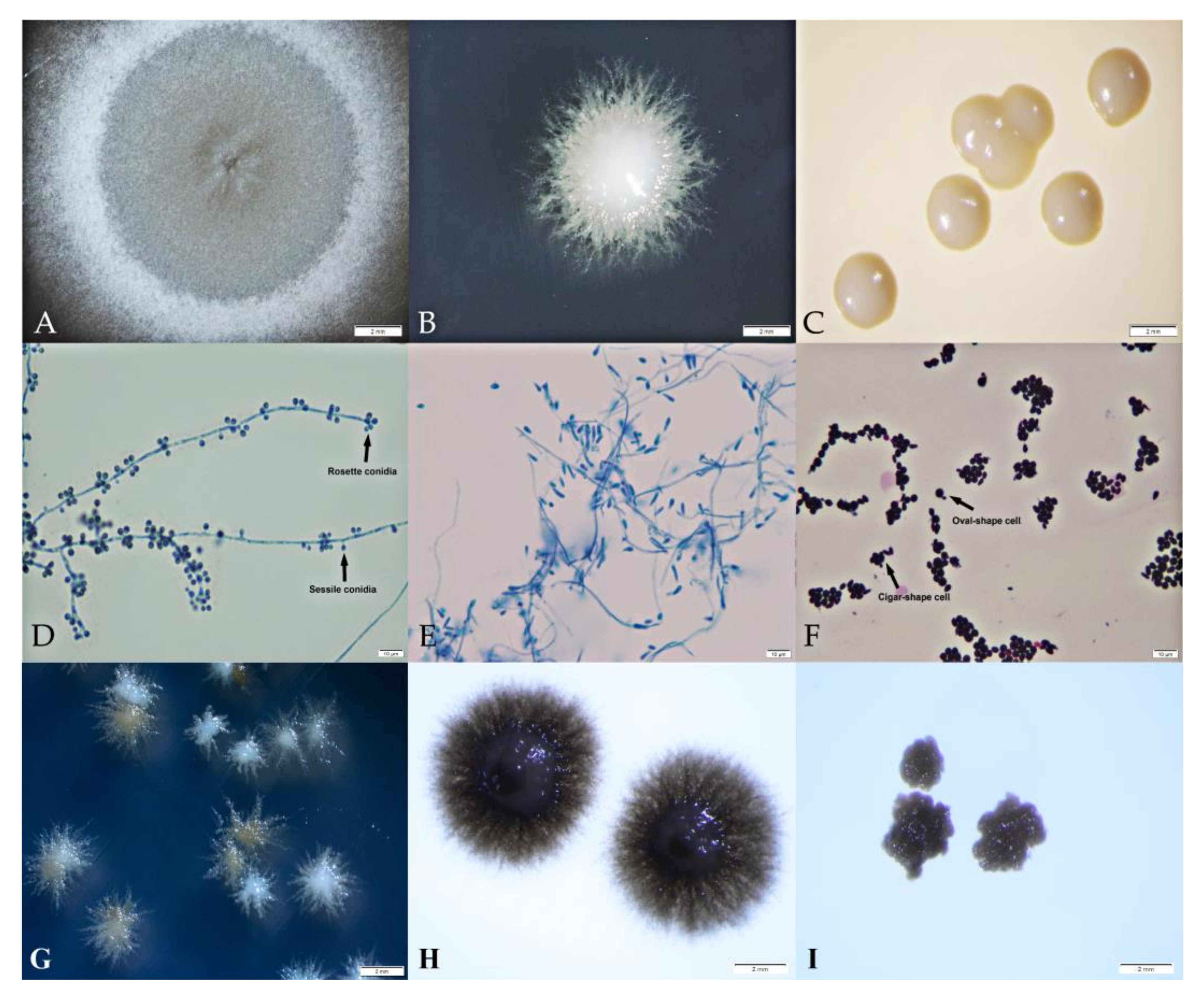
3.2. Phenotypic Characteristics of S. schenckii Thai Isolates

All isolates included in the phenotypic study (n = 26) were able to grow at 30 and 35 °C. The growth was inhibited at 40 °C, and 31% of isolates were stunted earlier at 37 °C. Three unique colony appearances (mold, yeast-like, and yeast) were consistently exhibited and associated with the culture medium and temperature, as presented in Figure 2. The mold colony was observed as white to brownish aerial mycelia (Figure 2A); the yeast-like colony was white to brownish, with a smooth or wrinkled surface and a fine filamentous edge (Figure 2B); and the yeast colony was white to cream colored, moist, and smooth with a round margin (Figure 2C).
Mold colonies showed a microscopic morphology of hyaline septate hyphae, less differentiated conidiophores, and abundant pigmented, thick-walled, globose to ovoid, and 2.39–5.45 µm long by 1.91–2.96 µm wide sessile conidia (Figure 2D). The yeast-like colonies microscopically revealed septate, smooth, thin-walled, hyaline hyphae, conidiophores not differentiated from vegetative hyphae, and produced abundant clavate to elongate pyriform, which were slightly curved and wider rounded at apex conidia (Figure 2E). The microscopic morphology of the yeast form was similar at all temperatures: 2.39 to 5.64 µm in size and mixed oval and cigar-shaped yeast cells with narrow-based budding (Figure 2F). Some of our isolates (9/26) started producing light brown colonies (Figure 2G), which became heavily dark brown in mold form (Figure 2H) and yeast form (Figure 2I) after 14 days of incubation.
Table 3 shows the progression of the mold, yeast-like, and yeast phases, including the colony sizes in relation to the culture medium and temperature. The mold phase was detected in only 19% of all isolates. The largest colony sizes in the mold phase were observed on SDA and PDA at 30 °C, with diameters of 6.99 to 9.34 mm. The smaller colony sizes presented at 35 °C on SDA and PDA with 4% growth. The yeast-like form, observed as a mix of mycelial and yeast cells, was the most common appearance of S. schenckii isolates in our study. This morphology was found in a broad range of growth conditions, especially at 30 °C on SDA/PDA (81%) and BHIA (50%), with a large colony diameter size (3.41 to 4.11 mm), and at 35 ˚C on SDA/PDA (73 to 85%), and with a smaller diameter size (1.74 to 1.97 mm). About 50% of all isolates were able to convert to the yeast phase at 30 °C on BHIA, and the highest conversion percentage was found at 35 °C on BHIA (85%). At higher temperatures (35 and 37 °C), the transition from the mold to the yeast phase occurred on SDA and PDA, but the sizes of the yeast colonies were smaller than those grown on BHIA. The average % GI values at temperatures of 30 to 37 °C were 90.80% on SDA, 95.23% on PDA, and 83.12% on BHIA. Therefore, S. schenckii isolates in our study were classified as a low-thermotolerant strain.

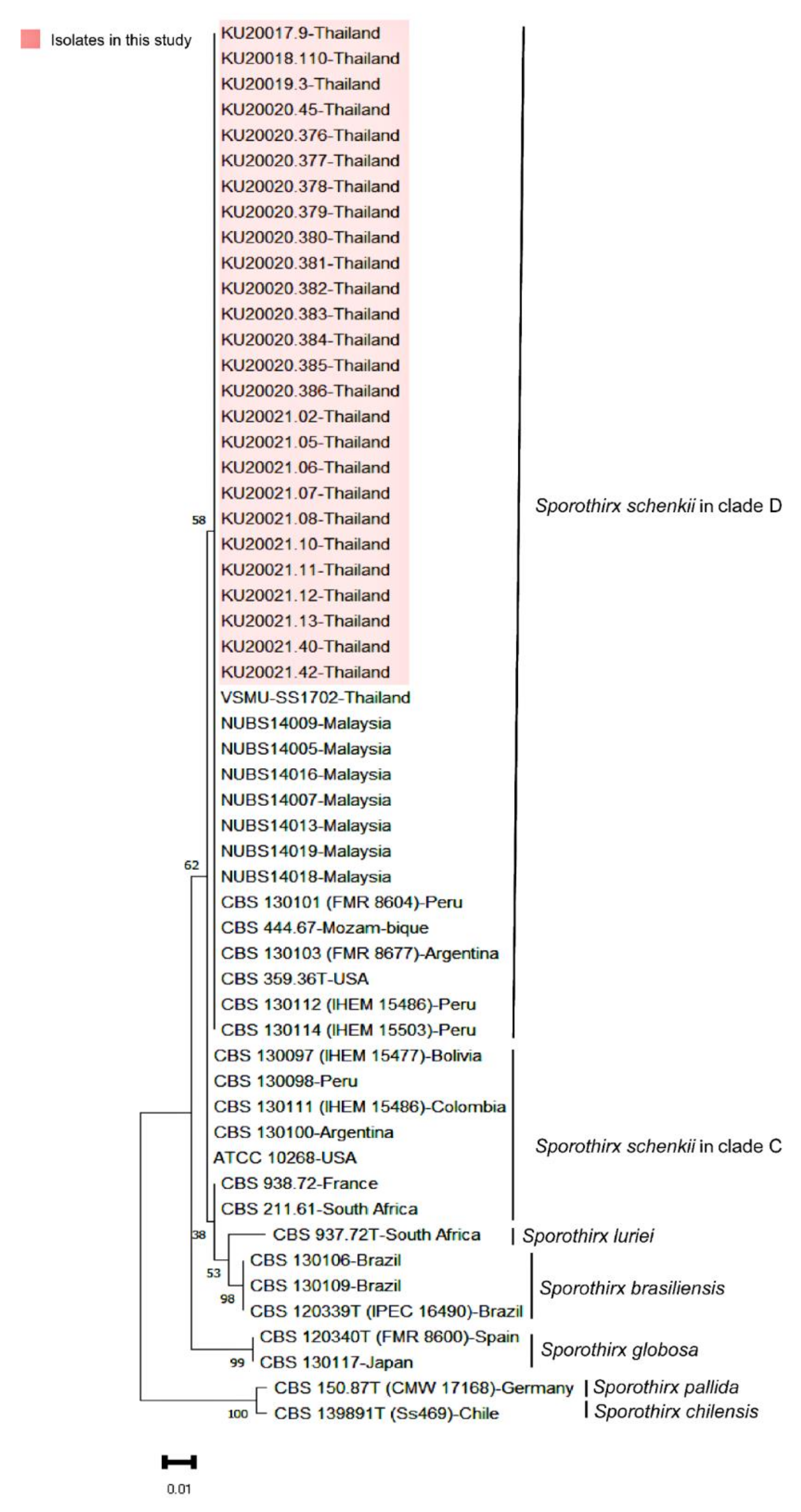
3.3. Phylogenetic Analysis of S. schenckii Thai Isolates

The sequence of ITS was approximately 655 nucleotides, and all sequences were matched with S. schenckii (identity range = 99.83 to 100%). The sequence of CAL was approximately 911 nucleotides, and all strains were matched with S. schenckii (identity range = 95.4 to 100%). The sequence of b-TUB was approximately 404 nucleotides, and all sequences were matched with S. schenckii (identity range = 99.76 to 100%).
Phylogenetic trees constructed based on the sequences of each gene showed the distribution of a clinical clade of Sporothrix spp. into four distinctive species: S. luriei, S. globosa, S. brasiliensis, and S. schenckii. All Thai clinical strains were co-clustered with S. schenckii strains of different geographic origins, including all reported continents. Based on the ITS sequences, the Thai strains were grouped with clade D of S. schenckii, which consists of isolates from Malaysia, the United States, Peru, Argentina, and Brazil (Figure 3). We also aligned the ITS sequences of Thai strains against isolates from Malaysia and other countries. The nucleotide variable sites were detected and showed consistency with the results of the phylogenetic tree analysis. S. schenckii clade C and D had a variable site at position 500 (Figure S1). All Thai and Malaysian strains revealed the presence of nucleotide T at position 162, while nucleotide C was noted among other strains in clade D (Figure S2). The single nucleotide polymorphism between the Thai and Malaysian strains was detected once at position 40—read adenine (A) from the Thai strains and thymine (T) from the Malaysian strains (Figure S3).
Based on a phylogenetic analysis of the concatenated CAL gene and b-TUB gene, the S. schenckii isolates were divided into five groups according to their location on phylogenetic clades. Group I was a group of clades composed of isolates from various hosts (human and environment) and geographic areas (United States, Japan, and Mexico). Group II was a monophyletic clade of Thai isolates that were mainly from cats, with one from a human. Group III was a clade from South America (Columbia, Peru, Bolivia, and Argentina) and North America (Guatemala), mainly isolated from humans. Group IV was a clade of isolates from Peru, Argentina, and South Africa, mostly from humans. Finally, Group V was a clade of environmental isolates from Mexico (Figure 4).

3.4. Antifungal Susceptibility

The in vitro antifungal activities of eight antifungal agents against 26 S. schenckii isolates were evaluated based on MIC and MFC. We analyzed the MIC ranges, geometric mean values, the MIC50, and the MIC90 during both the yeast and mold phases (Table 4). For most antifungal drugs, including AMB, ITR, POS, KET, and MCZ, slightly higher MICs (one dilution) were found for the yeast form, but the opposite was true for TERB.
For AMB, MICs ranged from 0.25 to 4 µg/mL and the MIC50 were between 0.5 to 1 µg/mL for the mold and yeast phases, respectively. An MIC of 4 µg/mL was required to inhibit the growth of most isolates (MIC90), and fungicidal activity was observed for MFCs ranging from 0.25 to ≥32 µg/mL and an MFC90 of 16 µg/mL. For ITR and POS, the MIC ranged from 0.25 to ≥32 µg/mL in the yeast phase and 0.0625 to ≥32 µg/mL in the mold phase. The MIC50 were between 0.5 to 1 µg/mL in the yeast phase and 0.25 to 0.5 µg/mL in the mold phase. A higher MIC was required to inhibit the growth of most isolates (MIC90), at 4 µg/mL for ITR and 8 µg/mL for POS. Interestingly, much higher MFCs of ITR and POS, ranging from 0.5 µg/mL to ≥32 µg/mL, were observed for fungicidal activity.
TERB inhibited the growth of isolates at concentrations ranging from 0.0156 to 2 µg/mL in the yeast phase and 0.0312 to 4 µg/mL in the mold phase. The MIC50 concentrations were 0.5 and 0.75 µg/mL for the yeast and mold phases, respectively, and most of our isolates in both phases required an MIC90 of 2 µg/mL to inhibit growth. However, TERB showed fungicidal activity at MFCs ranging from 1 µg/mL to >8 µg/mL, an MFC50 of 8 µg/mL, and an MFC90 of >8 µg/mL. KET and MCZ had the lowest MIC ranges (0.0039 to 1 µg/mL), MIC50 (0.25 µg/mL), and MIC90 (0.5 µg/mL). The efficacy of FLU against isolates in both phases had an MIC ranging from 32 to >256 µg/mL; the MIC50 and MIC90 were >256 µg/mL.

4. Discussion

Sporotrichosis is a mycotic infectious disease in humans and animals that is often reported in cats. In this study, we assessed the present outbreak of sporotrichosis in and around Bangkok, Thailand.
Our sample mainly comprised domestic cats that had free access to the surrounding area of their houses in Bangkok. All cases and isolates in our study were obtained from the culture collection of the Microbiology Testing Services of the Department of Microbiology and Immunology, which commonly supports cases from the Kasetsart University Veterinary Teaching Hospital. Therefore, the geographic distribution might not be representative of the feline sporotrichosis outbreak in Bangkok or Thailand. However, we noticed that in 2021, there was a widening distribution of cases from Bangkok to the neighboring provinces to the north, including Nonthaburi, Pathum Thani, and Ayutthaya. A similar geographic distribution was observed in Brazil, which suggested the rapid expansion of zoonotic and feline sporotrichosis cases from the South toward the Northeast [29]. In Thailand, the first case was reported in December 2017 in Bangkok and was followed by 38 cases during 2018–2021 reported in southern Thailand, close to Malaysia, the country with the most reported cases of human and feline sporotrichosis in Southeast Asia [14,24,30]. However, the transboundary expansion of this pathogen from Malaysia to Thailand has not yet been proven.
Most cats with sporotrichosis included in our study were young adults (mean age of 3.96 years), male, and domestic short hairs with uncontrolled outdoor access, similar to previous reports [31,32,33]. Specific demographic characteristics such as age and outdoor access, as well as natural and sexual behaviors of cats, may play a critical role in the transmission of sporotrichosis from cat to cat, cat to dog, and cat to human. An age of 3 years is linked to sexual maturity, and being in heat possibly increased the chance of cats fighting or having contact with other infected animals that bear Sporothrix in skin lesions, nails, and oral mucosa [34]. Other cat behaviors that possibly increased their risk of sporotrichosis infection include burying their excrement, climbing trees, or sharpening their claws on trees [35].
The severity of sporotrichosis infections or treatment outcomes were not associated with the underlying presence of FeLV/FIV in our cases, consistent with previous reports [3,36]. In contrast, HIV infection in humans aggravates sporotrichosis, with a higher incidence of severe disseminated cases and a higher number of hospitalizations and deaths [37]. Mortality in our sporotrichosis cases was high among those cats with severe skin lesions—on ≥3 sites of the body (50%)—and those with nasal involvement (21%). Therefore, nasal involvement, respiratory signs, and high fungal loads in skin lesions are prognostic factors of treatment failure in feline sporotrichosis, as suggested by others [38,39]. Other factors may be associated with the failure of treatment, such as low income of the owners, difficulty in administering medication for cats at home, and difficulty in confining cats at home for a long period [33].
Thermotolerance has been known as a factor influencing the virulence of a disease, especially in terms of clinical manifestations. In Brazil, Mexico, and Guatemala where most human cases presented with lymphocutaneous form, strains of S. brasiliensis and S. schenckii recovered there have shown more resistance to 37 °C [12,40]. A previous report of human sporotrichosis in Malaysia revealed that 7 of 19 cases had a history of cat bites/scratches, and lymphocutaneous sporotrichosis was the most common presentation observed there [41]. We identified a phenotypic trait of high-temperature intolerance and a requirement for lower conversion temperatures in our Thai strains, similar to other Thai and Malaysian strains [24,42]. Therefore, the dissemination via lymphatics or blood circulation or its zoonotic potential was yet a concern among these S. schenckii s. str. Thai and Malaysian strains. Furthermore, the low-thermotolerant trait may be related to the common skin lesions in cooler body parts such as ear tips, nasal planum, and nasal cavities in cats [11]. Interestingly, colonization of Sporothrix spp. at sites with a lower body temperature, such as the nasal and oral cavities and the nails, in the infected and noninfected cats of endemic areas is suggested to be a potential source of zoonotic transmission [34]. Moreover, melanin contributes to the virulence of fungal agents by protecting them from the host defense response, oxidizing agents, and hydrolytic enzymes, and reduces phagocytosis or the induction of cell death [43]. Some of our isolates started presenting melanized colonies after seven days of incubation and became dark brown pigmented after 14 days of incubation. These relate to S. brasiliensis, which is most associated with feline and human sporotrichosis in Brazil; this species has rapid melanization and a high level of pigmentation. On the other hand, S. globosa, the most endemic species, caused nearly 99.3% of sporotrichosis in Asia. Most isolates of S. globasa were recovered from fixed cutaneous and lymphocutaneous lesions, and this species has never been reported to cause infections in animals. Furthermore, S. globosa isolates from Asia and all geographical regions are generally considered less virulent than S. schenckii s. str. by presenting very low thermotolerance (97–100% of inability to grow at 37 °C), a low percentage of yeast conversion in lesion and culture, and a low level of melanin production [12,44]. Therefore, further studies on the virulence and pathogenicity of S. schenckii s. str., the most isolated species causing emerging feline sporotrichosis in Southeast Asia with zoonotic potential, should be extended in the future.
Phenotypic identification based on micromorphology, physiology, and biochemical tests is inadequate for the species-level identification of Sporothrix spp., which is related to geographic distribution, host range, virulence factors, and antifungal susceptibility [45]. Based on the sequencing of the ITS region of rDNA, all isolates were identified as S. schenckii s. str. with a very high identity match (99.83–100%) and were clustered in clinical clade D together with strains originally reported in Malaysia, southern Thailand, and globally. In terms of S. schenckii s. str., intraspecies sequence-based typing has been performed using various methods, such as, for example, mating-type idiomorphs [46], mitochondria DNA (mtDNA) typing [47], and multi-locus sequence analysis (MLSA) of concatenated genes (CAL, b-TUB, ITS, or translation elongation factor [TEF] encoding genes), which is commonly used to determine the genetic relatedness of whole groups of the genus Sporothrix [48,49,50]. To demonstrate the geographic relationship of the isolates from Thailand and other geographic origins, a phylogenetic tree was built with the concatenated sequences of the CAL and b-TUB markers. According to the phylogenetic trees, our isolates and feline clinical isolates from another study from the southern part of Thailand were grouped together in the same clade (Group II) with strong bootstrap support (98%), supporting their close genetic relationship and clearly dividing them from others in the phylogenetic tree [24]. There have also been studies of feline and human clinical isolates from a nearby geographic area in Southeast Asia (Malaysia) [23,26]. Even though there is a lack of concordance of gene selection with our analysis, the previous research showed that the Malaysian and Thai S. schenckii s. str. isolates were closely related in CAL gene phylogeny, regardless of whether they were from humans or cats [46]. Interestingly, Group IV on the tree was composed of the isolates from different geographic areas (Argentina, Peru, and South Africa), which was the nearest clade from the S. brasiliensis outgroup, but this arrangement of clades conformed with previous studies that used two different base methods. One of these methods is amplified fragment length polymorphism, which is a well-acknowledged DNA fingerprint typing method in the Sporothrix species [42], and the other is MLSA sequence-based typing of the concatenated chitin synthase (CHS), b-TUB, and CAL genes.
Although the ITS is the most used and most sufficient in distinguishing the species of Sporothrix, the bootstrap value of our phylogenetic tree showed that the CAL was the most accurate for clustering the clinically relevant Sporothrix spp. These results are supported by previous studies, including Zhang et al. [43], which reported the success rates of multi-locus gene sequencing (ITS, CAL, TEF1, and TEF3) data analysis. They demonstrated that identification with the CAL gene had a high-resolution power by obtaining small intraspecific sequence variability compared with barcoding gaps between species. We suggest using the ITS region for routine identification because the ITS gene is a universal DNA barcode for the identification of fungi species [51]. Using the CAL gene is preferred for epidemiologically sorting Sporothrix clinical species.
For antifungal susceptibility testing, we observed higher MICs in the yeast phase, which differed from previous studies [52,53,54] and could possibly be explained by the inoculum sizes used. We followed the CLSI M38-A2 guidelines for both the yeast and mold phases; therefore, the yeast inocula were 40 times higher than the usual recommended use in BMD yeast testing (CLSI M27-A3). Interestingly, consistency in the MIC90s obtained in both phases was detected by following the inoculum size in the CLSI M38-A2. BMD testing against S. schenckii is officially described in the CLSI M38-A2 for only the filamentous phase, whereas the infected form in the host body as the yeast phase has not been standardized yet. The correlation between the lower MICs of the yeast phase according to the CLSI M27-A3 of previous studies and the emergence of in vitro resistant strains of S. schenckii and in vivo treatment failure is still in question [55]. The MIC breakpoints of Sporothrix are not established yet in the CLSI M38-A2 document [28]. Species-specific epidemiological cutoff values (ECVs) for S. schenckii and S. brasiliensis have been proposed to identify the non-wild type isolate that is equal to a potentially resistant isolate. However, the ECVs for S. schenckii were only available for AMB (4 µg/mL), ITR (2 µg/mL), POS (2 µg/mL), and voriconazole (64 µg/mL), and not for KET or TERB yet [56]. In accordance with Espinel-Ingroff et al. [55], we preferred to interpret the antifungal-resistance profile of our isolates using the ECVs versus the MIC, MIC50, and MIC90 values.
For AMB, all our isolates were classified as wild type strains by the MIC50 and MIC90. They were similar to S. schenckii isolates found worldwide but different from the non-wild type or AMB-resistant strains that have been identified in Brazil [57]. For ITR, our strains were classified as wild type by the MIC50 but exhibited a non-wild type MIC90 value, similar to the S. schenckii from Brazil but different from isolates worldwide that exhibited non-wild type MIC50 and MIC90 values. However, we found four ITR-resistant strains (4/26, 15.38%) with MICs of 4 µg/mL (n = 2) and >32 µg/mL (n = 2). Previous reports of antifungal susceptibilities of S. schenckii isolates from southern Thailand showed high resistance to AMB (34.21%) and ITR (26.32%), while studies in Malaysia reported 5% of isolates were ITR-resistant strains [24,42]. ITR is the drug of choice for feline sporotrichosis treatment; however, S. schenckii has been found to be less susceptible to ITR than S. brasiliensis [58]. In cats, the bioavailability of ITR was observed to be better in oral solution than in tablets or capsules, but it is more expensive and less accessible. Concerns regarding the use of capsules or tablets for treatment in feline sporotrichosis include treatment failure and drug resistance [59]. For POS, our isolates were identified as wild type and non-wild type strains by MIC50 and MIC90, similar to S. schenckii reported worldwide [57]. POS is a new triazole drug with fungicidal effects against Aspergillus spp., Candida spp., and zygomycetes [60]. In our S. schenckii isolates, POS was more active than ITR, but POS-resistant and ITR–POS-resistant strains were detected. To the best of our knowledge, POS has never been used in Thai veterinary practices, so the resistance remains questionable.
Because there were ECVs of only four antifungal drugs available for identifying the wild type and non-wild type strains of S. schenckii, the susceptibility of other antifungal drugs tested in our strains was compared with the MIC of mode (the most frequent MIC detected in S. schenckii worldwide) published by Espinel-Ingroff et al. [55]. Various studies have reported that TERB was the most effective antifungal drug against S. schenckii, with low MICs. Data obtained from worldwide testing of 118 isolates had an MIC range of ≤0.03–1 µg/mL, a mode of 0.5 µg/mL (43/118), and the highest reported MIC since 2017 at 1 µg/mL (6/118) [55,61,62,63,64]. Our isolates demonstrated high MICs for TERB at 1 µg/mL (7/26), 2 µg/mL (6/26), and 4 µg/mL (1/26). High MICs for TERB were also reported from cat isolates in Malaysia: 4 µg/mL (23/40 isolates) and 8 µg/mL (4/40 isolates). Since being reported in the 1990s, Malaysian isolates of S. schenckii s. str. have exhibited low susceptibility against all antifungal drugs [42]. In our hospital, TERB has been used more frequently, especially in combination with ITR, to treat sporotrichosis cases with low responsiveness. For other less commonly used antifungal drugs, isolates in Thailand were susceptible to KET and MCZ, but the many side effects of KET, such as anorexia, depression, nausea, vomiting, diarrhea, or elevated liver enzymes, are factors to consider before using these drugs [65]. The emerging resistance to commonly used antifungal drugs, including ITR, TERB, and AMB, should be a concern in refractory cases, and antifungal susceptibility testing might be required.
Cat-transmitted sporotrichosis has garnered worldwide public concern, and a One Health approach has been implemented in South American countries to control and prevent the spread of the disease in animals, humans, and the environment. Veterinarians play an important role in controlling this disease, including investigating and addressing the outbreak; educating cat owners on the importance and difficulties of treatment, management, and control of their infected cat; understanding the risks and sources of zoonosis; and decontaminating and responsibly handling deceased cats [66]. Since 2018, Thailand has faced a feline sporotrichosis outbreak. However, the true burden of this disease in cats and humans has not been investigated. Further studies and programs focused on epidemiology, surveillance, and controlling the spread of sporotrichosis among the cat population may help reduce the risk of cat-transmitted sporotrichosis to other animals and humans.

5. Conclusions

We described 38 cases in the recent feline sporotrichosis outbreak caused by S. schenckii s. str. in Bangkok and the surrounding provinces, which are in the central part of Thailand. Our Thai isolates reported here share phenotypic and genotypic characteristics with those distributed in Malaysia and southern Thailand. Susceptibility testing of the Thai isolates showed that AMB, ITR, and POS MIC50s were within the limit of ECVs and compatible with the wild type traits. However, high MICs and MFCs of ITR and POS have been detected in some non-wild type or resistant strains. The MICs of TERB observed within our isolates were higher than those frequently reported worldwide. We recommend that veterinarians perform fungal cultures in every possible case and reserve antifungal drug susceptibility tests for cats that are unresponsive to antifungal drugs. This approach would be not only beneficial for treatment outcomes but also essential for controlling and preventing the spread of disease from infected cats to other animals and humans in Thailand.

Supplementary Materials

The following supporting information can be downloaded at: https://www.mdpi.com/article/10.3390/jof9050590/s1. Supplementary Figure S1: The specific variable nucleotide base at position 500 in multiple sequence alignment of the ITS gene of S. schenckii from different origins. Supplementary Figure S2: The specific variable nucleotide base at position 162 in multiple sequence alignment of the ITS gene of S. schenckii from different origins. Supplementary Figure S3: The specific variable nucleotide base between our isolates and Malaysia isolates at position 40 in multiple sequence alignment of the ITS gene of S. schenckii from different origins.

Author Contributions

Conceptualization, C.Y.; methodology and resources, K.Y., O.L., S.S., W.N. and K.L; Writing—original draft, K.Y. and K.L.; Writing—review and editing, C.Y.; Visualization, K.Y.; Supervision, C.Y.; Funding acquisition, C.Y. All authors have read and agreed to the published version of the manuscript.

Funding

This research was funded by the Faculty of Veterinary Medicine, Kasetsart University, Thailand (62-2/03 and VET 61/SRF 02).

Institutional Review Board Statement

Not applicable.

Informed Consent Statement

Informed consent was obtained from all subjects involved in the study.

Data Availability Statement

The data presented in this study are available within the article and Supplementary Materials.

Acknowledgments

We would like to thank Theerapong Krajaejun for an initial review of this manuscript.

Conflicts of Interest

The authors declare no conflict of interest.

References

  1. Téllez, M.D.; Batista-Duharte, A.; Portuondo, D.; Quinello, C.; Bonne-Hernandez, R.; Carlos, I. Sporothrix schenckii complex biology: Environment and fungal pathogenicity. Microbiology 2014, 160, 2352–2365. [Google Scholar] [CrossRef] [PubMed]
  2. Rodrigues, A.M.; de Melo Teixeira, M.; de Hoog, G.S.; Schubach, T.M.P.; Pereira, S.A.; Fernandes, G.F.; Bezerra, L.M.L.; Felipe, M.S.; de Camargo, Z.P. Phylogenetic analysis reveals a high prevalence of Sporothrix brasiliensis in feline sporotrichosis outbreaks. PLoS Negl. Trop. Dis. 2013, 7, e2281. [Google Scholar] [CrossRef] [PubMed]
  3. Schubach, T.M.; Schubach, A.; Okamoto, T.; Barros, M.B.; Figueiredo, F.B.; Cuzzi, T.; Fialho-Monteiro, P.C.; Reis, R.S.; Perez, M.A.; Wanke, B. Evaluation of an epidemic of sporotrichosis in cats: 347 cases (1998–2001). J. Am. Vet. Med. Assoc. 2004, 224, 1623–1629. [Google Scholar] [CrossRef] [PubMed]
  4. Barros, M.B.d.L.; de Almeida Paes, R.; Schubach, A.O. Sporothrix schenckii and sporotrichosis. Clin. Microbiol. Rev. 2011, 24, 633–654. [Google Scholar] [CrossRef]
  5. Barros, M.B.; Schubach, T.P.; Coll, J.O.; Gremião, I.D.; Wanke, B.; Schubach, A. Sporotrichosis: Development and challenges of an epidemic. Rev. Panam. Salud. Publica. 2010, 27, 455–460. [Google Scholar]
  6. Madrid, H.; Cano, J.; Gené, J.; Bonifaz, A.; Toriello, C.; Guarro, J. Sporothrix globosa, a pathogenic fungus with widespread geographical distribution. Rev. Iberoam. Micol. 2009, 26, 218–222. [Google Scholar] [CrossRef]
  7. Rodrigues, A.M.; Cruz Choappa, R.; Fernandes, G.F.; de Hoog, G.S.; de Camargo, Z.P. Sporothrix chilensis sp. nov. (Ascomycota: Ophiostomatales), a soil-borne agent of human sporotrichosis with mild-pathogenic potential to mammals. Fungal. Biol. 2016, 120, 246–264. [Google Scholar] [CrossRef]
  8. Marimon, R.; Cano, J.; Gené, J.; Sutton, D.A.; Kawasaki, M.; Guarro, J. Sporothrix brasiliensis, S. globosa, and S. mexicana, three new Sporothrix species of clinical interest. J. Clin. Microbiol. 2007, 45, 3198–3206. [Google Scholar] [CrossRef]
  9. Marimon, R.; Gené, J.; Cano, J.; Guarro, J. Sporothrix luriei: A rare fungus from clinical origin. Med. Mycol. 2008, 46, 621–625. [Google Scholar] [CrossRef]
  10. Chakrabarti, A.; Bonifaz, A.; Gutierrez-Galhardo, M.C.; Mochizuki, T.; Li, S. Global epidemiology of sporotrichosis. Med. Mycol. 2015, 53, 3–14. [Google Scholar] [CrossRef]
  11. Siew, H. The current status of feline sporotrichosis in Malaysia. Med. Mycol. J. 2017, 58, E107–E113. [Google Scholar] [CrossRef] [PubMed]
  12. Almeida-Paes, R.; de Oliveira, L.C.; Oliveira, M.M.E.; Gutierrez-Galhardo, M.C.; Nosanchuk, J.D.; Zancopé-Oliveira, R.M. Phenotypic characteristics associated with virulence of clinical isolates from the Sporothrix complex. BioMed. Res. Int. 2015, 2015, 212308. [Google Scholar] [CrossRef] [PubMed]
  13. Ferreira, C.P.; Nery, J.A.; de Almeida, A.C.; Ferreira, L.C.; Corte-Real, S.; Conceição-Silva, F. Parinaud’s oculoglandular syndrome associated with Sporothrix schenckii. ID Cases 2014, 1, 38–39. [Google Scholar] [CrossRef]
  14. Duangkaew, L.; Yurayart, C.; Limsivilai, O.; Chen, C.; Kasorndorkbua, C. Cutaneous sporotrichosis in a stray cat from Thailand. Med. Mycol. Case Rep. 2018, 23, 46–49. [Google Scholar] [CrossRef]
  15. O’Donnell, K.; Nirenberg, H.I.; Aoki, T.; Cigelnik, E. A Multigene phylogeny of the Gibberella fujikuroi species complex: Detection of additional phylogenetically distinct species. Mycoscience 2000, 41, 61–78. [Google Scholar] [CrossRef]
  16. Stielow, J.B.; Lévesque, C.A.; Seifert, K.A.; Meyer, W.; Iriny, L.; Smits, D.; Renfurm, R.; Verkley, G.J.; Groenewald, M.; Chaduli, D.; et al. One fungus, which genes? Development and assessment of universal primers for potential secondary fungal DNA barcodes. Persoonia 2015, 35, 242–263. [Google Scholar] [CrossRef] [PubMed]
  17. Tamura, K.; Stecher, G.; Kumar, S. MEGA11: Molecular evolutionary genetics analysis version 11. Mol. Biol. Evol. 2021, 38, 3022–3027. [Google Scholar] [CrossRef] [PubMed]
  18. Kwangsukstith, C.; Vanittanakom, N.; Khanjanasthiti, P.; Uthammachai, C. Cutaneous sporotrichosis in Thailand: First reported case. Mycoses 1990, 33, 513–518. [Google Scholar] [CrossRef]
  19. Tamura, K. Estimation of the number of nucleotide substitutions when there are strong transition-transversion and G+C-content biases. Mol. Biol. Evol. 1992, 9, 678–687. [Google Scholar] [CrossRef]
  20. Minh, B.Q.; Schmidt, H.A.; Chernomor, O.; Schrempf, D.; Woodhams, M.D.; Von Haeseler, A.; Lanfear, R. IQ-TREE 2: New models and efficient methods for phylogenetic inference in the genomic era. Mol. Biol. Evol. 2020, 37, 1530–1534. [Google Scholar] [CrossRef]
  21. Letunic, I.; Bork, P. Interactive tree of life (iTOL) v5: An online tool for phylogenetic tree display and annotation. Nucleic Acids Res. 2021, 49, W293–W296. [Google Scholar] [CrossRef]
  22. Zhou, X.; Rodrigues, A.; Feng, P.; Hoog, S. Global ITS diversity in the Sporothrix schenckii complex. Fungal Divers. 2013, 66, 153–165. [Google Scholar] [CrossRef]
  23. Kamal Azam, N.K.; Selvarajah, G.T.; Santhanam, J.; Abdul Razak, M.F.; Ginsapu, S.J.; James, J.E.; Suetrong, S. Molecular epidemiology of Sporothrix schenkii isolates in Malaysia. Med. Mycol. 2020, 58, 617–625. [Google Scholar] [CrossRef] [PubMed]
  24. Indoung, S.; Chanchayanon, B.; Chaisut, M.; Buapeth, K.-O.; Morteh, R.; Jantrakajorn, S. Feline sporotrichosis caused by Sporothrix schenckii sensu stricto in Southern Thailand: Phenotypic characterization, molecular identification, and antifungal susceptibility. Med. Mycol. 2022, 60, myac075. [Google Scholar] [CrossRef]
  25. Matos, A.M.F.; Moreira, L.M.; Barczewski, B.F.; de Matos, L.X.; de Oliveira, J.B.V.; Pimentel, M.I.F.; Almeida-Paes, R.; Oliveira, M.G.; Pinto, T.C.A.; Lima, N.; et al. Identification by MALDI-TOF MS of Sporothrix brasiliensis isolated from a subconjunctival infiltrative lesion in an immunocompetent patient. Microorganisms 2020, 8, 22. [Google Scholar]
  26. Kano, R.; Okubo, M.; Siew, H.H.; Kamata, H.; Hasegawa, A. Molecular typing of Sporothrix schenckii isolates from cats in Malaysia. Mycoses 2015, 58, 220–224. [Google Scholar] [CrossRef] [PubMed]
  27. Marimon, R.; Gené, J.; Cano, J.; Trilles, L.; Dos Santos Lazéra, M.; Guarro, J. Molecular phylogeny of Sporothrix schenckii. J. Clin. Microbiol. 2006, 44, 3251–3256. [Google Scholar] [CrossRef] [PubMed]
  28. Indira, G. In Vitro antifungal susceptibility testing of 5 antifungal agents against dermatophytic species by CLSI (M38-A) micro dilution method. Clin. Microbiol. Open Access 2014, 3, 1–5. [Google Scholar] [CrossRef]
  29. Rodrigues, A.M.; Della Terra, P.P.; Gremião, I.D.; Pereira, S.A.; Orofino-Costa, R.; de Camargo, Z.P. The threat of emerging and re-emerging pathogenic Sporothrix species. Mycopathologia 2020, 185, 813–842. [Google Scholar] [CrossRef]
  30. Tovikkai, D.; Maitrisathit, W.; Srisuttiyakorn, C.; Vanichanan, J.; Thammahong, A.; Suankratay, C. Sporotrichosis: The case series in Thailand and literature review in Southeast Asia. Med. Mycol. Case Rep. 2020, 27, 59–63. [Google Scholar] [CrossRef]
  31. Jessica, N.; Sonia, R.L.; Rodrigo, C.; Isabella, D.F.; Tânia, M.P.; Jeferson, C.; Anna, B.F.; Sandro, A. Diagnostic accuracy assessment of cytopathological examination of feline sporotrichosis. Med. Mycol. 2015, 53, 880–884. [Google Scholar] [CrossRef] [PubMed]
  32. Schubach, T.M.; de Oliveira Schubach, A.; dos Reis, R.S.; Cuzzi-Maya, T.; Blanco, T.C.; Monteiro, D.F.; Barros, B.M.; Brustein, R.; Zancopé-Oliveira, R.M.; Fialho Monteiro, P.C.; et al. Sporothrix schenckii isolated from domestic cats with and without sporotrichosis in Rio de Janeiro, Brazil. Mycopathologia 2002, 153, 83–86. [Google Scholar] [CrossRef] [PubMed]
  33. Andrade, E.H.P.; Moreira, S.M.; Paiva, M.T.; Zibaoui, H.M.; Salvato, L.A.; Azevedo, M.I.; Oliveira, C.S.F.; Soares, D.F.M.; Keller, K.M.; Magalhães, S.L.; et al. Characterization of animal sporotrichosis in a highly urbanized area. Comp. Immunol. Microbiol. Infect. Dis. 2021, 76, 101651. [Google Scholar] [CrossRef]
  34. Macêdo-Sales, P.A.; Souto, S.; Destefani, C.A.; Lucena, R.P.; Machado, R.L.D.; Pinto, M.R.; Rodrigues, A.M.; Lopes-Bezerra, L.M.; Rocha, E.M.S.; Baptista, A.R.S. Domestic feline contribution in the transmission of Sporothrix in Rio de Janeiro State, Brazil: A comparison between infected and non-infected populations. BMC Vet. Res. 2018, 14, 19. [Google Scholar] [CrossRef] [PubMed]
  35. Schubach, T.M.; Valle, A.C.; Gutierrez-Galhardo, M.C.; Monteiro, P.C.; Reis, R.S.; Zancopé-Oliveira, R.M.; Marzochi, K.B.; Schubach, A. Isolation of Sporothrix schenckii from the nails of domestic cats (Felis catus). Med. Mycol. 2001, 39, 147–149. [Google Scholar] [CrossRef] [PubMed]
  36. Gremião, I.D.F.; Martins da Silva da Rocha, E.; Montenegro, H.; Carneiro, A.J.B.; Xavier, M.O.; de Farias, M.R.; Monti, F.; Mansho, W.; de Macedo Assunção Pereira, R.H.; Pereira, S.A.; et al. Guideline for the management of feline sporotrichosis caused by Sporothrix brasiliensis and literature revision. Braz. J. Microbiol. 2021, 52, 107–124. [Google Scholar] [CrossRef]
  37. Freitas, D.F.S.; Valle, A.C.F.d.; da Silva, M.B.T.; Campos, D.P.; Lyra, M.R.; de Souza, R.V.; Veloso, V.G.; Zancopé-Oliveira, R.M.; Bastos, F.I.; Galhardo, M.C.G. Sporotrichosis: An emerging neglected opportunistic infection in HIV-infected patients in Rio de Janeiro, Brazil. PLoS Negl. Trop. Dis. 2014, 8, e3110. [Google Scholar] [CrossRef] [PubMed]
  38. Souza, E.; Borba, C.; Pereira, S.; Gremião, I.; Langohr, I.; Oliveira, M.; Oliveira, R.; Cunha, C.; Zancope-Oliveira, R.; Monteiro de Miranda, L.; et al. Clinical features, fungal load, coinfections, histological skin changes, and itraconazole treatment response of cats with sporotrichosis caused by Sporothrix brasiliensis. Sci. Rep. 2018, 8, 9074. [Google Scholar] [CrossRef]
  39. de Miranda, L.H.M.; Silva, J.N.; Gremião, I.D.F.; Menezes, R.C.; Almeida-Paes, R.; Dos Reis, É.G.; de Oliveira, R.V.C.; de Araujo, D.; Ferreiro, L.; Pereira, S.A. Monitoring fungal burden and viability of Sporothrix spp. in skin lesions of cats for predicting antifungal treatment response. J. Fungi 2018, 4, 92. [Google Scholar] [CrossRef]
  40. Mesa-Arango, A.C.; Del Rocío Reyes-Montes, M.; Pérez-Mejía, A.; Navarro-Barranco, H.; Souza, V.; Zúñiga, G.; Toriello, C. Phenotyping and genotyping of Sporothrix schenckii isolates according to geographic origin and clinical form of Sporotrichosis. J. Clin. Microbiol. 2002, 40, 3004–3011. [Google Scholar] [CrossRef]
  41. Tang, M.M.; Tang, J.J.; Gill, P.; Chang, C.C.; Baba, R. Cutaneous sporotrichosis: A six-year review of 19 cases in a tertiary referral center in Malaysia. Int. J. Derm. 2012, 51, 702–708. [Google Scholar] [CrossRef]
  42. Han, H.S.; Kano, R.; Chen, C.; Noli, C. Comparison of two in vitro antifungal sensitivity tests and monitoring during therapy of Sporothrix schenckii sensu stricto in Malaysian cats. Vet. Derm. 2017, 28, 156–e132. [Google Scholar] [CrossRef] [PubMed]
  43. Jacobson, E.S. Pathogenic roles for fungal melanins. Clin. Microbiol. Rev. 2000, 13, 708–717. [Google Scholar] [CrossRef] [PubMed]
  44. Rudramurthy, S.M.; Shankarnarayan, S.A.; Hemashetter, B.M.; Verma, S.; Chauhan, S.; Nath, R.; Savio, J.; Capoor, M.; Kaur, H.; Ghosh, A.K.; et al. Phenotypic and molecular characterisation of Sporothrix globosa of diverse origin from India. Braz. J. Microbiol. 2021, 52, 91–100. [Google Scholar] [CrossRef] [PubMed]
  45. De Carolis, E.; Posteraro, B.; Sanguinetti, M. Old and new insights into Sporothrix schenckii complex biology and identification. Pathogens 2022, 11, 297. [Google Scholar] [CrossRef]
  46. de Carvalho, J.A.; Pinheiro, B.G.; Hagen, F.; Gonçalves, S.S.; Negroni, R.; Kano, R.; Bonifaz, A.; de Camargo, Z.P.; Rodrigues, A.M. A new duplex PCR assay for the rapid screening of mating-type idiomorphs of pathogenic Sporothrix species. Fungal Biol. 2021, 125, 834–843. [Google Scholar] [CrossRef]
  47. Kawasaki, M.; Anzawa, K.; Mochizuki, T.; Ishizaki, H. New strain typing method with Sporothrix schenckii using mitochondrial DNA and polymerase chain reaction restriction fragment length polymorphism (PCR–RFLP) technique. J. Dermatol. 2012, 39, 362–365. [Google Scholar] [CrossRef]
  48. Zhang, Y.; Hagen, F.; Stielow, B.; Rodrigues, A.M.; Samerpitak, K.; Zhou, X.; Feng, P.; Yang, L.; Chen, M.; Deng, S.; et al. Phylogeography and evolutionary patterns in Sporothrix spanning more than 14 000 human and animal case reports. Persoonia 2015, 35, 1–20. [Google Scholar] [CrossRef]
  49. Sasaki, A.A.; Fernandes, G.F.; Rodrigues, A.M.; Lima, F.M.; Marini, M.M.; dos S. Feitosa, L.; de Melo Teixeira, M.; Felipe, M.S.S.; da Silveira, J.F.; de Camargo, Z.P. Chromosomal polymorphism in the Sporothrix schenckii complex. PLoS ONE 2014, 9, e86819. [Google Scholar]
  50. de Carvalho, J.A.; Monteiro, R.C.; Hagen, F.; Camargo, Z.P.d.; Rodrigues, A.M. Trends in molecular diagnostics and genotyping tools applied for emerging Sporothrix species. J. Fungi 2022, 8, 809. [Google Scholar] [CrossRef]
  51. Irinyi, L.; Serena, C.; Garcia-Hermoso, D.; Arabatzis, M.; Desnos-Ollivier, M.; Vu, D.; Cardinali, G.; Arthur, I.; Normand, A.C.; Giraldo, A.; et al. International society of human and animal mycology (ISHAM)-ITS reference DNA barcoding database--the quality controlled standard tool for routine identification of human and animal pathogenic fungi. Med. Mycol. 2015, 53, 313–337. [Google Scholar] [CrossRef] [PubMed]
  52. Córdoba, S.; Isla, G.; Szusz, W.; Vivot, W.; Hevia, A.; Davel, G.; Canteros, C.E. Molecular identification and susceptibility profile of Sporothrix schenckii sensu lato isolated in Argentina. Mycoses 2018, 61, 441–448. [Google Scholar] [CrossRef] [PubMed]
  53. Mahmoudi, S.; Zaini, F.; Kordbacheh, P.; Safara, M.; Heidari, M. Sporothrix schenckii complex in Iran: Molecular identification and antifungal susceptibility. Med. Mycol. 2016, 54, 593–599. [Google Scholar] [CrossRef] [PubMed]
  54. Trilles, L.; Fernández-Torres, B.; dos Santos Lazéra, M.; Wanke, B.; de Oliveira Schubach, A.; de Almeida Paes, R.; Inza, I.; Guarro, J. In vitro antifungal susceptibilities of Sporothrix schenckii in two growth phases. Antimicrob. Agents Chemother. 2005, 49, 3952–3954. [Google Scholar] [CrossRef] [PubMed]
  55. Espinel-Ingroff, A.; Abreu, D.P.B.; Almeida-Paes, R.; Brilhante, R.S.N.; Chakrabarti, A.; Chowdhary, A.; Hagen, F.; Córdoba, S.; Gonzalez, G.M.; Govender, N.P.; et al. Multicenter, international study of MIC/MEC distributions for definition of epidemiological cutoff values for Sporothrix species identified by molecular methods. Antimicrob. Agents Chemother. 2017, 61, e01057-17. [Google Scholar] [CrossRef]
  56. Espinel-Ingroff, A.; Turnidge, J. The role of epidemiological cutoff values (ECVs/ECOFFs) in antifungal susceptibility testing and interpretation for uncommon yeasts and moulds. Rev. Iberoam. Micol. 2016, 33, 63–75. [Google Scholar] [CrossRef]
  57. Waller, S.B.; Dalla Lana, D.F.; Quatrin, P.M.; Ferreira, M.R.A.; Fuentefria, A.M.; Mezzari, A. Antifungal resistance on Sporothrix species: An overview. Braz. J. Microbiol. 2021, 52, 73–80. [Google Scholar] [CrossRef]
  58. Rodrigues, A.; Hoog, S.; Pires, D.; Brihante, R.; Sidrim, J.; Gadelha, M.; Colombo, A.; Camargo, Z. Genetic diversity and antifungal susceptibility profiles in causative agents of sporotrichosis. BMC Infect. Dis. 2014, 14, 219. [Google Scholar] [CrossRef]
  59. Liang, C.; Shan, Q.; Zhong, J.; Li, W.; Zhang, X.; Wang, J.; Cao, C.; Zeng, Z. Pharmacokinetics and bioavailability of itraconazole oral solution in cats. J. Feline Med. Surg. 2016, 18, 310–314. [Google Scholar] [CrossRef]
  60. Farowski, F.; Vehreschild, J.J.; Cornely, O.A. Posaconazole: A next-generation triazole antifungal. Future Microbiol. 2007, 2, 231–243. [Google Scholar] [CrossRef]
  61. Pereira Silveira, C.; Torres-Rodríguez, J.M.; Alvarado-Ramírez, E.; Murciano-Gonzalo, F.; Dolande, M.; Panizo, M.; Reviakina, V. MICs and minimum fungicidal concentrations of amphotericin B, itraconazole, posaconazole and terbinafine in Sporothrix schenckii. J. Med. Microbiol. 2009, 58, 1607–1610. [Google Scholar] [CrossRef] [PubMed]
  62. Oliveira, D.C.; Lopes, P.G.; Spader, T.B.; Mahl, C.D.; Tronco-Alves, G.R.; Lara, V.M.; Santurio, J.M.; Alves, S.H. Antifungal susceptibilities of Sporothrix albicans, S. brasiliensis, and S. luriei of the S. schenckii complex identified in Brazil. J. Clin. Microbiol. 2011, 49, 3047–3049. [Google Scholar] [CrossRef] [PubMed]
  63. Marimon, R.; Serena, C.; Gené, J.; Cano, J.; Guarro, J. In vitro antifungal susceptibilities of five species of Sporothrix. Antimicrob. Agents Chemother. 2008, 52, 732–734. [Google Scholar] [CrossRef] [PubMed]
  64. Sanchotene, K.O.; Brandolt, T.M.; Klafke, G.B.; Poester, V.R.; Xavier, M.O. In vitro susceptibility of Sporothrix brasiliensis: Comparison of yeast and mycelial phases. Med. Mycol. 2017, 55, 869–876. [Google Scholar] [CrossRef] [PubMed]
  65. Adams, H.R. Veterinary Pharmacology and Therapeutics; Iowa State University Press: Ames, IA, USA, 2001. [Google Scholar]
  66. Rossow, J.A.; Queiroz-Telles, F.; Caceres, D.H.; Beer, K.D.; Jackson, B.R.; Pereira, J.G.; Ferreira Gremião, I.D.; Pereira, S.A. A one health approach to combatting Sporothrix brasiliensis: Narrative review of an emerging zoonotic fungal pathogen in South America. J. Fungi 2020, 6, 247. [Google Scholar] [CrossRef] [PubMed]
Figure 1. Geographic distribution of 38 cats with sporotrichosis in Bangkok and the Bangkok metropolitan regions of Thailand during 2020 and 2021. Each data point represents a single case in a given year.
Jof 09 00590 g001
Figure 2. Features of S. schenckii colonies in this study incubated at 30 and 35 °C for 14 days. The colonies exhibited different morphologies: the mold form was white aerial mycelia (A), the yeast-like form had a smooth surface at the center with a filamentous edge (B), and the yeast form presented as white to cream-colored, with a smooth surface and round margin (C). Microscopic morphologies are also shown in the rosette and sessile conidia (D), the yeast-like cell and filament (E), and the yeast cells (F). After 7 days of incubation, some isolates were observed to have a light brown color (G), and for 14 days, the isolates turned into dark brown colors in mold form (H) and yeast form (I).
Jof 09 00590 g002
Figure 3. Phylogenetic analysis of the ITS region of rDNA Sporothrix spp. Sequences obtained during this study are presented in the orange box. The species, country, and strain are indicated to the right of the sequence. Evolutionary analyses were conducted in MEGA11, using 1000 bootstrap replicates.
Jof 09 00590 g003
Figure 4. Phylogenetic (ML) analysis, based on the concatenated calmodulin and beta-tubulin genes of S. schenckii plus 4 S. brasiliensis and 19 S. globosa sequences as the closely related outgroup. The color of the squares indicates the host of the isolates, and the color of the circles shows the continent of origin. The strain names in green color represented strains isolated in this study.
Jof 09 00590 g004
Table 1. Sporothrix spp. isolates used for phylogenetic analysis in this study.
SpeciesStrainSourceOriginAccession Number
ITSCALb-TUB
S. schenckiiKU20017.9CatThailandMW819850MZ215901MZ215921
S. schenckiiKU2001810CatThailandMW819852MZ215902MZ215922
S. schenckiiKU20019.3CatThailandMW819851MZ215903MZ215923
S. schenckiiKU20020.45CatThailandMW857103MZ215906MZ215926
S. schenckiiKU20020.376CatThailandMW857105MZ215907MZ215927
S. schenckiiKU20020.377CatThailandMW857106MZ215908MZ215928
S. schenckiiKU20020.378CatThailandMW857108MZ215909MZ215929
S. schenckiiKU20020.379CatThailandMW857109MZ215910MZ215930
S. schenckiiKU20020.380CatThailandMW857111MZ215911MZ215931
S. schenckiiKU20020.381CatThailandMW857112MZ215912MZ215932
S. schenckiiKU20020.382CatThailandMW857113MZ215913MZ215933
S. schenckiiKU20020.383HumanThailandMW857114MZ215914MZ215934
S. schenckiiKU20020.384CatThailandMW857115MZ215915MZ215935
S. schenckiiKU20020.385CatThailandMW857126MZ215916MZ215936
S. schenckiiKU20020.386CatThailandMW857128MZ215917MZ215937
S. schenckiiKU20021.02CatThailandMW857129MZ215919MZ215939
S. schenckiiKU20021.05CatThailandMZ882155OK065908OK065918
S. schenckiiKU20021.06CatThailandMZ882257OK065909OK065919
S. schenckiiKU20021.07CatThailandMZ882288OK065910OK065920
S. schenckiiKU20021.08CatThailandMZ882289OK065911OK065921
S. schenckiiKU20021.10CatThailandMZ882290OK065912OK065922
S. schenckiiKU20021.11CatThailandMZ882291OK065913OK065923
S. schenckiiKU20021.12CatThailandMZ882292OK065914OK065924
S. schenckiiKU20021.13CatThailandMZ882293OK065915OK065925
S. schenckiiKU20021.40CatThailandMZ882294OK065916OK065926
S. schenckiiKU20021.42CatThailandMZ882295OK065917OK065927
S. schenckiiUTHSC
04-1064
HumanUSA-AM399014AM747285
S. schenckiiUTHSC
04-771
HumanUSA-AM398985AM747287
S. schenckiiUdeA_0001HumanColumbia-ON060845MG953922
S. schenckiiUdeA_0007NDColumbia-ON060848ON023043
S. schenckiiUdeA_0008NDColumbia-ON060849ON023044
S. schenckiiUdeA_22NDColumbia-ON060860MG953936
S. schenckiiUdeA_220HumanColumbia-ON060861MG953935
S. schenckiiUdeA 1023HumanColumbia-ON060850MG953933
S. schenckiiUdeA 1027NDColumbia-ON060851MG953934
S. schenckiiUdeA_3017HumanColumbia-ON060863MG953916
S. schenckiiUdeA_4015NDColumbia-ON060864ON023050
S. schenckiiUdeA_4023NDColumbia-ON060865MG953917
S. schenckiiUdeA_4047NDColumbia-ON060866ON023051
S. schenckiiUdeA_6450HumanColumbia-ON060870MG953918
S. schenckiiUdeA_6514NDColumbia-ON060871ON023052
S. schenckiiUdeA_7011NDColumbia-ON060872ON023053
S. schenckiiUdeA_7027HumanColumbia-ON060873MH174665
S. schenckiiUdeA_8004HumanColumbia-ON060874MG953919
S. schenckiiUdeA_9043HumanColumbia-ON060876MH174668
S. schenckiiUdeA 10937HumanColumbia-ON060852MG953927
S. schenckiiUdeA 11024HumanColumbia-ON060853MG953925
S. schenckiiUdeA 15565HumanColumbia-ON060857MG953924
S. schenckiiUdeA 15741HumanColumbia-ON060858MG953920
S. schenckiiUdeA 20008HumanColumbia-ON060859MG953921
S. schenckiiPSUV_SS18-1CatThailand-ON211523ON211485
S. schenckiiPSUV_SS18-2CatThailand-ON211527ON211489
S. schenckiiPSUV_SS18-3CatThailand-ON211510ON211472
S. schenckiiPSUV_SS18-4CatThailand-ON211508ON211470
S. schenckiiPSUV_SS18-6CatThailand-ON211517ON211479
S. schenckiiPSUV_SS18-7CatThailand-ON211511ON211473
S. schenckiiPSUV_SS18-8CatThailand-ON211520ON211482
S. schenckiiPSUV_SS18-9CatThailand-ON211519ON211481
S. schenckiiPSUV_SS18-10CatThailand-ON211529ON211491
S. schenckiiPSUV_SS19-1CatThailand-ON211515ON211477
S. schenckiiPSUV_SS19-2CatThailand-ON211513ON211475
S. schenckiiPSUV_SS19-3 CatThailand-ON211514ON211476
S. schenckiiPSUV_SS19-5 CatThailand-ON211542ON211504
S. schenckiiPSUV_SS19-6CatThailand-ON211530ON211492
S. schenckiiPSUV_SS19-7CatThailand-ON211540ON211502
S. schenckiiPSUV_SS19-8CatThailand-ON211524ON211486
S. schenckiiPSUV_SS19-9CatThailand-ON211518ON211480
S. schenckiiPSUV_SS19-10CatThailand-ON211529ON211491
S. schenckiiPSUV_SS20-3CatThailand-ON211516ON211478
S. schenckiiPSUV_SS20-4CatThailand-ON211543ON211505
S. schenckiiPSUV_SS20-5CatThailand-ON211525ON211487
S. schenckiiPSUV_SS20-6CatThailand-ON211534ON211496
S. schenckiiPSUV_SS20-7CatThailand-ON211539ON211501
S. schenckiiPSUV_SS20-8CatThailand-ON211521ON211483
S. schenckiiPSUV_SS20-9CatThailand-ON211522ON211484
S. schenckiiPSUV_SS21-1CatThailand-ON211535ON211497
S. schenckiiPSUV_SS21-2CatThailand-ON211537ON211499
S. schenckiiPSUV_SS21-3 CatThailand-ON211538ON211500
S. schenckiiPSUV_SS21-5CatThailand-ON211544ON211506
S. schenckiiPSUV_SS21-6CatThailand-ON211545ON211507
S. schenckiiPSUV_SS20-10CatThailand-ON211541ON211503
S. schenckiiPSUV_SS20-11CatThailand-ON211526ON211488
S. schenckiiPSUV_SS20-12CatThailand-ON211528ON211490
S. schenckiiPSUV_SS20-13CatThailand-ON211512ON211474
S. schenckiiPSUV_SS20-14CatThailand-ON211531ON211493
S. schenckiiPSUV_SS20-15CatThailand-ON211533ON211495
S. schenckiiPSUV_SS20-16CatThailand-ON211532ON211494
S. schenckiiPSUV_SS20-17CatThailand-ON211536ON211498
S. schenckiiNBRC_8158NDJapan-AM117438AM116910
S. schenckiiIHEM_3774HumanColombia-AM117447AM116921
S. schenckiiIHEM_3787NDSouth Africa-AM117435AM116925
S. schenckiiIHEM_15502HumanPeru-AM117427AM116927
S. schenckiiIHEM_15503HumanPeru-AM117433AM116930
S. schenckiiIHEM_15508HumanPeru-AM117443AM116924
S. schenckiiIHEM_15511HumanPeru-AM117440AM116917
S. schenckiiIHEM_15477HumanBolivia-AM117444AM116916
S. schenckiiIHEM_15486HumanPeru-AM117432AM116929
S. schenckiiIHEM_15489HumanPeru-AM117430AM116926
S. schenckiiIHEM_15499HumanPeru-AM117434AM116928
S. schenckiiFMR_8604HumanPeru-AM117429AM116914
S. schenckiiFMR_8605HumanPeru-AM117442AM116923
S. schenckiiFMR_8606HumanPeru-AM117431AM116913
S. schenckiiFMR_8608HumanPeru-AM117441AM116919
S. schenckiiFMR_8609HumanPeru-AM117439AM116918
S. schenckiiFMR_8677HumanArgentina-AM117436AM116915
S. schenckiiFMR_8678HumanArgentina-AM117446AM116920
S. schenckiiFMR_8679HumanArgentina-AM117445AM116922
S. schenckiiEH-195ENVMexico-ON060840ON023042
S. schenckiiEH-197HumanMexico-ON060841MH174664
S. schenckiiEH-230HumanGuatemala-ON060842MG953930
S. schenckiiEH-251 ENVMexico-ON060843MG953938
S. schenckiiEH-254ENVMexico-ON060844MG953937
S. schenckii1099-18HumanUSA-JF313360AXCR01000005
S. schenckiiVSMU-SS1702HumanThailandMG270182--
S. schenckiiNUBS14005CatMalaysiaLC012513--
S. schenckiiNUBS14007CatMalaysiaLC012518--
S. schenckiiNUBS14009CatMalaysiaLC012520--
S. schenckiiNUBS14013CatMalaysiaLC012599--
S. schenckiiNUBS14016CatMalaysiaLC012602--
S. schenckiiNUBS14018CatMalaysiaLC012604--
S. schenckiiNUBS14019CatMalaysiaLC012605--
S. schenckiiFMR 8609HumanPeru-AM117439AM116918
S. schenckiiCBS 359.36THumanUSAKP017100AM117437AM116911
S. schenckiiCBS 444.67NDMozambiqueKP017099--
S. schenckiiCBS 130097HumanBoliviaKC113218--
S. schenckiiCBS 130098HumanPeruKP017091--
S. schenckiiATCC 10268HumanUSAAB122038--
S. schenckiiCBS 130111HumanColombiaKC113219--
S. schenckiiCBS 130103HumanArgentinaKC113222--
S. schenckiiCBS 130114HumanPeruKC113221--
S. schenckiiCBS 130101HumanPeruKP017095--
S. schenckiiCBS 130112HumanPeruKC113223--
S. schenckiiCBS 211.61NDSouth AfricaKP017093--
S. schenckiiCBS 938.72HumanFranceKP017094--
S. schenckiiCBS 130100HumanArgentinaKC113217--
S. globosaCBS 130117HumanJapanKC113229--
S. globosaCBS 120340 THumanSpainKC113229KP101459.WOEH00000000
S. globosaCBS 129720NDChina-KP101463KC113237
S. globosaCBS 129722HumanChina-KP101464KC113236
S. globosaCBS 129724HumanChina-KP101466KC113238
S. globosaUdeA 0003NDColumbia-ON060846MG953948
S. globosaUdeA 0004NDColumbia-ON060847MG953945
S. globosaUdeA 64NDColumbia-ON060869MG953946
S. globosaUdeA 5048NDColumbia-ON060867MG953939
S. globosaUdeA 6004NDColumbia-ON060868MG953940
S. globosaUdeA 8029NDColumbia-ON060875MH174666
S. globosaUdeA 9051NDColumbia-ON060877MG953941
S. globosaUdeA 12926NDColumbia-ON060855MH174667
S. globosaNCCPF 220126HumanIndia-MN243809KX881724
S. globosaNCCPF 220127HumanIndia-MN243810KX881725
S. globosaNCCPF 220129HumanIndia-MN243811KX881701
S. globosaNCCPF 220135HumanIndia-MN243812KX881721
S. globosaNCCPF 220136HumanIndia-MN243813KX881722
S. globosaNCCPF 220146HumanIndia-MN243822KX881719
S. globosaNCCPF 220149HumanIndia-MN243823MN257636
S. lurieiCBS 937.72 THumanSouth AfricaAB128012--
S. brasiliensisCBS 120339 THumanBrazilKP017087--
S. brasiliensisCBS 130106HumanBrazilKC113212--
S. brasiliensisCBS 130109HumanBrazilKC113213--
S. brasiliensisCF2018NDArgentina-MK850446MK850450
S. brasiliensisCHS2018NDArgentina-MK850448MK850452
S. brasiliensisCN2018NDArgentina-MK850447MK850451
S. brasiliensisIPEC_16919HumanBrazil-AM116898AM116934
S. pallidaCBS 150.87 TENVGermanyEF127879--
S. chilensisCBS 139891 THumanChileKP711811--
Sequences obtained during this study are indicated in bold. b-TUB: Beta-tubulin; CAL: Calmodulin; ENV: environment; ITS: internal transcribed spacer region of the nuclear ribosomal DNA gene; ND: not defined; T: Type strain of species.
Table 2. Geographic distribution, demographic characteristics, and clinical data.
Characteristic Number (%)
Case number 38
Geographic originBangkok32 (85)
Nonthaburi2 (5)
Pathum Thani2 (5)
Ayutthaya2 (5)
Age (years)mean (±standard deviation)3.96 (±3.93)
Youngest6 M
Oldest14 Y 10 M
SexMale23 (61)
Female15 (39)
BreedDomestic short hair36 (95)
Purebred 2 (5)
CastrationYes22 (58)
No16 (42)
Cat rearingIndoor only9 (24)
Outdoor access29 (76)
Possible transmissionFights with other cats19 (50)
None11 (29)
Unknown8 (21)
FeLV/FIV infectedYes2 (5)
No21 (55)
No data15 (40)
Number of lesions19 (24)
211 (29)
≥318 (47)
The form of lesionUlcer35 (92)
Granulomatous nodular3 (8)
Other organ involvementRespiratory system8 (21)
Eyes lids2 (5)
Bone 1 (3)
None27 (71)
TherapeuticsAntifungal drugs31 (82)
Combined surgy and anti-fungal drugs7 (18)
OutcomeRecovery20 (53)
Stable disease and continue treatment4 (10)
death14 (37)
FeLV: feline leukemia virus; FIV: feline immunodeficiency virus; M: month; Y: year.
Table 3. Average colony diameter (mm) after 14 days of incubation and number (%) of S. schenckii Thai strains by culture medium and temperature.
Growth ConditionGrowth (Number, %) and Average Colony Diameter (mm)
No GrowthMoldYeast-LikeYeast
30 °C
SDA05, 19 (6.99)21, 81 (3.41)0
PDA05, 19 (9.34)21, 81 (4.11)0
BHIA0013, 50 (4.11)13, 50 (4.15)
35 °C
SDA01, 4 (0.74) 22, 85 (1.74)3, 12 (2.24)
PDA01, 4 (1.77)19, 73 (1.97) 6, 23 (2.15)
BHIA004, 15 (3.91)22, 85 (3.33)
37 °C
SDA8 (31)06, 23 (0.39)12, 46 (0.48)
PDA08, 31 (0.18)10, 38 (0.38)
BHIA03, 11 (0.46)15, 58 (0.98)
The numbers in the table indicate the number and percentage of isolates. The values in parentheses are average colony diameter (mm). BHIA: brain heart infusion agar; PDA: potato dextrose agar; SDA: Sabouraud dextrose agar.
Table 4. Antifungal susceptibility of 26 S. schenckii Thai strains in both yeast and mold forms.
AgentsYeast PhaseMold Phase
MIC RangeModeMIC50MIC90MIC RangeModeMIC50MIC90
AMB0.25 to 42140.25 to 40.50.54
ITR0.25 to >321140.0625 to ≥320.50.54
POS0.25 to >320.50.580.0625 to ≥320.250.258
TERB0.0156 to 210.520.0312 to 410.752
KET0.0039 to 10.50.250.50.0039 to 10.250.250.5
MCZ0.0039 to 10.50.250.50.0039 to 10.250.250.5
FLU32 to >256>256>256>25632 to >256>256>256>256
GRI0.0625 to ≥3244320.0625 to ≥324416
AMB: amphotericin B; FLU: fluconazole; GRI: griseofulvin; ITR: itraconazole; KET: ketoconazole; MCZ: miconazole; MIC: minimum inhibitory concentration; POS: posaconazole; TERB: terbinafine. Mode is the MIC that represents the most frequently obtained MIC. MIC50 and MIC90 values were recognized as the minimum concentrations of antifungal agents at which 50% and 90% of the growth of S. schenckii strains were inhibited.
Disclaimer/Publisher’s Note: The statements, opinions and data contained in all publications are solely those of the individual author(s) and contributor(s) and not of MDPI and/or the editor(s). MDPI and/or the editor(s) disclaim responsibility for any injury to people or property resulting from any ideas, methods, instructions or products referred to in the content.

