Article

Studies on the Genus Pyrenopolyporus (Hypoxylaceae) in Thailand Using a Polyphasic Taxonomic Approach

by
Sarunyou Wongkanoun
1,
Boonchuai Chainuwong
1,
Noppol Kobmoo
2,
Sittiruk Roytrakul
3,
Sayanh Somrithipol
2,
Jennifer Luangsa-ard
2,
Esteban Charria-Girón
4,5,
Prasert Srikitikulchai
1,* and
Marc Stadler
4,5,*
1
National Biobank of Thailand (NBT), National Science and Technology Development Agency (NSTDA), 111 Thailand Science Park, Phahonyothin Road, Khlong Nueng, Khlong Luang, Pathum Thani 12120, Thailand
2
Plant Microbe Interaction Research Team (APMT), Integrative Crop Biotechnology and Management Research Group, National Center for Genetic Engineering and Biotechnology (BIOTEC), 113 Thailand Science Park, Phahonyothin Road, Khlong Nueng, Khlong Luang, Pathum Thani 12120, Thailand
3
Functional Proteomics Technology (IFPT), Functional Ingredients and Food Innovation Research Group, National Center for Genetic Engineering and Biotechnology (BIOTEC), 113 Thailand Science Park, Phahonyothin Road, Khlong Nueng, Khlong Luang, Pathum Thani 12120, Thailand
4
Department of Microbial Drugs, Helmholtz Centre for Infection Research GmbH (HZI), and German Centre for Infection Research Association (DZIF), Partner Site Hannover-Braunschweig, Inhoffenstraße 7, 38124 Braunschweig, Germany
5
Institute of Microbiology, Technische Universität Braunschweig, Spielmannstraße 7, 38106 Braunschweig, Germany
*
Authors to whom correspondence should be addressed.
J. Fungi 2023, 9(4), 429; https://doi.org/10.3390/jof9040429
Submission received: 2 February 2023 / Revised: 6 March 2023 / Accepted: 28 March 2023 / Published: 30 March 2023
(This article belongs to the Special Issue Phylogeny and Taxonomy of Ascomycete Fungi)

Abstract

:
Over the past two decades, hypoxylaceous specimens were collected from several sites in Thailand. In this study, we examined their affinity to the genus Pyrenopolyporus using macroscopic and microscopic morphological characters, dereplication of their stromatal secondary metabolites using ultrahigh performance liquid chromatography coupled to diode array detection and ion mobility tandem mass spectrometry (UHPLC-DAD-IM-MS/MS), and molecular phylogenetic analyses. We describe and illustrate five novel species and a new record for the country, present multi-locus phylogenetic analyses that show the distinction between the proposed species, and provide proteomic profiles of the fungi using matrix associated laser desorption ionization-time-of-flight mass spectrometry (MALDI-TOF/MS) for the first time. Based on our findings, this strategy is useful as a complementary tool to distinguish species between Daldinia and Pyrenopolyporus in a consistent way with the phylogenetic analysis.

Graphical Abstract

1. Introduction

The genus Pyrenopolyporus was erected by Lloyd in 1917 [1] based on morphological characters of stromata, reminiscent of the macroscopic appearance of polyporaceous basidiomycetes. The earliest taxonomic name for its type species, P. hunteri, however, was Penzigia polyporus Starbäck. Pyrenopolyporus hunteri was previously treated as Hypoxylon polyporum by Ju and Rogers in their monograph of Hypoxylon [2]. The genus Pyrenopolyporus was resurrected by Wendt et al. [3] and included other species that were also previously placed in Hypoxylon by Ju and Rogers [2]. This group of hypoxylaceous pyrenomycetes had historically been regarded as an intermediate form between Hypoxylon and Daldinia (cf. [2,3,4]). However, the ITS-based phylogenies of the aforementioned studies did not provide conclusive evidence that would justify the separation of Pyrenopolyporus from Hypoxylon [5,6,7]. Their phylogeny was finally resolved by Wendt et al. [3] who demonstrated that three Pyrenopolyporus species constituted a distinct monophyletic clade as a sister group to Daldinia. Moreover, Pyrenopolyporus species are characterized by having massive, often discoid to peltate stromata forming long tubular perithecia. They differ from the species of Daldinia, of which the stromata possess no internal concentric zones such as in D. korfii [4] and D. placentiformis which have ascospores with indehiscent perispores in KOH solution [3]. Where this is known, the species of Pyrenopolyporus also differ from those of Daldinia in their anamorphic branching patterns and the production of certain secondary metabolites in their cultures [5] (Figure 1). Pyrenopolyporus spp. have a characteristic virgariella-like conidial stage and produce cochliodinol and 8-methoxy-1-naphtol but no chromones, eutypinols, and phytotoxic lactones of the “Ab-5046” type, which are characteristic of Daldinia [5]. The basis for corroborating the phylogenetic affinities of Pyrenopolyporus and allied genera has been recently established [3,8]. By using multi-locus phylogenetic studies of the type and authentic specimens of the stromatic Xylariales, a phylogenetic backbone for these pyrenomyceteous genera was provided for the first time. Likewise, phylogenomic studies of representatives of the Xylariales have further confirmed the placement of Pyrenopolyporus in the Hypoxylaceae and provided a starting point in the establishment of a stable phylogeny in the Xylariales [9]. The availability of high quality genomic data for representatives of this family has as well enabled the study of their biosynthetic diversity, revealing the presence of 783 different biosynthetic pathways across only 14 species, from which the majority of biosynthetic gene clusters had no clear links to the previously reported secondary metabolites from the Hypoxylaceae [10].
Recently, peptide mass fingerprint (PMF) created by matrix-assisted laser desorption/ionization time-of-flight mass spectrometry (MALDI-TOF/MS) has been widely used to support systematics and taxonomy (identification of microbial species and strains in medical mycology and bacteriology) [11,12]. This technique has emerged as an additional tool to identify isolates of filamentous fungi. During our taxonomic studies of Xylariales in Thailand, we discovered five new species of Pyrenopolyporus and a new record for the country. The current study is dedicated to their phenotypic description and illustration, and also provides evidence on their phylogenetic position. Furthermore, we have conducted, for the first time, a proteomics profiling via MALDI-TOF/MS for Pyrenopolyporus, which showed a resolution power to the species level, suggesting the use of this method as a complementary identification tool.

2. Materials and Methods

2.1. Survey and Sample Collection

The fungal specimens of this study were collected from several sampling sites in Thailand. Dark purple or dark grey, hemispherical or flattened, hard or velvety stromata occurring on dead fallen dicotyledonous wood and bamboos were carefully excised from the substrate and placed separately in paper bags and brought to the laboratory. Macroscopic features, including stromata appearance in the natural habitat, were examined using a Canon 60D digital camera (Canon Inc., Tokyo, Japan). Fungal cultures were obtained using a multi-spore isolation method accordance with Ju and Rogers [2]. Germinated ascospores were transferred to new agar plates. Pure cultures were deposited in Thailand Bioresource Research Center (TBRC, BCC) and National Biobank of Thailand (NBT), whereas the dried specimens were deposited at the BIOTEC Bangkok Herbarium (BBH). Scanning electron microscopy (SEM) was carried out using a conventional procedure described by Kuhnert et al. [13].

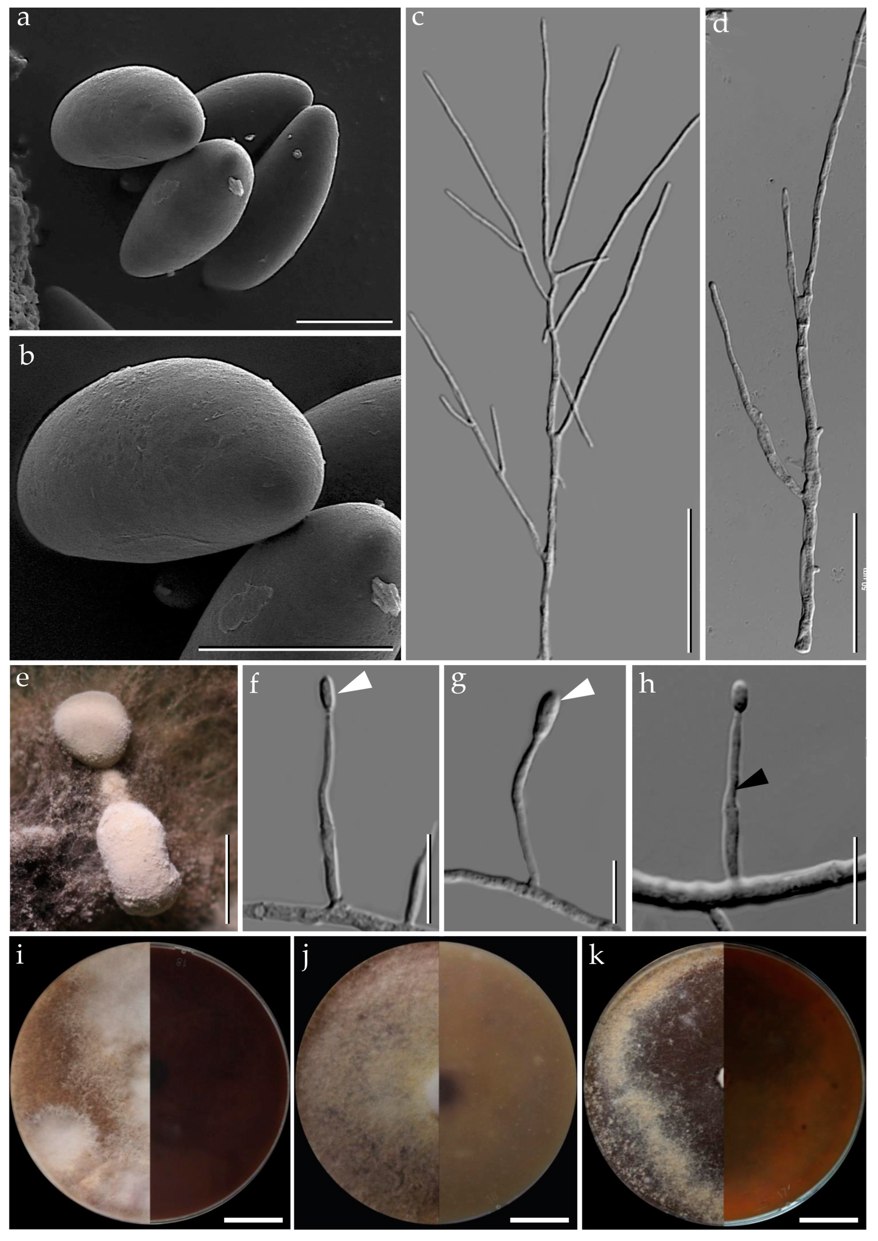
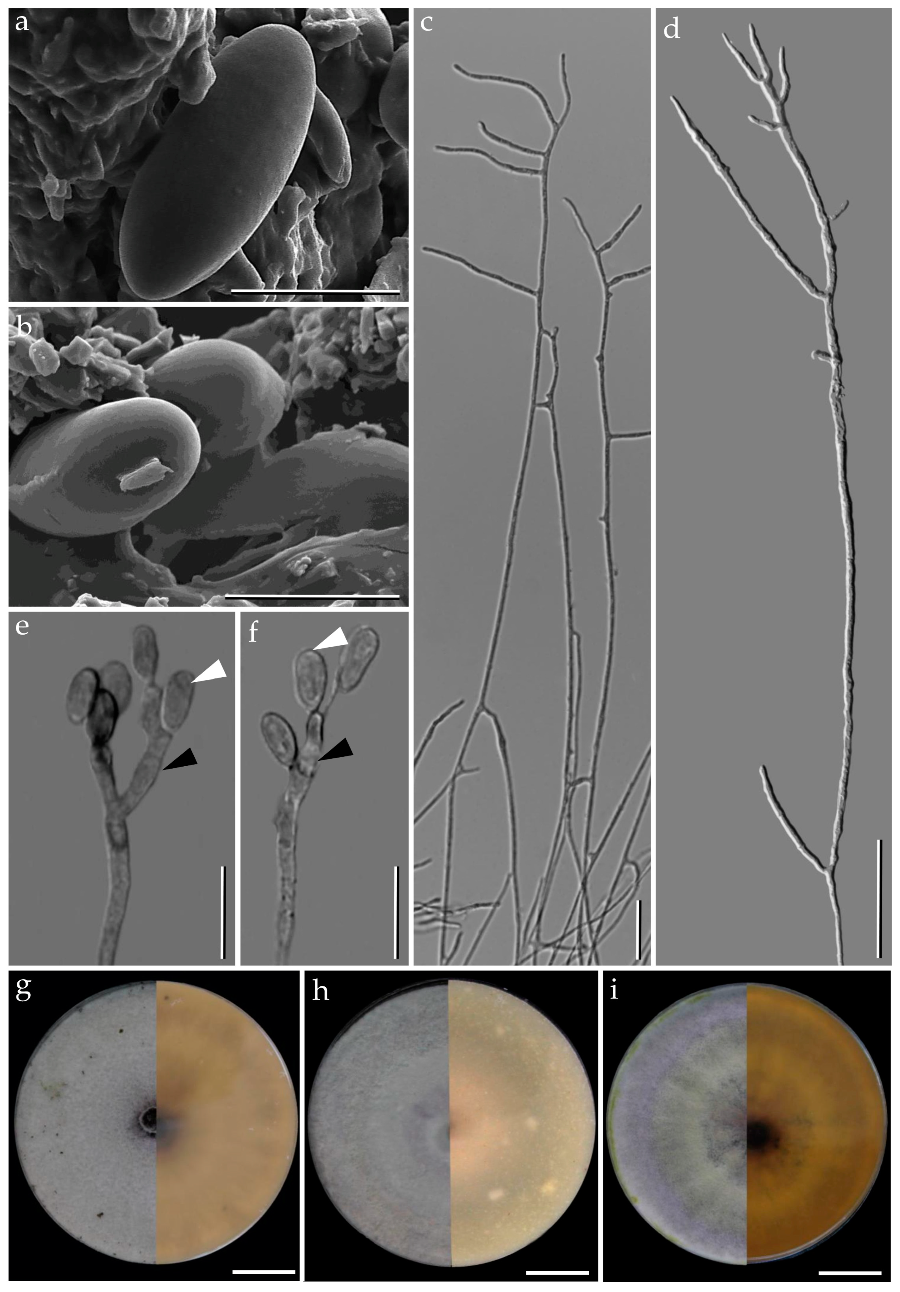
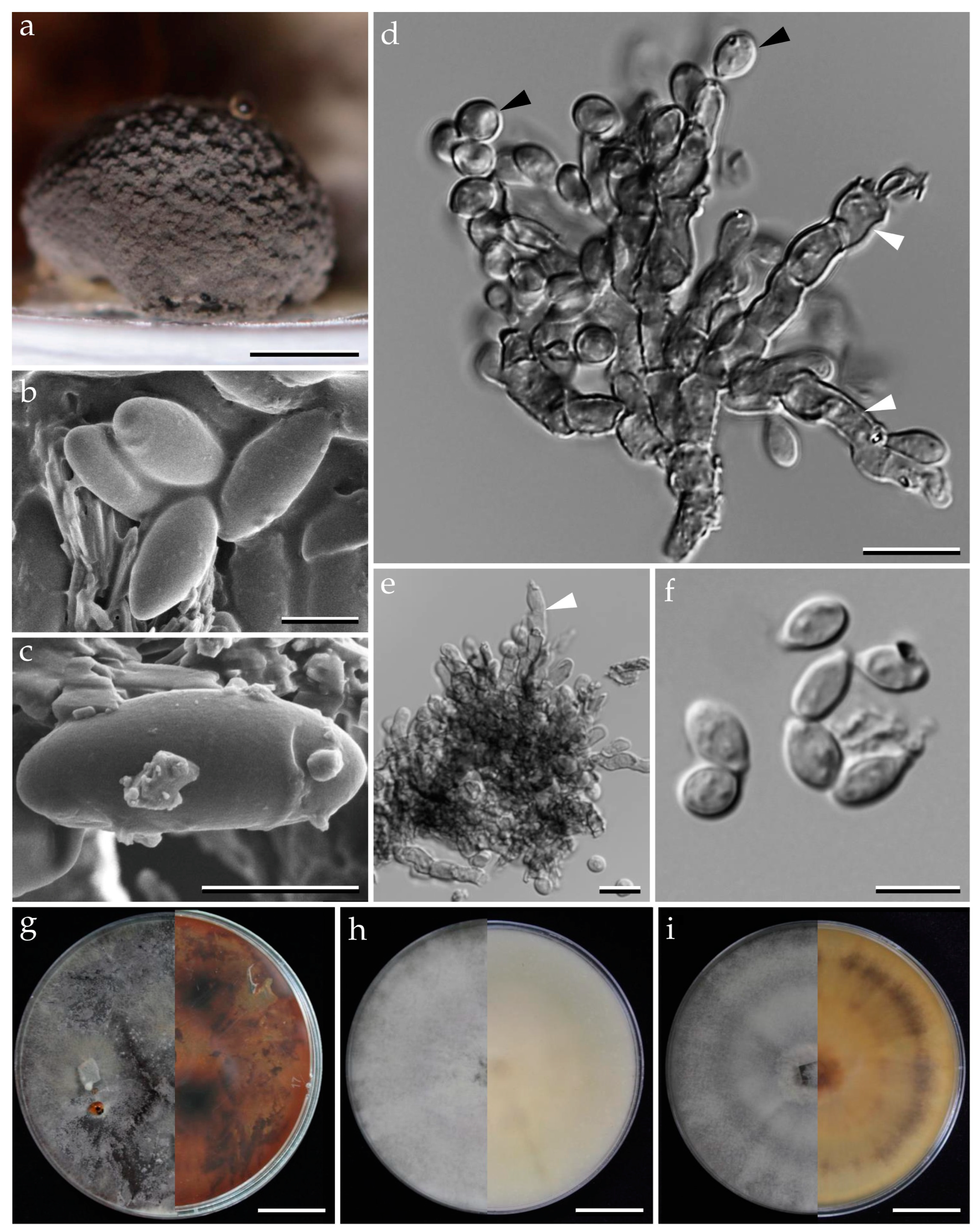
2.2. Morphological Characterization and HPLC Profiling

Morphological features, such as stromatal size and shape, perithecia, asci, apical apparatus, and ascospores were examined in accordance with Ju and Rogers [2] using a Nikon (Bangkok, Thailand) Eclipse Ni connected with a Nikon microscope camera DS-Ri2 and a stereo dissecting microscope Olympus SZ61 (Olympus, Bangkok, Thailand). Fungal cultures were grown on several media, i.e., Oatmeal Agar (Difco OA; Becton Dickinson, Carlsbad, CA, USA), Potato Dextrose Agar (Difco PDA), and Yeast Malt Glucose Agar (1% malt extract, 0.4% glucose and 0.4% yeast extract; agar 1%; YMGA). The morphological studies were carried out on 9 cm Petri dishes. Conidiogenous cells and conidiophore branching patterns of the anamorph were examined as proposed by Ju and Rogers [2]. Furthermore, the colors of stromata, KOH-extractable pigments, and cultures were documented following the color chart of Rayner [14]. For chemotaxonomic studies, stromatal secondary metabolites were extracted with acetone and analyzed using ultrahigh performance liquid chromatography coupled to diode array detection and ion mobility tandem mass spectrometry (UHPLC-DAD-IM-MS/MS) as described concurrently [15].

2.3. DNA Extraction, Polymerase Chain Reaction (PCR)

A modified method based on cetyltrimethyl ammonium bromide (CTAB) was used to isolate total genomic DNA from mycelia (pure cultures) grown for 5 days on PDA as previously described in Mongkolsamrit et al. [16]. The internal transcribed spacer regions (ITS), and partial sequences of the large subunit of the rDNA (LSU), RNA polymerase II (RPB2), and beta tubulin (TUB2) were amplified, using standard primers introduced by White at al. [17] (ITS4 and ITS5 for ITS [18], (LR5), Rehner and Samuels [19] (LROR) for LSU, Liu et al. [20] (RPB2–5F and 7Cr for RPB2), and O’Donnell and Cigelnik [21] (T1 and T22) for TUB2. PCR was conducted in 25 µL reaction volumes consisting of 1× PCR buffer, 200 μM of each of the four dNTPs, 2.5 mM MgCl2, 1 U Taq DNA Polymerase recombinant (Thermo Scientific, USA), 0.5 µM of each primer, and 50–100 ng of DNA template. The PCR conditions were performed as follows: 94 °C for 2 min, followed by 35 cycles of denaturation at 94 °C for 1 min, annealing at a suitable temperature for 1 min, extension at 72 °C for 2 min, and a final extension of 72 °C for 10 min. The annealing temperature of each gene was 55 °C for ITS and LSU; 54 °C for RPB2; and 53 °C for TUB2. PCR products were purified and subsequently sequenced with PCR amplification primers.

2.4. Sequencing Methods

A total of 5 µL of a post-PCR product was combined with 2 µL of ExoSAP-IT™ reagent for a 7 µL reaction total volume. When treating PCR product volumes greater than 5 µL, we simply increased the amount of ExoSAP-IT™ reagent proportionally. The mix was incubated at 37 °C for 15 min, followed by 15 min at 80 °C to degrade remaining primers and dinucleotides. DNA templates were processed for the DNA sequencing using the ABI-PRISM BigDye Terminator (version 3.1; Applied Biosystems, Foster, CA, USA) with both forward and reverse sequence-specific primers. Purified PCR products were used in a 20 µL sequencing reaction solution containing 8 µL of BigDye Terminator and 0.1 M of the same PCR primer. Sequencing reactions were performed using a 2 min initial denaturation at 96 °C, followed by 25 cycles for 10 s at 94 °C, 15 s at 50 °C, and 3 min at 60 °C. Sequence data were generated with the ABI PRISM 3100 DNA Analyzer (Applied Biosystems). Sequences were analyzed by Sequencer 3.1.1 software (Applied Biosystems) to compare variations. DNA sequences were checked manually and assembled using BioEdit v. 7.2.5 [22]. All newly generated sequences were submitted to GenBank (https://www.ncbi.nlm.nih.gov/ accessed on 21 December 2022) and listed in Table 1.

2.5. Phylogenetic Analyses

All sequences were aligned in Multiple Sequence Comparison by Log-Expectation program (MUSCLE) [37] and refined by direct examination. Multiple sequence alignments were analyzed with closely matched sequences and other reference taxa obtained from GenBank as shown in Table 1. Sequences were analyzed using maximum parsimony (MP), maximum likelihood (ML), and Bayesian inference (MB). The MP analysis was performed in PAUP*4.0b10 [38] and all characters were equally weighted, and gaps were treated as missing data. The most parsimonious trees were obtained from heuristic searches: 500 replicates of stepwise random addition and tree-bisection-reconnection (TBR) as a branch swapping algorithm.
Maximum parsimony bootstrap supports (MPBS) were estimated by 1000 replicates (10 replicates of stepwise random sequence addition). Tree length, consistency index (CI), retention index (RI), relative consistency index (RC), and homoplasy index (HI) were estimated. The ML tree and bootstrap analyses (MLBS) were conducted through the CIPRES Science Gateway V. 3.3 [39] using RAxML 8.2.4 [40] with the BFGS method to optimize GTR rate parameters. Bayesian posterior probabilities (BPP) of the branches were computed using MrBayes 3.0B4 [41] with the best-fit model (GTR + I + G), selected using the Akaike information criterion (AIC) in Mr Modeltest 2.2 [42] and tested with hierarchical likelihood ratios (hLRTs). Five million generations were run in four Markov chains and sampled every 100 generations with a burn-in value set at 5000 sampled trees. Sequences of Graphostroma platystomum CBS 270.87 and Xylaria hypoxylon CBS12260 were used as out groups.

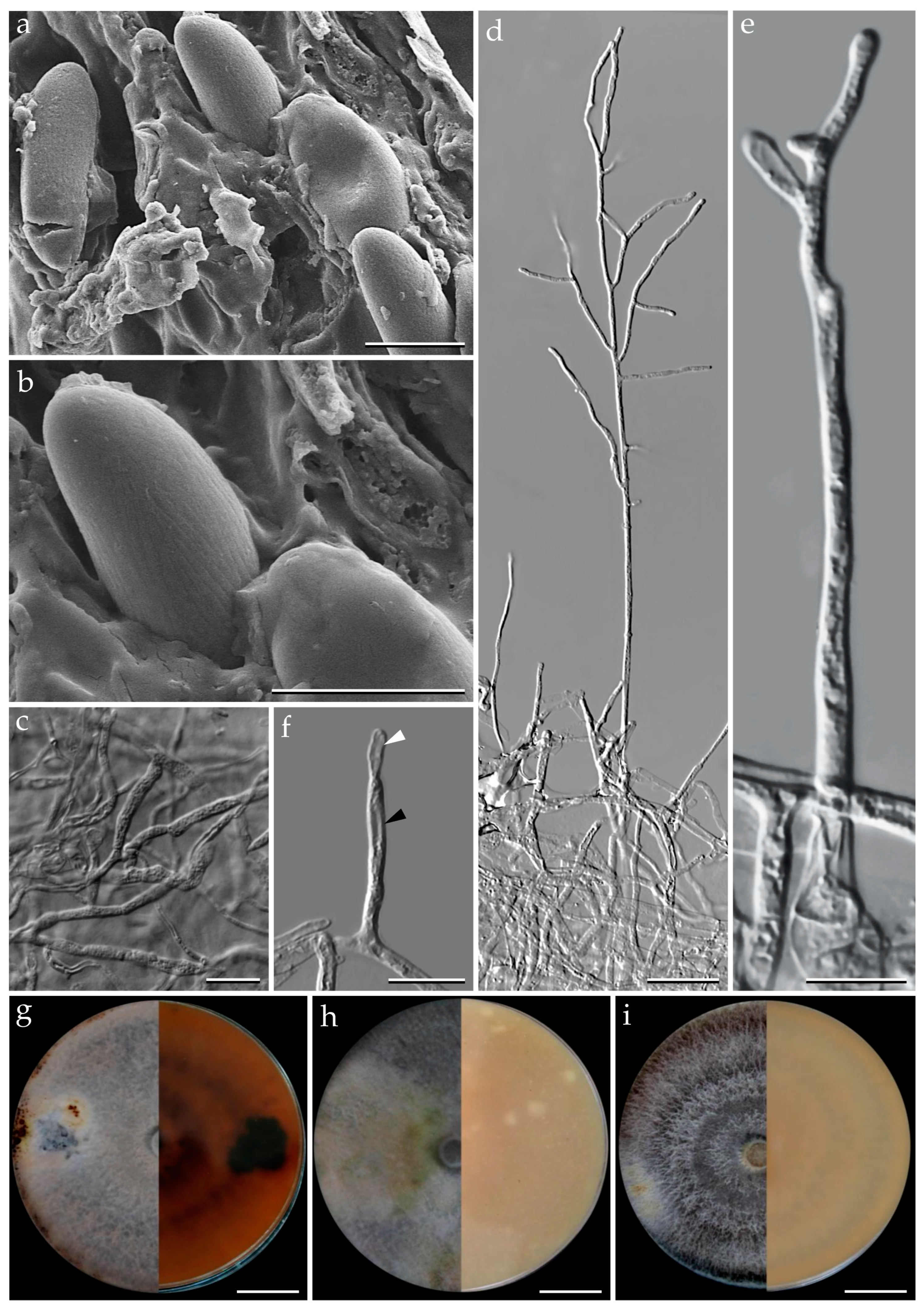
2.6. Cultivation of Fungal Strains for MALDI-TOF MS Analyses

The following strains were used for comparison in the MALDI-TOF/MS (for details see taxonomic part): Pyrenopolyporus bambusicola BCC89335; P. cinereopigmentosus BCC33615 and BCC89375; P. hunteri MUCL49209 (ex-epitype) and MUCL49339; P. laminosus BCC82043 and BCC89388; P. macrosporus: BCC89373; P. papillatus BCC20324 and BCC33622; P. tonngachangensis BCC31553 and BCC31555; Daldinia flavogranulata: BCC89367 and BCC82045; D. bambusicola BCC33677.
For the fermentation of Pyrenopolyporus and Daldinia spp., the seed culture were realized in 50 mL centrifuge tube containing 20 mL potato dextrose broth (Difco, PDB). Five pieces (ca. 20 mm) of a well grown agar plate of the fungi were used to inoculate each tube. The tubes were incubated for 3 days on a shaker (25 °C, under 12 h of fluorescent light at 150 rpm).

2.7. MALDI-TOF MS Analysis

The fungal mycelia were mixed thoroughly with 300 µL distilled water, and with absolute ethanol (900 µL). The content was then centrifuged at 13,000 rpm for 5 min; the supernatant was discarded, and the pellet was air dried. Approximately 50 μL of the pellet was mixed thoroughly with 100 μL of trifluoroacetic acid (80%), and centrifuged at 13,000 rpm for 15 min. The protein concentration in the obtained supernatant was adjusted to 0.4–0.8 mg/μL with standard solvent (50% acetonitrile and 2.5% trifluoroacetic acid) and then 1 µL was placed on an MSP 96 target polished steel BC (Ref. 1011025092). Subsequently, eight sample positions (including one Bruker Bacterial Test Standard position) were overlaid with 1 µL of a matrix (HCCA portioned; Bruker Daltonics GmbH, Bremen, Germany) consisting of a saturated solution of α-cyano-4 hydroxycinnamic acid (HCCA) in 50% acetonitrile, 47.5% water, and 2.5% trifluoroacetic acid (final concentration:10 mg HCCA/mL) and air-dried at room temperature. MALDI-TOF/MS measurement was conducted on a Microflex LT bench-top instrument operated by FlexControl software (Bruker Daltonics GmbH, Bremen, Germany). Spectra were acquired in linear positive mode at a laser frequency of 200 Hz by using the standard FlexControl and AutoX methods within a mass range of 2000 to 20,000 Da. Spectra were accumulated in the MS/parent mode (240 shots) resulting in 24 MALDI spectra per strain.
Raw spectra from fungal extracts were loaded into the ClinProTools software (version 3; Bruker) and processed for analysis using the following parameters: 800 resolution, Top Hat baseline subtraction with a 10% minimal baseline width and no data reduction. Null spectra and noise spectra exclusion with a noise threshold of 2.00 were both enabled, and spectra grouping was also supported. Peak selection and average peak list calculation ranged from 2000 to 10,000 mass to charge ratio values (m/z), and recalibration was performed with a 1000 parts per million (ppm) maximal peak shift and 30% match to mass calibrant peaks. Non-recalibrated spectra were excluded. A final set of 82 peaks were retained. Mass to charge ratio values (m/z) from average spectra were identified according to their statistical significance, as determined by the different statistical tests realized in ClinProTools: ANOVA test and Wilcoxon/Kruskal–Wallis test (PWKW). Statistical analyses through principal component analysis (PCA) were performed using the obtained feature table containing the averaged peak areas/intensities values from the final set of 82 peaks. ClinProTools can also automatically select the two most discriminating peaks between classes of samples as defined by users. Therefore, the software picked the two most discriminating peaks between (1) all taxa, (2) Pyrenopolyporus cinereopigmentosus and P. macrosporus, (3) P. hunteri, P. papillatus and P. tonngachangensis, (4) P. bambusicola and P. laminosus. The ex-epitype species of the genus was included in each of these statistical analyses.

3. Results

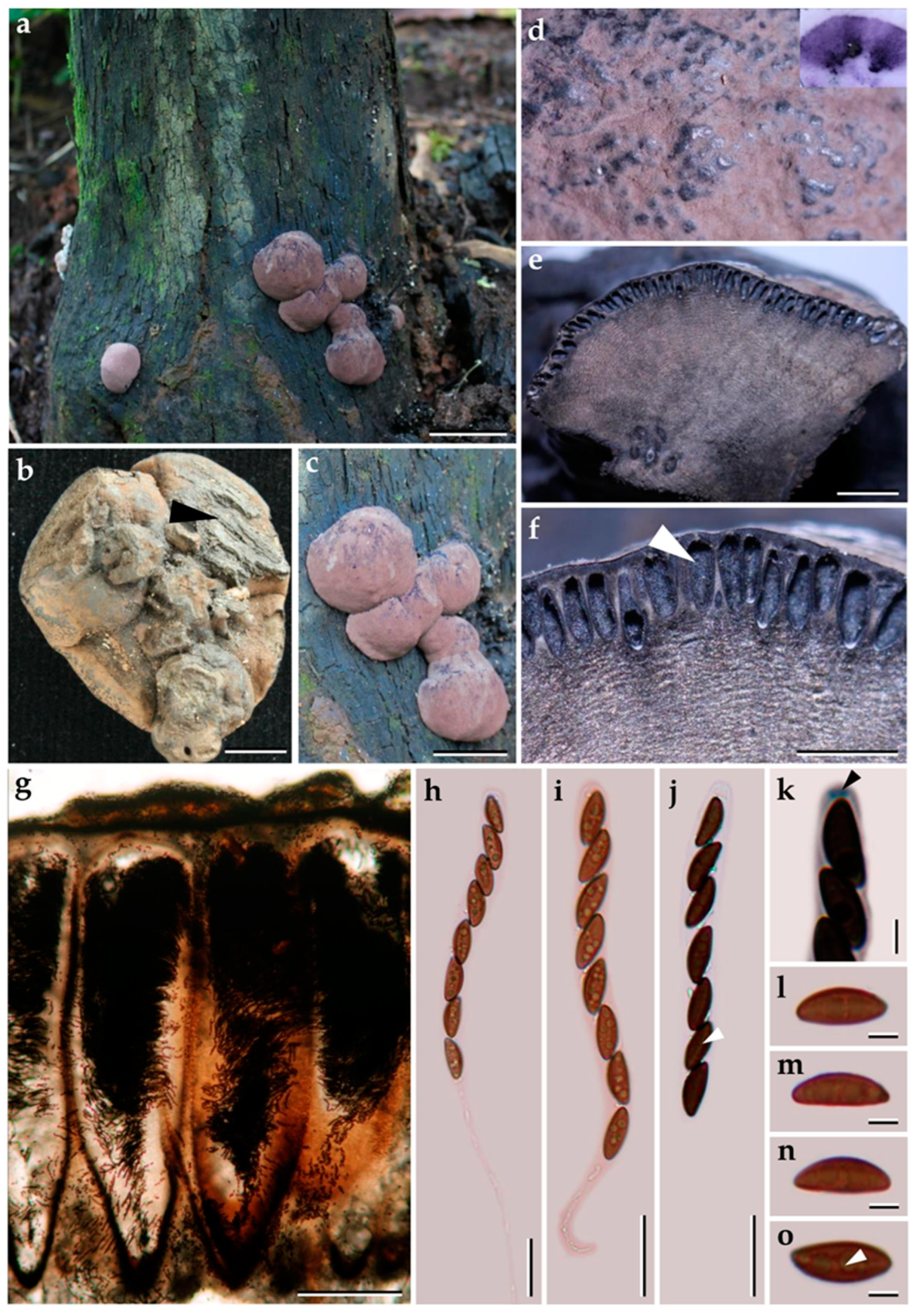
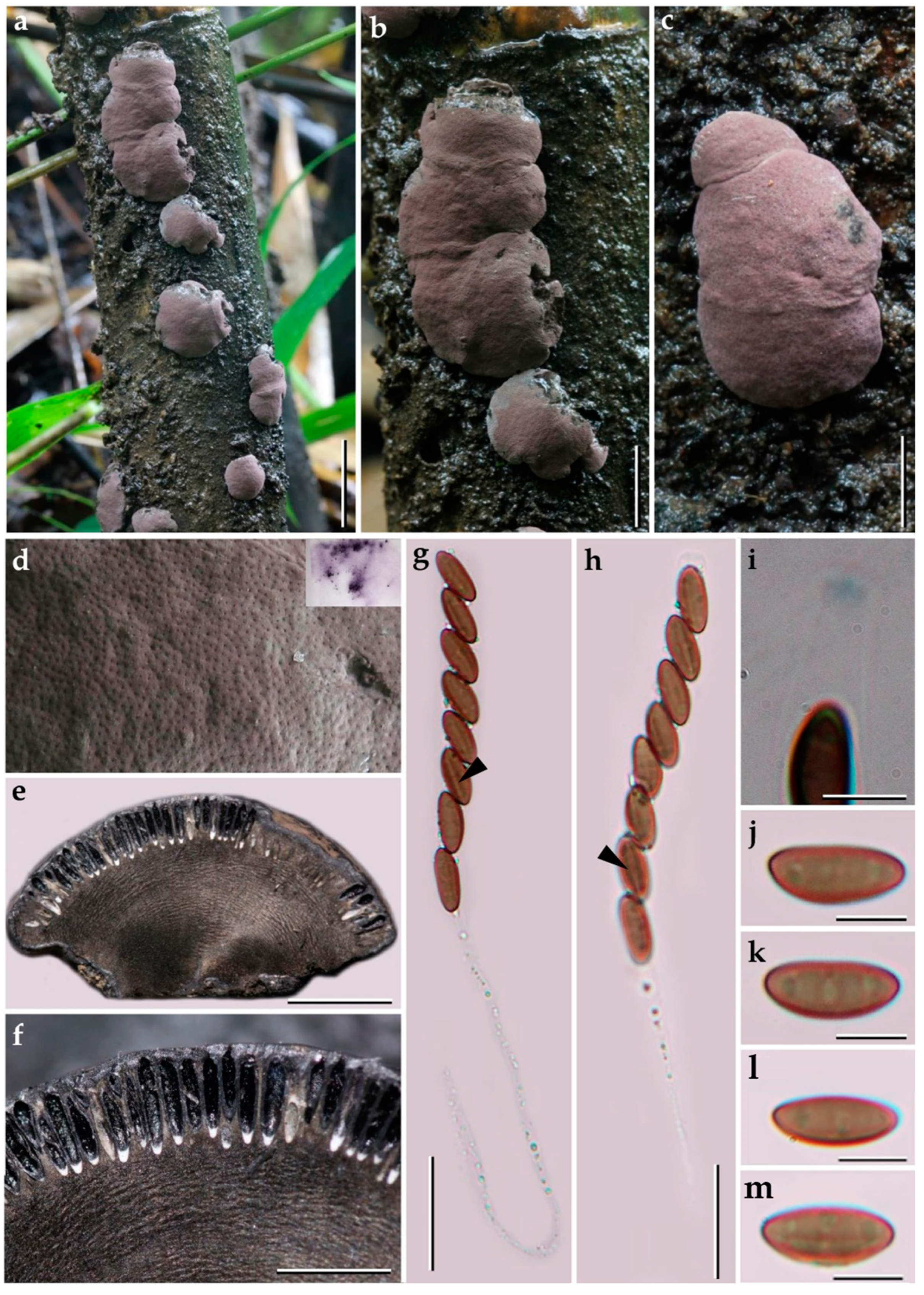
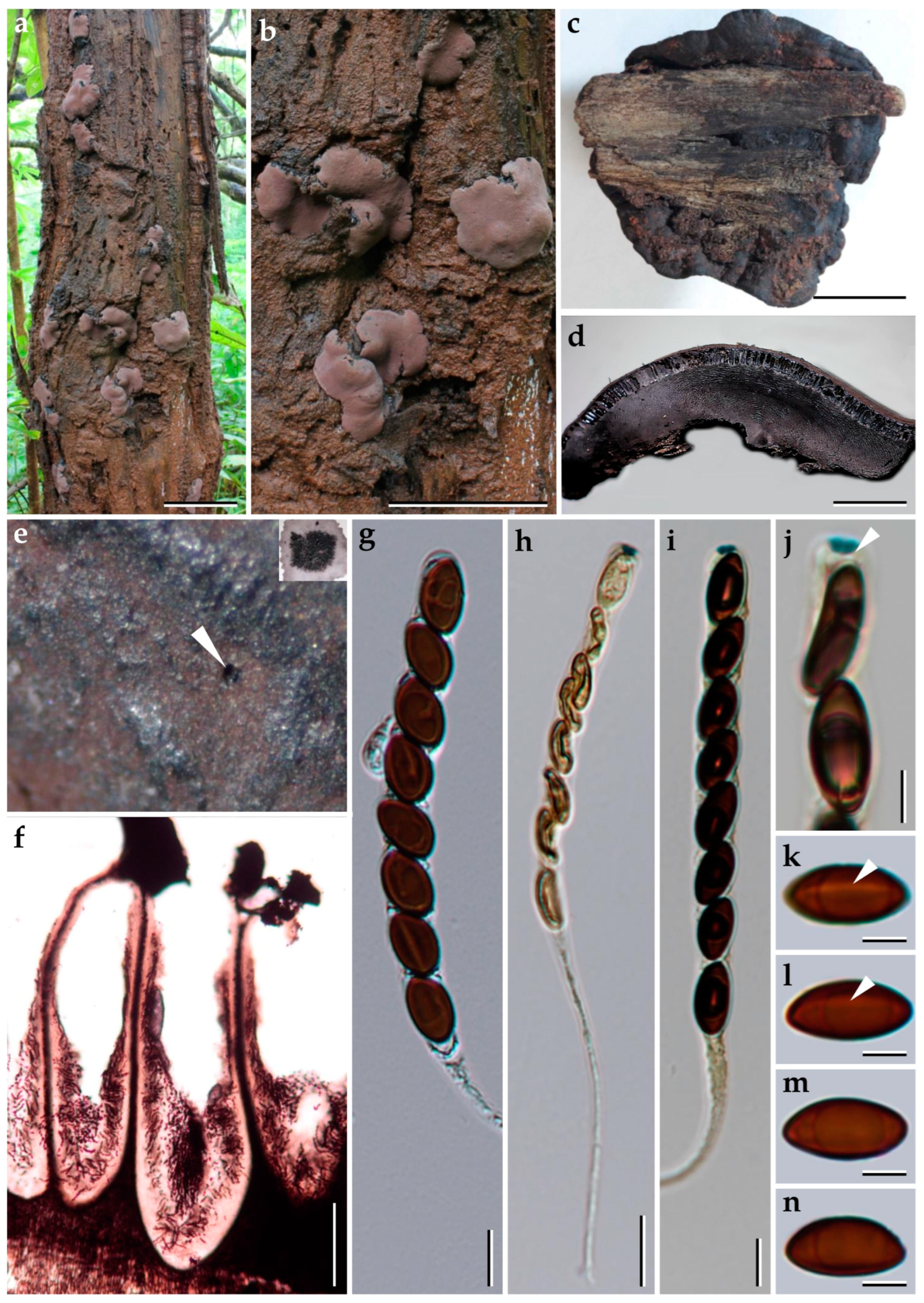
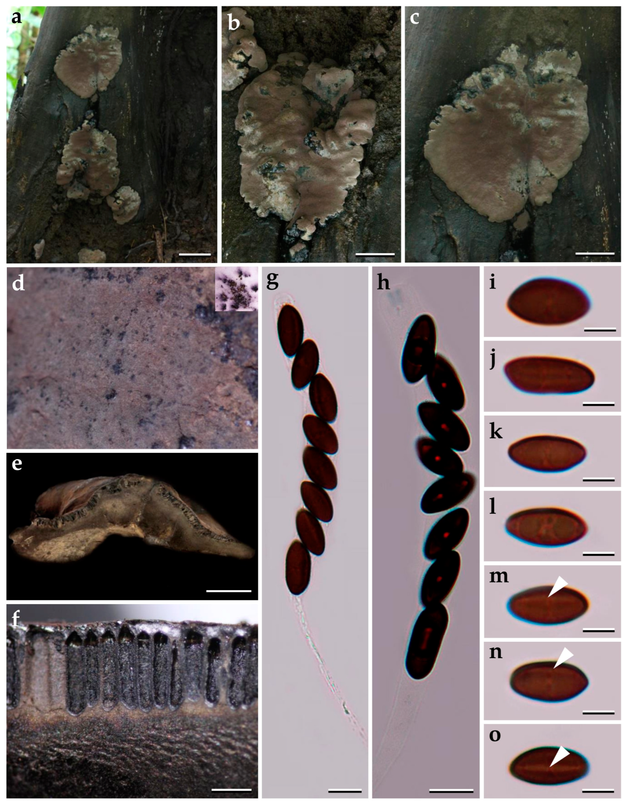
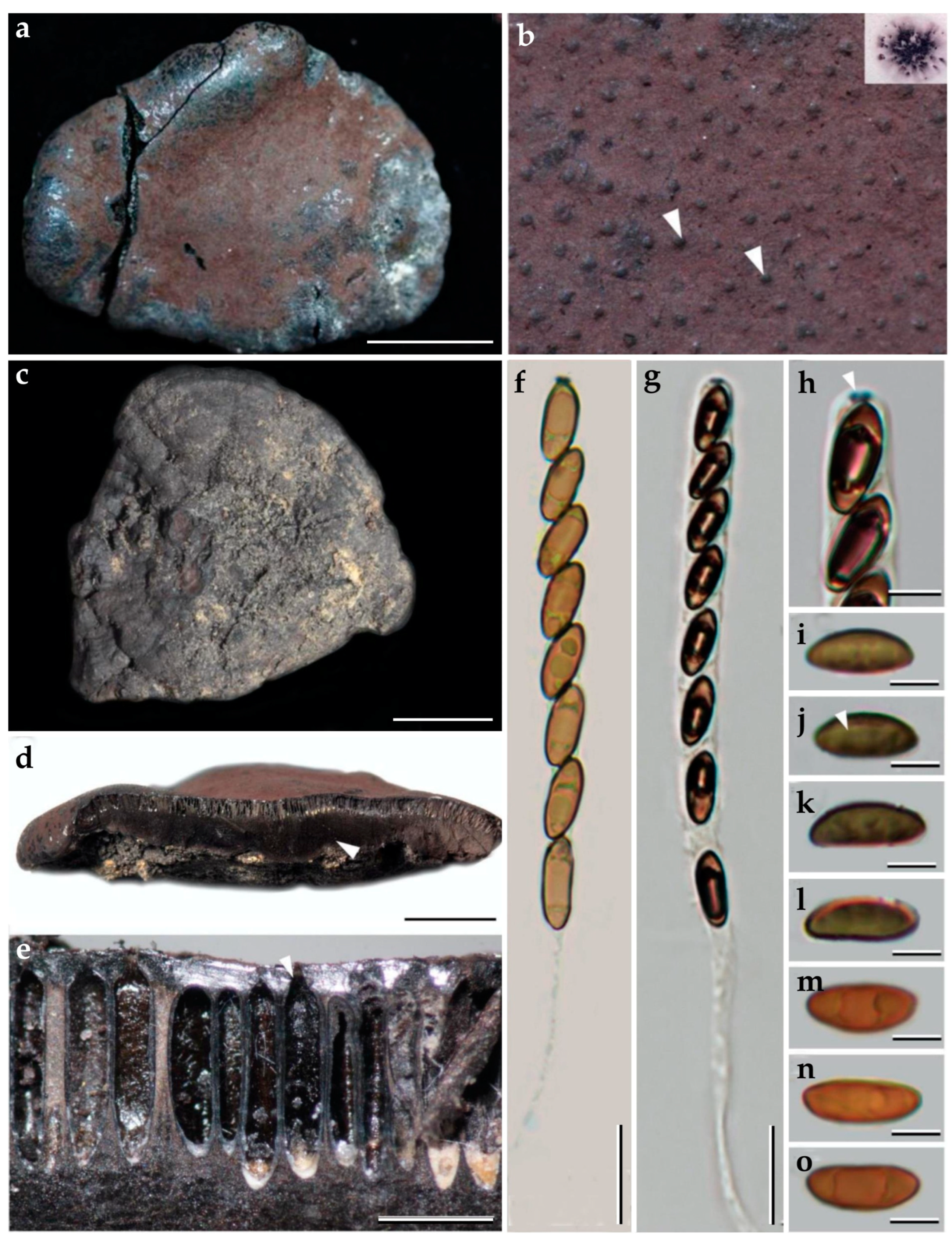
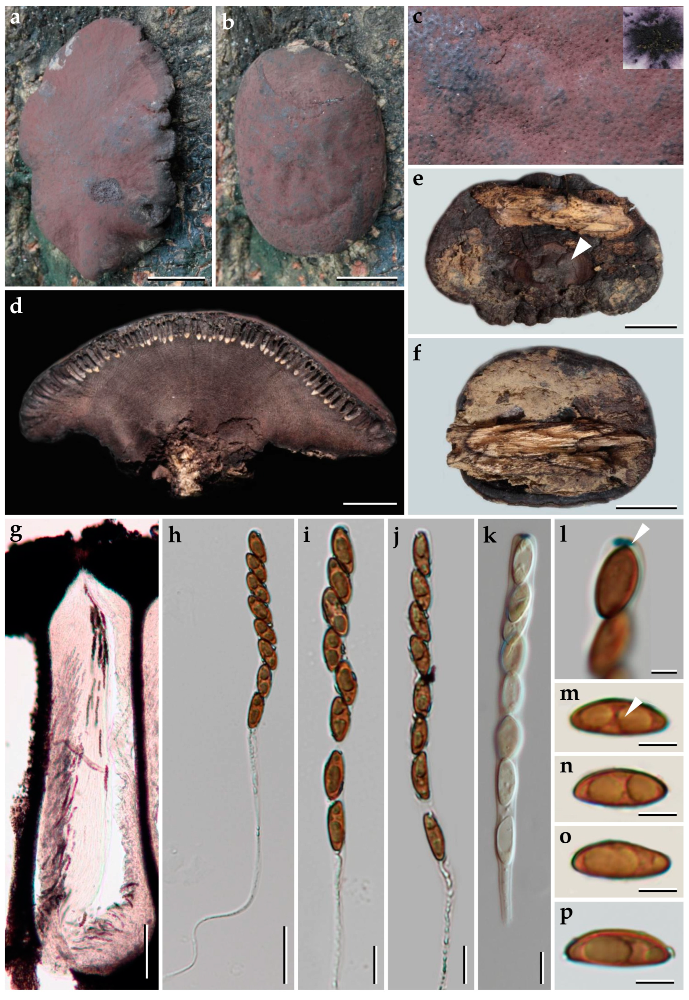
3.1. Morphological Characterization

The morphological features of the five novel species and the new record of Pyrenopolyporus and the phylogenetic positions of these taxa according to the multi-locus genealogy are described further below.
Pyrenopolyporus laminosus (J. Fourn., Kuhnert and M. Stadler) M. Stadler, Kuhnert and L. Wendt, Mycological Progress 17 (1–2): 150 (2017).               Figure 2 and Figure 3.
Material studied. Thailand: Tak Province, Pa Daeng Mine’s area, 16°41′46″ N, 98°36′56″ E, reforestation forest, on decaying wood, 13 December 2017, P. Srikitikulchai (P.S.), S. Wongkanoun (S.W.), (BBH47928); (strain, BCC89383); DNA sequences of the Thai strain: (ITS = MN153855), (LSU = MN153872), (RPB2 = MN172210), (TUB2 = MN172199).
Teleomorph. Stromata solitary to coalescent, hemispherical to depressed-spherical, widely attached to the substrate, very rarely substipitate, smooth or with inconspicuous perithecial outlines, 18–39 × 14–24 mm; surface Mouse Grey (116), Purplish Grey (126), and Vinaceous Grey (126); dark brown granules immediately beneath the surface, with KOH-extractable pigments Livid Violet (79) or Greyish Lavender (98), often rather dilute, especially in fully mature to overly mature specimens; the tissue between perithecia greyish brown to brown, pithy to woody; the tissues below the perithecia layer greyish, soft-textured, with a blackish line separating the perithecial layer from the sterile internal tissue, interior blackish brown, soft-textured, solid, with a lamellate structure consisting of densely intricate small black and golden-brown lines, 8–14 mm thick. Perithecia lanceolate, 0.2–0.3 × 0.7–0.9 mm ( x = 0.3 × 0.8 µm; n = 20). Ostioles umbilicate to slightly raised, discoid. Asci cylindrical, eight-spored, 170.0−207.5 µm in length, the spore-bearing parts, 75.0−87.5 µm long, 5.0−7.5 µm broad, the stipes, 87.5−137.5 µm long; with amyloid apical apparatus bluing in Melzer’s reagent, discoid, 1−2 µm high, 3 µm broad. Ascospores ellipsoid-inequilateral with narrowly rounded ends, (12–) 13–14 (–15) × 4–5 ( x = 13.1 × 4.8 µm; n= 50); with straight to rarely slightly sigmoid germ slit covering much less than spore-length or nearly spore-length on the convex side; perispore indehiscent in 10% KOH, epispore smooth.
Cultures and anamorph. Colonies on OA covering a 9 cm Petri dish in 1 week, at first whitish becoming velvety to felty, azonate with entire margin, Rosy Vinaceous (58), reverse Olivaceous (46). Colonies on YMGA covering a 9 cm Petri dish in 1 week, azonate, at first aerial mycelium whitish becoming velvety to felty, azonate with entire margin, Olivaceous (48) and Dark Brick (66); reverse Sepia (63). Colonies on PDA covering a 9 cm Petri dish in 1 week, at first whitish becoming velvety to felty, azonate with entire margin, Dark Brick (66); reverse Dark Vinaceous (82), Sepia (63), and Dark Brick (66). Primordia cylindrical to somewhat clavate, unbranched or sometimes branched, 2.8 × 1.3 mm. Conidiogenous structures with virgariella-like branching patterns as defined by Ju and Rogers [2], main axis hyaline to pale brown, finely roughened. Conidiogenous cells produced holoblastically, cymbiform, obovoid, hyaline, 15–20 × 2–3 μm, each cell producing one or several conidia. Conidia hyaline, smooth, subglobose, obovoid, ellipsoid, (6–) 7–8 × 3–4 µm ( x = 7.28 × 2.97 µm; n = 10).
Additional specimens examined. Thailand: Chiang Mai Province, Ban Hua Thung community forest, 18°51′17″ N, 99°16′57″ E, hill evergreen forest, on bamboo, 22 August 2016; P.S. and S.W., (BBH47916, BCC82043; BBH47917, BCC82044). Chiang Mai Province, Ban Hua Thung community forest, 18°51′17″ N, 99°16′57″ E, hill evergreen forest, on bamboo, 3 November 2016, P.S. and S.W., (BBH42275, BCC82671). Tak Province, Pa Daeng Mine’s area, 16°41′46″ N, 98°36′56″ E, restoration forest, on bamboo, 4 September 2018, P.S. and S.W., (BCC89388).
Secondary metabolites. Stromata contain hypoxylone (1), BNT (2), and an unknown hydroxy derivative of hypoyxlone (3: [M + H]+ = 349.07041 Da; C20H12O6, Figure S8) as major constituents (Figure S7 and Table S2).
Notes. The Thai specimens of Pyrenopolyporus laminosus correspond well to the description by Kuhnert et al. [28]. This species is distinctive for its stromatal morphology and the characteristic tissue below the perithecia layer is without any internal concentric zones. Herein we reexamined the type of material of P. laminosus (syn. Hypoxylon laminosus) and compared it with the Thai material, matching the data originally reported by Kuhnert et al. [28]. Our phylogeny based on multi-locus analyses showed that the Thai strains grouped with Pyrenopolyporus laminosus with high statistical supports MP, ML, and BPP, confirming that this species is not only present in the neotropics but also occurs in Thailand.
Pyrenopolyporus bambusicola Srikitikulchai, Wongkanoun, M. Stadler and Luangsa–ard, sp. nov.                                             Figure 4 and Figure 5.
MycoBank. MB846446.
Etymology.bambusicola” refer to the bambusicolous habit.
Holotype. Thailand: Tak Province, Pa Daeng Mine’s area, 16°41′46″ N, 98°36′56″ E, restoration forest, on bamboo trunk in fire damaged area, 4 September 2018, P.S. and S.W., (BBH47923).
Ex-type culture. BCC89355. DNA sequences of ex-type culture: (ITS = OP304856), (LSU = OP304876), (RPB2 = OP981624), (TUB2 = OQ101839).
Teleomorph. Stromata solitary to coalescent, peltate to hemispherical with a short and broadly attached central base, the margin almost inseparable from host surface with the host surface, 11–16 mm long, 8–13 mm wide, 4–9 mm thick; surface Pale Mouse Grey (117) to Mouse Grey (116) and Pale Purplish Grey (127) with KOH-extractable pigments Livid Violet (79) and Greyish Lavender (98); dark brown to black tissue forming a thin layer above perithecia; the tissue between perithecia grey or blackish brown; the tissue below the perithecial layer without internal concentric zones, grey, 3–8 mm thick, with a lamellate structure consisting of densely intricate small black and golden brown lines; lacking the dark brown line below the perithecia layer. Perithecia tubular, 0.75–0.90 mm high, 0.30–0.35 mm broad. Ostioles umbilicate conspicuous. Asci cylindrical, very long-stipitate, eight-spored, 154–160 μm in length, the spore-bearing parts, 62–64 μm long, 4–5 μm broad; with amyloid apical apparatus, bluing in Melzer’s reagent, discoid in outline, 1.0–1.2 μm high, 1 μm broad. Ascospores brown to blackish brown, ellipsoid with narrowly rounded ends, 10–11 (–12) × (3–) 4–5 μm ( x = 10.56 × 4.04 μm, n = 50), with a straight spore-length germ slit on the most convex side; perispore indehiscent in KOH, epispore smooth.
Cultures and anamorph. Colonies on OA covering a 9 cm Petri dish in 1 week, at first whitish becoming velvety to felty, azonate with entire margin, Herbage Green (17), reverse Olivaceous (48). Colonies on YMGA covering a 9 cm Petri dish in 1 week, at first whitish becoming velvety to felty, inconspicuous zonate with entire margin, Olivaceous (48) and Dark Brick (66) reverse Olivaceous (48). Colonies on PDA covering a 9 cm Petri dish in 1 week, at first whitish becoming velvety to felty, zonate with entire margin, Olivaceous (48); reverse Greyish Sepia (106) and Olivaceous Grey (121). Conidiogenous structures with virgariella-like branching patterns as defined by Ju and Rogers [2]. Conidiogenous cells cylindrical, hyaline, finely roughened, 14−15 × 2.5−3.0 µm. Conidia hyaline, smooth, ellipsoid, 5−6 × 3−4 µm.
Additional specimens examined. Thailand: Tak Province, Pa Daeng Mine’s area, 16°41′46″ N, 98°36′56″E, reforestation forest, on bamboo trunk (Bambusoideae) in fire damaged area, 4 September 2018, P.S. and S.W., (BCC89369, BBH47923; BCC89360).
Secondary metabolites. Stromata contain hypoxylone (1), BNT (2), and an unknown hydroxy derivative of hypoyxlone (3: [M + H]+ = 349.07041 Da; C20H12O6; Figure S8) as major constituents (Figures 18 and S7 and Table S2).
Notes. Our new fungus Pyrenopolyporus bambusicola showed a close relationship to P. laminosus which is associated with Bambusoideae but differs by the ascospore size range [10–11 (–12) × (3–) 4–5 (P. bambusicola) vs. 11–13.5 × 4.2–4.5 µm (P. laminosus)]. The tissue below the perithecial layer of P. laminosus has a blackish line separating the perithecial layer from the sterile internal tissue, a characteristic lacking in P. bambusicola. Additionally, the stromatal secondary metabolites found in P. bambusicola resemble the ones found in P. laminosus.
Pyrenopolyporus cinereopigmentosus Srikitikulchai, Wongkanoun, M. Stadler and Luangsa-ard, sp. nov.                                            Figure 6 and Figure 7.
MycoBank. MB846447.
Etymology. from the Latin “cinereus” in reference to its grey KOH-extractable pigments of the stromatal surface.
Holotype. Thailand: Tak Province, Pa Daeng Mine’s area, 16°41′46″ N, 98°36′56″ E, restoration forest, on decaying wood in fire-damaged area, 4 September 2018, P.S. and S.W., (BBH47927).
Ex-type culture. BCC89382. DNA sequences of ex-type culture: (ITS = OP304860), (LSU = OP304882), (RPB2 = OP981627), (TUB2 = OQ101843).
Teleomorph. Stromata solitary to coalescent, effused-pulvinate, attached on substrate, the margin almost in contact with the host surface, 25–36 mm diam, 5–19 mm thick; surface Pale Mouse Grey (117), Mouse Grey (116), and Fuscous Black (104); dark brown to blackish brown immediately beneath the stromatal surface, with KOH-extractable pigment Dark Mouse Grey (119) and Iron Grey (122); the tissue between perithecia Pale Olivaceous Grey (120) or Olivaceous Grey (121); the tissue below the perithecia layer massive, Olivaceous Grey (121) and or Olivaceous Black (108), 3.6–5 mm thick. Perithecia tubular 0.9–1.1 mm high, 0.3–0.4 mm broad. Ostioles inconspicuous, umbilicate. Asci cylindrical, eight-spored, 180–248 µm in length, the spore bearing part, 83–98 µm long, 7–8 µm broad; apical apparatus bluing in Melzer’s reagent rectangular shape, 3–4 × 1–2 µm. Ascospores dark brown to blackish brown, unicellular, ellipsoid with narrowly to broadly rounded ends, (12–) 13–14 (–15) × 6–7 µm ( x = 13.62 × 6.42 µm; n = 25) with straight germ slit covering full spore length on convex side, perispore indehiscent in 10% KOH.
Cultures and anamorph. Colonies on OA covering a 9 cm Petri dish in 1 week, at first whitish becoming velvety to felty, inconspicuous zonate with entire margin, Sepia (63) and Dark Brick (66); reverse Olivaceous (48), Dull Green (70), and Sepia (68). Colonies on YMGA covering a 9 cm Petri dish in 1 week, at first aerial mycelium whitish becoming velvety, azonate with entire margin, Sepia (68); reverse Sepia (68) and Dark Vinaceous (82). Colonies on PDA covering a 9 cm Petri dish in 1 week, at first whitish becoming velvety to felty, inconspicuous zonate with entire margin, Sepia (62); reverse Dark Vinaceous (82), Sepia (63), and Brown Vinaceous (84). Conidiogenous structures with virgariella-like branching patterns as defined in Ju and Rogers [2], main axis hyaline and the cell walls roughed or smooth, dark brown to blackish brown. Conidiogenous cells cylindrical, hyaline, finely roughened, 10−15 × 2−3 µm. Conidia hyaline, smooth, ellipsoid, 5−8 × 2−3 µm.
Additional specimens examined. Thailand: Tak Province, Pa Daeng Mine’s area, 16°41′46″ N, 98°36′56″ E, reforestation forest, on decaying wood in fire-damaged area, 4 September 2018, P.S. and S.W., (BCC89355, BBH47920; BCC89360).
Secondary metabolites. Stromata contain hypoxylone (1), BNT (2), two isobaric unknown compounds (4: [M + Na]+ = 258.10997 Da; C13H17NO3), and other unknown metabolites (5: [M + H]+ = 633.45371; C30H60N6O8) as major constituents (Figure S7 and Table S2).
Notes. Molecular phylogenetic assessment via a multi-locus supermatrix approach led to the placement of our new fungus Pyrenopolyporus cinereopigmentosus as a sister species to P. macrosporus. Morphologically, P. cinereopigmentosus closely resembles the above-mentioned species by having pale brown to dark brown ascospore color but differs by the ascospore morphology and size range. Pyrenopolyporus macrosporus produces a highly variable shape of ascospore as shown in the Figure 7i−o, while the ascospore length is much larger than P. cinereopigmentosus as follows [(14–) 16–17 × (6–) 7–8 vs. (12–) 13–14 (–15) × 6–7 µm]. Pyrenopolyporus cinereopigmentosus differs from P. hunteri in the KOH-extractable pigment and the ascospores size range is as follows (12–) 13–14 (–15) × 6–7 (P. cinereopigmentosus) vs. 11.5–14.0 × 5.0–5.5 µm (P. hunteri)]. Our phylogenetic multi-locus analysis showed that our new species is clearly separated from P. hunteri with high statistical support. Similarly, the chemical characterization of this new fungus showed the additional presence of unknown compounds not presence among the major constituents of P. laminosus and P. bambusicola. However, its secondary metabolite profile resembles the one obtained for P. macrosporus.
Pyrenopolyporus macrosporus Srikitikulchai, Wongkanoun, M. Stadler and Luangsa-ard, sp. nov.                                             Figure 8 and Figure 9.
MycoBank. MB846448.
Etymology.macrosporus” based on the large ascospores when compared with other Pyrenopolyporus species.
Holotype. Thailand: Tak Province, Pa Daeng Mine’s area, 16°41′46″ N, 98°36′56″ E, restoration forest, on decaying wood in fire-damaged area, 4 September 2018, P.S. and S.W., (BBH47924).
Ex-type culture. BCC89373; DNA sequences of ex-type culture: (ITS = OP304870), (LSU = OP304879), (RPB2 = OP981621), (TUB2 = OQ101844).
Teleomorph. Stromata solitary or coalescent, effused-pulvinate, attached on substrate, 20–70 mm long, 21–29 mm wide, 6–11 mm thick; surface Pale Olivaceous Grey (120), Olivaceous Grey (121), and Iron Grey (122); carbonaceous immediately beneath of the stromatal surface with KOH-extractable pigment Livid Violet (79) and Greyish Lavender (98); the tissue between perithecia greyish brown to brown or pithy to woody; with a woody line separating the perithecial layer from the sterile internal tissue; interior blackish brown or dark brown, 2.1–2.4 mm thick. Perithecia tubular, 0.7–0.9 mm high, 0.3–0.4 mm broad. Ostioles lower than the stromatal surface, inconspicuous. Asci unitunicate, cylindrical, eight-spored, 177–206 µm in length; the spore bearing part, 91–105 µm long, 7–8 µm broad; apical apparatus bluing in Melzer’s reagent, rectangular shape, 2 × 1 µm. Ascospores dark brown to blackish brown, unicellular, ellipsoid, inequilateral, highly variable with narrowly to broadly rounded ends, (14–) 16–17 × (6–) 7–8 µm ( x = 15.67 × 7.14 µm, n = 25) with straight germ slit covering full spore length on convex side, perispore indehiscent in 10% KOH, epispore smooth.
Cultures and anamorph. Colonies on OA covering a 9 cm Petri dish in 1 week, inconspicuous zonate with entire margin, at first whitish becoming Sepia (63) and Dark Brick (66); reverse Olivaceous (48), Dull Green (70), and Sepia (68). Colonies on covering a 9 cm Petri dish in 1 week, at first whitish becoming velvety to felty, zonate with entire margin, Sepia (68); reverse, Sepia (68), and Dark Vinaceous (82). Colonies on PDA covering a 9 cm Petri dish in 1 week, at first aerial mycelium whitish becoming velvety to felty, inconspicuous zonate with entire margin, Sepia (62); reverse Dark Vinaceous (82), Sepia (63), and Brown Vinaceous (84). Conidiogenous structures with virgariella-like branching patterns as defined in Ju and Rogers [2], main axis hyaline to hyaline to pale brown, finely roughened. Conidiogenous cells cylindrical, hyaline, finely roughened, 1−2 × 1−1.5 µm. Conidia hyaline, smooth, subglobose, 4−5 × 2.5−4.0 µm.
Additional specimens examined. Thailand: Tak Province, Pa Daeng Mine’s area, 16°41′46″ N, 98°36′56″ E, reforestation forest, on decaying wood in fire-damaged area, 4 September 2018, P.S. and S.W., (BCC89374, BBH47925).
Secondary metabolites. Stromata contain hypoxylone (1), BNT (2), two isobaric unknown compounds (4: [M + Na]+ = 258.10997 Da; C13H17NO3), and other unknown metabolite (5: [M + H]+ = 633.45371; C30H60N6O8) as major constituents (Figure S7 and Table S2).
Notes. Pyrenopolyporus macrosporus is clearly distinct from other members of the genus based on the phylogenetic placement as well as its morphological features. Pyrenopolyporus macrosporus is very similar to P. symphyon by having the stromatal surface with KOH-extractable purple color, the ascospores with narrowly to broadly rounded ends. However, P. macrosporus differs from P. symphyon by having larger ascospores [(14–) 16–17 × (6–) 7–8 (P. macrosporus) vs. 9.5–12 (−13) × 4–5 µm (P. symphyon)]. The original description of the type specimen shows tubular perithecia, 1.3 mm long, 0.3−0.4 mm broad, with dark brown, ovoid ascospores, 10 × 4–5 µm [43]. Pyrenopolyporus macrosporus differs from the previous descriptions of P. symphyon by Ju and Rogers [2] and Möller [43], and is confirmed as a new member of the genus.
Pyrenopolyporus papillatus Srikitikulchai, Wongkanoun, M. Stadler and Luangsa-ard, sp. nov.                                                  Figure 10 and Figure 11.
MycoBank. MB846449.
Etymology.papillatus” based on the papillated ostioles.
Holotype. Thailand: Nakhon Si Thammarat Province, Nopphitam, Khao Luang National Park, 8°22′07″ N, 99°44′06″ E, tropical rainforest, on decaying wood, 21 February 2006, P.S., (BBH15197).
Ex-type culture. BCC20324; DNA sequences of ex-type culture: (ITS = OP304854), (LSU = OP304874), (RPB2 = OP981619), (TUB2 = OQ101846).
Teleomorph. Stromata hemispherical to depressed-spherical, widely attached to the substrate, very rarely substipitate, smooth or with inconspicuous perithecial outlines, 25 –33 mm wide, 7–8 mm thick; surface Vinaceous Grey (116), Fuscous Black (104), and Mouse grey (116); blackish brown granules immediately beneath the stromatal surface, with KOH-extractable pigments Purplish Grey (126) or Vinaceous Grey (116); the tissue between perithecia greyish brown to brown, pithy to woody; the tissue below the perithecial layer massive, blackish brown or dark brown, 3.1–3.3 mm thick. Perithecia lanceolate, 1.1–1.4 mm long, 0.2–0.3 mm broad. Ostioles papillate. Asci cylindrical, eight-spored, 168−170 µm in length, the spore-bearing parts, 77−87 µm long, 7−8 µm broad; apical apparatus bluing in Melzer’s reagent, 0.9−1.2 µm long, 2.3−2.7 µm broad. Ascospores light brown, ellipsoid, slightly inequilateral, favorably variable, or irregularly shaped, narrowly rounded ends, (11–) 12–13 (–14) × 4–5, ( x = 12.05 × 4.89, n = 25 µm); with straight to rarely slightly sigmoid germ slit much less than spore-length or nearly spore-length on the convex side; perispore indehiscent in 10% KOH, epispore smooth.
Cultures and anamorph. Colonies on OA covering a 9 cm Petri dish in 1 week, at first whitish becoming velvety to felty, azonate with distinct margins, Pale Green Grey (98); reverse Grey Olivaceous (107). Colonies on YMGA covering a 9 cm Petri dish in 1 week, at first whitish becoming velvety to felty, inconspicuous zonate, Pale Greenish Grey (123); reverse Grey Olivaceous (107). Colonies on PDA, covering a 9 cm Petri dish in 1 week, at first whitish, becoming Pale Greenish Grey (123); reverse Pale Vinaceous (85). Conidiogenous structures with virgariella-like branching patterns as defined in Ju and Rogers [2], main axis hyaline to pale brown, finely roughened. Conidiogenous cells cylindrical, hyaline, finely roughened, 9−10 × 4−5 µm. Conidia hyaline, smooth, ellipsoid, 6−11 × 3−4 µm.
Additional specimens examined. Thailand: Nakhon Si Thammarat, Khao Nan National Park, 8°46′14″ N, 99°48′20″ E, tropical rainforest, on decaying wood, 29 October 2008, P.S., (BCC33622, BBH25144).
Secondary metabolites. Stromata contain hypoxylone (1), BNT (2), two isobaric unknown compounds (4: [M + Na]+ = 258.10997 Da; C13H17NO3), and an unknown hydroxyl derivative of hypoxylone (3: [M + H]+ = 349.07041 Da; C20H12O6; Figure S8) as major constituents (Figure S7 and Table S2).
Notes. Pyrenopolyporus papillatus is morphologically similar to P. nicaraguensis by having purple KOH-extractable pigment from the stromatal surface but differs from the latter by having light brown to pale brown ascospores. Pyrenopolyporus papillatus differs from P. nicaraguensis by having smaller ascospores than P. nicaraguensis [(11–) 12–13 (–14) × 4–5 (P. papillatus) vs. (11–) 12–15 (–16) × 5.0–6.5 µm (P. nicaraguensis)]. Morphologically, P. hunteri closely resembles our new fungus but differs by the ascospore size range [11.5–14.0 × 5.0–5.5 (P. hunteri) vs. (11–) 12–13 (–14) × 4–5 µm (P. papillatus)]. Morphologically, our new species is quite similar to P. hunteri and P. nicaraguensis, but the molecular phylogeny clearly separates it from the previously reported species. The morphological features and secondary metabolites of Pyrenopolyporus species and the allied genus Daldinia are summarized in Table S1.
Pyrenopolyporus tonngachangensis Srikitikulchai, Wongkanoun, Stadler and Luangsa-ard, sp. nov.                                            Figure 12 and Figure 13.
MycoBank. MB846674.
Etymology. “tonngachangensis” referring to the locality “Ton Nga Chang Wildlife Sanctuary” where the type specimen was collected.
Holotype. Thailand: Songkhla Province, Hat Yai, Ton Nga Chang Wildlife Sanctuary, 6°57′06” N, 100°13′57” E, tropical rainforest, on decaying wood in the forest, 10 August 2008, P.S., (BBH25392).
Ex-type culture. BCC31553; DNA sequences of ex-type culture: (ITS = OP304865), (LSU = OP304887), (RPB2 = OP981632), (TUB2 = OQ101847).
Teleomorph. Stromata solitary or coalescent, hemispherical to depressed-spherical, widely attached to the substrate, very rarely sub-stipitate, smooth or with inconspicuous perithecial outlines, 30–45 mm long, 24–30 mm wide, 5–7 mm thick; surface Purple Slate (102), Fuscous Black (104), Vinaceous Grey (116), and Mouse Grey (118); carbonaceous immediately beneath the stromatal surface, with KOH-extractable pigments Livid Violet (79) or Greyish Lavender (98) and Violet Slate (99); the tissue between perithecia is greyish brown to brown, pithy to woody; the tissues below the perithecial layer are greyish brown, soft-textured, with a lamellate structure consisting of densely intricate small black and golden-brown lines, 5.0–5.7 mm thick. Perithecia lanceolate, 1.1–1.2 mm high, 0.2–0.3 mm broad. Ostioles conspicuous, umbilicate. Asci cylindrical, eight-spored, 170.0−207.5 µm in length, the spore-bearing parts, 75−83 µm long, 7 µm broad; apical apparatus bluing in Melzer’s reagent, 1.5–1.7 µm long, 3.4–3.8 µm broad. Ascospores light brown, ellipsoid, slightly inequilateral or irregularly shaped, narrowly rounded ends, (12–) 13–14 (–16) × 4–5 ( x = 13.48 × 4.91 µm, n = 25); with straight germ slit covering ca. 2/3 length on the convex side; perispore indehiscent in 10% KOH, epispore smooth.
Cultures and anamorph. Colonies on OA covering a 9 cm Petri dish in 1 week, inconspicuous zonate with distinct margins, at first whitish becoming Pale Greenish Grey (123) and Pale Olivaceous Grey (120); reverse Pale Olivaceous Grey (120). Immature stromata hemispherical, 5.7 × 5 mm. Colonies on YMGA covering a 9 cm Petri dish in 1 week, zonate, at first aerial mycelium whitish becoming velvety to felty, Pale Greenish (123); reverse Green Olivaceous (107) and Smoke Grey (105). Colonies on PDA covering a 9 cm Petri dish in 1 week, inconspicuous zonate, at first aerial mycelium whitish becoming Sepia (63), Dark Vinaceous (82), and Dark Brick (66); reverse Brown Vinaceous (84). Primordia hemispherical, 5.7 × 5.0 mm. Conidiophores loosely arranged, branched, undetermined in length, 2–3 μm broad. Conidiogenous cells produced holoblastically, cymbiform, rarely subglobose to obovoid, hyaline, 9–10 × 4–5 μm, each cell producing one or several conidia. Conidia hyaline, smooth, subglobose, obovoid, ellipsoid with flattened base, (4–) 5–6 (–7) × (3–) 4–5 µm. ( x = 5.44 × 4.11 µm, n = 25).
Additional specimens examined. Thailand: Songkhla Province, Hat Yai, Ton Nga Chang Wildlife Sanctuary, 6°57′06″ N, 100°13′57″ E, tropical rainforest forest, on decaying wood, 10 August 2008, P.S., (BCC31555). Chiang Mai Province, Ban Saluang Nok Community Forest, 19°01′06″ N, 98°53′26″ E, hill evergreen forest, on decaying wood, 8 October 2019, P.S., (BCC91227).
Secondary metabolites. Stromata contain hypoxylone (1), BNT (2), and an unknown hydroxyl derivative of hypoxylone (3: [M + H]+ = 349.07041 Da; C20H12O6; Figure S8) as major constituents (Figure S7 and Table S2).
Notes. The morphological features of Pyrenopolyporus tonngachangensis closely resembles P. hunteri and P. papillatus with the light brown color of ascospores and produces dark livid purple KOH-extractable pigment on the stromatal surface. However, the morphological features of P. tongngachangensis differ from P. hunteri by having conspicuous umbilicate ostioles. The ascospores of P. tonngachangensis are also larger than P. hunteri [(12–)13–14(–16) × 4–5 for P. tonngachangensis vs. 11.5–14 × 5–5.5 µm for P. hunteri]. Pyrenopolyporus papillatus differs from P. tonngachangenis by showing conspicuous papillate ostioles. Our molecular phylogeny also confirmed the above phenotypic data.

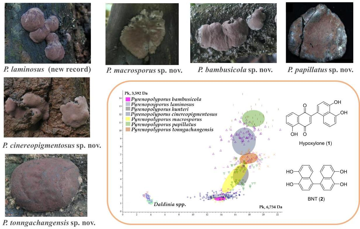
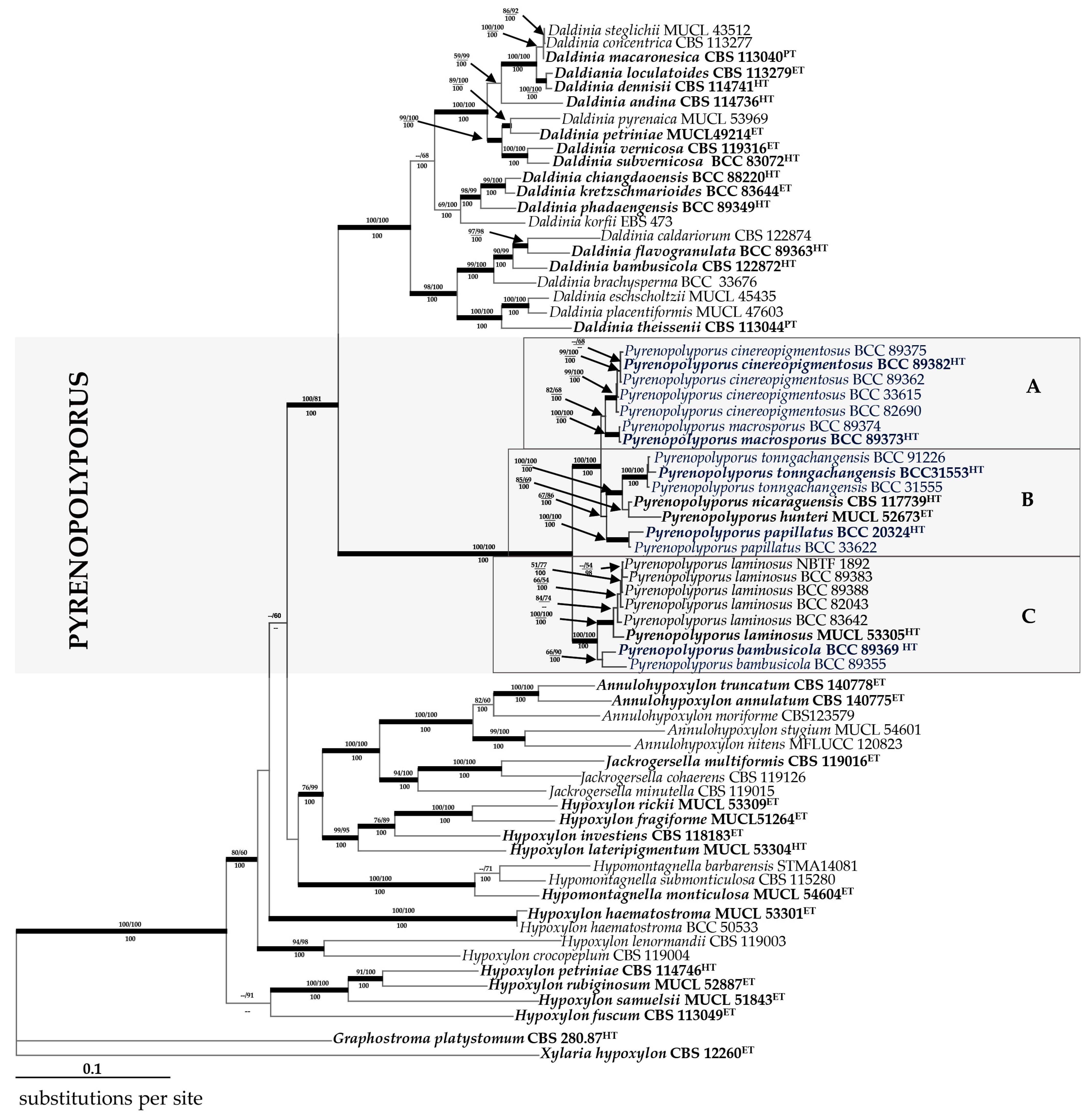
3.2. Molecular Phylogeny (Figure 14)

After providing the full taxonomic description of the five novel species and a new record of Pyrenopolyporus sp. for Thailand, we have also confirmed their taxonomic position through multi-locus phylogenetic analyses as shown in Figure 14 and single-gene analyses as shown in Figures S1–S5. The 70 newly generated ITS, LSU, RPB2, and TUB2 sequences were compared with data from the GenBank NCBI nucleotide database. This was performed to clarify the phylogenetic placement of newly collected Thai specimens of Hypoxylaceae and to distinguish them from other species and genera in the Xylariales (PCR amplifications yielded approximately 500 bp, 1000 bp, 800 bp, and 1000 bp of ITS rDNA, LSU rDNA, RPB2, TUB2 sequences, respectively). The phylogenetic relationships were estimated using the MP, ML, and MB analyses. The dataset of the multi-locus DNA sequences included 66 taxa from the Hypoxylaceae: Annulohypoxylon (5), Daldinia (21), Hypoxylon (12), Hypomontagnella (3), Jackrogersella (3), and Pyrenopolyporus (22). The combined dataset consisted of 5131 characters, of which 2954 were constant, 1722 were parsimony informative, and 455 were uninformative. The MP analysis yielded 11650 trees with a CI of 0.333, a RI of 0.648, and a HI of 0.667. The best phylogenetic tree inferred from RAxML had a likelihood of −56675.811. The alignment had 2456 distinct alignment patterns, with 23.49% undetermined characters or gaps. Estimated base frequencies were as follows: A = 0.239, C = 0.264, G = 0.261, T = 0.234; substitution rates were AC = 1.713, AG = 4.654, AT = 1.599, CG = 1.104, CT = 7.949, GT = 1.000; gamma distribution shape parameter was α 0.841. The likelihood of the Bayesian tree was −65650.640. As shown in Figure 14, the sequences of the new Thai strains are well separated from the previously proposed Pyrenopolyporus species, while the Thai specimens of P. laminosus clustered with the holotype that was originally reported from the Caribbean by Kuhnert et al. [28]. As the topology of the RAxML tree is practically identical to the one presented by Wendt et al. [3], from which most DNA sequence data were included in our study and analyzed using essentially the same methodology, we restrict our discussion on the phylogenetic positions of the new taxa.
Figure 14. Phylogeny of the Hypoxylaceae. RAxML tree was generated based on multiple loci alignment of concatenated ribosomal (ITS and LSU) and proteinogenic (TUB2 and RPB2) sequence data. Support values were calculated via MP, ML, and Bayesian analyses and are indicated above (MPBS/MLBS) and below (BPP) the respective branches. Branches of significant support (BS ≥ 70% and PP ≥ 100) are thickened. New species are indicated in blue and the clade comprising the sequences of the Pyrenopolyporus spp. is marked by a grey rectangle consisting of subclades A, B and C; ET indicates ex-epitype, HT ex-holotype, and PT ex-paratype strains are highlighted in bold letters.
Jof 09 00429 g014

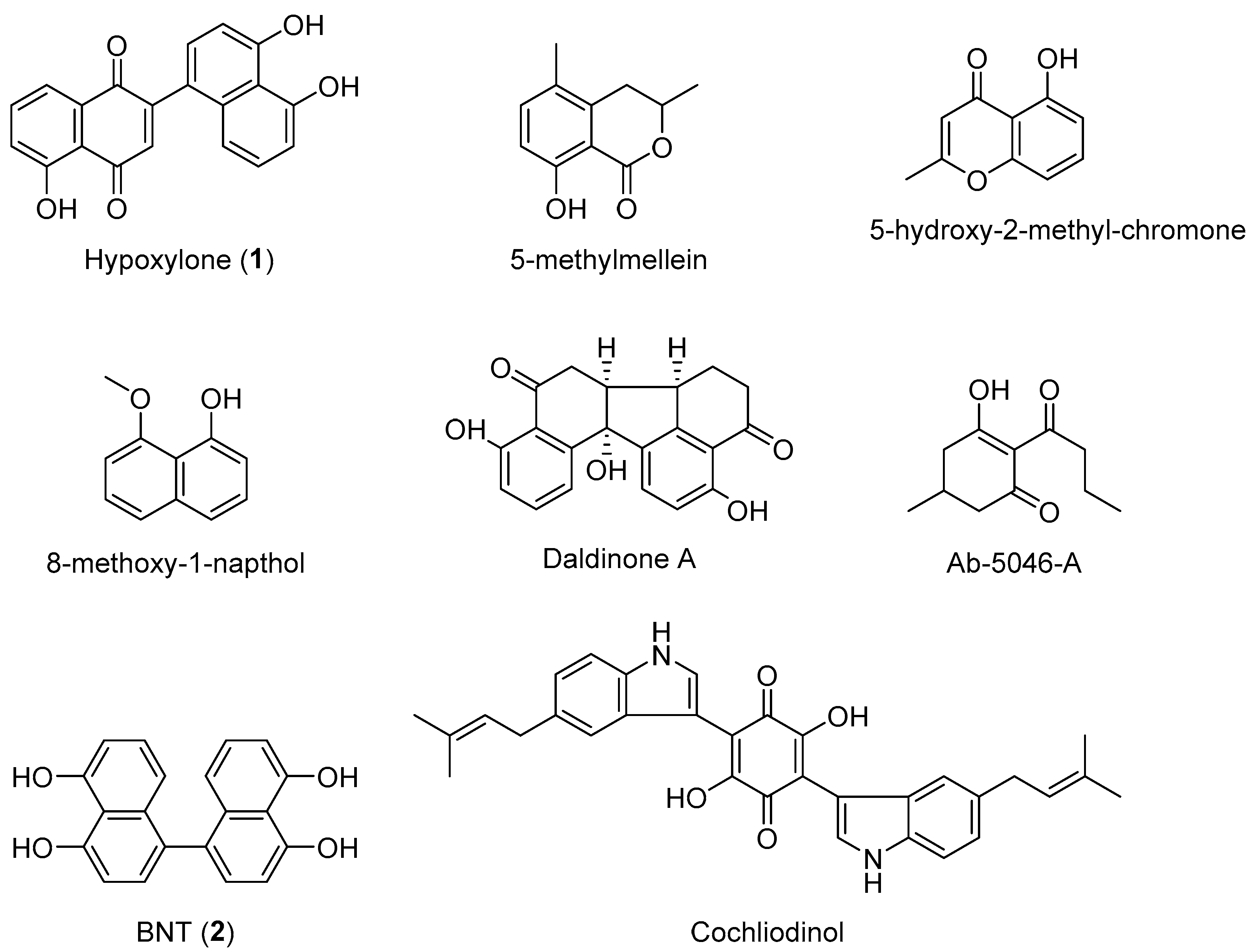
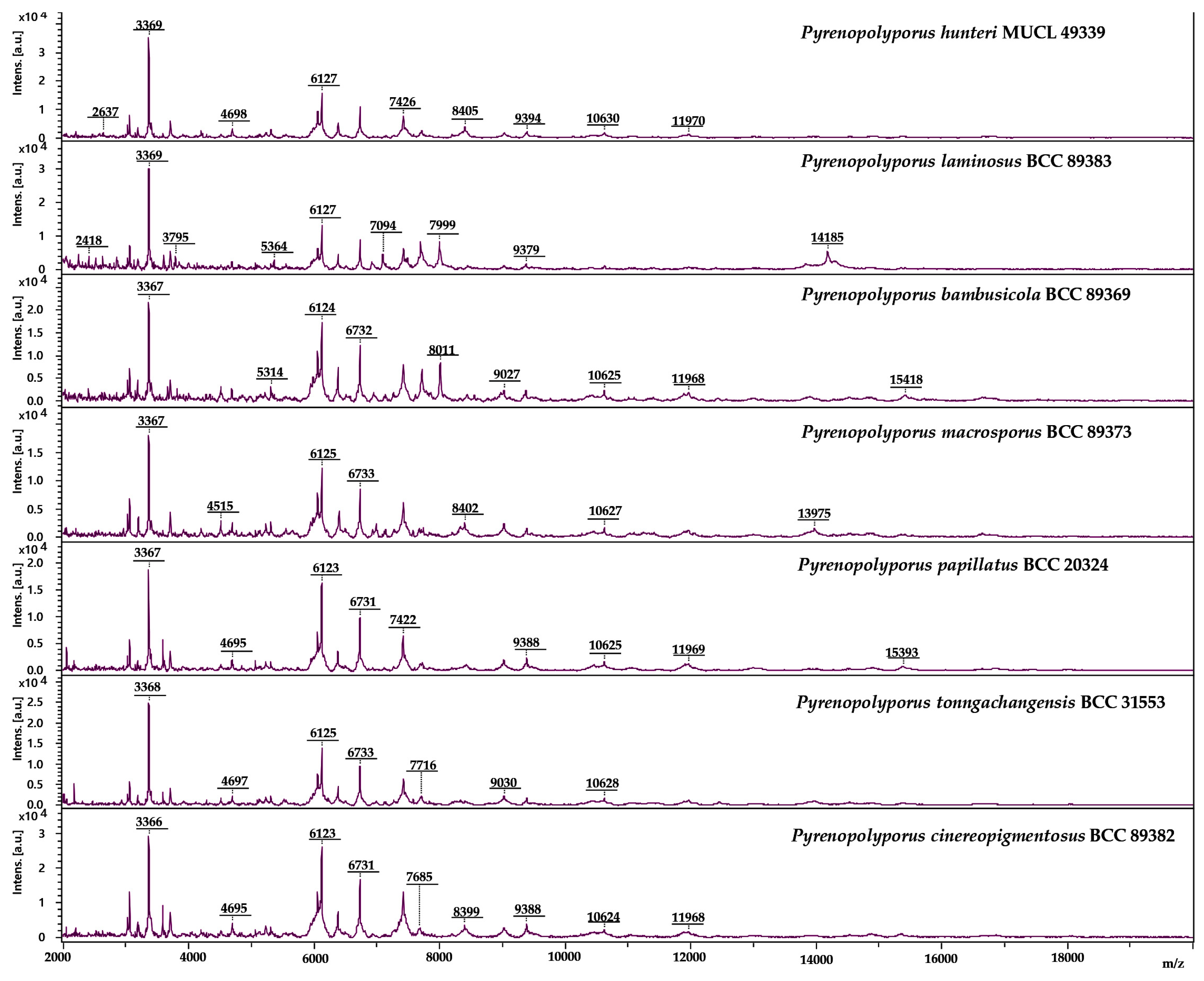
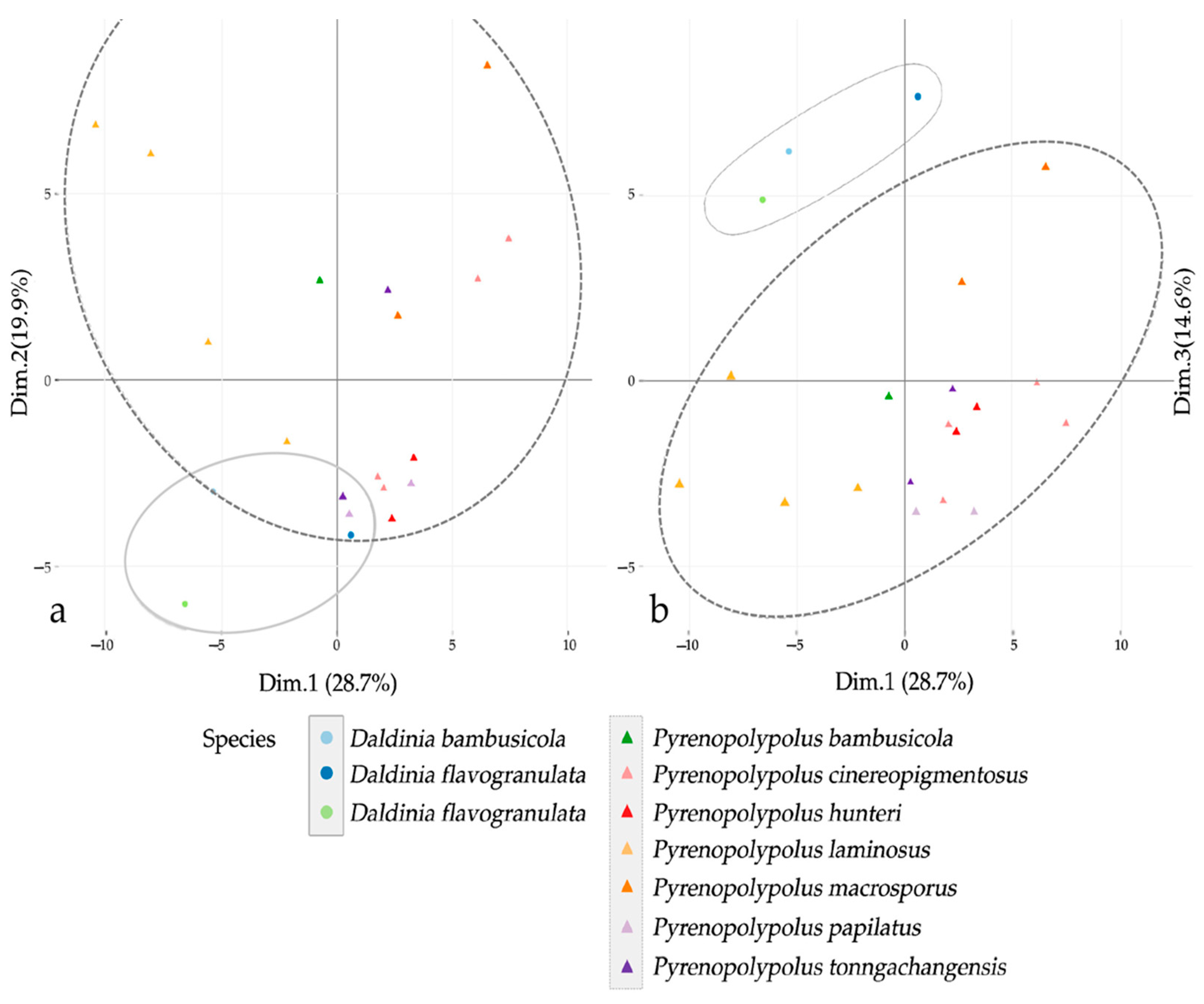
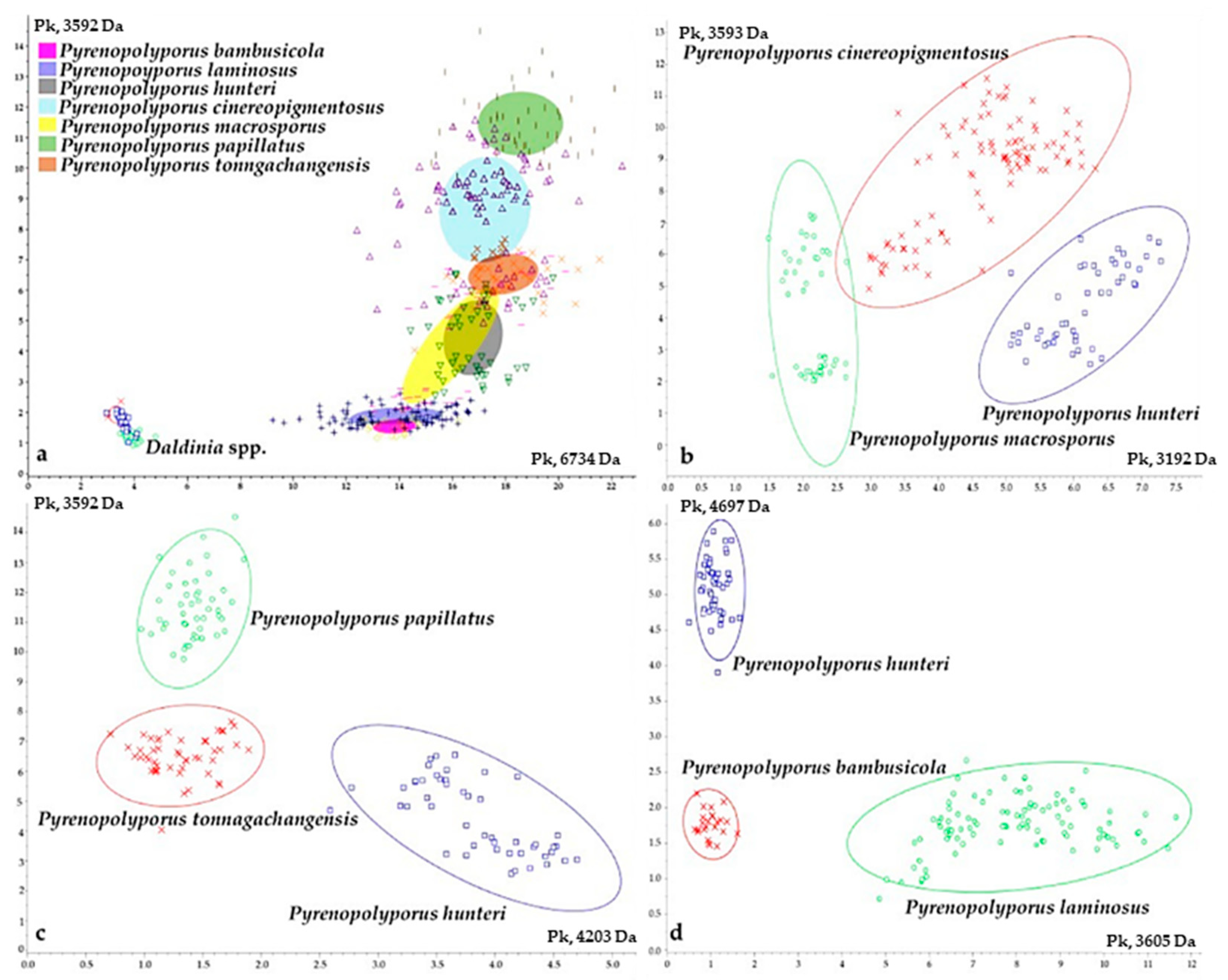
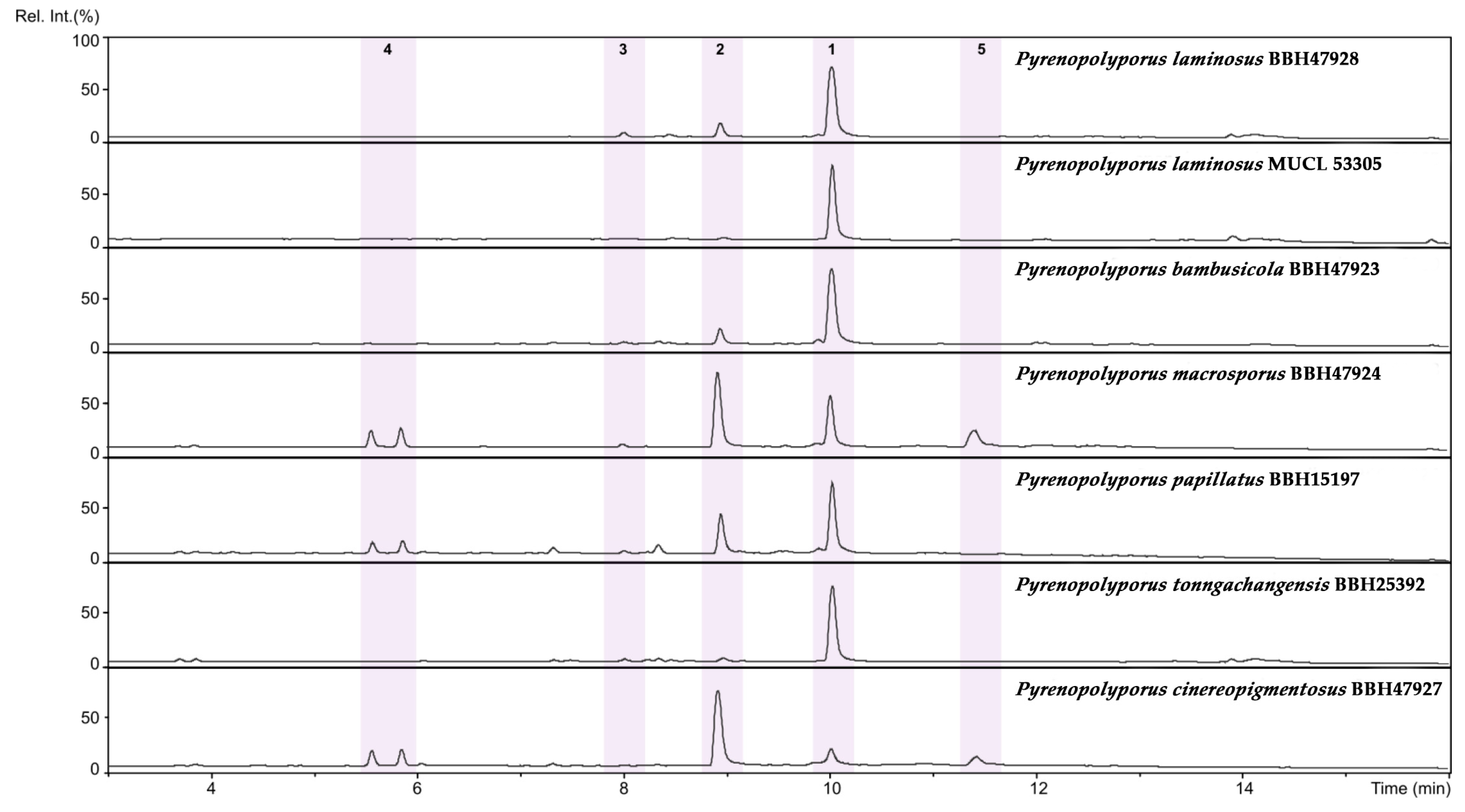
3.3. MALDI-TOF Mass Spectrometry (Figure 15, Figure 16 and Figure 17)

We also investigated the peptide mass fingerprint (PMF) via MALDI-TOF MS of our samples after providing full taxonomic characterization to support our hypothesis regarding the discrimination of closely related species. Representative samples of each species from Pyrenopolyporus, including the ex-epitype culture of the type species of the genus (P. hunteri; Figure 15), were analyzed using MALDI-TOF MS; three Daldinia spp. isolates were also included for this comparison. All samples delivered high quality MALDI spectra (peak rich) as shown in Figure S6. The principal component analysis (PCA) of the 82 final peaks (after denoising, recalibration, and negative-control subtraction) showed a clear difference between the genera Pyrenopolyporus and Daldinia; statistical analyses based on 2D peak distribution using the software ClinProTools gave results in agreement with the PCA mentioned above by revealing two proteomic markers that can be used for discriminating Pyrenopolyporus spp. from Daldinia spp. at 6734 and 3592 Da (Figure 17a).
Despite an overall similarity between Pyrenopolyporus species as revealed by the PCA, ClinProTools was able to give the two most discriminating molecules between some pairs of species (Figure 17b–d). The species within Pyrenopolyporus appeared more or less overlapped except for P. laminosus and P. macrosporus (Figure 16). By dividing the samples into three groups following taxonomic position of our gene multi loci analyses, group A comprised P. cinereopigmentosus and P. macrosporus; group B comprised P. hunteri, P. papillatus, and P. tonngachangensis; group C comprised P. bambusicola and P. laminosus. There is an increased resolution in the discrimination of the species.
Figure 15. Schematic representation of MALDI-TOF chromatograms from mycelial peptide extracts of Pyrenopolyporus spp. evaluated in this study. All spectra shown are baseline-subtracted, smoothed and with the y-axis auto-scaled covering the mass range from 2 kDa to 20 kDa (with x-axis scale increments of 2 kDa).
Jof 09 00429 g015
Figure 16. Principal component analysis (PCA) of denoised, recalibrated, and negative-control subtracted 82 MALDI-TOF mass spectra; (a) the coordinate plane between the first (Dim.1) and second (Dim.2) components; (b) the coordinate plane between the first (Dim.1) and the third components (Dim.3). The ovals on the figures represent 95% confidence concentration ellipses.
Jof 09 00429 g016
Group A (Pyrenopolyporus cinereopigmentosus and P. macrosporus) shared common peaks at 6734 Da while they can be discriminated from each other by the averaged peak area/intensity distribution pattern of the molecules at 3192 and 3594 Da (Figure 17b). The molecular phylogenetic placement was also confirmed in that P. cinereopigmentosus was clearly distinct from P. macrosporus, with full supports for all phylogenetic inferences (MP, ML, MB).
Group B (Pyrenopolyporus hunteri, P. papillatus, and P. tonngachangensis) shared common peaks at 6734 Da; they can be clearly discriminated from each other by the averaged peak area/intensity distribution pattern of the molecules at 3592 and 4203 Da (Figure 17c). Our molecular phylogeny also confirmed that P. papillatus and P. tonngachangensis were clearly distinct from P. hunteri with full support for all phylogenetic inferences (MP, ML, MB).
Figure 17. Principal component analysis (PCA) of the two most discriminant mass spectra peaks derived from the fungal isolates included in this study; (a) PCA for all Pyrenopolyporus and Daldinia strains. P. bambusicola (pink), P. laminosus (purple), P. hunteri (grey), P. cinereopigmentosus (greyish blue), P. macrosporus (yellow), P. papillatus (green), P. tonngachangensis (orange); (b) PCA including only P. cinereopigmentosus (red), P. hunteri (blue), and P. macrosporus (green); (c) PCA including only P. hunteri (blue), P. papillatus (green), and P. tonngachangensis (red); (d) PCA including only P. bambusicola (red), P. hunteri (blue), and P. laminosus (green).The ovals on the figures represent 95%-confidence concentration ellipses.
Jof 09 00429 g017
Group C was phylogenetically segregated from other species with high statistical support (Clade C, Figure 14). The two species within this group (Pyrenopolyporus bambusicola and P. laminosus) are very challenging for identification using morphological features. The proteomic profiling also showed the similarity between these two species; they seemed to share common peaks at 6734 and 3592 Da while being discriminated by the two molecules at 3605 and 4697 Da (Figure 17d). In general, this resolution was much better than the chemotaxonomic study by HPLC-DAD/MS (Figure 18).
  • Dichotomous key to the species of Pyrenopolyporus
  • 1a. Ascospores highly variable in shape, ellipsoid to slightly ellipsoid-inequilateral……...2
  • 1b. Ascospores less variable in shape, ellipsoid-inequilateral ….............................................4
  • 2a. KOH-extractable pigments without purple shades; ascospores 9.5–14(–16) × 4–6 µm with straight to slightly sigmoid germ slit; much less than spore length on the more convex side ....................................................................................................................……P. tortisporus
  • 2b. KOH-extractable pigments purple, ascospores with straight germ slit less than spore length………………………………...……………………………………………………………3
  • 3a. Ascospores 9.5–12 (−13) × 4–5 µm with straight germ slit less than spore length frequently on the more flattened side..........................................................................P. symphyon
  • 3b. Ascospores (14–) 16–17 × (6–) 7–8 µm with straight germ slit covering full spore length on convex side…………………………………………………………………... P. macrosporus
  • 4a. Ascospores pale brown to light brown……………..………………………....…………....5
  • 4b. Ascospores brown to dark brown…………………………………………..……...……….7
  • 5a. Ostioles umbilicate; ascospores (12–) 13–14 (–16) × 4–5 µm with straight to rarely slightly sigmoid germ slit much less than spore-length or nearly spore-length on the convex side…...…………..…………………..……………...………..….….…P. tonngachangensis
  • 5b. Ostioles lower than stromatal surface, punctiform, papillate ……………...…………….6
  • 6a. Ostioles punctiform, slightly lower than the stromal surface, ascospores 11.5–14 × 5–5.5 µm with straight germ slit much less than spore-length……………..……………..P. hunteri
  • 6b. Ostioles papillate; ascospores, (11–) 12–13 (–14) × 4–5 µm with straight germ slit much less than spore-length ……..……………………...………………………...…….. P. papillatus
  • 7a. Species occurring on bamboos………………………….………...….……………..……….8
  • 7b. Species on woody, dicot substrates…………………………………………………………9
  • 8a. Stromata found in fire-damaged areas; ostioles conspicuous umbilicate; perithecia long tubular, 0.75–0.9 mm high; ascospores 10–11 (–12) × (3–) 4–5 µm ………..…P. bambusicola
  • 8b. Stromata not found in fire-damaged area; ostioles umbilicate black to inconspicuous; perithecia long tubular 0.75–0.90 mm high, ascospores 11.0–13.5 × 4.2–4.5 µm...............................................................................................................................P. laminosus
  • 9a. KOH-extractable pigment Dark Livid and Livid Purple; perithecia 0.8–1.5 mm high, ascospores (11.5–) 12–15(−16) × 5–6.5 μm…………………….……..………..P. nicaraguensis
  • 9b. KOH-extractable pigment Dark Mouse Grey or Iron Grey; perithecia tubular 0.9–1.1 mm high ascospores (12–) 13–14 (–15) × 6–7 μm ……………………...P. cinereopigmentosus

4. Discussion

Most Pyrenopolyporus species are morphologically highly similar, which makes species delimitation and identification based on morphology alone difficult and confusing [2]. Recently, much progress has been achieved thanks to DNA sequence data, particularly of protein-coding genes such as RPB2 or TUB2, which have superior resolution compared to ITS or LSU [3,8]. However, an obstacle for an improved species delimitation and identification is the lack of sequences for type materials or well-identified reference specimens in GenBank. Pyrenopolyporus hunteri, the type species of the genus is a good example of these problems. Its taxonomy has been re-investigated by Wendt et al. [3]. In this study, we examined the phylogenetic relationships of our fresh collections with the species of Pyrenopolyporus spp. for which multi-gene sequence data are available. We have performed a multi-gene analysis using ITS, LSU, RPB2, and TUB2 sequence data to determine the phylogenetic placement of our specimens. Pyrenopolyporus clearly forms a monophyletic clade within Hypoxylaceae, distinct from the genus Daldinia, which is in accordance with the extensive results of Wendt et al. [3]. Considering our molecular phylogenetic analyses, the clade Pyrenopolyporus is split into three strongly supported subclades and formed a sister group to the genus Daldinia.
Subclade A is comprised of Pyrenopolyporus cinereopingmentosus and P. macrosporus, which share similar morphological features such as having darker ascospore color. Considering the molecular phylogeny, the two new species including are closely related, but strongly segregated into two distinct monophyletic clades with high supports. The morphological comparisons between these two new species demonstrates very similar features with dark brown ascospores and purple stromal KOH-extractable pigment, as well as similar stromatal secondary metabolites. However, P. macrosporus has the largest ascospores with very diverse forms compared to P. cinereopigmentosus. Hence, our combination between morphological characterization and multi-locus phylogeny supports the status of distinct species between them. However, the proteomics and metabolomics data could not allow a clear distinction to closely differentiate between both species. Morphologically, P. macrosporus is also similar to P. tortisporus and P. symphyon but differs by its ascospore morphology and stromatal KOH-extractable pigments that we have already mentioned in the notes accompanying the species description. The pantropical species, P. tortisporus, was first reported by Ju and Rogers [2]. The type specimen of this species originated from NY as specimen no. WSP69643. The phenotypic features of this fungus are clearly distinctive from P. macrosporus and other species by having frequently deformed ascospores and producing an olivaceous pigment in 10% KOH solution. Fournier et al. [44] also provided a new illustration of a specimen discovered in the French West Indies, whose morphological features fitted well with Ju and Rogers’ description. Pyrenopolyporus symphyon was first reported by Möller [43] but has no appropriate specimen for reexamination since Ju and Rogers [2] reported the type specimen to be immature. The fungus thus needs to be collected in a fresh state for epitypification of the species.
Subclade B is comprised of the type species Pyrenopolyporus hunteri along with its sister species P. nicaraguensis and other closely related species including P. tonngachangensis and P. papillatus. Pyrenopolyporus hunteri and P. nicaraguensis closely resemble P. papillatus and P. tonngachangensis regarding the appearance of the morphological characterization (see in the notes of taxonomic description). Kuhnert et al. [28] found hypoxylone (a naphthoquinone) from fresh specimens of P. laminosus, similar to the finding in P. hunteri and P. nicaraguensis Bitzer et al. [6]. This naphthoquinone could represent an additional chemotaxonomic marker for the species group comprising P. laminosus and its closely related species. Pyrenopolyporus hunteri and P. nicaraguensis were listed (under the epithets H. polyporum and H. nicaraguense) by Ju and Rogers [2] as members of the genus Hypoxylon, and regarded as closely related to Hypoxylon sclerophaeum [45]. These species were considered as part of the “H. placentiforme line” circumscribed by Ju and Rogers [2], characterized by massive semiglobose to peltate stromata with a solid lamellate interior at times with radiating black strands, in contrast to the interior zonate characteristic of the genus Daldinia. Despite morphological differences, H. placentiforme has been transferred to Daldinia (as D. placentiformis) based on the phylogenetic analyses by Hsieh et al. [25], corroborated by chemotaxonomic evidence [5].
The molecular phylogenetic analyses also showed that the species within the subclade B (P. hunteri, P. nicaraguensis, P. papillatus, and P. tonngachangensis) were clearly segregated from the other species. We did not have any axenic cultures from P. nicaraguensis to test whether its proteomic profile would be different from the other species of this subclade; whereas, the MALDI-TOF/MS data allowed a distinction between P. hunteri, P. papillatus and P. tonngachangensis, consistent with the molecular phylogenetic results. Therefore, our study, through the MALDI-TOF/MS data, does not only confirm the distinction between Daldinia and Pyrenopolyporus, but also the differences between the species within the subclade B in those for whom cultures are available. In contrast, the stromatal metabolite profile for P. papillatus and P. tonngachangensis showed high similarity to the profiles obtained for species in the subclade C.
Subclade C consists exclusively of bambusicolous species, Pyrenopolyporus bambusicola and P. laminosus. Pyrenopolyporus laminosus is well discriminated by daldinioid stromata with violet KOH-extractable pigments and light brown ascospores with a spore-lengthed germ slit and indehiscent perispore in 10% KOH, and by the occurrence on bamboos. Amongst Pyrenopolyporus spp. having peltate stromata with violet KOH-extractable pigments, P. nicaraguensis is the most similar to P. laminosus as it has ascospores with a germ slit covering almost the entire spore length. The lamellate structure of the interior tissue of the stroma of P. nicaraguensis has a similar appearance to that of P. laminosus. However, P. nicaraguensis differs in having raised discoid ostioles and broader ellipsoid ascospore and it has been almost exclusively reported to occur on dicotyledons [2,28]. Our novel species, P. bambusicola, was found only on bamboos and is distinguished from other members of this genus by deeply umbilicate ostioles and the ascospore size range. The phylogenetic analyses clearly support its distinctiveness. Furthermore, the MALDI-TOF/MS seems to support its difference from the other Pyrenopolyporus species. In particular, P. laminosus can be discriminated from its sister species P. bambusicola. These two sister species are highly similar for their morphology.
MALDI-TOF/MS has been demonstrated to be a highly adaptable approach for efficient identification and classification of bacteria and yeasts in clinical laboratories, presenting a complementary technique to traditional microscopic and molecular biology methods [11,12]. However, this technique has not been used extensively for the identification or classification of filamentous fungi for various reasons, including the difficulty of fungal protein extraction and the necessary high capital investment. In this study, the technique served well to discriminate a complicated species complex and provided corroborating evidence to other data that were obtained by studying classical morphology, chemotaxonomy, and molecular phylogeny. The MALDI-TOF/MS analysis allowed a good discrimination between the different Pyrenopolyporus species, and particularly high resolution between Daldinia and Pyrenopolyporus, coherently with our phylogenetic analysis. Although the sampling might be limited in our study, the data encourage further investigations with more samples of the same genus, or even other species complexes in the Hypoxylaceae in order to further prove the robustness of the technique.
Moreover, our study advocates that the PMF obtained via MALDI-TOF/MS can be used as a reliable tool for species discrimination in Pyrenopolyporus. Despite the highly similar morphological traits of the fungi in this group, the PMF data demonstrated that Pyrenopolyporus species are distinguishable, consistent with molecular phylogenetic data. Cryptic morphology in fungal species complexes has long been a problem for taxonomists. Our study showed that molecular phylogenies based on multi-locus analyses and PMF could contribute to resolve species identification.
Recently, some strains representing important lineages of the Hypoxylaceae have been selected for a phylogenomic study relying on high quality genomes [9], revealing the occurrence of ITS polymorphisms [46] and thus the necessity to use more than ITS for species identification and classification in this family and the order Xylariales in general. This accomplishment has offered a significant starting point for the development of a stable phylogeny of this order, as well as studies on evolution, ecological guilds, and natural product biosynthesis.

Supplementary Materials

The following supporting information can be downloaded at https://www.mdpi.com/article/10.3390/jof9040429/s1, Figure S1: Phylogeny of the Hypoxylaceae. The phylogenetic relationships inferred from Bayesian analysis based on multiple genetic loci of nuclear ribosomal LSU and proteinogenic (TUB2 and RPB2) sequences. Support values of more than 50% (MPBS/MLBS) or 95 (BPP) were calculated via MP, ML, and Bayesian analysis and are indicated above (MPBS/MLBS) and below (BPP) the respective branches. Branches of significant support (MPBS, MLBS = 70% and BPP = 95) are thickened. New species are indicated in blue and the clade comprising the sequences of the Pyrenopolyporus spp. which is marked by a grey rectangle, and ET indicates ex-epitype, HT ex-holotype, and PT ex-paratype strains; Figure S2: Phylogeny of the Hypoxylaceae. The phylogenetic relationships depicted as maximum parsimony tree generated based on ITS sequences. Support values of more than 50% (MPBS/MLBS) or 95 (BPP) were calculated via MP, ML, and Bayesian analysis and are indicated above (MPBS/MLBS) and below (BPP) the respective branches. Branches of significant support (MPBS, MLBS = 70% and BPP = 95) are thickened. New species are indicated in blue and the clade comprising the sequences of the Pyrenopolyporus spp. is marked by a grey rectangle, and ET indicates ex-epitype, HT ex-holotype, and PT ex-paratype strains; Figure S3: Phylogeny of the Hypoxylaceae. The phylogenetic relationships depicted as maximum parsimony tree are generated based on LSU sequences. Support values of more than 50% (MPBS/MLBS) or 95 (BPP) were calculated via MP, ML, and Bayesian analysis and are indicated above (MPBS/MLBS) and below (BPP) the respective branches. Branches of significant support (MPBS, MLBS =70% and BPP = 95) are thickened. New species are indicated in blue and the clade comprising the sequences of the Pyrenopolyporus spp. is marked by a grey rectangle, and ET indicates ex-epitype, HT ex-holotype, and PT ex-paratype strains; Figure S4: Phylogeny of the Hypoxylaceae. The phylogenetic relationships inferred from Bayesian analysis based on proteinogenic (RPB2) sequences. Support values of more than 50% (MPBS/MLBS) or 95 (BPP) were calculated via MP, ML, and Bayesian analysis and are indicated above (MPBS/MLBS) and below (BPP) the respective branches. Branches of significant support (MPBS, MLBS = 70% and BPP = 95) are thickened. New species are indicated in blue and the clade comprising the sequences of the Pyrenopolyporus spp. is marked by a grey rectangle, and ET indicates ex-epitype, HT ex-holotype, and PT ex-paratype strains; Figure S5: Phylogeny of the Hypoxylaceae. The phylogenetic relationships inferred from Bayesian analysis based on proteinogenic (TUB2) sequences. Support values of more than 50% (MPBS/MLBS) or 95 (BPP) were calculated via MP, ML, and Bayesian analysis and are indicated above (MPBS/MLBS) and below (BPP) the respective branches. Branches of significant support (MPBS, MLBS = 70% and BPP = 95) are thickened. New species are indicated in blue and the clade comprising the sequences of the Pyrenopolyporus spp. which is marked by a grey rectangle, and ET indicates ex-epitype, HT ex-holotype, and PT ex-paratype strains; Figure S6: MALDI-TOF MASS SPECTRA; Figure S7: DAD spectra of the major metabolites (1−5) depicted in the stromata of the evaluated Pyrenopolyporus spp.; Figure S8: MS/MS spectra comparison between hypoxylone (1) and compound 3. The MS/MS similarity score between the two metabolites is <500, but both spectra share analogous neutral losses for the major fragment ions; Table S1: Comparison of morphological and chemotaxonomic features of Hypoxylaceae species with massive stroma and long tubular perithecia; Table S2: Dereplicated metabolites from the stromatal extracts of the Pyrenopolyporus spp. and in-house standards.

Author Contributions

S.W.: Analysis and interpretation of phylogenetic data morphological and proteomic studies, visualization, drafting of the paper, critical review of the draft. B.C.: DNA extraction, PCR amplification, DNA Sequencings. N.K.: investigation, analysis, and interpretation of data, critical review of the draft. S.S.: Critical review of the draft. E.C.-G. and M.S.: Chemical analyses, and interpretation of the data, critical review of the draft. S.R.: Conception and design for MALDI-TOF. M.S.: Conception and design of the overall study, critical review of the draft. J.L.-a. Critical review of the draft. P.S.: resources, supervision, investigation, conceptualization. All authors have read and agreed to the published version of the manuscript.

Funding

This research benefitted from funding by the European Union’s Horizon 2020 research and innovation program (RISE) under the Marie Skłodowska-Curie grant agreement No. 101008129, project acronym “MycoBiomics” (lead beneficiaries J.J.L and M.S.). Esteban Charria-Girón was supported by the HZI POF IV Cooperativity and Creativity Project Call. This work was also supported by the National Science and Technology Development Agency (NSTDA), National S & T Infrastructure (NSTI) grant No. P2250737, and the Technology and Innovation Management Department grant number P-18-50644 in the Pa Daeng Mine’s area, Tak Province, Thailand.

Institutional Review Board Statement

Not applicable.

Informed Consent Statement

Not applicable.

Data Availability Statement

All newly generated sequences have been submitted to the public domain as GenBank.

Acknowledgments

We acknowledge Satinee Suetrung and Sissades Thongsima (NBT-NSTDA) for their continuous support in this present study. The authors thank Jirawan Kumsao for sample collections in Ban Hua Thung community forest in northern Thailand. Our warmest thanks go to Ulrike Beutling and Janthima Jaresitthikunchai for their expert assistance in the lab.

Conflicts of Interest

The authors declare no conflict of interest.

References

  1. Lloyd, C.G. Synopsis of some genera of the large pyrenomycetes. Mycological Notes 50. Mycol. Writ. 1917, 5, 701–716. [Google Scholar]
  2. Ju, Y.M.; Rogers, J.D. Mycologia Memoir n° 20. In A Revision of the Genus Hypoxylon; APS Press: St. Paul, MN, USA, 1996; p. 365. [Google Scholar]
  3. Wendt, L.; Sir, E.B.; Kuhnert, E.; Heitkämper, S.; Lambert, C.; Hladki, A.I.; Romero, A.I.; Luangsa-ard, J.J.; Srikitikulchai, P.; Peršoh, D. Resurrection and emendation of the Hypoxylaceae, recognised from a multigene phylogeny of the Xylariales. Mycol. Prog. 2018, 17, 115–154. [Google Scholar] [CrossRef] [Green Version]
  4. Helaly, S.E.; Thongbai, B.; Stadler, M. Diversity of biologically active secondary metabolites from endophytic and saprotrophic fungi of the ascomycete order Xylariales. Nat. Prod. Rep. 2018, 35, 992–1014. [Google Scholar] [CrossRef] [PubMed]
  5. Sir, E.; Lambert, C.; Wendt, L.; Hladki, A.; AI, R.; Stadler, M. A New Species of Daldinia (Xylariaceae) from the Argentine Subtropical Montane Forest. Mycosphere 2016, 7, 1378–1388. [Google Scholar] [CrossRef]
  6. Bitzer, J.; Laessoe, T.; Fournier, J.; Kummer, V.; Decock, C.; Tichy, H.V.; Piepenbring, M.; Persoh, D.; Stadler, M. Affinities of Phylacia and the daldinoid Xylariaceae, inferred from chemotypes of cultures and ribosomal DNA sequences. Mycol. Res. 2008, 112, 251–270. [Google Scholar] [CrossRef] [PubMed]
  7. Triebel, D.; Peršoh, D.; Wollweber, H.; Stadler, M. Phylogenetic relationships among Daldinia, Entonaema and Hypoxylon as inferred from ITS nrDNA sequences. Nova Hedwig. 2005, 80, 25–43. [Google Scholar] [CrossRef]
  8. Daranagama, D.A.; Hyde, K.D.; Sir, E.B.; Thambugala, K.M.; Tian, Q.; Samarakoon, M.C.; McKenzie, E.H.C.; Jayasiri, S.C.; Tibpromma, S.; Bhat, J.D.; et al. Towards a natural classification and back-bone tree for Graphostromataceae, Hypoxylaceae, Lopadostomataceae and Xylariaceae. Fungal Divers. 2018, 88, 1–165. [Google Scholar] [CrossRef]
  9. Wibberg, D.; Stadler, M.; Lambert, C.; Bunk, B.; Spröer, C.; Rückert, C.; Kalinowski, J.; Cox, R.J.; Kuhnert, E. High quality genome sequences of thirteen Hypoxylaceae (Ascomycota) strengthen the phylogenetic family backbone and enable the discovery of new taxa. Fungal Divers. 2021, 106, 7–28. [Google Scholar] [CrossRef]
  10. Kuhnert, E.; Navarro-Muñoz, J.; Becker, K.; Stadler, M.; Collemare, J.; Cox, R. Secondary metabolite biosynthetic diversity in the fungal family Hypoxylaceae and Xylaria hypoxylon. Stud. Mycol. 2021, 99, 100118. [Google Scholar] [CrossRef]
  11. Čmoková, A.; Kolařík, M.; Dobiáš, R.; Hoyer, L.L.; Janouškovcová, H.; Kano, R.; Kuklová, I.; Lysková, P.; Machová, L.; Maier, T.; et al. Resolving the taxonomy of emerging zoonotic pathogens in the Trichophyton benhamiae complex. Fungal Divers. 2020, 104, 333–387. [Google Scholar] [CrossRef]
  12. Patel, R. A Moldy Application of MALDI: MALDI-ToF Mass Spectrometry for Fungal Identification. J. Fungi 2019, 5, 4. [Google Scholar] [CrossRef] [PubMed] [Green Version]
  13. Kuhnert, E.; Surup, F.; Sir, E.; Lambert, C.; Hyde, K.; Hladki, A.; Romero, A.; Stadler, M. Lenormandins A–G, new azaphilones from Hypoxylon lenormandii and Hypoxylon jaklitschii sp. nov., recognised by chemotaxonomic data. Fungal Divers. 2015, 71, 165–184. [Google Scholar] [CrossRef]
  14. Rayner, R.W. Commonwealth Mycological Institute (Great Britain). In A Mycological Colour Chart; British Mycological Society: Manchester, UK, 1970. [Google Scholar]
  15. Cedeño-Sanchez, M.; Charria-Girón, E.; Lambert, C.; Luangsa-ard, J.J.; Decock, C.; Franke, R.; Brönstrup, M.; Stadler, M. Segregation of the genus Parahypoxylon (Hypoxylaceae, Xylariales) from Hypoxylon by a polyphasic taxonomic approach. Mycokeys 2023, 95, 131–162. [Google Scholar] [CrossRef]
  16. Mongkolsamrit, S.; Luangsa-ard, J.J.; Spatafora, J.W.; Sung, G.H.; Hywel-Jones, N.L. A combined ITS RDNA and β-tubulin phylogeny of Thai species of Hypocrella with non-fragmenting ascospores. Mycol. Res. 2009, 113, 684–699. [Google Scholar] [CrossRef] [PubMed]
  17. White, T.J.; Bruns, T.; Lee, S.; Taylor, J. Amplification and direct sequencing of fungal ribosomal RNA genes for phylogenetics. In PCR Protocols; Academic Press: Cambridge, MA, USA, 1990; pp. 315–322. [Google Scholar]
  18. Vilgalys, R.; Hester, M. Rapid genetic identification and mapping of enzymatically amplified ribosomal DNA from several Cryptococcus species. J. Bacteriol. 1990, 172, 4238–4246. [Google Scholar] [CrossRef] [Green Version]
  19. Rehner, S.A.; Samuels, G.J. Taxonomy and phylogeny of Gliocladium analysed from nuclear large subunit ribosomal DNA Sequences. Mycol. Res. 1994, 98, 625–634. [Google Scholar] [CrossRef]
  20. Liu, Y.J.; Whelen, S.; Hall, B.D. Phylogenetic relationships among ascomycetes: Evidence from an RNA polymerse II subunit. Mol. Biol. Evol. 1999, 16, 1799–1808. [Google Scholar] [CrossRef] [Green Version]
  21. O’Donnell, K.; Cigelnik, E. Two divergent intragenomic rDNA ITS2 types within a monophyletic lineage of the fungus Fusarium are nonorthologous. Mol. Phylogenet. Evol. 1997, 7, 103–116. [Google Scholar] [CrossRef]
  22. Hall, T.A. BioEdit: A user-friendly biological sequence alignment editor and analysis program for Windows 95/98/NT. Nucleic Acids Symp. Ser. 2013, 41, 95–98. [Google Scholar]
  23. Kuhnert, E.; Sir, E.B.; Lambert, C.; Hyde, K.D.; Hladki, A.I.; Romero, A.I.; Rohde, M.; Stadler, M. Phylogenetic and chemotaxonomic resolution of the genus Annulohypoxylon (Xylariaceae) including four new species. Fungal Divers. 2017, 85, 1–43. [Google Scholar] [CrossRef]
  24. Daranagama, D.A.; Camporesi, E.; Tian, Q.; Liu, X.; Chamyuang, S.; Stadler, M.; Hyde, K.D. Anthostomella is polyphyletic comprising several genera in Xylariaceae. Fungal Divers. 2015, 73, 203–238. [Google Scholar] [CrossRef]
  25. Hsieh, H.M.; Ju, Y.M.; Rogers, J.D. Molecular phylogeny of Hypoxylon and closely related genera. Mycologia 2005, 97, 844–865. [Google Scholar] [CrossRef] [PubMed]
  26. Wongkanoun, S.; Becker, K.; Boonmee, K.; Srikitikulchai, P.; Boonyuen, N.; Chainuwong, B.; Luangsa-ard, J.; Stadler, M. Three novel species and a new record of Daldinia (Hypoxylaceae) from Thailand. Mycol. Prog. 2020, 19, 1113–1132. [Google Scholar] [CrossRef]
  27. U’Ren, J.M.; Miadlikowska, J.; Zimmerman, N.B.; Lutzoni, F.; Stajich, J.E.; Arnold, A.E. Contributions of North American endophytes to the phylogeny, ecology, and taxonomy of Xylariaceae (Sordariomycetes, Ascomycota). Mol. Phylogenetics Evol. 2016, 98, 210–232. [Google Scholar] [CrossRef] [Green Version]
  28. Kuhnert, E.; Fournier, J.; Peršoh, D.; Luangsa-ard, J.J.D.; Stadler, M. New Hypoxylon species from Martinique and new evidence on the molecular phylogeny of Hypoxylon based on ITS rDNA and β-tubulin data. Fungal Divers. 2014, 64, 181–203. [Google Scholar] [CrossRef]
  29. Stadler, M.; Laessoe, T.; Fournier, J.; Decock, C.; Schmieschek, B.; Tichy, H.V.; Persoh, D. A polyphasic taxonomy of Daldinia (Xylariaceae). Stud. Mycol. 2014, 77, 1–143. [Google Scholar] [CrossRef] [Green Version]
  30. Wongkanoun, S.; Wendt, L.; Stadler, M.; Luangsa-Ard, J.; Srikitikulchai, P. A novel species and a new combination of Daldinia from Ban Hua Thung community forest in the northern part of Thailand. Mycol. Prog. 2019, 18, 553–564. [Google Scholar] [CrossRef] [Green Version]
  31. Johannesson, H.; Laessøe, T.; Stenlid, J. Molecular and morphological investigation of the genus Daldinia in northern Europe. Mycol. Res. 2000, 104, 275–280. [Google Scholar] [CrossRef]
  32. Zhang, N.; Castlebury, L.A.; Miller, A.N.; Huhndorf, S.M.; Schoch, C.L.; Seifert, K.A.; Rossman, A.Y.; Rogers, J.D.; Kohlmeyer, J.; Volkmann-Kohlmeyer, B.; et al. An overview of the systematics of the Sordariomycetes based on a four-gene phylogeny. Mycologia 2006, 98, 1076–1087. [Google Scholar] [CrossRef]
  33. Koukol, O.; Kelnarová, I.; Černý, K. Recent observations of sooty bark disease of sycamore maple in Prague (Czech Republic) and the phylogenetic placement of Cryptostroma corticale. For. Pathol. 2015, 45, 21–27. [Google Scholar] [CrossRef]
  34. Lambert, C.; Wendt, L.; Hladki, A.I.; Romero, A.I.; Stadler, M.; Sir, E.B. Hypomontagnella (Hypoxylaceae): A new genus segregated from Hypoxylon by a polyphasic taxonomic approach. Mycol. Prog. 2019, 18, 187–201. [Google Scholar] [CrossRef]
  35. Stadler, M.; Kuhnert, E.; Persoh, D.; Fournier, J. The Xylariaceae as model example for a unified nomenclature following the “One Fungus-One Name” (1F1N) Concept. Mycology 2013, 4, 5–21. [Google Scholar] [CrossRef]
  36. Sir, E.B.; Kuhnert, E.; Lambert, C.; Hladki, A.I.; Romero, A.I.; Stadler, M. New species and reports of Hypoxylon from Argentina recognized by a polyphasic approach. Mycol. Prog. 2016, 15, 42. [Google Scholar] [CrossRef]
  37. Edgar, R.C. MUSCLE: Multiple sequence alignment with high accuracy and high throughput. Nucleic Acids Res. 2004, 32, 1792–1797. [Google Scholar] [CrossRef] [Green Version]
  38. Swofford, D.L. PAUP* 4.0b10: Phylogenetic Analysis Using Parsimony (*and other Methods); Sinauer Associates: Sunderland, UK, 2002. [Google Scholar] [CrossRef]
  39. Miller, M.A.; Pfeiffer, W.; Schwartz, T. Creating the CIPRES Science Gateway for inference of large phylogenetic trees. In Proceedings of the Gateway Computing Environments Workshop (GCE), New Orleans, LA, USA, 14 November 2010; pp. 1–8. [Google Scholar]
  40. Stamatakis, A. RAxML version 8: A tool for phylogenetic analysis and post-analysis of large phylogenies. Bioinformatics 2014, 30, 1312–1313. [Google Scholar] [CrossRef] [PubMed] [Green Version]
  41. Huelsenbeck, J.P.; Ronquist, F. MRBAYES: Bayesian inference of phylogenetic trees. Bioinformatics 2001, 17, 754–755. [Google Scholar] [CrossRef] [Green Version]
  42. Nylander, J. MrModeltest V2; Uppsala Universitit: Uppsala, Sweden, 2004. [Google Scholar]
  43. Möller, A. Phycomyceten und Ascomyceten. Untersuchungen aus Brasilien; G. Fischer: Schaffhausen, Swiss, 1901; Volume 9, pp. 1–319. [Google Scholar]
  44. Fournier, J.; Lechat, C.; Courtecuisse, R. The genus Hypoxylon (Xylariaceae) in Guadeloupe and Martinique (French West Indies). Ascomycete Org. 2016, 7, 145–212. [Google Scholar]
  45. Miller, J.H. A Monograph of the World Species of Hypoxylon; Univ. Georgia Press: Athens, Georgia, USA, 1961; p. 158. [Google Scholar]
  46. Stadler, M.; Lambert, C.; Wibberg, D.; Kalinowski, J.; Cox, R.J.; Kolařík, M.; Kuhnert, E. Intragenomic polymorphisms in the ITS region of high-quality genomes of the Hypoxylaceae (Xylariales, Ascomycota). Mycol. Prog. 2020, 19, 235–245. [Google Scholar] [CrossRef] [Green Version]
Figure 1. Chemical structures of stromatal metabolites detected in this study as well as representative metabolites from Daldinia, Hypoxylon, and other genera of the Hypoxylaceae as reported by Wendt et al. [3].
Jof 09 00429 g001
Figure 2. Morphological characteristics of Pyrenopolyporus laminosus from Thailand (BBH47928). (a,c) Stromata with natural substrate; (b) detail of stromal insertion point; (d) stromal surface and ostioles with KOH-extractable pigments in 10% KOH; (e) longitudinal section of the stroma showing perithecia and the tissue below the perithecial layer; (f) Perithecia (white arrow); (g) Perithecia under light microscope; (hj) asci; (k) apical apparatus, bluing in Melzer’s reagent (black arrow); (ln) ascospores. (o) Ascospore showing germ slit (white arrow); scale bars: (a,c) = 5 cm; (b) = 0.5 cm; (e) = 5 mm; (f) = 1 mm; (g) = 0.25 mm; (hj) = 20 µm; (ko) = 5 µm.
Jof 09 00429 g002
Figure 3. Pyrenopolyporus laminosus from Thailand (strain BCC89383). (a,b) Ascospores by SEM; (c,d) aerial mycelium showing branching pattern; (e) primordia in culture; (f) conidia (white arrows); (h) conidiogenous cell (black arrow); (i) colony on PDA after one month; (j) colony on OA after one month; (k) colony on YMGA after one month. Scale bars: (a,b) = 5 µm; (c,d) = 50 µm; (e) = 0.5 mm; (fh) = 10 µm; (ik) = 2 cm.
Jof 09 00429 g003
Figure 4. Morphological characteristics of Pyrenopolyporus bambusicola (BBH47923). (ac) Stroma on bamboos; (d) stromal surface and ostioles with KOH-extractable pigments in 10% KOH; (e) longitudinal section of stroma showing perithecia and the tissue below the perithecial layer; (f) Perithecia; (g,h) asci showing germ slit (white arrows); (i) apical apparatus, bluing in Melzer’s reagent; (jm) ascospores. Scale bars: (a) = 10 mm; (b,c) = 5 mm; (e) = 2 mm; (f) = 1 mm (g,h) = 20 µm; (im) = 5 µm.
Jof 09 00429 g004
Figure 5. Pyrenopolyporus bambusicola strain BCC89369. (a,b) Ascospores by SEM; (c) vegetative mycelium; (d,e) aerial mycelium showing branching pattern; (f) conidia (white arrow) and conidiogenous cell (black arrow); (g) colony on PDA after one month; (h) colony on OA after one month; (i) colony on YMGA after one month. Scale bars: (a,b) = 5 µm; (c,e,f) = 10 µm; (d) = 20 µm; (gi) = 2 cm.
Jof 09 00429 g005
Figure 6. Morphological characteristics of Pyrenopolyporus cinereopigmentosus (BBH47927). (a,b) Stromata on natural habitat; (c) stromata showing ventral surface. (d) Longitudinal section of the stroma showing the tissue below the perithecial layer; (e) stromatal surface showing ostioles (white arrow) with KOH-extractable pigment; (f) longitudinal section of the stromata showing perithecia under light microscope; (gi) asci. (j) Apical apparatus bluing in Melzer’s reagent (white arrow); (k,l) ascospores showing germ slit (white arrow); (m,n) ascospores. Scale bars (a,b) = 5 cm; (c) = 1 cm; (d) = 5 mm; (f) = 0.25 mm; (gl) = 10 µm; (jn) = 5 µm.
Jof 09 00429 g006
Figure 7. Pyrenopolyporus cinereopigmentosus strain BCC89382. (a,b) Ascospores by SEM; (c,d) aerial mycelium showing branching pattern; (e) conidiogenous cell; (f) conidia; (g) colony on PDA after one month; (h) colony on OA after one month; (i) colony on YMGA after on month. Scale bars: (a,b) = 5 µm; (c,d) = 20 µm; (e,f) = 10 µm; (gi) = 2 cm.
Jof 09 00429 g007
Figure 8. Morphological characteristics of Pyrenopolyporus macrosporus (BBH47924). (ac) Stroma on natural habit; (d) stromal surface and ostioles with KOH-extractable pigments in 10% KOH; (e) longitudinal section of stroma showing the tissue below the perithecial layer; (f) Perithecia; (g) ascus in distilled water; (h) ascus in Melzer’s reagent showing apical apparatus; (jo) ascospores with highly variable shapes. Scale bars: (a) = 2 cm; (b,c) = 1 cm; (e,f) = 0.5 mm; (g,h) = 10 µm; (io) = 5 µm.
Jof 09 00429 g008
Figure 9. Pyrenopolyporus macrosporus strain (BCC89373). (a,b) Ascospores by SEM; (c) aerial mycelium showing branching pattern; (df) conidia (white arrow) and conidiogenous cells (black arrow); (g) colony on PDA after one month; (h) colony on OA after one month; (i) colony on YMGA after one month. Scale bars: (a,b) = 5 µm; (c) = 20 µm; (df) = 10 µm; (gi) = 2 cm.
Jof 09 00429 g009
Figure 10. Morphological characteristics of Pyrenopolyporus papillatus (BBH15197). (a) Stroma; (b) stromatal surface showing ostiole (white arrows) and KOH-extractable pigment; (c) detail of stromal insertion point; (d) longitudinal section showing the tissue below the perithecial layer (arrow); (e) Perithecia (arrow); (f,g) asci in Melzer’s iodine regent; (g) apical apparatus bluing in Melzer’s reagent (arrow); (il) ascospores in 10% KOH with showing germ slit (arrow); (m,n) ascospore in distilled water. Scale bars: (a,b) = 1 cm; (d) = 5 mm; (e) = 0.5 mm (f,g) = 20 µm; (ho) = 5 µm.
Jof 09 00429 g010
Figure 11. Pyrenopolyporus papillatus strain (BCC20324). (a,b) Ascospores by SEM; (c,d) aerial mycelium showing branching pattern; (e,f) conidia (white arrow) and conidiogenous cells (black arrow); (g) colony on PDA after one month; (h) colony on OA after one month; (i) colony on YMGA after on month. Scale bars: (a,b) = 5 µm; (c,d) = 20 µm; (e,f) = 10 µm; (gi) = 2 cm.
Jof 09 00429 g011
Figure 12. Morphological characteristics of Pyrenopolyporus tonngachangensis (BBH25392); (ad) stroma on natural habit; (c) stromal surface and ostioles with KOH-extractable pigments in 10% KOH; (d) longitudinal section of stroma showing perithecia and the tissue below the perithecial layer; (e,f) detail of stromal insertion point (white arrow); (g) longitudinal section perithecia under the light microscope; (hj) asci in distilled water; (k) young ascus in Melzer’s reagent; (l) apical apparatus, bluing in Melzer’s reagent (black arrow); (m) ascospore showing germ slit (white arrow); (np) ascospores. Scale bars: (a,b,e,f) = 1 cm; (d) = 0.5 cm; (h) = 20 µm; (ik) = 10 µm (lp) = 5 µm.
Jof 09 00429 g012
Figure 13. Pyrenopolyporus tonngachangensis strain BCC31553. (a) Primordia; (b,c) ascospores by SEM; (d,e) conidiogenous cells (indicated by white arrows) and conidia (indicated by black arrows); (g) colony on PDA after one month; (h) colony on OA after one month; (i) colony on YMGA after one month. Scale bars: (a) = 2 mm; (b,c) = 5 µm; (df) = 10 µm; (gi) = 2 cm.
Jof 09 00429 g013
Figure 18. HPLC–UV/vis chromatograms (210 nm) of stromatal extracts of the evaluated Pyrenopolyporus spp. in this study. 1: Hypoxylone [M + H]+: 333.07588 Da; C20H12O5; 2: BNT [M + H]+: 319.09643 Da; C20H14O4; 3: Unknown hypoxylone derivative [M + H]+: 349.07041 Da; C20H12O6; 4: Unknown isobaric metabolites [M + H]+: 258.10997 Da; C13H17NO3; 5: Unknown compound [M + H]+: 633.45371 Da.
Jof 09 00429 g018
Table 1. List of all taxa used in the current phylogenetic study.
TaxaStrain/StatusOriginGenBank Accession NumberReference
ITSLSURPB2TUB2
Annulohypoxylon annulatumCBS 140775/ETTexasKY610418KY610418KY624263KX376353ITS, LSU, RPB2: [3]; TUB2: [23]
A. moriformeCBS 123579MartiniqueKX376321KY610425KY624289KX271261ITS, TUB2: [23]; RPB2, LSU: [3]
A. nitensMFLUCC 12.0823ThailandKJ934991KJ934992KJ934994KJ934993[24]
A. stygiumMUCL 54601French GuianaKY610409KY610475KY624292KX271263[3]
A. truncatumCBS 140778/ETTexasKY610419KY610419KY624277KX376352TUB2: [23]; ITS, LSU, RBP2: [3]
Daldinia andinaCBS 114736/HTEcuadorAM749918KY610430KY624239KC977259ITS: [5]; TUB2: [23]; LSU, RPB2: [3]
D. bambusicolaCBS 122872/HTThailandKY610385KY610431KY624241AY951688TUB2: [25]; ITS, LSU, RPB2: [3]
D. brachyspermaBCC33676ThailandMN153854MN153871N/a MN172205[26]
D. caldariorumCBS122874USAKU683756KU683756KU684289KU684128[27]
D. chiangdaoensisBCC88220/HTThailandMN153850MN153867MN172208MN172197[26]
D. concentricaCBS 113277GermanyAY616683KY610434KY624243KC977274ITS: [7]; TUB2: [28]; LSU, RPB2: [3]
D. dennisiiCBS 114741/HTAustraliaJX658477KY610435KY624244KC977262ITS: [29]; TUB2: [28]; LSU, RPB2: [3]
D. eschscholtziiMUCL 45435BeninJX658484KY610437KY624246KC977266ITS: [29]; TUB2: [28]; LSU, RPB2: [3]
D. flavogranulataBCC89363/HTThailandMN153856MN153873MN172211MN172200[26]
D. korfiiEBS 067ArgentinaKY204018N/AN/AKY204014[5]
D. kretzschmarioidesTBRC 8875/ETThailandMH938531MH938540MK165425MK165416[30]
D. loculatoidesCBS 113279/ETUKAF176982KY610438KY624247KX271246ITS: [31]; LSU, RPB2, TUB2: [3]
D. macaronesicaCBS 113040/PTSpainKY610398KY610477KY624294KX271266[3]
D. padaengensisBCC89349/HTThailandMN153852MN153869MN172206MN172195[26]
D. petriniaeMUCL 49214/ETAustriaAM749937KY610439KY624248KC977261ITS: [5]; TUB2: [28]; LSU, RPB2: [3]
D. placentiformisMUCL 47603MexicoAM749921KY610440KY624249KC977278ITS: [5]; TUB2: [28]; LSU, RPB2: [3]
D. pyrenaicaMUCL 53969FranceKY610413KY610413KY624274KY624312[3]
D. steglichiiMUCL 43512Papua New GuineaKY610399KY610479KY624250KX271269[3]
D. subvernicosaTBRC 8877/HTThailandMH938533MH938542MK165430MK165421[30]
D. theisseniiCBS 113044/PTArgentinaKY610388KY610441KY624251KX271247[3]
D. vernicosaCBS 119316/ETGermanyKY610395KY610442KY624252KC977260TUB2: [28]; ITS, LSU, RPB2: [3]
Graphostroma platystomumCBS 270.87/ETFranceJX658535DQ836906KY624296HG934108ITS: [29]; LSU: [32]; TUB2: [33], RPB2: [3]
Hypomontagnella barbarensisSTMA 14081/HTArgentinaMK131720MK131718MK135891MK135893[34]
Hy. monticulosaMUCL 54604/ETFrench GuianaKY610404KY610487KY624305KX271273[34]
Hy. submonticulosaCBS 115280FranceKC968923KY610457KY624226KC977267ITS, TUB2: [28]; LSU, RPB2: [3]
Hypoxylon crocopeplumCBS 119004FranceKC968907KY610445KY624255KC977268ITS, TUB2: [28]; LSU, RPB2: [3]
H. fragiformeMUCL 51264/ETGermanyKC477229KM186295KM186296KX271282ITS: [35]; LSU, RPB2: [13]; TUB2: [3]
H. fuscumCBS 113049/ETFranceKY610401KY610482KY624299KX271271[3]
H. haematostromaMUCL 53301/ETMartiniqueKC968911KY610484KY624301KC977291ITS, TUB2: [28]; LSU, RPB2: [3]
H. haematostromaBCC50533ThailandMN153866MN153883MN172221MN172204[30]
H. investiensCBS 118183/ETMalaysiaKC968925KY610450KY624259KC977270ITS, TUB2: [28]; LSU, RPB2: [3]
H. lateripigmentumMUCL 53304/HTMartiniqueKC968933KY610486KY624304KC977290ITS, TUB2: [28]; LSU, RPB2: [3]
H. lenormandiiCBS 119003EcuadorKC968943KY610452KY624261KC977273ITS, TUB2: [28]; LSU, RPB2: [3]
H. petriniaeCBS 114746/HTFranceKY610405KY610491KY624279KX271274TUB2: [28]; ITS, LSU, RPB2, TUB2: [3]
H. rickiiMUCL 53309/ETMartiniqueKC968932KY610416KY624281KC977288ITS, TUB2: [28]; LSU, RPB2: [3]
H.rubiginosumMUCL 52887/ETGermanyKC477232KY610469KY624266KY624311ITS: [35]; LSU, RPB2, TUB2: [3]
H. samuelsiiMUCL 51843/ETGuadeloupeKC968916KY610466KY624269KC977286ITS, TUB2: [28]; LSU, RPB2: [3]
J. cohaerensCBS 119126GermanyKY610396KY610497KY624270KY624314[3]
J. minutellaCBS 119015PortugalKY610381KY610424KY624235KX271240TUB2: [28]; ITS, LSU, RPB2: [3]
J. multiformisCBS 119016/ETGermanyKC477234KY610473KY624290KX271262ITS: [28]; TUB2: [23]; LSU, RPB2: [3]
Pyrenopolyporus bambusicolaBCC89355/HTThailandOP304856OP304876OP981624OQ101839This study
P. bambusicolaBCC89369ThailandOP304858OP304878OP981623OQ101840This study
P. cinereopigmentosusBCC89362ThailandOP304857OP304877OP981625OQ101841This study
P. cinereopigmentosusBCC89375ThailandOP304859OP304881OP981626OQ101842This study
P. cinereopigmentosusBCC89382/HTThailandOP304860OP304882OP981627OQ101843This study
P. cinereopigmentosusBCC33615ThailandOP304867OP304889OP981628OQ101839This study
P. cinereopigmentosusBCC82690ThailandOP304868OP304890OP981629OQ101840This study
P. hunteriMUCL 52673/ETIvory CoastKY610421KY610472KY624309KU159530TUB2: [20]; ITS, LSU, RPB2: [3]
P. laminosusMUCL 53305MartiniqueKC968934KY610485KY624303KC977292ITS, TUB2: [28]; LSU, RPB2: [3]
P. laminosusNBTF1892Thailand OP304864OQ123731N/AOQ032514This study
P. laminosusBCC89383Thailand MN153855MN153872MN172210MN172199[26]
P. laminosusBCC89388ThailandOP304861OP304883OP981634OQ032513This study
P. laminosusBCC82043ThailandOP304855OP304875OP981633OQ032515This study
P. laminosusBCC83642ThailandOP304863OP304885OP981635OQ032516This study
P. macrosporusBCC89373/HTThailandOP304870OP304879OP981621OQ101844This study
P. macrosporusBCC89374ThailandOP304871OP304880OP981622OQ101845This study
P. nicaraguensisCBS 117739/HTBurkina FasoAM749922KY610489KY624307KC977272ITS: [5]; TUB: [28]; LSU, RPB2: [3]
P. papillatusBCC20324/HTThailandOP304854OP304874OP981619OQ101846This study
P. papillatusBCC33622ThailandOP304869OP304891OP981620N/AThis study
P. tonngachangensisBCC31553/HTThailandOP304865OP304887OP981632OQ101847This study
P. tonngachangensisBCC31555ThailandOP304866OP304888OP981630OQ101848This study
P. tonngachangensisBCC91226ThailandOP304862OP304884OP981631OQ101849This study
Xylaria hypoxylonCBS12260/HTSwedenKY610407KY610495KY624231KX271279TUB2: [36]; ITS, LSU, RPB2: [3]
New taxa proposed in this study are in bold. ET indicates epitype, HT holotype, and PT paratype. N/A, Data not available. Acronyms of culture collections: BCC, BIOTEC Culture Collection, Pathum Thani, Thailand; CBS, Centraalbureau voor Schimmelcultures, CBS-KNAW Culture, Utrecht, Netherlands; EBS, Fundación Miguel Lillo, San Miguel de Tucumán, Argentina; MFLUCC, Mae Fah Luang culture collection; MUCL, Laboratory of Mycology, which is part of the Earth and Life Institute (ELI), in particular the Pole of Applied Microbiology (ELIM) of the Université catholique de Louvain (UCLouvain); NBTF, National Biobank of Thailand, Pathum Thani, Thailand; STMA, HZI culture collection, Helmholtz Centre for Infection Research, Braunschweig, Germany.
Disclaimer/Publisher’s Note: The statements, opinions and data contained in all publications are solely those of the individual author(s) and contributor(s) and not of MDPI and/or the editor(s). MDPI and/or the editor(s) disclaim responsibility for any injury to people or property resulting from any ideas, methods, instructions or products referred to in the content.

