Article

Genome-Wide Association Study Reveals CLEC7A and PROM1 as Potential Regulators of Paracoccidioides brasiliensis-Induction of Cytokine Production in Peripheral Blood Mononuclear Cells

by
Ana Marina B. de Figueiredo
1,†,
Jéssica Cristina dos Santos
2,†,
Brenda Kischkel
2,3,
Edwin Ardiansyah
2,
Marije Oosting
2,
Grazzielle Guimarães Matos
1,
Iara Barreto Neves Oliveira
1,
Frank van de Veerdonk
2,
Mihai G. Netea
2,
Célia Maria de Almeida Soares
4,‡,
Fátima Ribeiro-Dias
1,‡ and
Leo A. B. Joosten
2,*,‡
1
Laboratório de Imunidade Natural (LIN), Instituto de Patologia Tropical e Saúde Pública, Universidade Federal de Goiás, Goiânia 74605-050, Goiás, Brazil
2
Department of Internal Medicine, Radboud Institute for Molecular Sciences (RILMS) and Radboud Center of Infectious Diseases (RCI), Radboud University Medical Center, 6524 Nijmegen, The Netherlands
3
Departamento de Microbiologia, Instituto de Ciência Biomédicas, Universidade de São Paulo, São Paulo 05508-000, São Paulo, Brazil
4
Laboratório de Biologia Molecular, Instituto de Ciências Biológicas, Universidade Federal de Goiás, Goiânia 74690-900, Goiás, Brazil
*
Author to whom correspondence should be addressed.
These authors contributed equally to this work.
J. Fungi 2023, 9(4), 428; https://doi.org/10.3390/jof9040428
Submission received: 6 January 2023 / Revised: 25 March 2023 / Accepted: 27 March 2023 / Published: 30 March 2023
(This article belongs to the Section Fungal Pathogenesis and Disease Control)

Abstract

:
Paracoccidioidomycosis (PCM) is a systemic mycosis caused by fungi of the genus Paracoccidioides and the different clinical forms of the disease are associated with the host immune responses. Quantitative trait loci mapping analysis was performed to assess genetic variants associated with mononuclear-cells-derived cytokines induced by P. brasiliensis on 158 individuals. We identified the rs11053595 SNP, which is present in the CLEC7A gene (encodes the Dectin-1 receptor) and the rs62290169 SNP located in the PROM1 gene (encodes CD133) associated with the production of IL-1β and IL-22, respectively. Functionally, the blockade of the dectin-1 receptor abolished the IL-1β production in P. brasiliensis-stimulated PBMCs. Moreover, the rs62290169-GG genotype was associated with higher frequency of CD38+ Th1 cells in PBMCs cultured with P. brasiliensis yeasts. Therefore, our research indicates that the CLEC7A and PROM1 genes are important for the cytokine response induced by P. brasiliensis and may influence the Paracoccidioidomycosis disease outcome.

1. Introduction

Paracoccidioidomycosis (PCM) is a systemic fungal infection endemic in Latin America, where South America is responsible for the most cases of the disease [1]. The disease is caused by the thermodimorphic fungi of the genus Paracoccidioides. The first species was described as Paracoccidioides brasiliensis [2] and this species has a wide distribution in Latin America, from Mexico to Argentina [3].
Humans infected with Paracoccidioides spp. may be asymptomatic or manifest nonspecific symptoms. A very small percentage of infected individuals progress to the disease presenting different clinical forms. The acute/subacute form (or juvenile form) predominantly affects children, adolescents and young adults. It is characterized by a rapid clinical evolution and wide dissemination of the fungus to different organs. The chronic form (or adult form), responsible for most cases of PCM, is characterized by a slow clinical evolution, generally affecting adults aged 30 to 60 years and is predominant in males. The main clinical manifestations are due to the impairment of lungs; however, it can cause lesions in the mucosa of the upper aerodigestive tract and in the skin or other organs due to fungal dissemination [1,4,5]. Individuals with PCM may also present additional clinical forms such as the regressive form of the disease, rarely diagnosed, with mild clinical manifestations and clinical regression even without treatment; the mixed form, where there are clinical manifestations of both the acute and chronic forms; the isolated organic form, which affects only one organ, but does not meet the criteria for classification as acute or chronic form; and the residual form, which consists of sequel resulting from PCM, mainly characterized by fibrosis and emphysema [6]
Pattern recognition receptors (PRRs) such as dectin-1, Toll like receptor (TLR)-2 and TLR4 interact with pathogen-associated molecular patterns (PAMPs) of P. brasiliensis, which induce innate immune activation. Further, acquired immune responses result in a granulomatous inflammatory process that is responsible for the containment or elimination of the fungus as well as the tissue damage observed in PCM [7]. The human immune response with subsequent cytokine profile is an important factor for the clinical outcome of the disease. In fact, patients with the acute/juvenile form of the disease are presented with a T helper type 2 (Th2) and Th9 profile, with low production of interferon-gamma (IFNγ), high levels of interleukin (IL)-4, IL-5, IL-10 and IL-9, and they do not control the infection [8,9,10]. In the chronic/adult form of the disease, a Th17/Th22 response is predominantly detected in peripheral blood mononuclear cells (PBMCs), with important contribution from Th1 cells, presenting production of tumor necrosis factor (TNF), IL-17, IL-22 and IFNγ [10]. IL-17 has been detected in the inflammatory process in skin and mucosal lesions of patients with PCM [11]. The role of Th17-associated cytokines in P. brasiliensis infection has been demonstrated in mice with IL-17 receptor A or IL-6 and IL-23 deficiencies. In these animals, there is impairment in the well-organized granuloma formation and great susceptibility to P. brasiliensis infection, suggesting a protective role of Th17 profile [12]. In fact, IL-1β and IL-6 contribute to the development of the Th17 cells, being important for the control of P. brasiliensis infection [12,13,14]. In asymptomatic individuals, there is usually a Th1 response, with production of TNF and IFNγ contributing to the containment of the fungal dissemination [8,10]. However, compared to these individuals that do not develop PCM, patients with the chronic form present higher expression of TNF, IL-17, IL-22 and IFNγ, i.e., these cytokines are associated with severe disease [10]. This difference is probably due to the production of cytokines from the Th17 cells. Although these cells contribute to partial resistance to infection, Th17 cytokines can lead to an intense inflammatory response and, consequently, tissue damage and disease complication [13].
In PBMCs, human monocytes react to pathophysiological changes in human body; thus, these cells are suitable samples when searching for biomarkers as cytokines and genes/molecules involved in their genetic control [15]. Some studies have shown that the cytokines IFNγ and TNF are important to control the growth of P. brasiliensis in human monocytes and macrophages [16,17,18]. In turn, human monocytes from healthy individuals exposed to viable yeasts of P. brasiliensis produced IL-1β, IL-6, IL-10 and TNF [19]. Sonicated P. brasiliensis also induces IL-32 production in PBMCs from healthy individuals, a cytokine associated with the monocyte capacity to control the fungus [20]. Moreover, PBMCs from individuals cured of PCM, stimulated ex vivo with the P. brasiliensis gp43 protein, produced increased levels of IL-2, IFNγ and IL-10; however, PBMCs from patients with active PCM (chronic or acute form) produced low levels of IL-2 and IFNγ with substantial amounts of IL-10 in response to stimulation with gp43 [21]. From these studies, Th1 immune response appears to control the P. brasiliensis infection while mixed Th responses can contribute for partial resistance and chronic inflammatory process.
Genetic polymorphisms such as single nucleotide polymorphisms (SNPs) can be responsible for variability in the immune responses to infections and, consequently, can be associated with the increase of disease severity or resistance as well as large spectrum of clinical manifestations [22]. Studies have sought to elucidate whether the presence of SNPs can explain the variability in the immune response of individuals with PCM. In this sense, it was shown that the presence of the A allele (genotypes A/A and G/A) in the promoter region of the TNF gene (−308 G/A) and IL10 gene (−1082 G/A) as well as the presence of the C allele (−590 C/T) in the IL4 gene promoter were related to increased production of these cytokines by PBMCs from patients with PCM [23,24]. In addition, a polymorphism at intron-3 microsatellite region of IL4 gene seems to be involved in IL-4 production and the different genotypes were associated with risk or resistance to PCM [25]. Another study evaluated SNPs, rs4804803 and rs7975232, in the DC-SIGN receptor gene (CD209), which is a C-type lectin receptor, and in the vitamin D receptor gene (VDR), respectively. The C allele of rs7975232 and the GG genotype of rs4804803 were associated with a high susceptibility to oral PCM [26]. In addition, Sato et al. [27] observed that the A allele of the SNP rs1946518 (−607) in the IL18 gene is related to high disease severity in individuals with the acute or chronic multifocal form.
Genetics is one of the host factors that contribute to the interindividual variability during immune responses to infectious microorganisms [28]. The presence of genetic variations regulating the production of innate- and adaptive-derived cytokines induced by P. brasiliensis has been poorly investigated. Thus, the aim of the present study was to perform a genome-wide association study (GWAS) to investigate the influence of host genetic polymorphisms in the production of cytokines induced by P. brasiliensis in PBMCs from healthy individuals. We describe the potential causal genes controlling IL-1β and IL-22 induced by P. brasiliensis.

2. Material and Methods

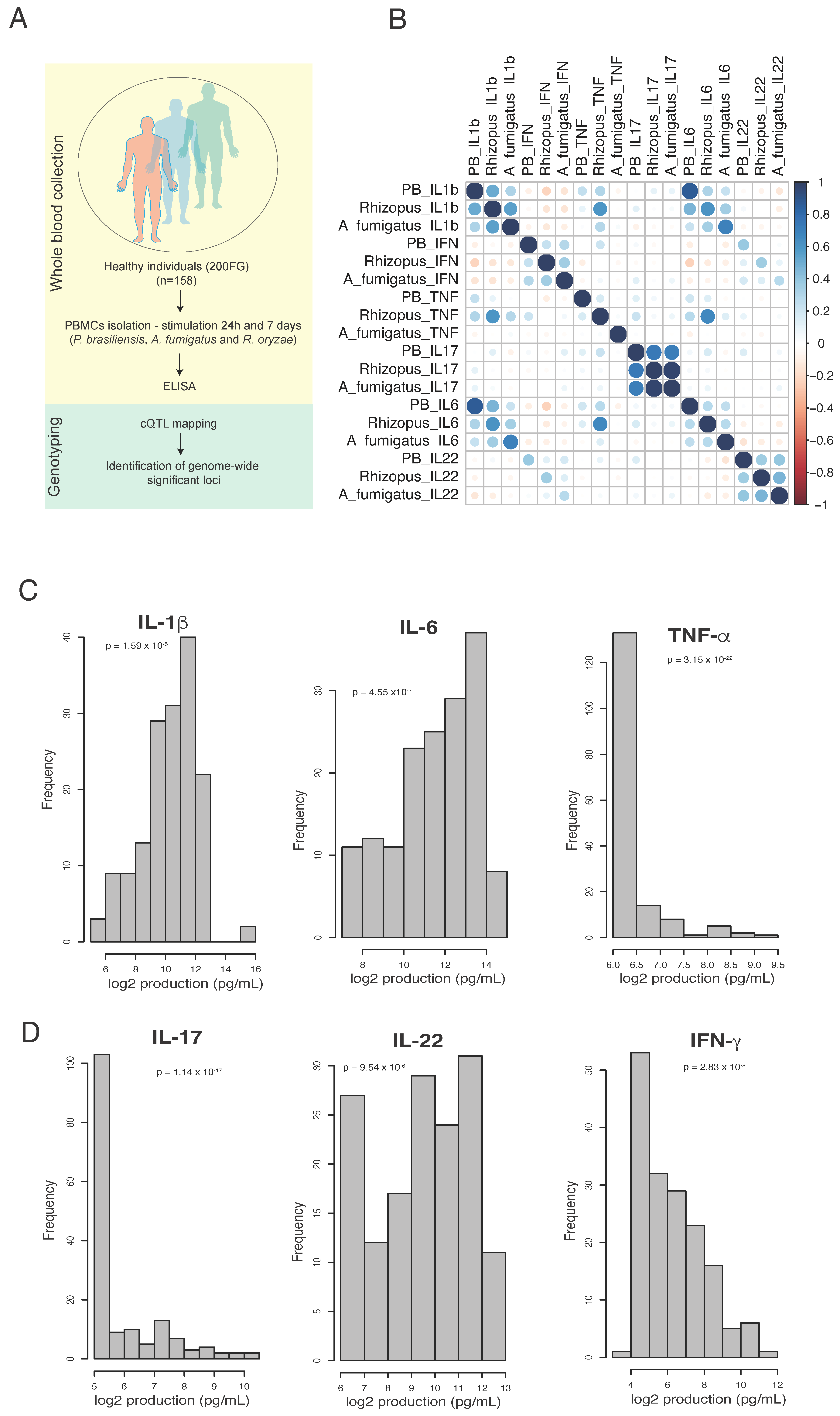
2.1. Study Cohort and Ethical Approval

The study was performed in a cohort of healthy individuals of Western ancestry, which are part of the Human Functional Genomics Project (www.humanfunctionalgenomics.org (accessed on 15 June 2020 )) [29]. The age of the individuals goes from 23 to 73 years old, in which 77% are males and 23% are females.
All volunteers provided written informed consent prior to blood collection and the study was approved by the ethical review board of Radboud University Nijmegen (no. 42561.091.12). In addition, healthy volunteers from Blood Bank of Instituto Goiano de Hematologia e Oncologia (Brazil) participated (only in CLEC7a functional validation experiments) after signing the written informed consent as approved by the Ethical Committee of Hospital de Doenças Tropicais Dr. Anuar Auad-HDT/HAA, CAAE: 81316417.1.3001.0034, Brazil.

2.2. Microorganisms and Culture Conditions

Yeast cells of the P. brasiliensis Pb18 strain were grown in complete semi-solid Fava-Netto medium, composed by 1% peptone (Kasvi, São José dos Pinhais, PR, Brazil), 0.5% (w/w) yeast extract (BD, Franklin Lakes, NJ, USA), 0.3% (w/v) proteose peptone (Sigma-Aldrich, St. Louis, MO, USA), 0.5% (w/v) meat extract (BD), 0.5% (w/v) NaCl (Sigma-Aldrich), 4% (w/w) glucose (Sigma-Aldrich), and 1.2% (w/v) agar (BD). The pH was adjusted to 7.2 and the medium was supplemented with 5% (v/v) fetal bovine serum (FBS, Cripion, Andradina, SP, Brazil) and 1 μg/mL gentamicin (Sigma-Aldrich). The culture was maintained at 37 °C as described by Bastos et al. [30] and subcultivations were made every seven days.
To obtain heat-killed P. brasiliensis for stimulation of PBMCs, the colonies were expanded in an infused medium of the brain and liquid heart (BD) supplemented with 4% (w/w) glucose, 5% (v/v) FBS, and 1 mg/mL gentamicin (Complete BHI medium). The expansion was maintained in a shaker with 150 rpm, at 37 °C, for three days. After incubation, the yeasts were centrifuged (1400× g, 5 min), washed three times with PBS (phosphate-buffered saline solution), and the supernatant was discarded. The quantification was performed on a hematocytometer, and the cell viability was assessed by vital staining with 0.1% (w/v) trypan blue in PBS. The yeasts were autoclaved for 40 min at 120 °C and heat-killed yeast cells (4 × 10⁷/mL) were stored at −20 °C.
The conidia forms of Aspergillus fumigatus, strain Af293 (ATCC 46645) and Rhizopus oryzae, strain Af1163, were prepared and heat inactivated as described previously [31]. A suspension of 1 × 10⁶ conidia/mL was used in the experiments.

2.3. Peripheral Blood Mononuclear Cells and Cytokine Measurement

To obtain PBMCs, peripheral blood from healthy individuals was collected in tubes with EDTA. Whole blood was centrifuged (200× g, 15 min, 4 °C), plasma was discarded, and blood was diluted 1:2 in PBS containing 0.01 mM EDTA (PBS-EDTA) and centrifuged (1400× g, 15 min, 4 °C) on Percoll gradient (1.077 density; 61.5%) (Percoll, Sigma-Aldrich). Alternatively, Ficoll (Ficoll-Paque Plus; GE healthcare, Zeist, The Netherlands) gradient centrifugation was used to isolate PBMCs. The mononuclear cell layer was harvested, and the cells were washed twice in PBS-EDTA (600× g in the first and 200× g in the second centrifugation, 10 min, 4 °C). Then, they were resuspended in RPMI 1640 medium supplemented with 100 U/mL of penicillin (Sigma-Aldrich) and 100 μg/mL of streptomycin (Sigma-Aldrich), 2 mM L-glutamine (Sigma-Aldrich), and 11 mM of sodium bicarbonate (Sigma-Aldrich). The cells were quantified on a hematocytometer and cell viability (≥90%) was assessed using 0.1% (w/v) trypan blue. The cell density was adjusted to 5 × 10⁶/mL and 5 × 10⁵ cells in 100 μL/well were seeded in round bottom 96-well plate (Corning, New York, NY, USA). The PBMCs were incubated with heat-killed P. brasiliensis (2.5 PBMCs:1 yeast), A. fumigatus or R. oryzae (2.5 PBMCs:1 conidia) for 24 h or seven days. For seven-day cultures, the medium was supplemented with 10% (v/v) autologous serum. After incubation period, the supernatants were collected for cytokine measurement (TNF, IL-6, IL-1β, IL-17, IL-22, IFNγ). Cytokines were measured by using ELISA commercial kits according to manufacturer’s instructions (R&D Systems, Minneapolis, MN, USA).

2.4. Genotyping, Quality Control and Cytokine QTL Mapping

DNA samples of 200 individuals were genotyped using a commercially available SNP chip, Illumina HumanOmniExpressExome-8 v1.0. Genotype calling was performed using Opticall 0.7.0 using default settings [32]. The imputation and quality control were performed as in the previous study [28]. In brief, samples with a call rate ≤ 0.99 were excluded, as were variants with Hardy–Weinberg equilibrium (HWE) ≤ 0.0001, and minor allele frequency (MAF) ≤ 0.1. Strands of variants were aligned and identified against the 1000 Genome reference panel using Genotype Harmonizer. In total, there were 158 samples (Western ancestry) with both DNA and cytokine measurements. Cytokine levels were log-transformed and the R/MatrixEQTL package was used to conduct the genome-wide association. We used a linear regression model with age and gender as covariates. We considered a p-value < 5 × 10⁻⁸ as the genome-wide significant threshold. In addition, a p-value of <5 × 10⁻⁶ was considered as the suggestive threshold for downstream analysis. To investigate the genomic context around genome-wide significant associations, summary statistics for each phenotype were uploaded to the LocusZoom [33] server to visualize regional QTL mapping scan results, using hg19 and 1000 Genomes Nov 2014 EUR as reference genomes. Enrichment analysis of biological pathways was performed using Gene Set Enrichment Analysis (GSEA) [34,35].

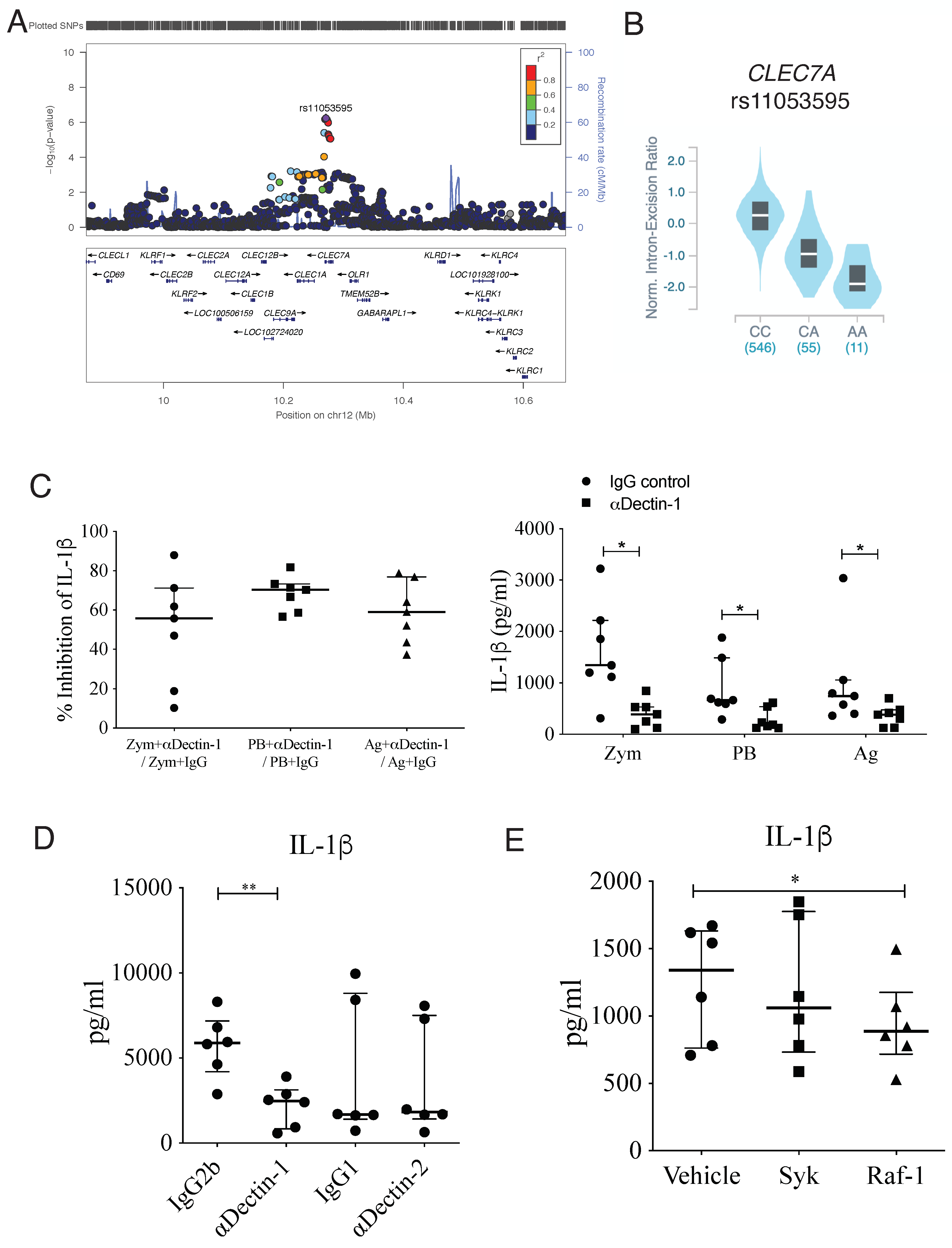
2.5. Functional Validation of Potential Causal Genes (CLEC7A, PROM1)

To validate potential causal genes related to cytokine variation in response to P. brasiliensis, we selected the SNPs that show a p-value of <5 × 10⁻⁶. Among the identified SNPs, rs11053595-CLEC7A (encodes Dectin-1) associated with the production of IL-1β, and rs62290169-PROM1 (encodes Prominin 1 or CD133) associated with the production of IL-22, were selected for further functional validation.
For blocking the Dectin-1 and Dectin-2 receptors, PBMCs (2.5 × 10⁵ cells/well in 100 μL) were incubated 1 h with 10 μg/mL of anti-Dectin-1 (clone GE2; Catalog MA5-16692; Invitrogen, Thermo Fisher Scientific and clone 259931; Catalog MAB1859; R&D Systems) and anti-dectin-2 (clone #545925; Catalog MAB3114; R&D Systems) neutralizing antibodies or with isotype control antibodies (10 μg/mL) (Catalog I5381; Sigma-Aldrich; IgG2b clone #20116; Catalog MAB004; R&D Systems; IgG1 clone #11711; Catalog MAB002; R&D Systems). For inhibition of downstream signalling pathways molecules, the cells were treated with 1 µM of Raf-1 (GW5074, Sigma-Aldrich) and 50 nM of Syk inhibitor (574711, EMD-Millipore, Burlington, MA, USA). PBMCs were stimulated with heat-killed yeast cells of P. brasiliensis (2.5 PBMCs: 1 yeast cell), particulated Pb18 antigens (20 μg/mL; prepared as in Guimarães de Matos et al. [20]), LPS (100 ng/mL, E. coli 011:B4, Sigma-Aldrich) or Zymosan (50 μg/mL; Sigma-Aldrich). The cell culture was incubated for 24 h at 37 °C. After incubation, the supernatant was collected to measure IL-1β by immunoenzymatic assay (ELISA), using a commercial kit according to the instructions of manufacturer (BD or R&D Systems).
Flow cytometry was used to determine the CD38 surface expression in PBMCs. Cells from volunteers carrying the different genotypes of the rs62290169 SNP (PROM1 gene) were used. The cells (2.5 × 10⁵ cells per well in 100 μL) were incubated with heat-killed yeast cells of P. brasiliensis (MOI 2.5 cells:1 fungus) for 24 h or seven days. After the incubation period, cells with or without stimulus were labeled with the following antibodies: anti-HLA-DR (clone G46-6, BUV661, BD Biosciences, Franklin Lakes, NJ, USA), anti-CD56 (clone NCAM16.2, BUV737, BD Biosciences), anti-CD3 (clone UCHT1, PerCP, Biolegend, San Diego, CA, USA), anti-CD4 (clone RPA-TA, AF594, Biolegend), anti-CD8 (clone B9.11, PacBlue, Beckman Coulter, Indianapolis, IN, USA), anti-CD45 (clone J3.3, KrO, Beckman Coulter), anti-CXCR5 (clone J252D4, BV605, Beckman Coulter), anti-CD38 (clone HB-7, BV786, Biolegend), anti-CXCR3 (clone G025H7, AF488, Beckman Coulter), anti-CCR4 (clone L291H4, PC5, Beckman Coulter), anti-CCR6 (clone B-R35, PC7, Beckman Coulter) and ViaKrome as viability dye (IR885/40, C36628, Beckman Coulter). Afterwards, the cells were washed and resuspended in FACS buffer (PBS with 2% bovine serum albumin) and a minimum of 9000 and maximum of 16,000 events were acquired based on CD45 expression. The samples were assessed in a CytoFLEX flow cytometer (Beckman Coulter) and the results were analyzed using Kaluza analysis software (Beckman Coulter, IN, USA).

2.6. Statistical Analysis

Statistical analyses were performed in R (v3.6.0). The cytokine levels obtained for GWAS were transformed using inverse-normal transformation and the p-value was assessed by Shapiro test. Unsupervised hierarchical clustering was performed using Spearman’s correlation as the measure of similarity. R-package Matrix-eQTL [36] was used for cytokine QTL mapping, where the linear model was applied with age and gender information included as covariates. Manhattan plot and Q-Q plot for each association analysis were generated by package qqman [10.21105/joss.00731] using default parameter settings. In the validation, the results for cytokine IL-1β (pg/mL) are presented as medians with interquartile ranges, analyzed by One-way ANOVA Kruskal–Wallis, followed by post hoc Dunn’s test, Wilcoxon’s paired test, or Mann–Whitney. GraphPad Prism 6.0 Software Inc. (San Diego, CA, USA) was used for the analysis. The level of significance was set at p < 0.05.

3. Results

3.1. The Profile of P. brasiliensis-Induced IL-1β, IL17 and IL-22 Is Similar to Aspergillus fumigatus and Rhizopus oryzae

To determine whether P. brasiliensis induced a pathogen specific cytokine profile, PBMCs from 158 volunteers of the 200FG cohort were stimulated for 24 h or seven days, and we assessed the correlation between the levels of cytokines induced by P. brasiliensis or A. fumigatus and R. oryzae as the measure of similarity (Figure 1A). Overall, among the cytokines related to innate immunity (TNF, IL-6 and IL-1β) produced after 24 h of exposure to the three fungi, only the production profile of IL-1β induced by P. brasiliensis showed a strong resemblance to R. oryzae-induced IL-1β production (Figure 1B). For this cytokine, R. oryzae and A. fumigatus also presented similar profiles of responses (Figure 1A). It was not detected similarities in the TNF profile induced by the three fungi, but for IL-6, a significant correlation was observed among the three fungi (Figure 1B). For the T-helper cytokines produced after seven days of culture, strong, moderate and weak correlations were detected for IL-17, IL-22 and IFNγ, respectively, among the three fungi (Figure 1A). The production of cytokines induced by each specific fungus, IL-1β and IL-6, were strongly correlated independent of the fungus genus. IL-1β and TNF were strongly correlated when PBMCs were stimulated with R. oryzae, but for P. brasiliensis this correlation was weak and there was no correlation between these cytokines when A. fumigatus was the stimulus. A moderate correlation between IFNγ and IL-22 levels was detected when P. brasiliensis or R. oryzae stimulated PBMCs, whereas A. fumigatus stimulation led to a weak association between these cytokines (Figure 1B). The IL-17 levels were not associated to other cytokines independent of the fungus genus. In addition, no correlations were detected between levels of monocyte- and Th-produced cytokines upon stimulation with any fungal type.
Next, we assessed how the cytokines levels were distributed across the individuals of our cohort upon P. brasiliensis exposure by displaying the log-transformed values. We observed a similar distribution of individuals with high and low IL-1β and IL-6 response to P. brasiliensis, whereas the majority of the individuals produced low levels of TNF (Figure 1C). Moreover, a substantial number of low responders were seen for IL-17 in comparison to IL-22 and IFNγ (Figure 1D). The inter-individual variability for each cytokine produced after fungal stimulations is shown in Supplementary Figure S1.

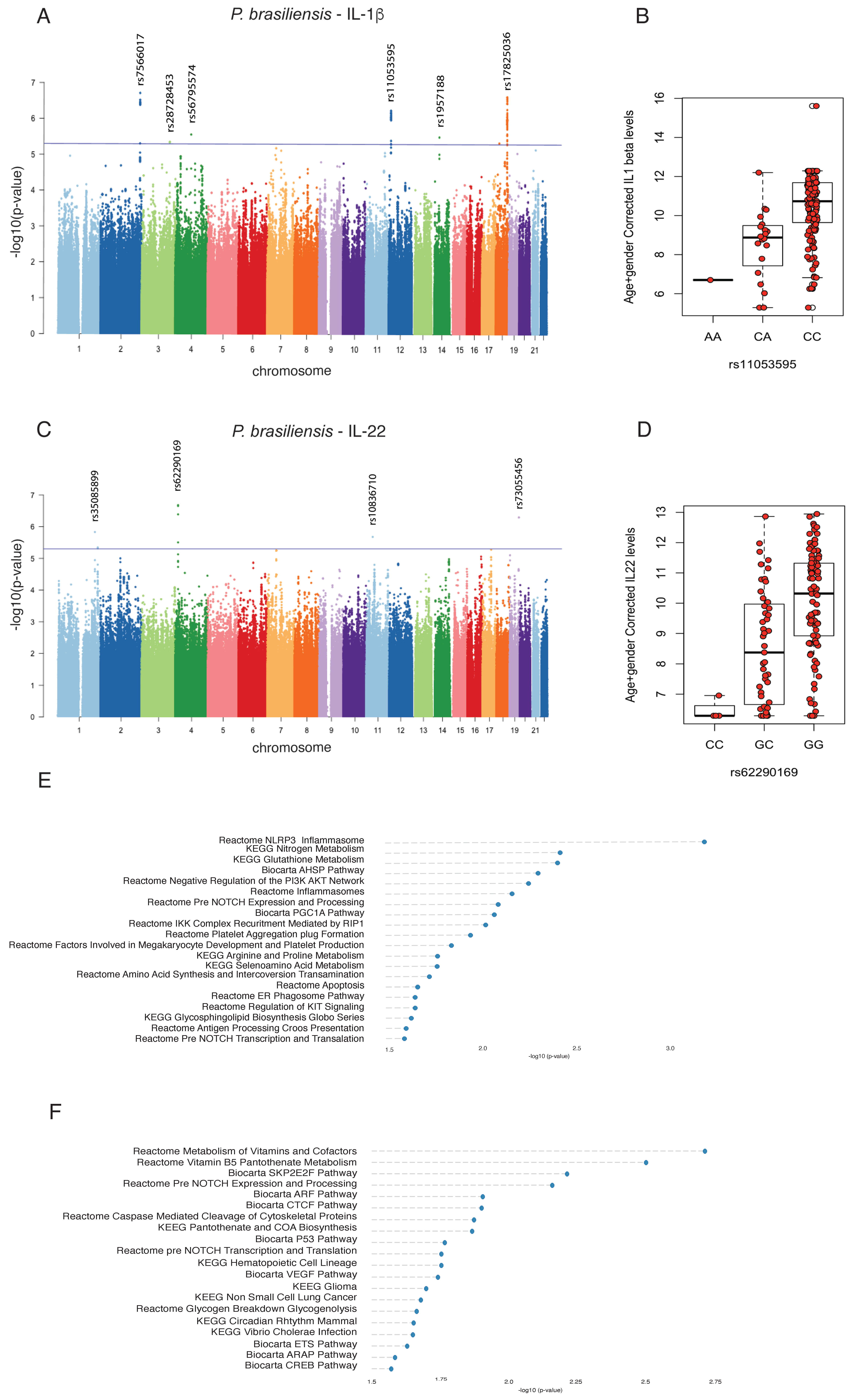
3.2. Genome-Wide Mapping Identifies Genetic Variations Associated with Innate- and Adaptive-Derived Cytokines Induced by P. brasiliensis

In order to study the genetic influence in P. brasiliensis-induced cytokine production, we performed cytokine production quantitative trait loci (cQTLs) analysis as described in Li et al. [28]. After multiple testing corrections in which the threshold for genome-wide significance was set at <5 × 10⁻⁸, no genome-wide significant cQTL was identified. To increase our sensitivity for cQTL influencing P. brasiliensis-induced cytokine production, we analyzed cQTLs with p values < 5 × 10⁻⁶ for IL-6, IL-1β, INFγ and IL-22 (Table 1). cQTL analysis were not performed for TNF and IL-17 as the levels of these cytokines were very low for most of the individuals evaluated (cytokine production below detection limit).
We identified an SNP (rs11053595) in the 3′ UTR region of the CLEC7A gene (at chromosome 12), which encodes the Dectin-1 protein receptor, associated with IL-1β production (p = 5.82 × 10⁻⁷) upon P. brasiliensis exposure (Figure 2A). There was only one homozygous individual for A allele of rs11053595 SNP in which lower amounts of IL-1β were observed after stimulation with heat-killed P. brasiliensis yeasts compared to homozygous individuals for C allele (Figure 2B).
We also identified an SNP (rs62290169) in an intronic region of the PROM1 gene (at chromosome 4), which encodes the CD133 transmembrane protein. This protein has been used as a marker to isolate cancer stem cells in several tumors. Although it is highly expressed in embryonic stem cells, its function in these cells remains unknown [37,38]. This cQTL was associated with the production of IL-22 (p = 2.09 × 10⁻⁷) upon P. brasiliensis exposure (Figure 2C). The homozygous individuals for G allele of rs62290169 SNP produced more amounts of IL-22 after stimulation with heat-killed P. brasiliensis yeasts compared to homozygous individual for C allele (Figure 2D). Therefore, the results indicate that the CLEC7A and PROM1 genes are causal genes affected by rs11053595 and rs62290169 SNPs, respectively, whose allelic variations result in disturbances of the cytokine production influencing the innate immune response towards infection caused by P. brasiliensis.
Further, we tested whether the identified cQTLs are associated with biological pathways. Interestingly, among the various pathways enriched for the SNPs that affected IL-1β production, we identified the association of IL-1β with NLRP3 inflammasome and glutathione metabolism (Figure 2E). Moreover, the biological pathways associated with IL-22 QTLs were related to metabolism of vitamins, pantothenate metabolism and CTCF, a protein involved in the regulation of chromatin architecture (Figure 2F).

3.3. IL-1β Production Induced by Heat-Killed Yeasts Cells or Particulated P. brasiliensis Antigens Is Dependent on Dectin-1 Receptor

We have identified an SNP (rs11053595) located in the CLEC7A gene (chromosome 12) that influenced the IL-1β production upon P. brasiliensis exposure (Figure 3A). eQTL analysis demonstrated the different genotypes of rs11053595 influencing CLEC7A expression in whole blood (data obtained from GTEX Portal, www.gtexportal.org (accessed on 24 march 2023)) (Figure 3B). As this gene encodes the Dectin-1 receptor, we performed biological assays to confirm its influence on cytokine production. In fact, blocking of Dectin-1, but not Dectin-2, with neutralizing antibodies significantly decreased the production of IL-1β induced by heat-killed yeasts or particulated P. brasiliensis antigens (Figure 3C,D, Supplementary Figure S2). In Figure 3C, it was shown that, after treatment with anti-Dectin-1, the IL-1β production induced by heat-killed yeasts or particulated antigens, as well as by zymosan, was 60% to 70% inhibited in comparison with controls (IgG control) in most individuals (Figure 3C left). The absolute values for IL-β concentration are also shown (Figure 3C right). In addition, inhibition of Raf-1, but not Syk (p value = 0.06; Wilcoxon test), which are both well-known molecules downstream of the dectin-1 pathway [39,40], decreased the production of IL-1β induced by heat-killed P. brasiliensis (Figure 3E). Thus, our data show that the rs11053595 SNP affects the function of the Dectin-1 receptor, which in turn is crucial for the induciton of IL-1β during the innate response to P. brasiliensis.

3.4. The PROM1 rs62290169 SNP Regulates the Surface Expression of CD38 in T-Helper Lymphocytes

We identified an SNP in the intronic region of PROM1 related with IL-22 production (Figure 4A). However, the PROM1 gene is a poorly expressed gene in whole blood in comparison to other tissues in the body (Figure 4B). The PROM1 gene is located in close proximity with the gene that encodes the CD38 molecule, which is a marker of T cell activation [41]. Furthermore, three-dimensional chromatin structures, known as topologically associating domains (TADs), show that chromatin in the eukaryotic nucleus is divided into regions enriched in chromosomal contacts [42]. Publicly available chromosome conformation capture (Hi-C) data obtained from K562 cells (hematopoeietic cell line) revealed the presence of a region containing the CTCF architectural protein (Figure 4C), which is capable of promoting the interaction of DNA by forming loops that brings proximal regions in close proximity [43]. Based on this information, we hypothesized that PROM1 makes contact with CD38 through CTCF-dependent loop formation. In order to validate this conection, we stimulated PBMCs from health donors with P. brasiliensis for 24 h or seven days and evaluated the expression of CD38 by flow cytometry (Supplementary Figure S3). All gate strategies for flow cytometry evaluation are described in Supplementary Figure S4. A significant increase in the frequency of CD38+ cells was observed after seven days of stimulation with heat-killed P. brasiliensis in comparison to non-stimulated cells (Supplementary Figure S3A). Additionally, our results have shown that individuals carrying the GG genotype of the rs62290169 SNP in PROM1 gene produced higher amounts of IL-22 than CC genotype (Figure 2D). In order to study the relationship between PROM1 and CD38, healthy individuals carrying the different genotypes of the rs62290169 SNP from the 200FG cohort were evaluated for CD38 expression in T-helper lymphocyte subsets upon P. brasiliensis exposure. The results showed that individuals with GG genotype presented higher frequency of CD38+ Th1 cells when PBMCs were cultured with P. brasiliensis in comparison to non-stimulated cultures (Figure 4D). We did not detect significant alterations in the frequency of CD38+ Th2 (Supplementary Figure S3B) or Th17 cells after exposure to the fungus (Figure 4D). Of note, although we only had one individual carrying the CC genotype, we observed that the percentage of Th1, Th2 and Th17 expressing CD38 was lower than GC and GG genotypes. After evaluating CD38 mean fluorescence intensity (MFI) in Th1, Th2 and Th17 cells, the results have shown that as for percentages P. brasiliensis exposure led to an increased CD38 MFI in comparison to non-stimulated cells (Supplementary Figure S3C). Moreover, in the individual carrying the CC genotype, the decrease in CD38 MFI was observed only in Th1 cells. Together, our results indicate that CD38 expression in T lymphocytes is modulated by P. brasiliensis in PBMCs, a process that appears to be modulated by the contact of PROM1 and nearby regulatory elements with CD38.

4. Discussion

The present study aimed to identify host genetic polymorphisms that are involved in the cytokine response of human PBMCs induced by P. brasiliensis using GWAS. First, we evaluated the cytokine profile induced by heat-killed Pb18 yeasts in PBMCs from healthy individuals after 24 h and seven days of culture, respectively. After, we performed a correlation test between cytokine levels induced by P. brasiliensis and the fungi A. fumigatus and R. oryzae, which are among the most relevant agents of the fungal human diseases, aspergillosis and mucormycosis, respectively [44,45,46]. Despite these fungi causing distinct diseases, there was a similar profile of IL-1β production induced by P. brasiliensis and by R.oryzae after 24 h of culture. A similar profile was also detected for IL-17 and IL-22 induced by P. brasiliensis and the other fungi in the seven-day cultures, thus indicating that IL-1β, IL-17 and IL-22 are similarly induced by the three fungi evaluated in the present study. On the other hand, the absence of similarity between profiles of TNF and weak correlations for IL-6 and INF-γ production induced by the three fungi suggests that different mechanisms are associated in the induction of these cytokines. Considering the cytokines induced by a specific fungus, for P. brasiliensis it is noticeable that there is a strong positive correlation between IL-1β and IL-6 production. The same pattern was observed for A. fumigatus and R. oryzae, but to a lower extent. Nevertheless, IL-1β and TNF were also strongly associated when R. oryzae stimulated PBMCs, whereas in P. brasiliensis-stimulated cultures, these cytokines were weakly associated. In opposite, A. fumigatus induced IL-1β and TNF, but no correlation was detected between the two cytokines, suggesting that distinct monocyte-derived cytokines can be associated depending on the fungus genus. The production of IFNγ and IL-22 were positively associated after stimulation of the PBMCs with each of the fungus, although for A. fumigatus, the correlation was weak. Altogether, these data suggest that PAMPs of the three fungi trigger the production of cytokines involved in Th17/Th22 profiles (IL-1β, IL-17, IL-22), which, together with host genetic factors, can explain the inter-individual variability in the induction of immune responses.
In this study, by evaluating the variability of cytokine levels in response to heat-killed P. brasiliensis-exposed PBMCs, we observed the presence of good and poor responders, ranging from individuals with ability to produce very high or low levels of IL-1β and IL-6 as well as IFNγ and IL-22. The TNF and IL-17 production profiles, on the other hand, were composed by individuals producing very low levels of these cytokines in response to P. brasiliensis, therefore impairing the possibility of studying the genetic contribution associated to the inter-individual variation for these cytokines. As genetic polymorphisms can contribute for host cytokine variability [28,47], GWAS analyses were done and, in agreement with previous study [28], we initially set the genome-wide significance threshold at p-value < 5 × 10⁻⁸; however, we did not identify any significant variation, which could be due the fact that we used a small cohort composed by healthy volunteers of Western European ethnicity. Next, we used a suggestion threshold, p-value < 5 × 10⁻⁶, to determine the cQTLs associated with the production of IL-6, IL-1β, IFN-γ and IL-22. We identified several SNPs associated with the production of these cytokines induced by heat-killed P. brasiliensis. Among the several cQTL identified, we decided to focus on SNPs associated with IL-1β and IL-22 production, components of Th17/Th22 profiles, which have been associated with resistance to P. brasiliensis infection [10,12,48,49,50].
The SNP (rs11053595) is located in the CLEC7A gene (3’UTR region), which encodes the Dectin-1 receptor and was significantly associated with IL-1β production. The AA genotype of rs11053595 SNP was related to low amounts of IL-1β after stimulation with heat-killed P. brasiliensis yeasts compared to CC genotype. The SNP can influence resistance or susceptibility to PCM since Dectin-1 is known as a receptor that is crucial for the recognition of β-glucan present in the P. brasiliensis-cell wall [49,51,52]. We showed that blocking Dectin-1, but not Dectin-2, significantly decreased IL-1β production induced by heat-killed yeast or P. brasiliensis particulate antigens. It is known that Dectin-2 triggering by P. brasiliensis induces the production of IL-1β in human plasmacytoid dendritic cells (pDCs) [53], which was not observed in PBMCs in the present study. In our experiments, there was a partial inhibition of IL-1β production induced by heat-killed yeast or particulate antigen (60% to 70%) after Dectin-1 blockade, suggesting the participation of other receptors in P. brasiliensis recognition.
Our results showed that P. brasiliensis-induced IL-1β production was dependent on Raf-1 signaling pathway (p < 0.05), although Syk appears to participate in less extension (p value = 0.06). It is known that Syk and Raf-1 are independent pathways that converge at NF-κB activation level to induce cytokine production after Dectin-1 triggering in human DCs [39]. Moreover, inhibition of Syk completely abolishes the production of cytokine induced through activation of Dectin-1 alone, but when TLR is involved in the activation, Raf-1 mediates the Dectin-1 signaling [39,54]. Thus, our results suggest that Dectin-1 can be acting together TLR to induce IL-1β in PBMCs exposed to heat-killed P. brasiliensis. In human monocytes and human and mouse DCs, the evaluation of Dectin-1/TLR2/TLR4 in the immune response against P. brasiliensis has been reported, but the cooperation among these receptors to the production of IL-1β is missing [49,55,56]. However, de Castro et al. [49] showed that blocking only TLR2 in human DCs stimulated with P. brasiliensis had no effect on IL-1β production, whereas Dectin-1/Syk pathway was crucial for NLRP3 inflammasome-dependent cytokine production (IL-1β, IL-18). These results suggest that P. brasiliensis acting on TLR2 and Dectin-1 can induce pro-IL-1β through NFkB pathway, but only Dectin-1/Syk is crucial for inflammasome activation and IL-1β release [49]. Further studies aiming to investigate pro-IL-1β and bioactive IL-1β production can reveal the most relevant pathways to induce higher levels of IL-1β after stimulation with P. brasiliensis than with other fungi, as shown here for R. oryzae and A. fumigatus. To our knowledge, no study has been performed with R. oryzae and inflammasome; however, A. fumigatus activates two inflammasomes, AIM2 and NLRP3, to induce IL-1β production and fungal control [57,58,59]. Thus, additional experiments should be performed to elucidate possible cooperation between innate receptors for IL-1β production during the immune response against P. brasiliensis as well as the mechanisms responsible for different levels of IL-1β induced by P. brasiliensis and A. fumigatus.
Our results confirmed Dectin-1 as an important receptor for IL-1β production during the response to P. brasiliensis. Other studies have also shown the relevance of Dectin-1 in this fungal infection. Loures et al. [60] showed that Dectin-1 plays an important protective role in the lungs during infection with P. brasiliensis in mice. Dectin-1 deficiency impaired murine macrophage fungicidal mechanisms by decreasing nitric oxide (NO) and increasing IL-10. The disease was severe in the absence of Dectin-1, with increased lung pathology and mortality. Deficiency in Dectin-1 also contributed to a decrease in Th1/Th2/Th17 cytokines, decreased Th17 cell differentiation and increased T regulatory cell expansion. Ex vivo, Silva et al. [61] blocked Dectin-1 in human monocytes and neutrophils before infection with P. brasiliensis yeasts, which resulted in a decreased production of hydrogen peroxide and increased fungal load in these cells. As IL-1β is produced after Dectin-1 activation, this cytokine can be at least partially responsible for Dectin-1 effects in the fungal resistance. Tavares et al. [14] showed that IL-1β-induced signaling is important for the control of P. brasiliensis infection in murine macrophages. This cytokine also plays an important role in the inflammatory process and contributes to the development of a Th17/Th22 responses [13,62], which appears to contribute for P. brasiliensis infection resistance [10,12,50].
It has been demonstrated by eQTL analysis that the different genotypes of rs11053595 SNP impact the expression of CLEC7A in whole blood (data obtained from the GTEX Portal, www.gtexportal.org), despite the SNP being located at the 3’UTR. It is possible that this difference in the expression of CLEC7A is related to differences in mRNA stability, in the degradation process, as well as in the translation of this mRNA, thus regulating the levels of dectin-1. Additionally, it is possible that the different genotypes present mRNA isoforms containing different 3’UTRs, which can lead to post-translational modifications as protein conformation [63].
Strengthening our data about the relevance of the SNP in CLEC7A and IL-1β production, when we analyzed whether cQTLs were associated with biological pathways, we identified the association of IL-1β cQTL with the NLRP3 inflammasome and glutathione metabolism pathways. Dectin-1 activation is related to the NLRP3 inflammasome activation. After the recognition of β-glucan by Dectin-1/Syk phosphorylation, NLRP3 inflammasome is activated and, in turn, activates caspase-1 that processes pro-IL-1β into bioactive IL-1β [14,49,51]. Dectin-1/caspase 8 pathway also regulates IL-1β production during P. brasiliensis infection [64]. This pathway has been reported in P. brasiliensis infection as a mechanism that is critical for resistance against the fungus by inducing IL-1β and promoting Th1/Th17 responses [14,48,51,64]. In turn, glutathione metabolism is important in several biological processes such as protection against oxidative stress caused by reactive oxygen species (ROS), since glutathione in its reduced form interacts with reactive metabolites and helps in their removal [65,66]. In fungal infection, Seegren et al. [67] showed that, during phagocytosis of Candida albicans and zymosan by murine macrophages, there was stimulation of ROS production and this was crucial for fungal control. Furthermore, the study of Santos et al. [68] shows that the P. brasiliensis Pb18 strain is capable of inducing ROS in murine polymorphonuclear cells and Tavares et al. [14] showed that this fungus induces ROS/inflammasome in murine macrophages. Therefore, we could speculate that heat-killed P. brasilienis yeasts can induce ROS production in PBMCs which, consequently, leads to activation of the NLRP3 inflammasome and glutathione metabolism.
Associated with IL-22 production, we identified an SNP (rs62290169) located in the PROM1 gene, which encodes the CD133 protein. Despite this protein being highly expressed in embryonic stem cells, its function in these cells remains unknown [37,38]. The identified SNP is located in a non-coding and junction region with the gene encoding the CD38 molecule. In lymphocytes, CD38 is a glycoprotein that acts as an enzyme and a multifunctional receptor, which is important in the processes of activation, proliferation, and secretion of cytokines [69]. CD38 also plays a role in DC migration and survival as well as Th1 differentiation profile [70]. It was shown that, in the cQTL where PROM1 is located, there is a region that contains the CTCF architectural protein, which is capable of promoting DNA interaction through the formation of loops [43]. As the topologically associated domain (TAD) map showed a connection between CD38 with PROM1 and it was shown that CD38 can control IL-22 production [71], we hypothesize that PROM1 makes contact with CD38 through CTCF-dependent loop formation to control production of IL-22.
To validate our hypothesis, we evaluated the expression of CD38 on T cells from healthy individuals with the different PROM1 SNP rs62290169 genotypes after stimulation with heat-killed P. brasiliensis yeast. The 7-day exposure to the fungus induced a significant increase in CD38 expression compared to the negative control. Flow cytometry showed that the individual carrying the CC genotype had lower percentages of CD38+ Th1, Th2 and Th17 lymphocytes, suggesting that SNP PROM1 C allele can affect CD38 expression. In addition, CD38 MFI in Th1 cells of the individual carrying the CC genotype was shown to have the same pattern observed for percentages, whereas this was not the case for Th2 and Th17 cells. We had shown that the individual carrying the CC genotype produced lower levels of IL-22 than homozygous GG individuals. Consistent with our results, Gangemi et al. [71] showed that patients with chronic B lymphocytic leukemia who had high CD38 expression presented higher levels of IL-22 than patients with low CD38 expression. Therefore, our data suggest that during P. brasiliensis infection PROM1 variant rs62290169 can down regulate the CD38 expression, which leads to a decrease of the IL-22 production. Concerning Th lymphocyte subsets, however, CD38 was highly expressed on Th1 cells from GG carriers after P. brasililensis exposure. As we did not identify the cell source of IL-22 in our experiments, PROM1 can regulate the production of IL-22 in Th1, Th22, Th17 or even other cells present in PBMCs as potential sources of IL-22 [72,73]; moreover, this gene can be also connected with IFNγ production, via or not CD38 [70], since Th1 cells produce IFNγ and IL-22 [73,74]. These suggestions need to be further evaluated as well as to investigate whether and how the PROM1 really controls CD38 expression. We were not able to detect PROM1 mRNA expression in PBMCs, confirming that this gene is too low expressed in whole blood (Figure 4).
The biological pathways associated with IL-22 cQTLs were related to vitamin metabolism, pantothenate (vitamin B5) metabolism and the CTCF architectural protein. IL-22 is a cytokine produced by Th lymphocyte subsets and innate lymphoid cells, which is essential for host defense in skin and mucosal immune responses as well as tissue repair [73,75]. It is known that adequate intake of vitamins is important for the proper functioning of the immune system [76]. Cha et al. [77] demonstrated that vitamin A deficiency in a murine model impairs the differentiation of T cells towards a Th17 profile, reducing the expression of cytokines as IL-22. Our group has also recently demonstrated that vitamin D can improve the fungicidal activity of PBMCs infected with P. brasiliensis [20]. There are only two studies about P. brasiliensis infection and IL-22. In mice, reduction of Th22 cells was associated with an increase of fungal burden in the lungs, enhanced lung pathology and mortality, thus suggesting a protective role for Th22 in pulmonary experimental PCM [50]. Evidence that IL-22 is produced after P. brasiliensis infection was shown by de Castro et al. [78] in co-cultures of human macrophages and T lymphocytes. This scarce information about IL-22 in PCM, together our results, indicates that evaluation about the role of this cytokine in this fungal disease deserves further study.
One of the limitations of the present study is the fact that we performed genome-wide association analysis in a cohort consisting of a relatively small sample size, which might explain the absence of the identification of genome-wide significant loci. In addition, the use of heat-killed P. brasiliensis cannot represent the overall immune response to the live fungus, and only one strain of P. brasiliensis (the Pb18, considered of intermediate/high virulence) was used, whereas some studies have shown differences in virulence among strains in mouse infection as a factor that can influence the induction of cytokines [19,52,79,80]. Another limitation in this study is that the evaluation of patients with PCM to check the influence of the SNPs studied here as well as other CLEC7A gene SNPs is missing. Some SNPs in CLEC7A gene can influence the expression of different dectin-1 isoforms as well as alter the receptor ability to interact with the ligand [81,82,83]. The SNP rs16910526 (Y238X) leads to an early stop codon, impairing the binding of β-glucan to the receptor and the production of cytokines during the response to C. albicans [81]. Therefore, the evaluation of CLEC7A SNPs in a cohort of patients with PCM is crucial to determine the contribution of Dectin-1 polymorphisms to P. brasiliensis infection. In addition, besides nonopsonic phagocytosis mediated by β-glucan/Dectin-1, opsonophagocytosis mediated by complement and pentraxin are relevant in fungal infections [84]. In human neutrophils, intracellular expression of Dectin-1 can be upregulated by C. albicans opsonized by mannose binding lectin (MBL); intracellular Dectin-1 binds to β-glucan, which increases ROS [85]. Thus, in addition to the contribution of complement activation to increase opsonophagocytosis of P. brasiliensis [86], the effects of CLEC7A SNPs in Dectin-1-mediated phagocytosis in concert with other receptors to fungal opsonophagocytosis deserves further evaluation.
Despite the difficulties in addressing all these questions, the findings of the present study are strengthened by the addition of functional data; moreover, the genetic variability among Paracoccidioides spp. has been shown not to influence the development of PCM clinical forms [52]. Lastly, the profile of cytokines induced by heat-killed Pb18 was similar to what is detected in PBMCs from patients with adult/chronic PCM, the most common disease form, where mixed Th1/Th17/Th22 responses are reported [10].

5. Conclusions

In conclusion, our results showed the influence of the CLEC7A and PROM1 genes on the production of cytokines IL-1β and IL-22, respectively, in response to P. brasiliensis. A more detailed study should be carried out to better understand the role of CD38 in PCM and to elucidate the biological pathways related to IL-22 production. The role of IL-22 in PCM is not yet completely established. As PROM1 is highly expressed in the lungs (Figure 4B) and associated with the production of IL-22 as well as IL-1β induces IL-22 [87], to evaluate the PROM1 and CLEC7A variants described in this study in PCM patients, future work should contribute to understanding mechanisms involved in resistance or susceptibility to the disease, revealing novel targets for treatment of PCM. In addition, other possible causal genes associated with P. brasiliensis-induced cytokine production identified in this study can be validated in future studies showing other genetic variants as determinants of susceptibility or resistance to PCM. Thus, our future aims are to validate other putative causal genes identified in this study, to better investigate the relationship between PROM1, CD38 and IL-22 in immune response to P. brasiliensis, and to explain how the rs11053595 SNP in the CLEC7A gene influences the function of the dectin-1.

Supplementary Materials

The following supporting information can be downloaded at: https://www.mdpi.com/article/10.3390/jof9040428/s1, Figure S1: Inter-individual variation upon stimulation.; Figure S2: Blocking of Dectin-1 decreases IL-1β production induced by P. brasiliensis.; Figure S3: Frequency of CD38 expressing cells.; Figure S4: Representative dot plots of the indicated receptors in PBMCs upon P. brasiliensis exposure for 7 days.

Author Contributions

Conceptualization, A.M.B.d.F., J.C.d.S., F.R.-D. and L.A.B.J.; Methodology, A.M.B.d.F., J.C.d.S., B.K., E.A., G.G.M. and I.B.N.O.; Validation, J.C.d.S.; Formal analysis, A.M.B.d.F., J.C.d.S. and E.A.; Investigation, J.C.d.S.; Resources, L.A.B.J.; Data curation, J.C.d.S.; Writing—original draft, A.M.B.d.F., J.C.d.S. and B.K.; Writing—review & editing, J.C.d.S., M.O., M.G.N., C.M.d.A.S., F.R.-D. and L.A.B.J.; Supervision, F.v.d.V., M.G.N., C.M.d.A.S., F.R.-D. and L.A.B.J.; Project administration, F.R.-D.; Funding acquisition, C.M.d.A.S. and L.A.B.J. All authors have read and agreed to the published version of the manuscript.

Funding

This study was financed in part by the Coordenação de Aperfeiçoamento de Pessoal de Nível Superior–Brasil (CAPES)–Finance Code 001; A.M.B.F. is a CAPES fellow. PRONEM (2017/10267000516) and National Institute of Science and Technology of the strategies in host-pathogen interaction (INCT-IPH; 465771/2014-9) grants are from Fundação de Amparo à Pesquisa do Estado de Goiás (FAPEG), Brazil; F.R-D and C.M.A.S. are fellow researchers of National Council for Scientific and Technological Development (CNPq), Brazil.

Institutional Review Board Statement

The study was conducted in accordance with the Declaration of Helsinki, and approved by the ethical review board of Radboud University Nijmegen (no. 42561.091.12) and by the Ethical Committee of Hospital de Doenças Tropicais Dr. Anuar Auad -HDT/HAA (CAAE: 81316417.1.3001.0034, 15 January 2020), Brazil.

Informed Consent Statement

All volunteers provided written informed consent prior to blood collection.

Acknowledgments

The authors thank Ezio T. Fok and Musa Mhlanga for their support during the experiments performed in order to investigate the relation between PROM1 and CD38. The authors are grateful to CAPES, CNPq and FAPEG for the financial support provided to this study. In addition, we are grateful to Murilo Barros Silveira and Rodrigo Saar Gomes for their technical assistance.

Conflicts of Interest

The authors declare no conflict of interest.

References

  1. Martinez, R. New Trends in Paracoccidioidomycosis Epidemiology. J. Fungi 2017, 3, 1. [Google Scholar] [CrossRef] [Green Version]
  2. Almeida, F.P. Comparative studies of coccidioic granuloma in the United States and Brazil. New genus for Brazilian parasite. Ann. Fac. Med. Univ. 1930, 5, 125–141. [Google Scholar]
  3. Ashraf, N.; Kubat, R.C.; Poplin, V.; Adenis, A.A.; Denning, D.W.; Wright, L.; McCotter, O.; Schwartz, I.S.; Jackson, B.R.; Chiller, T.; et al. Re-Drawing the Maps for Endemic Mycoses. Mycopathologia 2020, 185, 843–865. [Google Scholar] [CrossRef] [PubMed]
  4. Paniago, A.M.M.; Aguiar, J.I.A.; Aguiar, E.S.; da Cunha, R.V.; Pereira, G.R.D.O.L.; Londero, A.T.; Wanke, B. Paracoccidioidomicose: Estudo clínico e epidemiológico de 422 casos observados no Estado de Mato Grosso do Sul. Rev. Soc. Bras. Med. Trop. 2003, 36, 455–459. [Google Scholar] [CrossRef]
  5. Shikanai-Yasuda, M.A.; Mendes, R.P.; Colombo, A.L.; de Queiroz Telles, F.; Kono, A.; Paniago, A.M.M.; Nathan, A.; do Valle, A.C.F.; Bagagli, E.; Benard, G.; et al. II Consenso Brasileiro em Paracoccidioidomicose-2017. Epidemiol. Serviços Saúde 2018, 27, 1–34. [Google Scholar] [CrossRef] [PubMed] [Green Version]
  6. Mendes, R.P.; Cavalcante, R.D.S.; Marques, S.A.; Marques, M.E.A.; Venturini, J.; Sylvestre, T.F.; Paniago, A.M.M.; Pereira, A.C.; da Silva, J.D.F.; Fabro, A.T.; et al. Paracoccidioidomycosis: Current Perspectives from Brazil. Open Microbiol. J. 2017, 11, 224–282. [Google Scholar] [CrossRef] [PubMed] [Green Version]
  7. Fortes, M.R.P.; Miot, H.A.; Kurokawa, C.S.; Marques, M.E.A.; Marques, S.A. Imunologia Da Paracoccidioidomicose. An. Bras. Dermatol. 2011, 86, 516–524. [Google Scholar] [CrossRef]
  8. Oliveira, S.J.; Mamoni, R.L.; Musatti, C.C.; Papaiordanou, P.M.O.; Blotta, M.H.S.L. Cytokines and Lymphocyte Proliferation in Juvenile and Adult Forms of Paracoccidioidomycosis: Comparison with Infected and Non-Infected Controls. Microbes Infect. 2002, 4, 139–144. [Google Scholar] [CrossRef] [PubMed]
  9. Marques Mello, L.; Silva-Vergara, M.L.; Rodrigues, V. Patients with Active Infection with Paracoccidioides Brasiliensis Present a Th2 Immune Response Characterized by High Interleukin-4 and Interleukin-5 Production. Hum. Immunol. 2002, 63, 149–154. [Google Scholar] [CrossRef]
  10. De Castro, L.F.; Ferreira, M.C.; da Silva, R.M.; Blotta, M.H.D.S.L.; Longhi, L.N.A.; Mamoni, R.L. Characterization of the Immune Response in Human Paracoccidioidomycosis. J. Infect. 2013, 67, 470–485. [Google Scholar] [CrossRef]
  11. Pagliari, C.; Fernandes, E.R.; Stegun, F.W.; da Silva, W.L.F.; Seixas Duarte, M.I.; Sotto, M.N. Paracoccidioidomycosis: Cells Expressing IL17 and Foxp3 in Cutaneous and Mucosal Lesions. Microb. Pathog. 2011, 50, 263–267. [Google Scholar] [CrossRef] [PubMed]
  12. Tristão, F.S.M.; Rocha, F.A.; Carlos, D.; Ketelut-Carneiro, N.; Souza, C.O.S.; Milanezi, C.M.; Silva, J.S. Th17-Inducing Cytokines IL-6 and IL-23 Are Crucial for Granuloma Formation during Experimental Paracoccidioidomycosis. Front. Immunol. 2017, 8, 949. [Google Scholar] [CrossRef] [PubMed] [Green Version]
  13. Lee, Y.; Awasthi, A.; Yosef, N.; Quintana, F.J.; Xiao, S.; Peters, A.; Wu, C.; Kleinewietfeld, M.; Kunder, S.; Hafler, D.A.; et al. Induction and Molecular Signature of Pathogenic TH17 Cells. Nat. Immunol. 2012, 13, 991–999. [Google Scholar] [CrossRef] [PubMed] [Green Version]
  14. Tavares, A.H.; Magalhães, K.G.; Almeida, R.D.N.; Correa, R.; Burgel, P.H.; Bocca, A.L. NLRP3 Inflammasome Activation by Paracoccidioides Brasiliensis. PLoS Negl. Trop. Dis. 2013, 7, e2595. [Google Scholar] [CrossRef] [PubMed] [Green Version]
  15. Alexovič, M.; Lindner, J.R.; Bober, P.; Longuespée, R.; Sabo, J.; Davalieva, K. Human Peripheral Blood Mononuclear Cells: A Review of Recent Proteomic Applications. Proteomics 2022, 22, 2200026. [Google Scholar] [CrossRef]
  16. Moscardi-Bacchi, M.; Brummer, E.; Stevens, D.A. Support of Paracoccidioides Brasiliensis Multiplication by Human Monocytes or Macrophages: Inhibition by Activated Phagocytes. J. Med. Microbiol. 1994, 40, 159–164. [Google Scholar] [CrossRef] [Green Version]
  17. Calvi, S.A.; Peraçoli, M.T.S.; Mendes, R.P.; Marcondes-Machado, J.; Fecchio, D.; Marques, S.A.; Soares, A.M.V.C. Effect of Cytokines on the in Vitro Fungicidal Activity of Monocytes from Paracoccidioidomycosis Patients. Microbes Infect. 2003, 5, 107–113. [Google Scholar] [CrossRef]
  18. Parise-Fortes, M.R.; Marques, S.A.; Soares, A.M.V.C.; Kurokawa, C.S.; Marques, M.E.A.; Peracoli, M.T.S. Cytokines Released from Blood Monocytes and Expressed in Mucocutaneous Lesions of Patients with Paracoccidioidomycosis Evaluated before and during Trimethoprim-Sulfamethoxazole Treatment: Cytokines in Lesions of Paracoccidioidomycosis. Br. J. Dermatol. 2006, 154, 643–650. [Google Scholar] [CrossRef]
  19. Kurokawa, C.S.; Araujo, J.P.; Soares, A.M.V.C.; Sugizaki, M.F.; Peraçoli, M.T.S. Pro- and Anti-Inflammatory Cytokines Produced by Human Monocytes Challenged In Vitro with Paracoccidioides Brasiliensis. Microbiol. Immunol. 2007, 51, 421–428. [Google Scholar] [CrossRef]
  20. Guimarães de Matos, G.; Barroso de Figueiredo, A.M.; Diniz Gonçalves, P.H.; Luiz de Lima Silva, L.; Bastista, A.C.; Borges, C.L.; Maria de Almeida Soares, C.; Joosten, L.A.B.; Ribeiro-Dias, F. Paracoccidioides Brasiliensis Induces IL-32 and Is Controlled by IL-15/IL-32/Vitamin D Pathway in vitro. Microb. Pathog. 2021, 154, 104864. [Google Scholar] [CrossRef]
  21. Benard, G.; Romano, C.C.; Cacere, C.R.; Juvenale, M.; Mendes-Giannini, M.J.S.; Duarte, A.J.S. Imbalance of IL-2, IFN-γ and IL-10 Secretion in the Immunosuppression Associated with Human Paracoccidioidomycosis. Cytokine 2001, 13, 248–252. [Google Scholar] [CrossRef] [PubMed]
  22. Prokunina, L.; Alarcón-Riquelme, M.E. Regulatory SNPs in Complex Diseases: Their Identification and Functional Validation. Expert Rev. Mol. Med. 2004, 6, 1–15. [Google Scholar] [CrossRef] [PubMed]
  23. Bozzi, A.; Pereira, P.P.N.; Reis, B.S.; Goulart, M.I.; Pereira, M.C.N.; Pedroso, E.P.; Leite, M.F.; Goes, A.M. Interleukin-10 and Tumor Necrosis Factor–α Single Nucleotide Gene Polymorphism Frequency in Paracoccidioidomycosis. Hum. Immunol. 2006, 67, 931–939. [Google Scholar] [CrossRef]
  24. Bozzi, A.; Reis, B.S.; Pereira, P.P.; Pedroso, E.P.; Goes, A.M. Interferon-Gamma and Interleukin-4 Single Nucleotide Gene Polymorphisms in Paracoccidioidomycosis. Cytokine 2009, 48, 212–217. [Google Scholar] [CrossRef]
  25. Mendonça, M.S.; Peraçolli, T.S.; Silva-Vergara, M.L.; Ribeiro, S.C.; Oliveira, R.F.; Mendes, R.P.; Rodrigues, V., Jr. High Interleukin-4 Expression and Interleukin-4 Gene Polymorphisms Are Associated with Susceptibility to Human Paracoccidioidomycosis. Mem. Inst. Oswaldo Cruz 2015, 110, 781–785. [Google Scholar] [CrossRef] [PubMed] [Green Version]
  26. Alves Pereira Neto, T.; Costa Pereira, A.A.; Costa Hanemann, J.A.; Coelho, L.F.L.; Malaquias, L.C.C. DC-SIGN and VDR Polymorphisms Are Associated with Chronic Form of Paracoccidioidomycosis with Oral Manifestations. Mycoses 2019, 62, 186–192. [Google Scholar] [CrossRef]
  27. Sato, P.K.; Busser, F.D.; Carvalho, F.M.D.C.; Gomes dos Santos, A.; Sadahiro, A.; Diogo, C.L.; Kono, A.S.G.; Moretti, M.L.; Luiz, O.D.C.; Shikanai-Yasuda, M.A. Polymorphism in the Promoter Region of the IL18 Gene and the Association With Severity on Paracoccidioidomycosis. Front. Immunol. 2020, 11, 542210. [Google Scholar] [CrossRef]
  28. Li, Y.; Oosting, M.; Deelen, P.; Ricaño-Ponce, I.; Smeekens, S.; Jaeger, M.; Matzaraki, V.; Swertz, M.A.; Xavier, R.J.; Franke, L.; et al. Erratum: Corrigendum: Inter-Individual Variability and Genetic Influences on Cytokine Responses to Bacteria and Fungi. Nat. Med. 2016, 22, 1192. [Google Scholar] [CrossRef] [Green Version]
  29. Netea, M.G.; Joosten, L.A.B.; Li, Y.; Kumar, V.; Oosting, M.; Smeekens, S.; Jaeger, M.; ter Horst, R.; Schirmer, M.; Vlamakis, H.; et al. Understanding Human Immune Function Using the Resources from the Human Functional Genomics Project. Nat. Med. 2016, 22, 831–833. [Google Scholar] [CrossRef] [PubMed]
  30. Bastos, K.P.; Bailão, A.M.; Borges, C.L.; Faria, F.P.; Felipe, M.S.; Silva, M.G.; Martins, W.S.; Fiúza, R.B.; Pereira, M.; Soares, C.M. The Transcriptome Analysis of Early Morphogenesis in Paracoccidioides Brasiliensis Mycelium Reveals Novel and Induced Genes Potentially Associated to the Dimorphic Process. BMC Microbiol. 2007, 7, 29. [Google Scholar] [CrossRef] [PubMed] [Green Version]
  31. Bruno, M.; Dewi, I.M.W.; Matzaraki, V.; ter Horst, R.; Pekmezovic, M.; Rösler, B.; Groh, L.; Röring, R.J.; Kumar, V.; Li, Y.; et al. Comparative Host Transcriptome in Response to Pathogenic Fungi Identifies Common and Species-Specific Transcriptional Antifungal Host Response Pathways. Comput. Struct. Biotechnol. J. 2021, 19, 647–663. [Google Scholar] [CrossRef]
  32. Shah, T.S.; Liu, J.Z.; Floyd, J.A.B.; Morris, J.A.; Wirth, N.; Barrett, J.C.; Anderson, C.A. OptiCall: A Robust Genotype-Calling Algorithm for Rare, Low-Frequency and Common Variants. Bioinformatics 2012, 28, 1598–1603. [Google Scholar] [CrossRef] [PubMed] [Green Version]
  33. Pruim, R.J.; Welch, R.P.; Sanna, S.; Teslovich, T.M.; Chines, P.S.; Gliedt, T.P.; Boehnke, M.; Abecasis, G.R.; Willer, C.J. LocusZoom: Regional Visualization of Genome-Wide Association Scan Results. Bioinformatics 2010, 26, 2336–2337. [Google Scholar] [CrossRef] [PubMed] [Green Version]
  34. Subramanian, A.; Tamayo, P.; Mootha, V.K.; Mukherjee, S.; Ebert, B.L.; Gillette, M.A.; Paulovich, A.; Pomeroy, S.L.; Golub, T.R.; Lander, E.S.; et al. Gene Set Enrichment Analysis: A Knowledge-Based Approach for Interpreting Genome-Wide Expression Profiles. Proc. Natl. Acad. Sci. USA 2005, 102, 15545–15550. [Google Scholar] [CrossRef] [PubMed] [Green Version]
  35. Mootha, V.K.; Lindgren, C.M.; Eriksson, K.-F.; Subramanian, A.; Sihag, S.; Lehar, J.; Puigserver, P.; Carlsson, E.; Ridderstråle, M.; Laurila, E.; et al. PGC-1α-Responsive Genes Involved in Oxidative Phosphorylation Are Coordinately Downregulated in Human Diabetes. Nat. Genet. 2003, 34, 267–273. [Google Scholar] [CrossRef] [PubMed]
  36. Shabalin, A.A. Matrix EQTL: Ultra Fast EQTL Analysis via Large Matrix Operations. Bioinformatics 2012, 28, 1353–1358. [Google Scholar] [CrossRef] [PubMed] [Green Version]
  37. Gopisetty, G.; Xu, J.; Sampath, D.; Colman, H.; Puduvalli, V.K. Epigenetic Regulation of CD133/PROM1 Expression in Glioma Stem Cells by Sp1/Myc and Promoter Methylation. Oncogene 2013, 32, 3119–3129. [Google Scholar] [CrossRef] [Green Version]
  38. Wang, H.; Gong, P.; Li, J.; Fu, Y.; Zhou, Z.; Liu, L. Role of CD133 in Human Embryonic Stem Cell Proliferation and Teratoma Formation. Stem Cell Res. Ther. 2020, 11, 208. [Google Scholar] [CrossRef]
  39. Gringhuis, S.I.; den Dunnen, J.; Litjens, M.; van der Vlist, M.; Wevers, B.; Bruijns, S.C.M.; Geijtenbeek, T.B.H. Dectin-1 Directs T Helper Cell Differentiation by Controlling Noncanonical NF-ΚB Activation through Raf-1 and Syk. Nat. Immunol. 2009, 10, 203–213. [Google Scholar] [CrossRef]
  40. Jin, Y.; Li, P.; Wang, F. β-Glucans as Potential Immunoadjuvants: A Review on the Adjuvanticity, Structure-Activity Relationship and Receptor Recognition Properties. Vaccine 2018, 36, 5235–5244. [Google Scholar] [CrossRef]
  41. Savarino, A.; Bottarel, F.; Malavasi, F.; Dianzani, U. Role of CD38 in HIV-1 Infection: An Epiphenomenon of T-Cell Activation or an Active Player in Virus/Host Interactions? AIDS 2000, 14, 1079–1089. [Google Scholar] [CrossRef] [PubMed]
  42. Fanucchi, S.; Mhlanga, M.M. Lnc-Ing Trained Immunity to Chromatin Architecture. Front. Cell Dev. Biol. 2019, 7, 2. [Google Scholar] [CrossRef]
  43. Ong, C.-T.; Corces, V.G. CTCF: An Architectural Protein Bridging Genome Topology and Function. Nat. Rev. Genet. 2014, 15, 234–246. [Google Scholar] [CrossRef] [Green Version]
  44. Ibrahim, A.S.; Spellberg, B.; Avanessian, V.; Fu, Y.; Edwards, J.E. Rhizopus Oryzae Adheres to, Is Phagocytosed by, and Damages Endothelial Cells In Vitro. Infect. Immun. 2005, 73, 778–783. [Google Scholar] [CrossRef] [PubMed] [Green Version]
  45. Dagenais, T.R.T.; Keller, N.P. Pathogenesis of Aspergillus Fumigatus in Invasive Aspergillosis. Clin. Microbiol. Rev. 2009, 22, 447–465. [Google Scholar] [CrossRef] [Green Version]
  46. Vasudevan, B.; Hazra, N.; Shijith, K.P.; Neema, S.; Vendhan, S. Mucormycosis: The Scathing Invader. Indian J Derm. 2021, 66, 393–400. [Google Scholar]
  47. Boahen, C.K.; Joosten, L.A.B.; Netea, M.G.; Kumar, V. Conceptualization of Population-Specific Human Functional Immune-Genomics Projects to Identify Factors That Contribute to Variability in Immune and Infectious Diseases. Heliyon 2021, 7, e06755. [Google Scholar] [CrossRef]
  48. Feriotti, C.; de Araújo, E.F.; Loures, F.V.; da Costa, T.A.; Galdino, N.A.D.L.; Zamboni, D.S.; Calich, V.L.G. NOD-Like Receptor P3 Inflammasome Controls Protective Th1/Th17 Immunity against Pulmonary Paracoccidioidomycosis. Front. Immunol. 2017, 8, 786. [Google Scholar] [CrossRef] [PubMed] [Green Version]
  49. de Castro, L.F.; Longhi, L.N.A.; Paião, M.R.; Justo-Júnior, A.D.S.; de Jesus, M.B.; Blotta, M.H. de S.L.; Mamoni, R.L. NLRP3 Inflammasome Is Involved in the Recognition of Paracoccidioides Brasiliensis by Human Dendritic Cells and in the Induction of Th17 Cells. J. Infect. 2018, 77, 137–144. [Google Scholar] [CrossRef]
  50. de Araújo, E.F.; Preite, N.W.; Veldhoen, M.; Loures, F.V.; Calich, V.L.G. Pulmonary Paracoccidioidomycosis in AhR Deficient Hosts Is Severe and Associated with Defective Treg and Th22 Responses. Sci. Rep. 2020, 10, 11312. [Google Scholar] [CrossRef]
  51. Feriotti, C.; Bazan, S.B.; Loures, F.V.; Araújo, E.F.; Costa, T.A.; Calich, V.L.G. Expression of Dectin-1 and Enhanced Activation of NALP3 Inflammasome Are Associated with Resistance to Paracoccidioidomycosis. Front. Microbiol. 2015, 6, 913. [Google Scholar] [CrossRef] [Green Version]
  52. Chaves, A.F.A.; Navarro, M.V.; de Barros, Y.N.; Silva, R.S.; Xander, P.; Batista, W.L. Updates in Paracoccidioides Biology and Genetic Advances in Fungus Manipulation. J. Fungi 2021, 7, 116. [Google Scholar] [CrossRef] [PubMed]
  53. Preite, N.W.; Feriotti, C.; Souza de Lima, D.; da Silva, B.B.; Condino-Neto, A.; Pontillo, A.; Calich, V.L.G.; Loures, F.V. The Syk-Coupled C-Type Lectin Receptors Dectin-2 and Dectin-3 Are Involved in Paracoccidioides Brasiliensis Recognition by Human Plasmacytoid Dendritic Cells. Front. Immunol. 2018, 9, 464. [Google Scholar] [CrossRef]
  54. Rogers, N.C.; Slack, E.C.; Edwards, A.D.; Nolte, M.A.; Schulz, O.; Schweighoffer, E.; Williams, D.L.; Gordon, S.; Tybulewicz, V.L.; Brown, G.D.; et al. Syk-Dependent Cytokine Induction by Dectin-1 Reveals a Novel Pattern Recognition Pathway for C Type Lectins. Immunity 2005, 22, 507–517. [Google Scholar] [CrossRef] [Green Version]
  55. Bonfim, C.V.; Mamoni, R.L.; Souza, M.H.; Blotta, L. TLR-2, TLR-4 and Dectin-1 Expression in Human Monocytes and Neutrophils Stimulated by Paracoccidioides Brasiliensis. Med. Mycol. 2009, 47, 722–733. [Google Scholar] [CrossRef] [PubMed]
  56. Loures, F.V.; AraÃjo, E.F.; Feriotti, C.; Bazan, S.B.; Calich, V.L.G. TLR-4 Cooperates with Dectin-1 and Mannose Receptor to Expand Th17 and Tc17 Cells Induced by Paracoccidioides Brasiliensis Stimulated Dendritic Cells. Front. Microbiol. 2015, 6, 261. [Google Scholar] [CrossRef]
  57. Saïd-Sadier, N.; Padilla, E.; Langsley, G.; Ojcius, D.M. Aspergillus Fumigatus Stimulates the NLRP3 Inflammasome through a Pathway Requiring ROS Production and the Syk Tyrosine Kinase. PLoS ONE 2010, 5, e10008. [Google Scholar] [CrossRef] [PubMed] [Green Version]
  58. Karki, R.; Man, S.M.; Malireddi, R.K.S.; Gurung, P.; Vogel, P.; Lamkanfi, M.; Kanneganti, T.-D. Concerted Activation of the AIM2 and NLRP3 Inflammasomes Orchestrates Host Protection against Aspergillus Infection. Cell Host Microbe 2015, 17, 357–368. [Google Scholar] [CrossRef] [Green Version]
  59. Briard, B.; Fontaine, T.; Samir, P.; Place, D.E.; Muszkieta, L.; Malireddi, R.K.S.; Karki, R.; Christgen, S.; Bomme, P.; Vogel, P.; et al. Galactosaminogalactan Activates the Inflammasome to Provide Host Protection. Nature 2020, 588, 688–692. [Google Scholar] [CrossRef] [PubMed]
  60. Loures, F.V.; Araújo, E.F.; Feriotti, C.; Bazan, S.B.; Costa, T.A.; Brown, G.D.; Calich, V.L.G. Dectin-1 Induces M1 Macrophages and Prominent Expansion of CD8+IL-17+ Cells in Pulmonary Paracoccidioidomycosis. J. Infect. Dis. 2014, 210, 762–773. [Google Scholar] [CrossRef] [Green Version]
  61. de Quaglia e Silva, J.C.; Della Coletta, A.M.; Gardizani, T.P.; Romagnoli, G.G.; Kaneno, R.; Dias-Melicio, L.A. Involvement of the Dectin-1 Receptor upon the Effector Mechanisms of Human Phagocytic Cells against Paracoccidioides Brasiliensis. J. Immunol. Res. 2019, 2019, 1529189. [Google Scholar] [CrossRef] [PubMed] [Green Version]
  62. Xiao, C.; Zhou, Q.; Li, X.; Li, H.; Meng, T.; Zhong, Y.; Pu, J.; Zhu, M.; Xu, Y.; Gan, L.; et al. Differentiation and recruitment of IL-22-producing helper T cells in lgA nephropathy. Am. J. Transl. Res. 2016, 8, 3872–3882. [Google Scholar]
  63. Mayr, C. What Are 3′ UTRs Doing? Cold Spring Harb Perspect Biol 2019, 11, a034728. [Google Scholar] [CrossRef] [PubMed] [Green Version]
  64. Ketelut-Carneiro, N.; Ghosh, S.; Levitz, S.M.; Fitzgerald, K.A.; da Silva, J.S. A Dectin-1-Caspase-8 Pathway Licenses Canonical Caspase-1 Inflammasome Activation and Interleukin-1β Release in Response to a Pathogenic Fungus. J. Infect. Dis. 2018, 217, 329–339. [Google Scholar] [CrossRef] [PubMed]
  65. Anderson, M.E. Glutathione: An Overview of Biosynthesis and Modulation. Chem.-Biol. Interact. 1998, 111–112, 1–14. [Google Scholar] [CrossRef]
  66. Townsend, D.M.; Tew, K.D.; Tapiero, H. The Importance of Glutathione in Human Disease. Biomed. Pharmacother. 2003, 57, 145–155. [Google Scholar] [CrossRef] [PubMed]
  67. Seegren, P.V.; Downs, T.K.; Stremska, M.E.; Harper, L.R.; Cao, R.; Olson, R.J.; Upchurch, C.M.; Doyle, C.A.; Kennedy, J.; Stipes, E.L.; et al. Mitochondrial Ca2+ Signaling Is an Electrometabolic Switch to Fuel Phagosome Killing. Cell Rep. 2020, 33, 108411. [Google Scholar] [CrossRef]
  68. Santos, L.A.; Rosalen, P.L.; Dias, N.A.; Grisolia, J.C.; Nascimento Gomes, B.J.; Blosfeld-Lopes, L.; Ikegaki, M.; Alencar, S.M.d.; Burger, E. Brazilian Red Propolis Shows Antifungal and Immunomodulatory Activities against Paracoccidioides Brasiliensis. J. Ethnopharmacol. 2021, 277, 114181. [Google Scholar] [CrossRef]
  69. Deaglio, S.; Vaisitti, T.; Serra, S.; Audrito, V.; Bologna, C.; D’Arena, G.; Laurenti, L.; Gottardi, D.; Malavasi, F. CD38 in chronic lymphocytic leukemia: From bench to bedside? Mini. Rev. Med. Chem. 2011, 11, 503–507. [Google Scholar] [CrossRef]
  70. Frasca, L.; Fedele, G.; Deaglio, S.; Capuano, C.; Palazzo, R.; Vaisitti, T.; Malavasi, F.; Ausiello, C.M. CD38 Orchestrates Migration, Survival, and Th1 Immune Response of Human Mature Dendritic Cells. Blood 2006, 107, 2392–2399. [Google Scholar] [CrossRef] [Green Version]
  71. Gangemi, S.; Allegra, A.; Alonci, A.; Pace, E.; Cannavò, A.; Ferraro, M.; Penna, G.; Saitta, S.; Gerace, D.; Musolino, C. Interleukin 22 Is Increased and Correlated with CD38 Expression in Patients with B-Chronic Lymphocytic Leukemia. Blood Cells Mol. Dis. 2013, 50, 39–40. [Google Scholar] [CrossRef]
  72. Wolk, K.; Kunz, S.; Asadullah, K.; Sabat, R. Cutting Edge: Immune Cells as Sources and Targets of the IL-10 Family Members? J. Immunol. 2002, 168, 5397–5402. [Google Scholar] [CrossRef] [Green Version]
  73. Rutz, S.; Eidenschenk, C.; Ouyang, W. IL-22, Not Simply a Th17 Cytokine. Immunol. Rev. 2013, 252, 116–132. [Google Scholar] [CrossRef]
  74. Gurney, A.L. IL-22, a Th1 Cytokine That Targets the Pancreas and Select Other Peripheral Tissues. Int. Immunopharmacol. 2004, 4, 669–677. [Google Scholar] [CrossRef] [PubMed]
  75. Wolk, K.; Witte, E.; Witte, K.; Warszawska, K.; Sabat, R. Biology of Interleukin-22. Semin. Immunopathol. 2010, 32, 17–31. [Google Scholar] [CrossRef] [PubMed]
  76. Maggini, S.; Wintergerst, E.S.; Beveridge, S.; Hornig, D.H. Selected Vitamins and Trace Elements Support Immune Function by Strengthening Epithelial Barriers and Cellular and Humoral Immune Responses. Br. J. Nutr. 2007, 98, S29–S35. [Google Scholar] [CrossRef] [PubMed]
  77. Cha, H.-R.; Chang, S.-Y.; Chang, J.-H.; Kim, J.-O.; Yang, J.-Y.; Kim, C.-H.; Kweon, M.-N. Downregulation of Th17 Cells in the Small Intestine by Disruption of Gut Flora in the Absence of Retinoic Acid. J. Immunol. 2010, 184, 6799–6806. [Google Scholar] [CrossRef] [PubMed] [Green Version]
  78. De Castro, L.F.; de Araújo Mathias, K.; Nunes, J.V.; Galastri, A.L.B.; da Silva, D.H.L.; Longhi, L.N.A.; de Souza Lima Blotta, M.H.; Mamoni, R.L. Ethanol Modulates the Effector Functions of Human Monocyte-Derived Macrophages in Response to Paracoccidioides Brasiliensis Yeast Cells. Med. Mycol. 2021, 59, 773–783. [Google Scholar] [CrossRef]
  79. Kurokawa, C.S.; Lopes, C.R.; Sugizaki, M.F.; Kuramae, E.E.; Franco, M.F.; Peraçoli, M.T.S. Virulence Profile of Ten Paracoccidioides Brasiliensis Isolates: Association with Morphologic and Genetic Patterns. Rev. Inst. Med. Trop. S. Paulo 2005, 47, 257–262. [Google Scholar] [CrossRef] [PubMed] [Green Version]
  80. Singer-Vermes, L.M.; Vaz, C.A.; Calich, V.L. Comparative Studies on the Antibody Repertoire Produced by Susceptible and Resistant Mice to Virulent and Nonvirulent Paracoccidioides Brasiliensis Isolates. Am. J. Trop. Med. Hyg. 1998, 59, 971–977. [Google Scholar] [CrossRef] [Green Version]
  81. Ferwerda, B.; Ferwerda, G.; Plantinga, T.S.; Willment, J.A.; van Spriel, A.B.; Venselaar, H.; Elbers, C.C.; Johnson, M.D.; Cambi, A.; Huysamen, C.; et al. Human Dectin-1 Deficiency and Mucocutaneous Fungal Infections. N. Engl. J. Med. 2009, 361, 1760–1767. [Google Scholar] [CrossRef] [PubMed] [Green Version]
  82. Kalia, N.; Kaur, M.; Sharma, S.; Singh, J. A Comprehensive in Silico Analysis of Regulatory SNPs of Human CLEC7A Gene and Its Validation as Genotypic and Phenotypic Disease Marker in Recurrent Vulvovaginal Infections. Front. Cell. Infect. Microbiol. 2018, 8, 65. [Google Scholar] [CrossRef] [PubMed]
  83. Kalia, N.; Singh, J.; Kaur, M. The Role of Dectin-1 in Health and Disease. Immunobiology 2021, 226, 152071. [Google Scholar] [CrossRef]
  84. Parente, R.; Doni, A.; Bottazzi, B.; Garlanda, C.; Inforzato, A. The Complement System in Aspergillus Fumigatus Infections and Its Crosstalk with Pentraxins. FEBS Lett. 2020, 594, 2480–2501. [Google Scholar] [CrossRef] [Green Version]
  85. Li, D.; Dong, B.; Tong, Z.; Wang, Q.; Liu, W.; Wang, Y.; Liu, W.; Chen, J.; Xu, L.; Chen, L.; et al. MBL-Mediated Opsonophagocytosis of Candida Albicans by Human Neutrophils Is Coupled with Intracellular Dectin-1-Triggered ROS Production. PLoS ONE 2012, 7, e50589. [Google Scholar] [CrossRef]
  86. Jiménez, M.D.P.; Restrepo, A.; Radzioch, D.; Cano, L.E.; GarcÃa, L.F. Importance of Complement 3 and Mannose Receptors in Phagocytosis of Paracoccidioides Brasiliensis Conidia by Nramp1 Congenic Macrophages Lines. FEMS Immunol. Med. Microbiol. 2006, 47, 56–66. [Google Scholar] [CrossRef] [Green Version]
  87. Wu, W.-J.H.; Kim, M.; Chang, L.-C.; Assie, A.; Saldana-Morales, F.B.; Zegarra-Ruiz, D.F.; Norwood, K.; Samuel, B.S.; Diehl, G.E. Interleukin-1β Secretion Induced by Mucosa-Associated Gut Commensal Bacteria Promotes Intestinal Barrier Repair. Gut Microbes 2022, 14, 2014772. [Google Scholar] [CrossRef] [PubMed]
Figure 1. Similarities in innate and acquired cytokine profiles induced by P. brasiliensis and other fungi. (A) Overview of the study. (B) Spearman correlation coefficients between IL-1β, TNF, IL-6 (24 h of incubation), IFNγ, IL-22 and IL-17 (seven days of incubation) levels after PBMC stimulation with heat-killed yeasts of P. brasiliensis, conidia of A. fumigatus and or R. oryzae (2.5 PBMCs:1 fungus; cytokines were measured by ELISA in pg/mL); (C,D) Distribution of P. brasiliensis-induced cytokines after 24 h (C) and seven days (D), Log2-transformed values were used, and p-value was assessed by Shapiro test.
Jof 09 00428 g001
Figure 2. Genome-wide QTL mapping and association genotype with IL-1β and IL-22 levels induced by Paracoccidioides brasiliensis, and enriched immune pathways. (A,C) Manhattan plots showing the cQTL mapping results for P. brasiliensis-induced IL-1β and IL22, respectively. The y-axis represents −log10 p-values and the x-axis shows the data of each chromosome. Horizontal blue lines correspond to p-value < 5 × 10⁻⁶ for both IL-1β and IL-22 Manhattan plots. (B,D) Log2 of age- and gender-corrected cytokine production capacity in the different genotype groups for SNP rs11053595 (CLEC7A gene) (p value = 1.873 × 10⁻⁵; Kruskal–Wallis test) and rs62290169 (PROM1 gene) (p value = 4.429 × 10⁻⁶; Kruskal–Wallis test). The data is displayed as box-plots with median, quartiles with maximal and minimal values; (E,F) Enrichment analysis of biological pathways associated to IL-1β and IL-22 QTL.
Jof 09 00428 g002
Figure 3. Regional association plot for IL-1β QTL and relevance of Dectin-1 for IL-1β induced by Paracoccidioides brasililensis. (A) The regional association plot for the IL-1β production-associated SNP (rs11053595, purple diamond), which is located in the 3′ UTR region of the CLEC7A gene at chromosome 12. Each dot represents an SNP, and the linkage disequilibrium of neighboring SNPs with the top SNP is color-indicated. The y-axis represents −log10 p values of SNPs. The x-axis shows chromosome 12 positions. (B) Whole blood GTEX expression data obtained from individuals carrying different genotypes of the rs11053595 SNP (www.gtexportal.org (accessed on 24 March 2023)). (C) Inhibition of IL-1β by neutralizing Dectin-1 antibodies for each stimulus. PBMCs (2.5 × 10⁵ cells/well) obtained from healthy donors were incubated in the absence or presence of neutralizing anti-Dectin-1 antibodies (αDectin-1 at 10 μg/mL) or control IgG antibodies (IgG at 10 μg/mL) for 1 h. After, the cells were stimulated with 50 μg/mL of Zymosan (Zym), 1 × 10⁶ yeasts/mL (2.5 PBMCs:1 yeast) of heat-killed yeasts P. brasiliensis (PB), or 20 μg/mL of particulate antigens from P. brasiliensis yeasts (Ag) for 24 h. The supernatant was collected to measure the amount of IL-1β by ELISA. (n = 7). On the left, results showed the inhibition of IL-1β production (stimulus + αDectin-1) in relation to the IgG antibody control (stimulus + IgG); on the right are shown the absolute values of IL-1β concentration after Dectin-1 blocking. (D,E) PBMCs (2.5 × 10⁵ cells/well) obtained from healthy donors were incubated in the absence or presence of neutralizing anti-Dectin antibodies (αDectin-1and αDectin-2 at 10 μg/mL) or isotype control antibodies (IgG2b and IgG1 at 10 μg/mL), 1 µM of Raf-1, 50 nM of Syk inhibitor or vehicle for 1 h. Cells were stimulated with heat-killed yeasts of P. brasiliensis (2.5 PBMCs:1 yeast) for 24 h and IL1β production was assessed in the supernatant by ELISA. (n = 6). The data are presented as individual values and medians and interquartile ranges (Wilcoxon paired test, * p < 0.05, ** p < 0.001).
Jof 09 00428 g003
Figure 4. Regional association plot for IL22 QTL and regulation of Paracoccidioides brasiliensis-induced CD38 expression by PROM1 gene. (A) The regional association plot for the IL-22 production-associated SNP, rs62290169 (purple diamond), which is located at the intronic region of the PROM1 gene at chromosome 4. Each dot represents an SNP, and the linkage disequilibrium of neighboring SNPs with the top SNP is color-indicated. The y-axis represents –log10 p values of SNPs. The x-axis shows chromosome 5 positions. (B) GTEX expression data obtained from different tissues (www.gtexportal.org accessed on 24 March 2023). (C) Topologically associating domains (TADs) of PROM1 locus. (D) CD38 protein expression upon 7 days P. brasiliensis exposure assessed by flow cytometry in Th1, and Th17 cells of individuals part of the 200 FG cohort carrying the different genotypes of the rs62290169 SNP (PROM1 gene) CC (n = 1), GC (n = 4), GG (n = 6) (Wilcoxon paired test, * p < 0.05).
Jof 09 00428 g004
Table 1. Results of cQTL mapping analyses (p < 5 × 10⁻⁶).
SNPCytokinep ValueCHROMPOSREFALTGene
rs56795574IL-66.90 × 10−8491745027AGCCSER1
rs10850949IL-61.94 × 10−712118442266GTRFC5
rs17058173IL-62.20 × 10−71873512093CTLOC339298
rs35049004IL-69.03 × 10−71676913364GAMIR4719
rs76262774IL-63.04 × 10−61733048609CTTMEM132E
rs7566017IL-1 beta1.92 × 10−72231536848CTLOC151475
rs17825036IL-1 beta2.39 × 10−71865170712TGDSEL
rs11053595IL-1 beta5.82 × 10−71210270822CACLEC7A
rs56795574IL-1 beta2.75 × 10−6491745027AGCCSER1
rs28728453IL-1 beta4.42 × 10−63163725033AGMIR1263
rs1957188IL-1 beta9.59 × 10−61446509206TCRPL10L
rs369304721IFN gamma1.34 × 10−62251221190GARABL2B
rs111342688IFN gamma1.58 × 10−6578330613TGDMGDH
rs7185240IFN gamma2.29 × 10−6168272911GATMEM114
rs2245968IFN gamma2.43 × 10−6107528542TGTIH5
rs74439139IFN gamma3.23 × 10−67113757643TGFOXP2
rs62290169IL-222.09 × 10−7416005374GCPROM1
rs73055456IL-225.10 × 10−71954794580GCLILRA3
rs35085899IL-221.47 × 10−61216889197TCESRRG
rs7252511IL-227.98 × 10−6192396243CTTMPRSS9
rs10836710IL-222.11 × 10−61137256074TGSNORA31
Abbreviations: CHROM, chromosome; IL, interleukin; IFN, Interferon SNP, single-nucleotide polymorphisms; POS, genomic position; REF, reference allele; ALT, altered allele.
Disclaimer/Publisher’s Note: The statements, opinions and data contained in all publications are solely those of the individual author(s) and contributor(s) and not of MDPI and/or the editor(s). MDPI and/or the editor(s) disclaim responsibility for any injury to people or property resulting from any ideas, methods, instructions or products referred to in the content.

