Article

Vacuole Proteins with Optimized Microtubule Assembly Is Required for Fum1 Protein Localization and Fumonisin Biosynthesis in Mycotoxigenic Fungus Fusarium verticillioides

1
Department of Plant Pathology and Microbiology, Texas A&M University, College Station, TX 77843, USA
2
College of Plant Protection & Hunan Provincial Key Laboratory for Biology and Control of Plant Diseases and Plant Pests, Hunan Agricultural University, Changsha 410128, China
*
Authors to whom correspondence should be addressed.
These authors contributed equally to this work.
J. Fungi 2023, 9(2), 268; https://doi.org/10.3390/jof9020268
Submission received: 13 December 2022 / Revised: 9 February 2023 / Accepted: 13 February 2023 / Published: 16 February 2023
(This article belongs to the Special Issue Mycotoxins in Food: Biosynthesis, Detection, and Control)

Abstract

:
Fumonisin contamination of corn caused by Fusarium verticillioides is a major concern worldwide. While key genes involved in fumonisin biosynthesis are known, the location within the fungal cell where this process occurs has yet to be fully characterized. In this study, three key enzymes, i.e., Fum1, Fum8, and Fum6, associated with early steps of fumonisin biosynthesis pathway, were tagged with GFP, and we examined their cellular localization. Results showed that these three proteins co-localized with the vacuole. To further understand the role of the vacuole in fumonisin B1 (FB1) biosynthesis, we disrupted two predicted vacuole associated proteins, FvRab7 and FvVam7, resulting in a significant reduction of FB1 biosynthesis and a lack of Fum1-GFP fluorescence signal. Furthermore, we used the microtubule-targeting drug carbendazim to show that proper microtubule assembly is critical for proper Fum1 protein localization and FB1 biosynthesis. Additionally, we found that α1 tubulin is a negative regulator in FB1 biosynthesis. We concluded that vacuole proteins with optimized microtubule assembly play a crucial role in proper Fum1 protein localization and fumonisin production in F. verticillioides.

1. Introduction

Secondary metabolites are low molecular weight compounds produced by microbes, algae, plants, and animals, which are not essential for maintaining normal growth but are likely important for interaction with other organisms. Examples of such compounds include antibiotics and mycotoxins, which are produced by fungi and can be either beneficial or harmful to humans. Genes responsible for secondary metabolisms biosynthesis and regulation typically occur in clusters [1]. To fully utilize or control these compounds, it is critical to understand where the specific secondary metabolites are synthesized. The possible cellular localization of these biosynthesis enzymes is cytosol, peroxisome, endoplasmic reticulum (ER), golgi, vesicle, and vacuole [2]. To date, subcellular localization of several key secondary metabolites biosynthetic proteins has been studied in filamentous fungi. In Penicillium chrysogenum, penicillin biosynthetic pathway enzymes were shown to be in multiple cell compartments including vesicles, vacuoles, peroxisome, and cytosol [3,4,5,6]. In Aspergillus parasiticus, aflatoxin biosynthetic enzymes localized to peroxisomes, vesicles, and vacuoles, whereas vesicles play a more critical role in aflatoxin production [7,8,9,10]. In Fusarium graminearum, the deoxynivalenol (DON) biosynthetic enzymes are localized in ‘toxisome’, an ovoid and spherical structure composed of the remodeled ER and myosin I-actin cytoskeleton [11,12].
Fusarium verticillioides (teleomorph: Gibberella moniliformis Wineland) is a fungal pathogen of maize that causes ear rot and stalk rot diseases and thus poses significant risks in food production, quality, and safety. Significantly, F. verticillioides produces a range of mycotoxins and biologically active secondary metabolites including fumonisins, fusaric acid, and fusarins in infested maize. While several Fusarium species can produce fumonisins, F. verticillioides is the most prevalent species with a significant economic impact. A total of 28 fumonisin analogs have been discovered, which are grouped into the A, B, C, and P series of fumonisins [13]. Deeper investigations have been conducted on the B series fumonisin, especially fumonisin B1 (FB1), since these are known as the most pervasive and toxic forms. Fumonisins are a group of polyketide-derived mycotoxins that share structural similarities with sphinganine and are hazardous to humans and animals [14,15]. Governmental agencies around the world limit the amount of fumonisins in grains and foods intended for human and animal consumption due to health concerns.
Fumonisins are secondary metabolites with 19–20 carbon backbone polyketide. The fumonisin biosynthesis gene cluster (also known as the “FUM cluster”) was initially proposed by Proctor et al. (1999) [16], which is comprised of 16 genes encoding biosynthetic enzymes and regulatory proteins (Proctor et al., 2013). All FUM genes, except for FUM15-FUM18, are critical for fumonisin production in F. verticillioides. Specifically, a polyketide synthase (PKS) gene FUM1, along with the aminotransferase gene FUM8, contribute to the initial stage of fumonisin production. The third step in FB1 biosynthesis pathway is thought to be carried out by a cytochrome P450 monooxygenase FUM6, whereas Knockout mutants fum1, fum8, and fum6 exhibited complete abolishment in FB1 production [16,17].
The identification of FUM gene cluster allowed researchers to gain a better understanding of the biosynthetic mechanism associated with fumonisins contamination in maize Fusarium ear rot. Previous studies demonstrated that multiple components, environmental and epigenetic factors, that participate in this complex biological process [18,19]. However, the fundamental question of where in F. verticillioides cell fumonisins are synthesized remained elusive. Here, our study characterizes the cellular localization of key Fum enzymes in F. verticillioides, and how it contributes to FB1 production. Specifically, we show that Fum proteins important for early FB1 biosynthesis steps co-localized in vacuole with FM4-64 staining. To confirm that Fum proteins co-localizing with FM4-64 are functionally related to the vacuole, we disrupted two genes involved in vacuole functions and monitored Fum1-GFP localization and FB1 production. Additionally, with the treatment of carbendazim, a drug that binds tubulin and inhibits microtubule (MT) assembly, we found that microtubule assembly is required for regulating Fum1 protein localization in vacuoles and proper FB1 production. Furthermore, we identified α1 tubulin as a negative regulator in FB1 biosynthesis.

2. Materials and Methods

2.1. Strains and Culture Conditions

F. verticillioides wild-type (WT) strain M3125 and all mutants were cultured at room temperature on 0.2 × PDA medium. Spores were harvested and determined by a hemocytometer. Minimal medium (MM liquid: 10 g of D-glucose, 50 mL of 20 × nitrate salts, 1 mL Trace elements, 1 mL 1% thiamine, adjust pH = 5.9) and FB1 biosynthesis induction medium (myro liquid: 1 g of NH4H2PO4, 3 g of KH2PO4, 2 g of MgSO4·7H2O, 5 g of NaCl, 40 g of sucrose per liter, adjust pH = 5.9) were prepared as previously described [20]. A growth assay was performed on V8 agar medium [20]. Each experiment was repeated three times.

2.2. Fum Proteins GFP and mCherry Tagging

A double fusion PCR-based method was used to generate split fragments which targets the native locus in WT strain for Fum1, Fum6, and Fum8 fluorescent strains construction (Figure S1A). Briefly, around 1 kb upstream of stop codon and 3′ flank of the gene of interest were amplified. GFP and hygromycin B phosphotransferase gene (hygromycin B phosphotransferase gene is referred to as HPH, HPH divided into two parts, named HP and PH for later PCR) were amplified from pKNTG and pBP15 plasmids, respectively. Three fragments (1 kb upstream, GFP and HP) were fused by PCR. PH and about 1 kb 3′ flanking region were joined as well. Two independent joint-PCR fragments were transformed into WT protoplast. In this way, GFP and HPH were inserted to replace the targeted gene stop codon. Targeted genes were expressed under their native promoter and a single copy of the gene existing in the genome. Strains created were Fum1-GFP (FVEG_00316, 2586 aa), Fum6-GFP (FVEG_00317, 1048 aa), and Fum8-GFP (FVEG_14634, 553 aa). The same approach was employed to create Fum6-mCherry in Fum1-GFP background. mCherry and geneticin (GE and EN) genes were amplified from pKNT-mCherry and pBSG plasmids, respectively. All fragments were amplified by Q5® High-Fidelity DNA Polymerase (New England Biolabs, lpswich, MA, USA). GeneJET Gel Extraction Kit and GeneJET Plasmid Miniprep Kit were used to purify DNA fragments or isolate plasmids in this study (Thermo Scientific, Waltham, MA, USA). Phire plant direct PCR kit (Thermo Scientific, Waltham, MA, USA) was used to confirm the correct single insertion in this study (Figure S1B). All primers used in this study are shown in Table S1.

2.3. Construction of FvVam7-mCherry and mCherry-FvRab7 Plasmids

To construct FvVam7-mCherry, FvVAM7 (FVEG_05495) was amplified with primers Vam7_mcherry-F/R and cloned into PKNT-mCherry, using KpnI and HindIII sites by Gibson assembly (New England Biolabs, lpswich, MA, USA). FvRab7 (FVEG_04809) was tagged with mCherry at the N-terminus. The mCherry was amplified from PKNT-mCherry. All fluorescence constructs in the study used its native promoter and gene fragments amplified from F. verticillioides genomic DNA by Q5® High-Fidelity DNA Polymerase (New England Biolabs, lpswich, MA, USA). The purified products were introduced to the HindIII and BamHI sites of pKNT using Gibson assembly (New England Biolabs, lpswich, MA, USA). The plasmid was sequenced and transformed into the Fum1-GFP strain for localization study.

2.4. Gene Deletion, FB1 and Split Luciferase Complementation Assay

The deletion mutants of ΔFvvam7, ΔFvrab7, ΔFvα1 (FVEG_00557), ΔFvα2 (FVEG_00855), ΔFvβ1 (FVEG_04081), and ΔFvβ2 (FVEG_05512) were generated in Fum1-GFP background via split marker approach described before by replacing the gene with the geneticin (Figure S2A) [21]. Respective drug-resistant colonies were screened through two sets of PCR screenings, as depicted in Figure S2B, using the Phire plant direct PCR kit (Thermo Scientific, Waltham, MA, USA). RNA from knockout mutant candidates was extracted and further confirmed by qPCR (Figure S2C). All primers used in this study are listed in Table S1. For FB1 assay, autoclaved cracked corn kernels (2 g) were inoculated with spore solutions (200 μL, 106/mL) and incubated at room temperature for eight days [21]. FB1 and ergosterol were extracted and determined as previously described [22]. Split luciferase complementation assay was performed as described previously [21,23]. Statistical test was performed using prism software (San Diego, CA, USA, GraphPad Software), and details were shown in the figure legends.

2.5. Live Cell Imaging and Staining

Fum1-GFP, Fum6-GFP, and Fum8-GFP spores were grown in 5 mL of MM or myro liquid medium on a 25 mL flask at 28 °C with constant shaking at 150 rpm. The conidia inoculum concentration was 105/mL for this study. Strains were followed by microscopy observation and imaging. For time points study, cultures were assayed at 12, 24, 36, and 48 h. For the FM4-64 staining vacuole assay in Fum1-GFP, mycelia from 48 h culture were stained with 8 μm FM4-64 for 30 min. The cells were washed and observed under a fluorescent microscope. For the FM4-64 staining vacuole assay in WT, ΔFvvam7, ΔFvrab7, ΔFvα1, ΔFvα2, ΔFvβ1, and ΔFvβ2, three-day-old colony was excised from a V8 plate and put on a glass slide with 35 μL of 25 μM FM4-64 for 90 min before observation. Images in this study were prepared using ImageJ software [24].

3. Results

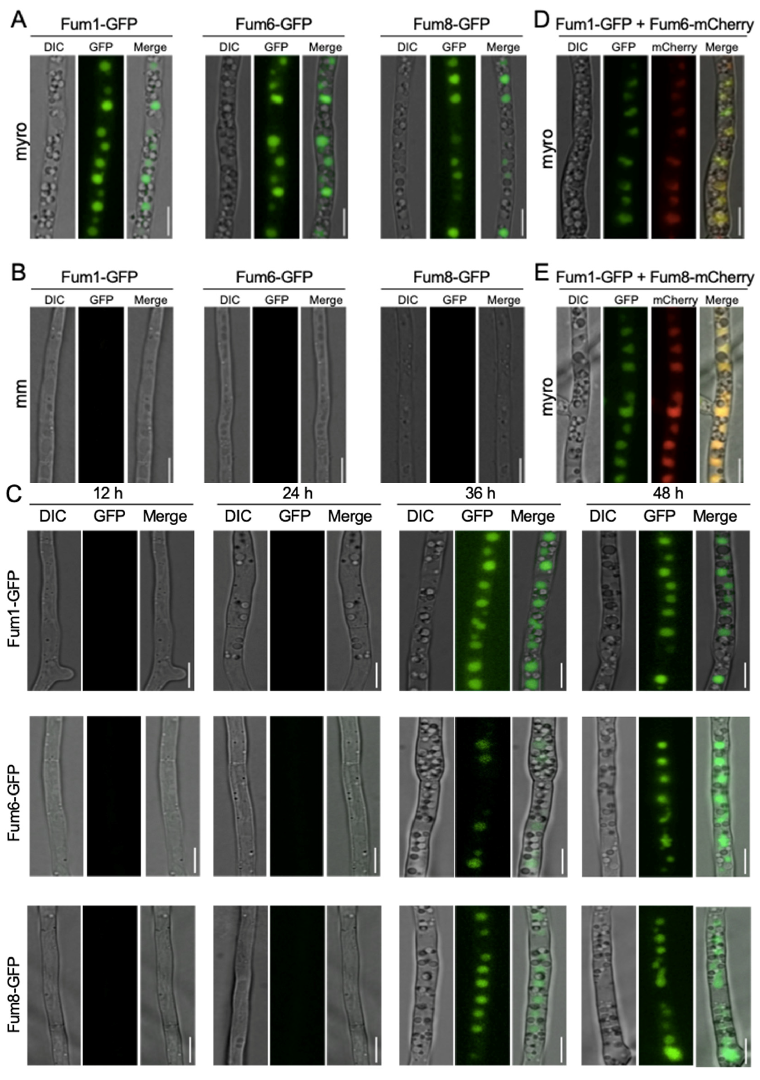
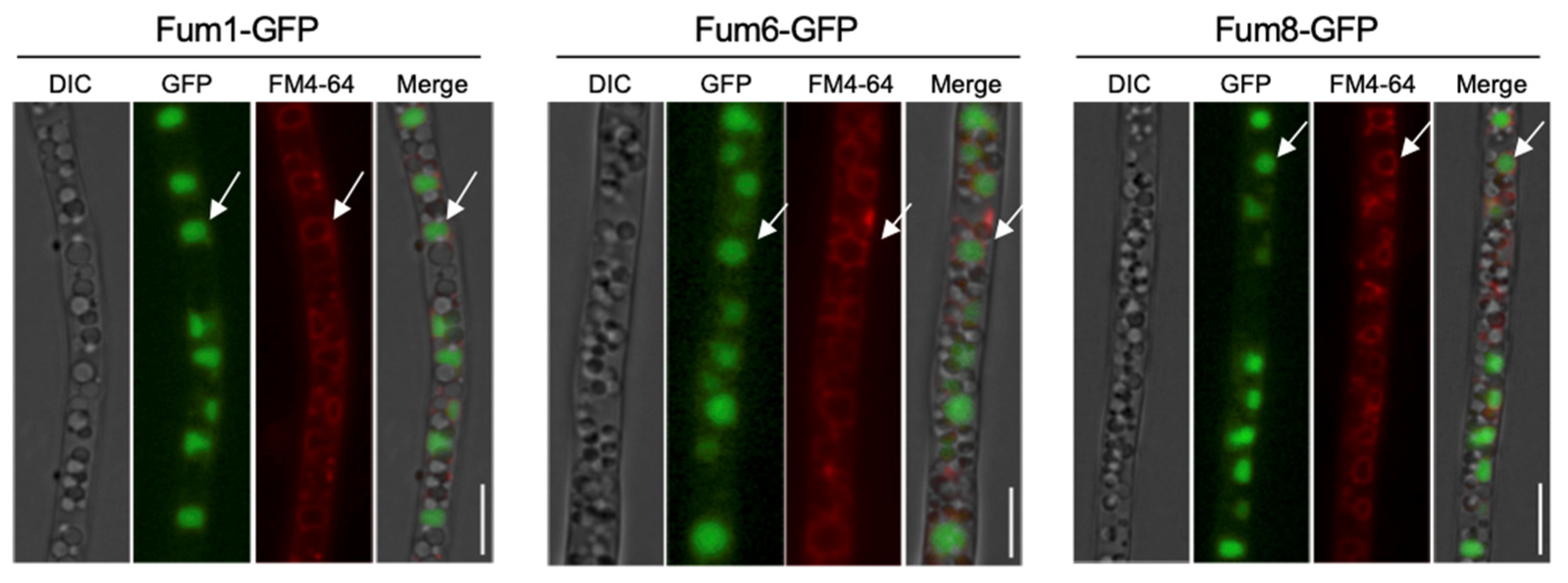
3.1. Subcellular Localization of Three FB1 Biosynthesis Enzymes

To determine the localization of FB1 biosynthesis enzymes, the first three Fum proteins (Fum1, Fum8, and Fum6) in the proposed FB1 biosynthetic pathway were studied [18,25]. Fum1, Fum8, and Fum6 were tagged with GFP at their C-terminus under its endogenous locus in F. verticillioides (Figure S1). We first employed live-cell imaging to visualize Fum1-GFP, Fum8-GFP, and Fum6-GFP fluorescence under toxin-inducing medium (myro) and toxin non-inducing minimal liquid medium (MM). The results showed that all three tagged Fum proteins were highly induced and localized to punctate structures in hyphae in myro broth, while no green fluorescent was observed in MM after 48 h (Figure 1A,B). To verify that the Fum-protein fluorescence are medium-dependent, we included a SNARE protein tagged strain, Syn1-GFP, from our previous study as a control [26]. Syn1-GFP showed fluorescence signals in both myro and MM (Figure S3). We hypothesized that Fum proteins display different localization at earlier time points. However, our time course study (12, 24, 36, 48 h) demonstrated that Fum1-GFP, Fum6-GFP, and Fum8-GFP localized to similar structures at 36 h and 48 h, but no fluorescence appeared at 12 h and 24 h when cultured in myro medium (Figure 1C). We reasoned that Fum-protein fluorescence first appeared between 24 and 36 h. Indeed, detectable Fum1-GFP, Fum6-GFP, and Fum8-GFP protein fluorescence appeared approximately after 26 h incubation, and fluorescence intensity increased afterwards (data not shown). Additionally, we noted that Fum-GFP signal did not appear in the newly synthesized branching hyphae and apical areas (Figure S4). These data indicate that key Fum proteins Fum1, Fum6, and Fum8 localized to similar punctate structures, and the localizations are regulated in time- and medium-dependent manners.

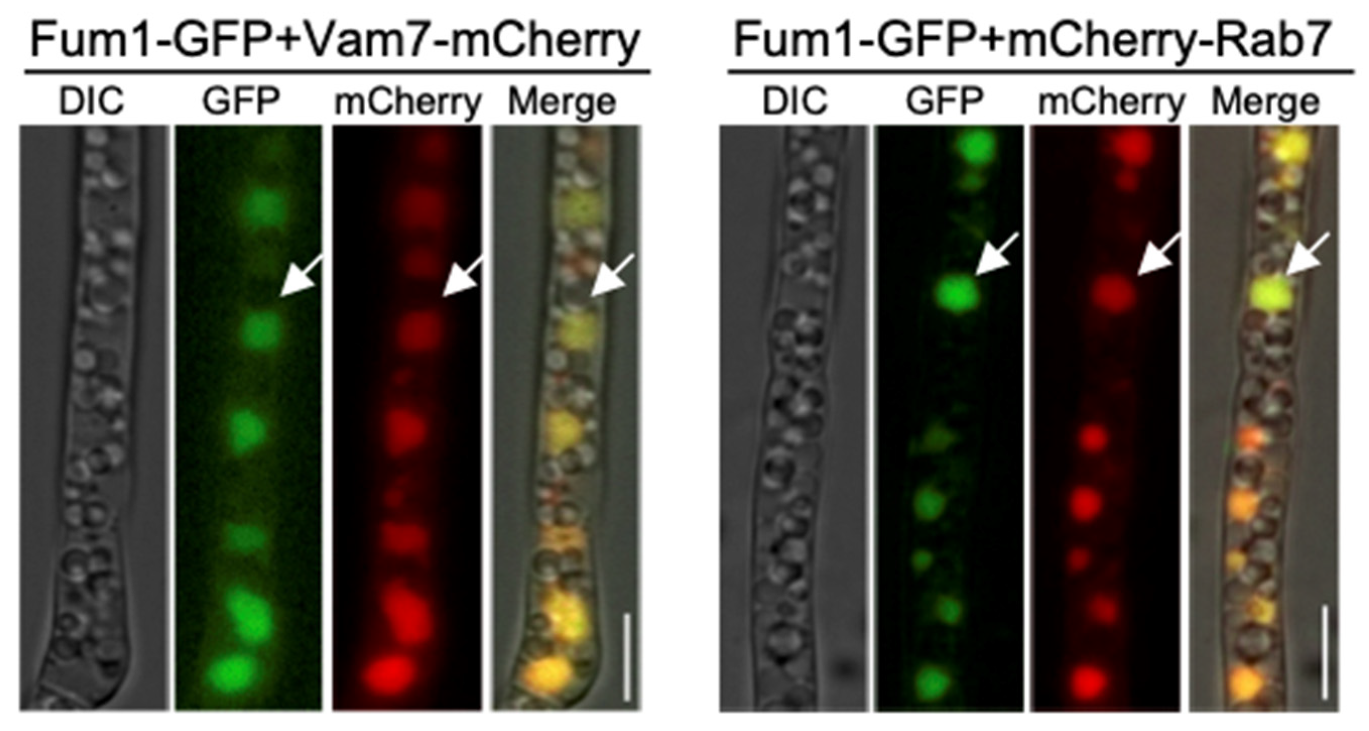
3.2. Co-Localization of Fum1, Fum6, and Fum8 in Vacuoles

Our observation of Fum1, Fum6, or Fum8 localized to similar spherical punctate structures raised the question of whether or not they are expressed in the same organelle. To test this idea, we first tagged Fum6 or Fum8 with mCherry and transformed into Fum1-GFP strain to study the co-localizations of Fum6 or Fum8 with Fum1. As expected, Fum6-mCherry or Fum8-mCherry were co-localized with Fum1-GFP in the same circular organelle (Figure 1D,E). Next, to address the precise structure where Fum proteins localize, we employed lipophilic dye FM4-64 which is well known for staining plasma membranes, Spitzenkörper and vacuolar membrane, with varied incubation time [27]. Fum1-GFP, Fum6-GFP, and Fum8-GFP cells were stained with fluorescent endocytic marker FM4-64 for 30 min for vacuolar membrane stain. We found that Fum1-GFP, Fum6-GFP, and Fum8-GFP overlapped well with FM4-64 labeled vacuoles (Figure 2). In conclusion, our fluorescence microscopic results demonstrated Fum1-GFP, Fum6-GFP, and Fum8-GFP are co-localized with FM4-64 stained vacuoles.

3.3. Two Vacuole Proteins Are Required for FB1 Biosynthesis

In F. graminearum, two vacuole proteins, FgRab7 and FgVam7, are reported to be localized in the vacuole and essential for vacuole morphology and proper functions [28,29]. We identified these two vacuole protein homologues, FvRab7 and FvVam7, in F. verticillioides. To validate our previous localization result from FM4-64 staining, we first expressed FvRab7 and FvVam7 with mCherry in Fum1-GFP background strain. Due to the same location of Fum1, Fum6, and Fum8, we decided to use Fum1 for validation. The results showed that Fum1-GFP displayed co-localization with FvVam7-mCherry and mCherry-FvRab7 on large vacuole structure (Figure 3), which further confirmed the Fum1 localization in the vacuole. Next, we disrupted FvVAM7 and FvRAB7 genes in Fum1-GFP strain independently to elucidate the role of the vacuole in the expression of Fum1. Fluorescence microscopy study showed no detectable Fum1-GFP fluorescence signal in ∆Fvvam7 and ∆Fvrab7 mutants in myro medium (Figure 4C), which demonstrated that FvVAM7 and FvRAB7 genes are important for Fum1 expression. Furthermore, growth assay showed that ∆FvVAM7 and ∆FvRAB7 exhibited severe hyphal growth defect compared to the wild-type progenitor (Figure 4A). Additionally, these two mutants exhibited a small and fragmented vacuole morphology phenotype, with vacuoles unevenly distributed throughout the cells in contrast to the WT (Figure 4D). FB1 production assay showed that FB1 levels were repressed about 40 folds in ∆Fvvam7 and 20 folds in ∆Fvrab7 compared to the wild-type progenitor (Figure 4B). We tested the expression of 15 PKS genes through RT-qPCR to further verify the role of FvVam7 and FvRab7 in secondary metabolism (Table S2). As expected, almost all tested mRNA levels of PKS genes were altered in ∆Fvvam7 and ∆Fvrab7 mutants. More importantly, PKS11 (FUM1), which is required for the first step of FB1 biosynthesis, was remarkably downregulated in the absence of FvVam7 and FvRab7. Based on these results, we concluded that Fum1 localized in the vacuole and that proper functions of FvVam7 and FvRab7 are indispensable for Fum1 protein expression and FB1 production.

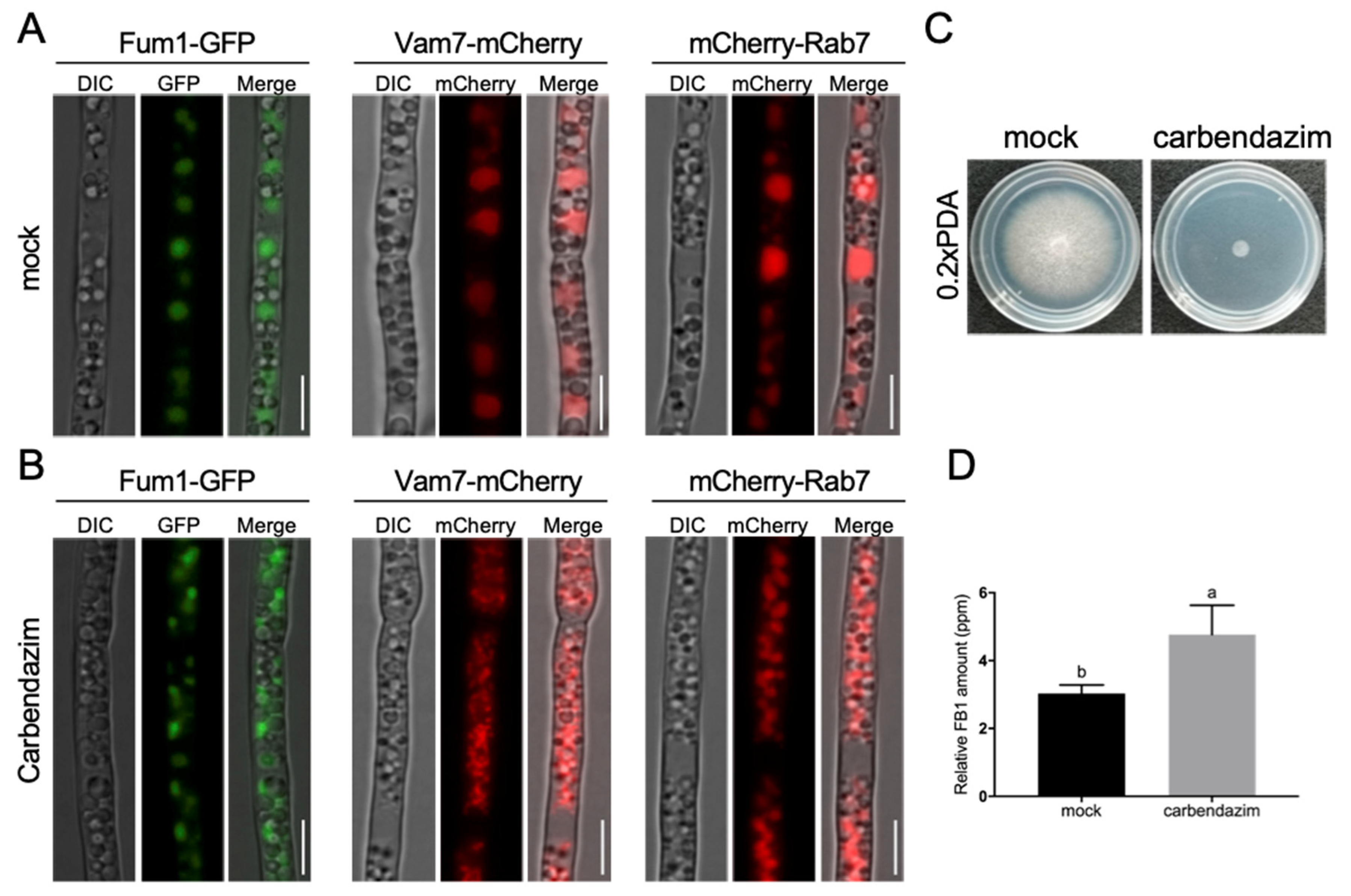
3.4. Proper Microtubule Assembly Regulates Vacuole Structure and FB1 Biosynthesis

Previous research showed that MT cytoskeleton interacts with vacuole and regulates its morphology and function [30,31,32,33,34]. To investigate the roles of MT network in vacuole structure and Fum protein expression in F. verticillioides, carbendazim was used to treat Fum1-GFP, Vam7-mCherry, and mCherry-Rab7 strains to observe their localization and vacuole structure. Our findings showed that Fum1, Vam7, and Rab7 localized to the large vacuole structure in the mock treatment. However, with the treatment of 1.0 μg/mL carbendazim for 24 h, Fum1-GFP was localized to irregular and punctate structures in the cytoplasm, while Vam7-mCherry and mCherry-Rab7 exhibited small and fragmented vacuoles with irregular spheroids structures (Figure 5A,B). The results suggested that proper microtubule assembly is required for large vacuole structure and Fum1 localization. Furthermore, FB1 production level was significantly enhanced by 60% when treated with 1.0 μg/mL carbendazim compared to the mock-treated wild-type, while the mycelial growth was severely inhibited on 0.2 × PDA plate containing carbendazim (Figure 5C,D). Our results suggested that MT network plays important roles in regulating vacuole structure and FB1 biosynthesis in F. verticillioides.

3.5. Tubulin Fvα1 Is a Negative Regulator in FB1 Biosynthesis

Carbendazim is a classical tubulin polymerization inhibitor. To better define the role of MT network in FB1 biosynthesis, four F. verticillioides tubulins, Fvα1, Fvα2, Fvβ1, and Fvβ2, were knocked out in Fum1-GFP background. Fum1-GFP signals in the four mutants and wild-type cultured in myro medium for 48 h were monitored. As shown in Figure 6A, Fum1-GFP were highly induced in ΔFvα1 and ΔF1, while weak signals were observed in ΔFvα2 and ΔFvβ2 mutants. Examination of vacuole morphology in these tubulin mutants revealed that ΔFvα1 and ΔFvβ1 were unable to produce large vacuoles like the wild-type strain (Figure 6B). To verify these results, we tested the transcription levels of FvFUM1 in wild-type and tubulin deletion mutants. The transcription level of FvFUM1 was significantly increased in ΔFvα1 and dramatically reduced in ΔFvα2, ΔFvβ1, and ΔFvβ2, especially in ΔFvα2 and ΔFvβ2 (Figure 6C). Consistent with FUM1 transcript level, FB1 level in ΔFvα1 was significantly higher, whereas that in ΔFvα2 and ΔFvβ2 were dramatically lower than the wild-type control (Figure 6D). Meanwhile, we observed a lower FB1 level in ΔFvβ1, although not statistically significant as FUM1 transcript level. Further growth assay showed that the mutants ΔFvα1 and ΔFvβ1 exhibited significantly reduced growth rate (Figure 7A). To assess how these four proteins interact, we lastly performed split luciferase complementation assay in F. verticillioides under a FB1-producing condition to assess the interaction of these four proteins. Luciferase activity was detected in samples Fvα1:Fvβ1, Fvα1:Fvβ2, Fvα2:Fvβ1, and Fvα2:Fvβ2 (Figure 7B), which suggested that four types of tubulin heterodimer can be formed in F. verticillioides under FB1-producing condition. In addition, qRT-PCR revealed that the expression of other tubulin subunits was significantly upregulated in Fvα1 and Fvβ1 mutants (Figure 7C). Our results suggest that Fvα2 and Fvβ2 play a role in regulating FB1 biosynthesis through translocation of Fum enzymes to vacuole. However, more importantly, we concluded that Fvα1 serves as a negative regulator of FB1 production, but the mechanism associated with how Fvα1 deletion impacts Fum genes needs further examination.

4. Discussion

F. verticillioides genetic and biochemical studies associated with fumonisin biosynthesis in the past three decades have provided valuable insights into how fumonisins are synthesized. However, the organization and compartmentation of fumonisin biosynthetic enzymes remained ambiguous. In this work, we tagged the first three Fum proteins in the proposed FB1 biosynthesis pathway. Under FB-producing conditions, we found that FvFum1, FvFum6, and FvFum8 localized to round, vacuole-like organelles. Further co-localization study demonstrated that FvFum1-GFP co-localizes with FvFum6-mCherry and FvFum8-mCherry in a live cell imaging assay, which suggested that these three Fum proteins occupy the same localization. Further characterization indicated that the organelle is the vacuole compartment since it showed positive localization with FM4-64. Genes involved in FB1 biosynthesis are highly arranged in gene clusters [16,17], and our results showed that proteins encoded by these genes are spatially well organized in vacuoles. Thus, we reasoned that F. verticillioides keeps those enzymes in the same organelle for optimum reaction and prevention of self-toxicity. However, the results of our localization study are different from a recent report which showed that Ds-Red::Fum8 localized in ER-derived vesicles under the control of constitutive A. nidulans promoter PoliC [35]. This report also showed that Fum17 and Fum18 share the same localization with Ds-Red::Fum8 with strong promoters Pgln1 and PoliC, respectively. The different Fum proteins localizations in different organisms suggested that Fum proteins could be organism specific, or constitutive promoters may be a factor that leads to different localization.
The vacuole is a dynamic cellular compartment that exerts many functions. Plant vacuoles were shown to permanently or temporarily store diverse and numerous secondary metabolites (>200,000 compounds) including chemicals used as defense or attractants [36]. In fungi, vacuoles are primarily recognized as a key site for degradation as well as for the storage of small molecules. To rule out misinterpretation of the Fum protein localization results, we carried out three additional experiments. We first conducted a time course study to monitor Fum protein localization. Fum proteins were localized in vacuole as well in earlier time points, but no fluorescence signal appeared until about 26 h incubation. Second, we used Syn1-GFP as a control which showed different localization pattern in the cell than Fum1-GFP [26]. Lastly, further experiment with glycine treatment, a major nitrogen, was also performed [37]. The results showed that Fum1-GFP localized to vacuoles, whereas fluorescence diminished after treatment of glycine. However, glycine treatment did not affect the localization of vacuole-related proteins Vam7-mCherry and mCherry-Rab7 (Figure S5). This is in line with our early finding that the addition of nitrogen repressed FB1 production [37]. Taken together, our results strongly support our novel finding that the key Fum proteins display vacuole localization in toxin-inducing broth. In addition, we expressed vacuole-associated proteins FvRab7 and FvVam7 with mCherry and analyzed their spatial relationship with FvFum1-GFP. In both A. nidulans and F. gramminerum, a small GTPase, Rab7, was shown to be localized to the vacuolar membrane [28,38]. We predicted that FvRab7 has the same localization since FvRab7 and FgRab7 are 100% identical in amino acid level. As expected, all these vacuole-associated proteins exhibited co-localization with FvFum1 under fumonisin-producing condition, demonstrating that vacuole is the organelle for fumonisin biosynthesis in F. verticillioide. However, we found that mCherry-FvRab7 co-localized with Fum1-GFP on the whole vacuole structure instead of appearing only on vacuolar membrane. To exclude the effect of incubation time on FvRab7 localization, we monitored the expression at 12h, 24h, 36h, and 48 h and found no mere vacuolar membrane localization (Figure S6). As a key FB1 biosynthesis level marker, Fum1-GFP expression was not detected in ΔFvrab7 mutant, and FB1 production was significantly reduced in the absence of Fvrab7. This result in F. verticillioides is in line with the data in F. gramminerum that ΔFgrab7 mutant showed significantly lower DON production [39]. However, in A. parasiticus, deletion mutant of vb1 (Rab7 homolog) led to a 7-fold higher of aflatoxin compared to WT in an aflatoxin-inducing condition [7]. Those strongly revealed the functional importance of the vacuole in mycotoxin production.
We also examined other factors that may potentially influence vacuole’s function. As an important cytoskeleton, MT and tubulin heterodimers interact with a variety of cellular components (metabolic enzymes or cellular organelles), thereby regulating intracellular metabolic pathways. Evidence revealed that MT binds to most of the glycolytic enzymes and regulates their activities [40,41,42]. In HeLa cell, MT is crucial for the spatial distribution and biological functions of purinosomes [43]. Our recent study demonstrated that MT participated in toxisomes formation and DON biosynthesis in F. graminearum [44]. In this work, we analyzed the regulatory role of MT network in vacuole morphology via carbendazim treatment. The results demonstrated that fragmented vacuoles were observed in hyphae under carbendazim treatment with 1.2 μg/mL (100% inhibition against mycelial growth of F. verticillioides) for 24 h in F. graminearum. This is consistent with vacuole localization in F. verticillioides. We found that MT network participated in vacuole morphology regulation in F. verticillioides. Clear evidence was also found in some plant species that vacuole morphology is regulated by MT [30,34].
Interestingly, we found that FB1 production significantly increased when treated with carbendazim in F. verticillioides. However, the expression of FvFum1 showed no significant difference when compared to the control group by RT-qPCR analysis. The reason is still unclear. In A. parasiticus, two important aflatoxin synthetic enzymes were localized in vesicles. When deleting vb1 (a member of Vps tethering complex) in A. parasiticus or using Vps tethering complex inhibitor Sortin3 to treat A. parasiticus, fragmented vacuoles and increased aflatoxin production were observed, but the expression of aflatoxin biosynthetic genes showed no significant changes in vb1 mutants or under Sortin3 treatment [7]. We speculated that fragmented vacuoles could facilitate FB1 biosynthesis, yet there is no close correlation between the expression levels of FvFum1 and vacuole morphology in F. verticillioides. Additionally, in F. graminearum, low concentrations of carbendazim boosted DON production, but high concentrations significantly reduced it [44]. Our experiments utilized a 1 μg/mL carbendazim concentration, resulting in increased FB1 production. The effects of carbendazim on FB1 production in F. verticillioides could also relate to the concentrations of carbendazim.
Our previous study showed that α1-β2 tubulin heterodimer is critical for toxisome formation and DON biosynthesis in F. graminearum [44]. However, gene deletion assays in F. verticillioides revealed that three tubulin subunits were involved in FB1 biosynthesis. ΔFvα1 showed significantly increased FB1 production, whereas FB1 production in ΔFvα2 and ΔFvβ2 mutants was dramatically reduced. The differences between F. verticillioides and F. graminearum indicated that the regulatory roles of tubulin subunits in secondary metabolism vary among species. Further research is needed to unveil the different FB1 regulating mechanisms in F. verticillioides.
In summary, this report revealed the localization of Fum proteins in vacuole. Two vacuole proteins, FvRab7 and FVam7, are required for FB1 biosynthesis. Further study showed that disrupted MT assembly caused the change in Fum1 localization and enhanced FB1 production. Fvα1 tubulin is a negative regulator in FB1 production. However, further studies are needed to better understand the effect of MT on promoting FB1 production and how the protein–protein interaction network of tubulins interact with MT in regulating FB1 production.

Supplementary Materials

The following supporting information can be downloaded at: https://www.mdpi.com/article/10.3390/jof9020268/s1, Figure S1: Schematic description and confirmation of the split marker strategy to generate Fum1 GFP strain. Figure S2: Schematic representation of the gene deletion and mutant screening. Figure S3: Localization of Syn1-GFP in myro and MM broth. Figure S4: Fum1-GFP and Fum8-GFP absent localization on growing apical area. Figure S5: Nitrogen repression of Fum1-GFP expression. Figure S6: Time courses of Vam7-mCherry and mCherry-Rab7. Table S1: Primers used in this study. Table S2: Relative expression level of PKS genes in wild-type, ΔFvrab7 and ΔFvvam7.

Author Contributions

H.Y., Z.Z. and W.B.S. conceived and designed the experiments. H.Y., Z.Z. and H.Z. performed experiments. H.Y., Z.Z., H.Z. and W.B.S. analyzed the data. H.Y., Z.Z., H.Z. and W.B.S. wrote the manuscript. All authors have read and agreed to the published version of the manuscript.

Funding

This research was funded in part by the Agriculture and Food Research Initiative Competitive Grants Program Grant (2013-68004-20359) from the USDA National Institute of Food and Agriculture and the Department of Plant Pathology and Microbiology at Texas A&M University.

Institutional Review Board Statement

Not applicable.

Informed Consent Statement

Not applicable.

Data Availability Statement

All data generated or analyzed in this study are included in this published article and its supplementary information.

Acknowledgments

The authors also would like to acknowledge China Scholarship Council support (201906850058) for Z. Zhou. The authors thank Brian Shaw and Joe Vasselli in the Department of Plant Pathology & Microbiology, Texas A&M University, for assisting in microscopy. The authors thank Wenhui Zheng (Fujian Agriculture and Forestry University) for providing pKNTG and pKNT-mCherry plasmids.

Conflicts of Interest

The authors declare no conflict of interest.

References

  1. Keller, N.P.; Turner, G.; Bennett, J.W. Fungal secondary metabolism—From biochemistry to genomics. Nat. Rev. Microbiol. 2005, 3, 937–947. [Google Scholar] [CrossRef] [PubMed]
  2. Kistler, H.C.; Broz, K. Cellular compartmentalization of secondary metabolism. Front. Microbiol. 2015, 6, 68. [Google Scholar] [CrossRef] [Green Version]
  3. Evers, M.E.; Trip, H.; van den Berg, M.A.; Bovenberg, R.A.L.; Driessen, A.J.M. Compartmentalization and transport in beta-lactam antibiotics biosynthesis. In Molecular Biotechnology of Fungal Beta-Lactam Antibiotics and Related Peptide Synthetases; Springer: Berlin/Heidelberg, Germany, 2004; Volume 88, pp. 111–135. [Google Scholar]
  4. Muller, W.H.; Bovenberg, R.A.L.; Groothuis, M.H.; Kattevilder, F.; Smaal, E.B.; Vandervoort, L.H.M.; Verkleij, A.J. Involvement of Microbodies in Penicillin Biosynthesis. Biochim. Biophys. Acta 1992, 1116, 210–213. [Google Scholar] [CrossRef] [PubMed]
  5. Kurylowicz, W.; Kurzatkowski, W.; Kurzatkowski, J. Biosynthesis of Benzylpenicillin by Penicillium-Chrysogenum and its Golgi-Apparatus. Arch. Immunol. Ther. Exp. 1987, 35, 699–724. [Google Scholar]
  6. Lendenfeld, T.; Ghali, D.; Wolschek, M.; Kubicekpranz, E.M.; Kubicek, C.P. Subcellular Compartmentation of Penicillin Biosynthesis in Penicillium-Chrysogenum—The Amino-Acid Precursors Are Derived from the Vacuole. J. Biol. Chem. 1993, 268, 665–671. [Google Scholar] [CrossRef] [PubMed]
  7. Chanda, A.; Roze, L.V.; Kang, S.; Artymovich, K.A.; Hicks, G.R.; Raikhel, N.V.; Calvo, A.M.; Linz, J.E. A key role for vesicles in fungal secondary metabolism. Proc. Natl. Acad. Sci. USA 2009, 106, 19533–19538. [Google Scholar] [CrossRef] [PubMed] [Green Version]
  8. Hong, S.-Y.; Linz, J.E. Functional expression and subcellular localization of the aflatoxin pathway enzyme Ver-1 fused to enhanced green fluorescent protein. Appl. Environ. Microbiol. 2008, 74, 6385–6396. [Google Scholar] [CrossRef] [Green Version]
  9. Hong, S.-Y.; Linz, J.E. Functional expression and sub-cellular localization of the early aflatoxin pathway enzyme Nor-1 in Aspergillus parasiticus. Mycol. Res. 2009, 113, 591–601. [Google Scholar] [CrossRef] [Green Version]
  10. Lee, L.W.; Chiou, C.H.; Klomparens, K.L.; Cary, J.W.; Linz, J.E. Subcellular localization of aflatoxin biosynthetic enzymes Nor-1, Ver-1, and OmtA in time-dependent fractionated colonies of Aspergillus parasiticus. Arch. Microbiol. 2004, 181, 204–214. [Google Scholar] [CrossRef]
  11. Menke, J.; Weber, J.; Broz, K.; Kistler, H.C. Cellular development associated with induced mycotoxin synthesis in the filamentous fungus Fusarium graminearum. PLoS ONE 2013, 8, e63077. [Google Scholar] [CrossRef] [Green Version]
  12. Tang, G.; Chen, Y.; Xu, J.R.; Kistler, H.C.; Ma, Z. The fungal myosin I is essential for Fusarium toxisome formation. PLoS Pathog. 2018, 14, e1006827. [Google Scholar] [CrossRef] [PubMed] [Green Version]
  13. Rheeder, J.P.; Marasas, W.F.; Vismer, H.F. Production of fumonisin analogs by Fusarium species. Appl. Environ. Microbiol. 2002, 68, 2101–2105. [Google Scholar] [CrossRef] [PubMed] [Green Version]
  14. Gelderblom, W.C.; Jaskiewicz, K.; Marasas, W.F.; Thiel, P.G.; Horak, R.M.; Vleggaar, R.; Kriek, N.P. Fumonisins--novel mycotoxins with cancer-promoting activity produced by Fusarium moniliforme. Appl. Environ. Microbiol. 1988, 54, 1806–1811. [Google Scholar] [CrossRef] [PubMed] [Green Version]
  15. Nelson, P.E.; Desjardins, A.E.; Plattner, R.D. Fumonisins, mycotoxins produced by fusarium species: Biology, chemistry, and significance. Annu. Rev. Phytopathol. 1993, 31, 233–252. [Google Scholar] [CrossRef]
  16. Proctor, R.H.; Desjardins, A.E.; Plattner, R.D.; Hohn, T.M. A polyketide synthase gene required for biosynthesis of fumonisin mycotoxins in Gibberella fujikuroi mating population A. Fungal Genet. Biol. 1999, 27, 100–112. [Google Scholar] [CrossRef] [Green Version]
  17. Seo, J.A.; Proctor, R.H.; Plattner, R.D. Characterization of four clustered and coregulated genes associated with fumonisin biosynthesis in Fusarium verticillioides. Fungal Genet. Biol. 2001, 34, 155–165. [Google Scholar] [CrossRef]
  18. Woloshuk, C.P.; Shim, W.B. Aflatoxins, fumonisins, and trichothecenes: A convergence of knowledge. FEMS Microbiol. Rev. 2013, 37, 94–109. [Google Scholar] [CrossRef] [Green Version]
  19. Blacutt, A.A.; Gold, S.E.; Voss, K.A.; Gao, M.; Glenn, A.E. Fusarium verticillioides: Advancements in Understanding the Toxicity, Virulence, and Niche Adaptations of a Model Mycotoxigenic Pathogen of Maize. Phytopathology 2018, 108, 312–326. [Google Scholar] [CrossRef] [Green Version]
  20. Yan, H.; Huang, J.; Zhang, H.; Shim, W.B. A Rab GTPase protein FvSec4 is necessary for fumonisin B1 biosynthesis and virulence in Fusarium verticillioides. Curr. Genet. 2020, 66, 205–216. [Google Scholar] [CrossRef]
  21. Yan, H.; Shim, W.B. Characterization of non-canonical G beta-like protein FvGbb2 and its relationship with heterotrimeric G proteins in Fusarium verticillioides. Environ. Microbiol. 2020, 22, 615–628. [Google Scholar] [CrossRef]
  22. Christensen, S.; Borrego, E.; Shim, W.B.; Isakeit, T.; Kolomiets, M. Quantification of fungal colonization, sporogenesis, and production of mycotoxins using kernel bioassays. J. Vis. Exp 2012, 3727. [Google Scholar] [CrossRef] [Green Version]
  23. Kim, H.K.; Cho, E.J.; Jo, S.; Sung, B.R.; Lee, S.; Yun, S.H. A split luciferase complementation assay for studying in vivo protein-protein interactions in filamentous ascomycetes. Curr. Genet. 2012, 58, 179–189. [Google Scholar] [CrossRef] [PubMed]
  24. Schneider, C.A.; Rasband, W.S.; Eliceiri, K.W. NIH Image to ImageJ: 25 years of image analysis. Nat. Methods 2012, 9, 671–675. [Google Scholar] [CrossRef] [PubMed]
  25. Alexander, N.J.; Proctor, R.H.; McCormick, S.P. Genes, gene clusters, and biosynthesis of trichothecenes and fumonisins in Fusarium. Toxin Rev. 2009, 28, 198–215. [Google Scholar] [CrossRef]
  26. Zhang, H.; Yan, H.; Shim, W.B. Fusarium verticillioides SNARE protein FvSyn1 harbours two key functional motifs that play selective roles in fungal development and virulence. Microbiology 2019, 165, 1075–1085. [Google Scholar] [CrossRef]
  27. Fischer-Parton, S.; Parton, R.M.; Hickey, P.C.; Dijksterhuis, J.; Atkinson, H.A.; Read, N.D. Confocal microscopy of FM4-64 as a tool for analysing endocytosis and vesicle trafficking in living fungal hyphae. J. Microsc. 2000, 198, 246–259. [Google Scholar] [CrossRef] [Green Version]
  28. Li, Y.; Li, B.; Liu, L.; Chen, H.; Zhang, H.; Zheng, X.; Zhang, Z. FgMon1, a guanine nucleotide exchange factor of FgRab7, is important for vacuole fusion, autophagy and plant infection in Fusarium graminearum. Sci. Rep. 2015, 5, 18101. [Google Scholar] [CrossRef] [Green Version]
  29. Zhang, H.; Li, B.; Fang, Q.; Li, Y.; Zheng, X.; Zhang, Z. SNARE protein FgVam7 controls growth, asexual and sexual development, and plant infection in Fusarium graminearum. Mol. Plant Pathol. 2016, 17, 108–119. [Google Scholar] [CrossRef]
  30. Oda, Y.; Hirata, A.; Sano, T.; Fujita, T.; Hiwatashi, Y.; Sato, Y.; Kadota, A.; Hasebe, M.; Hasezawa, S. Microtubules regulate dynamic organization of vacuoles in Physcomitrella patens. Plant Cell Physiol. 2009, 50, 855–868. [Google Scholar] [CrossRef] [Green Version]
  31. Ligrone, R.; Duckett, J.G. The leafy stems of Sphagnum (Bryophyta) contain highly differentiated polarized cells with axial arrays of endoplasmic microtubules. New Phytol. 1998, 140, 567–579. [Google Scholar] [CrossRef]
  32. Pressel, S.; Ligrone, R.; Duckett, J.G. Cellular differentiation in moss protonemata: A morphological and experimental study. Ann. Bot. 2008, 102, 227–245. [Google Scholar] [CrossRef] [PubMed] [Green Version]
  33. Vedrenne, C.; Hauri, H.P. Morphogenesis of the endoplasmic reticulum: Beyond active membrane expansion. Traffic 2006, 7, 639–646. [Google Scholar] [CrossRef] [PubMed] [Green Version]
  34. Ligrone, R.; Duckett, J.G. Polarity and endoplasmic microtubules in food-conducting cells of mosses: An experimental study. New Phytol. 1996, 134, 503–516. [Google Scholar] [CrossRef]
  35. Janevska, S.; Ferling, I.; Jojic, K.; Rautschek, J.; Hoefgen, S.; Proctor, R.H.; Hillmann, F.; Valiante, V. Self-Protection against the Sphingolipid Biosynthesis Inhibitor Fumonisin B1 Is Conferred by a FUM Cluster-Encoded Ceramide Synthase. mBio 2020, 11, e00455-20. [Google Scholar] [CrossRef]
  36. de Brito Francisco, R.; Martinoia, E. The Vacuolar Transportome of Plant Specialized Metabolites. Plant Cell Physiol. 2018, 59, 1326–1336. [Google Scholar] [CrossRef] [Green Version]
  37. Shim, W.B.; Woloshuk, C.P. Nitrogen repression of fumonisin B1 biosynthesis in Gibberella fujikuroi. FEMS Microbiol. Lett. 1999, 177, 109–116. [Google Scholar] [CrossRef]
  38. Abenza, J.F.; Galindo, A.; Pinar, M.; Pantazopoulou, A.; de los Rios, V.; Penalva, M.A. Endosomal maturation by Rab conversion in Aspergillus nidulans is coupled to dynein-mediated basipetal movement. Mol. Biol. Cell 2012, 23, 1889–1901. [Google Scholar] [CrossRef] [Green Version]
  39. Zheng, H.; Zheng, W.; Wu, C.; Yang, J.; Xi, Y.; Xie, Q.; Zhao, X.; Deng, X.; Lu, G.; Li, G.; et al. Rab GTPases are essential for membrane trafficking-dependent growth and pathogenicity in Fusarium graminearum. Environ. Microbiol. 2015, 17, 4580–4599. [Google Scholar] [CrossRef]
  40. Keller, A.; Peltzer, J.; Carpentier, G.; Horvath, I.; Olah, J.; Duchesnay, A.; Orosz, F.; Ovadi, J. Interactions of enolase isoforms with tubulin and microtubules during myogenesis. Biochim. Biophys. Acta 2007, 1770, 919–926. [Google Scholar] [CrossRef]
  41. Cassimeris, L.; Silva, V.C.; Miller, E.; Ton, Q.; Molnar, C.; Fong, J. Fueled by microtubules: Does tubulin dimer/polymer partitioning regulate intracellular metabolism? Cytoskeleton 2012, 69, 133–143. [Google Scholar] [CrossRef]
  42. Durrieu, C.; Bernier-Valentin, F.; Rousset, B. Microtubules bind glyceraldehyde 3-phosphate dehydrogenase and modulate its enzyme activity and quaternary structure. Arch. Biochem. Biophys. 1987, 252, 32–40. [Google Scholar] [CrossRef] [PubMed]
  43. An, S.; Deng, Y.; Tomsho, J.W.; Kyoung, M.; Benkovic, S.J. Microtubule-assisted mechanism for functional metabolic macromolecular complex formation. Proc. Natl. Acad. Sci. USA 2010, 107, 12872–12876. [Google Scholar] [CrossRef] [PubMed] [Green Version]
  44. Zhou, Z.; Duan, Y.; Zhang, J.; Lu, F.; Zhu, Y.; Shim, W.B.; Zhou, M. Microtubule-assisted mechanism for toxisome assembly in Fusarium graminearum. Mol. Plant Pathol. 2021, 22, 163–174. [Google Scholar] [CrossRef] [PubMed]
Figure 1. Localization of Fum1, Fum6, and Fum8 in hyphae in toxin inducing and non-inducing media. (A) Fum1-GFP, Fum6-GFP, and Fum8-GFP are localized to the spherical structures in hyphae grown in the toxin inducing medium (myro). (B) GFP signals were not detected in the minimal medium (mm). All images were taken after 48 h incubation at 28 °C with constant shaking. (C) Localization of three key Fum proteins grown in myro broth at different time points (12, 24, 36, 48 h). (D) Fum6-mCherry co-localized with Fum1-GFP in hyphae grown in myro liquid medium (E) Fum8-mCherry co-localized with Fum1-GFP in myro liquid medium. Bar = 5 μm.
Jof 09 00268 g001
Figure 2. Subcellular localization of Fum1-GFP, Fum6-GFP, or Fum8-GFP with FM4-64 on vacuoles. Fluorescent images indicate co-localization of Fum1-GFP, Fum6-GFP, or Fum8-GFP with FM4-64 labeling on vacuoles. Arrows indicate co-localization. Bars = 5 μm.
Jof 09 00268 g002
Figure 3. Co-localization of Fum1 with FvVam7 or FvRab7 proteins. FvFum1-GFP was co-localized with FvVam7-mCherry or FvRab7-mCherry in hyphae cultured in myro broth. Images were taken after 48 h constant shaking at 28 °C. Bar = 5 μm.
Jof 09 00268 g003
Figure 4. Colony growth and FB1 production of ΔFvvam7 and ΔFvrab7 mutants. (A) Wild-type (WT), ΔFvvam7, and ΔFvrab7 strains were cultured on V8 agar plates for 7 days at 28 °C. (B) FB1 levels in WT and mutant strains were normalized with fungal ergosterol levels. WT, Δfvvam7, and ΔFvrab7 strains were grown on autoclaved corn kernels for 8 days at room temperature before assay. Three replicates were performed for each strain. Different letters on the bars indicated significance according to prism Ordinary one-way ANOVA multiple comparisons at p < 0.05. (C) Fum1-GFP localization was determined in ∆Fvvam7 and ∆Fvrab7 compared to in WT strain. (D) FM4-64 was used to stain WT, ∆Fvvam7, and ∆Fvrab7 for vacuoles. Bar = 5 μm.
Jof 09 00268 g004
Figure 5. Carbendazim impacts on FB1 biosynthesis. Fum1-GFP, Vam7-mCherry, and Rab7-mCherry strains were inoculated in myro liquid medium without (A) and with (B) 1 μg/mL carbendazim treatment. Scale bar = 5 μm. (C) Sensitivity to carbendazim was shown by culturing WT on 0.2 × PDA or 0.2 × PDA + carbendazim (1 μg/mL) for 7 plates at 28 °C. (D) FB1 production in WT were tested after being cultured in myro broth with and without 1μg/mL carbendazim for 8 days of shaking at room temperature. Different letters on the bars indicated significance according to prism unpaired t-test at p < 0.05.
Jof 09 00268 g005
Figure 6. Fvα1 is negatively associated with FvFUM1 expression and FB1 biosynthesis. (A) Fum1-GFP localization in WT, ΔFvα1, ΔFvα2, ΔFvβ1, and ΔFvβ2 strains. Scale bar = 5 μm. (B) FM4-64 was used to stain WT, ΔFvα1, ΔFvα2, ΔFvβ1, and ΔFvβ2 for vacuoles. Bar = 5 μm. (C) RT-qPCR results for FvFUM1 under treatment of 1 μg/mL carbendazim relative to levels in myro broth. Significant differences were indicated with different letters on the bars according to prism two-way ANOVA at p < 0.05. (D) WT, ΔFvα1, ΔFvα2, ΔFvβ1, and ΔFvβ2 were cultivated on autoclaved corn kernels for 8 days at room temperature. FB1 levels were normalized with fungal ergosterol levels. Different letters on the bars indicated significance according to prism Ordinary one-way ANOVA multiple comparisons at p < 0.05.
Jof 09 00268 g006
Figure 7. Growth and gene interaction assays using four microtubule deletion mutants. (A) WT, ΔFvα1, ΔFvα2, ΔFvβ1, and ΔFvβ2 strains were cultivated on V8 plates. (B) Luminescence activity was obtained from three replicates. Multiple negative controls (Fvα1-Cluc + NLuc, Fvα2-Cluc + NLuc, NLuc + Fvβ1-CLuc, NLuc + Fvβ2-CLuc, NLuc-CLuc) were included in the study. Different letters on the bars indicated significance according to prism Ordinary one-way ANOVA multiple comparisons at p < 0.05. (C) Fvα1, Fvα2, Fvβ1, and Fvβ2 relative expression in WT, ΔFvα1, ΔFvα2, ΔFvβ1, and ΔFvβ2 strains were determined by RT-qPCR. Significant differences were indicated with different letters on the bars according to prism two-way ANOVA at p < 0.05.
Jof 09 00268 g007
Disclaimer/Publisher’s Note: The statements, opinions and data contained in all publications are solely those of the individual author(s) and contributor(s) and not of MDPI and/or the editor(s). MDPI and/or the editor(s) disclaim responsibility for any injury to people or property resulting from any ideas, methods, instructions or products referred to in the content.

