Article

Genotypic Analysis of the Population Structure in Malassezia globosa and Malassezia restricta

1
Laboratory of Fungal and Parasitic Molecular Biology, School of Medicine, University of Sfax, Sfax 3029, Tunisia
2
Faculty of Science, University of Gabes, Gabes 6029, Tunisia
3
Laboratory of Parasitology—Mycology, UH Habib Bourguiba, Sfax 3029, Tunisia
*
Author to whom correspondence should be addressed.
J. Fungi 2023, 9(2), 263; https://doi.org/10.3390/jof9020263
Submission received: 20 October 2022 / Revised: 22 December 2022 / Accepted: 23 December 2022 / Published: 15 February 2023
(This article belongs to the Special Issue Molecular Markers for Fungal Detection and Identification)

Abstract

:
The molecular characterization of Malassezia spp. isolates from animals and humans has not been thoroughly studied. Although a range of molecular methods has been developed for diagnosing Malassezia species, they have several drawbacks, such as inefficiency in differentiating all the species, high cost and questionable reproducibility. The present study aimed to develop VNTR markers for genotyping Malassezia isolated from clinical and animal samples. A total of 44 M. globosa and 24 M. restricta isolates were analyzed. Twelve VNTR markers were selected on seven different chromosomes (I, II, III, IV, V, VII and IX), six for each Malassezia species. The highest discriminatory power for a single locus was obtained with the STR-MG1 marker (0.829) and STR-MR2 marker (0.818) for M. globosa and M. restricta, respectively. After the analysis of multiple loci, 24 genotypes were noted among 44 isolates in M. globosa, with a discrimination index D of 0.943 and 15 genotypes were noted among 24 isolates in M. restricta, with a discrimination index D of 0.967. An endogenous infection was detected in two patients. Different genotypes of M. globosa strains colonized one patient. Interestingly, VNTR markers analysis revealed a carriage between a breeder and his dog in three cases for M. globosa and two for M. restricta. The FST (0.018 to 0.057) values indicate a low differentiation between the three populations of M. globosa. These results suggest a dominant clonal mode of reproduction in M. globosa. The typing of M. restricta showed a genotypic diversity of the strains, which can cause various skin pathologies. However, patient five was colonized with strains having the same genotype collected from different body parts (back, shoulder). VNTR analysis was capable of identifying species with high accuracy and reliability. More importantly, the method would facilitate monitoring Malassezia colonization in domestic animals and humans. It was shown that the patterns are stable and the method is discriminant, making it a powerful tool for epidemiological purposes.

1. Introduction

The genus Malassezia comprises lipophilic species that are normal parts of the human and animal skin flora. These yeasts are associated with various superficial dermatoses, including pityriasis versicolor, seborrheic dermatitis, folliculitis, and some variants of psoriasis and atopic dermatitis [1,2]. The genus Malassezia currently consists of 18 species [3], 10 of which have been isolated from human skin, including Malassezia furfur, Malassezia pachydermatis, Malassezia sympodialis, Malassezia slooffiae, Malassezia globosa, Malassezia obtusa, Malassezia restricta, Malassezia dermatis, Malassezia japonica and Malassezia yamatoensis [4]. M. furfur, M. globosa and M. sympodialis are the most commonly isolated species. M. globosa and M. restricta are the predominant species found on the scalp. M. globosa was found in pityriasis versicolor [5], while M. restricta was associated with seborrheic dermatitis and psoriasis [6,7,8]. The species distribution on the skin varies between different Malassezia related diseases, but their worldwide distribution may also differ. For example, M. sympodialis considered the most prevalent species in Europe and M. restricta and M. globosa were the most predominant species in Asia [8]. Since phenotypic identification methods are not very accurate for typing Malassezia species, various molecular techniques such as direct sequencing, PCR–restriction fragment length polymorphism (RFLP) [9,10], AFLP [11], RAPD [12] and real-time PCR [13,14] are gradually replacing the morphologic methods.
The Multiple Locus Variable-number tandem-repeat Analysis (MLVA) is based on the polymorphism of tandemly repeated genomic sequences called VNTR (Variable-Number Tandem-Repeats). VNTRs are classically separated into microsatellites (up to eight bp) and minisatellites (nine bp and more) [15]. The MLVA technique allows a resolution of closely related isolates for the investigation of disease outbreaks and provides information on the phylogenetic patterns among isolates [16,17,18]. The usefulness of MLVA was demonstrated for Aspergillus fumigatus [19], Aspergillus flavus [20,21], Trichophyton interdigitale [22] and Candida parapsilosis [23].
Here, we report the use of VNTR markers polymorphism as a new tool for the typing of M. globosa and M. restricta strains isolated from clinical and animal samples.

2. Materials and Methods

2.1. Fungal Isolates

A total of 68 Malassezia isolates were included in the present study: M. globosa (44) and M. restricta (24). M. globosa strains were collected from 26 patients (folliculitis (16), pityriasis capitis (6) pityriasis versicolor (4)) and 18 were isolated from breeders (9) and their dogs (9). Samples were collected using sterile compress from shoulder (7), face (8), neck (2), scalp (6) and back (10).
M. restricta strains were collected from 13 patients (folliculitis (5), pityriasis capitis (3) pityriasis versicolor (5)) and were isolated from breeders (7) and their dogs (4). Samples were collected using sterile compress from shoulder (3), face (7), neck (5), scalp (3) and back (2). Auricular samples from dogs were collected by swabbing.
Patients were referred to laboratory of parasitology mycology, UH Habib Bourguiba of Sfax-Tunisia.
All samples were then cultured on the modified Dixon agar medium and incubated for 3 to 7 days at 32 °C. These Malassezia strains were identified by real-time PCR, as described previously [14].
For the specificity tests, we used six Malassezia species [M. globosa (KU597270), M. restricta (KU597273), M. pachydermatis (KU597280), M. slooffiae (KU597272), M. furfur (KU597271) and M. sympodialis (KU597266)], four Candida species [C. albicans (ATCC 3153), C. parapsilosis (ATCC 2219), C. glabrata (CBS 138) and C. tropicalis (ATCC 66029)] and four filamentous fungi [Aspergillus flavus (MH329791), Aspergillus oryzae (MH 675520), Rhizopus arrhizus (MH247234) and Fusarium prolificans (MH 329789)] for the specificity tests.

2.2. STR Design

We collected the published sequences of M. globosa and M. restricta genomes from NCBI databases (http://ncbi.nlm.nih.gov) (accessed on 26 January 2020). To identify the microsatellite and the minisatellite in M. globosa and M. restricta genome, we used the Tandem Repeats Finder software (http://tandem.bu.edu/trf/trf.html) (New York, NY, USA). For minisatellites, loci with tandem repeats consisting of more than 50 nucleotides and more than three repeats were selected. Six minisatellite and six microsatellite loci were finally selected. The Primer (version 3) software (http://frodo.wi.mit.edu) was used to design primers. BLASTn search (http://blast.ncbi.nlm.nih.gov/Blast.cgi) (accessed on 26 April 2020) was consulted to verify the specificity of M. globosa and M. restricta primers. Sequence manipulation suite (https://www.bioinformatics.org/sms2/) (Version 2, European Computer Manufacturers Association, University of Alberta, Edmonton, AB, Canada) was used to verify primers criteria selection.

2.3. DNA Extraction

DNA was extracted using a QIAamp DNA Mini Kit (QIAGEN, Hilden, Germany), as indicated by the manufacturer’s instructions. DNA concentrations were estimated with a spectrophotometer absorbance at 260 nm. DNA extracts were stored at −20 °C.

2.4. PCR Amplification of STR Markers

A multiplex-PCR amplification was performed in a final volume of 25 µL, containing 1 ng of genomic DNA, 5 µL of 5X reaction buffer (pH 8.5), 3 mM MgCl2, 0.2 mM (each) dATP, dCTP, dGTP and dTTP (Promega, Madison, WI, USA), 0.5 µM of each primer and 2.5 U of GoTaq DNA polymerase (Promega). The PCR was run on an Eppendorf thermocycler using these conditions: 94 °C for 5 min, 35 cycles of 94 °C for 30 s, 56 °C for 30 s, 72 °C for 1 min followed by 10 min at 72 °C. Microsatellite markers were amplified in PCR multiplex using 3 fluorescent labels (FAM, HEX and TET). PCR products were diluted tenfold with formamide. One microliter of the diluted amplification products was mixed with 15 µL of formamide and 0.5 µL of internal size marker LIZ 500 (Applied Biosystems Inc., Waltham, MA, USA). Following denaturation, the PCR products were resolved by capillary electrophoresis with polymer POP-7 in an ABI 3500 genetic analyzer (Applied Biosystems Inc., Waltham, MA, USA).
For minisatellite analysis, the amplification products of each minisatellite marker were then migrated on 3% agarose gel. The capture and analysis of the gels were performed by the Quantity One software package (Bio-Rad, Hercules, CA, USA) coupled to the imaging system Gel Doc XR+TM.

2.5. Data Analysis

Different DNA preparations of the same isolate with repeated analysis of the same DNA preparation were tested to evaluate the repeatability of the STR markers. We used the GENEPOP software version 4.2 to calculate genotype frequencies for each marker [24].
The Simpson index of diversity D was computed for each marker and each possible marker combination to determine the most parsimonious combination yielding a D value of 0.95, a sufficiently high discriminatory power recommended for typing experiments [25]. The genetic differentiation between both populations was measured using the pairwise fixation index (Fst) computed with the FSTAT V2.9.3 software [26]. To better visualize the relationship between the identified genotypes, we constructed a UPGMA dendrogram and a minimum-spanning tree performed using BioNumerics (version 7.6) software (Applied Maths, Sint-Martens-Latem, Belgium) [27].

3. Results

The use of the TRF program to study the distribution of VNTRs in the M. globosa genome allowed us to obtain 254 microsatellites and 467 minisatellites, of which 35.5% represent repeat motifs of one to eight nucleotides and the remainder (64.5%) represent repeat motifs greater than eight nucleotides. Using the TRF program to study the distribution of VNTRs in the M. restricta genome allowed us to obtain 120 microsatellites and 51 minisatellites, of which 85% represent repeat motifs of one to 12 nucleotides and the rest (15%) represent repeat units greater than 12 nucleotides.
Six microsatellites and six minisatellite markers were retained for further assays (Table 1).
No PCR product was generated after amplification of the 12 VNTR markers using the genomic DNA of four Malassezia species (M. pachydermatis (KU597280), M. slooffiae (KU597272), M. furfur (KU597271) and M. sympodialis (KU597266)), four Candida species (C. albicans (ATCC 3153), C. parapsilosis (ATCC 2219), Candida glabrata (CBS 138) and Candida tropicalis (ATCC 66029)) and four filamentous fungi (Aspergillus flavus (MH329791), Aspergillus oryzae (MH 675520), Rhizopus arrhizus (MH247234) and Fusarium prolificans (MH 329789)).
In addition, No PCR product was detected for M. globosa (KU597270) using M. restricta (KU597273) primers. The same is true for M. restricta. Thus, the 12 new loci were specific for the corresponding study: M. globosa and M. restricta (six for each).
For the specificity tests, we used six Malassezia species: (M. globosa (KU597270), M. restricta (KU597273), M. pachydermatis (KU597280), M. slooffiae (KU597272), M. furfur (KU597271) and M. sympodialis (KU597266).
Each marker’s reproducibility evaluated after repeated trials with different DNA preparations of the same isolate and with the same DNA preparation was 100%. These markers were specific to M. globosa and M. restricta.

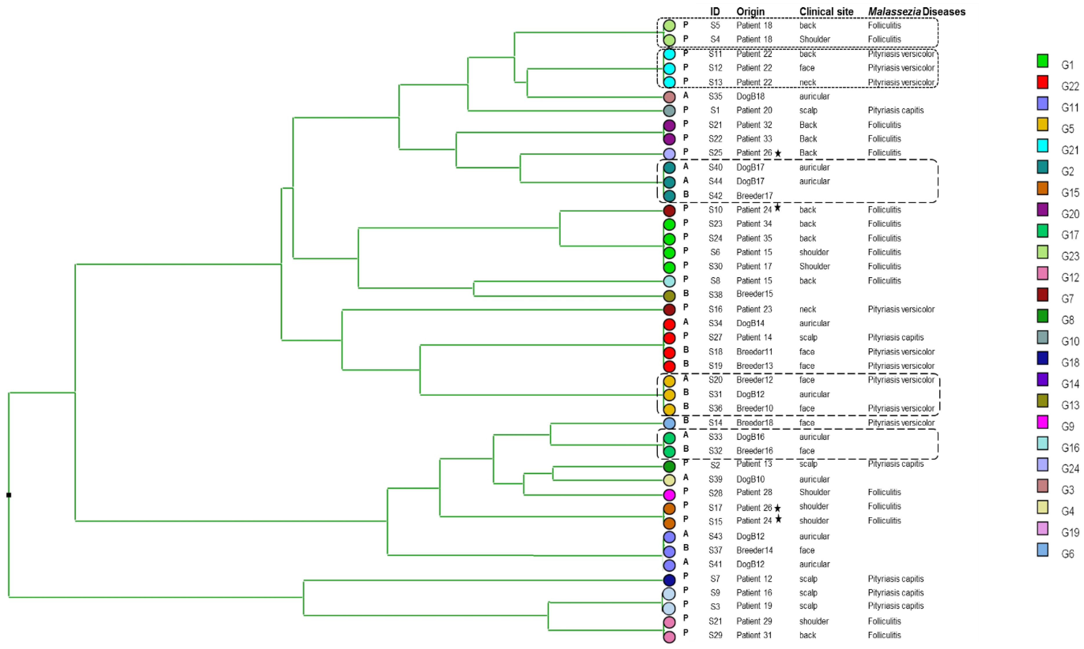
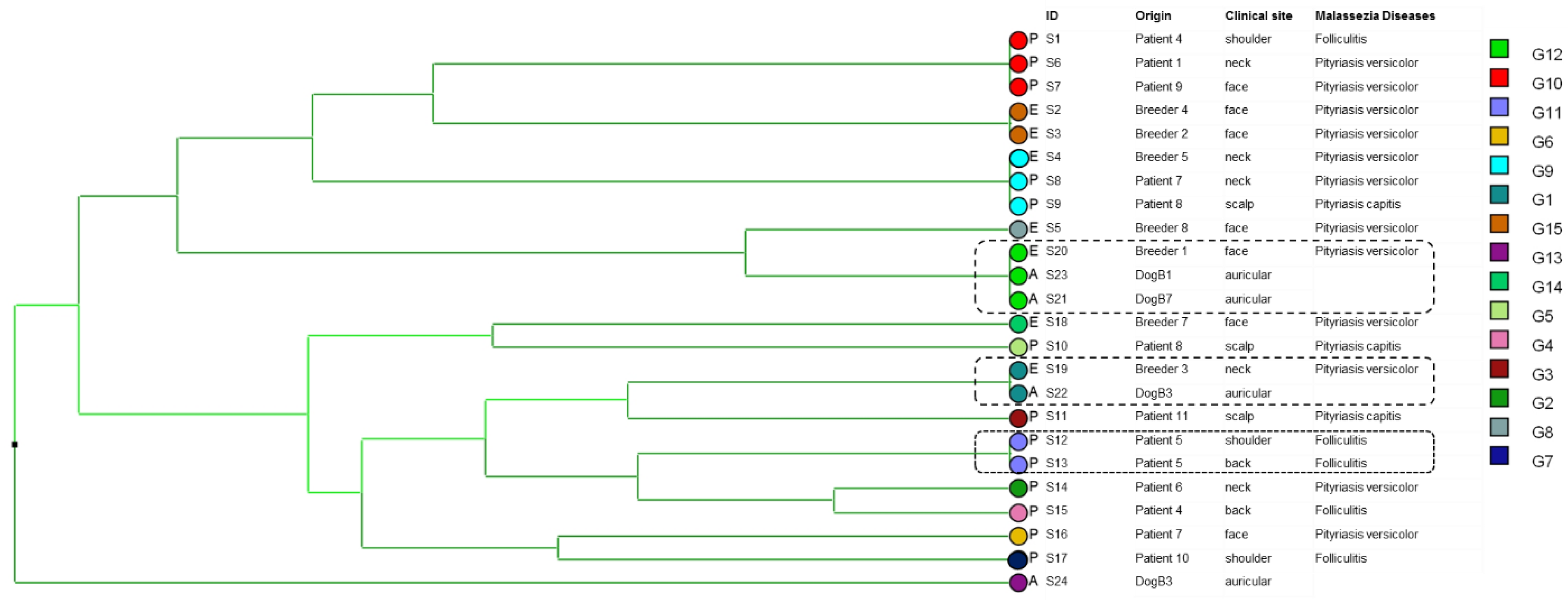
3.1. Genetic Diversity

Upon the analysis of 44 M. globosa isolates, 3–6 distinct alleles were detected for each microsatellite marker. The highest discriminatory power for a single locus was obtained with the STR-MG1 marker, which had six distinct alleles and a D value of 0.829 (Table 1). The highest discriminatory power for a single minisatellite locus was obtained with the STR-MG4 marker, which had six distinct alleles and a D value of 0.807 (Table 1). The combination of three minisatellite markers yielded 15 different genotypes with a 0.886 D value.
The STR-MG6 marker exhibited the lowest diversity index (0.581) for M. restricta (Table 1). The highest discriminating power for a single microsatellite marker was obtained with the STR-MR2 marker, which had six distinct alleles and a D value of 0.818. The STR-MR6 marker exhibited the lowest diversity index (0.528). The STR-MR4 minisatellite marker (0.775) presented the highest discriminating power for M. restricta (Table 1).
After the analysis of multiple loci, 24 genotypes were noted among 44 isolates in M. globosa, with a discrimination index D of 0.943 and 15 genotypes were noted among 24 isolates in M. restricta, with a discrimination index D of 0.967.

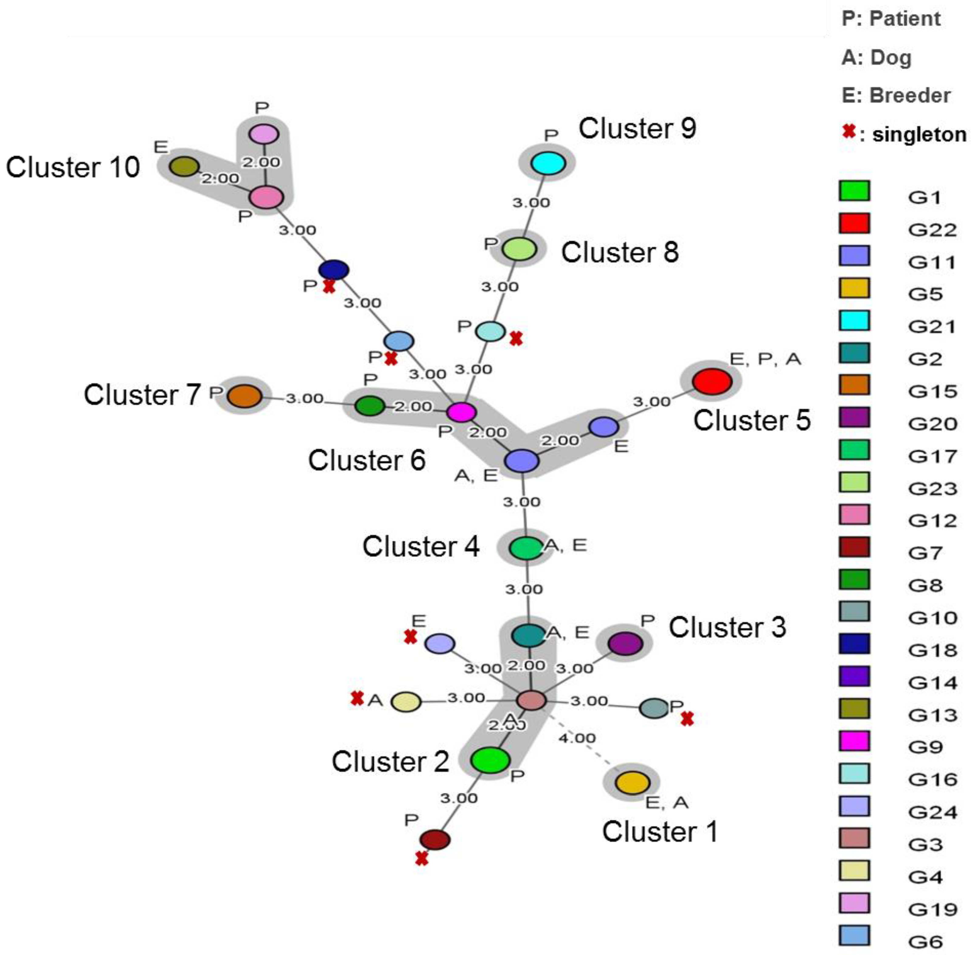
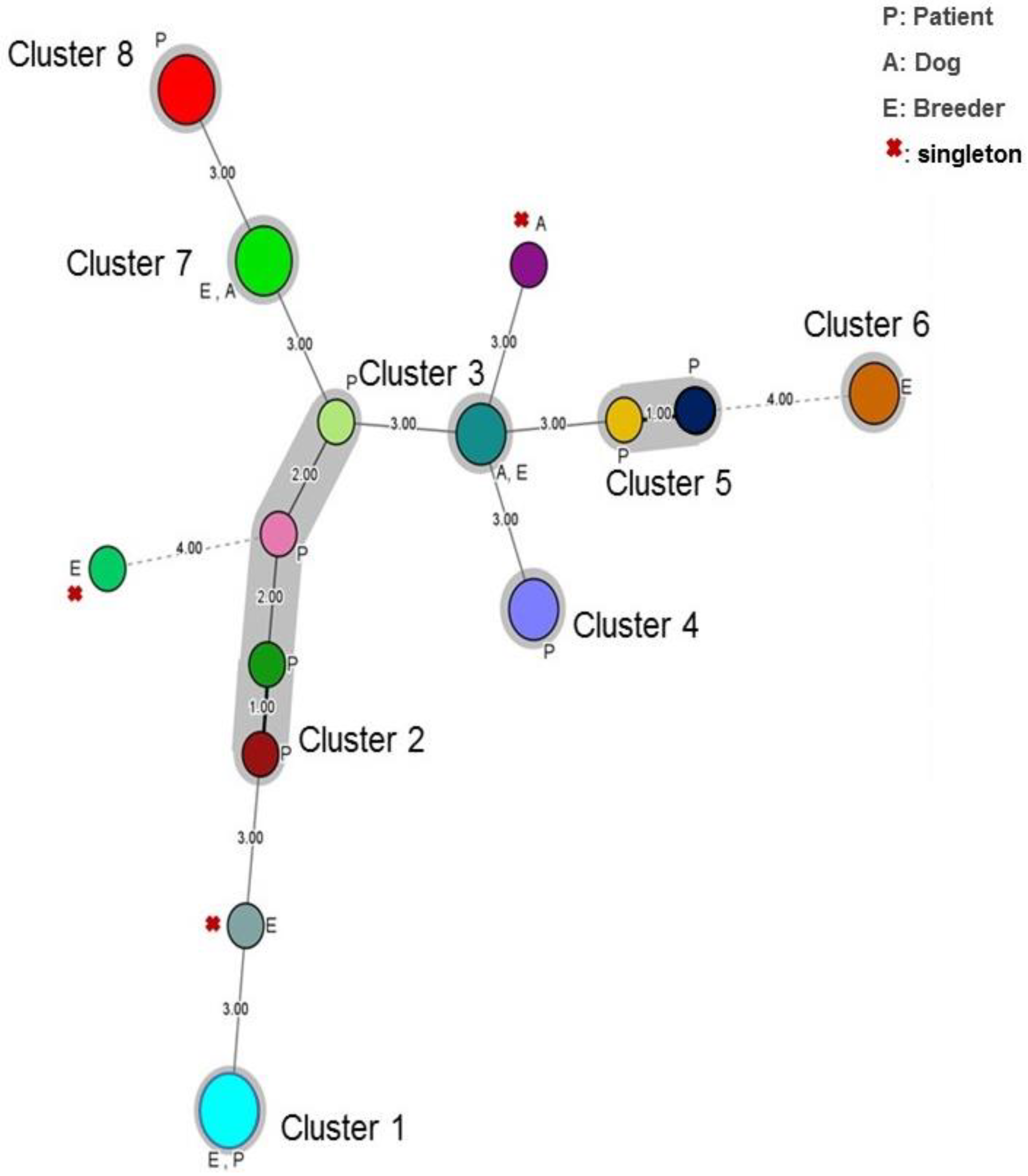
3.2. Cluster Analysis

The dendrogram generated after the analysis of six markers for 44 isolates of M. globosa revealed genetic heterogeneity with 24 distinct genotypes. For instance, all four isolates sampled from patient 24 (P24) and patient 26 (P26) were genetically unrelated. However, we were able to identify a clonal cluster that included isolates collected from breeders and their dogs (S40, S44, S42/S20, S31, S36/S33, S32), indicating a possible co-transmission. We also noted that isolates from different patients share the same genotype (S23, S24, S6, S30/S17, S15/S9, S3/S21, S22) (Figure 1).
Typing study by the analysis of six new markers of M. restricta showed a great genotypic diversity of the strains, which can cause various skin pathologies. However, patient five (P5) was colonized with strains having the same genotype collected from different sampling sites (back, shoulder) and two breeders shared the same genotype of their dogs (Figure 2).
The calculated genetic diversity per locus (FST) values for the three populations of M. globosa strains ranged from 0.018 to 0.057, indicating weak differentiation between them (Table 2). These results suggest a dominant clonal mode of reproduction in M. globosa.

3.3. Interpopulation Genetic Analysis

An MST analysis of the 24 distinct multilocus microsatellite genotypes was performed to illustrate the genetic structure of our M. globosa population (Figure 3).
Genetic cluster analyses suggested that our M. globosa population was formed by ten genetic clusters; three different and distant clusters indicate differences in three markers. Cluster three, cluster seven, cluster eight and cluster nine contain exclusively strains from the patient’s isolates. Cluster one and cluster four contains isolates col-lected from breeders and dogs. Cluster two, cluster five and cluster six comprised M. globosa genotypes isolated in breeders, dogs, and patients. Cluster ten comprised M. globosa isolates collected from breeders and patients. Nine unique genotypes formed singletons (Figure 3).
The minimum spanning tree (MStree) generated for the 24 M. restricta isolates showed that eight distant clusters formed our M. restricta population. These clusters differed by three or four markers (Figure 4). The first cluster contains strains collected from the breeder and patients. The second cluster is dominated by strains collected from patients with different Malassezia diseases. Clusters four, five and eight contain M. restricta patient isolates. For cluster six, it is composed of M. restricta breeders iso-lates. Finally, cluster seven and cluster three contain strains isolated from the breeder and dog (Figure 4).
Three unique genotypes formed singletons. They were isolated from breeders or dogs (Figure 4).
The Chi-square test results of the covariates’ effect on the cluster distribution are presented in Table 3. The isolation site was not significantly associated with genotype clusters (p > 0.05). However, we noted a significative association of singleton genotype with strains isolated from patients with pityriasis capitis (p > 0.01).
Finally, the genotype clusters were significantly associated (p < 0.05) with the year when they were collected.

4. Discussion

Despite the recent increase in studies focusing on Malassezia sp., little is still known concerning Malassezia genetic diversity. Molecular typing of Malassezia strains and the elucidation of their genetic relationships are essential to address important epidemiological issues such as tracking the transmission of infection and strain specificity for a specific disease manifestation.
Few molecular methods have been used for typing Malassezia strains, such as RFLP and multigene sequence analysis [28,29,30]. In previous studies, various molecular techniques found genetic variability within M. globosa, M. restricta, M. furfur [31] and M. pachydermatis [32]. Different Malassezia genotypes were identified as strictly related to the host, geographical origin and/or clinical manifestations [31,33].
However, the major problem with such techniques is the poor interlaboratory reproducibility and difficulty in exchanging the results obtained by these techniques. To our knowledge, the level of discrimination between strains obtained with the microsatellite method has never been reached previously. Our study represents the first report on the development of stable and discriminatory microsatellite markers for typing M. restricta and M. globosa.
The microsatellite markers are characterized by a high level of polymorphism, discrimination, reproducibility and feasibility. Moreover, microsatellite analysis may be more able than other genotyping methods to distinguish between strains with low degrees of sequence variation.
The reproducibility and specificity of our markers were found to be 100%. Microsatellite analysis requires small amounts of DNA (~one ng per reaction) compared to other typing methods. Moreover, a large number of isolates may be typed by using multiplex assays with primers labeled with different fluorescent dyes, resulting in higher throughput.
Microsatellite polymorphism analysis revealed a heterogeneous M. globosa and M. restricta genetic populations. After multiple loci analysis, 24 genotypes were noted among 44 isolates in M. globosa, with a discrimination index D of 0.943 and 15 genotypes were noted among 24 isolates in M. restricta, with a discrimination index D of 0.967. Therefore, the new VNTR markers proposed in the present study have shown promising results in terms of the identification of polymorphism of Malassezia strains. Further analyses can clarify the role of this yeast in disease development and give us clues about the sexual reproduction that might provide genetic variation in this genus [34].
Hiruma et al. [35] found that specific genotypic strains of M. globosa and M. restricta predominated in patients with dandruff.
Czyzenwska et al. identified 17 distinct genotypes of M. pachydermatis with 59 polymorphic sites by sequencing analysis; however, putatively virulent strains were more closely related, indicating a probable correlation between the genotype and the virulence potential [32].
In our study, we found, by cluster analysis of M. globosa species, a similarity between strains collected from the different clinical sites of the same patient (patient 22, patient 18). This revealed that it is an endogenous infection, also called autoinfection; the patient becomes infected with his strains.
However, clonal infection was also observed in different patients (P17, P28, P34 and P35) sharing the same genotype.
Instead, we found a diversity of Malassezia genotypes from isolates collected from the different clinical sites. Multiple genotypes of a species can colonize the same patient [33,36,37], but some genetic types might be linked to a particular body site or pathology, thus indicating an affiliation of Malassezia genotypes with host, geographical origin and/or clinical manifestations [38,39].
For S21 and S23, two dogs are colonized by M. restricta strains which share the same genotype. This could be explained by horizontal transmission of strains with direct contact between individuals but also by exposure to common susceptibility factors associated with fungal colonization of the skin.
Clinical isolate and animal isolate have the same genotype, suggesting that Malassezia infection was acquired from the dog to the breeder. This highlights an exogenous infection which is a cross-infection transmitted between the breeder and the dog which can occur through direct contact. The animal-to-human or human-to-animal carriage is still not well known and its risk factors must be determined. Microsatellite analyses have been efficient epidemiological instruments to make clear the origins of Malassezia infections.
Intra-species variations in DNA pattern of Malassezia isolates and the presence of specific genetic types in cattle, dogs or humans were observed in others studies. Genetic heterogeneity of these yeast in veterinary and human medicine studies is given considering a possible transmission animal to human or human to animal [12]. Phylogenetic analysis would facilitate monitoring of Malassezia spp. carriage in domestic animals and in humans.
Cluster analysis of the 24 M. restricta isolates also showed a genotypic diversity, particularly in strains that can cause various skin pathologies. Soares et al. [40] founded 27 Malassezia subtypes using RFLP and sequencing, mostly from M. restricta (13 subtypes), indicating variation among species and also intra-specific polymorphisms. Subtypes of a same species occur in different proportions depending on the sample, suggesting that each sample has its own subtypes community. Phylogenetic analysis revealed a large variety of Malassezia subtypes, indicating intra-specific diversity.
The calculated genetic diversity per locus (FST) values for the three populations of M. globosa strains ranged from 0.018 to 0.057, indicating weak differentiation between them. This result was supported by the MStree, showing that multilocus microsatellite genotypes contains isolates collected from breeders and patients, corresponding to human isolates were not separated from animal (dogs) ones. A dominant clonal mode of reproduction in M. globosa could be implicated. But also, genetic population structure analysis of human (patients and breeders) and animal Malassezia isolates carried out in a restricted geographic area could be suggested.

5. Conclusions

Malassezia genus have been widely studied in recent years due to their association with skin diseases. Different aspects have been analyzed, such as the specific fungal roles on the pathogenic process and the host immune response. The genomic sequences of M. globosa and M. sympodialis have been completely determined and the partial sequence of M. restricta is also available. Despite advances, little is still known concerning Malassezia genetic diversity. In this study, we describe the successful development of microsatellite PCR for genotyping M. globosa and M. restricta isolates from different sources and the subsequent application of this method to an extended number of other Malassezia species of clinical interest. Moreover, this method is a valuable tool for epidemiological studies, for the kinetics knowledge of the colonization-to-infection process and for the study of genotype’s antifungal susceptibility.

Author Contributions

Conceptualization, I.H. and S.N.; methodology, I.H. and N.K.; software, I.H.; validation, I.H., N.K. and A.I.; formal analysis, I.H. and S.N.; investigation, H.T. and H.S.; resources, H.T. and H.S.; data curation, I.H., N.K., H.T., H.S. and F.M.; writing—original draft preparation, I.H.; writing—review and editing, I.H.; visualization, S.N.; supervision, S.N.; project administration, F.M. and A.A. All authors have read and agreed to the published version of the manuscript.

Funding

Our study was supported by the Ministry of Education and scientific research and the Ministry of Health.

Institutional Review Board Statement

Not applicable.

Informed Consent Statement

Not applicable.

Data Availability Statement

All data are available within the manuscript.

Acknowledgments

We are grateful to Maher Kharrat for the genotyping work using the ABI 3500 Genetic Analyzer (platforme de recherche, Faculté de Médecine de Tunis).

Conflicts of Interest

The authors declare no conflict of interest.

References

  1. Sugita, T.; Nishikawa, A. Molecular and quantitative analyses of Malassezia microflora on the skin of atopic dermatitis patients and genotyping of M. globosa DNA. Nihon Ishinkin Gakkai Zasshi 2003, 44, 61–64. [Google Scholar] [CrossRef] [PubMed]
  2. Sugita, T.; Takashima, M.; Kodama, M.; Tsuboi, R.; Nishikawa, A. Description of a new yeast species, Malassezia japonica, and its detection in patients with atopic dermatitis and healthy subjects. J. Clin. Microbiol. 2003, 41, 4695–4699. [Google Scholar] [CrossRef] [PubMed] [Green Version]
  3. Theelen, B.; Cafarchia, C.; Gaitanis, G.; Bassukas, I.D.; Boekhout, T.; Dawson, T.L. Corrigendum: Malassezia ecology, pathophysiology, and treatment. Med. Mycol. 2019, 57, e2. [Google Scholar] [CrossRef] [PubMed] [Green Version]
  4. Prohic, A.; Jovovic Sadikovic, T.; Kuskunovic-Vlahovljak, S.; Baljic, R. Distribution of Malassezia Species in Patients with Different Dermatological Disorders and Healthy Individuals. Acta Dermatovenerol. Croat. 2016, 24, 274–281. [Google Scholar]
  5. Gupta, A.K.; Batra, R.; Bluhm, R.; Boekhout, T.; Dawson, T.L., Jr. Skin diseases associated with Malassezia species. J. Am. Acad. Dermatol. 2004, 51, 785–798. [Google Scholar] [CrossRef]
  6. Gupta, A.K.; Madzia, S.E.; Batra, R. Etiology and management of Seborrheic dermatitis. Dermatology 2004, 208, 89–93. [Google Scholar] [CrossRef]
  7. Gupta, A.K.; Bluhm, R. Seborrheic dermatitis. J. Eur. Acad. Dermatol. Venereol. 2004, 18, 13–26; quiz 19–20. [Google Scholar] [CrossRef]
  8. Paulino, L.C.; Tseng, C.H.; Blaser, M.J. Analysis of Malassezia microbiota in healthy superficial human skin and in psoriatic lesions by multiplex real-time PCR. FEMS Yeast Res. 2008, 8, 460–471. [Google Scholar] [CrossRef] [Green Version]
  9. Yim, S.M.; Kim, J.Y.; Ko, J.H.; Lee, Y.W.; Choe, Y.B.; Ahn, K.J. Molecular analysis of Malassezia microflora on the skin of the patients with atopic dermatitis. Ann. Dermatol. 2010, 22, 41–47. [Google Scholar] [CrossRef] [Green Version]
  10. Mirhendi, H.; Makimura, K.; Zomorodian, K.; Yamada, T.; Sugita, T.; Yamaguchi, H. A simple PCR-RFLP method for identification and differentiation of 11 Malassezia species. J. Microbiol. Methods 2005, 61, 281–284. [Google Scholar] [CrossRef]
  11. Gupta, A.K.; Boekhout, T.; Theelen, B.; Summerbell, R.; Batra, R. Identification and typing of Malassezia species by amplified fragment length polymorphism and sequence analyses of the internal transcribed spacer and large-subunit regions of ribosomal DNA. J. Clin. Microbiol. 2004, 42, 4253–4260. [Google Scholar] [CrossRef] [PubMed]
  12. Duarte, E.R.; Hamdan, J.S. RAPD differentiation of Malassezia spp. from cattle, dogs and humans. Mycoses 2010, 53, 48–56. [Google Scholar] [CrossRef] [PubMed]
  13. Akaza, N.; Akamatsu, H.; Sasaki, Y.; Takeoka, S.; Kishi, M.; Mizutani, H.; Sano, A.; Hirokawa, K.; Nakata, S.; Matsunaga, K. Cutaneous Malassezia microbiota in atopic dermatitis patients differ by gender and body part. Dermatology 2010, 221, 253–260. [Google Scholar] [CrossRef] [PubMed]
  14. Ilahi, A.; Hadrich, I.; Neji, S.; Trabelsi, H.; Makni, F.; Ayadi, A. Real-Time PCR Identification of Six Malassezia Species. Curr. Microbiol. 2017, 74, 671–677. [Google Scholar] [CrossRef]
  15. Vergnaud, G.; Denoeud, F. Minisatellites: Mutability and genome architecture. Genome Res. 2000, 10, 899–907. [Google Scholar] [CrossRef] [Green Version]
  16. Schouls, L.M.; van der Heide, H.G.; Vauterin, L.; Vauterin, P.; Mooi, F.R. Multiple-locus variable-number tandem repeat analysis of Dutch Bordetella pertussis strains reveals rapid genetic changes with clonal expansion during the late 1990s. J. Bacteriol. 2004, 186, 5496–5505. [Google Scholar] [CrossRef] [Green Version]
  17. Top, J.; Schouls, L.M.; Bonten, M.J.; Willems, R.J. Multiple-locus variable-number tandem repeat analysis, a novel typing scheme to study the genetic relatedness and epidemiology of Enterococcus faecium isolates. J. Clin. Microbiol. 2004, 42, 4503–4511. [Google Scholar] [CrossRef] [Green Version]
  18. Wang, Y.W.; Watanabe, H.; Phung, D.C.; Tung, S.K.; Lee, Y.S.; Terajima, J.; Liang, S.Y.; Chiou, C.S. Multilocus variable-number tandem repeat analysis for molecular typing and phylogenetic analysis of Shigella flexneri. BMC Microbiol. 2009, 9, 278. [Google Scholar] [CrossRef] [Green Version]
  19. Thierry, S.; Wang, D.; Arne, P.; Deville, M.; De Bruin, B.; Nieguitsila, A.; Pourcel, C.; Laroucau, K.; Chermette, R.; Huang, W.; et al. Multiple-locus variable-number tandem repeat analysis for molecular typing of Aspergillus fumigatus. BMC Microbiol. 2010, 10, 315. [Google Scholar] [CrossRef] [Green Version]
  20. Wang, D.Y.; Hadj-Henni, L.; Thierry, S.; Arne, P.; Chermette, R.; Botterel, F.; Hadrich, I.; Makni, F.; Ayadi, A.; Ranque, S.; et al. Simple and highly discriminatory VNTR-based multiplex PCR for tracing sources of Aspergillus flavus isolates. PLoS ONE 2012, 7, e44204. [Google Scholar] [CrossRef]
  21. Hadrich, I.; Neji, S.; Drira, I.; Trabelsi, H.; Mahfoud, N.; Ranque, S.; Makni, F.; Ayadi, A. Microsatellite typing of Aspergillus flavus in patients with various clinical presentations of aspergillosis. Med. Mycol. 2013, 51, 586–591. [Google Scholar] [CrossRef] [PubMed]
  22. Drira, I.; Hadrich, I.; Neji, S.; Mahfouth, N.; Trabelsi, H.; Sellami, H.; Makni, F.; Ayadi, A. Highly discriminatory variable-number tandem-repeat markers for genotyping of Trichophyton interdigitale strains. J. Clin. Microbiol. 2014, 52, 3290–3296. [Google Scholar] [CrossRef] [PubMed] [Green Version]
  23. Neji, S.; Hadrich, I.; Ilahi, A.; Trabelsi, H.; Chelly, H.; Mahfoudh, N.; Cheikhrouhou, F.; Sellami, H.; Makni, F.; Ayadi, A. Molecular Genotyping of Candida parapsilosis Species Complex. Mycopathologia 2018, 183, 765–775. [Google Scholar] [CrossRef]
  24. Rousset, F. genepop’007: A complete re-implementation of the genepop software for Windows and Linux. Mol. Ecol. Resour. 2008, 8, 103–106. [Google Scholar] [CrossRef] [PubMed]
  25. Hunter, P.R.; Gaston, M.A. Numerical index of the discriminatory ability of typing systems: An application of Simpson’s index of diversity. J. Clin. Microbiol. 1988, 26, 2465–2466. [Google Scholar] [CrossRef] [PubMed] [Green Version]
  26. Goudet, J. FSTAT Version 1.2: Un programme informatique pour calculer la statistique F. J. Hérédité 1995, 86, 485–486. [Google Scholar]
  27. Polzin, T.; Daneschmand, S.V. On Steiner trees and minimum spanning trees in hypergraphs. Oper. Res. Lett. 2003, 31, 12–20. [Google Scholar] [CrossRef]
  28. Dolatabadi, S.; de Hoog, G.S.; Meis, J.F.; Walther, G. Species boundaries and nomenclature of Rhizopus arrhizus (syn. R. oryzae). Mycoses 2014, 57 (Suppl. 3), 108–127. [Google Scholar] [CrossRef]
  29. Nyilasi, I.; Papp, T.; Csernetics, A.; Krizsan, K.; Nagy, E.; Vagvolgyi, C. High-affinity iron permease (FTR1) gene sequence-based molecular identification of clinically important Zygomycetes. Clin. Microbiol. Infect. 2008, 14, 393–397. [Google Scholar] [CrossRef] [Green Version]
  30. Puig, L.; Bragulat, M.R.; Castella, G.; Cabanes, F.J. Phenotypic and genetic diversity of Malassezia furfur from domestic and zoo animals. Med. Mycol. 2018, 56, 941–949. [Google Scholar] [CrossRef]
  31. Theelen, B.; Cafarchia, C.; Gaitanis, G.; Bassukas, I.D.; Boekhout, T.; Dawson, T.L., Jr. Malassezia ecology, pathophysiology, and treatment. Med. Mycol. 2018, 56, S10–S25. [Google Scholar] [CrossRef] [PubMed] [Green Version]
  32. Czyzewska, U.; Bartoszewicz, M.; Siemieniuk, M.; Tylicki, A. Genetic relationships and population structure of Malassezia pachydermatis strains isolated from dogs with otitis externa and healthy dogs. Mycologia 2018, 110, 666–676. [Google Scholar] [CrossRef]
  33. Cafarchia, C.; Gasser, R.B.; Latrofa, M.S.; Parisi, A.; Campbell, B.E.; Otranto, D. Genetic variants of Malassezia pachydermatis from canine skin: Body distribution and phospholipase activity. FEMS Yeast Res. 2008, 8, 451–459. [Google Scholar] [CrossRef] [PubMed] [Green Version]
  34. Celis, A.M.; Vos, A.M.; Triana, S.; Medina, C.A.; Escobar, N.; Restrepo, S.; Wosten, H.A.; de Cock, H. Highly efficient transformation system for Malassezia furfur and Malassezia pachydermatis using Agrobacterium tumefaciens-mediated transformation. J. Microbiol. Methods 2017, 134, 1–6. [Google Scholar] [CrossRef] [PubMed]
  35. Hiruma, M.; Cho, O.; Hiruma, M.; Kurakado, S.; Sugita, T.; Ikeda, S. Genotype analyses of human commensal scalp fungi, Malassezia globosa, and Malassezia restricta on the scalps of patients with dandruff and healthy subjects. Mycopathologia 2014, 177, 263–269. [Google Scholar] [CrossRef] [PubMed]
  36. Machado, M.L.; Cafarchia, C.; Otranto, D.; Ferreira, R.R.; Bianchi, S.P.; Latrofa, M.S.; Parisi, A.; Ferreiro, L. Genetic variability and phospholipase production of Malassezia pachydermatis isolated from dogs with diverse grades of skin lesions. Med. Mycol. 2010, 48, 889–892. [Google Scholar] [CrossRef] [PubMed] [Green Version]
  37. Ilahi, A.; Hadrich, I.; Goudjil, S.; Kongolo, G.; Chazal, C.; Leke, A.; Ayadi, A.; Chouaki, T.; Ranque, S. Molecular epidemiology of a Malassezia pachydermatis neonatal unit outbreak. Med. Mycol. 2018, 56, 69–77. [Google Scholar] [CrossRef]
  38. Cafarchia, C.; Latrofa, M.S.; Figueredo, L.A.; da Silva Machado, M.L.; Ferreiro, L.; Guillot, J.; Boekhout, T.; Otranto, D. Physiological and molecular characterization of atypical lipid-dependent Malassezia yeasts from a dog with skin lesions: Adaptation to a new host? Med. Mycol. 2011, 49, 365–374. [Google Scholar] [CrossRef] [Green Version]
  39. Cafarchia, C.; Gasser, R.B.; Figueredo, L.A.; Latrofa, M.S.; Otranto, D. Advances in the identification of Malassezia. Mol. Cell. Probes 2011, 25, 1–7. [Google Scholar] [CrossRef]
  40. Soares, R.C.; Zani, M.B.; Arruda, A.C.; Arruda, L.H.; Paulino, L.C. Malassezia intra-specific diversity and potentially new species in the skin microbiota from Brazilian healthy subjects and seborrheic dermatitis patients. PLoS ONE 2015, 10, e0117921. [Google Scholar] [CrossRef]
Figure 1. UPGMA dendrogram upon analysis of six VNTR markers obtained from 44 Malassezia globosa isolates. P: patient, A: animal and B: breeder. The highlighted isolates illustrate two epidemiological scenarios (S1 and S2): (S1) for P18 and P22 (square with dotted-line border), isolates collected from different clinical sites share the same genotype, indicating exposure to a clonal Malassezia population. For isolates highlighted by a square with a dashed-line border collected from breeders and their dogs present the same genotype indicating an animal–human co-transmission. (S2) P24 and P26 (star): each isolate has a distinct genotype, indicating exposure to a genetically highly heterogeneous clinical Malassezia population.
Jof 09 00263 g001
Figure 2. UPGMA dendrogram upon analysis of six VNTR markers obtained from 24 Malassezia restricta isolates. P: patient, A: animal and E: breeder. The highlighted isolates illustrate: for P5 (square with dotted-line border), isolates collected from the different clinical sites from the same patient share the same genotype. For isolates highlighted by a square with a dashed-line border collected from breeders and their dogs, present the same genotype indicating an animal–human co-transmission.
Jof 09 00263 g002
Figure 3. Minimum spanning tree showing the genotypic diversity of Malassezia globosa isolates (N = 44) based on VNTR data. Each circle shows a unique genotype and its size, the number of strains belonging to the same genotype. Connecting lines and numbers between circles show the similarity between genotypes: (1.00) indicates only one marker difference, (2.00) indicates a difference in two markers and (3.00) the difference in three markers.
Jof 09 00263 g003
Figure 4. Minimum spanning tree showing the genotypic diversity of Malassezia restricta isolates (N = 24) based on VNTR data. Each circle shows a unique genotype and its size, the number of strains belonging to the same genotype. Connecting lines and numbers between circles show the similarity between genotypes: (1.00) indicates only one marker difference, (2.00) indicates a difference in two markers and (3.00) a difference in three markers.
Jof 09 00263 g004
Table 1. Features of the polymorphic VNTR of M. globosa and M. restricta upon analysis of 68 isolates.
SpeciesMarker NamePrimer Sequences (5′ to 3′)DNA Motifs (bp)Number of AllelesFragment Size (bp)Simpson Diversity Index DMarker Location
M. globosaSTR-MG1F: FAM-CACGCTAAACTGCTGGATGA26242-256-270-286-296-3020.829Ch9
R: CGAATATGAACCCCATGGAC
STR-MG2F: HEX-CGGTAGTACGATTGGCCCTA25198-228-232-272-2820.741Ch3
R: CCACATCATACGAGCCACAC
STR-MG3F: TET-GGATGAAGAGGCTCGCTATG35168-177-189-213-2340.722Ch3
R: ACAGGCGTAGAAGCCAAAGA
STR-MG4F: GGCGGTGGTAAGCACTCTGG666250-300-350-400-450-5000.807Ch1
R: ATGGGTGGTCACCACATGG
STR-MG5F: AGGGGCTTCACGGTCATGTG864300-400-500-7000.649Ch3
R: CATTGGAGCCTGGTGTGACG
STR-MG6F: AACCAACGACGTCCAGAAAC1763300-450-7000.581Ch5
R: GTTAAGCTCGCTTGCTCGAAT
M. restrictaSTR-MR1F: FAM-TAGCACTATCCCAACGTACC25242-250-254-258-2860.774Ch7
R: GTTCCGTGTGTCATGATTGC
STR-MR2F: HEX-CGCAGAAAACCTAGAGACG35258-264-288-297-3360.818Ch3
R: CTCGTGCGTGTGAGTATTGG
STR-MR3F: TET-GCCTTGGATGCACTGGTATT34278-314-338-3410.642Ch2
R: CACGGCGTCAGGAACAAGAG
STR-MR4F: ACGGGTTCGAACGGTGAG665250-350-450-500-6000.775Ch4
R: ATGATTGCTTGCGTTGACTG
STR-MR5F: TATGGGTGCTGCCAGAGTCG1623300-600-7500.549Ch4
R: GTCGAAGGAGATTCACGGCG
STR-MR6F: CCCACCACCAACTAACAACA2283250-500-7500.521Ch5
R: AAACACGGACCACACAACAC
Table 2. M. globosa Fst values comparing patients, breeders and animals genotypes.
Pop EPop A
Pop P0.0180.045
Pop E 0.057
Pop P: population of patients; Pop E: population of breeders; Pop A: population of animals (Dogs).
Table 3. Association of covariates with the ten Malassezia genetic clusters identified by minimum spanning tree (MStree) analysis.
Cluster (%)IIIIIIIVVVIVIIVIIIIXXSingletonp
Patient-15.387.69-3.843/267.697.6911.537.6926.92>0.05
Breeder11.1111.11--11.1133.33---11.1111.110.16
Dog22.2233.33-11.1111.1111.11----11.110.16
Global Site effect >0.05
Malassezia folliculitis 2512.5--6.2512.512.5-12.518.75-
Pityriasis versicolor ---22.2222.22--33.3311.1111.11-
Pityriasis capitis ---2020----600.01
Healthy human25--25-25----25-
Global Year effect <0.05
201613.6318.18-9.0918.8813.63---9.0918.88-
2017-----40-40--20-
2018-23.5211.67--5.8811.67-17.645.8823.52-
Disclaimer/Publisher’s Note: The statements, opinions and data contained in all publications are solely those of the individual author(s) and contributor(s) and not of MDPI and/or the editor(s). MDPI and/or the editor(s) disclaim responsibility for any injury to people or property resulting from any ideas, methods, instructions or products referred to in the content.

