Article

Kojic Acid Gene Clusters and the Transcriptional Activation Mechanism of Aspergillus flavus KojR on Expression of Clustered Genes

1
Southern Regional Research Center, Agricultural Research Service, U. S. Department of Agriculture, 1100 Allen Toussaint Boulevard, New Orleans, LA 70124, USA
2
Western Regional Research Center, Agricultural Research Service, U. S. Department of Agriculture, 800 Buchanan Street, Albany, CA 94710, USA
3
School of Food Science and Engineering, Ocean University of China, Qingdao 266003, China
*
Author to whom correspondence should be addressed.
J. Fungi 2023, 9(2), 259; https://doi.org/10.3390/jof9020259
Submission received: 2 December 2022 / Revised: 8 February 2023 / Accepted: 11 February 2023 / Published: 15 February 2023
(This article belongs to the Special Issue Genomics Analysis of Fungi)

Abstract

:
Kojic acid (KA) is a fungal metabolite and has a variety of applications in the cosmetics and food industries. Aspergillus oryzae is a well-known producer of KA, and its KA biosynthesis gene cluster has been identified. In this study, we showed that nearly all section Flavi aspergilli except for A. avenaceus had complete KA gene clusters, and only one Penicillium species, P. nordicum, contained a partial KA gene cluster. Phylogenetic inference based on KA gene cluster sequences consistently grouped section Flavi aspergilli into clades as prior studies. The Zn(II)2Cys6 zinc cluster regulator KojR transcriptionally activated clustered genes of kojA and kojT in Aspergillus flavus. This was evidenced by the time-course expression of both genes in kojR-overexpressing strains whose kojR expression was driven by a heterologous Aspergillus nidulans gpdA promoter or a homologous A. flavus gpiA promoter. Using sequences from the kojA and kojT promoter regions of section Flavi aspergilli for motif analyses, we identified a consensus KojR-binding motif to be an 11-bp palindromic sequence of 5′-CGRCTWAGYCG-3′ (R = A/G, W = A/T, Y = C/T). A CRISPR/Cas9-mediated gene-targeting technique showed that the motif sequence, 5′-CGACTTTGCCG-3′, in the kojA promoter was critical for KA biosynthesis in A. flavus. Our findings may facilitate strain improvement and benefit future kojic acid production.

1. Introduction

Kojic acid (KA) is a secondary metabolite produced by fungi, including many species of Aspergillus and Penicillium [1]. It was originally isolated by Japanese microbiologist Kendo Saito in 1907 from “koji” starter inoculum, i.e., mycelia of Aspergillus oryzae grown on steamed rice used in oriental food fermentation [2,3]. The chemical structure was later determined to be 5-hydroxy-2-hydroxymethyl-4-pyrone [4]. KA is a well-known inhibitor of tyrosinase [5] and its mode of action is to chelate copper ions required for tyrosinase’s activity during melanin synthesis. Hence, it has long been used as a skin-whitening agent. KA and its derivatives also possess anti-bacterial, anti-fungal, anti-inflammatory, and antineoplastic properties [6]. In the food industry, KA is used as an antioxidant to prevent enzymatic browning and extend the shelf life of fruits and vegetables. Therefore, KA has wide applications in the cosmetics, pharmaceutical, and food industries [7].
KA has been on the market for over sixty-five years. Charles Pfizer and Company, Brooklyn, New York, USA, was the first company to announce its attempt to manufacture this product [8]. Though in the early years, demands for KA were low, in recent years, rapid growth has occurred in various industries, particularly in the cosmetic sector. The global market size of KA for different applications (cosmetics, food additives, and medicine materials) in 2021 was 35 million and is expected to reach 38 million in 2028 [9]. Efforts to increase KA production by fungi include optimization of medium compositions (C, N, and C/N ratios) or culturing conditions (temperature, pH, aeration, fermentation types), or both [10]. Various carbon sources from simple C2-C7 carbohydrates to starch of agricultural staples and agro-industrial by-products can all induce KA formation to different extents [8,11]. Strain development by UV and gamma-ray radiation or mutagen treatments also has been carried out extensively [12,13].
Despite being a secondary metabolite with an all-carbon backbone, KA is quite different from those in the polyketide category. Forming units and precursors for the biosynthesis of polyketides are breakdowns of primary metabolites, such as acetates. 14C-labeled isotope tracer studies have proven that KA is directly synthesized from glucose. A biosynthetic scheme for KA formation, which requires at most two or three enzymes, was then proposed [14]. In a later study, experimental evidence implied the involvement of glucose dehydrogenase and gluconate dehydrogenase in KA biosynthesis. This implication was inferred from a correlation of enzyme activity patterns under various conditions with amounts of KA produced. A biosynthetic pathway based on three alternative routes was thus suggested [15]. However, up until now, the enzymes or intermediates involved have not been conclusively identified. The precise nature of the KA biosynthetic process remains virtually unclear [12]. With the advent of the genomics era, gene function studies have identified three clustered genes, kojA-kojR-kojT, in the A. oryzae genome that are responsible for KA biosynthesis. These genes encode an oxidoreductase, a transcriptional activator, and a transporter, respectively [16,17].
In this study, we surveyed the presence of KA gene clusters in Aspergillus and Penicillium species, examined their gene organization structures, and delineated phylogenetic relationships among Aspergillus section Flavi species based on KA gene cluster sequences. We then explored the transactivation mechanism of the Aspergillus flavus KojR regulator on the expression of kojA and kojT using a heterologous gpdA promoter and a homologous gpiA promoter to drive kojR expression. Lastly, we identified a consensus KojR-binding motif in the promoter regions of section Flavi aspergilli to be an 11-bp palindromic sequence of 5′-CGRCTWAGYCG-3′ (R = A/G, W = A/T, Y = C/T). Using a CRISPR/Cas9-mediated gene-targeting approach, we showed that the motif sequence, 5′-CGACTTTGCCG-3′, in the A. flavus kojA promoter was critical for KA biosynthesis.

2. Materials and Methods

2.1. Media and Culture Conditions

For spore production, A. flavus was grown on V8 agar medium, which contains 5% V8 vegetable juice (Campbell Soup Company, Camden, NJ, USA) and 2% agar, pH 5.2. Kojic acid medium (KAM) for KA production [16] was modified slightly to contain 0.05% yeast extract, 0.1% K2HPO4·3H2O, 0.05% MgSO4·7H2O, and 2% glucose (pH 6.0). A potato dextrose agar medium (Difco, Detroit, MI, USA) supplemented with 1 mM ferric ion also was used for the examination of KA production. All fungal cultures were grown at 30 °C in the dark.

2.2. Identification of KA Gene Clusters in Aspergillus Species

The genome sequence of A. flavus NRRL3357 was retrieved from the National Center for Biotechnology Information (NCBI) genome database (https://www.ncbi.nlm.nih.gov/genome/). Analyses of the genome sequence was performed via CoGe (Comparative Genomics, https://genomevolution.org/coge/), an online platform for sequence comparison and retrieval. The extracted complete KA gene cluster sequence of NRRL3357, derived from comparisons to A. oryzae KA gene cluster sequences (kojA, kojR, and kojT) (https://www.genome.jp/kegg/), was used as the alignment template for BlastN genome assemblies of other aspergilli available in NCBI (Table 1). Aligned genomic sequences were downloaded for further analysis. The genes of the A. flavus KA gene cluster (kojA, kojR, and kojT), previously known as AFLA_096040, AFLA_096050, and AFLA_096060, are now updated as AFLA_009845, AFLA_009846, and AFLA_009847.

2.3. Phylogenetic Study of Section Flavi Aspergilli

The online system Mauve (http://darlinglab.org/mauve/mauve.html) for multiple genome alignments [18] was used to align the KA gene cluster sequences of twenty-three Aspergillus species (Table 1). Aligning sequences, extracting single nucleotide polymorphisms (SNP), filtering out noise (i.e., gaps and ambiguous bases), concatenating cleaned SNPs, and converting sequences to FASTA format were carried out using a custom JavaScript [19]. Phylogenetic inference with concatenated total SNP sequences was performed using the unweighted distance-matrix UPGMA method of the online program MAFFT (version 7) (https://mafft.cbrc.jp/alignment/server/phylogeny.html) [20].

2.4. Disruption of the kojR Gene in A. flavus

Gene deletion procedures via double-crossover recombination in A. flavus strains deficient in the NHEJ pathway have been described in detail [21]. This well-established protocol is routinely used for gene function studies by many researchers to obtain gene knockout mutants. The two 5′ and 3′ flanking fragments for targeting kojR were amplified by PCR using AccuPrimeTM Pfx Taq polymerase (Invitrogen, Carlsbad, CA, USA) with primers listed in Table S1. The two PCR fragments, after digestion with appropriate restriction enzymes, were sequentially cloned into a pyrG-containing plasmid, pPG28. The resulting deletion vector was linearized with SacI and SphI prior to polyethylene glycol-CaCl2 mediated fungal transformation [21]. The recipient strain, A. flavus SRRC1709 (=CA14PTS∆pyrG), was pyrithiamine-sensitive and uracil auxotrophic. All kojR deletion mutants were confirmed by diagnostic PCR and verified for their inability to produce KA. A deletion mutant, ∆kojR#4, was used for subsequent genetic complementation experiments.

2.5. Construction of kojR Expression Vectors with gpdA or gpiA Promoter

The pTR1-GPD-TRPC vector that contains the A. nidulans glyceraldehyde-3-phosphate dehydrogenase gene (gpdA, AN8041) promoter and trpC terminator and the A. oryzae pyrithiamine resistance gene (ptrA) as the selection marker [22] was modified slightly. Transcriptomic information based on RNA-Seq reads available from now-delisted AspGD (http://www.aspergillusgenome.org/) revealed that only the upstream 1.0 kb (579170 to 580140) is the gpdA promoter region, which is consistent with the previous report [23]. Therefore, the 1.1-kb upstream region in the vector that is not a part of the gpdA promoter but contains the phosphate transporter gene, AN8040, was removed by HindIII and XhoI digestion followed by self-ligation. This derived cloning vector was named pHgpdA-trpC-PTR in this study. A full-length A. flavus genomic kojR coding sequence was PCR amplified with primers kojR-OE and kojR-STOP (Table S1) by AccuPrimeTM Pfx polymerase and cloned into the NotI and SmaI sites of the shortened vector.
A. flavus gpiA (previously named AFLA_113120 and now as AFLA_007250 in FungiDB, https://fungidb.org/fungidb/app/), an orthologue of Saccharomyces cerevisiae ecm33, is a highly expressed gene [24,25]. An analysis of RNA-Seq reads related to this gene indicated that a 1.4-kb region, including the 5′ UTR, contained its promoter. A PCR fragment of the specified region was generated with primers GPI-H and GPI-Not (Table S1) to replace the gpdA promoter of pHgpdA-trpC-PTR to give pHgpiA-trpC-PTR. The vector had a unique NotI site and a unique SmaI site. Lastly, the full-length A. flavus kojR coding sequence was PCR amplified and cloned. The complementation vectors were individually transformed into the ∆kojR#4 mutant. Three KA-producing transformants from the A. nidulans gpdA promoter set (D-8, D-16, and D-20) and the A. flavus gpiA promoter set (I-5, I-9, and I-16) were selected based on visual inspection showing high levels of KA production (Figure S1). Their kojR copy numbers were determined, and they were used in a time-course expression study to examine how the restoration of kojR expression affected the expression of kojA and kojT.

2.6. Determination of KA Production and Fungal Mycelial Dry Weight

Spores of the six kojR-overexpressing strains and a control strain, KuPG, were harvested from five-day-old V8 agar plates, washed to remove debris, and counted on a hemocytometer. They were inoculated into 50 mL of KAM broth in 150 mL Erlenmeyer flasks at a final concentration of 105 per mL. Cultures in triplicate were incubated in a gyroshaker at 30 °C and shaken at 150 rpm for four days. At the end of growth, 10 mL aliquots of culture broth were collected and frozen at −20 °C. Mycelia were spun down, supernatants decanted, and transferred to 15-mL polypropylene tubes for lyophilization. After completion, mycelial dry weights were determined. For HPLC analysis of KA, frozen filtrates were thawed at room temperature, and a 1.5 mL aliquot of each was filtered through a 25 mm 0.45 µm nylon syringe filter. Each filtrate was diluted one to twenty times with water and a 20 µL portion was analyzed using a column of Intersil ODS-3, 5 μ, 4.6 × 250 mm (GL Sciences, Torrance, CA, USA) at a flow rate of 1.0 mL/min. The mobile phase was MeOH:0.1% H3PO4 (25:75), isocratic. The detection of KA was based on UV absorption at 265 nm. A linear standard curve constructed from standard KA concentrations was used to determine the amounts of KA in the filtrate samples.

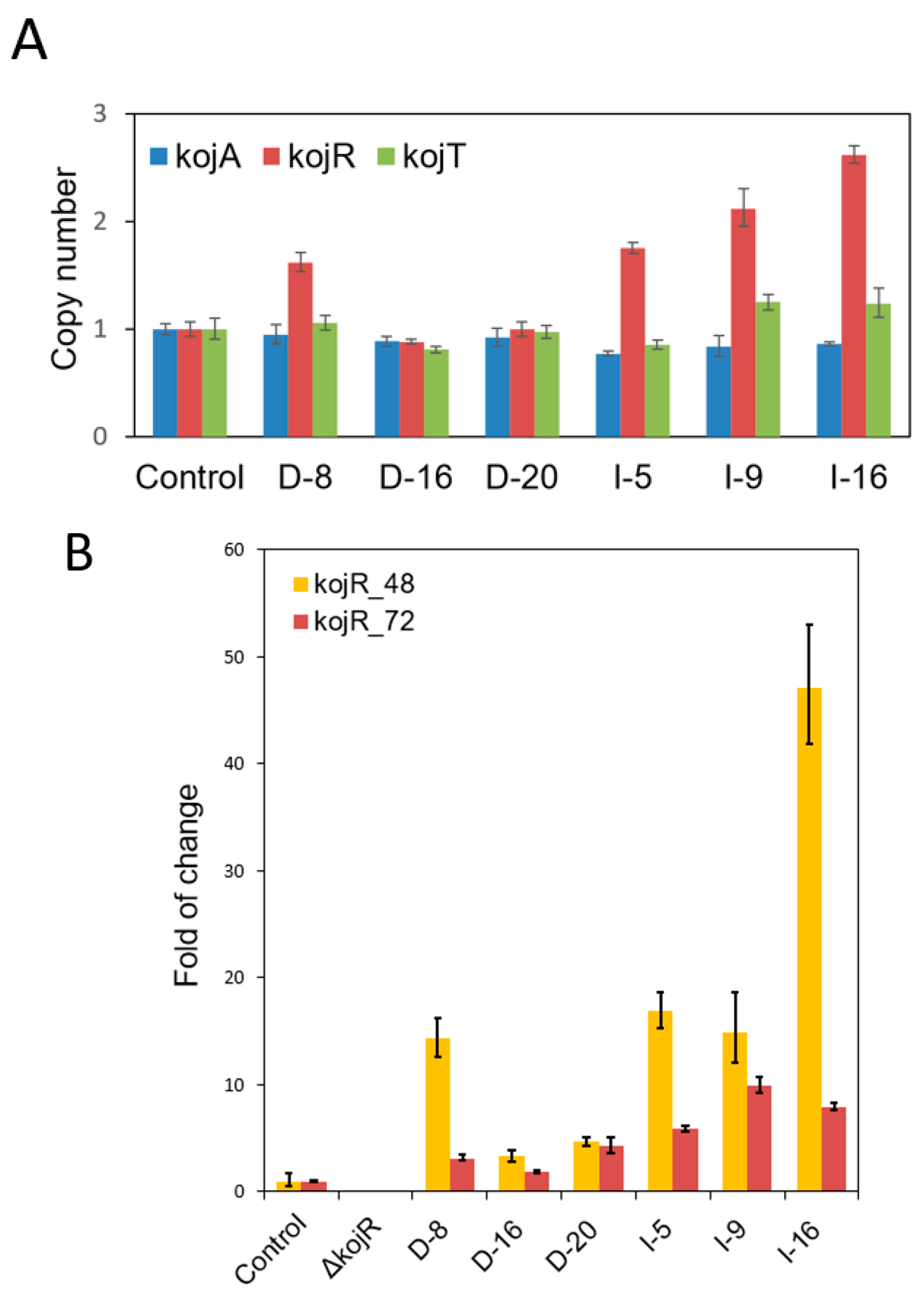
2.7. Determination of kojR Copy Numbers of gpdA and gpiA Promoter-Driven Overexpression Transformants

Spores of 1.0 × 10⁴ of the kojR-overexpressing and the KuPG control strains, prepared from V8 medium plates, were inoculated, respectively, into 1.0 mL PDB in 2-mL microfuge tubes. The cultures were grown stationary at 30 °C for two days. Mycelia were then harvested and disrupted with ZR bashing beads using a Mini-Beadbeater-8 cell disrupter (Biospec Products, Bartlesville, OK, USA). Genomic DNA was purified using a ZR Fungal/Bacterial DNA MiniPrep kit (Zymo Research, Orange County, CA, USA). The concentration of genomic DNA was determined with a Thermo Scientific™ NanoDrop™ 2000 spectrophotometer. qPCR was used to determine the copy numbers of overexpression strains using 20 ng of DNA as templates. Three technical replicates were used for each sample. The primers were kojR-F: AATACCGACGATTCCGGTCG and kojR-R: TTTCCTCTTGCGCAGTTTGC.

2.8. Time-Course Quantitative Reverse Transcription PCR (qRT-PCR) Analysis

Cultures for qRT-PCR analyses were grown on duplicate Petri dishes (100 × 15 mm), each containing 25 mL PDB and 1.0 × 10⁶ spores. At 48 and 72 h, mycelia were harvested, rinsed with sterilized distilled water, and ground with a mortar and pestle under liquid nitrogen. Total RNA was extracted with a Direct-Zol RNA Miniprep Kit (Zymo Research, Irvine, CA, USA) and followed by DNase I treatment. qRT-PCR was carried out in a 20 μL reaction volume with the LuminoCT SYBR Green qPCR ReadyMix (Sigma-Aldrich, St. Louis, MO, USA) and the reverse transcriptase enzyme mix (Applied Biosystems, Foster City, CA, USA) in an Applied Biosystems StepOneTM thermal cycler. The amplification conditions were as follows: an initial step of 48 °C for 30 min for reverse transcription reactions was followed by forty cycles, each consisting of 95 °C for 5 s and 60 °C for 20 s. Four technical replicates were used for each sample. Gene-specific primers used were listed in Table S1. Relative expression levels were calculated using the 2−ΔΔCt method.

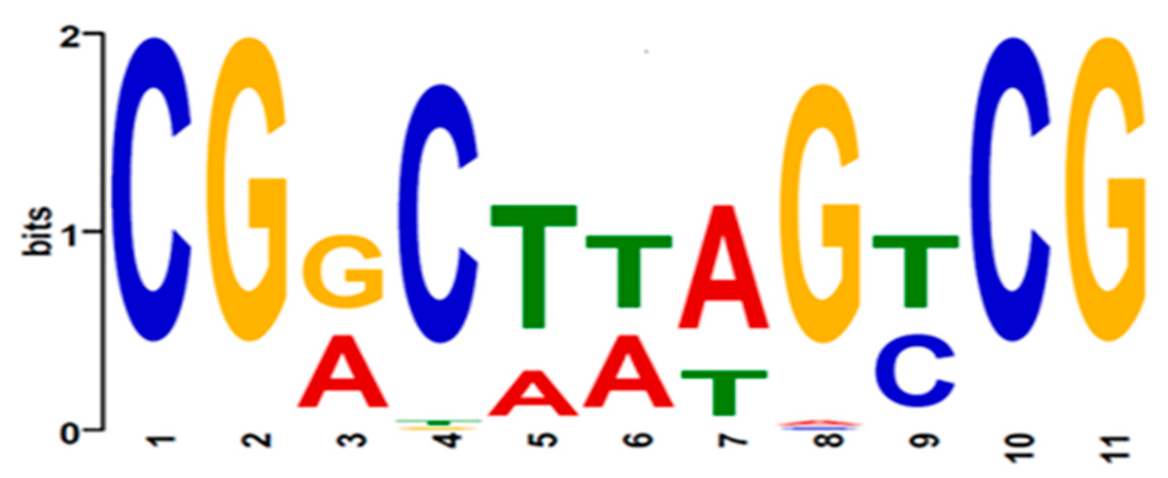
2.9. KojR DNA-Binding Motif Analysis

The motif finding program Multiple EM for Motif Elicitation (MEME, Version 5.3.0, http://meme-suite.org/tools/meme) [26] was used to predict the putative KojR-binding motif sequences in both kojA and kojT promoters of the twenty-three aspergilli in the section Flavi. Sequences of around 370 and 380 nucleotides in the respective 5’-untranslated regions of kojA and kojT were extracted from genome assemblies (Table S2) and uploaded into MEME for motif analysis.

2.10. Generation of Defects in Putative KojR DNA-binding Sites by the Established CRISPR/Cas9 Genome-Editing Approach

Targeted alteration of putative KojR DNA-binding sites was carried out using the established genome-editing CRISPR/Cas9 technology [27]. pAf-CRISPR-yA (Addgene plasmid #191015) was the template for making DNA fragments that encoded single guide RNAs (sgRNAs) to target motif sequences. The PCR fragments were cloned into the PstI and KpnI sites of pAsp-AMA-gpdA-ptr (Addgene plasmid #191016) to give the resulting gene-targeting vectors. The target sequences in kojA and kojT for the CRISPR/Cas9 complexes were GGTGGAATGAGCGGCAAAGTCGG and AAGCCATTCAGCGGCTAAGTCGG, respectively (motif sequences are underlined). To construct a sgRNA expression cassette, two target-specific DNA fragments were first generated by PCR with primer sets of U6-F-P/kojA_R or kojT_R and kojA_F or kojT_F/U6-R-K, respectively (Table S1). To this end, 40 pmol of each primer and 2 ng template were added to 20 μL of AccuPrime™ SuperMix (Invitrogen) and subjected to thirty cycles of PCR, which consisted of denaturation at 94 °C for 30 s, annealing at 55 °C for 30 s and extension at 72 °C for 1.0 min. The two PCR fragments, without further purification, were directly fused and amplified by another round of PCR using primers U6-F-P and U6-R-K. The annealing time was set for 2 min. The resulting fragment, after being cut with PstI and KpnI, was cloned to give a final vector. Gene-targeting vectors were transformed into the wild-type A. flavus CA14 recipient as previously described [21]. Selected primary pyrithiamine-resistant transformants were transferred onto KAM plates and grown at 30 °C for two to three days for direct PCR and sequencing analyses to confirm indel defects in the targeted kojA and kojT genes.

2.11. Identification of Molecular Defects in kojA and kojT Transformants

Sequencing was performed to reveal defects in kojA transformants that lost KA production and in kojT transformants that retained or lost KA production. Direct PCR using fungal mycelia as the genomic template and location-specific primers that encompassed targeted sequences was carried out with a Phire Plant Direct PCR Master Mix (Thermo Fisher Scientific, Waltham, MA, USA) [28]. The PCR protocol consisted of an initial denaturation at 98 °C for 5.0 min, followed by forty cycles of denaturation at 98 °C for 5 s, annealing at 60 °C for 5 s, and extension at 72 °C for 30 s. Purified PCR products were sequenced at the Genomics and Bioinformatics Research Unit of the Agricultural Research Service, US Department of Agriculture (Stoneville, MS, USA).

3. Results

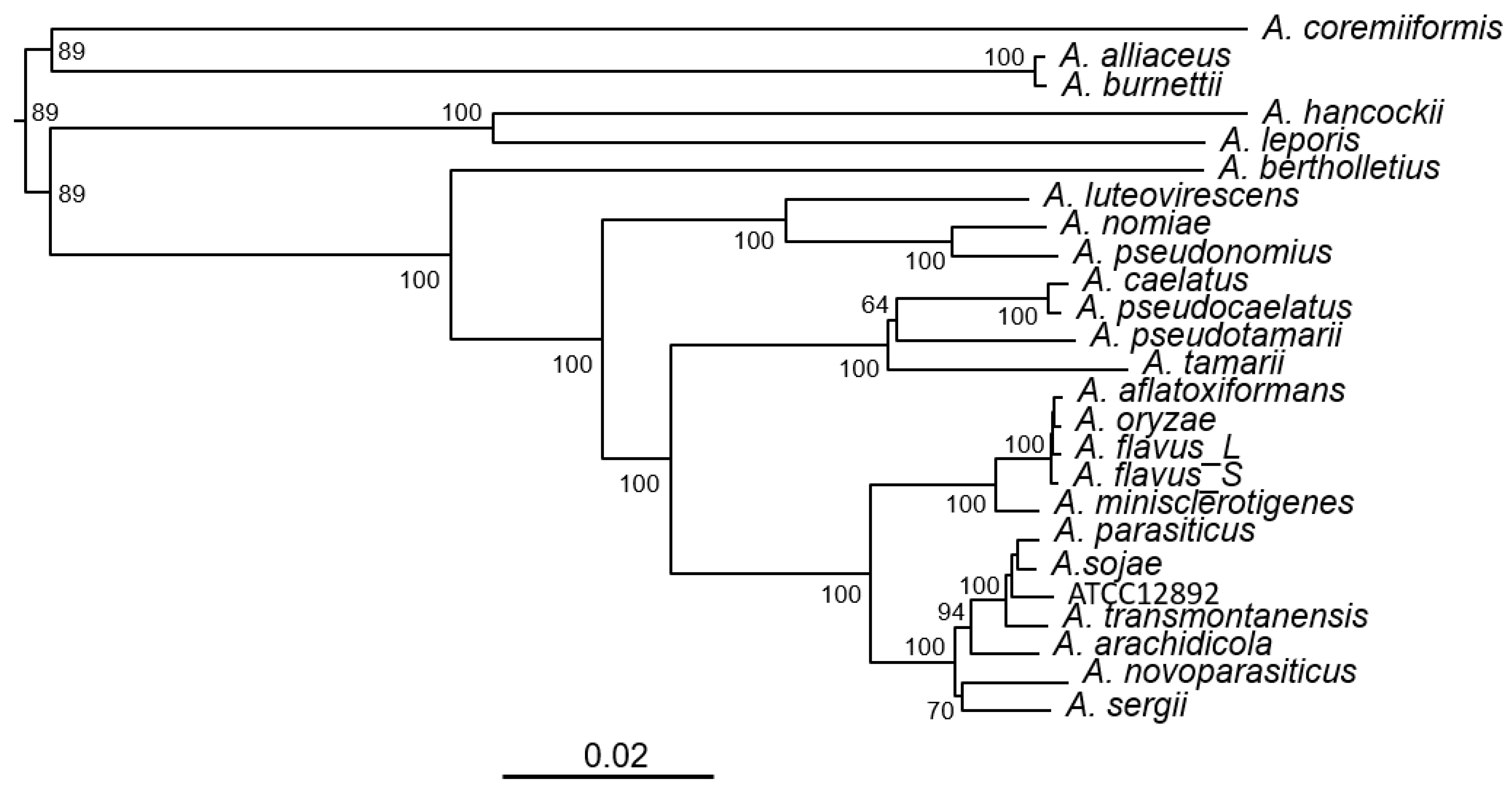
3.1. Phylogeny of Aspergillus Section Flavi Species Bases on Complete KA Gene Cluster Sequences

We investigated the presence of a complete KA gene cluster, that is, kojA-kojR-kojT in over 100 Aspergillus species, whose genome sequences are publicly available at NCBI. The sequence of the A. flavus NRRL3357 KA gene cluster (CP044621.1: 4359976 to 4365751), which resides on chromosome five, was the alignment template. We found that twenty-three known species contained the complete KA gene cluster (Table 1). They exclusively belonged to the Flavi section (“section” is a taxonomic rank in-between genus and species). Phylogenetic inference based on variations in conserved sites in the KA gene clusters showed that they were grouped into six clades (Figure 1). A. flavus, A. oryzae, A. minisclerotigenes and A. aflatoxiformans (=A. parvisclerotigenus) were in one clade, well separated from A. parasiticus, A. novoparasitcus, A. sojae, A. arachidicola and A. transmontanensis, which were in another clade. The phylogeny also showed that A. oryzae was closer to A. aflatoxiformans than to S-morphotype A. flavus, and it was adequately separated from A. minisclerotigenes. All known aflatoxin producers, A. flavus, A. pseudonomius, A. aflatoxiformans, A. parasiticus, A. novoparasiticus, A. arachidicola, A. minisclerotigenes, A. transmontanensis, A. nomiae, A. luteovirescens, and A. sergii had complete KA gene clusters.

3.2. Partial KA Gene Clusters in Other Aspergilli and Penicillia

The presence of a kojR homolog (An09g05060) in Aspergillus niger, which encodes a protein with 63% amino acid sequence identity to A. oryzae and A. flavus KojR proteins, has been reported [17]. In this study, we further confirmed that An09g05070, situated next to An09g05060 and encoding an MFS (major facilitator superfamily) transporter protein, was a kojT homolog with 78% amino acid sequence identity. However, we did not find homologous genes encoding the KojA oxidoreductase next to or adjacent to An09g05060. Therefore, A. niger contained only a partial KA gene cluster. We identified homologous proteins from other twenty-one aspergilli, including A. avenaceus, a section Flavi species (Table 2), using both annotated amino acid sequences of A. niger KojR and KojT as alignment templates. Surprisingly, no genes encoding a KojA oxidoreductase were present near kojR in A. avenaceus or the other twenty aspergilli not belonging to section Flavi. To identify possible KA gene clusters in Penicillium species, we carried out a BlastP search using the KojR and KojT sequences of A. steynii and A. tanneri, which had the lowest degrees of amino sequence identity to the homologs of the rest of the aspergilli. The rationale was that the low degrees may suggest that they are closer to Penicillium or other fungal genera than to other aspergilli. By this approach, we further identified KojR and/or KojT homologs from P. nordicum, P. freii, and P. polonicum, among over thirty Penicillium species. Using the P. nordicum KojR protein sequence (KOS46106.1) as an alignment template, we obtained the same results. The Penicillium KojR and KojT amino acid sequences were 50–56% and 70–73% identical to those of the homologs of A. steynii and A. tanneri, respectively). However, many of the originally annotated Penicillium KojR proteins varied greatly in their size, and some were mis-annotated. We found that only kojR and kojT of P. nordicum were organized as a partial gene cluster (Table 2).

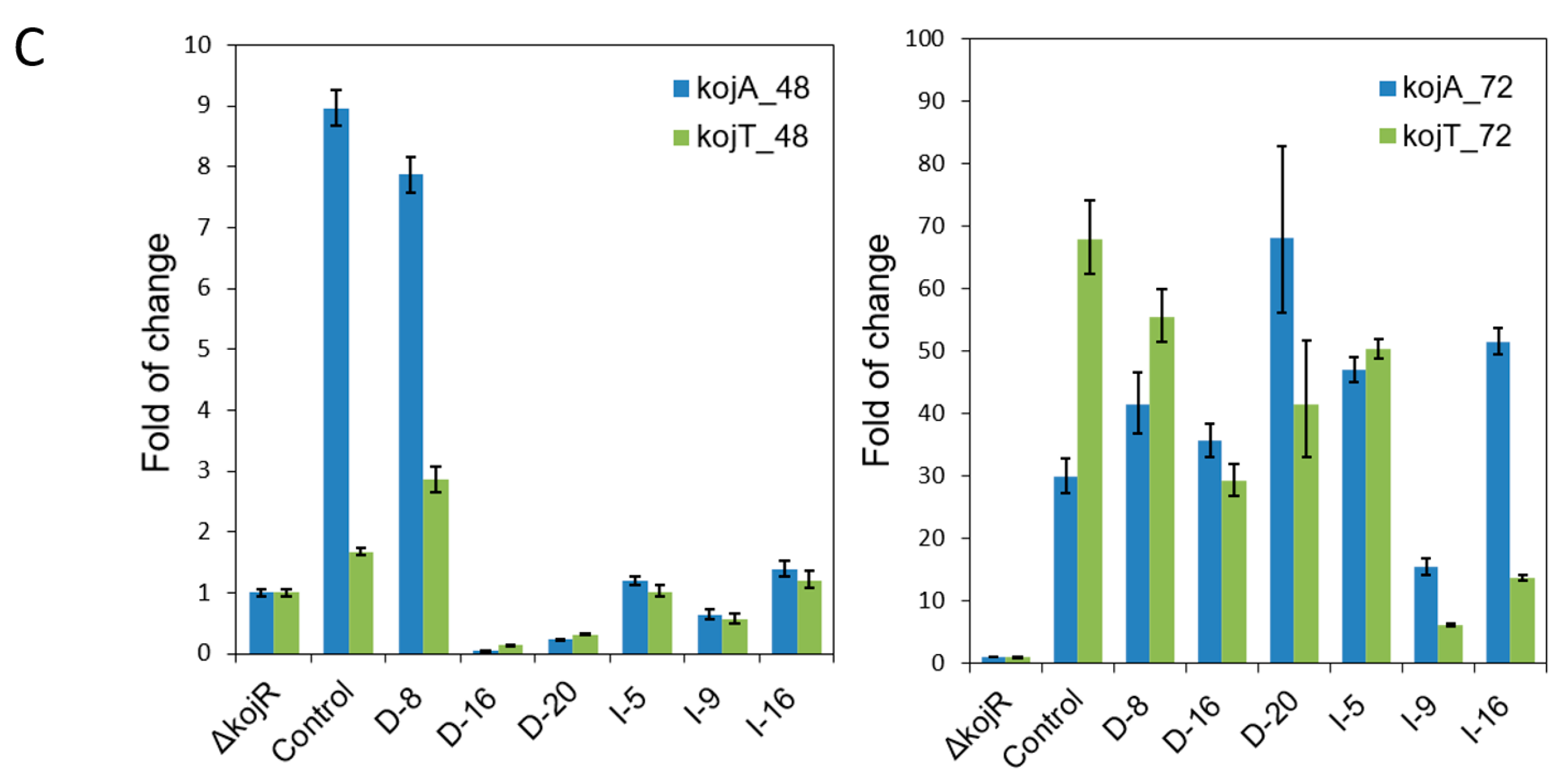
3.3. Expressing kojR Restored Expression of kojA and kojT in Overexpression Strains

The A. flavus kojR gene was involved in KA production, and kojR knockout strains were unable to produce KA. To investigate the transcriptional activation mechanism for kojA and kojT expression, we fused kojR with the gpdA or gpiA promoter and carried out overexpression experiments. Transformants exhibited varied abilities in terms of KA production on KAM or PDA supplemented with ferric ions (Figure S1). We selected three putative overexpression strains that visually showed high amounts of KA production from each set and determined their kojR copy numbers. Only one strain (D-8) in the gpdA promoter set contained more than one copy of kojR. In contrast, the strains of the gpiA set contained two to three copies of kojR (Figure 2A). In the time-course study that examined how kojR expression affected the expression of kojA and kojT, we found that the ∆kojR#4 mutant had lost kojR expression; its normalized expression levels of kojA and kojT at 72 h compared with those at 48 h decreased slightly and were within 40% (Table S3). For the KuPG control strain, normalized expression levels of kojA and kojT from 48 h to 72 h increased 2.0- and 35.2-fold, respectively. Noticeably, the kojR expression levels of the control and overexpression strains did not change much from 48 h to 72 h, and the variations were within 70%. Both the gpdA and gpiA promoters were able to drive higher kojR expression in the overexpression strains than the native kojR promoter in the control strain. We observed that kojR expression levels in all overexpression strains compared to those in the control strain were higher at 48 h than at 72 h. The relative expression levels at 48 h somewhat reflected the kojR copy numbers of these strains (Figure 2B). The normalized kojA and kojT expression levels in all overexpression strains at 48 h were low but increased significantly at 72 h (Table S3). Using the normalized kojA and kojT expression levels of the ∆kojR#4 mutant, which did not vary much from 48 h to 72 h (Table S3), as the basal expression levels (relative fold treated as one), we found that the relative kojA and kojT expression levels in most of the strains were low at 48 h except for D8, which were comparable to the control levels (Figure 2C). Noticeably, the kojA and kojT expression levels in D-16 and D-20 were significantly lower than those in the ΔkojR strain. These decreases seem to be related to delayed gene expression at 48 h in the two heterologous gpdA promoter set strains, which contained a single copy of kojR (Figure 2A), as compared to the kojA and kojT expression in the homologous gpiA promoter set strains at 48 h (Table S3). It is not known whether the developmental or physiological states of the two strains have a bearing on the observed variation. Expression levels of kojA and kojT at 72 h increased substantially from the basal levels but also varied greatly. Only the D8 relative expression patterns resembled those of the control (Figure 2C). Table 3 shows that at 96 h only strain D-16 produced 43% more KA than the control strain, whereas others produced slightly lower or higher amounts of KA.

3.4. Zn(II)2Cys6 Zinc Cluster Domains and Downstream Basic Regions of Aspergilli

We analyzed the amino-terminal 120 amino acid-containing regions of KojR proteins of the twenty-three-section Flavi aspergilli. Each region included a binuclear zinc-finger DNA-binding domain and the downstream basic sequence associated with dimerization. Using A. flavus KojR as a reference, we observed amino acid variations, including deletions and conservative and radical substitutions, in some aspergilli. All twenty-three aspergilli had identical Zn(II)2Cys6 zinc cluster domains, CETCKLRKRKCDGHEPCTYCLRYEYQC (Table 4). For the two aspergilli not belonging to section Flavi, A. phoenicis and A. welwitschiae (Table 2), they had the same Zn(II)2Cys6 zinc-cluster as A. flavus KojR. However, others showed minor variations. For example, the corresponding domain in A. niger was CETCKLRKRKCDGHEPC S F ̿ CL K ̿ YEY D ̿ C and contained four amino acid substitutions.

3.5. Identification of Putative KojR-Binding Sites in kojA and kojR Promoters of Aspergilli

The alignment of zinc-cluster domains in section Flavi aspergilli suggested that they recognized the same binding motif(s). To identify possible consensus KojR-binding site(s), we examined sequences in the kojA and kojT promoter regions of twenty-four aspergilli, including A. flavus L- and S-morphotype. Table 4 shows that the kojA and kojT promoter sequences of A. flavus shared various degrees of nucleotide sequence identity with those of the other twenty-two aspergilli, which ranged from 63.9% to 100% and 60.5% to 99.5%, respectively. We next performed motif analysis using MEME on sequences from half of the intergenic regions of kojA and kojR (370 bps upstream of kojA) and in the complete inter-gene regions of kojR and kojT (383 bps) (Table S2). We obtained a putative DNA-binding motif that contained an 11-nucleotide palindromic sequence, 5′-CGRCTWAGYCG-3′ (R = A/G, W = A/T, Y = C/T) (Figure 3). All putative motif sequences found in the kojA and kojT promoters were situated within 0.3 kb of the ATG start codon. Table 5 shows that these putative motif sequences (from 5′ to 3′) are on positive strands in the kojA promoters. However, only reverse complementary sequences that are like the aforementioned motif sequences (with one nucleotide substitution, i.e., position seven changed from T to A) are present on the positive strands in the kojT promoters (relative to the ATG codon). In other words, putative motif sequences were found on both strands when viewed because of their palindromic nature.

3.6. Involvement of the KojR-Binding Site in the A. flavus kojA Promoter in KA Production

To corroborate the significance of the identified motif sequences in the kojA and kojT promoters in KA biosynthesis, we carried out site mutation analyses in A. flavus using a CRISPR/Cas9-based genome editing technique. This technology is well-known for generating small deletions in targeted sequences. We analyzed twenty and thirty-four primary transformants of the kojA and kojT sets, respectively, for KA production by transferring them onto KAM plates. For the kojA set, no transformants were produced for KA. For the kojT set, twenty-three produced KA but eleven did not. PCR examination of the fifty-four transformants with location-specific primers (Table S1) revealed that seven transformants from the kojA set and seventeen transformants from the kojT set (fifteen KA producers and two KA non-producers) gave positive PCR products. Subsequent sequencing of these PCR products showed that the KA-nonproducing kojA transformants contained six types of sequence defects, which consisted of five types of deletions and one type of insertion. These defects disrupted various parts of the putative KojR-binding motif in the kojA promoter (Figure 4A and Figure S2). Similarly, KA-producing kojT transformants had nine types of defects, which consisted of six types of deletions and three types of insertions (one, six, and one hundred five nucleotides) that disrupted the putative KojR-binding motif in the kojT promoter. Figure 4B shows six types of them. The transformant T3 had half of the motif plus the downstream region extending beyond the kojT start site (404 bps) deleted (Figure S3). The two kojT transformants, T18 and T21, that did not produce KA, had large deletions of 999 and 1566 nucleotides, respectively. This started at the kojT target site and extended to the upstream kojR coding region (Figure S4), which indicated that the loss of KA production resulted primarily from the defective kojR. Figure 4C depicts the location of the identified functional KojR-binding site in the KA gene cluster. A defect at this site resulted in the loss of KA production.

4. Discussion

A list of thirty-three accepted species and their synonyms in Aspergillus section Flavi has been published [29]. Most recently, an additional species, A. burnettii, was identified [30]. Our analyses of the genome sequences of more than one hundred Aspergillus species showed that the KA gene cluster, kojA-kojR-kojT, was present nearly exclusively in Flavi section members (Table 1). Therefore, they appear to be the only aspergilli capable of producing KA. Phylogeny inferred from the KA gene cluster sequences showed that the derived clades were like those based on multi-locus sequences or 200 monocore (i.e., single copy) gene sequences [29,31]. The Aspergillus sp. ATCC 12892, officially designated as Aspergillus oryzae (Ahlburg) Cohn (ATCC® 12892™), turned out to be misidentified. It is most likely a strain of A. parasiticus or a closely related species. Its genome size (41.2 kb), which is 10% larger than those of A. oryzae RIB40 and A. flavus, also supports this notion. Misclassification of Apergillus species has been a concern, especially in the genomic era [32]. Classification of Aspergillus species by multi-locus sequence typing has been a conventional approach. The current study shows that using SNPs in the KA gene clusters is an equally valid method, and it can distinguish a misidentified species from correctly named aspergilli.
KA production by most sections Flavi aspergilli has been reported. However, not all Flavi species can produce KA, for example, A. coremiiformis and A. avenaceus [10,29]. The genome size of A. coremiiformis was 20% smaller than the average (Table 1). Nonetheless, it had a complete KA gene cluster. This KA gene cluster sequence, however, did not have a significant nucleotide identity to that of A. flavus. Cross-species sequence comparisons further showed that it shared 80–82% nucleotide identity with those of A. alliiaceus and A. burnettii in the same clade (Table 1 and Figure 1). We noticed that sequences in the promoter regions of A. coremiiformis kojR, kojA, and kojT were also highly variable (Table S2), which may explain its inability to produce KA. The A. oryzae KA gene cluster (kojA-kojR-kojT) identified in 2010 is likely an incomplete gene cluster. More than one enzyme is required for KA formation [15]. The kojA gene product, an oxidoreductase, should be directly involved in the conversion of the glucose precursor or intermediate metabolites. However, neither kojR nor kojT, which encode a Zn(II)2Cys6 zinc cluster regulator and a transporter protein (efflux pump), respectively, possess enzymatic activities. Therefore, we cannot exclude the possibility that the inability of A. coremiiformis to produce KA is due to defects in another yet-to-be-identified gene(s).
A. niger has been reported as being unable to produce KA [1]. In this study, we found that it only contained a partial KA gene cluster. Hence, the inability of A. avenaceus to produce KA is probably due to not having a complete KA gene cluster. Other aspergilli not belonging to section Flavi but containing partial KA gene clusters (Table 2) likely do not produce KA either. Up to now, only a few Penicillium species, such as P. citrinum, P. griseofulvum, P. purpurogenum, P. rubrum, P. jamesonlandense, and P. lanosum are known to produce KA [1,33]. Of the six species, the genome sequences of P. citrinum and P. griseofulvum are available in the NCBI genome database. Nonetheless, neither of the two penicillia contained a complete KA gene cluster. Our finding that only P. nordicum had a partial KA gene cluster suggests that most Penicillium KA biosynthesis genes, if they exist, do not form a gene cluster.
Zn(II)2Cys6 type regulators are unique to fungi and yeast [34,35,36]. They are involved in a diverse array of functions [37]. In the A. oryzae kojR-overexpressing strain, expression of kojA and kojT was barely detected at 24 h until 72 h, when the levels increased substantially [17]. In this study, the six A. flavus kojR-overexpressing strains also produced relatively low levels of kojA and kojT transcripts at 48 h, but the levels were greatly elevated at 72 h. Through transcriptional activation of kojA and kojT expression driven by the heterologous A. nidulans gpdA promoter and the homologous A. flavus gpiA promoter KA production was restored (Figure 2 and Table 3). However, it is not known if autoregulation, that is, a direct modulation of gene expression by the product of the corresponding gene (KojR) to activate or suppress its own transcription [38,39], is involved in kojR expression in wild-type A. flavus. The extents of variation in normalized kojR expression from 48 h to 72 h in the control and overexpression strains were much lower than those in kojA and kojT (Table S3). This implies that the pathway-specific kojR is tightly regulated, probably by unidentified global regulators. In the A. oryzae study [17], KA was reported to increase the transcription of its biosynthesis genes. Although kojA and kojR share the same intergenic region, the effect of KA addition was more prominent on kojA than on kojR. Feedback regulation, the process by which a metabolic pathway product influences its own production by controlling the amount and/or the activity of one or more involved enzymes [40], may have a bearing on KA biosynthesis.
A consensus motif sequence, 5′-AGTCGGG-3′ in upstream regions of A. oryzae kojA and kojT was proposed to be responsible for the regulation of kojA and kojT [17]. However, our analysis of the A. oryzae KA gene cluster showed that this heptanucleotide sequence resided on different strands, that is, on the negative strand upstream of kojA but on the positive strand upstream of kojT (Table 5). The difference in motif sequence orientation suggests that kojA and kojT might not be coordinately regulated by KojR. The proposed A. oryzae sequence (bold) was a part of 5′-CGGCAAAGTCGGG-3′on the positive strand upstream of kojR (underlined is the reverse complementary sequence of 5′-CGACTTTGCCG-3′, the motif identified in the A. flavus kojA promoter). Zn(II)2Cys6 type regulators like KojR bind as a dimer to both DNA strands [37]. The physical distance from the motif site to the kojR coding sequence start codon, ATG, was approximately twice that from the motif site to the kojA start codon (Table 5 and Figure 4C), which suggests that KojR is likely mainly involved in kojA regulation.
The CRISPR/Cas9 technology could be used as a complementary method to the ChIP-seq method for identifying DNA-binding motifs or the electrophoretic mobility shift assay (EMSA) for detecting protein-DNA interactions [41,42]. In this study, we showed that only the 11-bp sequence motif in the A. flavus kojA promoter was required for KA production (Figure 4A). Hence, the genuine KojR-binding motif sequence for aspergilli is likely longer than what was proposed for A. oryzae. For example, both the 18-bp insertion in the transformant A7 and the 8-bp deletion in the transformant A15 did not disrupt the A. flavus equivalent of the A. oryzae motif 5′-CCCGACTTTGCCG-3′ (bold is the reverse complementary sequence of 5′-AGTCGGG-3′) in their kojA promoters (Figure 4A). It was the deletions upstream of 5′-AGTCGGG-3′ that rendered the two A. flavus mutants unable to produce KA. For the A. flavus promoter, disruptions of the 11-bp palindromic motif 5′-CGGCTAAGTCG-3′ in transformants T2, T7, and T11 or a complete deletion of the proposed A. oryzae motif sequence 5′-AGTCGGG-3′ in transformants T3, T24, and T32 did not abolish KA production (Figure 4B). However, in the characterization of A. oryzae kojT by Terabayashi et al. [16], the kojT deletion mutant still produced a reduced amount of KA, as evidenced by the colony showing a pale red color. Therefore, we cannot rule out that KA production in these A. flavus mutants results from the presence of another unidentified transporter protein that can replace the function of KojT, as previously proposed. Nevertheless, the present work shows that the motif in the A. flavus kojA promoter is highly likely a KojR-binding site and is critical for KA production. Zn(II)2Cys6-type proteins act as pathway-specific regulators [37]. Three, all named AflR, are involved in the biosynthesis of sterigmatocystin and aflatoxin in A. nidulans and A. flavus/A. parasiticus, respectively, have been characterized; and their binding motifs are 11-bp palindromic sequences [43,44,45]. Apparently, A. flavus KojR has the same binding capacity. Despite the initial characterization of KojR-binding sites in the present study, the final proof of the identity of the KojR-binding motif, including its actual length, will have to come from direct evidence of protein-DNA interaction, such as the use of ChIP technology. Future characterization of those yet-to-be-identified structural genes that are directly involved in KA formation will shed light on genuine KojR-binding sequences in the KA biosynthesis gene promoters.

Supplementary Materials

The following supporting information can be downloaded at: https://www.mdpi.com/article/10.3390/jof9020259/s1, Figure S1: Examination of KA production by kojR complemented transformants of A. flavus ΔkojR#4; Figure S2: Sequencing chromatograms showing locations of deletions and an insertion in the KojR-binding motif of A. flavus kojA promoter; Figure S3: Sequencing chromatogram showing the breakpoint and sequence alignment for identifying a large deletion that disrupted the suggested motif in the kojT promoter and extended to the kojT-coding region in a KA-producing A. flavus mutant; Figure S4: Sequencing chromatogram showing the breakpoint and sequence alignment for identifying a large deletion that disrupted the suggested motif in the kojT promoter and extended to the upstream kojR-coding region in an A. flavus mutant that was unable to produce KA; Table S1: Primers used in vector construction, qRT-PCR, and sequencing; Table S2: Sequences of the kojA and kojT promoter regions used for the identification of KojR DNA-binding motif by MEME; Table S3: Normalized expression levels of KA genes in A. flavus strains.

Author Contributions

P.-K.C. and Q.K. designed experiments, analyzed results, and wrote the manuscript. L.L.S. and N.M. conducted experiments. All authors have read and agreed to the published version of the manuscript.

Funding

This work was supported by the United States Department of Agriculture, Agricultural Research Service.

Data Availability Statement

Raw data and materials described in this study will be shared upon reasonable request, in accordance with USDA policies and procedures.

Acknowledgments

Qing Kong was a visiting scientist, and this work was part of his research project conducted at the Southern Regional Research Center. The authors would like to thank Fanny Liu at Genomics and Bioinformatics Research Unit, Agricultural Research Service, U. S. Department of Agriculture in Stoneville, Mississippi for performing the sequencing of PCR fragments.

Conflicts of Interest

The authors declare no conflict of interest.

References

  1. Parrish, F.W.; Wiley, B.J.; Simmons, E.G.; Long, L., Jr. Production of aflatoxins and kojic acid by species of Aspergillus and Penicillium. Appl Microbiol 1966, 14, 139. [Google Scholar] [CrossRef] [PubMed]
  2. Katagiri, H.; Kitahara, K. The formation of kojic acid by Aspergillus oryzae: Part I. The formation of kojic acid from pentoses, sugar alcohols and gluconic acid. Bull. Agric. Chem. Soc. Jpn. 1929, 5, 38–47. [Google Scholar] [CrossRef]
  3. Bentley, R. From miso, sake and shoyu to cosmetics: A century of science for kojic acid. Nat. Prod. Rep. 2006, 23, 1046–1062. [Google Scholar] [CrossRef] [PubMed]
  4. Yabuta, T. LXXIII.—The constitution of kojic acid, a γ-pyrone derivative formed by Aspergillus oryzæ from carbohydrates. J. Chem. Soc. Trans. 1924, 125, 575–587. [Google Scholar] [CrossRef]
  5. Cabanes, J.; Chazarra, S.; Garcia-Carmona, F. Kojic acid, a cosmetic skin whitening agent, is a slow-binding inhibitor of catecholase activity of tyrosinase. J. Pharm. Pharm. 1994, 46, 982–985. [Google Scholar] [CrossRef]
  6. Brtko, J.; Rondahl, L.; Fickova, M.; Hudecova, D.; Eybl, V.; Uher, M. Kojic acid and its derivatives: History and present state of art. Cent. Eur. J. Public Health 2004, 12, S16–S18. [Google Scholar]
  7. Saeedi, M.; Eslamifar, M.; Khezri, K. Kojic acid applications in cosmetic and pharmaceutical preparations. Biomed. Pharm. 2019, 110, 582–593. [Google Scholar] [CrossRef]
  8. Mohamad, R.; Mohamed, M.S.; Suhaili, N.B.; Salleh, M.M.; Ariff, A.B. Kojic acid: Applications and development of fermentation process for production. Biotechnol. Mol. Biol. Rev. 2010, 5, 24–37. [Google Scholar]
  9. ResearchReportsWorld. Global Kojic Acid Market Growth (Status and Outlook) 2022–2028. 2022. Available online: https://www.researchreportsworld.com/global-kojic-acid-market-19888650 (accessed on 29 September 2022).
  10. Chaudhary, J.; Pathak, A.N.; Lakhawat, S. Production technology and applications of kojic acid. Annu. Res. Rev. Biol. 2014, 4, 3165–3196. [Google Scholar] [CrossRef]
  11. El-Kady, I.A.; Zohri, A.-N.A.; Hamed, S.R. Kojic Acid production from agro-industrial by-products using fungi. Biotechnol. Res. Int. 2014, 2014, 642385. [Google Scholar] [CrossRef] [Green Version]
  12. Feng, W.; Liang, J.-S.; Wang, B.; Chen, J. Improvement of kojic acid production in Aspergillus oryzae AR-47 mutant strain by combined mutagenesis. Bioprocess Biosyst. Eng. 2019, 42, 753–761. [Google Scholar] [CrossRef] [PubMed]
  13. Futamura, T.; Okabe, M.; Tamura, T.; Toda, K.; Matsunobu, T.; Park, Y.S. Improvement of production of kojic acid by a mutant strain Aspergillus oryzae, MK107-39. J. Biosci. Bioeng. 2001, 91, 272–276. [Google Scholar] [CrossRef]
  14. Arnstein, H.R.; Bentley, R. Kojic acid biosynthesis from 1-C14-glucose. Nature 1950, 166, 948–949. [Google Scholar] [CrossRef] [PubMed]
  15. Bajpai, P.; Agrawala, P.K.; Vishwanathan, L. Enzymes relevant to kojic acid biosynthesis in Aspergillus flavus. Microbiology 1981, 127, 131–136. [Google Scholar] [CrossRef] [Green Version]
  16. Terabayashi, Y.; Sano, M.; Yamane, N.; Marui, J.; Tamano, K.; Sagara, J.; Dohmoto, M.; Oda, K.; Ohshima, E.; Tachibana, K.; et al. Identification and characterization of genes responsible for biosynthesis of kojic acid, an industrially important compound from Aspergillus oryzae. Fungal Genet. Biol. 2010, 47, 953–961. [Google Scholar] [CrossRef] [PubMed]
  17. Marui, J.; Yamane, N.; Ohashi-Kunihiro, S.; Ando, T.; Terabayashi, Y.; Sano, M.; Ohashi, S.; Ohshima, E.; Tachibana, K.; Higa, Y.; et al. Kojic acid biosynthesis in Aspergillus oryzae is regulated by a Zn(II)2Cys6 transcriptional activator and induced by kojic acid at the transcriptional level. J. Biosci. Bioeng. 2011, 112, 40–43. [Google Scholar] [CrossRef]
  18. Darling, A.C.; Mau, B.; Blattner, F.R.; Perna, N.T. Mauve: Multiple alignment of conserved genomic sequence with rearrangements. Genome Res. 2004, 14, 1394–1403. [Google Scholar] [CrossRef] [Green Version]
  19. Chang, P.-K.; Chang, T.D.; Katoh, K. Deciphering the origin of Aspergillus flavus NRRL21882, the active biocontrol agent of Afla-Guard®. Lett. Appl. Microbiol. 2021, 72, 509–516. [Google Scholar] [CrossRef]
  20. Katoh, K.; Misawa, K.; Kuma, K.; Miyata, T. MAFFT: A novel method for rapid multiple sequence alignment based on fast Fourier transform. Nucleic Acids Res. 2002, 30, 3059–3066. [Google Scholar] [CrossRef] [Green Version]
  21. Chang, P.-K.; Scharfenstein, L.L.; Wei, Q.; Bhatnagar, D. Development and refinement of a high-efficiency gene-targeting system for Aspergillus flavus. J. Microbiol. Methods 2010, 81, 240–246. [Google Scholar] [CrossRef]
  22. Chang, P.-K.; Scharfenstein, L.L.; Mack, B.; Ehrlich, K.C. Deletion of the Aspergillus flavus orthologue of A. nidulans fluG reduces conidiation and promotes production of sclerotia but does not abolish aflatoxin biosynthesis. Appl. Env. Microbiol. 2012, 78, 7557–7563. [Google Scholar] [CrossRef] [Green Version]
  23. Punt, P.J.; Dingemanse, M.A.; Jacobs-Meijsing, B.J.; Pouwels, P.H.; van den Hondel, C.A. Isolation and characterization of the glyceraldehyde-3-phosphate dehydrogenase gene of Aspergillus nidulans. Gene 1988, 69, 49–57. [Google Scholar] [CrossRef]
  24. Chang, P.-K.; Zhang, Q.; Scharfenstein, L.; Mack, B.; Yoshimi, A.; Miyazawa, K.; Abe, K. Aspergillus flavus GPI-anchored protein-encoding ecm33 has a role in growth, development, aflatoxin biosynthesis, and maize infection. Appl. Microbiol. Biotechnol. 2018, 102, 5209–5220. [Google Scholar] [CrossRef] [PubMed]
  25. Umemura, M.; Kuriiwa, K.; Dao, L.V.; Okuda, T.; Terai, G. Promoter tools for further development of Aspergillus oryzae as a platform for fungal secondary metabolite production. Fungal Biol. Biotechnol. 2020, 7, 3. [Google Scholar] [CrossRef] [PubMed] [Green Version]
  26. Bailey, T.L.; Elkan, C. Fitting a mixture model by expectation maximization to discover motifs in biopolymers. Proc. Int. Conf. Intell. Syst. Mol. Biol. 1994, 2, 28–36. [Google Scholar] [PubMed]
  27. Jin, F.J.; Wang, B.T.; Wang, Z.D.; Jin, L.; Han, P. CRISPR/Cas9-based genome editing and its application in Aspergillus species. J. Fungi 2022, 8, 467. [Google Scholar] [CrossRef]
  28. Chang, P.-K.; Scharfenstein, L.L.; Abbas, H.K.; Bellaloui, N.; Accinelli, C.; Ebelhar, M.W. Prevalence of NRRL21882-like (Afla-Guard®) Aspergillus flavus on sesame seeds grown in research fields in the Mississippi Delta. Biocontrol Sci. Technol. 2020, 30, 1090–1099. [Google Scholar] [CrossRef]
  29. Frisvad, J.C.; Hubka, V.; Ezekiel, C.N.; Hong, S.B.; Novakova, A.; Chen, A.J.; Arzanlou, M.; Larsen, T.O.; Sklenar, F.; Mahakarnchanakul, W.; et al. Taxonomy of Aspergillus section Flavi and their production of aflatoxins, ochratoxins and other mycotoxins. Stud. Mycol. 2019, 93, 1–63. [Google Scholar] [CrossRef]
  30. Gilchrist, C.L.M.; Lacey, H.J.; Vuong, D.; Pitt, J.I.; Lange, L.; Lacey, E.; Pilgaard, B.; Chooi, Y.H.; Piggott, A.M. Comprehensive chemotaxonomic and genomic profiling of a biosynthetically talented Australian fungus, Aspergillus burnettii sp. nov. Fungal Genet. Biol. 2020, 143, 103435. [Google Scholar] [CrossRef]
  31. Kjaerbolling, I.; Vesth, T.; Frisvad, J.C.; Nybo, J.L.; Theobald, S.; Kildgaard, S.; Petersen, T.I.; Kuo, A.; Sato, A.; Lyhne, E.K.; et al. A comparative genomics study of 23 Aspergillus species from section Flavi. Nat. Commun. 2020, 11, 1106. [Google Scholar] [CrossRef] [Green Version]
  32. Arias, R.S.; Orner, V.A.; Martinez-Castillo, J.; Sobolev, V.S. Aspergillus Section Flavi, Need for a robust taxonomy. Microbiol. Resour. Announc. 2021, 10, e0078421. [Google Scholar] [CrossRef] [PubMed]
  33. Frisvad, J.C.; Larsen, T.O.; Dalsgaard, P.W.; Seifert, K.A.; Louis-Seize, G.; Lyhne, E.K.; Jarvis, B.B.; Fettinger, J.C.; Overy, D.P. Four psychrotolerant species with high chemical diversity consistently producing cycloaspeptide A, Penicillium jamesonlandense sp. nov., Penicillium ribium sp. nov., Penicillium soppii and Penicillium lanosum. Int. J. Syst. Evol. Microbiol. 2006, 56, 1427–1437. [Google Scholar] [CrossRef] [PubMed]
  34. Chang, P.-K.; Ehrlich, K.C. Genome-wide analysis of the Zn(II)2Cys6 zinc cluster-encoding gene family in Aspergillus flavus. Appl. Microbiol. Biotechnol. 2013, 97, 4289–4300. [Google Scholar] [CrossRef] [PubMed]
  35. Maicas, S.; Moreno, I.; Nieto, A.; Gomez, M.; Sentandreu, R.; Valentin, E. In silico analysis for transcription factors with Zn(II)2Cys6 binuclear cluster DNA-binding domains in Candida albicans. Comp. Funct. Genom. 2005, 6, 345–356. [Google Scholar] [CrossRef] [Green Version]
  36. Zhang, C.; Huang, H.; Deng, W.; Li, T. Genome-wide analysis of the Zn(II)2Cys6 zinc cluster-encoding gene family in Tolypocladium guangdongense and its light-induced expression. Genes 2019, 10, 179. [Google Scholar] [CrossRef] [Green Version]
  37. MacPherson, S.; Larochelle, M.; Turcotte, B. A fungal family of transcriptional regulators: The zinc cluster proteins. Microbiol. Mol. Biol. Rev. 2006, 70, 583–604. [Google Scholar] [CrossRef] [Green Version]
  38. Crews, S.T.; Pearson, J.C. Transcriptional autoregulation in development. Curr. Biol. 2009, 19, R241–R246. [Google Scholar] [CrossRef] [Green Version]
  39. Hoffmann, B.; Valerius, O.; Andermann, M.; Braus, G.H. Transcriptional autoregulation and inhibition of mRNA translation of amino acid regulator gene cpcA of filamentous fungus Aspergillus nidulans. Mol. Biol. Cell 2001, 12, 2846–2857. [Google Scholar] [CrossRef] [Green Version]
  40. Gerosa, L.; Sauer, U. Regulation and control of metabolic fluxes in microbes. Curr. Opin. Biotechnol. 2011, 22, 566–575. [Google Scholar] [CrossRef]
  41. Park, P.J. ChIP-seq: Advantages and challenges of a maturing technology. Nat. Rev. Genet. 2009, 10, 669–680. [Google Scholar] [CrossRef] [Green Version]
  42. Hellman, L.M.; Fried, M.G. Electrophoretic mobility shift assay (EMSA) for detecting protein-nucleic acid interactions. Nat. Protoc. 2007, 2, 1849–1861. [Google Scholar] [CrossRef] [PubMed]
  43. Kong, Q.; Chang, P.-K.; Li, C.; Hu, Z.; Zheng, M.; Sun, Q.; Shan, S. Identification of AflR binding sites in the genome of Aspergillus flavus by ChIP-Seq. J. Fungi 2020, 6, 52. [Google Scholar] [CrossRef] [PubMed]
  44. Ehrlich, K.C.; Montalbano, B.G.; Cary, J.W. Binding of the C6-zinc cluster protein, AFLR, to the promoters of aflatoxin pathway biosynthesis genes in Aspergillus parasiticus. Gene 1999, 230, 249–257. [Google Scholar] [CrossRef] [PubMed]
  45. Fernandes, M.; Keller, N.P.; Adams, T.H. Sequence-specific binding by Aspergillus nidulans AflR, a C6 zinc cluster protein regulating mycotoxin biosynthesis. Mol. Microbiol. 1998, 28, 1355–1365. [Google Scholar] [CrossRef] [PubMed]
Figure 1. Phylogenetic tree of twenty-four Aspergillus section Flavi species inferred from KA gene cluster sequences using NJ analysis. A total of 5776 conserved sites (i.e., concatenated sequences of total SNPs) from each species were used. Bootstrap values are shown at the nodes. The branch length scale is shown. Branch lengths represent genetic change; the longer the branch, the more divergence has occurred. The exact species name of ATCC12892, originally designated as A. oryzae, is not known.
Jof 09 00259 g001
Figure 2. Determination of kojR copy numbers and relative expression levels of kojR, kojA, and kojT of kojR-overexpressing strains. (A) kojR copy numbers of overexpression strains whose kojR expression was driven by the A. nidulans gpdA promoter or the A. flavus gpiA promoter. The copy numbers of kojA and kojT of the control and overexpression strains were used as single-gene-copy checks. (B) Relative expression levels of kojR to those of the control strain at 48 h and 72 h. (C) Relative expression levels of kojA and kojT to those of the ∆kojR strain, which presumably were the basal expression levels at 48 h and 72 h.
Jof 09 00259 g002aJof 09 00259 g002b
Figure 3. Putative KojR-binding motif identified by MEME using kojA and kojT promoter sequences of section Flavi aspergilli. The logo is the downloaded EPS (for publication) version from the MEME site, whose appearance is somewhat different from the PNG (for web) version in that all positions have the same baseline. The relative height indicates how certain it is to observe a particular nucleotide at a particular position, and high heights indicate high conservation/low uncertainty. In the MEME analysis, the maximum motif width was arbitrarily set at eleven and searched for palindromic motifs. Promoter sequences listed in Table S2 are the input sequences.
Jof 09 00259 g003
Figure 4. Identification of KojR-binding site in A. flavus kojA promoter. (A) Six indel defects in the motif of kojA, 5′- CGACTTTGCCG-3′, rendered the transformants unable to produce KA. (B) Six indel defects disrupted the identified motif of kojT, 5′-CGGCTAAGTCG-3′. However, they did not affect KA production of the transformants. The recipient strain used for the CRISPR/Cas9 work is wild-type A. flavus CA14. Wt represents wild-type sequences. Yellow-highlighted sequences are the target sites of the CRISPR/Cas9 complexes. Red trinucleotides CCG and CGG are protospacer adjacent motifs (PAM) that follow the regions targeted for cleavage by the Cas9 nuclease. Dash lines are deleted sequences. Gray-highlighted sequences are additional nucleotides inserted into respective motifs. The symbol ∆403 indicates a large deletion extending to the kojT-coding sequence (see Figure S3). The photos above the sequences are colony morphologies of six mutants on KAM agar plates, which are shown alternatively on their front and reverse sides. Colonies were grown at 30 °C for five days in the dark. Orange-red plates are KA-producing colonies. (C) Graphic representation showing the location of the functional KojR-binding site in the kojA and kojR intergenic regions inferred from the present study. The site is 266 nucleotides from the translation start codon of kojA and 466 nucleotides from the start codon of kojR.
Jof 09 00259 g004
Table 1. Genome sequences of Aspergillus section Flavi species and their KA gene clusters.
SpeciesStrainGenome (Mb)GenBank (WGS)Identity (%) aNote
A. aflatoxiformansCBS 121.6237.6SWAT00000000.199.8=A. parvisclerotigenus
A. alliaceusCBS 536.6540.2SWAS00000000.181.6=A. albertensis
A. arachidicolaCBS 11761038.9NEXV00000000.196.5
A. avenaceusIBT 1884233.8STFI00000000.1Not significant
A. bertholletiusIBT 2922837.0STFP00000000.187.2
A. burnettiiFRR 540041.0SPNV00000000.181.6
A. caelatusCBS 763.9740.0STFO00000000.192.5
A. coremiiformisCBS 553.7730.1STFN00000000.178.380.09% to A. leporis
81.53% to A. alliiaceus
81.53% to A. burnettii
A. flavusNRRL 335736.9AAIH00000000.3100.0L-morphotype
A. flavusAF1238.0NLCN00000000.199.8S-morphotype
A. hancockiiFRR 342539.9MBFL00000000.180.4
A. leporisCBS 151.6639.4SWBU00000000.180.9
A. luteovirescensNRRL 2601037.5LYCR00000000.191.6=A. bombycis
A. minisclerotigenesCBS 11763537.1SWDZ00000000.198.9
A. nomiaeNRRL 1313736.1JNOM00000000.191.2=A. nomius
A. novoparasiticusCBS 12684940.9SWDA00000000.196.3
A. oryzaeRIB4037.1JZJM00000000.199.8
A. parasiticusSU-139.5JZEE00000000.196.6
A. pseudocaelatusCBS 11761639.7STFS00000000.192.6
A. pseudonomiusCBS 11938837.8STFR00000000.191.3
A. pseudotamariiCBS 11762538.2STFH00000000.192.5
A. sergiiCBS 13001738.3STFL00000000.196.3
A. sojaeNBRC 423939.8BACA00000000.296.5
A. tamariiCBS 11762638.5STFJ00000000.192.1
A. transmontanensisCBS 13001539.3STFK00000000.196.5
A. oryzaebATCC 1289241.2NVQI00000000.196.5
a: Percentages of DNA sequence identity compared to A. flavus NRRL3357 KA gene cluster sequence, respectively. Not significant, no/low significant similarity found. b: The species was misidentified; see Results 3.1 and Discussion.
Table 2. Partial KA gene clusters in Aspergillus and Penicillium species.
SpeciesKojR(%) a#AAProtein ID bKojT(%) a#AAProtein ID b
A. niger CBS 513.88100.0561XP_001393818.1 c100.0572XP_001393819.2 c
A. welwitschiae CBS 139.54b99.5561XP_026625778.199.0585XP_026625777.1
A. phoenicis ATCC 1315799.1561RDK41302.199.3585RDK41301.1
A. brasiliensis CBS 10174090.2561OJJ69434.194.1572OJJ69433.1
A. tubingensis CBS 134.4888.8560OJI88168.191.5585OJI88169.1
A. neoniger CBS 11565687.9560XP_025482490.193.2567XP_025482489.1
A. luchuensis CBS 106.4787.7560OJZ82270.192.8563OJZ82269.1
A. piperis CBS 11281187.5560XP_025520632.192.7563XP_025520631.1
A. vadensis CBS 11336587.3560XP_025563047.193.2567XP_025563048.1
A. eucalypticola CBS 12271287.0560XP_025388882.192.3563XP_025388881.1
A. costaricaensis CBS 11557486.8560XP_025545016.193.4572XP_025545017.1
A. sclerotioniger CBS 11557276.7560XP_025467076.185.7574XP_025467075.1
A. carbonarius ITEM 501075.9559OOF94303.185.7659OOF94304.1
A. sclerotiicarbonarius CBS 12105774.9558PYI07967.185.3571PYI07968.1
A. ibericus CBS 12159374.5559XP_025576212.186.9573XP_025576213.1
A. transmontanensis CBS 13001562.9555KAE8316542.177.2564KAE8316543.1
A. avenaceus IBT 1884261.7553KAE8154428.175.0563KAE8154429.1
A. tanneri NIH100455.8545XP_033424679.173.3541XP_033424678.1
A. melleus CBS 546.6551.2465XP_045944549.173.2545XP_045944548.1
A. steynii IBT 2309649.4546XP_024706771.173.7546XP_024706772.1
P. nordicum DAOMC 185683100.0544KOS46106.1100.0548KOS46105.1
P. freii DAOM 24272390.8 c488KUM59906.193.1 c548KUM59902.1
P. polonicum IBT 450294.2 c292OQD63438.192.5 c557OQD63358.1
a: Percentages of amino acid sequence identity to the annotated KojR and KojT of A. niger CBS 513.88, respectively. b: Consecutive numbers indicate that the genes encoding the proteins are situated next to each other. c: Percentages of amino acid sequence identity to the annotated KojR and KojT of P. nordicum DAOMC 185683, which were treated as 100.0.
Table 3. KA production of kojR- overexpressing transformants driven by the gpdA or gpiA promoter.
Strainmg KA/g Dry Mycelia bRelative Amount
Control/(KuPG) a306.1 ± 187.01.00
D-8304.0 ± 24.10.99
D-16438.0 ± 57.81.43
D-20337.8 ± 15.51.10
I-5263.1 ± 35.20.86
I-9342.0 ± 19.11.12
I-16275.8 ± 39.20.90
a: KuPG is a pyrG+ parental strain of SRRC1709, which was the transformation recipient strain (see Materials and Methods). Cultures were grown at 30 °C for four days, and KA amounts in the supernatants were determined. b: Numbers are averages of triplicate samples.
Table 4. Comparison of zinc-finger DNA-binding domains of KojR and promoter regions of kojA and kojT of twenty-three aspergilli.
SpeciesZinc-Finger Domain and Basic RegionKojR akojAbkojTb
A. aflatoxiformansRAKRACETCKLRKRKCDGHEPCTYCLRYEYQCTFKPHPRRKPAASKS100.0100.099.2
A. alliaceusRAKRACETCKLRKRKCDGHEPCTYCLRYEYQCTFKPHPRRRPAVPKN91.7100.067.4
A. arachidicolaRAKRACETCKLRKRKCDGHEPCTYCLRYEYQCTFKPHPRRKPAASKS100.094.990.1
A. bertholletiusRAKRACETCKLRKRKCDGHEPCTYCLRYEYQCTFKPHPRRKPATSKS96.782.577.7
A. burnettiiRAKRACETCKLRKRKCDGHEPCTYCLRYEYQCTFKPHPRRRPAVPKN91.782.968.0
A. caelatusRAKRACETCKLRKRKCDGHEPCTYCLRYEYQCTFKPHPRRKPAASKS99.291.986.3
A. coremiiformisRAKRACETCKLRKRKCDGHEPCTYCLRYEYQCTFNPHPRRKPAPTKS91.763.960.5
A. flavus_LRAKRACETCKLRKRKCDGHEPCTYCLRYEYQCTFKPHPRRKPAASKS100.0100.0100.0
A. flavus_SRAKRACETCKLRKRKCDGHEPCTYCLRYEYQCTFKPHPRRKPAASKS100.0100.0100.0
A. hancockiiRAKRACETCKLRKRKCDGHEPCTYCLRYEYQCTFKPHPRRKPATSRS95.877.365.1
A. leporisRAKRACETCKLRKRKCDGHEPCTYCLRYEYQCTFKPHPRRKPAGSKS95.875.166.3
A. luteovirescensRAKRACETCKLRKRKCDGHEPCTYCLRYEYQCTFKPHPRRKPAASRS98.387.485.6
A. minisclerotigenesRAKRACETCKLRKRKCDGHEPCTYCLRYEYQCTFKPHPRRKPAASKS100.098.997.4
A. nomiaeRAKRACETCKLRKRKCDGHEPCTYCLRYEYQCTFKPHPRRKPAASRS95.888.483.6
A. novoparasiticusRAKRACETCKLRKRKCDGHEPCTYCLRYEYQCTFKPHPRRKPTASKS99.294.491.9
A. oryzaeRAKRACETCKLRKRKCDGHEPCTYCLRYEYQCTFKPHPRRKPAASKS100.0100.099.5
A. parasiticusRAKRACETCKLRKRKCDGHEPCTYCLRYEYQCTFKPHPRRKPAASKS100.095.491.9
A. pseudocaelatusRAKRACETCKLRKRKCDGHEPCTYCLRYEYQCTFKPHPRRKPAASKS99.292.386.3
A. pseudonomiusRAKRACETCKLRKRKCDGHEPCTYCLRYEYQCTFKPHPRRKPAASRS95.888.684.7
A. pseudotamariiRAKRACETCKLRKRKCDGHEPCTYCLRYEYQCTFKPHPRRKPAASKS99.390.287.8
A. sergiiRAKRACETCKLRKRKCDGHEPCTYCLRYEYQCTFKPHPRRKPAASKS100.094.392.7
A. sojaeRAKRACETCKLRKRKCDGHEPCTYCLRYEYQCTFKPHPRRKPAASKS100.095.291.7
A. tamariiRAKRACETCKLRKRKCDGHEPCTYCLRYEYQCTFKPHPRRKPTASKS99.289.387.8
A. transmontanensisRAKRACETCKLRKRKCDGHEPCTYCLRYEYQCTFKPHPRRKPAASKS100.095.791.9
a: The sequence of the first 120 amino acids of the N-terminus was compared to that of A. flavus. Percentages of amino acid sequence identity are shown. b: See Table S2 for promoter sequences of kojA and kojT, which were used for the MEME motif analysis. Percentages of nucleotide sequence identity are shown.
Table 5. Locations of putative KojR-binding sites in the kojA and kojT promoters of the twenty-three aspergilli.
SpecieskojA PromoterkojT Promoter
Start/StrandMotif SequenceStart/StrandMotif Sequence
A. aflatoxiformans-277/+CGACTTTGCCG-205/+CGGCTAAGTCG
A. alliaceus-275/+CGACTTTGCCG-205/+CGGCTATGTCG
A. arachidicola-277/+CGACTTTGCCG-204/+CGGCTAAGTCG
A. bertholletius-278/+CGACTTTGCCG-204/+CGGCTAAGTCG
A. burnettii-276/+CGACTTTGCCG-205/+CGGCTATGTCG
A. caelatus-277/+CGACTTTGCCG-206/+CGGCTAAGTCG
A. coremiiformis-282/+CGACTTTGCCG-212/+CGGGTAAGTCG
A. flavus-277/+CGACTTTGCCG-205/+CGGCTAAGTCG
A. hancockii-298/+CGACTTTGCCG-202/+CGGTTAAGTCG
A. leporis-289/+CGACTTTGCCG-207/+CGGCTAAGTCG
A. luteovirescens-276/+CGACTTTGCCG-204/+CGGTTAAGTCG
A. minisclerotigenes-278/+CGACTTTGCCG-205/+CGGCTAAGTCG
A. nomiae-276/+CGACTTTGCCG-204/+CGGCTAAGTCG
A. novoparasiticus-278/+CGACTTTGCCG-204/+CGGCTAAGTCG
A. oryzae-277/+CGACTTTGCCG-205/+CGGCTAAGTCG
A. parasiticus-277/+CGACTTTGCCG-205/+CGGCTAAGTCG
A. pseudocaelatus-278/+CGACTTTGCCG-206/+CGGCTAAGTCG
A. pseudonomius-277/+CGACTTTGCCG-207/+CGGCTAAGTCG
A. pseudotamarii-278/+CGACTTTGCCG-175/+CGGCTAAGTCG
A. sergii-277/+CGACTTTGCCG-205/+CGGCTAAGTCG
A. sojae-277/+CGACTTTGCCG-204/+CGGCTAAGTCG
A. tamarii-278/+CGACTTTGCCG-204/+CGGCTAAGTCG
A. transmontanensis-277/+CGACTTTGCCG-204/+CGGCTAAGTCG
These sites shown represent the 11-nucleotide imperfect palindromic sequence motifs found in the 5′ non-translated regions of respective genes. Negative number indicates the starting nucleotide position of each motif upstream of the ATG start codon (“A” treated as position zero). Strand designation “+” indicates the positive strand.
Disclaimer/Publisher’s Note: The statements, opinions and data contained in all publications are solely those of the individual author(s) and contributor(s) and not of MDPI and/or the editor(s). MDPI and/or the editor(s) disclaim responsibility for any injury to people or property resulting from any ideas, methods, instructions or products referred to in the content.

