Article

The Chromosome-Scale Genomes of Exserohilum rostratum and Bipolaris zeicola Pathogenic Fungi Causing Rice Spikelet Rot Disease

1
Key Laboratory of Agricultural Microbiology, College of Agriculture, Guizhou University, Guiyang 550025, China
2
Department of Entomology and MOA Key Lab of Pest Monitoring and Green Management, Plant Pathology Department, College of Plant Protection, China Agricultural University, Beijing 100193, China
*
Authors to whom correspondence should be addressed.
J. Fungi 2023, 9(2), 177; https://doi.org/10.3390/jof9020177
Submission received: 3 December 2022 / Revised: 12 January 2023 / Accepted: 19 January 2023 / Published: 28 January 2023
(This article belongs to the Special Issue Genomics of Fungal Plant Pathogens)

Abstract

:
Rice spikelet rot disease occurs mainly in the late stages of rice growth. Pathogenicity and biological characteristics of the pathogenic fungus and the infestation site have been the primary focus of research on the disease. To learn more about the disease, we performed whole-genome sequencing of Exserohilum rostratum and Bipolaris zeicola for predicting potentially pathogenic genes. The fungus B. zeicola was only recently identified in rice.We obtained 16 and 15 scaffolds down to the chromosome level for E. rostratum LWI and B. zeicola LWII, respectively. The length of LWI strain was approximately 34.05 Mb, and the G + C content of the whole genome was 50.56%. The length of the LWII strain was approximately 32.21 Mb, and the G + C content of the whole genome was 50.66%. After the prediction and annotation of E. rostratum LWI and B. zeicola LWII, we predicted that the LWI strain and LWII strain contain 8 and 13 potential pathogenic genes, respectively, which may be related to rice infection. These results improve our understanding of the genomes of E. rostratum and B. zeicola and update the genomic databases of these two species. It benefits subsequent studies on the mechanisms of E. rostratum and B. zeicola interactions with rice and helps to develop efficient control measures against rice spikelet rot disease.

1. Introduction

Rice is one of the world’s most important food crops, and the main rice-producing areas are concentrated in Asia; however, rice production and yield are affected by many pathogenic microorganisms found in nature. Two of the most serious rice diseases at present are rice blast caused by the fungus Magnaporthe grisea [1] and rice bacterial blight disease caused by the bacterial pathogen Xanthomonas oryzae pv. oryzae (Xoo) [2].However, in recent years, diseases of rice spikes have been increasing in China and are threatening rice production. These include rice brown spot caused by Bipolaris oryzae, which produces many brown spots on rice leaves, and rice spikelet rot disease caused by Fusarium graminearum, Alternaria altemata, and Nigrospora oryzae, which initially produces brown spots on rice spikes and white or pink mold on the grain in severe cases [3]. Exserohilum rostratum LWI and Bipolaris zeicola LWII, the species investigated in this study, also cause rice spikelet rot disease; however, E. rostratum LWI has rarely been reported to cause rice disease in China, and B. zeicola LWII was recently found on rice glumes for the first time [4].
Bipolaris Shoemaker and its related genera are mainly derived from Helminthosporium Link. The classification of Helminthosporium Link was vague in early studies, and after continuous research, in 1928, according to the shape of the conidia, the relationship of the conidia in the sexual stage, and the mode of germination of the conidia, the Helminthosporium Link was classified as Helminthosporium Link divided into two subgenera, Eu-Helminthosporium and Cylindro-Helminthosporium. The genus Exserohilum Leonard & Suggs was established by Leonard & Suggs in 1974, and the conidia with protruding umbilical points in Bipolaris and Drechslera were classified into the genus Exserohilum Leonard & Suggs [5]. Although Exserohilum is one of the close genera of Bipolaris, they can still be distinguished morphologically, the most obvious feature being the distinctive umbilical point of the spores of Exserohilum [6]. Particular fungi in the genera Bipolaris and Exserohilum can cause diseases in animals, plants, and humans. In plants, E. turcicum causes northern corn leaf blight (NCLB), a disease that in severe cases kills all foliar tissue, affecting the area of the leaf where photosynthesis takes place, which, in turn, leads to reduced yields [7]; northern corn leaf spot (NCLS) caused by B. zeicola is just as harmful as southern corn leaf blight and northern corn leaf blight. A major outbreak of NCLS can result in severe yield and quality losses, affecting leaves, ears, husks, and sheaves of corn [3].In humans, E. rostratum causes corneal and skin infections, and the funguscan cross-infect the plant and animal kingdoms [8]. Currently, only E. rostratum, E. mcginnisii, and E. longirostratum are pathogenic to humans [9].
Advances in sequencing technology enable us to further understand the evolution of fungal genomes and provide new insights into the evolutionary history of the eukaryotic community [10]. The genera Bipolaris and Exserohilum have been progressively studied in recent years, specifically with regard to molecular phylogenetic studies, the pathology of pathogenic infestations, and the determination of genome sequences. The phylogeographic study of the genus and its close genera was established using single- and multigene aggregation analyses. It was found that the constructed phylogenetic tree using ITS-GDPH-spliced sequences would provide a better phylogenetic analysis of the fungi of the genus Bipolaris. However, the genera Bipolaris and E. rostratum and E. corniculatum are treated as a polyphyletic group [7,11]. Morphological identification and molecular studies revealed that E. rostratum caused leaf blight in ginger, posing a threat to ginger cultivation. Combined with the fact that E. rostratum had previously infested plants such as rice and maize, it was hypothesized that many plants would be potential hosts for the fungus [12]. In 2013, the genome sequence of B. zeicola 26-R-13 (GCA_000523435.1) was analyzed to obtain 844 scaffolds [13], followed by the determination of 18contigsin B. zeicola GZL10 (GCA_016906865.1) from infested maize leaves [14]. E. rostratum isolated from a spinal abscess collected from a patient was sequenced and assembled to yield 256 contigs, while the control E. rostratum had 1121 contigs [15]. There are few genome assemblies of E. rostratum as a plant pathogen. Currently, only E. rostratum ZM170581 (GCA_024221855.1) isolated from maize has been reported, with a size of 36.34 Mb. This is the first reported genome sequence of E. rostratum isolated from maize [16].
The assembly and annotation of the whole-genome sequences of B. zeicola and E. rostratum are being carried out and refined. The amount of sequencing data obtained is gradually increasing, speeding up the process of studying the evolution, genetic diversity, and pathogenesis of pathogenic bacteria genomes. However, there are still shortcomings in the current study, such as the use of Illumina data for assembly only, the use of Illumina data and PacBio SMRT data for splicing and assembly is still not optimal, and no whole-genome sequence data of E. rostratum on rice. Therefore, in this study, E. rostratum and B. zeicola, which were isolated from rice glumes for the first time, were used to perform genome sequencing, prediction, annotation, and a comparative genome analysis of the two fungi using PacBio and Illumina high-throughput sequencing technologies to obtain annotated information, including a database of fungal virulence factors and a database of carbohydrate-active enzymes. These results will reveal the functions of rice spikelet rot disease causative genes and provide new directions for elucidating its pathogenesis, as well as important data for genomic studies of E. rostratum and B. zeicola. In turn, this will lay the foundation for preventing and regulating rice spikelet rot disease.

2. Materials and Methods

2.1. Collection of Isolates and Genomic DNA Extraction

Exserohilum rostratum LWI (Accession number: OQ199492) and Bipolaris zeicola LWII (Accession number: OQ199493) were isolated and identified from Oryza sativa L. Zhonghua 11 with typical infections collected from the field of the China Agricultural University experimental station in Beijing, kindly provided by Xujun Chen [4]. E. rostratum LWI and B. zeicola LWII were incubated on potato dextrose medium (PDA) for 7 days at 28 °C. Mycelium was scraped off the surface of the medium with a sterile scalpel, and total genomic DNA of the fungus was extracted using the Biomiga Fungal Genomic DNA Extraction Kit (GD2416, Biomiga, San Diego, CA, USA), and internal transcribed spacer (ITS) sequencing of both flanks was performed. Electrophoresis was performed on a 1% agarose electrophoresis gel, and two-way sequencing was performed by Sangon Biotechnology (Shanghai, China). The ITS sequences of E. rostratum LWI and B. zeicola LWII were compared with those of the standard strains to determine a match. Afterward, E. rostratum LWI and B. zeicola LWII were stored in 25% (v/v) glycerol at 4 °C for subsequent use.

2.2. Genome Survey and Repeat Sequence Annotation

JELLYFISH was chosen to assess the genomic heterozygosity, and the genomic heterozygosity was calculated using SOAPaligner/soap2 and SOAPsnp, filtered using the following settings: quality score of consensus genotype ≥ 20, rank–sum test p-value >0.05, and minor allele count (supported by ≥5 reads) [17]. Repeat sequences were annotated using RepeatMasker v1.323 [18] and RepeatModeler v1.0.8 (http://www.repeatmasker.org/RepeatModeler/, accessed on 20 June 2022). The genomic sequences were first compared with themselves using RepeatModeler v1.0.8 (parameter setting: -engine ncbi) to construct the repetitive sequence databases for E. rostratum LWI and B. zeicola LWII, and then RepeatMasker v1.323 (main parameter: -e ncbi) was used for the repetitive sequence analysis. The corresponding results were further counted, resulting in the fasta.sta file as the final statistical result. For de novo gene prediction, we chose Augustus v2.7 [19] and GeneMark + ES v4.0 [20], combining homology and RNA-Seq localization for the protein-coding regions of the E. rostratum LWI and B. zeicola LWII assemblies. The final gene model was obtained from EvidenceModeler v2012-06-25 [21].

2.3. Whole-Genome Sequencing and Assembly

Exserohilum rostratum LWI and Bipolaris zeicola LWII were cultured in potato dextrose broth liquid medium and placed in a shaker at 25 °C and 210 rpm for 5 days. After that, mycelia were extracted and collected in a fume hood using a filter flask. The collected mycelia were frozen with liquid nitrogen and stored in a refrigerator at −80 °C. Genome sequencing was performed on an Illumina HiSeq 2000 system of Novogene (Novogene, Beijing, China), using multiple DNA libraries with pair ends (180 and 500 bp) and mate ends (2, 5, and 10 kb). Trimmomatic v0.32 [22] is used for filtering to obtain high-quality read data. The PacBio Sequel sequencing platform of Novogene was used for whole-genome sequencing, and then, DNA fragmentation was carried out. The BluePippin system was used to recover DNA libraries of more than 20 Kb. After sequencing, the output sequences were filtered using SMRTlinkv5.0 (Pacific Biosciences Technology, Menlo Park, CA, USA) (-minReadScore = 0.8 and -minLength = 1000).For the genome size assessment, we used the software SOAPdenovov2.04 [23] (SOAPdenovo-127mer all -s config.txt -F -K 23 -p 50 -o out_put.), followed by SSPACEv3.0 [24]software to assemble high-quality Illumina reads. The assembled sequences were finally made complete using GapCloserv1.12 [24]. Data from PacBio Seqeul were corrected using Canu v1.5 [25], MECAT v1.3 [26], and NextDenovo v2.3.1(https://github.com/Nextomics/NextDenovo/, accessed on 4 June 2022)/NextPolish v1.3.1 [27]for genome splicing, followed by correction using pilon [28] in combination with Illumina data to improve the accuracy of the PacBio Sequel data. Finally, DBG2OLC [29] was used to mix and assemble the PacBio Sequel and Illumina data. The NextDenovo, Canu, and MECAT splicing results were screened for sequences with only complete 5′ (TTAGGG) and 3′ (CCCTAA) telomeres; after which, the sequences were selected using the MUMmer [30] program for comparative splicing, followed by visualization of the output using mummerplot.

2.4. Gene Prediction and Functional Analysis

For de novo gene prediction, we chose Augustus V2.7 [19] and GeneMark + ES V4.0 [20] combined with homology and RNA-Seq sequence localization for the protein-coding regions of the E. rostratum LWI and B. zeicola LWII assemblies and, finally, EvidenceModeler V2012-06-25 [21] to obtain the gene models. We chose to upload the E. rostratum LWI and B. zeicola LWII protein sequences to https://international.biocloud.net/, for annotation accessed on 12 July 2022 in the National Center for Biotechnology Information Non-Redundant Protein (Nr) and clusters of orthologous groups for eukaryotic complete genomes (KOG) with the parameters Total or Fungi. We also used the database of protein families (Pfam) for gene function annotation. We chose to do online annotation at http://eggnog-mapper.embl.de/, accessed on 14 July 2022 after TBtools [31] was chosen to analyze the annotation results of Pfam. The OMICSHARE cloud platform (http://www.omicshare.com/tools/Home/Soft/pathwaygsea/, accessed on 14 July 2022) was used to further analyze the protein sequences of E. rostratum LWI and B. zeicola LWII, the assignment of the reaction to the Kyoto Encyclopedia of Genes and Genomes (KEGG) secondary pathway, and the annotation of the Gene Ontology (GO) database. Protein sequences from E. rostratum LWI and B. zeicola LWII were uploaded to https://bcb.unl.edu/dbCAN2/blast.php for dbCAN, accessed on 14 July 2022 online annotation with the parameter selection HMMER: dbCAN (E-Value < 1e-15, coverage > 0.35) [32]. Drawing genome circle diagrams and visualizing partial annotation results of E. rostratum LWI and B. zeicola LWII genomes is possible with circus [33].

2.5. Secretome and Effectors Predictionand Toxicity Factor Prediction

Predictions of secreted proteins were made using SignalP v6.0 Server [34] to remove proteins without signal peptides, followed by TMHMM Server v1.0.10 [35] and Phobius v1.01 [36] to take intersections to remove proteins with transmembrane domains, ProtComp v9.0 [37] and WoLF PSORT [38] to take intersections to predict protein positions, and finally, PredGPI [39] to remove anchored proteins. The effector proteins were screened using the Klosterman standard, and the results of the secreted proteins were screened for amino acid numbers less than 300, followed by SnapGene software (https://www.snapgene.com/, accessed on 15 July 2022) to screen for genes with less than four cysteines. Finally, EffectorP 3.0 (https://effectorp.csiro.au/, accessed on 15 July 2022) was used to obtain the final effector proteins. The Pathogen–Host Interaction Database (PHI-base) can be searched at http://www.phi-base.org, accessed on 16 July 2022, and candidate virulence-associated genes were identified using BLASTp against PHI-base v4.3 [40].

2.6. Phylogenetic and Homology Analysis

OrthoMCL v2.0.9 [41] was used for the localization and annotation of direct homologs of E. rostratum LWI and B. zeicola LWII, followed by alignment using all-versus-all BLASTP (E-value ≤ 1 × 10⁻⁵, coverage ≥ 50%). Orthofinder [42] was used to locate single-copy genes, followed by MAFFT v7.22196 [43], and Gblocks v0.91b [44] was used to extract conserved loci from the alignment results. The phylogenetic tree was constructed using RA×ML v8.1.24 [45] based on the Maximum Likelihood, with Fusarium graminearum as the outgroup.

3. Results

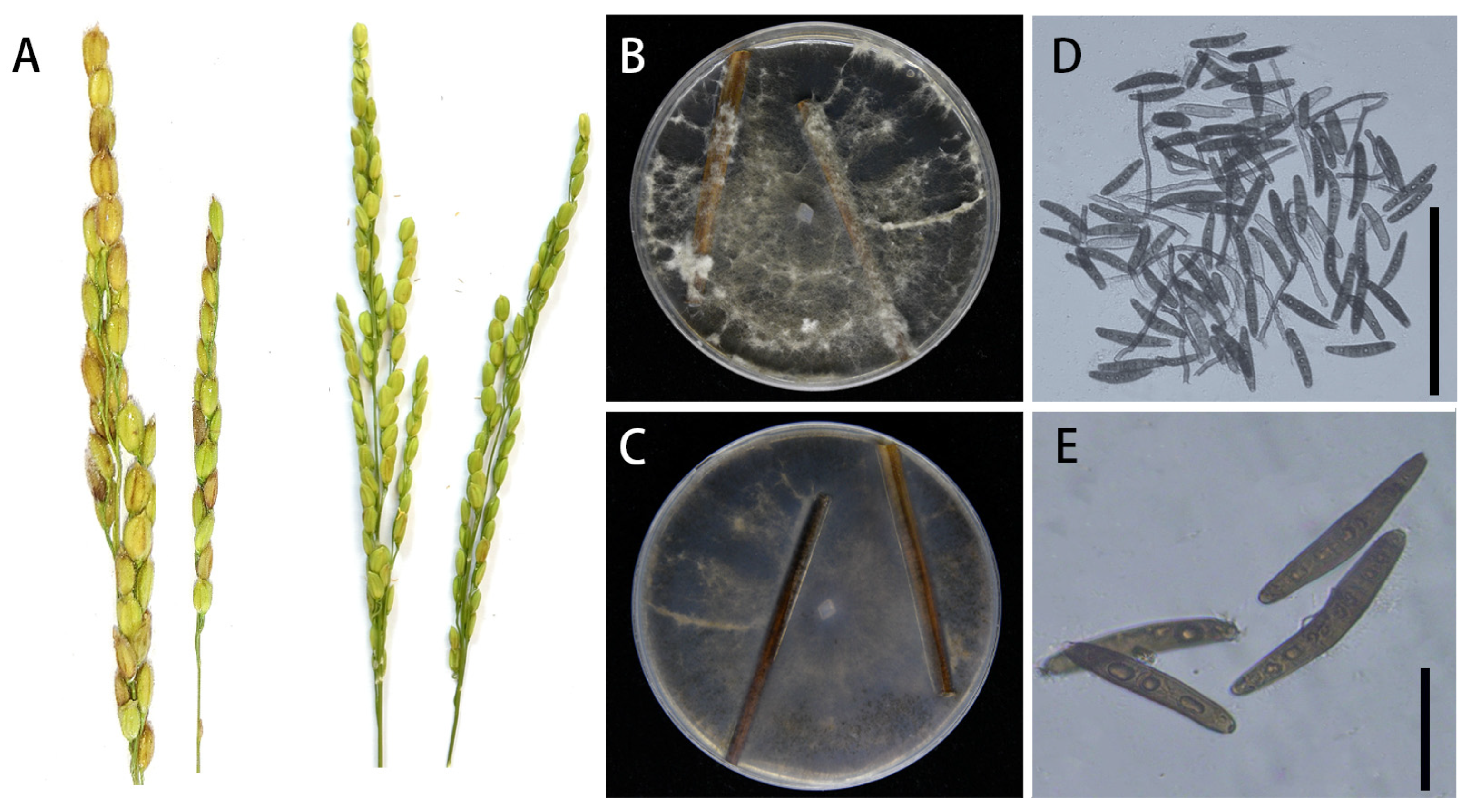
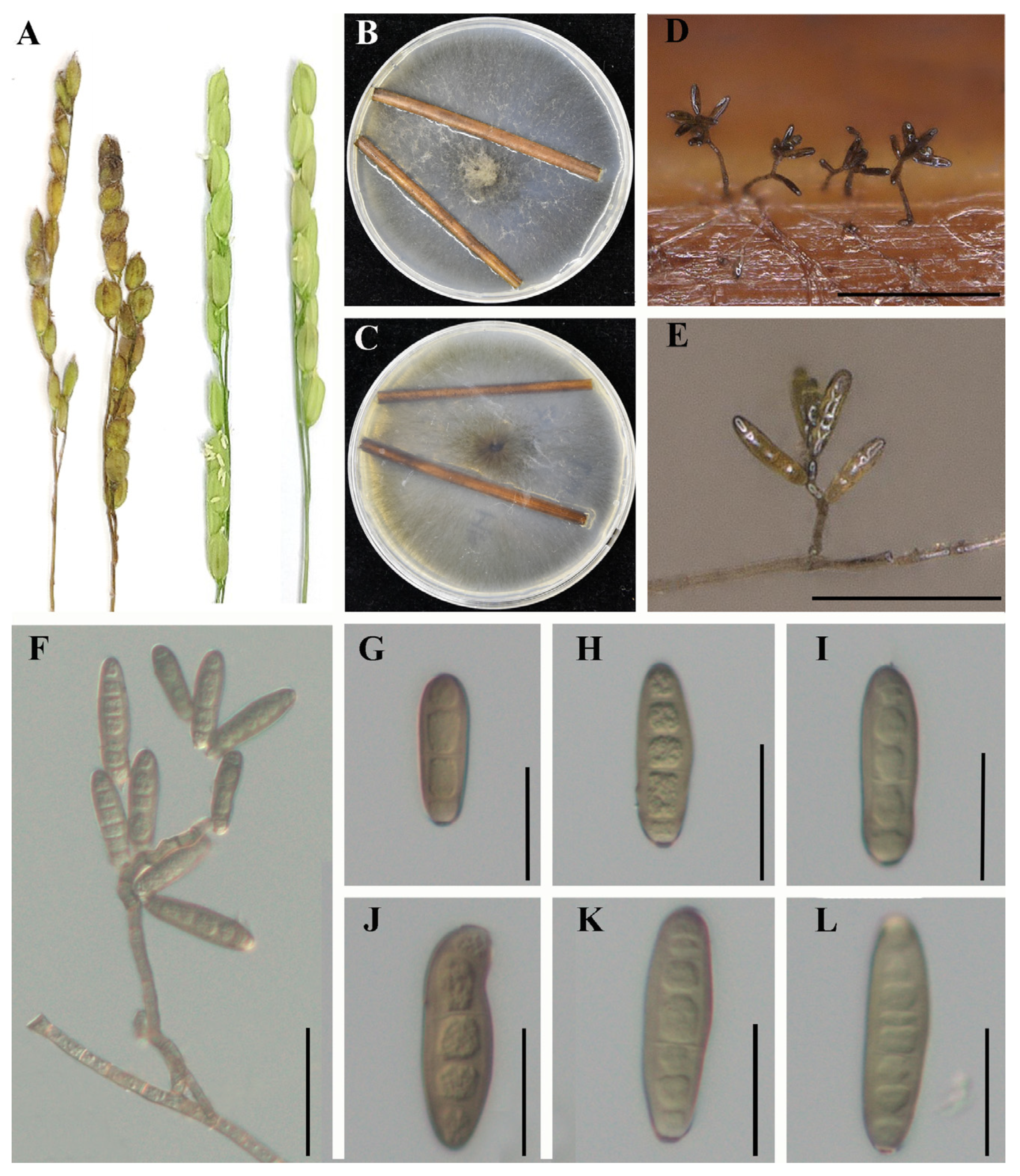
3.1. Pathogen Identification

Exserohilum rostratum LWI and Bipolaris zeicola LWII cause rice spikelet rot disease, in which the glumes of rice become chlorotic at the initial stage of the disease and brown spots develop over time, reducing rice yield and quality. E. rostratum LWI and B. zeicola LWII isolated from diseased spike parts were incubated on potato agar medium at 28 °C for 7 d in a 12 h photoperiod incubator, after which they were analyzed morphologically. In order to allow E. rostratum LWI and B. zeicola LWII to produce spores, water agar medium with wheat straw (TWA-W) was used. We used TWA-A medium at 28 °C for 5 d to produce conidia for E. rostratum LWI and B. zeicola LWII. The colonies of E. rostratum LWI were characterized by the production of a large number of white aerial hyphae with a raised center, which gradually turned brown in the center and white on the outer edge. When the conidia were observed under the microscope, they were brown in color and had an elongated oval shape with a central umbilical point at the base, with 3–8 septa and sizes of 48–80 μm × 9–19 μm. In contrast, the colonies of B. zeicola LWII had neat, dark grey edges, and the hyphae were white initially, then gradually became dark brown in color in the center. The conidia were observed to be slightly wider in the middle and taper at the ends, with bluntly rounded basal cells, brown in color, with 6–10 septa and sizes of 45–80 μm × 10–15 μm. The strainsLWI and LWII were later identified as E. rostratum and B. zeicola based on a phylogenetic tree constructed from the ITS and single-copy homologous genes results. E. rostratum LWI and B. zeicola LWII were inoculated on rice spikes by spray inoculation, respectively, and rice spikes sprayed with both showed typical symptoms, while the control group showed no symptoms (Figure 1A, Figure 2A), satisfying Koch’s hypothesis. Reisolation of the pathogenic fungi from infested rice spikes confirmed that these symptoms, which had the same morphological characteristics as the original pathogen, were consistent with the characteristics described for E. rostratum LWI and B. zeicola LWII.

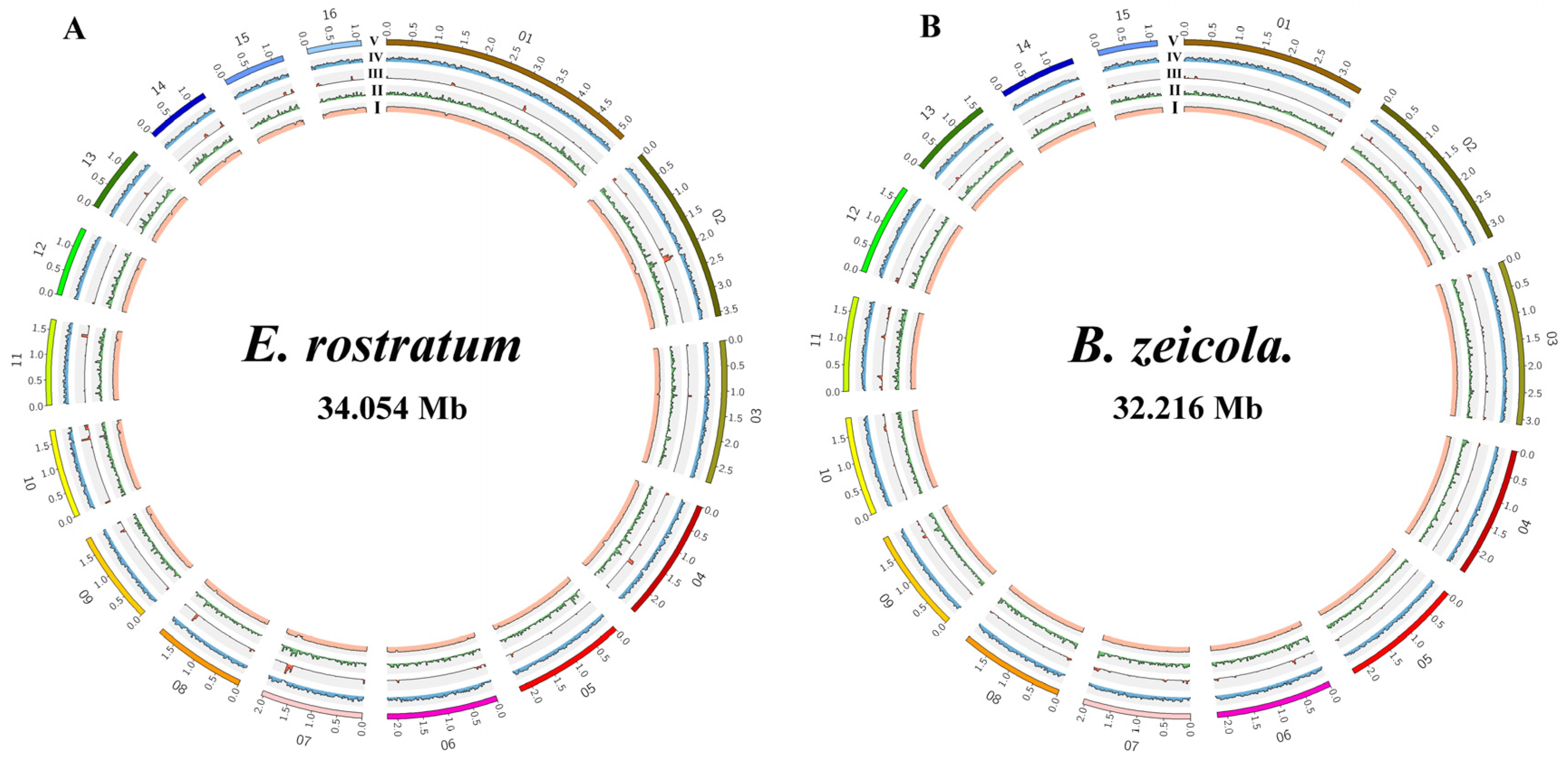
3.2. Genome Sequencing and Assembly

Based on Illumina reads, we sequenced and assembled E. rostratum LWI and B. zeicola LWII, and all high-quality data from E. rostratum LWI and B. zeicola LWII were evaluated using the k-mer analysis. The expected depths of the k-mers correspond to sequencing depths of 122x and 93.8x, respectively, and their sizes were estimated by the k-mer analysis. E. rostratum LWI was estimated to be 34.909 Mb with 93.8% non-repeated sequences, while B. zeicola LWII was 40.011 Mb with 74.2% non-repeated sequences (Figure S1). Afterward, Novogene’s PacBio Sequel sequencing platform was used for the whole-genome sequencing of E. rostratum LWI and B. zeicola LWII; the PacBio Sequel and Illumina data were mixed, and DBG2OLC was used for assembly. Finally, both E. rostratum LWI and B. zeicola LWII were assembled at the chromosome level, with 16 and 15 chromosomes, respectively. There were putative telomeric repeats 5′-(TTAGG)n-3′ at both their F-terminal and R-termini. After high-quality control, the total sizes of E. rostratum LWI and B. zeicola LWII assembled were 34,053,972 and 32,215,838 bp, the N50 lengths were 2,207,071 bp and 2,190,445 bp, and the average GC% was 50.56 and 50.66%, respectively, (Table 1, Figure 3). A comparison of genome assemblies showed that E. rostratum LWI was larger than B. zeicola LWII in terms of genome size and the number of predicted genes.
The repeated sequence of E. rostratum LWI was 1,259,007 bp and that of B. zeicola LWII was 3,747,661 bp, accounting for 3.70 and 11.63% of the genomeassembly, respectively. These repeats mainly included DNA repeats, long interspersed nuclear elements (LINEs), long terminal repeats (LTRs), and unclassified repeats. The genome of E. rostratum LWI had the largest proportion of LTRs at 1.58%, and the proportion of DNA transposons was 0.33%, while the proportion of DNA transposons in B. zeicola LWII was higher at 3.39%, and the proportion of LTRs was 2.35% (Table S1, Figure 3). Comparing the repeat sequence results of E. rostratum LWI and B. zeicola LWII, neither E. rostratum LWI nor B. zeicola LWII contained short-interspersed elements (SINEs); only B. zeicola LWII contained LINEs, B. zeicola LWII had more unclassified repeats than E. rostratum LWI, and only E. rostratum LWI contained small RNA. Finally, the G + C content, repeat sequence, LTR, gene density, and gene fragment sizes of E. rostratum LWI and B. zeicola LWII were visualized. Finally, the G + C content, repeat sequence, LTRs, gene density, and gene fragment size of B. zeicola LWII were visualized. An analysis of the differences between the two genomes indicated that the genome size and the number of predicted genes of E. rostratum LWI were larger than that of B. zeicola LWII, the genome fragment size of some LWI fragments was larger than that of B. zeicola LWII, and the LTRs of E. rostratum LWI and B. zeicola LWII differed (Figure 3).

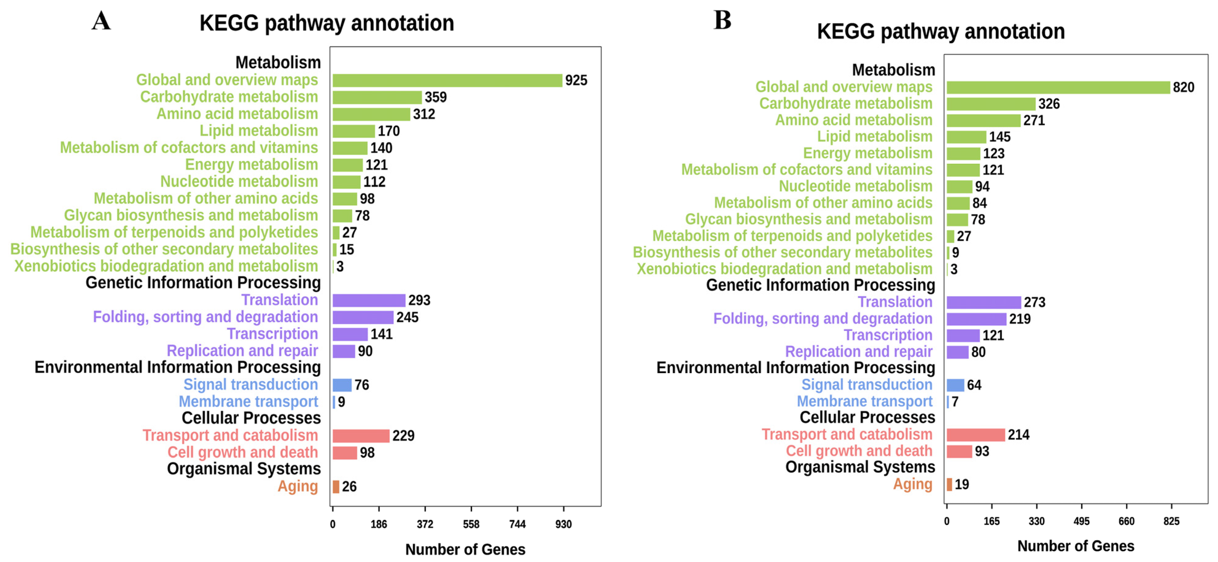
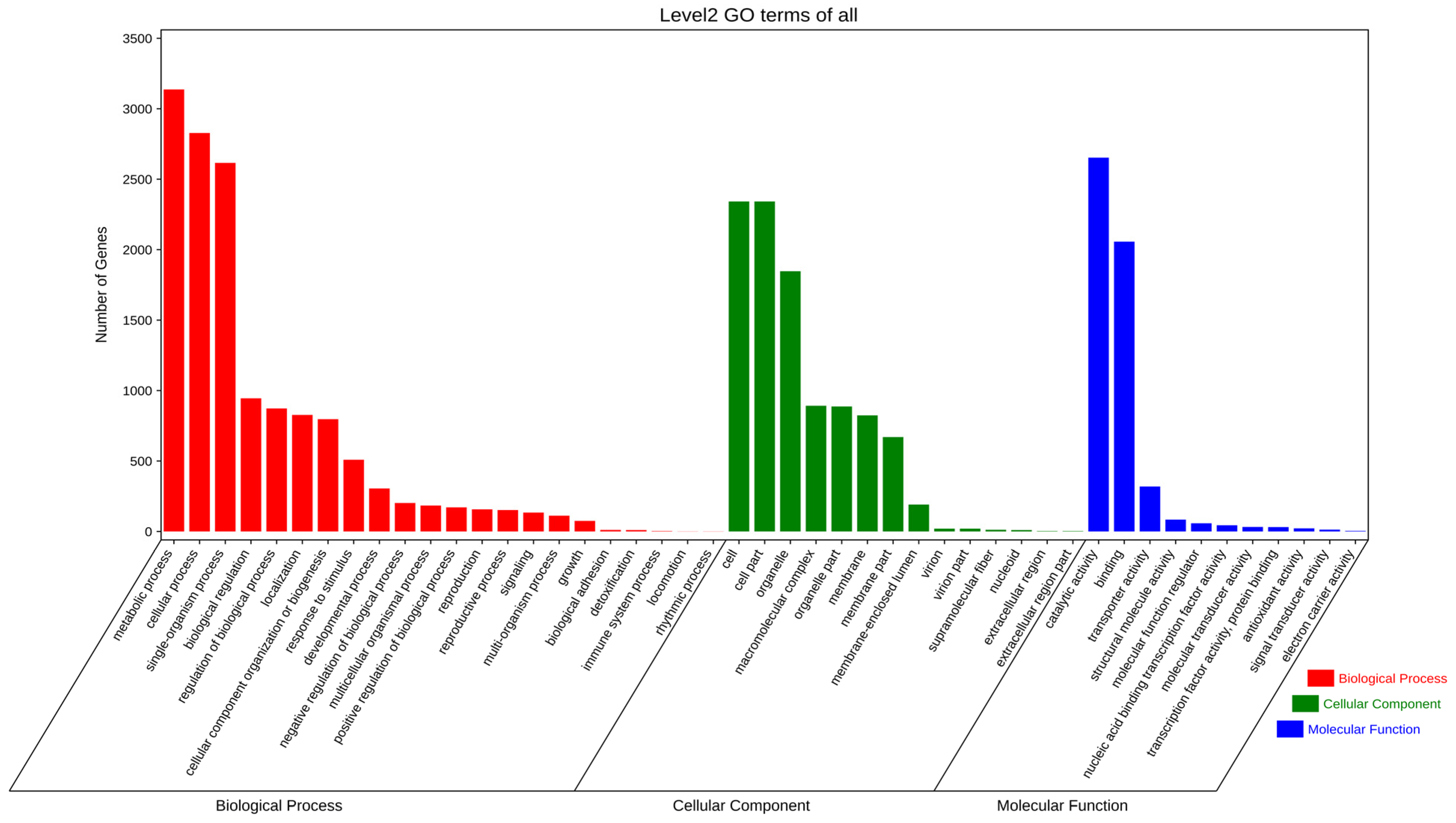
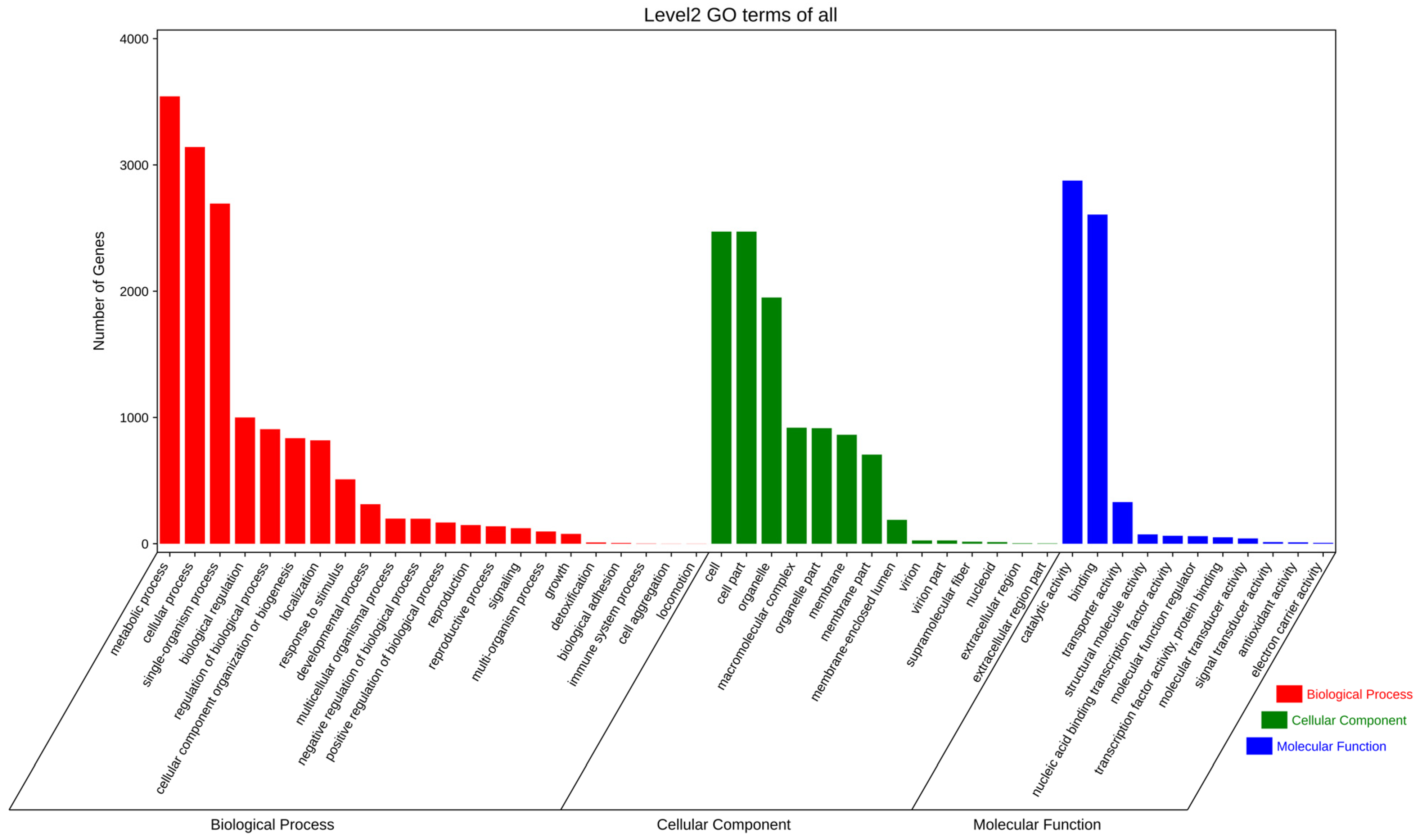
3.3. Gene Prediction and Annotation

Based on homology prediction and de novo prediction methods, we combined different software to identify and integrate protein-coding genes. According to our prediction, E. rostratum LWI and B. zeicola LWII contained 10,457 and 10,108 protein-coding genes, respectively, with an average length of 551 or 583 bases. We used different databases for annotation, such as the Nr, KEGG, GO, KOG, Pfam, PHI-base, and CAZy databases, to annotate E. rostratum LWI and B. zeicola LWII (Table 2).
In the Nr annotation, E. rostratum LWI had the highest matching degree with Setosphaeria turcica (6943), accounting for 67.92% of the total number of genes predicted by Nr, indicating that S. turcica has a close genetic relationship. B. zeicola LWII had the highest matching degree with B. zeicola (7648), accounting for 75.95% of the total genes predicted by Nr, indicating that B. zeicola is closely related to B. zeicola LWII. Among the top nine strains with a close genetic relationship to E. rostratum LWI, the genetic relationship between E. rostratum LWI and B. zeicola LWII was not high (363), indicating that the genetic relationship between E. rostratum LWI and B. zeicola LWII is not close. E. rostratum LWI was not included in the top nine strains closest to B. zeicola LWII, and the closest E. rostratum LWI relative, S. turcica, was very low (97), further suggesting that there is no close relationship between E. rostratum LWI and B. zeicola LWII (Figure S2).
Exserohilum rostratum LWI and Bipolaris zeicola LWII had 5204 and 4485 sequences, respectively, which were annotated into 25 KOG databases. In addition to some genes with unknown functions, the number of E. rostratum LWI annotations in the KOG database was much larger than that of B. zeicola LWII. The category with the most E. rostratum LWI and B. zeicola LWII annotations was “General function prediction only” (1016 and 1079), accounting for 19.52 and 24.06% of the total number of KOG annotations, followed by “Posttranslational modification, protein turnover, chaperones” (482 and 330) and “Translation, ribosomal structure, and biogenesis” (308 and 259) (Figure 4). In the Pfam database, there were 7881 and 7966 protein genes in E. rostratum LWI and B. zeicola LWII, respectively, which were similar to known proteins in the Pfam database.
Exserohilum rostratum LWI and Bipolaris zeicola LWII were annotated using the KEGG database. A total of 10,026 genes were annotated for E. rostratum LWI and 10,051 for B. zeicola LWII. The top 21 metabolic pathways annotated by E. rostratum LWI and B. zeicola LWII were very similar. Among the five categories of “Metabolism”, “Genetic Information Processing”, “Environmental Information Processing”, “Cellular Processes”, and “Organismal Systems”, the largest number was in “Global and overview maps” (925 and 820), followed by “Carbohydrate metabolism” (359 and 326). The number of annotations of the rest of the metabolic pathways was not much different, but in some metabolic pathways, the number of annotations of E. rostratum LWI was more than that of B. zeicola LWII (Figure 5). Except for sequences without subject annotations, the top 10 E. rostratum LWI sequences described in KEGG were all part of the glycoside hydrolases (GH) family (GH47, GH3, GH2, GH18, and GH76), followed by “Di-copper center-containing protein”, “tyrosinase”, “aldehyde dehydrogenase”, “amidase”, and “glutathione S-transferase” carbohydrate enzymes are closely related to the pathogenicity of E. rostratum LWI. The top 10 of LWII in the KEGG sequence description also included the GH family (GH18, GH47, GH2, GH3, and GH10), followed by “protein-arginine deiminase type-4”, “endo-1”, “aldehyde dehydrogenase”, and “cutinase”. The pathogenicity of B. zeicola LWII is not only related to carbohydrate enzymes but also associated with specific proteins.
Using the GO database to annotate the functions of E. rostratum LWI (Figure 6) and B. zeicola LWII (Figure 7), the 4582 and 5282 annotations of E. rostratum LWI and B. zeicola LWII were divided into three categories: “cellular components”, “molecular functions”, and “biological processes”. The clusters of E. rostratum LWI and B. zeicola LWII annotations were similar. The most annotated group of “Cellular component” was “cell and cell part”, the most annotated “Molecular function” was “catalytic activity and binding”, and the most annotated “Biological processes” was “metabolic process and cellular process”.

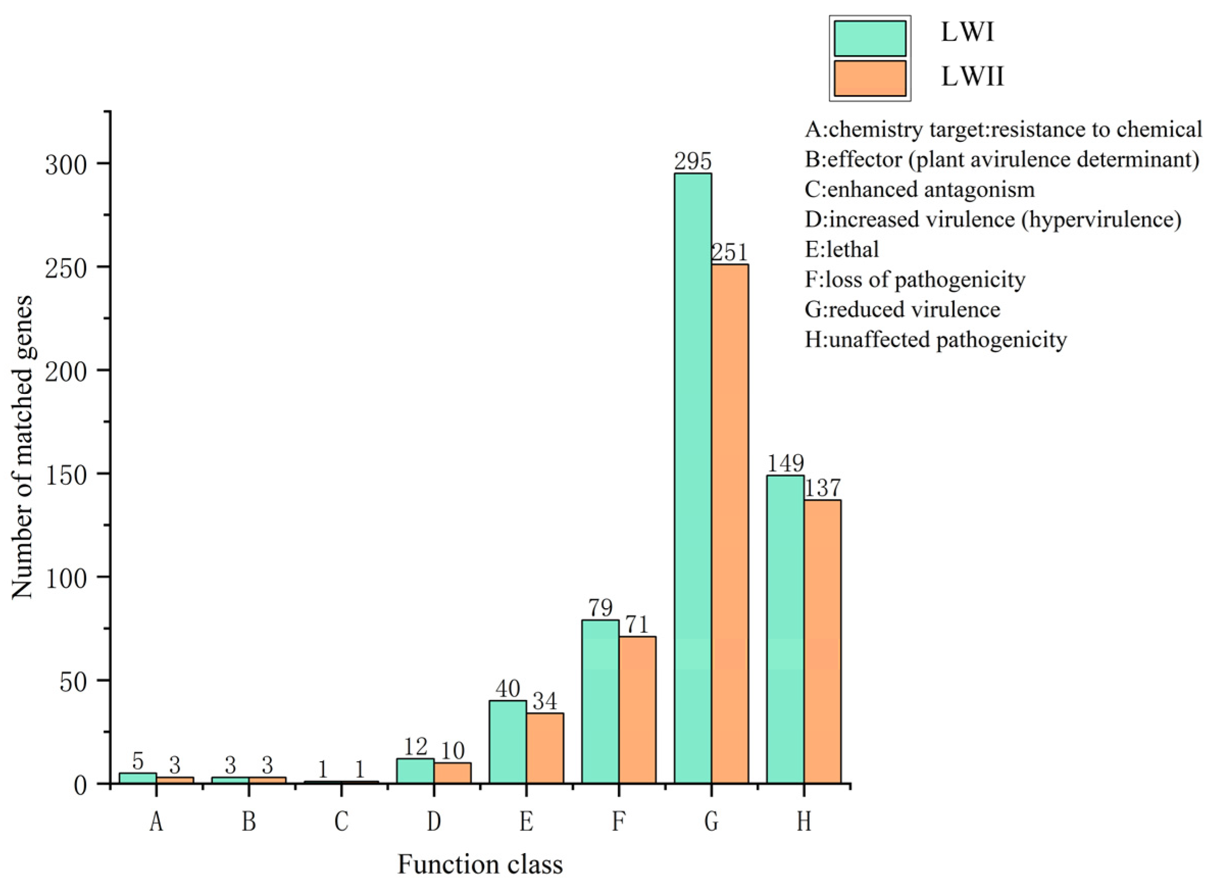
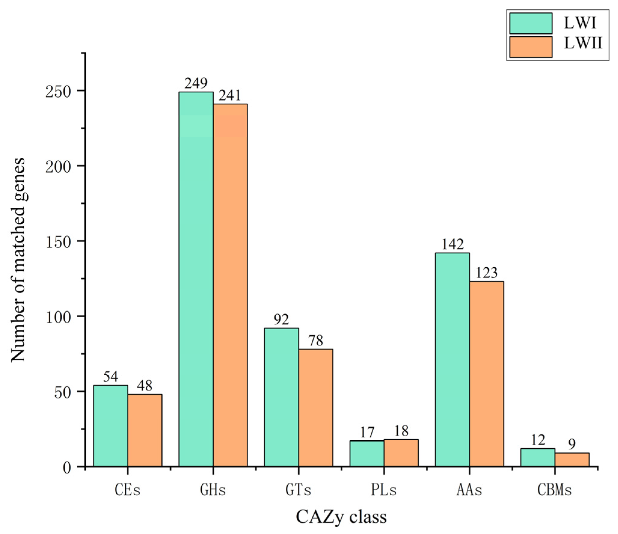
3.4. Prediction and Analysis of Pathogenicity-Related Genes

In the screening of secretory proteins, SingalP [34] was used to identify 1070 proteins containing secretory signals for E. rostratum LWI and 893 proteins for B. zeicola LWII. Next, the common regions of TMHMM [35] and Phiobius [36] were used to identify 915 proteins without a transmembrane structural domain in E. rostratum LWI and 711 in B. zeicola LWII. Further, a combination of the WOLF POSR [38] and ProtComp [37] analyses was used to find 504 proteins belonging to the extracellular secretory type for E. rostratum LWI and B. zeicola LWII, respectively, 420 proteins belonging to the extracellular secretory type for B. zeicola LWII, and the remaining 325 and 223 protein sequences for E. rostratum LWI and B. zeicola LWII with signal peptides but translocated to different organelles or plasma membranes in the cell. Finally, using Pred GPI [39] to remove ankyrins, 494 secreted proteins were found in E. rostratum LWI and 382 in B. zeicola LWII. The effector protein was then found from the screened secreted proteins. We first screened amino acids with less than 300 amino acids and used SnapGene software to screen more than four cysteine genes. Finally, using EffectorP 3.0, we screened out 164 and 123 effector proteins in E. rostratum LWI and B. zeicola LWII, respectively.
Exserohilum rostratum LWI and Bipolaris zeicola LWII were predicted to have 508 and 447 genes with ≥60% homology in the PHI database, respectively (Figure 8).Among these, E. rostratum LWI and B. zeicola LWII were the most enriched for “reduced virulence” (295 and 251) and “unaffected pathogenicity” (149 and 137).In other classifications, the difference in the number of E. rostratum LWI and B. zeicola LWII was not large. For E. rostratum LWI and B. zeicola LWII, the most critical annotated genes for pathogenicity (hypervirulence) were 12 and 10, respectively.
During the initial stages of infection, pathogens can use CAZymes primarily to degrade the polysaccharide components of the host cell wall [46,47]. We annotated E. rostratum LWI and B. zeicola LWII using the CAZy database to determine which specific enzymes were associated with the host range and pathogenesis. There were 566 and 517 CAZy annotations for E. rostratum LWI and B. zeicola LWII, respectively. In general, the difference in the number of genes assigned to the six categories between E. rostratum LWI and B. zeicola LWII was small. The CAZymes encoding gene models were divided into six major categories, with 54 and 48 carbohydrate esterases (CEs)for E. rostratum LWI and B. zeicola LWII, respectively, 249 and 241 occupied by glycoside hydrolases (GHs), 251 and 244 by glycosyltransferases (GTs) with 92 and 78, polysaccharide lyases (PLs) with 17 and 18, auxiliary modular enzymes (AAs) with 142 and 123, and carbohydrate-binding modules (CBMs) with 12 and 9, respectively (Figure 9).
Among the total CAZy annotated in E. rostratum LWI and B. zeicola LWII, CEs, PLs, and GHs accounted for 57.9% in E. rostratum LWI and 59.4% in B. zeicola LWII. The GH enzyme family breaks the “glycosidic bond” in carbohydrates or sugars. The number of GHs families in E. rostratum LWI and B. zeicola LWII was comparable; the most in E. rostratum LWI were GH3(16), GH18(13), and GH47(10) and in B. zeicola LWII were GH18(13), GH3(11), and GH31(9). The intersection analysis of E. rostratum LWI and B. zeicola LWII secreted protein genes, PHI genes, and CAZy gene annotation results; there were 8 genes in E. rostratum LWI and 13 genes in B. zeicola LWII in the intersection part (Figure S3). Combined with the characteristics of the secreted proteins and the annotation results of the two databases, we speculated that the gene IDs of the overlapping parts of E. rostratum LWI and B. zeicola LWII might be the key pathogenic genes of these two fungi infecting rice.

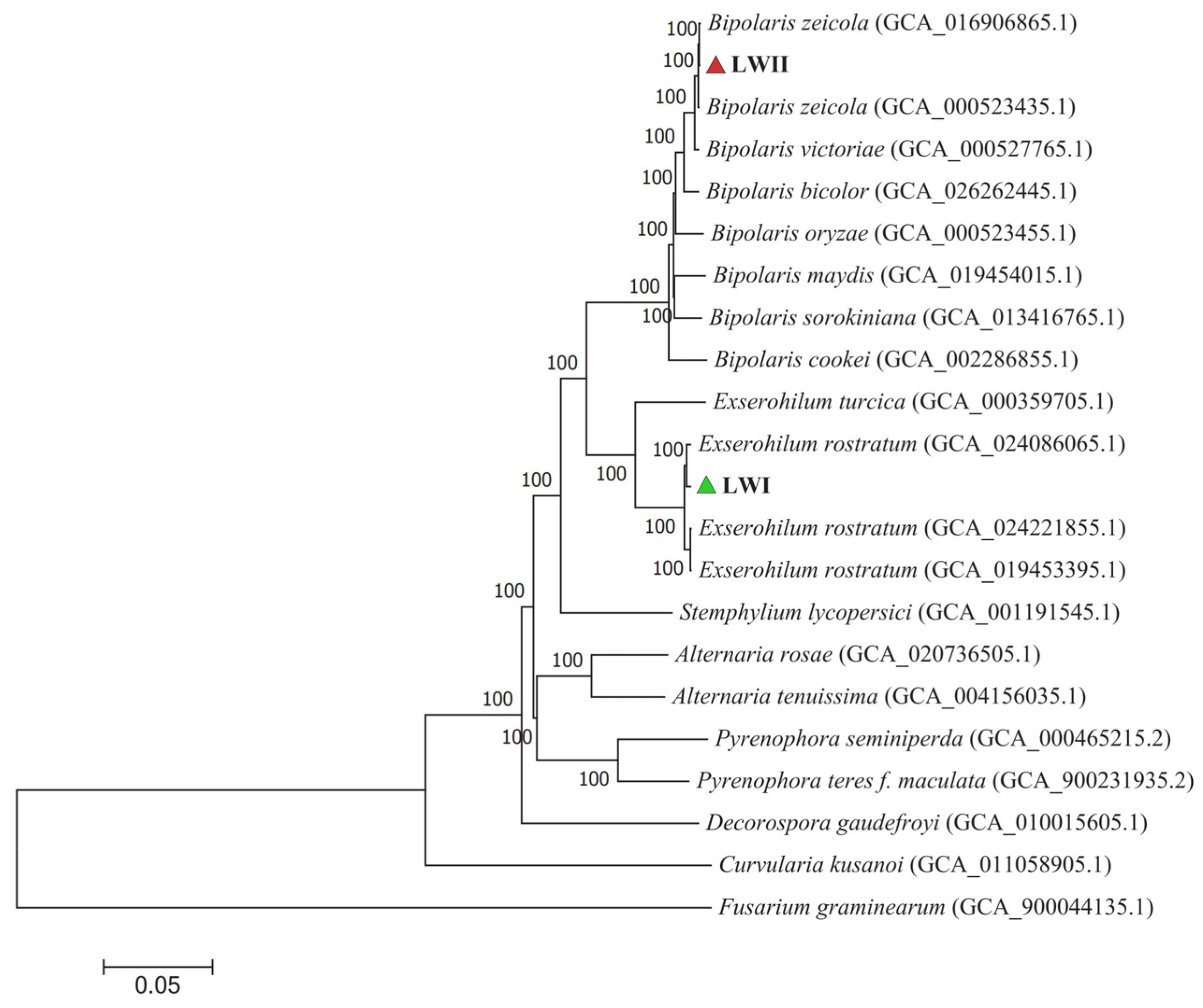
3.5. Phylogenomics Analysis

Clustering gene expression data thus provides important insights into gene coregulation and gene cellular function. A phylogenetic tree was constructed based on the results of single-copy homologous genes identified by gene family clustering, and the phylogenomic relationships between E. rostratum LWI and B. zeicola LWII and the remaining 20 strains were investigated using Fusarium graminearum PH-1 as the outgroup (Table S2). The genome-wide map of 2072 single-copy orthologue genes shared by the genomes with 18 strains was well supported, and all its branches had bootstrap values of 100, indicating the confidence level of the branch. From the phylogenetic tree, E. rostratum LWI and Exserohilum rostratum (Genome assembly: GCA_024086065.1) were clustered on one branch, and the support rate was as high as 100%, indicating that E. rostratum LWI is extremely closely related to this strain. B. zeicola LWII and Bipolaris zeicola (Genome assembly: GCA_016906865.1) were clustered on one branch, and the support rate was as high as 100%, indicating that B. zeicola LWII is closely related to this strain. The branches of E. rostratum LWI and B. zeicola LWII were far apart, indicating that their phylogenetic relationship is not close (Figure 10).

4. Discussion

In recent years, the impact of rice spikelet rot disease on human health and rice production cannot be underestimated. It is caused by various fungi in China [3]. However, rice spikelet rot disease caused by Exserohilumrostratum and Bipolariszeicola, investigated in the present study, has rarely been reported in China. E. rostratum is a plant pathogen with a wide range of hosts and has a high impact on grasses and Poaceae [48,49] and was first identified in rice in Venezuela [50]. Researchers in Algeria have found that E. rostratum is more invasive to maize than Bipolaris sorokiniana in pathogenicity tests of E. rostratum and B. sorokiniana [51]. In humans, the fungus has mainly caused keratitis and skin diseases [8,52]. E. rostratum also causes a number of diseases in animals, and a horse in Florida with chronic obstructive rhinitis was identified as the cause of E. rostratum [53]. Researchers in Brazil have, for the first time, isolated the pathogen E. rostratum in goats with rhinitis, which is unprecedented in goats [54]. At the same time, studies have shown that the Brn1 gene can help the study of intraspecific variations of E. rostratum, and the Brn1 gene can also identify E. rostratum [55]. B. zeicola can cause diseases in maize leaves and other tissues, ultimately leading to a reduced maize yield. Researchers from Korea developed species-specific primers for PCR (Bz-F/Bz-R) and recommended this method for rapid and accurate laboratory identification of B. zeicola and the diagnosis of maize diseases caused by B. zeicola [56,57]. Meanwhile, host-specific toxins produced by Cochliobolus species have been shown to enhance the virulence of pathogens [58]. The HC toxin is a non-ribosomal peptide generated by B. zeicola, and it induces a high acetylation of histones upon infection with maize types carrying just the susceptible gene [59,60]. Although B. zeicola is primarily pathogenic to maize, B. zeicola is also pathogenic to other gramineous crops. B. zeicola was first found to be pathogenic to barley in Argentina, and the symptoms were similar to those of B. zeicola to maize [61]. Egyptian researchers have found that B. zeicola can cause wilting, severe rot, and death in rice seedlings [62]. Rice spikelet rot disease caused by multiple fungi had a major impact on the production of rice in recent years and lacks effective preventive measures. However, due to the rapid development of high-throughput sequencing technology, genome sequencing, and the maturity of bioinformatics analysis tools, these techniques are now widely used to study pathogenic fungal pathogenicity and disease resistance. In this study, Illumina and PacBio Sequel were used for the whole-genome sequencing, assembly, and annotation of E. rostratum and B. zeicola.
The genome size of E. Rostratum LWI was 34,053,972 bp, assembled into 16 chromosomes, and the genome B. zeicola LWII was 32,215,838 bp, assembled into 15 chromosomes. To further understand the gene’s function, the annotation analysis of E. rostratum LWI and B. zeicola LWII was performed. Using GO terms, 4582 E. rostratum LWI and 5282 B. zeicola LWII genes were annotated in total. The protein sequences of E. rostratum LWI were mainly annotated in “Biological Process”, with a total of 14,050, and B. zeicola LWII were mainly annotated in “Cellular Component”, with a total of 10,574. E. rostratum LWI and B. zeicola LWII had 10,026 and 10,051 protein genes assigned to the KEGG pathway, respectively. The pathway with the largest proportion was “Global and overview maps”, with 925 in E. rostratum LWI and 820 in B. zeicola LWII. E. rostratum LWI and B. zeicola LWII had 5615 and 4485 genes annotated in the KOG database, respectively.
The cell wall of plants is the first barrier to prevent the invasion of pathogenic fungi. In order to successfully invade, pathogenic fungi degrade the cell wall, including upregulating carbohydrate hydrolases and enzymes related to plant cell wall degradation [63,64,65]. Studies have shown that the GH, PL, and CE superfamilies are closely related to pathogenicity. In this study, E. rostratum LWI and B. zeicola LWII had far more GH families than PL and CE families, and GH3 and GH18 were slightly higher than those of other GH families. GH3 plays a role in promoting the penetration of plant cell walls in the interactions between ascomycetes and plants [66], while GH18 is widely present in fungi, bacteria, insects, plants, and animals, and its role is to promote pathogenic bacteria. It is likely that these enzymes play a critical role in degrading plant cell walls in our two fungi by colonizing, inhibiting host immune responses, and even acting as virulence factors (3). To completely understand how pathogenic fungi degrade cell walls, it is vital to investigate the secretome and secreted effectors that play a role between hypha and host [67,68]. After a comprehensive analysis using various software packages, we identified 494 secreted proteins and 164 effectors proteins in E. rostratum LWI and 382 secreted proteins and 123 effector proteins in B. zeicola LWII. In the PHI annotation, E. rostratum LWI and B. zeicola LWII had 508 and 447 proteins associated with pathogenic genes, respectively. Finally, an integrated analysis of the secreted proteins, PHI, and CAZy of E. rostratum LWI and B. zeicola LWII was carried out. There were 8 and 13 genes in the intersection of the above three results in E. rostratum LWI and B. zeicola LWII, respectively. These genes are related to the invasion, colonization, and spread of fungi to plants. The disease process is closely related. As a result of these findings, we now have better knowledge of the interactions between E. rostratum LWI and B. zeicola LWII and rice, which provides more control options. At the same time, we assembled E. rostratum LWI and B. zeicola LWII at the chromosome level, greatly improving the assembly quality and laying the foundation for subsequent comparative genomics and resequencing.

5. Conclusions

In this study, we assembled E. rostratum LWI and B. zeicola LWII at the chromosomal level and achieved high-quality genomes of organisms, adding to and upgrading their respective genome databases. We investigated the two fungi’s pathogenic causes from the viewpoint of the genome using the whole-genome sequencing analysis; a comparison of functional databases showed that some genes might be crucial for fungus–host interactions. The results promote the pathogenic study of E. rostratum and B. zeicola and offer important data sources for investigating rice spikelet rot disease.

Supplementary Materials

The following supporting information can be downloaded at: https://www.mdpi.com/article/10.3390/jof9020177/s1: Figure S1: Assessing the genome size of E. rostratum LWI (A) and B. zeicola LWII (B). Figure S2: Predicted proteins between different fungal species using the Nr database for the E. rostratum LWI (A) and B. zeicola LWII (B) genomes. Figure S3: Intersection of the annotation results of the secreted protein genes, PHI genes, and CAZy genes of E. rostratum LWI and B. zeicola LWII. Table S1: Statistics of repeated sequence classification in the E. rostratum LWI and B. zeicola LWII genomes. Table S2: Strain data of 20 species used in a comparative genome analysis.

Author Contributions

Author Contributions: Conceptualization, K.H., C.Z., X.C. and C.L.; methodology, K.H., Q.Z. and C.L.; formal analysis, K.H. and C.Z.; investigation, K.H., X.W., M.Z., J.L., Q.Z., S.W. and C.L.; data curation, K.H. and C.L.; writing—original draft preparation, K.H., C.Z. and C.L.; writing—review and editing, K.H., J.L., C.Z., Q.Z., X.C., M.Z. and C.L.; supervision, C.L., Y.W. and X.C.; and software, K.H., Q.Z. and C.L. All authors have read and agreed to the published version of the manuscript.

Funding

This research was funded by the National Natural Science Foundation of China (Grant No. 32260653), Top Technology Talent Project from Guizhou Education Department of China (Grant No. Qian jiao ji [2022]074), Guizhou Provincial Science and Technology Projects of China (Grant No. ZK [2021]142), Talent Introduction Research Project of Guizhou University of China (Grant No. [2020]35), and the Cultivation Project of Guizhou University of China (Grant No. (2020)24 and Grant No. (2019)13).

Institutional Review Board Statement

Not applicable.

Informed Consent Statement

Not applicable.

Data Availability Statement

The genome sequence data and assemblies of E. rostratum LWI reported in this paper are associated with the NCBI BioProject: PRJNA902501, BioSample: SUB12290528, and Accession Numbers: CP111151–CP111166 in GenBank. The genome sequence data and assemblies of B. zeicola LWII reported in this paper are associated with the NCBI BioProject: PRJNA902512, BioSample: SUB12291265, and Accession Numbers: CP111167–CP111181 in GenBank.

Acknowledgments

The author is especially grateful to Xujun Chen (China Agricultural University) for providing two fungal strains, Novogene Technology for the sequencing of the fungal strains and the Key Laboratory of Mountain Microbiology of Guizhou University College of Agriculture for providing basic experimental equipment.

Conflicts of Interest

The authors declare no conflict of interest.

References

  1. Wang, C.; Yang, Y.; Yuan, X.; Xu, Q.; Feng, Y.; Yu, H.; Wang, Y.; Wei, X. Genome-wide association study of blast resistance in India rice. BMC Plant Biol. 2014, 14, 311. [Google Scholar] [CrossRef] [PubMed] [Green Version]
  2. Busungu, C.; Taura, S.; Sakagami, J.-I.; Anai, T.; Ichitani, K. High-resolution mapping and characterization of xa42, a resistance gene against multiple Xanthomonas oryzae pv. oryzae races in rice (Oryza sativa L.). Breed. Sci. 2018, 68, 188–199. [Google Scholar] [CrossRef] [Green Version]
  3. Huang, S.W.; Wang, L.; Liu, L.M.; Liu, E.Y.; Hou, E.Q.; Xiao, D.F.; Fan, Z.L. Isolation, Identification and Biological Characters of Pathogens of Rice Spikelet Rot Disease. Chin. J. Rice Sci. 2012, 26, 341. [Google Scholar] [CrossRef]
  4. Zhao, C.Y.; Liu, W.; Guo, Z.J.; Chen, X.J. Identification of pathogens causing brown spot disease on rice glume. Acta Phytophy Sin. 2022, 52, 999–1002. [Google Scholar] [CrossRef]
  5. Alcorn, J.L. Cochliobolus ellisii sp.nov. Trans. Br. Mycol. Soc. 1983, 81, 172–174. [Google Scholar] [CrossRef]
  6. Nelson, R.R. Cochliobolus vietoriae, the perfect state of Helminthosporium victoriae. Phytopathology 1960, 50, 774–775. [Google Scholar]
  7. Hernández-Restrepo, M.; Madrid, H.; Tan, Y.P.; Cunha, K.C.D.; Gené, J.; Guarro, J.; Crous, P.W. Multi-locus phylogeny and taxonomy of Exserohilum. Persoonia 2018, 41, 71–108. [Google Scholar] [CrossRef]
  8. Gauthier, G.M.; Keller, N.P. Crossover Fungal Pathogens: Crossover fungal pathogens: The biology and pathogenesis of fungi capable of crossing kingdoms to infect plants and humans. Fungal Genet. Biol. 2013, 61, 146–157. [Google Scholar] [CrossRef]
  9. Graham-Brown, R. Atlas of Clinical Dermatology, 3rd edn. Br. J. Dermatol 2003, 149, 445. [Google Scholar] [CrossRef]
  10. Yadav, V.; Yang, F.; Reza, M.H.; Liu, S.; Valent, B.; Sanyal, K.; Naqvi, N.I. Cellular Dynamics and Genomic Identity of Centromeres in Cereal Blast Fungus. MBio 2019, 10, e01581-19. [Google Scholar] [CrossRef] [Green Version]
  11. Manamgoda, D.S.; Cai, L.; McKenzie, E.H.C.; Crous, P.W.; Madrid, H.; Chukeatirote, E.; Shivas, R.G.; Tan, Y.P.; Hyde, K.D. A phylogenetic and taxonomic re-evaluation of the Bipolaris-Cochliobolus-Curvularia Complex. Fungal Divers. 2012, 56, 131–144. [Google Scholar] [CrossRef]
  12. Praveena, R.; Kozhamburath, A.; Jeevalatha, A.; Bhat, A.I.; Krishnamurthy, K.S. Association of Exserohilum rostratum with ginger: Morphological characterization, phylogenetic relationships and pathogenicity assays. Australas. Plant Path. 2022, 51, 333–344. [Google Scholar] [CrossRef]
  13. Condon, B.J.; Leng, Y.; Wu, D.; Bushley, K.E.; Ohm, R.A.; Otillar, R.; Martin, J.; Schackwitz, W.; Grimwood, J.; MohdZainudin, N.; et al. Comparative Genome Structure, Secondary Metabolite, and Effector Coding Capacity across Cochliobolus Pathogens. PLoS Genet. 2013, 9, e1003233. [Google Scholar] [CrossRef] [PubMed] [Green Version]
  14. Xia, X.; Wang, Y.; Zhou, S.; Liu, W.; Wu, H. Genome Sequence Resource for Bipolaris zeicola, the Cause of Northern Corn Leaf Spot Disease. Phytopathology 2022, 112, 1192–1195. [Google Scholar] [CrossRef] [PubMed]
  15. Litvintseva, A.P.; Hurst, S.; Gade, L.; Frace, M.A.; Hilsabeck, R.; Schupp, J.M.; Gillece, J.D.; Roe, C.; Smith, D.; Keim, P.; et al. Whole-Genome Analysis of Exserohilum rostratum from an Outbreak of Fungal Meningitis and Other Infections. J. Clin. Microbiol. 2014, 52, 3216–3222. [Google Scholar] [CrossRef] [PubMed] [Green Version]
  16. Ma, Q.; Cheng, C.; Geng, Y.; Zang, R.; Guo, Y.; Yan, L.; Wu, H.; Zhang, M.; Xu, C. The draft genome sequence and characterization of Exserohilum rostratum, a new causal agent of maize leaf spot disease in Chinese Mainland. Eur. J. Plant Pathol. 2023, 165, 57–71. [Google Scholar] [CrossRef]
  17. Marçais, G.; Kingsford, C. A fast, lock-free approach for efficient parallel counting of occurrences of k-mers. Bioinformatics 2011, 27, 764–770. [Google Scholar] [CrossRef] [Green Version]
  18. Chen, N. Using RepeatMasker to Identify Repetitive Elements in Genomic Sequences. Curr. Protoc. Bioinform. 2004, 5, 4–10. [Google Scholar] [CrossRef]
  19. Stanke, M.; Keller, O.; Gunduz, I.; Hayes, A.; Waack, S.; Morgenstern, B. AUGUSTUS: Ab initio prediction of alternative transcripts. Nucleic Acids Res. 2006, 34, W435–W439. [Google Scholar] [CrossRef] [Green Version]
  20. Ter-Hovhannisyan, V.; Lomsadze, A.; Chernoff, Y.O.; Borodovsky, M. Gene prediction in novel fungal genomes using an ab initio algorithm with unsupervised training. Genome Res. 2008, 18, 1979–1990. [Google Scholar] [CrossRef] [Green Version]
  21. Haas, B.J.; Salzberg, S.L.; Zhu, W.; Pertea, M.; Allen, J.E.; Orvis, J.; White, O.; Buell, C.R.; Wortman, J.R. Automated eukaryotic gene structure annotation using EVidenceModeler and the Program to Assemble Spliced Alignments. Genome Biol. 2008, 9, R7. [Google Scholar] [CrossRef] [PubMed]
  22. Bolger, A.M.; Lohse, M.; Usadel, B.T. Trimmomatic: A flexible trimmer for Illumina sequence data. Bioinformatics 2014, 30, 2114–2120. [Google Scholar] [CrossRef] [PubMed] [Green Version]
  23. Luo, R.B.; Liu, B.H.; Xie, Y.L.; Li, Z.Y.; Huang, W.H.; Yuan, J.Y.; He, G.Z.; Chen, Y.X.; Pan, Q.; Liu, Y.J.; et al. SOAPdenovo2: An empirically improved memory-efficient short-read de novo assembler. GigaScience 2012, 1, 2047–2217X. [Google Scholar] [CrossRef] [PubMed]
  24. Boetzer, M.; Henkel, C.V.; Jansen, H.J.; Butler, D.; Pirovano, W. Scaffolding pre-assembled contigs using SSPACE. Bioinformatics 2011, 27, 578–579. [Google Scholar] [CrossRef] [PubMed] [Green Version]
  25. Koren, S.; Walenz, B.P.; Berlin, K.; Miller, J.R.; Bergman, N.H.; Phillippy, A.M. Canu: Scalable and accurate long-read assembly via adaptive k-mer weighting and repeat separation. Genome Res. 2017, 27, 722–736. [Google Scholar] [CrossRef] [PubMed] [Green Version]
  26. Xiao, C.L.; Chen, Y.; Xie, S.Q.; Chen, K.N.; Wang, Y.; Luo, F.; Xie, Z. MECAT: An ultra-fast mapping, error correction and de novo assembly tool for single-molecule sequencing reads. Nat. Methods 2017, 14, 1072–1074. [Google Scholar] [CrossRef]
  27. Hu, J.; Fan, J.P.; Sun, Z.Y.; Liu, S.L. NextPolish: A fast and efficient genome polishing tool for long-read assembly. Bioinformatics 2020, 36, 2253–2255. [Google Scholar] [CrossRef]
  28. Walker, B.J.; Abeel, T.; Shea, T.; Priest, M.; Abouelliel, A.; Sakthikumar, S.; Cuomo, C.A.; Zeng, Q.; Wortman, J.; Young, S.K.; et al. Pilon: An Integrated Tool for Comprehensive Microbial Variant Detection and Genome Assembly Improvement. PLoS ONE 2014, 9, e112963. [Google Scholar] [CrossRef]
  29. Ye, C.X.; Hill, C.M.; Wu, S.G.; Ruan, J.; Ma, Z.S. (Sam). DBG2OLC: Efficient Assembly of Large Genomes Using Long Erroneous Reads of the Third Generation Sequencing Technologies. Sci. Rep. 2016, 6, 31900. [Google Scholar] [CrossRef] [Green Version]
  30. Delcher, A.L.; Kasif, S.; Fleischmann, R.D.; Peterson, J.; White, O.; Salzberg, S.L. Alignment of whole genomes. Nucleic Acids Res. 1999, 27, 2369–2376. [Google Scholar] [CrossRef] [Green Version]
  31. Chen, C.J.; Chen, H.; Zhang, Y.; Thomas, H.R.; Frank, M.H.; He, Y.H.; Xia, R. TBtools: An Integrative Toolkit Developed for Interactive Analyses of Big Biological Data. Mol. Plant. 2020, 13, 1194–1202. [Google Scholar] [CrossRef] [PubMed]
  32. Zhang, H.; Yohe, T.; Huang, L.; Entwistle, S.; Wu, P.Z.; Yang, Z.L.; Busk, P.K.; Xu, Y.; Yin, Y.B. dbCAN2: A meta server for automated carbohydrate-active enzyme annotation. Nucleic Acids Res. 2018, 46, W95–W101. [Google Scholar] [CrossRef] [PubMed] [Green Version]
  33. Hu, Y.; Yan, C.; Hsu, C.-H.; Chen, Q.-R.; Niu, K.; Komatsoulis, G.A.; Meerzaman, D. OmicCircos: A Simple-to-Use R Package for the Circular Visualization of Multidimensional Omics Data. Cancer Inform. 2014, 13, CIN.S13495. [Google Scholar] [CrossRef] [PubMed]
  34. Teufel, F.; Almagro Armenteros, J.J.; Johansen, A.R.; Gíslason, M.H.; Pihl, S.I.; Tsirigos, K.D.; Winther, O.; Brunak, S.; von Heijne, G.; Nielsen, H. SignalP 6.0 predicts all five types of signal peptides using protein language models. Nat. Biotechnol. 2022, 40, 1023–1025. [Google Scholar] [CrossRef] [PubMed]
  35. Hallgren, J.; Tsirigos, K.D.; Pedersen, M.D.; Armenteros, J.J.A.; Marcatili, P.; Nielsen, H.; Krogh, A.; Winther, O. DeepTMHMM predicts alpha and beta transmembrane proteins using deep neural networks. bioRxiv, 2022; preprint. [Google Scholar] [CrossRef]
  36. Käll, L.; Krogh, A.; Sonnhammer, E.L.L. Advantages of combined transmembrane topology and signal peptide prediction—The Phobius web server. Nucleic Acids Res. 2007, 35, W429–W432. [Google Scholar] [CrossRef] [Green Version]
  37. Emanuelsson, O.; Nielsen, H.; Brunak, S.; von Heijne, G. Predicting Subcellular Localization of Proteins Based on their N-terminal Amino Acid Sequence. J. Mol. Biol. 2000, 300, 1005–1016. [Google Scholar] [CrossRef] [Green Version]
  38. Horton, P.; Park, K.-J.; Obayashi, T.; Fujita, N.; Harada, H.; Adams-Collier, C.J.; Nakai, K. WoLF PSORT: Protein localization predictor. Nucleic Acids Res. 2007, 35, W585–W587. [Google Scholar] [CrossRef] [Green Version]
  39. Pierleoni, A.; Martelli, P.L.; Casadio, R. PredGPI: A GPI-anchor predictor. BMC Bioinform. 2008, 9, 392. [Google Scholar] [CrossRef] [Green Version]
  40. Urban, M.; Cuzick, A.; Seager, J.; Wood, V.; Rutherford, K.; Venkatesh, S.Y.; DeSilva, N.; Martinez, M.C.; Pedro, H.; Yates, A.D.; et al. PHI-base: The pathogen–host interactions database. Nucleic Acids Res. 2020, 48, D613–D620. [Google Scholar] [CrossRef]
  41. Li, L.; Stoeckert, C.J.; Roos, D.S. OrthoMCL: Identification of Ortholog Groups for Eukaryotic Genomes. Genome Res. 2003, 13, 2178–2189. [Google Scholar] [CrossRef]
  42. Emms, D.M.; Kelly, S.O. OrthoFinder: Phylogenetic orthology inference for comparative genomics. Genome Biol. 2019, 20, 238. [Google Scholar] [CrossRef] [PubMed] [Green Version]
  43. Katoh, K.; Misawa, K.; Kuma, K.; Miyata, T. MAFFT: A novel method for rapid multiple sequence alignment based on fast Fourier transform. Nucleic Acids Res. 2002, 30, 3059–3066. [Google Scholar] [CrossRef] [PubMed] [Green Version]
  44. Castresana, J. Selection of Conserved Blocks from Multiple Alignments for Their Use in Phylogenetic Analysis. Mol. Biol. Evol. 2000, 17, 540–552. [Google Scholar] [CrossRef] [PubMed] [Green Version]
  45. Stamatakis, A.; Aberer, A.J.; Goll, C.; Smith, S.A.; Berger, S.A.; Izquierdo-Carrasco, F. RAxML-Light: A tool for computing terabyte phylogenies. Bioinformatics 2012, 28, 2064–2066. [Google Scholar] [CrossRef] [Green Version]
  46. Bashyal, B.M.; Rawat, K.; Sharma, S.; Kulshreshtha, D.; Gopala Krishnan, S.; Singh, A.K.; Dubey, H.; Solanke, A.U.; Sharma, T.R.; Aggarwal, R. Whole Genome Sequencing of Fusarium Fujikuroi Provides Insight into the Role of Secretory Proteins and Cell Wall Degrading Enzymes in Causing Bakanae Disease of Rice. Front. Plant Sci. 2017, 8, 02013. [Google Scholar] [CrossRef] [Green Version]
  47. Ospina-Giraldo, M.D.; Griffith, J.G.; Laird, E.W.; Mingora, C. The CAZyome of Phytophthora spp.: A comprehensive analysis of the gene complement coding for carbohydrate-active enzymes in species of the genus Phytophthora. BMC Genom. 2010, 11, 525. [Google Scholar] [CrossRef] [PubMed] [Green Version]
  48. Brunings, A.M.; Datnoff, L.E.; Palmateer, A.J.; Locke, J.C.; Krause, C.R. Exserohilum Leaf Spot on Tiger Grass. Plant Health Prog. 2009, 10, 1. [Google Scholar] [CrossRef] [Green Version]
  49. Sharma, K.; Goss, E.M.; Dickstein, E.R.; Smith, M.E.; Johnson, J.A.; Southwick, F.S.; van Bruggen, A.H.C. Exserohilum rostratum: Characterization of a Cross-Kingdom Pathogen of Plants and Humans. PLoS ONE 2014, 9, e108691. [Google Scholar] [CrossRef]
  50. Cardona, R.; González, M.S. First Report of Exserohilum rostratum Associated with Rice Seed in Venezuela. Plant Dis. 2007, 91, 226. [Google Scholar] [CrossRef] [Green Version]
  51. Zibani, A.; Ali, S.; Benslimane, H. Corn diseases in Algeria: First report of three Bipolaris and two Exserohilum species causing leaf spot and leaf blight diseases. Cereal Res. Commun. 2022, 50, 449–461. [Google Scholar] [CrossRef]
  52. Bennett, H.Y.; Hayes, R.A.; O’Hagan, S. First two cases of Exserohilum rostratum keratitis in Australia. Clin. Exp. Ophthalmol. 2019, 47, 669–671. [Google Scholar] [CrossRef] [PubMed]
  53. More, S.N.; Hernandez, O.; Castleman, W.L. Mycotic Rhinitis and Sinusitis in Florida Horses. Vet. Pathol. 2019, 56, 586–598. [Google Scholar] [CrossRef] [PubMed]
  54. Firmino, M.O.; Alves, R.C.; Soares, K.L.; Silva, T.R.; Maruyama, F.H.; Dutra, V.; Galiza, G.J.N.; Dantas, A.F.M. Rhinitis in goat by Exserohilum rostratum (Setosphaeria rostrata). Ciência Rural 2023, 53, e20210807. [Google Scholar] [CrossRef]
  55. Zhang, Y.M.; Sun, G.Y. Evaluation of intraspecies variation of Exserohilum rostratum by Brn1 gene sequences. Chin. Agric. Sci. Bull. 2005, 21, 66–68. [Google Scholar]
  56. Kang, I.J.; Shim, H.K.; Roh, J.H.; Heu, S.; Shin, D.B. Simple Detection of Cochliobolus Fungal Pathogens in Maize. Plant Pathol. J. 2018, 34, 327–334. [Google Scholar] [CrossRef]
  57. Kamchenkov, A.V.; Tsvetkova, Y.V.; Kuznetsova, A.A.; Duchenko, I.P.; Usmanova, G.R. Evaluation of the applicability of classical and molecular methods for diagnosing the phytopathogen Bipolaris zeicola (Stout) Shoemaker in laboratory conditions. Plant Health Quar. 2022, 4, 33–45. [Google Scholar]
  58. Mapuranga, J.; Chang, J.Y.; Zhang, L.Y.; Zhang, N.; Yang, W.X. Fungal Secondary Metabolites and Small RNAs Enhance Pathogenicity during Plant-Fungal Pathogen Interactions. JoF 2023, 9, 4. [Google Scholar] [CrossRef]
  59. Brosch, G.; Ransom, R.; Lechner, T.; Walton, J.D.; Loidl, P. Inhibition of maize histone deacetylases by HC toxin, the host-selective toxin of Cochliobolus carbonum. Plant Cell 1995, 7, 1941–1950. [Google Scholar] [CrossRef] [Green Version]
  60. Ransom, R.F.; Walton, J.D. Histone hyperacetylation in maize in response to treatment with HC-toxin or infection by the filamentous fungus Cochliobolus carbonum. Plant Physiol. 1997, 115, 1021–1027. [Google Scholar] [CrossRef] [Green Version]
  61. Cipollone, J.; Mourelos, C.; Sisterna, M. First report of Bipolaris zeicola on barley worldwide. Crop Prot. 2020, 135, 105188. [Google Scholar] [CrossRef]
  62. EI-Shafey, R.A.S.; Attia, K.A.; Mostafa, F.A.; Elamawic, R.M. Incidence and molecular identification of Cochliobolus carbonum as causal organism of rice seedling blight. Beni-Suef Univ. J. Basic Appl. Sci. 2018, 7, 652–662. [Google Scholar] [CrossRef]
  63. Kang, Z.; Buchenauer, H. Cytology and ultrastructure of the infection of wheat spikes by Fusarium culmorum. Mycol. Res. 2000, 104, 1083–1093. [Google Scholar] [CrossRef]
  64. Kang, Z.; Buchenauer, H. Ultrastructural and Cytochemical Studies on Cellulose, Xylan and Pectin Degradation in Wheat Spikes Infected by Fusarium culmorum. J. Phytopathol. 2000, 148, 263–275. [Google Scholar] [CrossRef]
  65. Kang, Z.; Zingen-Sell, I.; Buchenauer, H. Infection of wheat spikes by Fusarium avenaceum and alterations of cell wall components in the infected tissue. Eur. J. Plant Pathol. 2005, 111, 19–28. [Google Scholar] [CrossRef]
  66. Tyler, L.; Bragg, J.N.; Wu, J.; Yang, X.; Tuskan, G.A.; Vogel, J.P. Annotation and comparative analysis of the glycoside hydrolase genes in Brachypodium distachyon. BMC Genom. 2010, 11, 600. [Google Scholar] [CrossRef] [PubMed] [Green Version]
  67. Valent, B.; Khang, C.H. Recent advances in rice blast effector research. Curr. Opin. Plant Biol. 2010, 13, 434–441. [Google Scholar] [CrossRef]
  68. Lo Presti, L.; Lanver, D.; Schweizer, G.; Tanaka, S.; Liang, L.; Tollot, M.; Zuccaro, A.; Reissmann, S.; Kahmann, R. Fungal Effectors and Plant Susceptibility. Annu. Rev. Plant Biol. 2015, 66, 513–545. [Google Scholar] [CrossRef]
Figure 1. Morphological characteristics of E. rostratum LWI. (A) Disease symptoms and experimental control group of the E. rostratum LWI strain on rice. (B,C) Growth of E. rostratum LWI on TWA-W agar (front and reverse). (D,E) Conidia produced by E. rostratum LWI on TWA-W agar. The (D) picture bar = 200 μm, and the (E) picture bar = 50 μm.
Jof 09 00177 g001
Figure 2. Morphological characteristics of B. zeicola LWII. (A) Disease symptoms and experimental control group of B. zeicola LWII on rice. (B,C) Growth of B. zeicola LWII on TWA-W agar (front and reverse). (D,E) Conidia produced by B. zeicola LWII on wheat straw on TWA-W agar. The (D) image bar = 200 μm, and the (E) image bar = 100 μm. (FL) Conidial structure and morphology of B. zeicola LWII. The (F) picture bar = 50 μm, and the (GL) picture bar = 25 μm.
Jof 09 00177 g002
Figure 3. Genome visualization map of partial gene annotation results of E. rostratum LWI (A) and B. zeicola LWII (B). I: GC content. II: Repeat sequence analysis. III: Long terminal repeats (LTRs). IV: Gene density. V: Genomic fragment sizes of E. rostratum LWI (A) and B. zeicola LWII (B).
Jof 09 00177 g003
Figure 4. KOG functional classification of the proteins of E. rostratum LWI and B. zeicola LWII.
Jof 09 00177 g004
Figure 5. Kyoto Encyclopedia of Genes and Genomes (KEGG) pathway annotation of E. rostratum LWI (A) and B. zeicola LWII (B).
Jof 09 00177 g005
Figure 6. Gene Ontology (GO) functional annotation of E. rostratum LWI.
Jof 09 00177 g006
Figure 7. Gene Ontology(GO) functional annotation of B. zeicola LWII.
Jof 09 00177 g007
Figure 8. Pathogen–Host Interactions Database (PHI) annotation results of E. rostratum LWI and B. zeicola LWII.
Jof 09 00177 g008
Figure 9. Carbohydrate-active Enzymes Database (CAZy) annotation results of E. rostratum LWI and B. zeicola LWII.
Jof 09 00177 g009
Figure 10. The phylogenetic relationship between E. rostratum LWI and B. zeicola LWIIwas constructed based on phylogenetic trees of the gene family analysis. There were 2072 single-copy homologous genes. The Bootstrap value indicates the confidence level of the branch in the phylogenetic tree. The triangular symbols indicate the positions of E. rostratum LWI (green) and B. zeicola LWII (red), and the information in parentheses indicates the gene assembly number.
Jof 09 00177 g010
Table 1. Genomic features of E. rostratum LWI and B. zeicola LWII.
Assembly FeatureLWILWII
Chromosomes1615
Total length (bp)34,053,97232,215,838
Longest scaffold length (bp)5,165,0243,456,296
Contigs N50 (bp)2,207,0712,190,445
Contigs N90 (bp)1,286,5391,573,589
Genome coverage120x120x
Genome GC %50.5650.66
Number of genes10,45710,108
Exon average length (bp)551583
Exon gene GC%34.5933.98
Total gene size (bp)5,761,019 5,896,621
Table 2. Statistics of E. rostratum LWI and B. zeicola LWII gene annotations.
DatabaseAnnotated Gene NumberAnnotation Ratio
LWILWIILWILWII
Nr10,24510,07997.97%99.71%
GO4582528243.82%52.26%
PHI5084470.05%0.04%
KOG5204448549.77%44.37%
KEGG10,02610,05195.88%99.45%
Pfam7881796675.42%78.81%
CAZy5665170.05%0.05%
Nr, National Center for Biotechnology Information Non-Redundant Protein Database; GO, Gene Ontology; KEGG, Kyoto Encyclopedia of Genes and Genomes; KOG, Eukaryotic Orthologous Groups; Pfam, Database of protein families; PHI, Pathogen–Host Interactions Database; CAZy, Carbohydrate-active Enzymes Database.
Disclaimer/Publisher’s Note: The statements, opinions and data contained in all publications are solely those of the individual author(s) and contributor(s) and not of MDPI and/or the editor(s). MDPI and/or the editor(s) disclaim responsibility for any injury to people or property resulting from any ideas, methods, instructions or products referred to in the content.

