Article

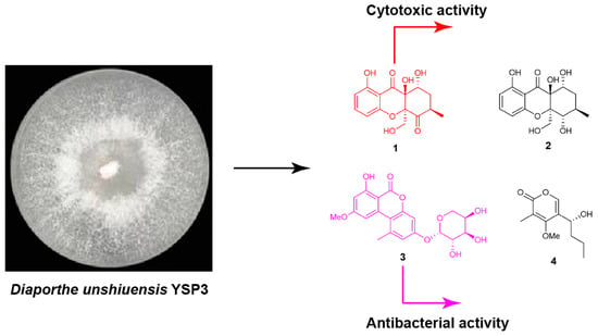
Chemical Investigation of Endophytic Diaporthe unshiuensis YSP3 Reveals New Antibacterial and Cytotoxic Agents

1
State & Local Joint Engineering Research Center of Green Pesticide Invention and Application, College of Plant Protection, Nanjing Agricultural University, Nanjing 210095, China
2
Key Laboratory of Integrated Management of Crop Diseases and Pests, Ministry of Education, Nanjing 210095, China
3
School of Life Sciences and Chemical Engineering, Jiangsu Second Normal University, Nanjing 211200, China
4
Department of Plant Pathology, University of Agriculture, Faisalabad 38000, Pakistan
*
Authors to whom correspondence should be addressed.
These authors contributed equally to this paper.
J. Fungi 2023, 9(2), 136; https://doi.org/10.3390/jof9020136
Submission received: 1 December 2022 / Revised: 13 January 2023 / Accepted: 17 January 2023 / Published: 19 January 2023
(This article belongs to the Special Issue Emerging Investigators in Bioactive Fungal Metabolites)

Abstract

:
Chemical investigation of the plant-derived endophytic fungus Diaporthe unshiuensis YSP3 led to the isolation of four new compounds (14), including two new xanthones (phomopthane A and B, 1 and 2), one new alternariol methyl ether derivative (3) and one α-pyrone derivative (phomopyrone B, 4), together with eight known compounds (512). The structures of new compounds were interpreted on the basis of spectroscopic data and single-crystal X-ray diffraction analysis. All new compounds were assessed for their antimicrobial and cytotoxic potential. Compound 1 showed cytotoxic activity against HeLa and MCF-7 cells with IC50 values of 5.92 µM and 7.50 µM, respectively, while compound 3 has an antibacterial effect on Bacillus subtilis (MIC value 16 μg/mL).

Graphical Abstract

1. Introduction

Microorganisms produce a wide range of secondary metabolites (SM), also known as natural products, which have an incredible and dignified success history concerning pharmaceutical potential and structural diversity [1]. Metabolites produced by endophytic fungal isolates are not only renowned for providing protection in the survival of their host but also have a phenomenal contribution to agriculture, medicine, and modern industry [2]. For pathogenic fungi, some natural products were used as chemical weapons to facilitate their invasion. For example, fusaoctaxin A was recently characterized as a virulence factor during the infection progress of Fusarium graminearum, which is a destructive wheat pathogen [3]. Moreover, for endophytic fungi, their metabolites can provide benefits to residing hosts by functioning as antibacterials, nutrition transporting agents, and plant growth regulators [3,4].
Species of Diaporthe (anamorph Phomopsis) comprise a diverse and widely distributed group of phytopathogens, saprophytes, endophytes, and pathogens of mammals [5]. As plant pathogens, they can infect a wide range of plant hosts (soybean, eggplant, grape, citrus, etc.) and cause diseases [6]. Many other species of Diaporthe are reported as non-pathogenic endophytes and recognized as producers of numerous secondary metabolites with a range of biological potencies, including antifungal [7], antibacterial [8], cytotoxic [9], anti-inflammatory [10], antioxidant [11], phytotoxic [12], and anti-influenza A virus (IAV) potencies [13]. Most of the compounds produced by this genus have been classified into polyketides, including xanthones, chromanones, benzofuranones, quinones, phenols and pyrones, terpenoids, steroids, lactones, alkaloids, and fatty acids [14,15].
As part of our continuous endeavor to search for structurally intriguing and/or bioactive natural products from endophytic fungi, an attempt was made to investigate the fungus Diaporthe unshiuensis YSP3 isolated from the leaves of Caesalpinia sepiaria. This work led to the isolation of two new xanthone derivatives (12), one new alternariol derivative (3), one new pyrone derivative (4), and eight known compounds. Herein, the isolation, structural elucidation, and bioactivity evaluation of metabolites 112 are described in detail.

2. Materials and Methods

2.1. General Experimental Procedures

Nuclear magnetic resonance (NMR) spectra were recorded on a Bruker Avance III 400 and 600 MHz NMR spectrometer (Bruker, Rheinstetten, Germany) in CDCl3 and acetone-d6 with TMS as an internal standard at room temperature. Ultraviolet (UV) spectra were recorded on a Hitachi U-3000 spectrophotometer (Hitachi, Tokyo, Japan). Optical rotations were measured in MeOH solution on a Rudolph Autopol III automatic polarimeter (Rudolph Research Analytical, NJ, USA). High-resolution-electrospray ionization-mass spectrometry (HR-ESI-MS) spectra were obtained on an Agilent 6210 TOF LC-MS spectrometer (Agilent, CA, USA). X-ray data were obtained on a Bruker APEX-II CCD diffractometer (Bruker, MA, USA). Column chromatography was performed on silica gel (200−300 mesh, Qingdao Marine Chemical Inc., Qingdao, China) and Sephadex LH-20 (Pharmacia Biotech, Uppsala, Sweden). High-performance liquid chromatography (HPLC) was performed on a Shimadzu LC-20AT instrument with an SPD-20A detector (Agilent, CA, USA) using an ODS column (ODS-2 HYPERSIL, 250 × 10 mm, 5 μm, Thermo Scientific, Shanghai, China). All chemicals used in this study were of analytical or HPLC grade.

2.2. Fungal Materials

The fungal strain YSP3 was isolated from the leaves of C. sepiaria collected in August 2016 from Nanjing Botanical Garden, Mem. Sun Yat-Sen, Nanjing, Jiangsu Province, People’s Republic of China. The strain was identified as D. unshiuensis (GenBank OP804247) based on a morphological characterization as well as phylogenetic analysis using five molecular markers (ITS, TEF1, HIS, CAL, and TUB) (Figure S1) [16]. The strain YSP3 was deposited in the culture collection bank of the Laboratory of Natural Products and Pesticide Chemistry, Nanjing Agricultural University (NAU).

2.3. Fermentation, Extraction, and Purification

Strain YSP3 was cultured on potato dextrose agar medium (PDA) for 5 days at 25 °C. Seed medium (potato 200 g, dextrose 20 g, distilled water 1000 mL) in 1000 mL Erlenmeyer flasks containing 400 mL broth was inoculated with pieces of the mycelium obtained from the colony strain YSP3 and incubated for 48 h at 25 °C on a rotating shaker (150 rpm). Fermentation was performed in 150 Erlenmeyer flasks containing PD medium (400 mL in each) inoculated with 10 mL seed solution and incubated at 25 °C for 14 days on a rotary shaker (150 rpm). The fermented broth (60 L) was filtered through muslin cloth, extracted thrice by adding EtOAc (v/v), and evaporated under vacuum to obtain crude extract (41 g). The EtOAc extract was separated into six fractions (Fr1-Fr6) on the silica gel column (350 g, 200−300 mesh), eluted with a gradient solvent system of CH2Cl2-MeOH (v/v 100:0, 100:1, 100:2, 100:4, 100:8, 100:16, 0:100). Fr2 was subjected to the silica gel (300−400 mesh), eluted with a gradient of petroleum ether–EtOAc (v/v, 100:0 0:100) to give five subfractions (Fr2.1 to Fr2.5). Fr2.2 was chromatographed over a Sephadex LH-20 (MeOH) column and then separated by semipreparative HPLC (MeOH/H2O, v/v, 50:50, 0.05% trifluoroacetic acid (TFA)) to obtain compound 2 (10 mg, tR = 35.7 min). Fr2.3 was further purified by Sephadex LH-20 (MeOH) and semipreparative HPLC using 42% (MeOH) in water to obtain 3 (7.2 mg, tR = 16.3 min) and 4 (38 mg, tR = 32 min). Fr3 was separated by silica gel chromatography (petroleum ether–EtOAc v/v, 100:0 to 0:100) to give five subfractions (Fr3.1 to Fr3.5). Fr3.2 was separated by HPLC (54% MeOH in H2O, flow rate = 2 mL/min, UV = 254 nm) to yield 5 (6.7 mg, tR = 13.2 min), 6 (5.9 mg, tR = 30.7 min), and 9 (8.7 mg, tR = 24.2 min). Fr3.3 was subjected to the Sephadex LH-20 (MeOH) chromatography and further purified by HPLC (MeOH/H2O, v/v, 45:55, 0.05 % TFA) to yield 1 (12.4 mg, tR = 19.3 min), 7 (6.2 mg, tR = 21 min), and 8 (7 mg, tR = 16.9 min). Fr3.4 was further separated by HPLC eluted with a gradient of (MeOH/H2O, v/v, 60:40) to afford compound 11 (7.8 mg, tR = 26 min). Fr4 was divided into four subparts (Fr4.1 to Fr4.4) by silica gel column with a gradient elution of petroleum ether–EtOAc (v/v, 100:0–0:100). Fr4.2 was subjected to a Sephadex LH-20 (MeOH) column and further purified by semipreparative HPLC (MeOH/H2O, v/v, 65:35) to afford 10 (7.2 mg, tR = 35.7 min) and 12 (12 mg, tR = 49 min).

2.4. Formation of the (S)- and (R)-MTPA Esters of 4

Compound 4 (2.0 mg) was dissolved in 500 μL of anhydrous pyridine, and 20 μL of (S)-(+)-MTPA chloride was added to the reaction mixture. After 48 h reaction in dark, the residue was purified by HPLC (CH3CN/H2O, 85:15) to give (R)-(+)-MTPA ester (1.7 mg). (R)-(−)-MTPA ester (1.8 mg) was obtained using the same step from (S)-(−)-MTPA chloride.

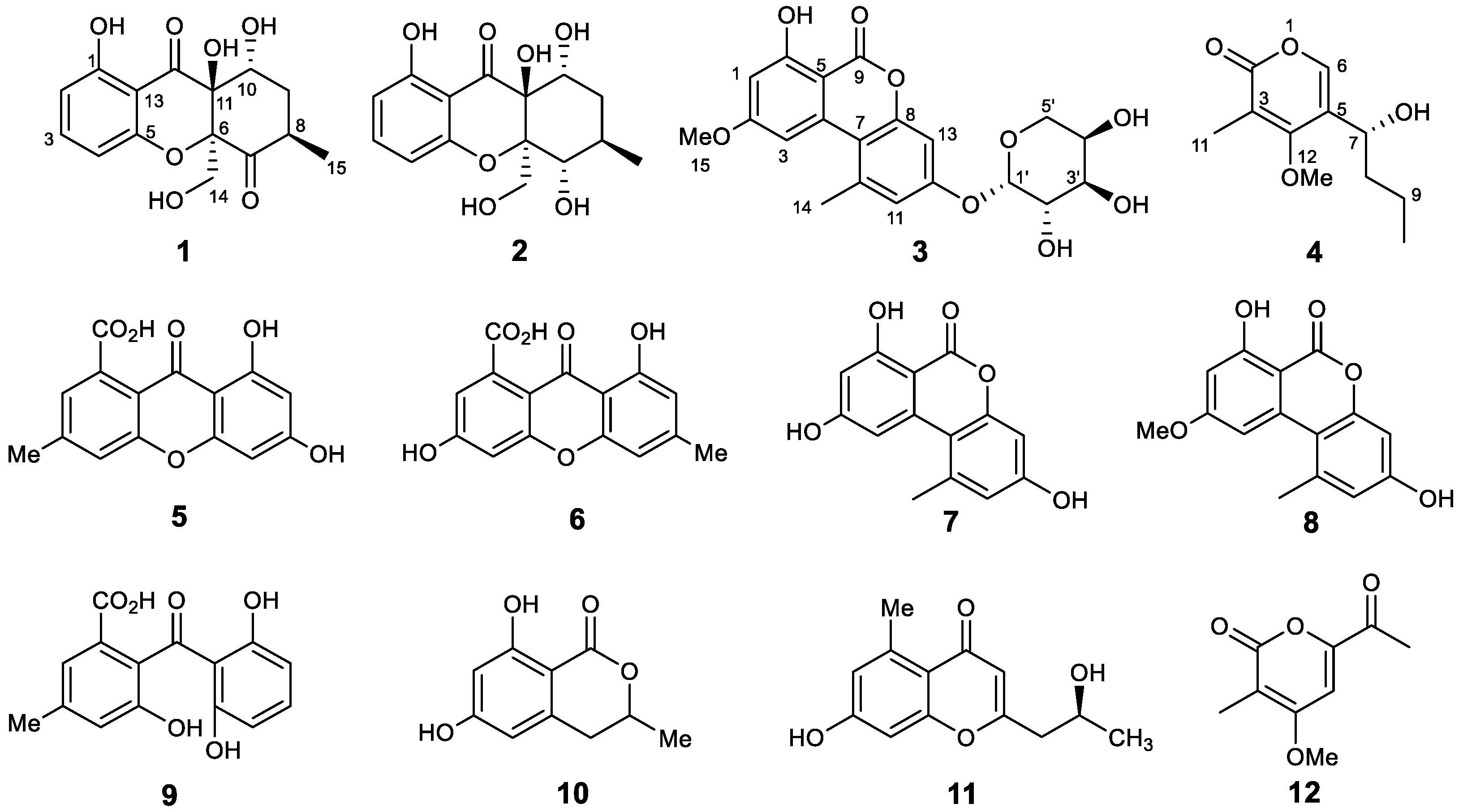
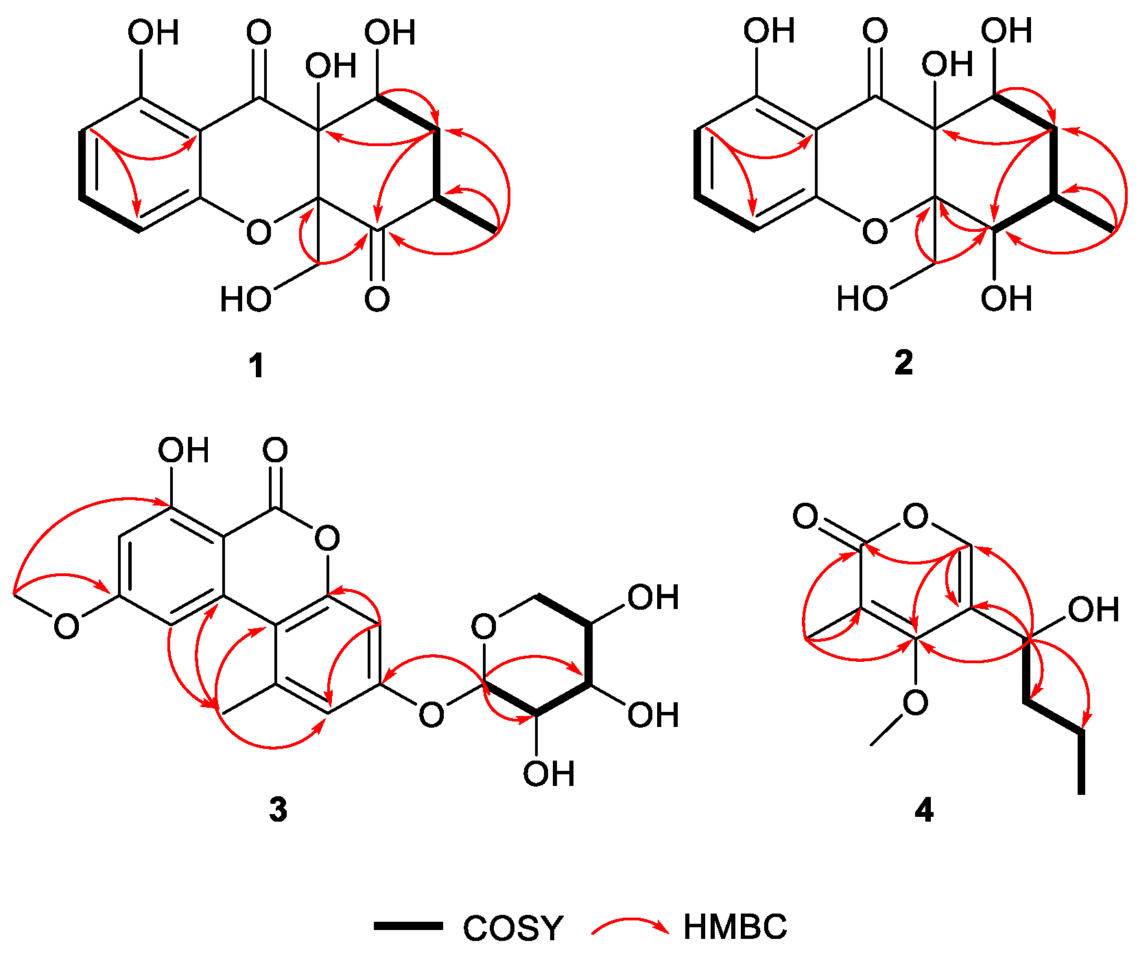
2.5. Phomopthane A (1)

Colorless crystals; [α]20D -96.0 (c 0.1, MeOH); UV (MeOH) λmax (log ε) 210 (1.9), 280 (1.0), 360 (0.4) nm; 1H and 13C NMR data, see Table 1; HR-ESI-MS m/z 331.0789 [M + Na]+ (cacld for C15H18O3Na, 331.0788).

2.6. Phomopthane B (2)

Colorless crystals; [α]20D +4.0 (c 0.1, MeOH); UV (MeOH) λmax (log ε) 210 (1.8), 280 (1.0), 360 (0.4) nm; 1H and 13C NMR data, see Table 1; HR-ESI-MS m/z 333.0950 [M + Na]+ (cacld for C15H18O4Na, 333.0945).

2.7. Alternariol Methyl Ether-12-O-α-D-arabinoside (3)

Yellow powder; [α]20D +77.9 (c 0.1, MeOH); UV (MeOH) λmax (log ε) 589 nm; 1H and 13C NMR spectroscopic data, see Table 2; HR-ESI-MS m/z 427.1006 [M + Na]+ (cacld for C20H20O9Na, 427.1005).

2.8. Phomopyrone B (4)

Pale yellow oil; [α]20D -41.4 (c 0.2, MeOH); UV (MeOH) λmax (log ε) 589 nm; 1H and 13C NMR spectroscopic data, see Table 2; HR-ESI-MS m/z 213.1129 [M + H]+ (cacld for C11H17O4, 213.1127).

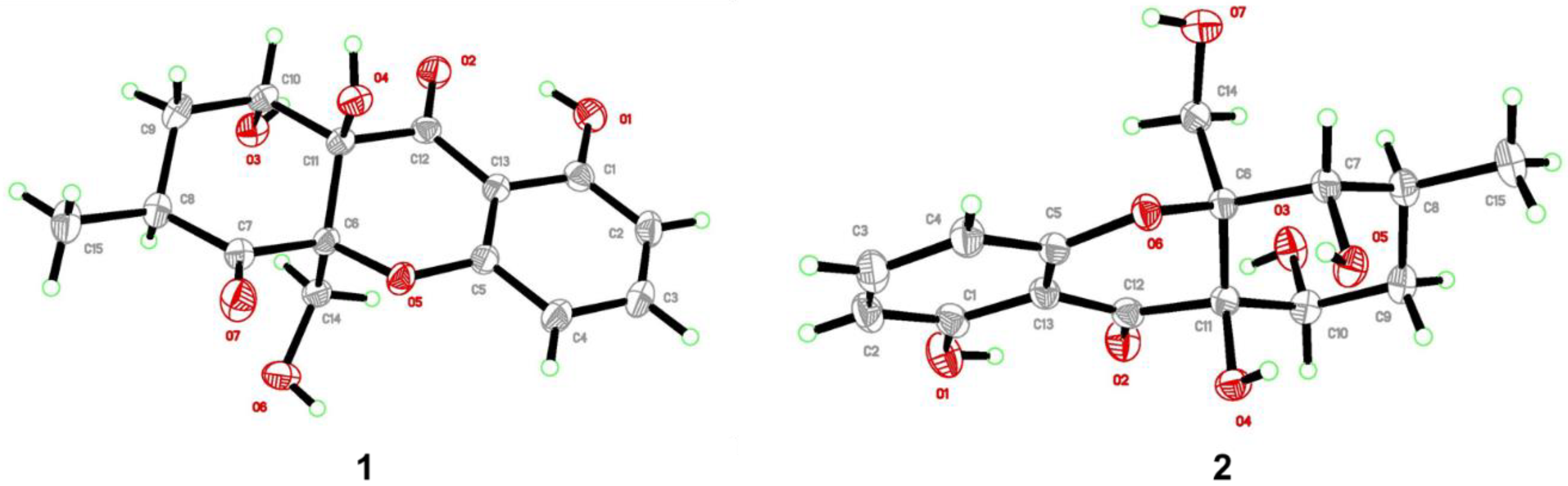
2.9. X-ray Crystallographic Analysis

The single colorless crystals of compounds 1 and 2 were shaped from the MeOH + CH2Cl2 mixture solvent (v/v, 1:1/2) at 4 °C after 25 or 27 days of slow solvent evaporation. Crystal diffraction data were collected on a Bruker APEX-II CCD diffractometer using Cu Kα radiation (λ = 1.5418 Å). These structures were solved by direct methods in OLEX2–1.3 software, followed by the refinement method of full-matrix least-squares calculations on F2 using SHELXL-2018 [17,18]. All crystal data of these compounds have been deposited in the Cambridge Crystallographic Data Centre (CCDC).

2.10. Crystal Data of 1

Triclinic, space group P-1, a = 8.2397 (3) Å, b = 8.5629 (3) Å, c = 11.4399 (4) Å, α = 69.676 (2)°, β = 86.302 (2)°, γ = 75.660 (2)°, V = 733.11 (5) Å3, Z = 2, T = 296 (2) K, Dcalc = 1.478 g/cm3, μ(Cu Kα) = 1.035 mm−1, F (000) = 344.0, Crystal size = 0.12 × 0.11 × 0.09 mm3, 4349 reflections collected, 2407 independent reflections (Rint = 0.0183). Final R1 = 0.0355, wR2 = 0.1007 [I >= 2σ(I)]. Final R1 = 0.0374, wR2 = 0.1024 (all data). The goodness of fit on F2 was 1.071. CCDC number: 2083291.

2.11. Crystal Data of 2

Monoclinic, space group P21/c, a = 11.3791 (11) Å, b = 7.4153 (6) Å, c = 16.9366 (15) Å, α = 90°, β = 108.048 (5)°, γ = 90°, V = 1358.8 (2) Å3, Z = 4, T = 296 (2) K, Dcalc = 1.517 g/cm3, μ(Cu Kα) = 1.027 mm−1, F (000) = 658.6, Crystal size = 0. 12 × 0.11 × 0.09 mm3, 5641 reflections collected, 2426 independent reflections (Rint = 0.0213). Final R1 = 0.0374, wR2 = 0.1092 [I >= 2σ(I)]. Final R1 = 0.0393, wR2 = 0.1118 (all data). The goodness of fit on F2 was 1.074. CCDC number: 2083288.

2.12. Antimicrobial Assays

All isolated new compounds were evaluated for their antibacterial potencies against four bacterial strains (Xanthomonas oryzae pv. oryzae, Xanthomonas oryzae pv. oryzicola, Bacillus subtilis, and Ralstonia solanacearum) in sterile 96-well plates by a broth dilution method [19]. Antifungal potencies were performed against Rhizoctonia solani, Fusarium solani, F. graminearum, Botrytis cinerea, Sclerotinia sclerotiorum, Alternaria solani, and Phytophthora capsici referred to the method reported [20]. All strains were provided by the Laboratory of Natural Products and Pesticide Chemistry, Nanjing Agricultural University, Nanjing, Jiangsu, China.

2.13. Cytotoxic Assays

The inhibitory effects of isolated compounds on the HeLa and MCF-7 cells were assessed in vitro by using the MTT assay on a 96-well plate. Experimental details of test used in this study are summarized in previous reports [21,22]. The toxicities against normal LO2 and HaCaT cells were tested via the cell counting kit-8 (CCK-8) method, similar to the reported reference [23].

2.14. Statistical Analysis

All tests were repeated thrice to remove the experimental error, and the quantitative data were presented as mean values ± standard deviation. The data were analyzed using IBM SPSS 22.0 with the probit analysis, and the Duncan statistical test was used for variance analysis between means. The value p ≤ 0.05 was considered statistically significant.

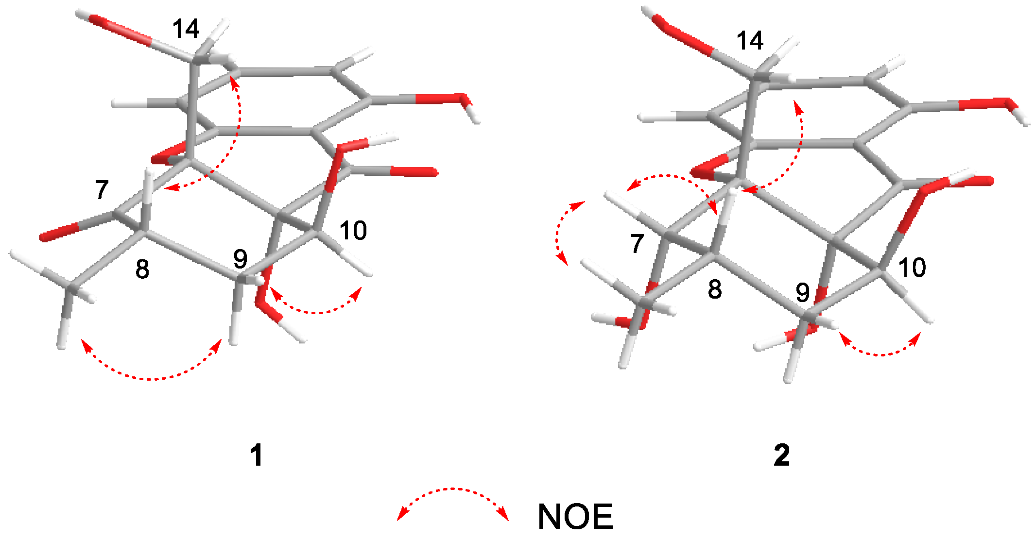
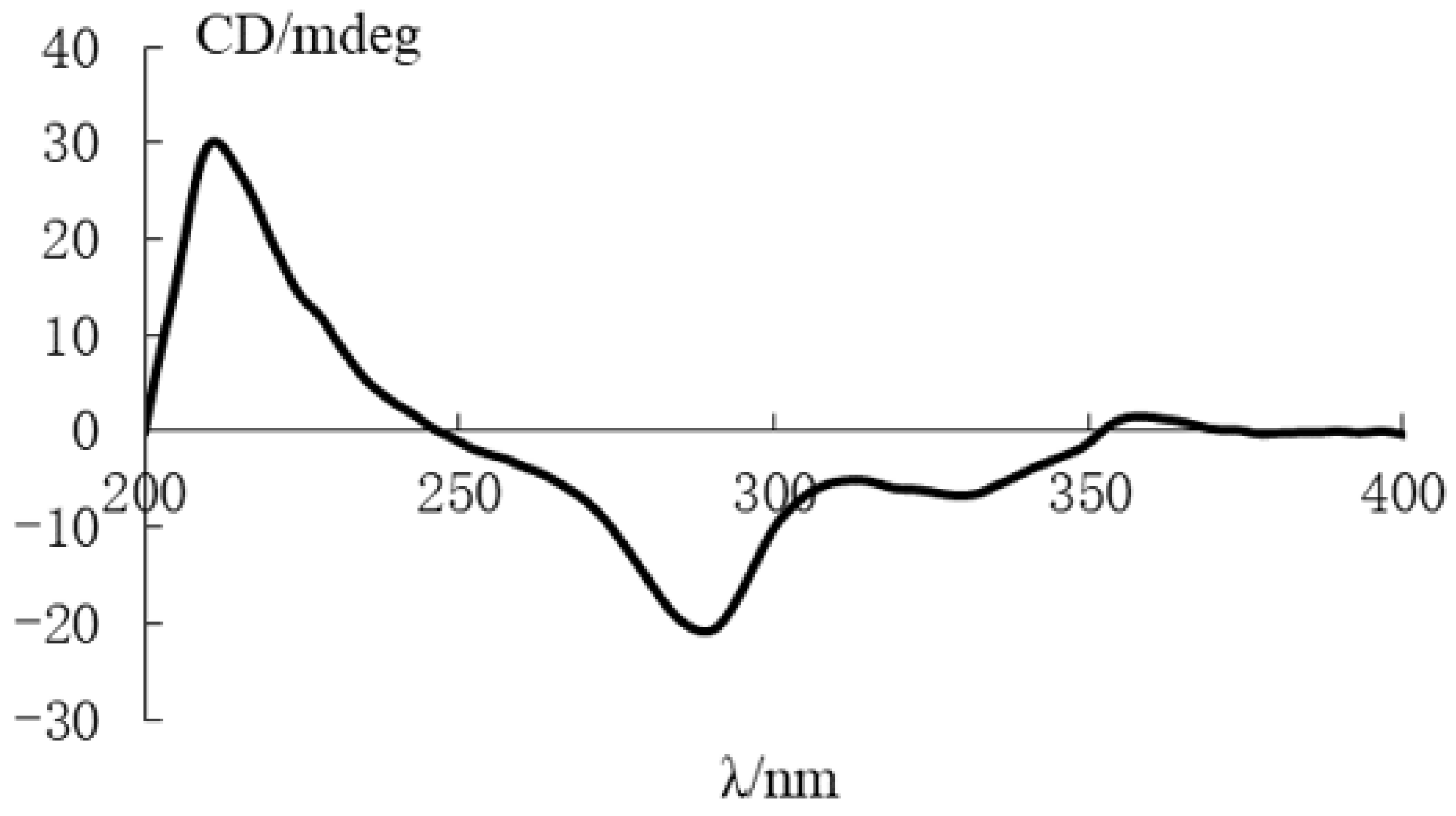
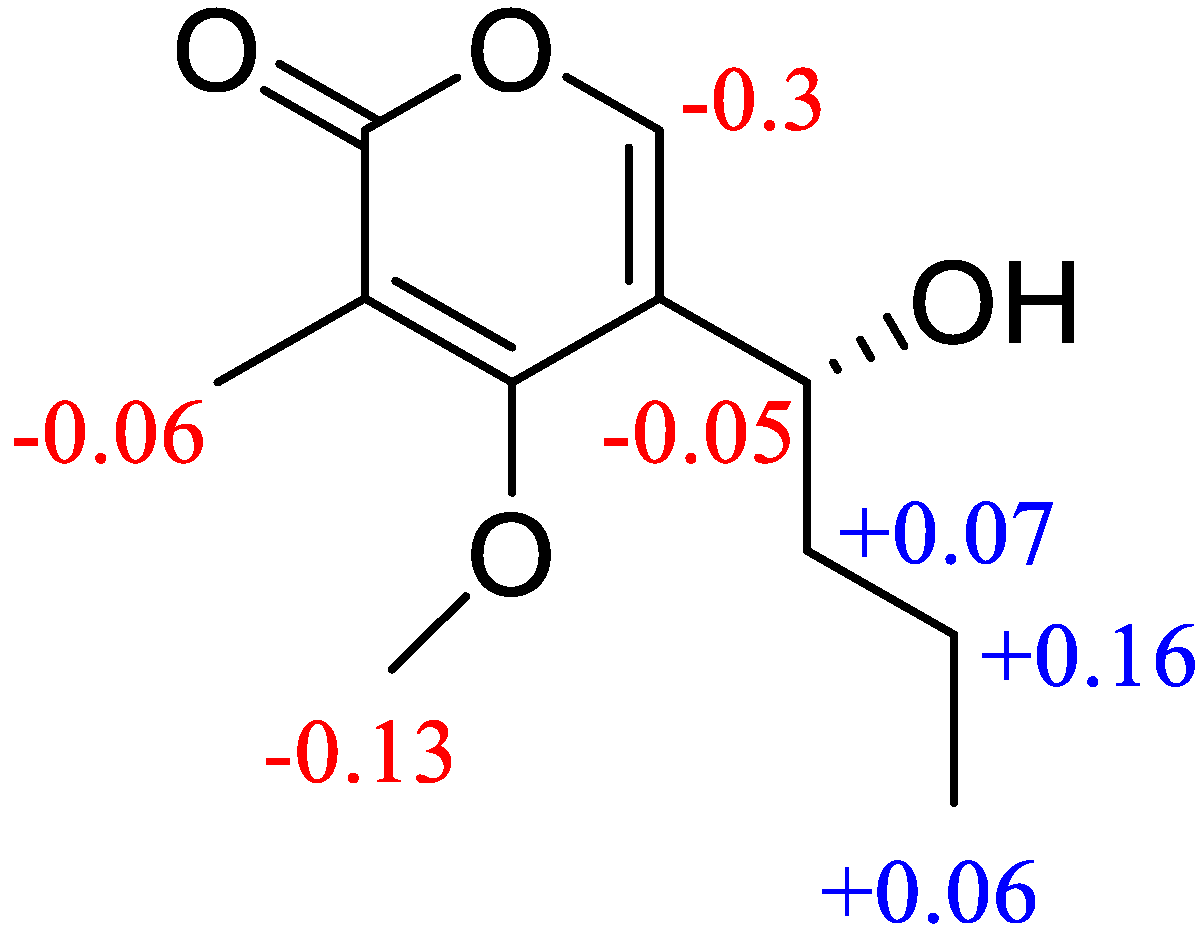
3. Results and Discussion

Phomopthane A (1) was obtained as colorless crystals with the molecular formula C15H16O7 as inferred from its HRESIMS, (m/z 331.0789 [M + Na]+, cacld for C15H16O7Na, 331.0788), indicating eight degrees of unsaturation. The 13C and DEPT NMR spectra exhibited 15 resonances resulting from one methyl, two methylenes, five methines, and seven quaternary carbons, including two carbonyl groups (δC 204.5 and δC 196.9). The 1H NMR spectrum revealed the presence of characteristic signals for three aromatic protons from a 1,2,3- trisubstituted benzene at δH 6.54 (d, J = 8.2 Hz, H-2), 6.48 (d, J = 8.2 Hz, H-4), and 7.47 (t, J = 8.2 Hz, H-3), one methylene at 2.14 (m, H-9), one oxygenated methylene at δH 4.83 (d, J = 13.1 Hz, H-14a) and 3.96 (d, J = 13.1 Hz, H-14b), two methines at δH 4.61 (br t, J = 2.5 Hz, H-10) and 3.12 (m, H-8), and one methyl group at δH 1.08 (d, J = 6.4 Hz, H-15). The 1H and 13C NMR spectra of 1 (Table 1) were closely similar to those of mangrovamide K [24], a xanthone derivative isolated from Penicillium sp.. The difference was attributed to the methyl group in mangrovamide K being changed to the oxygenated methylene at C-14 in structure 1 (Figure 1), which was further supported by the HMBC correlations from H3-15 to C-7, C-8, and C-9; H-14 to C-6 and C-7; and H-9 to C-7, C-11, and C-10 (Figure 2). Thus, the planar structure of 1 was proposed, as shown in Figure 1.
The relative configuration of 1 was partially determined by NOESY and 3JHH coupling data. H-10 showed a small coupling constant (2.5 Hz) to H-9, suggesting H-10 has an equatorial orientation. The NOESY correlation between H-8 and H-14 indicates that they both possess an axial position (Figure 3). There was no sufficient data to deduce the configuration at C-11. Fortunately, a single crystal X-ray study not only confirmed the planar structure but also determined the relative configuration of 1 (Figure 4).
The absolute configuration of 1 was assigned by comparing its electronic circular dichroism (ECD) spectrum with the structurally similar mangrovamide K, of which the absolute configuration has been established. Compound 1 contains the same chromophoric system as mangrovamide K but shows nearly a mirror image on ECD (Figure 5) [24], with a strong positive Cotton effect at 211 nm, negative bands in the 250–350 nm range, and a weak positive Cotton effect at 359 nm. Additionally, 1 has an opposite optical rotation ([α]20D −96.0) compared to mangrovamide K ([α]25D +72.9). Therefore, the absolute configuration of 1 was determined as 6R,8R,10S,11S.
Phomopthane B (2) was isolated as colorless crystals. Its molecular formula, C15H18O7, was deduced by HR-ESI-MS (m/z 333.0950, [M + Na]+, calcd for C15H18O7Na, 333.0945), which was two more mass units than 1. The close comparison of the 1D NMR data between 2 and 1 showed a general similarity (Table 1), except that the ketone at the C-7 position in 1 was replaced by an oxygenated methine at δH 4.42 (H-7) in 2. This assumption was supported by the key HMBC correlations of H3-15 to C-7, C-8, and C-9; H-14 to C-6 and C-7; and H-9 to C-7 and C-11 (Figure 2). H-7 was suggested to have an equatorial position, evidenced by its small J value (2.8 Hz). The relative configuration of the rest chiral centers in 2 was the same as in 1, based on their similar NOESY correlations and coupling constants (Figure 3 and Table 1). The above-mentioned structural elucidation was confirmed by a single X-ray analysis. Unlike 1, 2 crystallized in the monoclinic P21/c space group and has a near-zero optical rotation, suggesting that 2 was a racemic mixture [25,26].
Compound 3 was obtained as a gray powder with a molecular formula of C20H20O9 determined by the HR-ESI-MS spectrum (m/z 427.1006 [M + Na]+; calcd for C20H20O9Na, 427.1000), indicating 11 degrees of unsaturation. The 13C NMR, DEPT, and HSQC spectra revealed the presence of 20 carbons signals resulting from for one methoxy, one methyl, one methylene, eight methines, and nine quaternary carbons, including one lactonic ester group at δC 165.7 (C-9). The 1H-NMR data (Table 2) displayed characteristic resonances for four aromatic protons at δH 6.61 (d, J = 2.2 Hz, H-1), 7.00 (2H, m, H-11, and H-13), and 7.34 (d, J = 1.9 Hz, H-3), one methoxy group at δH 3.99 (s, H-15), one methyl group at δH 2.85 (s, H-14), one oxygenated methylene at δH 3.71 (m, H-5′), four oxygenated methines at δH 4.20 (2H), 4.31, and 5.80. The HMBC correlations (Figure 2) from H3-15 to C-2 and C-6; H3-14 to C-10, C-7, and C-11; H-13 to C-8 and C-11 suggested the presence of alternariol methyl ether in the chemical structure of 3 [27]. The NMR data (Table 2) of compound 3 were very similar to those of 8, except the existence of five more oxygenated carbons at δC 101.5 (C-1′), 73.1 (C-2′), 88.2 (C-3′), 70.8 (C-4′), and 62.9 (C-5′) in 3. Considering the molecular formula, it is speculated that there should be a monosaccharide structure in 3, which was further confirmed by the COSY correlations of H-1′/H-2′/H-3′/H-4′/H-5′ and HMBC correlations from H-1′ to C-2′, C-3′, and C-4′. The HMBC correlation of the anomeric proton at δH 5.80 (d, J = 4.3 Hz) with C-12 (δC 158.7) determined its linkage site in compound 3. The chemical shift of the anomeric carbon (δH 5.80 and δC 101.5) confirmed the α configuration. The sugar moiety was further determined as D-arabinoside by hydrolysis and compared to the standard. Thus, 3 was named as alternariol methyl ether-12-O-α-D-arabinoside shown in (Figure 1).
Compound 4 was isolated as a pale yellow oil. Its molecular formula, C11H16O4, was deduced by HR-ESI-MS (m/z 213.1129, [M + H]+, calcd for C11H17O4, 213.1121), indicating four degrees of unsaturation. The 13C and DEPT NMR data demonstrated 11 resonances resulting from two methyls, one methoxy, two methylenes, two methines, and four quaternary carbons signals, including an ester carbonyl group at (δC 164.9, C-2). The 1H-NMR spectral data (Table 2) revealed the signals for one olefinic proton at δH 7.48 (s, H-6), one methoxy group δH 3.95 (s, H-12), two methyl groups at δH 1.99 (s, H-11) and δH 0.92 (t, J = 7.4 Hz, H-10), two methylenes at δH 1.50 (m, H-9) and at δH 1.70 (m, H-8), and one methine at δH 4.63 (m, H-7). The α-pyrone ring structure was confirmed by HMBC correlations (Figure 2) from H-6 to C-2, C-4, and C-5. HMBC correlations from H-7 to C-8, C-9, and COSY correlations of H-7/H-8/H-9/H-10 determined the presence of a butyl side chain. The attachment of the butyl side chain at C-5 was confirmed by HMBC correlation from H-7 to C-4, C-5, and C-6. The attachment of the methoxy group (C-12) was also confirmed by the HMBC correlation between H-12 and C-4. Thus, the planar structure of 4 was deduced, as shown in Figure 1.
The absolute configuration of C-7 in 4 was confirmed by a modified mosher’s method [28,29]. Compound 4 reacted with (R)-(−)-MTPA chloride and (S)-(+)-MTPA chloride, respectively, to give the (S)- and (R)-MTPA esters. The Δδ values between R- and S-MTPA derivatives confirmed that C-7 was an R configuration (Figure 6). Finally, compound 4 was named phomopyrone B.
In addition, the other known compounds (512) were identified as 6,8-dihydroxy-3-methyl-9-oxo-9H-xanthene-1-carboxylic acid (5) [30], 3,8-dihydroxy-6-methyl-9-oxo-9H-xanthene-1-carboxylic acid (6) [31], alternariol (7), alternariol methyl ether (8) [32], monodictyphenone (9) [33], 3,4-dihydro-6,8-dihydroxy-3-methylisocoumarin (10) [34], 2-(2′S-hydroxypropyl)-7-hydroxychromone’s (11) [35], and wermopyrone (12) [36], respectively, by comparing their HRMS and NMR data to the literature.
To fully evaluate the antimicrobial potencies, newly isolated compounds (14) were screened for their antibacterial effect against four bacterial strains (three Gram-negative phytopathogenic bacteria and one Gram-positive bacterium) as well as for antifungal activities against seven fungal strains, respectively. Compound 3 showed the bactericidal effect against B. subtilis with an MIC value of 16 μg/mL, which was better than the positive control (streptomycin sulfate 64 μg/mL) (Table 3). The compounds (14) were also tested for their cytotoxic activities by the MTT test method. Compound 1 was active against HeLa and MCF-7 cell lines with IC50 values of 5.92 ± 0.04 µM and 7.50 ± 0.02 µM, respectively. Colchicine was used as a positive control (0.36 ± 0.07 µM for Hela and 0.44 ± 0.18 µM for MCF-7 cell lines) (Table 4). The other compounds were found to be ineffective (IC50 values > 20 µM). To explore the side effects of compound 1 on normal cells, 1 was tested for the toxicities towards normal LO2 and HaCaT cells using the CCK-8 assay. As shown in Table S3, compound 1 showed little effect on the viability of both LO2 and HaCat cells, which maintained high cell viability (~90 %) even at the compound concentration of 100 μM. These results indicated that 1 has a selective cytotoxic effect on tumor cell lines.

4. Conclusions

The current investigation reported the isolation and structural elucidation of twelve secondary metabolites, including two new xanthone derivatives (1 and 2), one new alternariol derivative (3), one new pyrone derivative (4), along with eight known compounds (512). All new compounds were evaluated for their bioactivities (antifungal, antibacterial, and cytotoxic). Compound 1 revealed potent cytotoxic potencies against human cancer cell lines HeLa and MCF-7, while compound 3 showed a bactericidal effect on B. subtilis. Additionally, 1 exhibited low toxicity to normal cells (LO2 and HaCaT). Thus, taking the importance of natural products, these findings enriched the structural diversity of secondary metabolites from Diaporthe species and highlighted their values for pharmaceutical or bactericidal applications.

Supplementary Materials

The following supporting information can be downloaded at: https://www.mdpi.com/article/10.3390/jof9020136/s1, Table S1: X-ray crystallographic data for 1; Table S2: X-ray crystallographic data for 2; Table S3: The inhibition rates of 1; against normal cells LO2 and HaCaT; Figure S1: Phylogenetic analysis of Diaporthe unshiuensis YSP3; Figure S2: The 1H-NMR spectrum of 1 in acetone-d6; Figure S3: The 13C-NMR spectrum of 1 in acetone-d6; Figure S4: The HSQC spectrum of 1 in acetone-d6; Figure S5: The HMBC spectrum of 1 in acetone-d6; Figure S6: The1H-1H COSY spectrum of 1 in acetone-d6; Figure S7: The NOESY spectrum of 1 in acetone-d6; Figure S8: The DEPT spectrum of 1 in acetone-d6; Figure S9: The 1H-NMR spectrum of 2 in CDCl3; Figure S10: The 13C-NMR spectrum of 2 in CDCl3; Figure S11: The HSQC spectrum of 2 in CDCl3; Figure S12: The HMBC spectrum of 2 in CDCl3; Figure S13: The 1H-1H COSY spectrum of 2 in CDCl3; Figure S14: The NOESY spectrum of 2 in CDCl3; Figure S15: The 1H-NMR spectrum of 3 in acetone-d6; Figure S16: The 13C-NMR spectrum of 3 in acetone-d6; Figure S17: The HSQC spectrum of 3 in acetone-d6; Figure S18: The HMBC spectrum of 3 in acetone-d6; Figure S19: The 1H-1H COSY spectrum of 3 in acetone-d6; Figure S20: The NOESY spectrum of 3 in acetone-d6; Figure S21: The DEPT spectrum of 3 in acetone-d6; Figure S22: The 1H-NMR spectrum of 4 in acetone-d6; Figure S23: The 13C-NMR spectrum of 4 in acetone-d6; Figure S24: The HSQC spectrum of 4 in acetone-d6; Figure S25: The HMBC spectrum of 4 in acetone-d6; Figure S26: The 1H-1H COSY spectrum of 4 in acetone-d6; Figure S27: The NOESY spectrum of 4 in acetone-d6; Figure S28:The DEPT spectrum of 4 in acetone-d6; Figure S29: 1H NMR of S-MTPA spectrum of 4 in acetone-d6; Figure S30: 1H NMR of R-MTPA spectrum of 4 in acetone-d6.

Author Contributions

Conceptualization, W.Y., Y.Y., W.W., G.L., and N.A.R.; investigation, B.K., Y.L., C.X., B.H., and C.Z.; writing—original draft preparation, B.K. and Y.L.; supervision, W.Y. and Y.Y.; project administration, W.Y. and Y.Y.; funding acquisition, W.Y., Y.Y., and G.L. All authors have read and agreed to the published version of the manuscript.

Funding

This research was funded by the Natural Science Foundation of Jiangsu Province (BK20211214, BK20201323, BK20221002, and BE2021303), the National Natural Science Foundation of China (32072443), the Guidance Foundation, Key Project of Natural Science Foundation of the Higher Education Institution of Jiangsu Province (19KJA430013). The APC was funded by the National Natural Science Foundation of China (32072443).

Institutional Review Board Statement

Not applicable.

Informed Consent Statement

Not applicable.

Data Availability Statement

The data presented in the manuscript are available on request from the corresponding authors.

Conflicts of Interest

The authors declare no competing financial interests.

References

  1. Newman, D.J.; Cragg, G.M. Natural products as sources of new drugs over the nearly four decades from 01/1981 to 09/2019. J. Nat. Prod. 2020, 83, 770–803. [Google Scholar] [CrossRef] [PubMed] [Green Version]
  2. Dissanayake, A.J.; Chen, Y.-Y.; Liu, J.-K. Unravelling Diaporthe species associated with woody hosts from karst formations (Guizhou) in China. J. Fungi 2020, 6, 251. [Google Scholar] [CrossRef]
  3. Jia, L.-J.; Tang, H.-Y.; Wang, W.-Q.; Yuan, T.-L.; Wei, W.-Q.; Pang, B.; Gong, X.-M.; Wang, S.-F.; Li, Y.-J.; Zhang, D. A linear nonribosomal octapeptide from Fusarium graminearum facilitates cell-to-cell invasion of wheat. Nat. Commun. 2019, 10, 922. [Google Scholar] [CrossRef] [Green Version]
  4. Strobel, G.; Daisy, B.; Castillo, U.; Harper, J. Natural products from endophytic microorganisms. J. Nat. Prod. 2004, 67, 257–268. [Google Scholar] [CrossRef]
  5. Gomes, R.; Glienke, C.; Videira, S.; Lombard, L.; Groenewald, J.; Crous, P. Diaporthe: A genus of endophytic, saprobic and plant pathogenic fungi. Pers. -Mol. Phylogeny Evol. Fungi 2013, 31, 1–41. [Google Scholar] [CrossRef] [PubMed] [Green Version]
  6. Udayanga, D.; Castlebury, L.A.; Rossman, A.Y.; Chukeatirote, E.; Hyde, K.D. Insights into the genus Diaporthe: Phylogenetic species delimitation in the D. eres species complex. Fungal Divers. 2014, 67, 203–229. [Google Scholar] [CrossRef] [Green Version]
  7. Tanney, J.B.; McMullin, D.R.; Green, B.D.; Miller, J.D.; Seifert, K.A. Production of antifungal and antiinsectan metabolites by the Picea endophyte Diaporthe maritima sp. nov. Fungal Biol. 2016, 120, 1448–1457. [Google Scholar] [CrossRef]
  8. Sebastianes, F.L.; Cabedo, N.; Aouad, N.E.; Valente, A.M.; Lacava, P.T.; Azevedo, J.L.; Pizzirani-Kleiner, A.A.; Cortes, D. 3-Hydroxypropionic acid as an antibacterial agent from endophytic fungi Diaporthe phaseolorum. Curr. Microbiol. 2012, 65, 622–632. [Google Scholar] [CrossRef]
  9. Sharma, V.; Singamaneni, V.; Sharma, N.; Kumar, A.; Arora, D.; Kushwaha, M.; Bhushan, S.; Jaglan, S.; Gupta, P. Valproic acid induces three novel cytotoxic secondary metabolites in Diaporthe sp., an endophytic fungus from Datura inoxia Mill. Bioorg. Med. Chem. Lett. 2018, 28, 2217–2221. [Google Scholar] [CrossRef]
  10. Cui, H.; Liu, Y.; Li, J.; Huang, X.; Yan, T.; Cao, W.; Liu, H.; Long, Y.; She, Z. Diaporindenes A–D: Four unusual 2, 3-dihydro-1 H-indene analogues with anti-inflammatory activities from the mangrove endophytic fungus Diaporthe sp. SYSU-HQ3. J. Org. Chem. 2018, 83, 11804–11813. [Google Scholar] [CrossRef]
  11. Niaz, S.I.; Khan, D.; Naz, R.; Safdar, K.; Abidin, S.Z.U.; Khan, I.U.; Gul, R.; Khan, W.U.; Khan, M.A.U.; Lan, L. Antimicrobial and antioxidant chlorinated azaphilones from mangrove Diaporthe perseae sp. isolated from the stem of Chinese mangrove Pongamia pinnata. J. Asian Nat. Prod. Res. 2021, 23, 1077–1084. [Google Scholar] [CrossRef] [PubMed]
  12. Reveglia, P.; Pacetti, A.; Masi, M.; Cimmino, A.; Carella, G.; Marchi, G.; Mugnai, L.; Evidente, A. Phytotoxic metabolites produced by Diaporthe eres involved in cane blight of grapevine in Italy. Nat. Prod. Res. 2021, 35, 2872–2880. [Google Scholar] [CrossRef] [PubMed]
  13. Luo, X.; Yang, J.; Chen, F.; Lin, X.; Chen, C.; Zhou, X.; Liu, S.; Liu, Y. Structurally diverse polyketides from the mangrove-derived fungus Diaporthe sp. SCSIO 41011 with their anti-influenza A virus activities. Front. Chem. 2018, 6, 282. [Google Scholar] [CrossRef] [PubMed] [Green Version]
  14. Xu, T.-C.; Lu, Y.-H.; Wang, J.-F.; Song, Z.-Q.; Hou, Y.-G.; Liu, S.-S.; Liu, C.-S.; Wu, S.-H. Bioactive secondary metabolites of the genus Diaporthe and anamorph Phomopsis from terrestrial and marine habitats and endophytes: 2010–2019. Microorganisms 2021, 9, 217. [Google Scholar] [CrossRef] [PubMed]
  15. Pu, H.; Liu, J.; Wang, Y.; Peng, Y.; Zheng, W.; Tang, Y.; Hui, B.; Nie, C.; Huang, X.; Duan, Y. Bioactive α-pyrone derivatives from the endophytic fungus Diaporthe sp. CB10100 as inducible nitric oxide synthase inhibitors. Front. Chem. 2021, 9, 679592. [Google Scholar] [CrossRef]
  16. Zhao, X.; Li, K.; Zheng, S.; Yang, J.; Chen, C.; Zheng, X.; Wang, Y.; Ye, W. Diaporthe Diversity and Pathogenicity Revealed from a Broad Survey of Soybean Stem Blight in China. Plant Dis. 2022, 106, 2892–2903. [Google Scholar] [CrossRef] [PubMed]
  17. Flack, H.; Bernardinelli, G. The use of X-ray crystallography to determine absolute configuration. Chirality: Pharmacol. Biol. Chem. Conseq. Mol. Asymmetry 2008, 20, 681–690. [Google Scholar] [CrossRef]
  18. Sheldrick, G.M. A short history of SHELX. Acta Crystallogr. Sect. A Found. Crystallogr. 2008, 64, 112–122. [Google Scholar] [CrossRef] [Green Version]
  19. Lu, S.; Sun, W.; Meng, J.; Wang, A.; Wang, X.; Tian, J.; Fu, X.; Dai, J.; Liu, Y.; Lai, D. Bioactive bis-naphtho-γ-pyrones from rice false smut pathogen Ustilaginoidea virens. J. Agric. Food Chem. 2015, 63, 3501–3508. [Google Scholar] [CrossRef]
  20. Yan, W.; Li, S.-J.; Guo, Z.-K.; Zhang, W.-J.; Wei, W.; Tan, R.-X.; Jiao, R.-H. New p-terphenyls from the endophytic fungus Aspergillus sp. YXf3. Bioorganic Med. Chem. Lett. 2017, 27, 51–54. [Google Scholar] [CrossRef]
  21. Khan, B.; Zhao, S.; Wang, Z.; Ye, Y.; Ahmed Rajput, N.; Yan, W. Eremophilane sesquiterpenes and benzene derivatives from the endophyte Microdiplodia sp. WGHS5. Chem. Biodivers. 2021, 18, e2000949. [Google Scholar] [CrossRef] [PubMed]
  22. Wang, K.; Li, W.; Rui, X.; Chen, X.; Jiang, M.; Dong, M. Structural characterization and bioactivity of released exopolysaccharides from Lactobacillus plantarum 70810. Int. J. Biol. Macromol. 2014, 67, 71–78. [Google Scholar] [CrossRef] [PubMed]
  23. Guo, Y.; Xu, T.; Bao, C.; Liu, Z.; Fan, J.; Yang, R.; Qin, S. Design and synthesis of new norfloxacin-1, 3, 4-oxadiazole hybrids as antibacterial agents against methicillin-resistant Staphylococcus aureus (MRSA). Eur. J. Pharm. Sci. 2019, 136, 104966. [Google Scholar] [CrossRef] [PubMed]
  24. Yang, B.; Tao, H.; Lin, X.; Wang, J.; Liao, S.; Dong, J.; Zhou, X.; Liu, Y. Prenylated indole alkaloids and chromone derivatives from the fungus Penicillium sp. SCSIO041218. Tetrahedron 2018, 74, 77–82. [Google Scholar] [CrossRef]
  25. Geng, C.-A.; Chen, X.-L.; Zhou, N.-J.; Chen, H.; Ma, Y.-B.; Huang, X.-Y.; Zhang, X.-M.; Chen, J.-J. LC-MS guided isolation of (±)-sweriledugenin A, a pair of enantiomeric lactones, from Swertia leducii. Org. Lett. 2014, 16, 370–373. [Google Scholar] [CrossRef]
  26. Lin, L.; Jiang, N.; Wu, H.; Mei, Y.; Yang, J.; Tan, R. Cytotoxic and antibacterial polyketide-indole hybrids synthesized from indole-3-carbinol by Daldinia eschscholzii. Acta Pharm. Sin. B 2019, 9, 369–380. [Google Scholar] [CrossRef]
  27. Li, X.-B.; Chen, G.-Y.; Liu, R.-J.; Zheng, C.-J.; Song, X.-M.; Han, C.-R. A new biphenyl derivative from the mangrove endophytic fungus Phomopsis longicolla HL-2232. Nat. Prod. Res. 2017, 31, 2264–2267. [Google Scholar] [CrossRef]
  28. Liu, C.-C.; Zhang, Z.-Z.; Feng, Y.-Y.; Gu, Q.-Q.; Li, D.-H.; Zhu, T.-J. Secondary metabolites from Antarctic marine-derived fungus Penicillium crustosum HDN153086. Nat. Prod. Res. 2019, 33, 414–419. [Google Scholar] [CrossRef]
  29. Mondol, M.A.M.; Surovy, M.Z.; Islam, M.T.; Schüffler, A.; Laatsch, H. Macrocyclic trichothecenes from Myrothecium roridum strain M10 with motility inhibitory and zoosporicidal activities against Phytophthora nicotianae. J. Agric. Food Chem. 2015, 63, 8777–8786. [Google Scholar] [CrossRef]
  30. Li, J.; Zhang, Y.; Chen, L.; Dong, Z.; Di, X.; Qiu, F. A new xanthone from Penicillium oxalicum. Chem. Nat. Compd. 2010, 46, 216–218. [Google Scholar] [CrossRef]
  31. Hanumaiah, T.; Rao, B.K.; Rao, C.; Rao, G.; Rao, J.; Rao, K.; Marshall, D.S.; Thomson, R.H. Naphthalenes and naphthoquinones from Ventilago species. Phytochemistry 1985, 24, 1811–1815. [Google Scholar] [CrossRef]
  32. Qiao, X.; Yin, J.; Yang, Y.; Zhang, J.; Shao, B.; Li, H.; Chen, H. Determination of Alternaria mycotoxins in fresh sweet cherries and cherry-based products: Method validation and occurrence. J. Agric. Food Chem. 2018, 66, 11846–11853. [Google Scholar] [CrossRef] [PubMed]
  33. Krick, A.; Kehraus, S.; Gerhäuser, C.; Klimo, K.; Nieger, M.; Maier, A.; Fiebig, H.-H.; Atodiresei, I.; Raabe, G.; Fleischhauer, J. Potential cancer chemopreventive in vitro activities of monomeric xanthone derivatives from the marine algicolous fungus Monodictys putredinis. J. Nat. Prod. 2007, 70, 353–360. [Google Scholar] [CrossRef] [PubMed]
  34. Ito, M.; Tsuchida, Y.; Mizoue, K.; Hanada, K. NG-011 AND NG-012, novel potentiators of nerve growth factor II. The structure determination of NG-011 AND NG-012. J. Antibiot. 1992, 45, 1566–1572. [Google Scholar] [CrossRef] [PubMed] [Green Version]
  35. Qian, S.-y.; Yang, C.-l.; Chen, R.-x.; Wu, M.-s.; Li, C.; Fu, S.-b.; Liu, J.-g. III Type Polyketone Compounds Produced by Streptomyces sp. FJS 31-2 from the Soil of Fanjing Mountain. Nat. Prod. Res. Dev. 2018, 30, 1160. [Google Scholar]
  36. Lu, X.; Xu, N.; Dai, H.-F.; Mei, W.-L.; Yang, Z.-X.; Pei, Y.-H. Three new compounds from endophytic fungus L10 of Cephalotaxus hainanensis. J. Asian Nat. Prod. Res. 2009, 11, 397–400. [Google Scholar] [CrossRef]
Figure 1. Structures of isolated compounds (112).
Jof 09 00136 g001
Figure 2. Key 1H-1H COSY and HMBC correlations of compounds 14.
Jof 09 00136 g002
Figure 3. Key NOESY correlations of 1 and 2.
Jof 09 00136 g003
Figure 4. X-ray crystallographic structures of 1 and 2.
Jof 09 00136 g004
Figure 5. The ECD spectrum of 1.
Jof 09 00136 g005
Figure 6. Δδ values (δSδR) obtained for the S- and R-MTPA esters of compound 4.
Jof 09 00136 g006
Table 1. 1H NMR (600 MHz) and 13C NMR (150 MHz) spectral data of 1 and 2.
1 (Acetone-d6)2 (CDCl3)
No.δC, TypeδH (J in Hz)δC, TypeδH (J in Hz)
1163.8, C 160.0, C
2110.6, CH6.54 (d, 8.2)105.3, CH6.54 (d, 8.1)
3140.2, CH7.47 (t, 8.2)136.1, CH7.42 (t, 8.1)
4110.5, C6.48 (d, 8.2)108.0, CH6.58 (d, 8.1)
5160.0, C 155.2, C
694.3, C 81.8, C
7204.5, C 72.2, CH4.42 (d, 2.1)
838.3, CH3.12 (m)25.3, CH2.35 (m)
939.5, CH22.14 (m)27.1, CH21.73 (m)
2.09 (m)
1067.8, CH4.61 (br t, 2.5)65.8, CH4.53 (s)
1179.6, C 71.9, C
12196.9, C 193.2, C
13108.4, C 105.0, C
1463.5, CH24.83 (d, 13.1)58.1, CH23.88 (d, 13.5)
3.96 (d, 13.1)4.20 (d, 13.5)
1515.4, CH31.08 (d, 6.4)15.1, CH31.17 (d, 6.7)
1-OH 10.8 (s)
Table 2. 1H NMR (600 MHz) and 13C NMR (150 MHz) spectral data of 3 and 4 in acetone-d6.
34
No.δC, TypeδH (J in Hz)δC, TypeδH (J in Hz)
1100.3, CH6.61 (d, 2.2)
2167.5, C 164.9, C
3105.1, CH7.34 (d, 1.9)110.5, C
4138.7, C 166.4, C
599.9, C 121.7, C
6165.8, C 146.4, CH7.48 (s)
7112.2, C 66.0, CH4.63 (m)
8153.7, C 39.4, CH21.70 (m)
9165.7, C 18.7, CH21.50 (m)
10139.3, C 13.2, CH30.92 (t, 7.4)
11119.5, CH7.00 (m)9.8, CH31.99 (s)
12158.7, C 60.7, CH33.95 (s)
13103.8, CH7.00 (m)
1425.7, CH32.85 (s)
1556.3, CH33.99 (s)
1’101.5, CH5.80 (d, 4.3)
2’73.1, CH4.31 (t, 5.2)
3’88.2, CH4.20 (m)
4’70.8, CH4.20 (m)
5’62.9, CH23.71 (m)
Table 3. Antibacterial potencies of compounds (14) against Bacillus subtilis.
CompoundsMIC (μg/mL)
Bacillus subtilis
1>100
2>100
316
4>100
Streptomycin sulfate64
Table 4. Cytotoxic effects of new compounds (14) on human cancer cell lines.
CompoundsGrowth Inhibition IC50 (µg/mL) Values
HelaMCF-7
15.92 ± 0.047.50 ± 0.02
2>20>20
3>20>20
4>20>20
Colchicine0.36 ± 0.070.44 ± 0.18
Disclaimer/Publisher’s Note: The statements, opinions and data contained in all publications are solely those of the individual author(s) and contributor(s) and not of MDPI and/or the editor(s). MDPI and/or the editor(s) disclaim responsibility for any injury to people or property resulting from any ideas, methods, instructions or products referred to in the content.

