Article

Phylogenetic Analyses and Morphological Studies Reveal Four New Species of Phellodon (Bankeraceae, Thelephorales) from China

1
Institute of Microbiology, School of Ecology and Nature Conservation, Beijing Forestry University, Beijing 100083, China
2
College of Forestry, Henan Agricultural University, Zhengzhou 450002, China
*
Author to whom correspondence should be addressed.
J. Fungi 2023, 9(1), 30; https://doi.org/10.3390/jof9010030
Submission received: 23 November 2022 / Revised: 19 December 2022 / Accepted: 21 December 2022 / Published: 23 December 2022
(This article belongs to the Special Issue Phylogeny and Diversity of Forestry Fungi)

Abstract

:
Phellodon is a genus of ectomycorrhizal fungi with important ecological roles and exploitable biological activities. In this study, four new species of Phellodon, P. caesius, P. henanensis, P. concentricus and P. subgriseofuscus, are described from China based on morphological characters and molecular evidence. The phylogenetic analyses of Phellodon were carried out based on the ITS + nLSU gene regions and the combined sequence dataset of ITS + nLSU + nSSU + RPB1 + RPB2 gene regions. Phellodon caesius is characterized by its dark bluish-grey, dark grey to black grey pileus, ash grey to dark bluish-grey spines, and the presence of both simple septa and clamp connections on generative hyphae of stipe. Phellodon concentricus is characterized by its zonate pileal surface, dark grey context in pileus, and spongy basidiomata. Phellodon henanensis is characterized by its ash grey, light vinaceous grey to light brown pileal surface, thin context in pileus, and the presence of both simple septa and clamp connections on generative hyphae of spines. Phellodon subgriseofuscus is characterized by its fuscous to black pileal surface, white to light brown spines, and vinaceous grey context. Illustrated descriptions and the ecological habits of the novel species are provided.

1. Introduction

Phellodon P. Karst., a genus of Bankeraceae, is a kind of stipitate hydnoid fungi. It was established by Petter Adolf Karsten in 1881 and typified by P. niger (Fr.) P. Karst [1]. Species in Phellodon are characterized by basidiome pileate and stipitate; pileus white to yellow-brown or grey-brown in various hues or olivaceous to black; basidia clavate, 4-spored, without basal clamp; spores broadly ellipsoid to subglobose, spinulose; cystidia lacking; odor of fenugreek when dried [2].
Species in Phellodon are a group of ectomycorrhizal fungi with important ecological roles [3]. The symbiotic relationship between mycorrhizal and host plants plays an essential role in nutrient cycling, energy flow, community species composition, biodiversity, and ecosystem change in forest ecosystems [4]. As significant ectomycorrhizal fungi, stipitate hydnoid fungi connected with plant roots can reflect the conservation state of forest ecosystems [5]. They can promote the absorption of nutrients by plants, which in turn promotes the circulation of materials in the ecosystem [3]. In addition, some species in Phellodon have exploitable biological activity. Stadler and Anke [6] conducted a study on Phellodon melaleucus (Sw. ex Fr.) P. Karst. and isolated a new antibiotic Phellodonic Acid from it. Reekie et al. [7] isolated a biologically active and highly functionalized hirsute derivative from the Tasmanian fungus Phellodon melaleucus and proposed the chemoenzymatic total synthesis of phellodonic acid. Fang et al. [8] isolated cyathane diterpenoids and nitrogenous terphenyl derivative from the fruiting bodies of basidiomycete Phellodon niger. Therefore, taxonomic and phylogenetic studies on Phellodon can lay the foundation for exploring their ecological functions and biological activities.
Fries originally placed the species of Phellodon in his tribe Mesopus, section Lignosa, which was made to include all tough mesopodous species of the Hydnaceae [9]. At that time, species of Phellodon were considered members of Hydnaceae. In 1961, Donk established Bankeraceae and made Bankera Coker and Beers and Phellodon members of the family [10]. Baird et al. [11] recombined Bankera fuligineoalba (J.C. Schmidt) Pouzar, the typified species of Bankera, to Phellodon. Since then, Bankera has been incorporated into Phellodon. From 1956 to 2005, morphological characteristics of Phellodon were systematically and deeply studied in North America and Europe [2,12,13,14,15,16,17,18,19,20,21,22]. Subsequently, with the development of molecular systematics, DNA sequence analysis was gradually introduced into the taxonomic and phylogenetic studies of the Bankeraceae [11,23,24,25]. However, these studies only focus on the internal transcribed Spacer (ITS) sequences, and there are still many unanswered questions. Baird et al. [11] reevaluated the species of stipitate hydnums from the southern United States and identified 41 distinct taxa of Hydnellum, Phellodon, and Sarcodon. They conducted a phylogenetic study based on ITS sequence and proved that Phellodon is independent of Hydnellum and Sarcodon. Li [26] conducted a systematic study of the Bankeraceae in Korea using ITS, the large subunit of nuclear ribosomal RNA gene (nLSU), and the second largest subunit of RNA polymerase II (RPB2) sequences, and 17 species were determined including the genus Phellodon. It was the first analysis of the family based on multigene sequences, but the number of species included in this phylogenetic analysis is relatively limited because many species do not have available sequences. In recent years, taxonomic and phylogenetic studies of Phellodon have been carried out in China, and multiple gene fragments of Phellodon have been provided. Song et al. [27] described four new species of Phellodon from southern China and provided the available sequences of nLSU, the small subunit of nuclear ribosomal RNA gene (nSSU), the small subunit of mitochondrial rRNA gene (mtSSU), the largest subunit of RNA polymerase II (RPB1), and RPB2 genes of Phellodon. Phylogenetic trees were constructed based on the combined ITS + nLSU + nSSU + RPB1 + RPB2 sequences, which confirmed the affinities of three new species and reveal the relationships of Phellodon species [28]. About 33 species have been described and transferred to the genus according to Index Fungorum (http://www.indexfungorum.org/ (accessed on 26 April 2022)). So far, eight species of Phellodon have been described in China [27,28,29], which means that the genus may have a relatively large distribution in China.
Macrofungi have important ecological and economical values. The species diversity, taxonomy, and phylogeny of macrofungi have been extensively investigated in recent years, and many new species have been discovered [30,31,32,33,34,35,36,37,38,39,40,41,42,43,44,45,46,47]. During our investigations of macrofungi in China, numerous specimens of Phellodon were collected. In the current study, the phylogenetic analyses of Phellodon were carried out based on the ITS + nLSU gene regions and the combined sequence dataset of ITS + nLSU + nSSU + RPB1 + RPB2 gene regions. Subsequent morphological and molecular studies uncovered four undescribed species. These species are described and illustrated below.

2. Materials and Methods

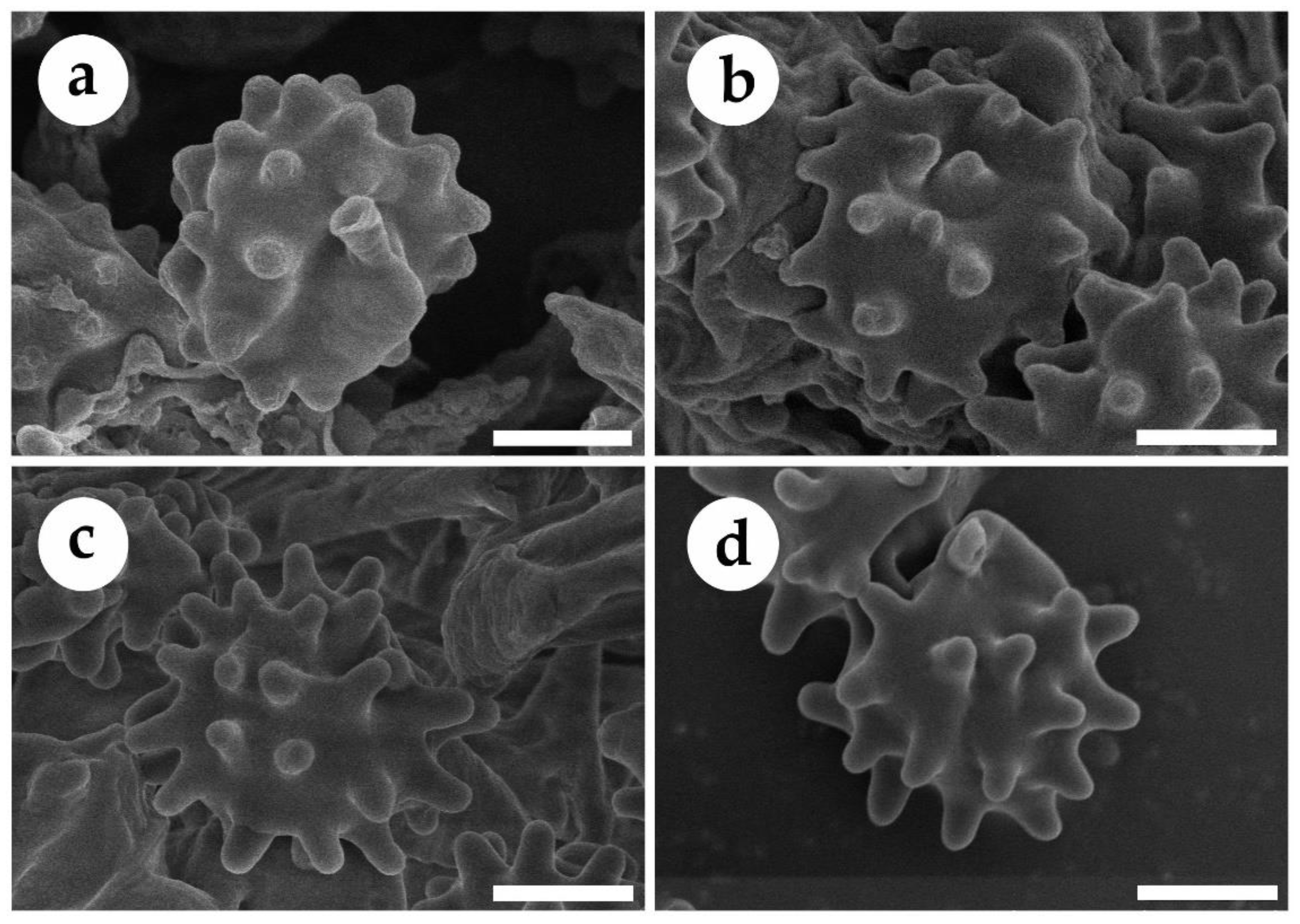
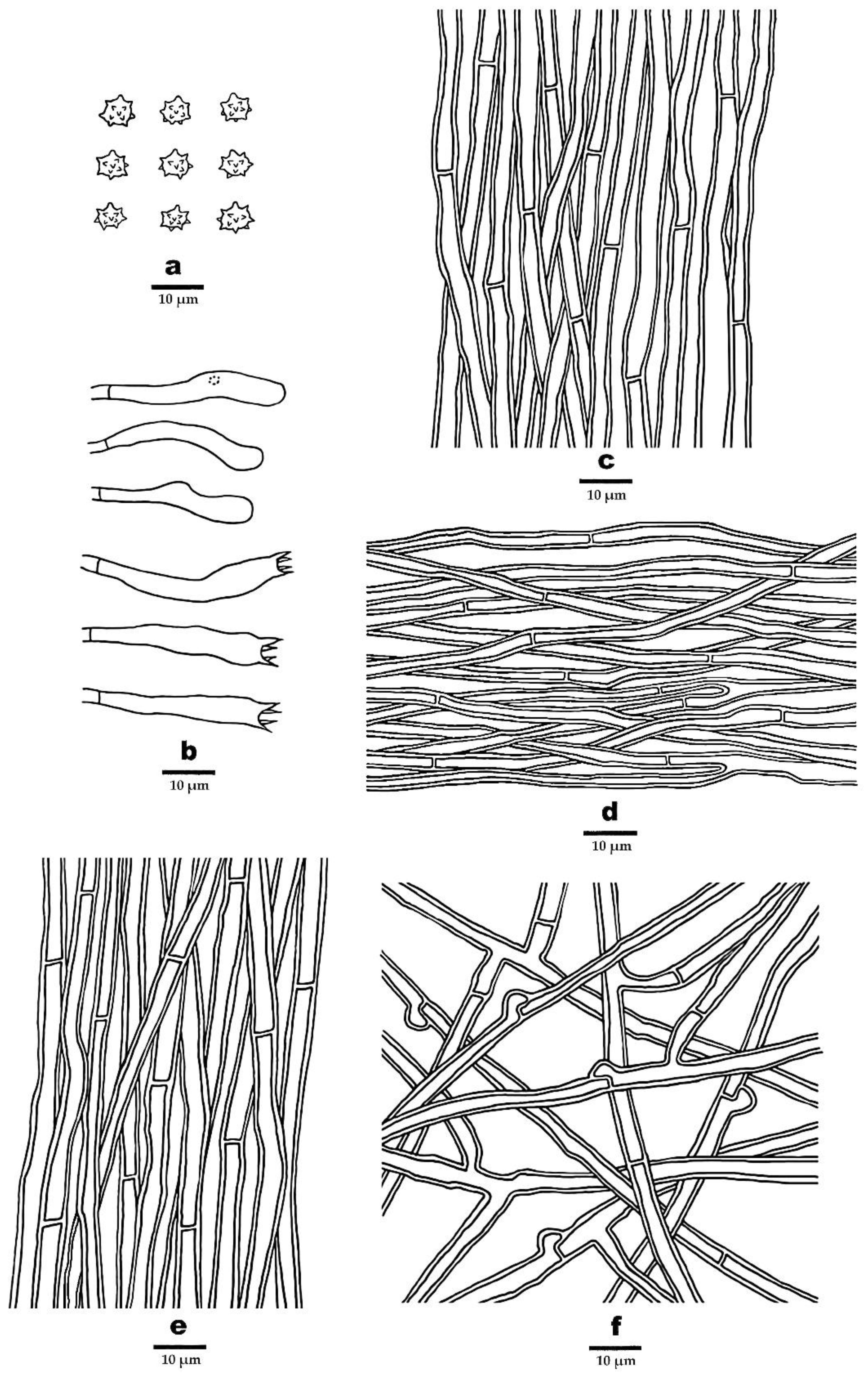
2.1. Morphological Studies

The specimens used in this study were collected during the annual growing season of macrofungi. At the same time, the specimen information, host trees, ecological habits, location, altitude, collector, and date were recorded, and photos of the fruiting bodies and growth environment were taken. The location information and ecological habits of the specimens mentioned above are stated in the results section. All samples examined in this study were deposited at the herbaria of the Institute of Microbiology, Beijing Forestry University, China (BJFC). Micro-morphological data were obtained from dried specimens and observed under a light microscope (Nikon Eclipse E 80i microscope, Nikon, Tokyo, Japan) following methods in Liu et al. [38].
Samples for microscopic examination were mounted in Cotton Blue, Melzer and 5% potassium hydroxide (KOH), separately. Basidiospores were measured from sections cut from the spines. The following abbreviations are used: IKI, Melzer’s reagent; IKI–, neither amyloid nor dextrinoid; KOH, 5% potassium hydroxide; CB, Cotton Blue; CB–, acyanophilous; L = mean spore length, W = mean spore width, Q = L/W ratio, n (a/b) = number of spores (a) measured from given number of specimens (b). A field Emission Scanning Electron Microscope (FESEM) Hitachi SU-8010 (Hitachi, Ltd., Tokyo, Japan) was used to film the spore’s morphology, and the materials were studied at up to 1800 times magnification, according to the method by Sun et al. [45].

2.2. DNA Extraction, PCR Amplification, and Sequencing

A CTAB plant genome rapid extraction kit-DN14 (Aidlab Biotechnologies Co., Ltd.) was employed for DNA extraction from dried specimens. The extracted DNA were used to perform the polymerase chain reaction (PCR) according to the manufacturer’s instructions with some modifications [34,40]. The primer pairs ITS5/ITS4, LR0R/LR7, NS1/NS4, AF/Cr, and 5F/7Cr were used to amplify ITS, nLSU, nSSU, RPB1, and RPB2 sequences [27,28]. The concentration of all primers is 1 g per mL. The final Polymerase Chain Reaction (PCR) volume was 30 μL; each tube contained 1 μL each primer, 1 μL extracted DNA, 12 μL ddH2O, and 15 μL 2 × EasyTaq PCR Supermix (TransGen Biotech Co., Ltd., Beijing, China). PCRs were performed on S1000™ Thermal Cycler (Bio-Rad Laboratories, California, USA). The PCR procedure for ITS was: initial denaturation at 95 °C for 3 min, followed by 34 cycles of denaturation at 94 °C for 40 s, annealing at 56 °C for 45 s and extension at 72 °C for 1 min, and a final extension at 72 °C for 10 min. The PCR process for nLSU and nSSU was as follows: initial denaturation at 94 °C for 1 min, followed by 35 cycles at 94 °C for 30 s, 50 °C for 1 min, 72 °C for 90 s, and a final extension of 72 °C for 10 min. The PCR process for RPB1 and RPB2 was as follows: initial denaturation at 94 °C for 2 min, 9 cycles at 94 °C for 45 s, 60 °C for 45 s, followed by 36 cycles at 94 °C for 45 s, 53 °C for 1 min, 72 °C for 90 s and a final extension of 72 °C for 10 min. The PCR products were purified and sequenced at the Beijing Genomics Institute, China, with the same primers. All sequences analyzed in this study were deposited at GenBank and listed in Table 1.

2.3. Phylogenetic Analyses

The phylogenetic relationships of Phellodon were analyzed by the datasets of combined ITS + nLSU sequences and ITS + nLSU + nSSU + RPB1 + RPB2 sequences. The ITS + nLSU sequences were used to infer the phylogeny of Phellodon. The 5-gene datasets more specifically showed the differences between Phellodon species. The sequences generated in this study and retrieved from GenBank were combined with ITS, nLSU, nSSU, RPB1, and RPB2 sequences of Phellodon and outgroups. Amaurodon aquicoeruleus Agerer (UK 452) and A. viridis (Alb. and Schwein.) J. Schröt (TAA 149664) were used as the outgroups, according to Song et al. [28]. The datasets were aligned in MAFFT 7 [46] and manually adjusted in BioEdit [47]. Alignments were spliced in Mesquite v. 3.2. [48]. The congruences of the 5-gene (ITS, nLSU, nSSU, RPB1, and RPB2,) were evaluated with the incongruence length difference (ILD) test [49] implemented in PAUP* version 4.0b10 [50], under heuristic search and 1000 homogeneity replicates. The best-fit evolutionary model was selected with AIC (Akaike Information Criterion) using jModelTest for each partition [51,52]. Phylogenetic analyses were carried out according to previous studies [31,40].
Maximum parsimony (MP) analysis was performed in PAUP*version 4.0b10 [50] with the heuristic search. All characters were equally weighted and gaps were treated as missing data. Trees were inferred using the heuristic search option with TBR branch swapping and 1000 random sequence additions. Max-trees was set to 5000, branches of zero length were collapsed and all parsimonious trees were saved. Clade robustness was assessed using a bootstrap analysis with 1000 replicates [53]. Descriptive tree statistics, tree length (TL), consistency index (CI), retention index (RI), rescaled consistency index (RC), and homoplasy index (HI) were calculated for each Maximum Parsimonious Tree (MPT) generated. Only the Maximum Parsimony best tree from all searches was kept. Maximum Likelihood (ML) analysis was performed in RAxmL v.7.2.8 with a GTR + G + I model [54]. All model parameters were estimated by the program, but only the best maximum likelihood tree from all searches was kept. MrModeltest 2.3 [55,56] was used to determine the best-fit evolution model for each dataset for Bayesian inference (BI). BI was performed using MrBayes 3.2.6 on Abe through the Cipres Science Gateway (www.phylo.org, accessed on 23 April 2022) with 2 independent runs, each one beginning from random trees with 4 simultaneous independent Chains, performing 2 million replicates, sampling one tree every 100 generations [57]. The first 25% of the sampled trees were discarded as burn-in and a majority rule consensus tree of all remaining trees was calculated.
Branches that received bootstrap supports for MP, ML greater than or equal to 50% and Bayesian inference (BI) greater than or equal to 0.95 were considered as significantly supported. Phylogenetic trees were visualized using FigTree v1.4.2.

3. Results

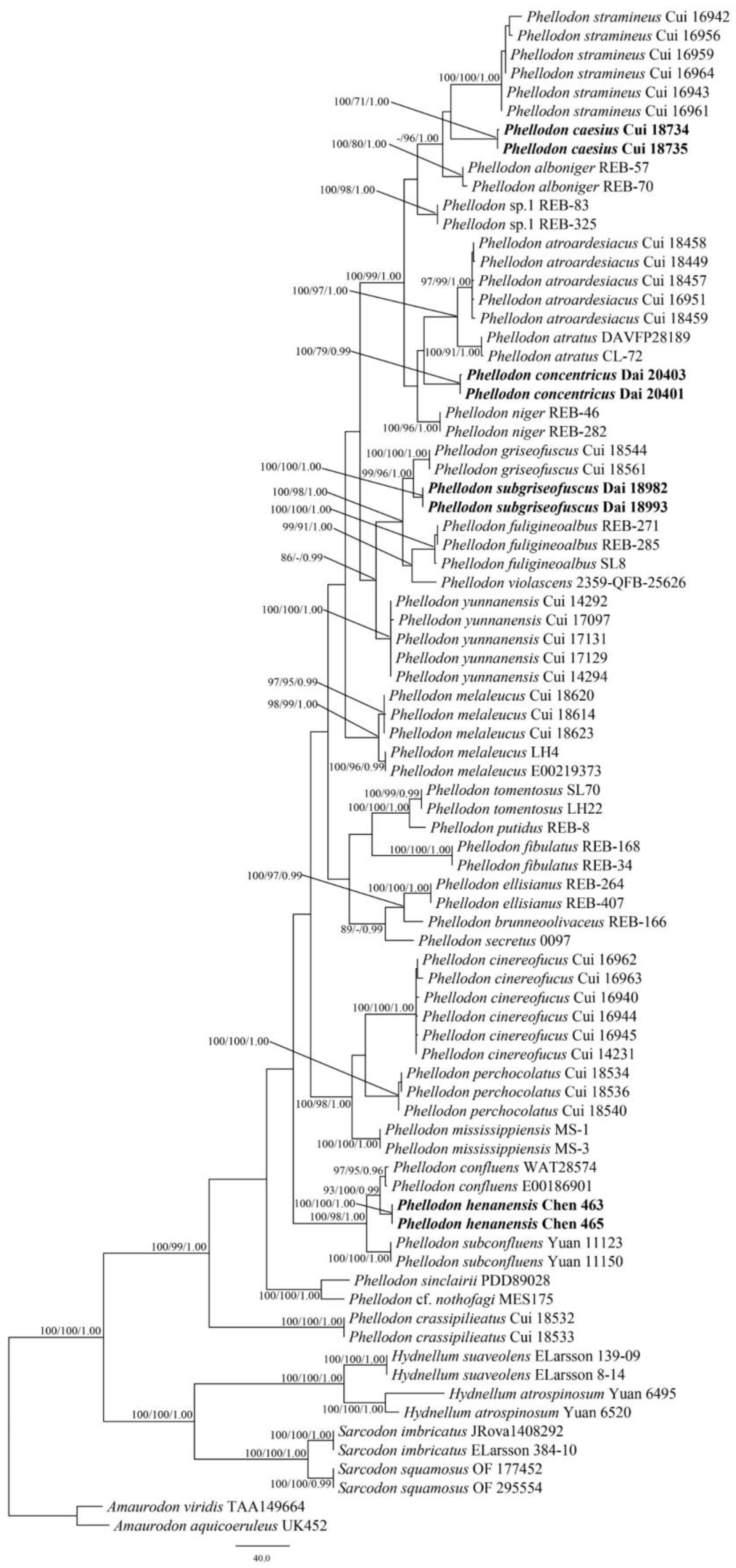
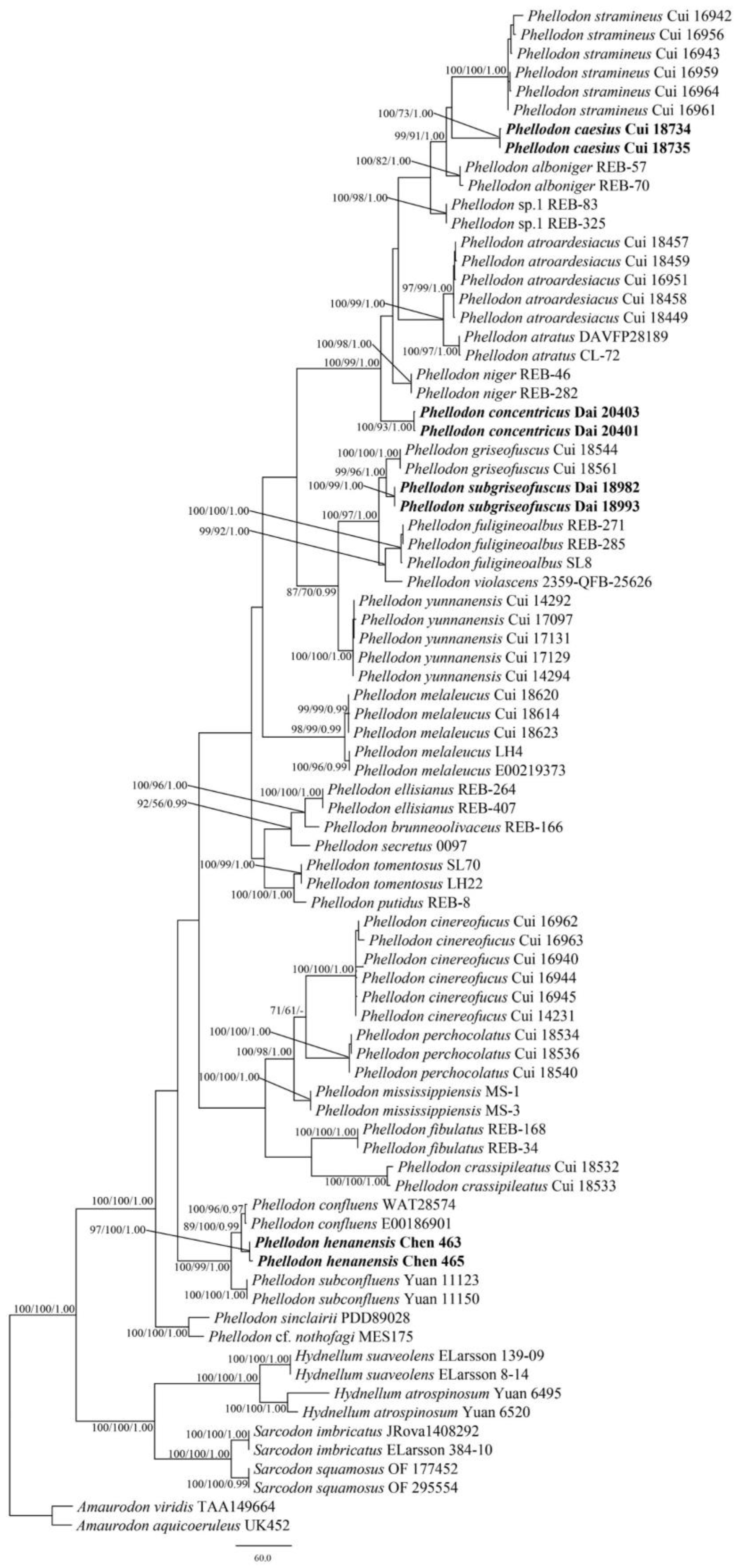
3.1. Phylogenetic Analyses
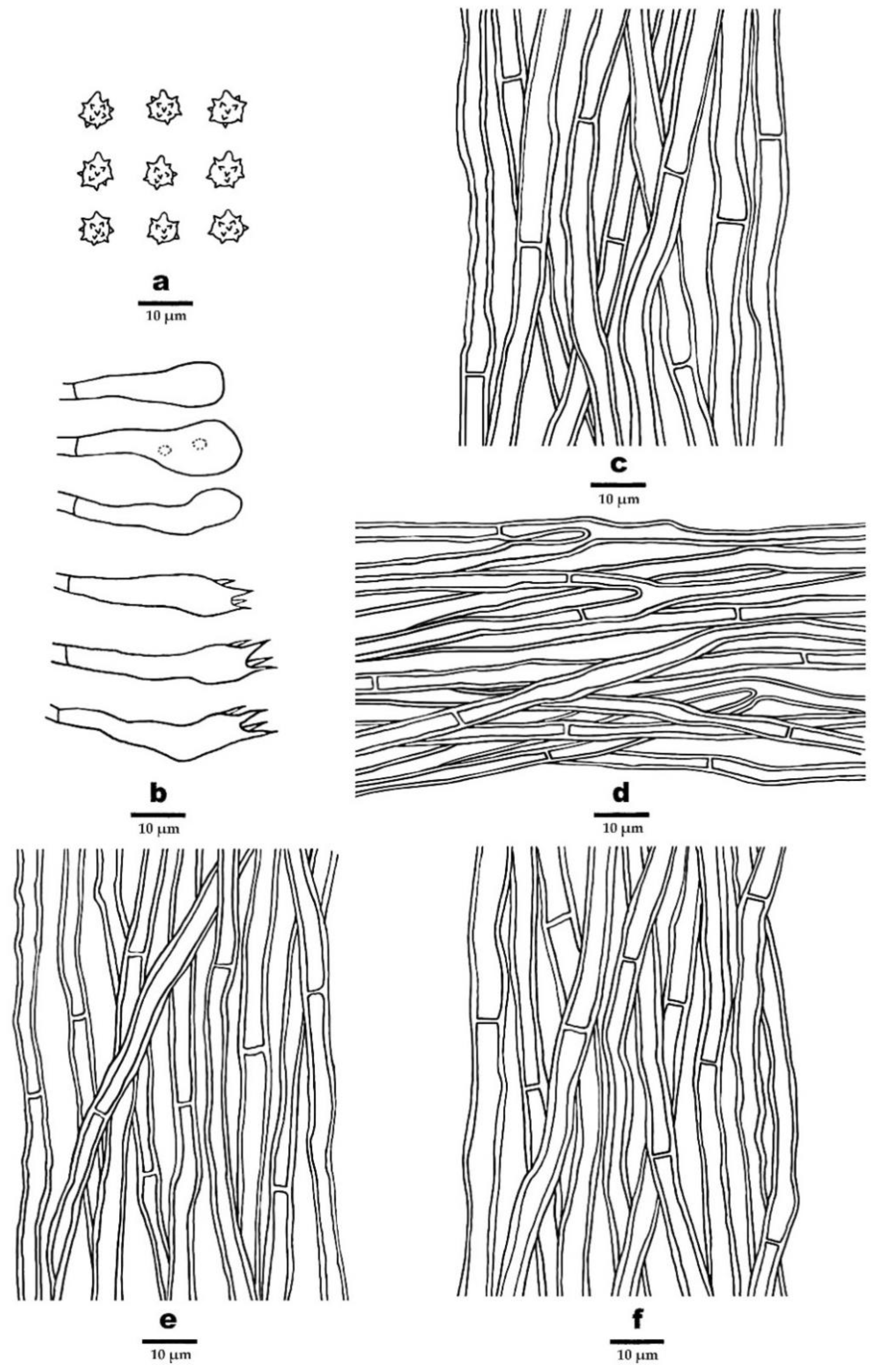
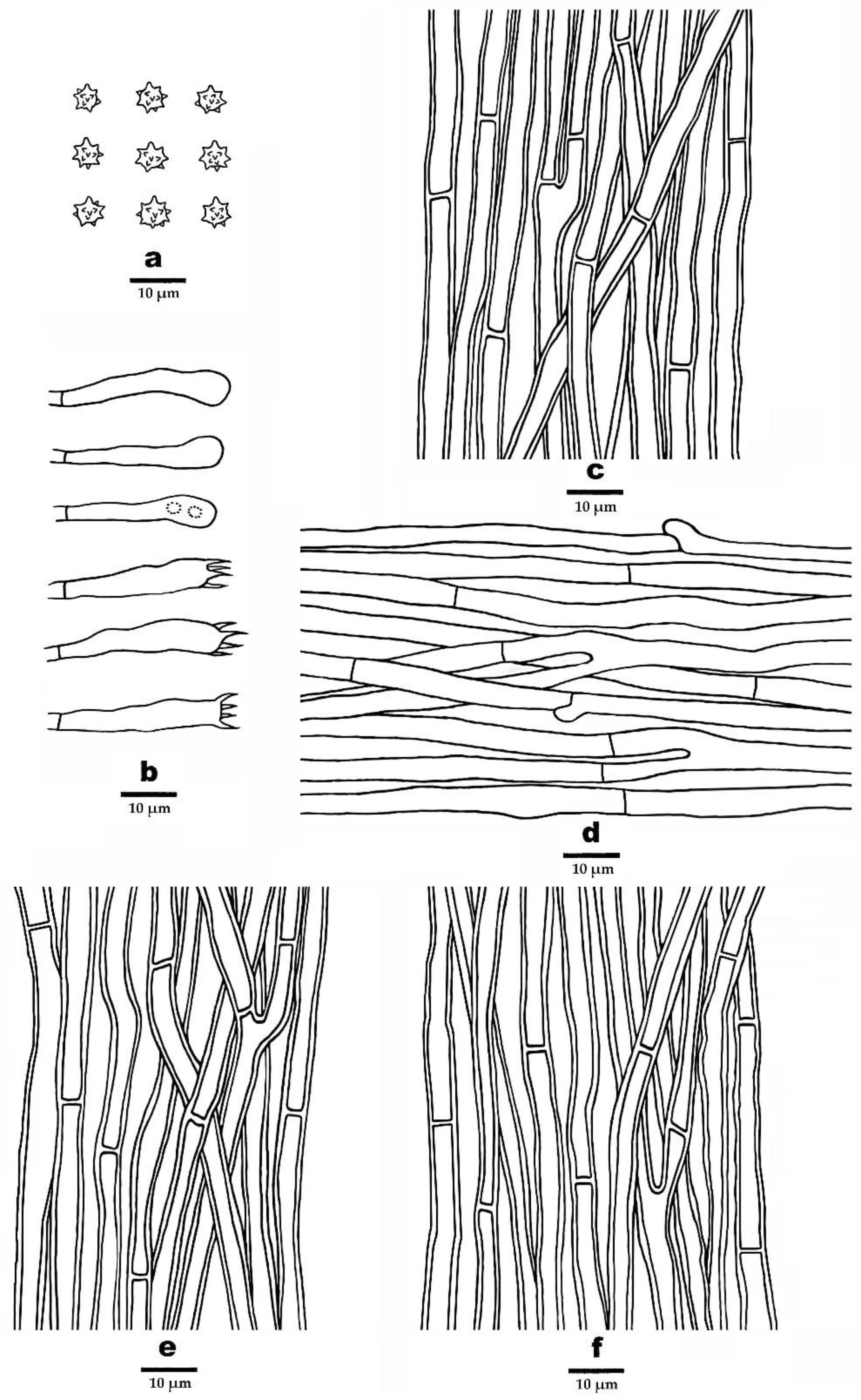
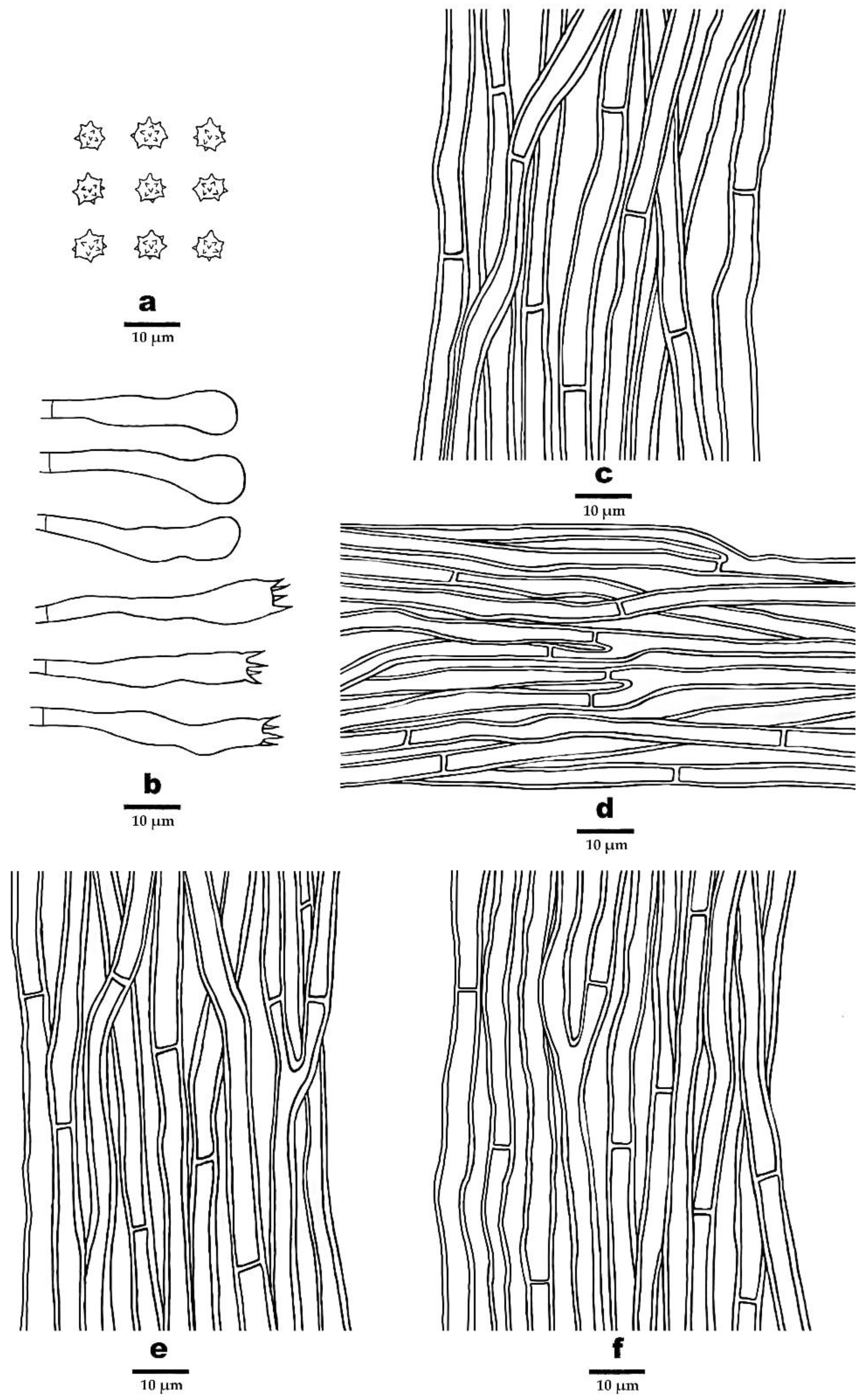
The combined ITS + nLSU dataset included sequences from 81 fungal samples representing 35 taxa. The dataset had an aligned length of 2244 characters, including gaps (865 characters for ITS, 1379 characters for nLSU), of which 1564 characters were constant, 69 were variable and parsimony-uninformative, and 611 were parsimony-informative. Maximum parsimony analysis yielded 1205 equally parsimonious trees (TL = 1894, CI = 0.548, RI = 0.843, RC = 0.462, HI = 0.452). The best models for each region of the combined ITS + nLSU sequence dataset estimated and applied in the Bayesian analysis were both GTR + I + G models. Bayesian and ML analysis resulted in a topology similar to that from MP analysis. The Bayesian analysis resulted in a concordant topology with an average standard deviation of split frequencies = 0.005071. Only the MP tree is provided in Figure 1, and the MP (≥50%), ML (≥50%), and BI (≥0.95) are shown at the nodes.
The combined 5-gene ITS + nLSU + nSSU + RPB1 + RPB2 dataset included sequences from 81 fungal samples representing 35 taxa. The dataset had an aligned length of 5597 characters, including gaps (865 characters for ITS, 1379 characters for nLSU, 1070 characters for nSSU, 1204 characters for RPB1, 1079 characters for RPB2), of which 4513 characters were constant, 199 were variable and parsimony-uninformative, and 885 were parsimony-informative. Maximum parsimony analysis yielded 2389 equally parsimonious trees (TL = 2389, CI = 0.615, RI = 0.857, RC = 0.527, HI = 0.385). The best-fit evolutionary models applied in Bayesian analyses were selected by jModelTest for each region of the five genes, the model for ITS, nLSU, nSSU, RPB1, and RPB2 was GTR + I+ G with an equal frequency of nucleotides. Bayesian and ML analysis resulted in a topology similar to that from MP analysis. The Bayesian analysis resulted in a concordant topology with an average standard deviation of split frequencies = 0.004330. Only the MP tree is provided in Figure 2, and the MP (≥50%), ML (≥50%), and BI (≥0.95) are shown at the nodes.
Both the ITS + nLSU dataset and the ITS + nLSU + nSSU + RPB1 + RPB2-based phylogenetic tree (Figure 1 and Figure 2) confirmed the affinities of the four new species within Phellodon. The four new species P. caesius, P. concentricus, P. henanensis, and P. subgriseofuscus formed distinct well-supported lineages distant from other species of Phellodon.

3.2. Taxonomy

Phellodon caesius B.K. Cui & C.G. Song, sp. nov., Figure 3a, Figure 4a and Figure 5.
MycoBank: 846978
Diagnosis—Differs from other Phellodon species by its bluish-grey, dark grey to black grey pileus, ash grey to dark bluish-grey spines, and the presence of both simple septa and clamp connections on generative hyphae of the surface layer of stipe.
Etymologycaesius (Lat.), refers to the bluish-grey pileus.
Holotype—CHINA. Sichuan Province, Xiaojin County, on the ground of forest dominated by Quercus aquifolioides, alt. 3320 m, 3 September 2021, Cui 18734 (BJFC 045001).
Fruitbody—Basidiomata annual, centrally or eccentrically stipitate, single to concrescent, with a light fenugreek odor when dry. Pileus slightly convex in the middle, plicate, up to 3.6 cm in diam, and 0.7 cm thick at the center. Pileal surface bluish-grey, dark bluish-grey to black grey when fresh and becoming pale mouse grey to mouse grey upon drying, azonate, fibrillose to spongy; margin white to ash grey when fresh, and becoming pale mouse grey upon drying, up to 2 mm wide. Context tough, dark violet to dark grey upon drying, up to 3 mm thick. Spines soft, white, ash grey to dark bluish-grey when fresh, becoming fragile, pale mouse grey to ash grey upon drying, up to 2 mm long. Stipe cylindrical, glabrous, dark grey to black in outer layer, black in the inner layer, up to 2.2 cm long, 1.2 cm in diam.
Hyphal structure—Hyphal system monomitic; generative hyphae in context, spines, and the inner layer of stipe with simple septa, generative hyphae in the surface layer of stipe mostly with simple septa, occasionally with clamp connections; all the hyphae IKI–, CB–; tissues turned olive green in KOH. Generative hyphae in context clay-buff, thick-walled, rarely branched, regularly arranged, 2.5–5 µm in diam. Generative hyphae in spines dark clay-buff, thick-walled, occasionally branched, regularly arranged, 2–3.5 µm in diam. Generative hyphae in the inner layer of stipe clay-buff to fuscous, thick-walled, rarely branched, regular arranged, 3–5 µm in diam; generative hyphae in the surface layer of stipe fuscous, thick-walled, branched, interwoven, 3–6 µm in diam.
Cystidia—Cystidia and other sterile hyphal elements absent.
Basidia—Clavate, bearing four sterigmata and a basal simple septum, 29–53 × 5.5–7 µm; sterigmata 2–5.5 µm long; basidioles similar to basidia in shape, but slightly smaller.
Spores—Basidiospores subglobose to globose, hyaline, thin-walled, echinulate, IKI–, CB–, 4–5.6(–6) × (3.8–)4–5.2 µm, L = 4.87 µm, W = 4.48 µm, Q = 1–1.25 (n = 60/2, without the ornamentation).
Additional specimen (paratype) examined—CHINA. Sichuan Province, Xiaojin County, on the ground of forest dominated by Quercus aquifolioides, alt. 3320 m, 3 September 2021, Cui 18735 (BJFC 045002).
Ecological habitsP. caesius was found on the ground of forest dominated by trees of Quercus aquifolioides, under a temperate climate at high altitude regions in Southwest China.
Phellodon concentricus B.K. Cui and C.G. Song, sp. nov., Figure 3b, Figure 4b and Figure 6.
MycoBank: 846979
Diagnosis—Differs from other Phellodon species by its zonate pileal surface, dark grey context in pileus, and spongy basidiomata.
Etymologyconcentricus (Lat.), refers to the concentric bands on pileal surface.
Holotype—CHINA. Yunnan Province, Yuxi County, Xinping, Mopanshan Forest Park, on the ground of forest dominated by Quercus sp., alt. 2088 m, 14 August 2019, Dai 20403 (BJFC 032071).
Fruitbody—Basidiomata annual, centrally or eccentrically stipitate, single to concrescent, with a strong fenugreek odor when dry. Pileus depressed, circular to irregular, up to 4.5 cm in diam, 0.3 cm thick at the center. Pileal surface deep olive to mouse grey upon drying, zonate, fibrillose to spongy at the center; margin fuscous to black upon drying, up to 5 mm wide. Context tough, dark grey upon drying, up to 1 mm thick. Spines soft when fresh, becoming fragile, ash grey upon drying, up to 2.5 mm long. Stipe cylindrical, spongy, deep olive, fuscous to black, up to 2.5 cm long, 1 cm in diam.
Hyphal structure—Hyphal system monomitic; generative hyphae with simple septa; all the hyphae IKI–, CB–; tissues turned light yellow-green to olive green in KOH. Generative hyphae in context dark yellowish-green, thick-walled, rarely branched, regularly arranged, 3–6.5 µm in diam. Generative hyphae in spines yellowish-brown to dark brown, slightly thick-walled, branched, regularly arranged, 2–4.5 µm in diam. Generative hyphae in stipe dark olive-green to black, thick-walled, rarely branched, regularly arranged, 2–6 µm in diam.
Cystidia—Cystidia and other sterile hyphal elements absent.
Basidia—Clavate, bearing four sterigmata and a basal simple septum, 25–44 × 5.2–6.8 µm; sterigmata 3.5–6 µm long; basidioles similar to basidia in shape but slightly smaller.
Spores—Basidiospores subglobose to globose, hyaline, thin-walled, echinulate, IKI–, CB–, 5–6.2 × 4.5–5.5(–5.7) µm, L = 5.48 µm, W = 4.99 µm, Q = 1–1.22 (n = 60/2, without the ornamentation).
Additional specimen (paratype) examined—CHINA. Yunnan Province, Yuxi County, Xinping, Mopanshan Forest Park, on the ground of forest dominated by Quercus sp., alt. 2088 m, 14 August 2019, Dai 20401 (BJFC 032069).
Ecological habitsP. concentricus was found in forest dominated by trees of Quercus sp., under a subtropical climate.
Phellodon henanensis B.K. Cui and C.G. Song, sp. nov., Figure 3c, Figure 4c and Figure 7.
MycoBank: 846980
Diagnosis—Differs from other Phellodon species by its ash grey, light vinaceous grey to light brown pileal surface, thin context in pileus, and the presence of both simple septa and clamp connections on generative hyphae of spines.
Etymologyhenanensis (Lat.), refers to the holotype locality of the species in Henan Province.
Holotype—CHINA. Henan Province, Luanchuan County, Laojun Mountain, Jindian, on the ground of mixed forest, alt. 2000 m, 8 September 2020, Chen 463 (BJFC 045003).
Fruitbody—Basidiomata annual, eccentrically stipitate, usually solitary, with a fenugreek odor when dry. Pileus depressed or shallow infundibuliform, up to 2.2 cm in diam, 0.3 cm thick at the center. Pileal surface ash grey, light vinaceous grey to light brown when fresh and becoming dark brown to black upon drying, azonate, fibrillose; margin cream to light brown when fresh, and becoming apricot-orange upon drying, up to 3 mm wide. Context tough, greyish-brown, up to 1 mm thick. Spines soft, ash grey to light brown when fresh, becoming fragile, vinaceous grey to greyish-brown upon drying, up to 1 mm long. Stipe cylindrical, glabrous, pale greyish-brown to pale mouse grey, up to 1.3 cm long, 0.2 cm in diam.
Hyphal structure—Hyphal system monomitic; generative hyphae in context and stipe with simple septa, generative hyphae in spines mostly with simple septa, occasionally with clamp connections; all the hyphae IKI–, CB–; all tissues turned olive green in KOH. Generative hyphae in context hyaline to clay-buff, thick-walled, occasionally branched, regularly arranged, 2–6 µm in diam. Generative hyphae in spines hyaline to clay-buff, thin-walled, occasionally branched, regularly arranged, 2–4 µm in diam. Generative hyphae in stipe occasionally hyaline to dark brown, thick-walled, branched, regularly arranged, 2.5–5 µm in diam.
Cystidia—Cystidia and other sterile hyphal elements absent.
Basidia—Clavate, bearing four sterigmata and a basal simple septum, 24–46 × 4.5–5.5 µm; sterigmata 2–5 µm long; basidioles similar to basidia in shape, but slightly smaller.
Spores—Basidiospores subglobose to globose, hyaline, thin-walled, echinulate, IKI–, CB–, (3.2–)3.8–5 × (3–)3.5–4.5(–4.8) µm, L = 4.17 µm, W = 3.84 µm, Q = 1–1.31 (n = 60/2, without the ornamentation).
Additional specimen (paratype) examined—CHINA. Henan Province, Luanchuan County, Laojun Mountain, Jindian, on the ground of mixed forest, alt. 2000 m, 8 September 2020, Chen 465 (BJFC 045004).
Ecological habitsP. henanensis was found on the ground with a thin layer of moss, under a warm temperate continental monsoon climate.
Phellodon subgriseofuscus B.K. Cui and C.G. Song, sp. nov., Figure 3d, Figure 4d and Figure 8.
MycoBank: 846981
Diagnosis—Differs from other Phellodon species by its fuscous to black pileal surface, white to light brown spines, and vinaceous grey context.
Etymologysubgriseofuscus (Lat.), refers to the new species resembling P. griseofuscus in morphology.
Holotype—CHINA. Gansu Province, Zhangye, Qilianshan Nature Reserve, Sidalong belay station, on the ground of forest dominated by Picea crassifolia, alt. 3000 m, 4 September 2018, Dai 18993 (BJFC 027462).
Fruitbody—Basidiomata annual, eccentrically stipitate, single to concrescent, with a fenugreek odor when dry. Pileus circular to irregular, up to 4.8 cm in diam, 1.2 cm thick at the center. Pileal surface fuscous to black when fresh and becoming dark brown to fuscous upon drying, zonate, glabrous, with radially aligned stripes; margin white to dark brown when fresh, and becoming white to cream upon drying, up to 3 mm wide. Context tough, vinaceous grey upon drying, up to 3 mm thick. Spines soft, white to light brown when fresh, becoming fragile, cream to buff-yellow upon drying, up to 2.5 mm long. Stipe cylindrical, glabrous, greyish-brown, dark brown to fuscous, up to 3.3 cm long, 1.5 cm in diam.
Hyphal structure—Hyphal system monomitic; generative hyphae with simple septa; all the hyphae IKI–, CB–; tissues turned olive green in KOH. Generative hyphae in context brown, thick-walled, rarely branched, regularly arranged, 2–6 µm in diam. Generative hyphae in spines hyaline to clay-buff, slightly thick-walled, branched, regularly arranged, 2–4 µm in diam. Generative hyphae in stipe hyaline to dark brown, thick-walled, occasionally branched, regularly arranged, 2–6 µm in diam.
Cystidia—Cystidia and other sterile hyphal elements absent.
Basidia—Clavate, bearing four sterigmata and a basal simple septum, 27–43 × 5–7 µm; sterigmata 2–5.5 µm long; basidioles similar to basidia in shape, but slightly smaller.
Spores—Basidiospores subglobose to globose, hyaline, thin-walled, echinulate, IKI–, CB–, 4–5 × (3–)3.2–4.8 µm, L = 4.47 µm, W = 3.9 µm, Q = 1–1.41 (n = 60/2, without the ornamentation).
Additional specimen (paratype) examined—CHINA. Gansu Province, Zhangye County, Qilianshan Nature Reserve, Xishui belay station, on the ground of forest dominated by Picea crassifolia, alt. 2250 m, 3 September 2018, Dai 18982 (BJFC 027451).
Ecological habitsP. subgriseofuscus was found on the ground of forest dominated by trees of Picea, under a continental alpine sub-humid mountain climate. This species grows in well-watered bryophytes.
Key to species of Phellodon from China
1.
Pileal surface straw buff-------------------------------------------------------------------------------P. stramineus
1.
Pileal surface differently colored--------------------------------------------------------------------2
2.
Pileal surface blackish-blue to dark grey or bluish-grey to dark bluish-grey-------------3
2.
Pileal surface differently colored--------------------------------------------------------------------4
3.
Clamp connections exist in spines------------------------------------------------------------------P. atroardesiacus
3.
Clamp connections do not exist in spines---------------------------------------------------------P. caesius
4.
Tissues color changed in KOH-----------------------------------------------------------------------5
4.
Tissues color unchanged in KOH-------------------------------------------------------------------P. subconfluens
5.
Pileal surface glabrous---------------------------------------------------------------------------------6
5.
Pileal surface not glabrous----------------------------------------------------------------------------8
6.
Pileal surface reddish-brown to cinnamon brown----------------------------------------------P. cinereofuscus
6.
Pileal surface differently colored--------------------------------------------------------------------7
7.
Pileal surface clay pink to brown-------------------------------------------------------------------P. yunnanensis
7.
Pileal surface fuscous to black-----------------------------------------------------------------------P. subgriseofuscus
8.
Clamp connections exist-------------------------------------------------------------------------------9
8.
Clamp connections absent----------------------------------------------------------------------------P. concentricus
9.
Pileal surface ash grey, light vinaceous grey to light brown---------------------------------P. henanensis
9.
Pileal surface differently colored-------------------------------------------------------------------10
10.
Clamp connections exist in spines----------------------------------------------------------------11
10.
Clamp connections do not exist in spines-------------------------------------------------------P. crassipileatus
11.
Spines brown after mature--------------------------------------------------------------------------P. griseofuscus
11.
Spines white after mature---------------------------------------------------------------------------P. perchocolatus

4. Discussion

Based on the phylogenetic analyses, 29 species of Phellodon grouped together (Figure 1 and Figure 2), including four new species from China: P. caesius, P. concentricus, P. henanensis, and P. subgriseofuscus. Our phylogenetic results are consistent with previous observations [27,28], and further information on the phylogeny and taxonomy of Phellodon is supplied. During the investigations of Phellodon, information on distribution areas and ecological habits was also obtained (Table 2).
Phellodon caesius is clustered together with P. alboniger and P. stramineus in our phylogenetic trees (Figure 1 and Figure 2). Morphologically, P. alboniger is similar to P. caesius in having a spongy pileal surface, and white, brownish-grey to burnt umber spines. However, P. alboniger differs from P. caesius by its white to greyish-orange pileal surface, and longer spines (up to 2 mm [11]). Phellodon stramineus is similar to P. caesius in having solitary or gregarious basidiomata, ash-grey spines, and similar-sized basidiospores [27]. However, P. stramineus differs from P. caesius in its straw-buff pileal surface and the absence of clamp connections in stipe [27]. Phellodon atroardesiacus B.K. Cui and C.G. Song are morphologically similar to P. caesius in their blackish-blue to dark grey pileal surface and the dark greyish-blue to ash grey spines [27]. Surprisingly, they are not closely related as demonstrated in our phylogenetic analyses (Figure 1 and Figure 2). Phellodon atroardesiacus differs from the newly described P. caesius by its smaller basidiospore size (4–5 × (3–)3.5–4.5 in P. atroardesiacus vs. 4–5.6(–6) × (3.8–)4–5.2 in P. caesius [27]).
Phellodon concentricus is closely related to P. niger in our phylogenetic analyses (Figure 1 and Figure 2). Morphologically, P. niger is similar to P. concentricus in having single to concrescent basidiomata, crowded spines, and a spongy pileal surface. However, P. niger can be distinguished by its dark blue to black context, white, grey, or bluish-grey spines, and the smaller basidiospores size (5–6 × 4–5 μm in P. niger vs. 5–6.2 × 4.5–5.5(–5.7) µm in P. concentricus [11]).
Phellodon henanensis and P. confluens were clustered together and then grouped with P. subconfluens in our phylogenetic analyses (Figure 1 and Figure 2). Phellodon confluens is similar to P. henanensis in having shallow infundibuliform pileus, and the same-colored pileus. However, P. confluens differs from P. henanensis by its larger pileus measuring 3–10 cm, and the smaller basidiospores size (3.6–4.3 × 3.3–4 µm in P. confluens vs. (3.2–)3.8–5 × (3–)3.5–4.5(–4.8) µm in P. henanensis [18]). Morphologically, P. subconfluens is similar to P. henanensis in having a fenugreek odor when dry and short spines (up to 1 mm). However, P. subconfluens differs from P. henanensis by its greyish-buff, brownish-orange to reddish-brown pileal surface, cream to greyish-buff spines, and the smaller basidiospores size ((3.0–)3.1–4.1(–4.8) × (2.5–)2.9–3.5(–3.8) μm in P. subconfluens vs. (3.2–)3.8–5 × (3–)3.5–4.5(–4.8) µm in P. henanensis [29]).
Phellodon subgriseofuscus is closely related to P. griseofuscus B.K. Cui and C.G. Song in our phylogenetic analyses (Figure 1 and Figure 2). Morphologically, P. griseofuscus is similar to P. subgriseofuscus in having dark brown or black pileal surface and white to light brown spines. However, P. griseofuscus can be distinguised by its shorter spines (up to 1 µm), and clamp connections in generative hyphae of pileus and stipe [28].
The diversity and evolutionary relationships of Phellodon species can be objectively revealed by combining traditional morphological observation with molecular systematics methods. In the past, only a few numbers of publications had used phylogenetic analyses of the Phellodon genus, and the majority of those studies had only used the ITS sequences of a few species [11,24,25,29]. Song et al. [27,28] conducted phylogenetic analysis of Phellodon based on 5-gene sequences (ITS + nLSU + nSSU + RPB1 + RPB2), which undoubtedly filled in the blank of multiple gene fragments of Phellodon. In this study, both ITS + LSU and ITS + LSU + SSU + RPB1 +RPB2 datasets share a similar topology with Song et al. [27,28] but with discrepant bootstrap values.
Phellodon species frequently grow beneath pine needles or oak leaves, which serve to prevent water loss, in damp woodlands covered in dense mosses. Specimens collected in China were gathered from forests of pinaceae, fagaceae, or mixed trees (Table 2). It revealed that Phellodon species are host-biased, providing an additional foundation for species discovery and identification. The specimens were collected from northeast, southwest, northwest, and central China at elevations ranging from 870 to 3320 m, which indicated that the genus is a widespread species.
With the addition of the species discussed above, there are now 12 taxa in Phellodon known from China. The identification and descriptions of stipitate hydnoid fungi in this paper can enrich the species diversity of Phellodon and promote the taxonomy and phylogeny of the genus. The combination of morphological and phylogenetic methods will contribute to the exploration of species diversity. Additionally, it suggested that other Phellodon species might be discovered by combining the evidence of morphological characters, molecular data, and ecological habits. A fully resolved phylogeny for species in Phellodon requires evolutionary information from more samples.

Author Contributions

B.-K.C. designed the research; B.-K.C., Y.-Y.C., Y.-F.S., S.L. and C.-G.S. prepared the samples; C.-G.S. and S.L. conducted the molecular experiments and analyzed the data; C.-G.S., Y.-F.S. and B.-K.C. drafted the manuscript. All authors have read and agreed to the published version of the manuscript.

Funding

The research is supported by the National Natural Science Foundation of China (Nos. 31870008, 32270010, 31900017) and Beijing Forestry University Outstanding Young Talent Cultivation Project (No. 2019JQ03016).

Institutional Review Board Statement

Not applicable.

Informed Consent Statement

Not applicable.

Data Availability Statement

The data and results of this study are available upon reasonable request. Please contact the main author of this publication.

Acknowledgments

We express our gratitude to Yu-Cheng Dai (Beijing Forestry University, China) for his help during field collections.

Conflicts of Interest

The authors declare no conflict of interest. The funders had no role in the design of the study; in the collection, analyses, or interpretation of data; in the writing of the manuscript, or in the decision to publish the results.

References

  1. Karsten, P.A. Enumeratio boletinearum et polyporearum fennicarum, systemate novo dispositarum. Rev. Mycol. 1881, 3, 16–19. [Google Scholar]
  2. Maas Geesteranus, R.A. Hydnaceous fungi of the eastern old world. Verh. Kon. Ned. Akad. Wetensch. Afd. Natuurk 1971, 60, 176. [Google Scholar]
  3. Erland, S.; Taylor, A.F.S. Resupinate ectomycorrhizal fungal genera. In Ectomycorrhizal Fungi Key Genera in Profile; Cairney, J.W.G., Chambers, S.M., Eds.; Springer: Berlin/Heidelberg, Germany, 1999; pp. 347–363. [Google Scholar]
  4. Brundrett, M.C. Mycorrhizal associations and other means of nutrition of vascular plants: Understanding the global diversity of host plants by resolving conflicting information and developing reliable means of diagnosis. Plant Soil 2009, 320, 37–77. [Google Scholar] [CrossRef]
  5. Van der Heijden, M.; Klironomos, J.; Ursic, M.; Moutoglis, P.; Streitwolf-Engel, R.; Boller, T.; Wiemken, A.; Sanders, I. Mycorrhizal fungal diversity determines plant biodiversity, ecosystem variability and productivity. Nature 1998, 396, 69–72. [Google Scholar] [CrossRef]
  6. Stadler, M.; Anke, T.; Dasenbrock, J.; Steglich, W. Phellodonic Acid, a new biologically active hirsutane derivative from Phellodon melaleucus (Thelephoraceae, Basidiomycetes). Z. Nat. C 1993, 48, 545–549. [Google Scholar] [CrossRef]
  7. Reekie, T.A.; Austin, K.A.; Banwell, M.G.; Willis, A.C. The chemoenzymatic total synthesis of phellodonic acid, a biologically active and highly functionalized hirsutane derivative isolated from the tasmanian fungus Phellodon melaleucus. Aust. J. Chem. 2008, 61, 94–106. [Google Scholar] [CrossRef]
  8. Fang, S.T.; Zhang, L.; Li, Z.H.; Li, B.; Liu, J.K. Cyathane diterpenoids and nitrogenous terphenyl derivative from the fruiting bodies of basidiomycete Phellodon niger. Chem. Pharm. Bull. 2010, 58, 1176–1179. [Google Scholar] [CrossRef] [Green Version]
  9. Banker, H.J. A contribution to a revision of the North American Hydnaceae. Mem. Torrey Bot. Club 1906, 12, 99–194. [Google Scholar]
  10. Donk, M.A. Four new families of Hymenomycetes. Persoonia 1961, 1, 405–407. [Google Scholar]
  11. Baird, R.E.; Wallace, L.E.; Baker, G.; Scruggs, M. Stipitate hydnoid fungi of the temperate southeastern United States. Fungal Divers. 2013, 62, 41–114. [Google Scholar] [CrossRef]
  12. Harrison, K.A. New or little known north American stipitate hydnums. Can. J. Bot. 1964, 42, 1205–1233. [Google Scholar] [CrossRef]
  13. Harrison, K.A. A new species of Phellodon possessing clamp connections. Can. J. Bot. 1972, 50, 1219–1221. [Google Scholar] [CrossRef]
  14. Hrouda, P. Hydnaceous fungi of the Czech Republic and Slovakia. Czech Mycol. 1999, 51, 99–155. [Google Scholar] [CrossRef]
  15. Hrouda, P. Bankeraceae in Central Europe. 1. Czech Mycol. 2005, 57, 57–78. [Google Scholar] [CrossRef]
  16. Hrouda, P. Bankeraceae in Central Europe. 2. Czech Mycol. 2005, 57, 279–297. [Google Scholar] [CrossRef]
  17. Coker, W.C.; Beers, A.H. The Stipitate Hydnums of the Eastern United States; University of North Carolina Press: Chapel Hill, NC, USA, 1951; p. 211. [Google Scholar]
  18. Pouzar, Z.D. Příspěvek k poznání našich kloboukatých lošáků. Čes. Mykol. 1956, 10, 65–76. [Google Scholar]
  19. Maas Geesteranus, R.A. The stipitate hydnums of the Netherlands. Fungus 1958, 28, 48–61. [Google Scholar]
  20. Maas Geesteranus, R.A. Notes on the hydnums. Persoonia 1960, 1, 341–384. [Google Scholar]
  21. Maas Geeseranus, R.A. Hyphal structures in the hydnums. Persoonia 1962, 2, 476. [Google Scholar]
  22. Maas Geesteranus, R.A. The terrestrial hydnums of Europe. Verh. Kon. Ned. Akad. Wetensch. Afd. Natuurk 1975, 65, 127. [Google Scholar]
  23. Parfitt, D.; Ainsworth, A.M.; Simpson, D.; Rogers, H.J.; Boddy, L. Molecular and morphological discrimination of stipitate hydnoids in the genera Hydnellum and Phellodon. Mycol. Res. 2007, 3, 761–777. [Google Scholar] [CrossRef] [PubMed]
  24. Ainsworth, A.M.; Parfitt, D.; Rogers, H.J.; Boddy, L. Cryptic taxa within European species of Hydnellum and Phellodon revealed by combined molecular and morphological analysis. Fungal Ecol. 2010, 3, 65–80. [Google Scholar] [CrossRef]
  25. Baird, R.E.; Wallace, L.E.; Baker, G. Stipitate hydnums of the southern United States 1: Phellodon mississippiensis sp. nov. Mycotaxon 2013, 123, 183–191. [Google Scholar] [CrossRef]
  26. Li, V. Phylogenetic Study of Family Bankeraceae in Korea. Master’s Thesis, Seoul National University, Seoul, Korea, 2017. [Google Scholar]
  27. Song, C.G.; Ji, X.; Liu, S.; He, X.L.; Cui, B.K. Taxonomy and molecular phylogeny of Phellodon (Thelephorales) with descriptions of four new species from Southwest China. Forests 2021, 12, 932. [Google Scholar] [CrossRef]
  28. Song, C.G.; Chen, Y.Y.; Liu, S.; Xu, T.M.; He, X.L.; Wang, D.; Cui, B.K. A phylogenetic and taxonomic study on Phellodon (Bankeraceae, Thelephorales) from China. J. Fungi 2022, 8, 429. [Google Scholar] [CrossRef]
  29. Mu, Y.H.; Wu, F.; Yuan, H.S. Hydnaceous fungi of China 7. Morphological and molecular characterization of Phellodon subconfluens sp. nov. from temperate, deciduous forests. Phytotaxa 2019, 414, 280–288. [Google Scholar] [CrossRef]
  30. Han, M.L.; Chen, Y.Y.; Shen, L.L.; Song, J.; Vlasák, J.; Dai, Y.C.; Cui, B.K. Taxonomy and phylogeny of the brown-rot fungi: Fomitopsis and its related genera. Fungal Divers. 2016, 80, 343–373. [Google Scholar] [CrossRef] [Green Version]
  31. Cui, B.K.; Li, H.J.; Ji, X.; Zhou, J.L.; Song, J.; Si, J.; Yang, Z.L.; Dai, Y.C. Species diversity, taxonomy and phylogeny of Polyporaceae (Basidiomycota) in China. Fungal Divers. 2019, 97, 137–392. [Google Scholar] [CrossRef]
  32. Mu, Y.H.; Yu, J.R.; Cao, T.; Wang, X.H.; Yuan, H.S. Multi-Gene phylogeny and taxonomy of Hydnellum (Bankeraceae, Basidiomycota) from China. J. Fungi 2021, 7, 818. [Google Scholar] [CrossRef] [PubMed]
  33. Shen, L.L.; Wang, M.; Zhou, J.L.; Xing, J.H.; Cui, B.K.; Dai, Y.C. Taxonomy and phylogeny of Postia. Multi-gene phylogeny and taxonomy of the brown-rot fungi: Postia (Polyporales, Basidiomycota) and related genera. Persoonia 2019, 42, 101–126. [Google Scholar] [CrossRef] [PubMed] [Green Version]
  34. Sun, Y.F.; Costa-Rezende, D.H.; Xing, J.H.; Zhou, J.L.; Zhang, B.; Gibertoni, T.B.; Gates, G.; Glen, M.; Dai, Y.C.; Cui, B.K. Multi-gene phylogeny and taxonomy of Amauroderma s. lat. (Ganodermataceae). Persoonia 2020, 44, 206–239. [Google Scholar] [CrossRef] [PubMed]
  35. Cao, T.; Hu, Y.P.; Yu, J.R.; Wei, T.Z.; Yuan, H.S. A phylogenetic overview of the Hydnaceae (Cantharellales, Basidiomycota) with new taxa from China. Stud. Mycol. 2021, 99, 100–121. [Google Scholar] [CrossRef] [PubMed]
  36. Deng, L.S.; Kang, R.; Zeng, N.K.; Yu, W.J.; Chang, Z.; Xu, F.; Deng, W.Q.; Qi, L.L.; Zhou, Y.L.; Fan, Y.G. Two new Inosperma (Inocybaceae) species with unexpected muscarine contents from tropical China. MycoKeys 2021, 85, 87–108. [Google Scholar] [CrossRef] [PubMed]
  37. Deng, L.S.; Yu, W.J.; Zeng, N.K.; Zhang, Y.Z.; Wu, X.P.; Li, H.J.; Xu, F.; Fan, Y.G. A new muscarine-containing Inosperma (Inocybaceae, Agaricales) species discovered from one poisoning incident occurring in tropical China. Front. Microbiol. 2022, 13, 923435. [Google Scholar] [CrossRef]
  38. Liu, S.; Han, M.L.; Xu, T.M.; Wang, Y.; Wu, D.M.; Cui, B.K. Taxonomy and phylogeny of the Fomitopsis pinicola complex with descriptions of six new species from east Asia. Front. Microbiol. 2021, 12, 644979. [Google Scholar] [CrossRef]
  39. Liu, S.; Shen, L.L.; Wang, Y.; Xu, T.M.; Gates, G.; Cui, B.K. Species diversity and molecular phylogeny of Cyanosporus (Polyporales, Basidiomycota). Front. Microbiol. 2021, 12, 631166. [Google Scholar] [CrossRef]
  40. Liu, S.; Song, C.G.; Xu, T.M.; Ji, X.; Wu, D.M.; Cui, B.K. Species diversity, molecular phylogeny, and ecological habits of Fomitopsis (Polyporales, Basidiomycota). Front. Microbiol. 2022, 13, 859411. [Google Scholar] [CrossRef]
  41. Liu, S.; Chen, Y.Y.; Sun, Y.F.; He, X.L.; Song, C.G.; Si, J.; Liu, D.M.; Gates, G.; Cui, B.K. Systematic classification and phylogenetic relationships of the brown-rot fungi within the Polyporales. Fungal Divers. 2022. [Google Scholar] [CrossRef]
  42. Zhang, M.; Wang, C.Q.; Buyck, B.; Deng, W.Q.; Li, T.H. Multigene phylogeny and morphology reveal unexpectedly high number of new species of Cantharellus subgenus Parvocantharellus (Hydnaceae, Cantharellales) in China. J. Fungi 2021, 7, 919. [Google Scholar] [CrossRef]
  43. Ji, X.; Zhou, J.L.; Song, C.G.; Xu, T.M.; Wu, D.M.; Cui, B.K. Taxonomy, phylogeny and divergence times of Polyporus (Basidiomycota) and related genera. Mycosphere 2022, 13, 1–52. [Google Scholar] [CrossRef]
  44. Wang, Y.; Tuo, Y.L.; Wu, D.M.; Gao, N.; Zhang, Z.H.; Rao, G.; Wang, X.M.; Wang, J.; Dai, D.; Li, Y.; et al. Exploring the relationships between four new species of boletoid fungi from Northern China and their related species. J. Fungi 2022, 8, 218. [Google Scholar] [CrossRef] [PubMed]
  45. Sun, Y.F.; Xing, J.H.; He, X.L.; Wu, D.M.; Song, C.G.; Liu, S.; Vlasák, J.; Gates, G.; Gibertoni, T.B.; Cui, B.K. Species diversity, systematic revision and molecular phylogeny of Ganodermataceae (Polyporales, Basidiomycota) with an emphasis on Chinese collections. Stud. Mycol. 2022, 101, 287–415. [Google Scholar] [CrossRef] [PubMed]
  46. Katoh, K.; Standley, D.M. MAFFT multiple sequence alignment software version 7: Improvements in performance and usability. Mol. Biol. Evol. 2013, 30, 772–780. [Google Scholar] [CrossRef] [PubMed] [Green Version]
  47. Hall, T.A. Bioedit: A user-friendly biological sequence alignment editor and analysis program for windows 95/98/NT. Nucleic Acids Symp. Ser. 1999, 41, 95–98. [Google Scholar]
  48. Maddison, W.P.; Maddison, D.R. Mesquite: A Modular System for Evolutionary Analysis. Version 3.2. 2017. Available online: http://mesquiteproject.org (accessed on 19 December 2022).
  49. Farris, J.S.; Källersjö, M.; Kluge, A.G.; Kluge, A.G.; Bult, C. Testing significance of incongruence. Cladistics 1994, 10, 315–319. [Google Scholar] [CrossRef]
  50. Swofford, D.L. PAUP*: Phylogenetic Analysis Using Parsimony (*and Other Methods), Version 4.0b10; Sinauer Associates: Sunderland, MA, USA, 2002.
  51. Guindon, S.; Gascuel, O. A simple, fast, and accurate algorithm to estimate large phylogenies by maximum likelihood. Syst. Biol. 2003, 52, 696–704. [Google Scholar] [CrossRef] [PubMed] [Green Version]
  52. Darriba, D.; Taboada, G.L.; Doallo, R.; Posada, D. jModelTest 2: More models, new heuristics and parallel computing. Nat. methods. 2012, 9, 772. [Google Scholar] [CrossRef] [Green Version]
  53. Felsenstein, J. Confidence intervals on phylogenetics: An approach using bootstrap. Evolution 1985, 39, 783–791. [Google Scholar]
  54. Stamatakis, A. RAxML-VI-HPC: Maximum likelihood-based phylogenetic analysis with thousands of taxa and mixed models. Bioinformatics 2006, 22, 2688–2690. [Google Scholar] [CrossRef] [Green Version]
  55. Posada, D.; Crandall, K.A. Modeltest: Testing the model of DNA substitution. Bioinformatics 1998, 14, 817–818. [Google Scholar] [CrossRef] [Green Version]
  56. Nylander, J.A.A. MrModeltest v2. Program. Distributed by the Author; Evolutionary Biology Center, Uppsala University: Uppsala, Sweden, 2004. [Google Scholar]
  57. Ronquist, F.; Huelsenbeck, J.P. MrBayes 3: Bayesian phylogenetic inference under mixed models. Bioinformatics 2003, 19, 1572–1574. [Google Scholar] [CrossRef] [PubMed]
Figure 1. Maximum parsimony (MP) phylogram of the Phellodon species based on ITS + nLSU sequences data. The supported branches are labeled with parsimony bootstrap values higher than 50%, maximum likelihood bootstrap values higher than 50%, and Bayesian posterior probabilities more than 0.95. Bold names = New species.
Jof 09 00030 g001
Figure 2. Maximum parsimony (MP) phylogram of the Phellodon species based on ITS + nLSU + nSSU + RPB1 + RPB2 sequences data. The supported branches are labeled with parsimony bootstrap values higher than 50%, maximum likelihood bootstrap values higher than 50%, and Bayesian posterior probabilities more than 0.95. Bold names = New species.
Jof 09 00030 g002
Figure 3. Basidiomata of Phellodon species. (a) P. caesius, (b) P. concentricus, (c) P. henanensis, and (d) P. subgriseofuscus. Scale bars: 2 cm.
Jof 09 00030 g003
Figure 4. SEM of basidiospores of Phellodon species. (a) P. caesius, (b) P. concentricus, (c) P. henanensis, and (d) P. subgriseofuscus. Scale bars: 1.5 µm.
Jof 09 00030 g004
Figure 5. Microscopic structures of P. caesius (drawn from the holotype). (a) Basidiospores, (b) Basidia and basidioles, (c) Hyphae from context, (d) Hyphae from spines, (e) Hyphae from inner layer of stipe, and (f) Hyphae from surface layer of stipe.
Jof 09 00030 g005
Figure 6. Microscopic structures of P. concentricus (drawn from the holotype). (a) Basidiospores, (b) Basidia and basidioles, (c) Hyphae from context, (d) Hyphae from spines, (e) Hyphae from inner layer of stipe, and (f) Hyphae from surface layer of stipe.
Jof 09 00030 g006
Figure 7. Microscopic structures of P. henanensis (drawn from the holotype). (a) Basidiospores, (b) Basidia and basidioles, (c) Hyphae from context, (d) Hyphae from spines, (e) Hyphae from inner layer of stipe, and (f) Hyphae from surface layer of stipe.
Jof 09 00030 g007
Figure 8. Microscopic structures of P. subgriseofuscus (drawn from the holotype). (a) Basidiospores, (b) Basidia and basidioles, (c) Hyphae from context, (d) Hyphae from spines, (e) Hyphae from inner layer of stipe, and (f) Hyphae from surface layer of stipe.
Jof 09 00030 g008
Table 1. A list of species, specimens, and GenBank accession numbers of sequences used in this study.
SpeciesSpecimen No.LocalityGenBank Accession No.
ITSnrLSUnuSSURPB1RPB2
Amaurodon aquicoeruleusUK 452AustraliaAM490944AM490944---
A. viridisTAA 149664RussiaAM490942AM490942---
Hydnellum atrospinosumYuan 6520ChinaMW579912-MW579912--
H. atrospinosumYuan 6495ChinaMW579938MW579885MW579911--
H. suaveolensELarsson 139-09NorwayMK602734MK602734---
H. suaveolensELarsson 8-14SwedenMK602735MK602735---
P. albonigerREB-70USAKC571749----
P. albonigerREB-57USAJN135206----
P. atratusCL-72CanadaMK281471----
P. atratusDAVFP 28189CanadaHQ650766----
P. atroardesiacusCui 18449ChinaMZ221189MZ225598MZ225636- -
P. atroardesiacusCui 18457ChinaMZ225577MZ225599MZ225637--
P. atroardesiacusCui 18458ChinaMZ225633MZ225600MZ225638--
P. atroardesiacusCui 18459ChinaMZ225634MZ225601MZ225639--
P. atroardesiacusCui 16951ChinaMZ225632MZ225597MZ225635MZ343209MZ343197
P. brunneoolivaceusREB-166USAKC571752----
P. caesiusCui 18734ChinaOP751005OP751407OP751414OP755302OP755305
P. caesiusCui 18735China-OP751408OP751415OP755303-
P. cinereofuscusCui 14231ChinaMZ225579----
P. cinereofuscusCui 16940AustraliaMZ225580MZ225602MZ225640MZ343210MZ343198
P. cinereofuscusCui 16944ChinaMZ225581MZ225603MZ225641MZ343211MZ343199
P. cinereofuscusCui 16945ChinaMZ225582MZ225604MZ225642--
P. cinereofuscusCui 16962ChinaMZ225583MZ225605MZ225643MZ352084MZ343200
P. cinereofuscusCui 16963ChinaMZ225584MZ225606MZ225644MZ352085MZ343201
P. concentricusDai 20401China-OP751406OP751413OP755301-
P. concentricusDai 20403ChinaOP751004OP751405OP751412--
P. confluensWAT 28574UKEU622361----
P. confluensE00 186901UKEU622362----
P. crassipilieatusCui 18532ChinaOL449267OL439037OL439027--
P. crassipilieatusCui 18533ChinaOL449268OL439038OL439028--
P. ellisianusREB-264USAKC571757----
P. ellisianusREB-407USAKC571759----
P. fibulatusREB-168USAJN135205----
P. fibulatusREB-34USAKC571761----
P. fuligineoalbusREB-271USAKC571760----
P. fuligineoalbusREB-285USAJN135196----
P. fuligineoalbusSL8-EU622316----
P. griseofuscusCui 18544ChinaOL449265OL439035OL439025OL456229OL449087
P. griseofuscusCui 18561ChinaOL449266OL439036OL439026--
P. henanensisChen 463ChinaOP751002-OP751410--
P. henanensisChen 465ChinaOP751003OP751404OP751411--
P. melaleucusLH4UKEU622368----
P. melaleucusE00219373UKEU622369----
P. melaleucusCui 18614ChinaOL449262OL439032OL439022 -
P. melaleucusCui 18620ChinaOL449263OL439033OL439023--
P. melaleucusCui 18623ChinaOL449264OL439034OL439024--
P. mississippiensisMS-1USAJN247563----
P. mississippiensisMS-3USAJN247564----
P. nigerREB-46USAJN135202----
P. nigerREB-282USAKC571766----
P. cf. nothofagi MES-175ChileMH930224----
P. perchocolatusCui 18534ChinaOL449259OL439029OL439020 -
P. perchocolatusCui 18536ChinaOL449260OL439030---
P. perchocolatusCui 18540ChinaOL449261OL439031OL439021--
P. putidusREB-8USAJN135200----
P. secretus0097RussiaMG597404----
P. sinclairiiPDD 89028New ZealandGU222291----
P. stramineusCui 16942ChinaMZ225585MZ225607MZ225645MZ352086-
P. stramineusCui 16943ChinaMZ225586MZ225608MZ225646MZ352087MZ343202
P. stramineusCui 16956ChinaMZ225587MZ225609MZ225647MZ352088MZ343203
P. stramineusCui 16959ChinaMZ225588MZ225610MZ225648MZ352089MZ343204
P. stramineusCui 16961ChinaMZ225589MZ225611MZ225649MZ352090MZ343205
P. stramineusCui 16964ChinaMZ225590MZ225612MZ225650MZ352091-
P. subconfluensYuan 11123ChinaMK677464----
P. subconfluensYuan 11150ChinaMK677465----
P. subgriseofuscusDai 18982ChinaOP751000----
P. subgriseofuscusDai 18993ChinaOP751001OP751403OP751409-OP755301
Phellodon sp.1REB-83USAKC571747----
Phellodon sp.1REB-325USAKC571748----
P. tomentosusSL70UKEU622381----
P. tomentosusLH22UKEU622382 ---
P. yunnanensisCui 14292ChinaMZ225591----
P. yunnanensisCui 14294ChinaMZ225592----
P. yunnanensisCui 17097ChinaMZ225593MZ225613MZ225651-MZ343206
P. yunnanensisCui 17129ChinaMZ225594MZ225614MZ225652-MZ343207
P. yunnanensisCui 17131ChinaMZ225595MZ225615MZ225653-MZ343208
P. violascens2359-QFB-25626-KM406977----
Sarcodon imbricatusJRova 1408292SwedenMK602746MK602746---
S. imbricatusELarsson 384-10NorwayMK602747MK602747---
S. squamosusOF 177452NorwayMK602768MK602768---
S. squamosusOF 295554NorwayMK602769MK602769---
New sequences are shown in bold.
Table 2. The main morphological characteristics of species in Phellodon described in China.
SpeciesDistribution in ChinaEcological
Habits
Alt.Pileal
Surface
Spines ColorSpines Size (mm)Clamp
Connection
Basidios-Pores (µm)References
P. atroardesiacusXizang Autonomous Regionin Pinus densata forest2900 mblackish-blue to dark grey when freshdark greyish-blue to ash grey when freshup to 5occasionally with clamp connections in spines4–5 × (3–) 3.5–4.5Song et al., 2021 [27]
P. caesiusSichuan Provinceon the ground of forest dominated by Quercus aquifolioides3320 mbluish-grey, dark bluish-grey to black-grey when freshwhite, ash grey to dark bluish-grey when freshup to 2occasionally with clamp connections in the surface layer of stipe4–5.6(–6) × (3.8–)4–5.2This study
P. cinereofuscusYunnan Provinceon the ground of forest dominated by Pinus and Fagaceae forest, and mixed forest1800–2250 mreddish-brown to cinnamon brown when freshgreyish-brown to white when freshup to 6unclamped4–5 × (3.5–) 4–4.5Song et al., 2021 [27]
P. concentricusYunnan Provinceon the ground of forest dominated by Quercus2088 mdark olive to mouse grey when dryash grey when dryup to 2.5unclamped5–6.2 × 4.5–5.5(–5.7)This study
P. crassipileatusSichuan Provinceon the ground of forest dominated by Quercus1190 mpale brown to dark brown when freshwhite when freshup to 3with clamp connections in pileus and stipe(3.5–) 4–5 × 4–5Song et al., 2022 [28]
P. griseofuscusSichuan Provincei on the ground of forest dominated by Pinus and Picea2400 mdark brown to black when freshwhite when young and brown with age when freshup to 1with clamp connections in spines4–5 × 3.5–4.5Song et al., 2022 [28]
P. henanensisHenan Provinceon the ground of mixed forest2000 mash grey, light vinaceous grey to light brown when freshash grey to light brown when freshup to 1occasionally with clamp connections in spines(3.2–)3.8–5 × (3–)3.5–4.5(–4.8)This study
P. perchocolatusSichuan Provinceon the ground of forest dominated by Quercus1190 mbrown to greyish-brown when freshwhite when freshup to 3with clamp connections in spines4–5 (–5.5) × (3.5–) 4–4.5 (–5)Song et al., 2022 [28]
P. stramineusYunnan Provinceon the ground of forest dominated by Pinus yunnanensis and Fagaceae2250 mstraw buff when freshdark grey to ash grey when freshup to 3unclamped4–5.5 (–6) × 4–5 (–5.5)Song et al., 2021 [27]
P. subconfluensLiaojing Provinceon the ground of forest dominated by Quercus870 mgreyish-buff, brownish-orange to reddish-brown when freshcream to greyish-buff when freshup to 1unclamped(3.0–) 3.1–4.1 (–4.8) × (2.5–) 2.9–3.5 (–3.8)Mu et al., 2019 [29]
P. subgriseofuscusGansu Provinceon the ground of forest dominated by Picea crassifolia2250–3000 mdark brown to black when freshwhite to light brown when freshup to 2.5unclamped4–5 × (3–)3.2–4.8This study
P. yunnanensisYunnan Provinceon the ground of Pinus and Fagaceae forest or Pinus forest or Pinus armandii and Rhododendron forest2300–2600 mclay pink to brown when freshpale brown to white when freshup to 5occasionally with clamp connections in stipe3.5–4.5(–5) × 3–4 (–4.5)Song et al., 2021 [27]
Disclaimer/Publisher’s Note: The statements, opinions and data contained in all publications are solely those of the individual author(s) and contributor(s) and not of MDPI and/or the editor(s). MDPI and/or the editor(s) disclaim responsibility for any injury to people or property resulting from any ideas, methods, instructions or products referred to in the content.

