Article

Four New Species and New Records of Orbilia from China Based on Molecular and Morphological Data

1
Institute of Applied Microbiology, College of Agriculture, Guangxi University, Nanning 530005, China
2
Key Laboratory of Beibu Gulf Environment Change and Resources Utilization of Ministry of Education, Nanning Normal University, Nanning 530001, China
3
Guangxi Forest Inventory & Planning Institute, Nanning 530011, China
*
Author to whom correspondence should be addressed.
J. Fungi 2022, 8(11), 1188; https://doi.org/10.3390/jof8111188
Submission received: 31 August 2022 / Revised: 5 November 2022 / Accepted: 6 November 2022 / Published: 11 November 2022

Abstract

:
This study reports four new species and three new record species of Orbiliaceous fungi from China. Orbilia baisensis, O. hanzhongensis, O. nanningensis and O. pinea are described as new species and O. crenatomarginata, O. vinosa and O. vitalbae are described as new record species. All the studied species were identified by morphological characteristics and phylogenetic analysis of internal transcribed spacer (ITS) and large subunit (LSU) sequences. Four new species are described based on their sexual and asexual states, and their differences with the close relatives were compared and discussed.

1. Introduction

The family Orbiliaceae is characterized by producing tiny, waxy, translucent, light-colored, sessile to sub-stipitate apothecia with small ascospores, which are asymmetrically globose to sub-fusoid [1]. Members of family Orbiliaceae are widely distributed in the environment and sporadically in arid habitats as saprophytic, parasitic or superficial on tree bark, deadwood, withered leaf and animals’ excrement [2,3]. The most prominent feature of the family Orbiliaceae is the presence of a plasmatic spore body which is a strongly refractive vacuolar in the ascospore and is only visible in the living state [4]. The genus of Orbilia was established to accommodate Orbilia leucostigma [5], and the family Orbiliaceae was recognized by Nannfeldt [6] and assigned to the order Helotiales, which was revised to the class Leotiomycetes [4,7]. Attributed to the morphological features and molecular phylogenetic evidence, Orbiliaceae was transferred to the order Orbiliales and the class Orbiliomycetes, comprising two teleomorphic genera, Orbilia Fr. and Hyalorbilia Baral [8]. The third teleomorphic genus Pseudorbilia includes only one species carrying the characteristics intermediate between Orbilia and Hyalorbilia [9,10]. In the past decade, there is a continuous documentation of new species and taxonomic reforms, depicting the evolutionary changes in the diversity of the genus Orbilia [1,4,11,12,13,14,15,16,17,18,19,20]. Seven sexual-type genera and three asexual-type genera are accepted in the family Orbiliaceae; the teleomorph-typified include Amphosoma, Bryorbilia, Liladisca, Lilapila, Pseudorbilia, Hyalorbilia and Orbilia, while the anamorph-typified include Lecophagus, Mycoceros, Retiarius, and 415 species of Orbilia have been assigned among these genera [3].
The internal transcribed spacer (ITS) [21] and the large subunit gene of the rDNA (LSU) [22] region are extensively employed in phylogeny studies of fungi and these two markers have been proven to be effective to study the phylogenetic relationship in Orbiliaceous fungi. More recently, in order to overcome the ambiguities associated with Orbilia leucostigma and Orbilia xanthostigma, Baral revealed the high variation of ITS and LSU and presented distinct genotypes [23]. However, some studies have also made use of several other genes, which include the translation elongation factor 1-alpha (TEF) [24], beta-tubulin (TUB) [25,26], RNA polymerase second largest subunit (RPB2) [27,28] and chitin synthase 1 (CHS-1) [29], etc.
The concept of sexual and asexual states in Orbiliomycetes was first established by Brefeld [30] but it was well explained by Pfister [31]. The asexual states of Orbiliaceae include Arthrobotrys, Dactylella, Dactylellina, Dicranidion, Drechslerella, Helicoon, Tridentaria, Trinacrium, etc., while the Arthrobotrys, Dactylellina and Drechslerella belong to nematode-trapping fungi. Harkness established Dicranidion based on Dicranidion fragile Harkness [32], while the genus Dicranidion was placed into the section 2 of Hyphomycetes by Hughes [33]. The conidia of Dicranidion consist of two or three lobes, and in some species, conidia have multiple lobes, the lobes are equal or unequal, parallel or unparallel, septate or non- septate [34]. Brefeld firstly reported Dicranidion sp. that isolated from Orbilia, Berthet described the conidia of Orbilia xanthostigma with illustrations [35].
China is rich in endemic species resources and biological diversity owing to its varying environmental and geographic regions. During surveys of the orbiliaceous fungi from Guangxi Province of Southwestern China and Shaanxi province of Northwestern China, seven species of Orbilia were found and identified based on morphological evidence together with LSU and ITS sequence data. Among them, Orbilia baisensis, Orbilia hanzhongensis, Orbilia nanningensis and Orbilia pinea are described as new species, and Orbilia crenatomarginata, Orbilia vinosa and Orbilia vitalbae are described as new Chinese record.

2. Materials and Methods

2.1. Morphological Studies

Fresh specimens were collected from decayed and fallen tree branches and wood logs from Guangxi and Shaanxi provinces, China. In the description, the symbols were adopted as follows: * = living state, † = dead state. The specimens were dried and deposited in the GXU (Herbarium of Institute of Applied Microbiology, Guangxi University, China).
To obtain a pure culture, a fresh apothecium was fixed to the lid of a petri dish with the hymenia facing downward, allowing the ascospores to shoot on the surface of the water agar (18 g agar, 1 L distilled water). After germination, the ascospores deposits were transferred onto PDA (Potato Dextrose Agar) plates [36], MEA (Malt Extract Agar) plates, CMA (Corn Meal Agar) plates and LY (Lactose-Yeast Extract Agar) plates and incubated for 5–10 days at 25 °C. Cultures was deposited in the Institute of Applied Microbiology, Guangxi University, China. Observations and photographs were taken with a Nikon Eclipse 80i microscope (Nikon Corporation, Tokyo, Japan) equipped with Nikon Digital Sight DS-L1 microphotographic system. All the morphological measurements were recorded from 20 elements in water mounts employing Spot32 software v4.0.8 (Diagnostic Instruments, Sterling Heights, MI, USA).

2.2. DNA Extraction, PCR Amplification and Sequencing

Mycelia from the fresh cultures were inoculated in the potato dextrose broth (PDB) and were cultured under dark conditions in a thermostable shaker at 25 °C. After 2 weeks of shaking, mycelia were collected and washed with sterile distilled water and were used to extract DNA by CTAB method [37]. For those species without pure cultures, the sequencing DNA was directly amplified from the hydrated apothecia as described by Vitória et al. [38]. Briefly, the apothecia were placed in a PCR tube using a needle and stored at −80 °C for 12–24 h. One apothecium was transferred to a PCR tube containing 3 μL Cell Lysis Buffer, and vortexed for 2 min at maximum speed followed by incubation at 80 °C for 15 min. The samples were preserved at −20 °C for later use or directly used for PCR amplification.
Sequence data were generated from the internal transcriber spacer region of nuclear ribosomal DNA (ITS) and the large subunit of the rDNA genes (LSU) using primer pairs ITS1/ITS4 [21] and LROR/LR5 [22]. PCR amplification was performed in a reaction mixture of 50 μL containing 25 μL 2X Taq-Plus PCR Master Mix, 1 μL of each primer, 22 μL of doubly distilled H2O, 1 μL of DNA template and the total. PCR reaction conditions were as follows: initial denaturation at 94 °C for 5 min; followed by 35 cycles of denaturation at 94 °C for 1 min, annealing at 56 °C for 1 min and extension at 72 °C for 1 min; and a final extension at 72 °C for 5 min. Amplified PCR products were separated on 1% agarose gel and examined under the UV light. PCR products were sequenced from the Beijing Genomics Institute (BGI).

2.3. Phylogenetic Analysis

Thirteen new sequences were generated in this study. To establish the preliminary identification of the studied species, the acquired sequences were first carefully examined for intactness and then blasted in the NCBI nucleotide sequence blast and were compared with the already published data. The obtained sequences of ITS and LSU were then used for phylogenetic studies. Related sequences of similar species were downloaded from NCBI GenBank and the sequences data sets were aligned using Clustal X 1.83 [39] and converted to FASTA files and constructed maximum likelihood tree by MEGA version 6.06 using the Kimura 2-parameter model [40]. The sequences were converted to NEXUS files by Phylosuite [41] and the partition homogeneity test was performed with 1000 replicates in PAUP*4 [42]. Nucleotide substitution models were selected by MrModeltest 2.31 [43]. The corresponding phylogenetic trees were constructed using the maximum likelihood and Bayesian inference analyses. Maximum likelihood analyses were performed with MEGA version 6.06, and Bayesian inference analyses were carried out using MrBayes v3.2.2 [44]. The tree was viewed in Fig Tree v1.4.4 [45]. The maximum likelihood bootstrap proportions (MLBP) were above 50% and Bayesian inference posterior probabilities (BIPP) greater than 0.95 at nodes. GenBank accession numbers are given in Table 1.

3. Results

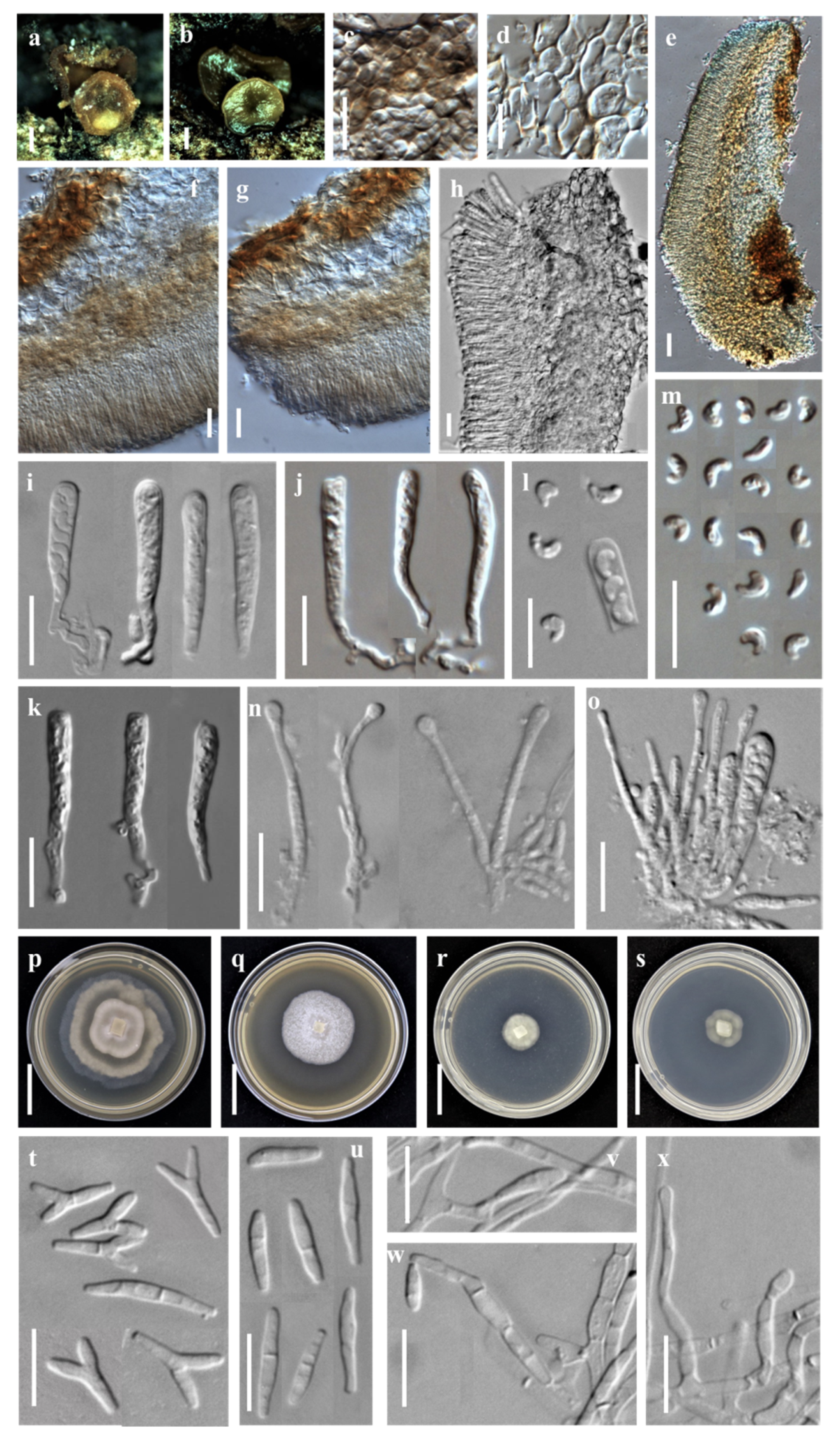
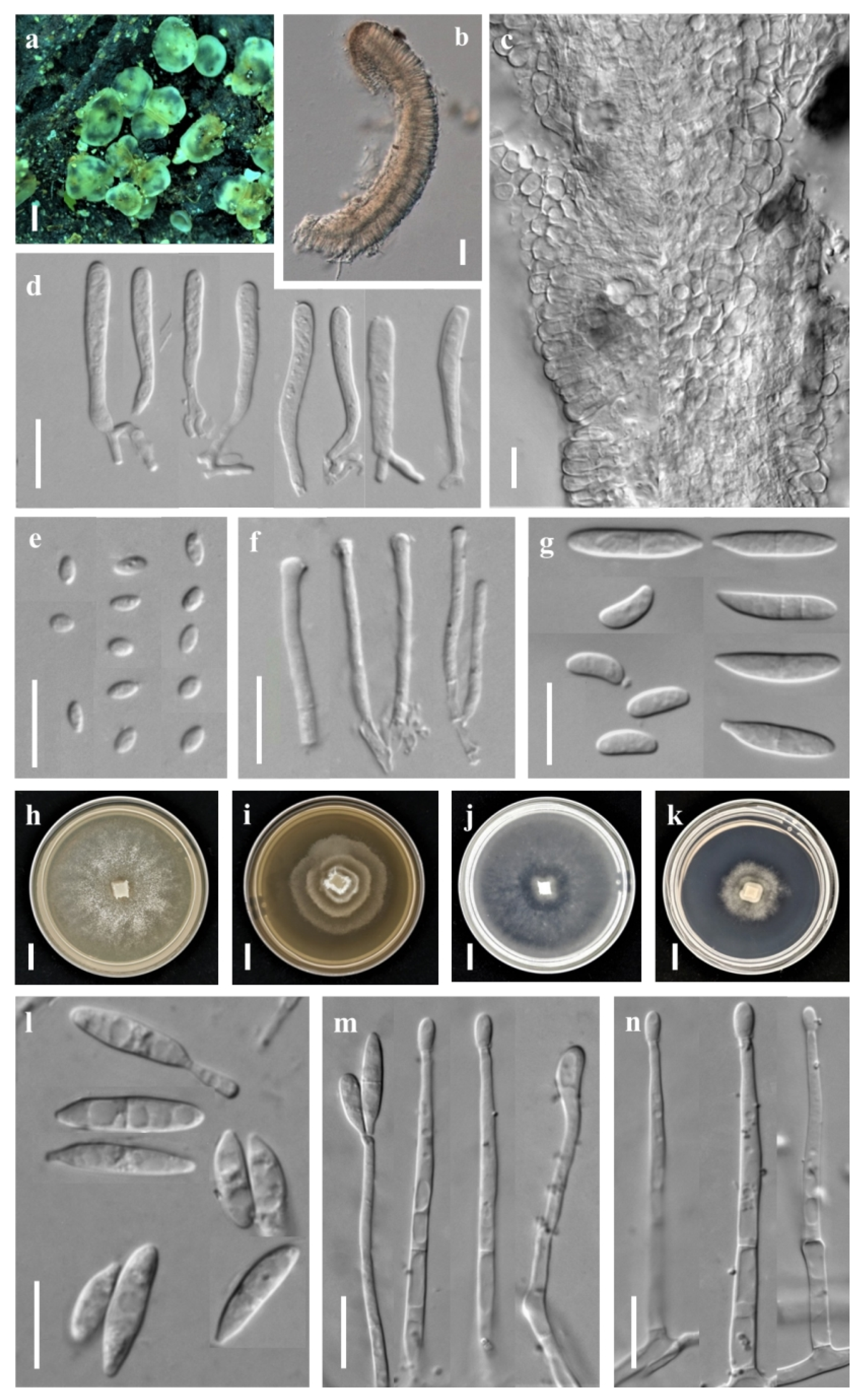
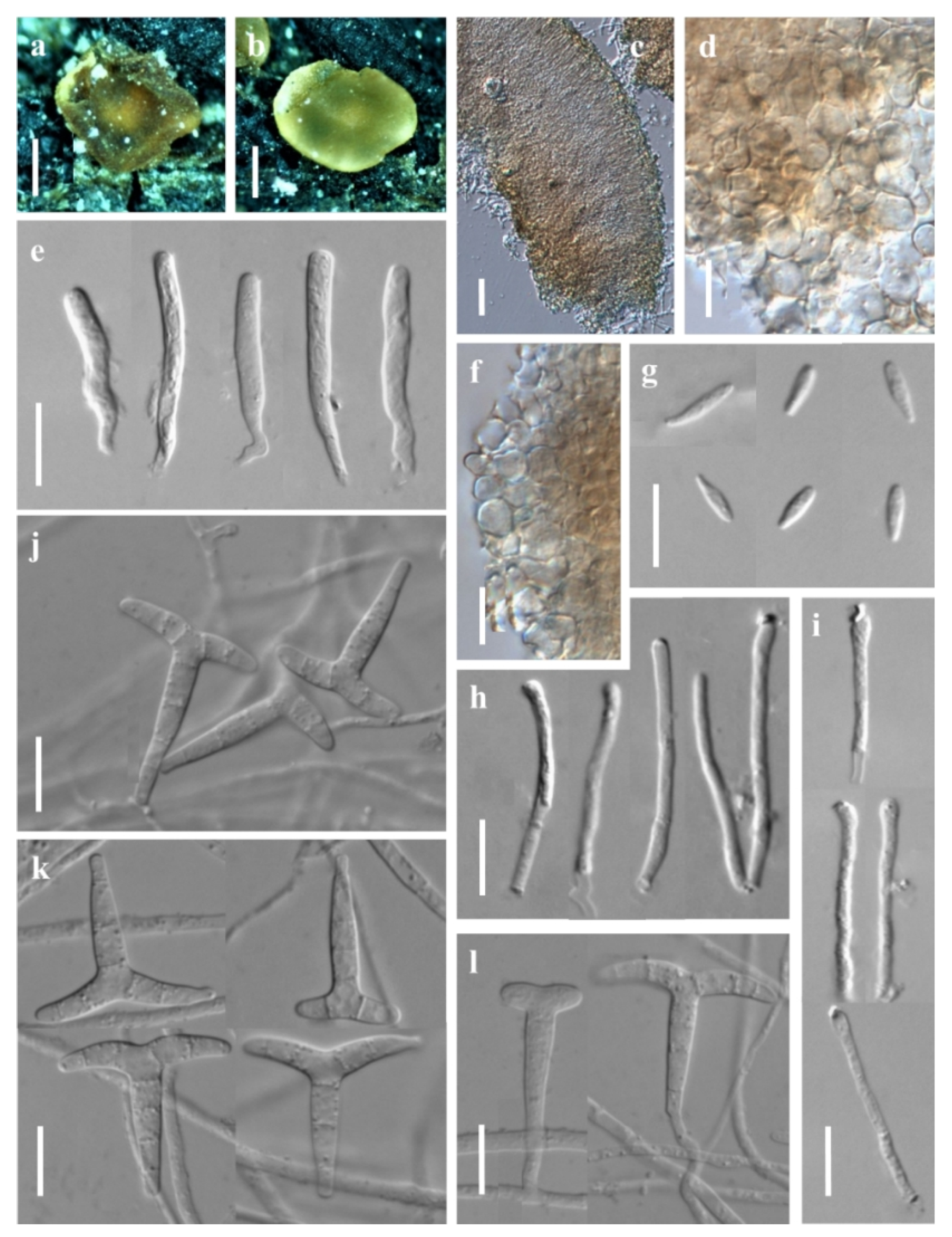
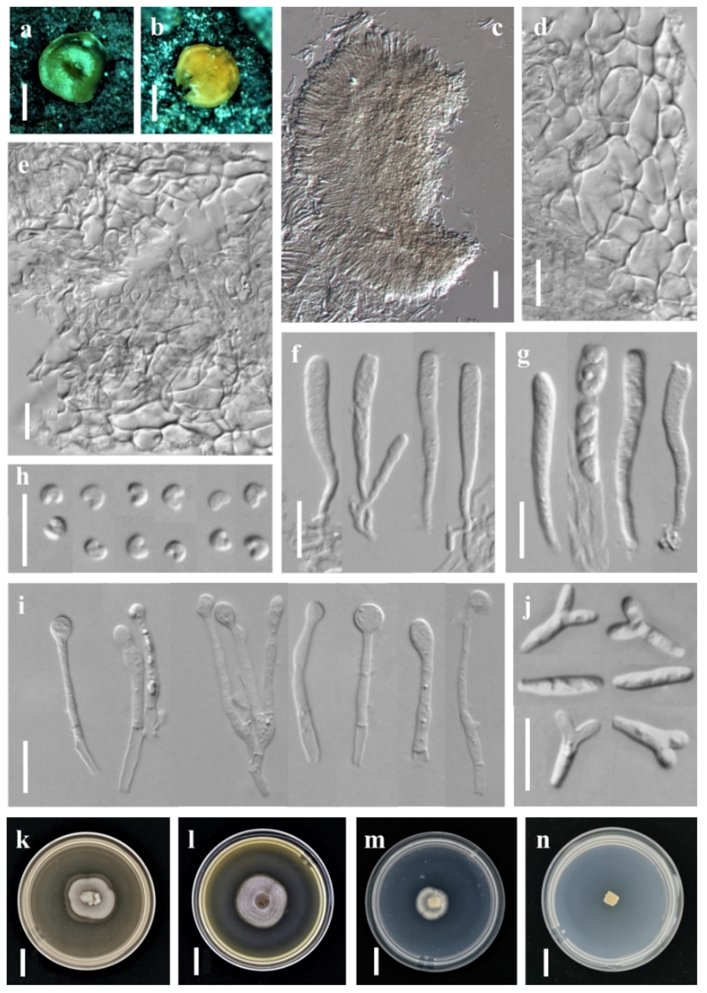
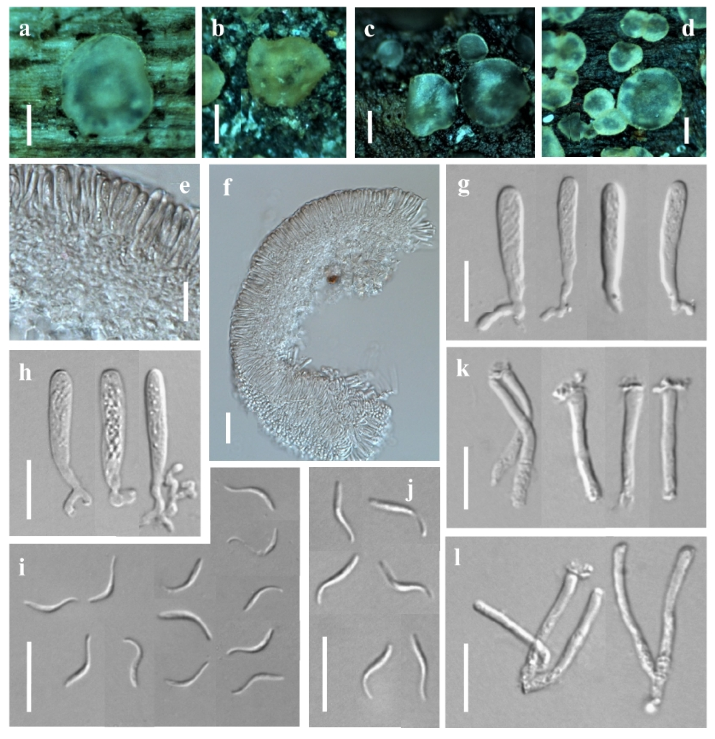
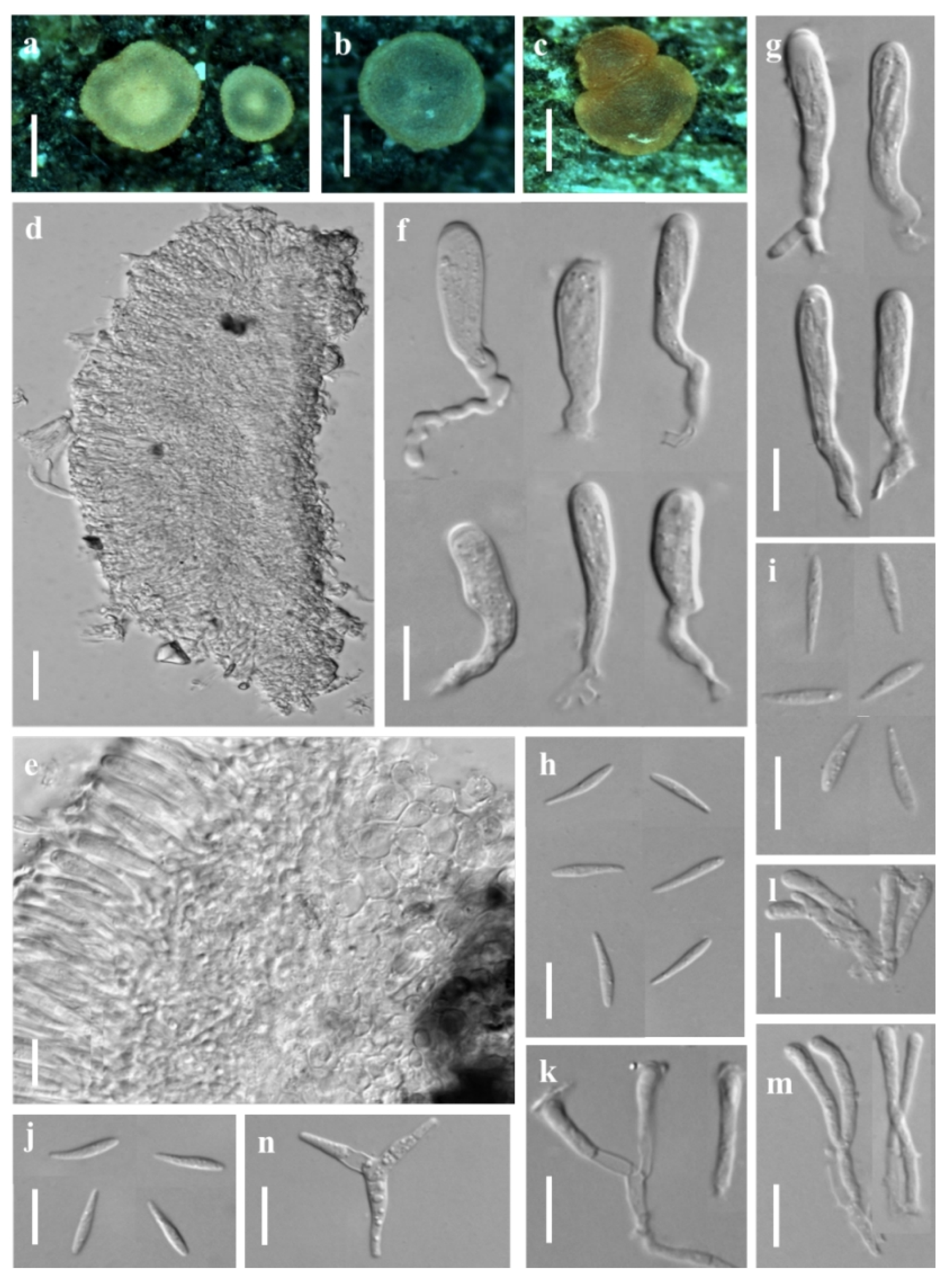
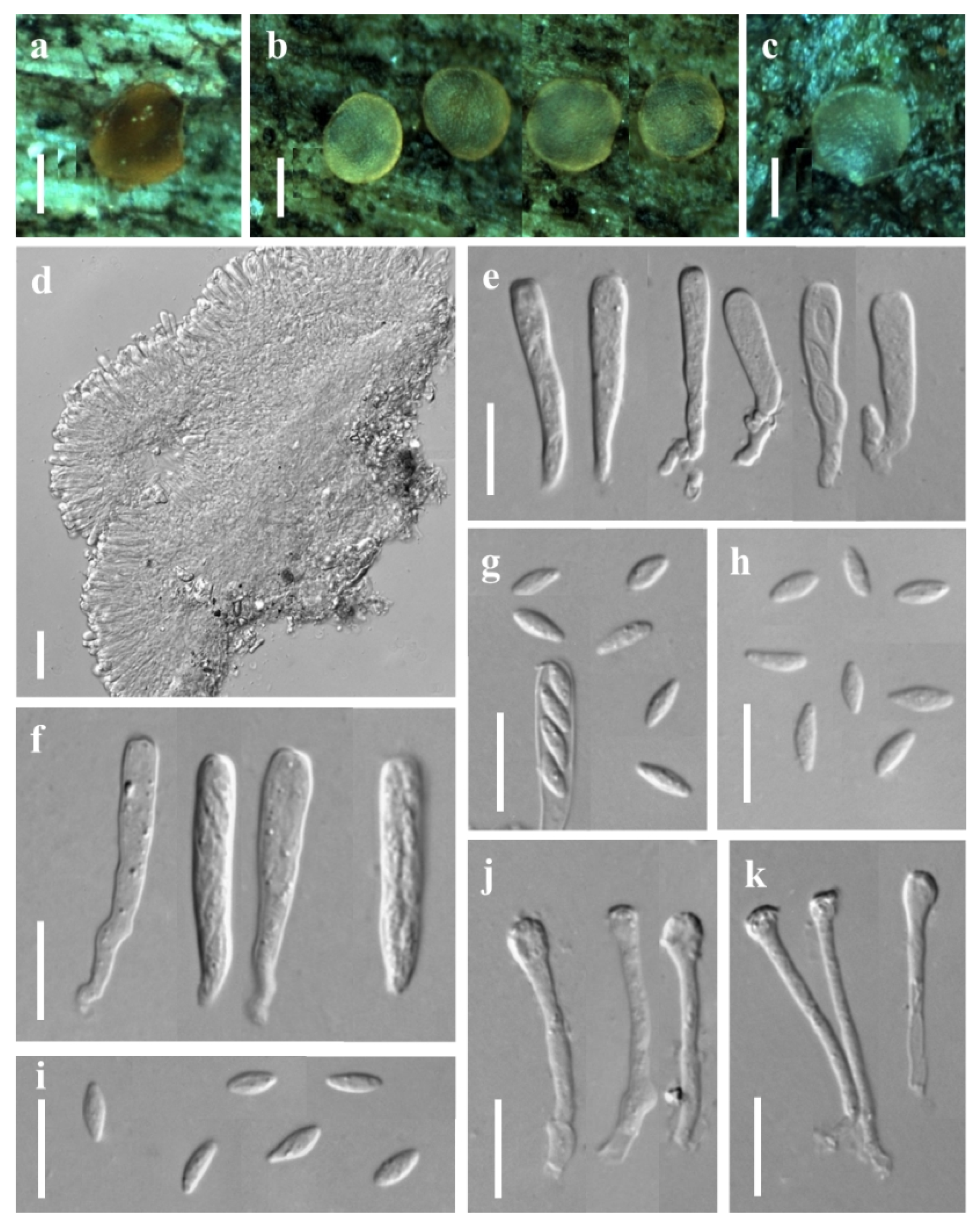
3.1. Taxonomic Description

3.1.1. New Species

Orbilia baisensis X.Y. Ou and Bin Liu, sp. nov. (Figure 1).
MycoBank: MB 846093.
Etymology: from the geographical origin, Baise (Guangxi).
Holotype: CHINA, Guangxi province, Baise city, Dawangling drift scenic spot, from deadwood of Castanea mollissima on the ground, 11 July 2016, X.Y. Ou, GXU2279. Strain DL17 was isolated from GXU2279.
Sexual state: Apothecia superficial on the deadwood of Castanea mollissima, 0.2–1.4 mm in diameter, gregarious in groups or scattered, waxy, translucent, smooth, disc slight concave to flat, margin not protruding, sessile, yellow when fresh or rehydrated, turned yellow to orange when dry. Asci †12.2–39.3 × 2.3–4.6 μm, cylindric-clavate, 8-spored, pars sporifera †18.5–23.6 μm long, truncate to hemispherical at the apex, gradually narrowing towards to the base, flexuous stalk, forked to L-, T- or Y-shaped. Ascospores †2.9–4.6 × 1.6–2.1 μm, hyaline, non-septate, smooth, pronounced reniform, strongly curved ~90–150°, one end round to obtuse, the other end small with a short pointed base, pairs of ascospores arranged in the ascus; containing one refractive globose SB (spore body) at the end close to the wall in alive mature ascospores, 0.6–1.1 μm diameter. Paraphyses apically inflated to mostly capitate (-clavate), †21.9–29.0 × 0.8–1.9 μm, basally branched and expanding to 1.8–3.1 µm in diameter at the apex. Hymenium 70.5–192.3 μm thick; medullary excipulum 25.8–40.6 μm thick, of ± loose textura intricata with ± inflated cells, sharply delimited. Ectal excipulum composed of textura globulosa-angularis, thin-walled to slightly gelatinized, 38.8–146.6 μm thick, cells †3.2–7.9 × 2.0–5.5 μm diam., ovate to spherical.
Asexual state: Dicranidion-like.
Colonies beige-white on PDA, 34.8 mm in diameter at 25 °C after 10 d, strongly keratinized, aerial hyphae absent; beige-white on MEA, 24.8 mm diam., aerial hyphae sparse; white on CMA and LY, 11.8–12.0 mm diam., aerial mycelium absent; Hyphae hyaline, septate, branched, smooth and 1.4–3.2 μm wide. Conidiophores hyaline, erected or slightly bent, septate, unbranched, *5.7–33.1 μm long, *2.0–2.8 μm wide at the base and gradually tapering to *0.9–1.5 μm wide at the tip where bearing 1 apical spore. Conidia thallic, hyaline, Y-shaped, consisted of a spindle and two equal or unequal lobes; the spindle *6.0–11.5 μm long, 1–2 septate; the lobe *2.0–6.7 × 1.5–2.3 μm, 1 septate; in addition, columnar conidia *6.3–20.0 × 1.8–2.6 μm, 1–3 septate.
Additional specimen examined: China, Shaanxi province, Hanzhong city, Foping County, Wangjiawan, from deadwood, 23 July 2017, X.Y. Ou, GXU2373.
Notes: Orbilia baisensis is clustered with O. renispora Y.Y. Shao, Quijada, Baral, Haelew. and Bin Liu, O. leucostigma (Fr.) Fr., O. xanthostigma (Fr.) Fr. and O. cf. xanthostigma (as O. delicatula) by having reniform to nephroid or C-shaped ascospores and their asexual states are belonging to Dicranidion. However, O. baisensis features on 8-spored asci, flexuous base and forked to L-, T- or Y-shaped, pronounced reniform ascospores, round and larger at one end, the other end with a short pointed base, the middle section being the widest and capitate paraphyses. Orbilia renispora differs from O. baisensis by the smaller (3.0–3.6 × 1.5–1.8) and lower curl ascospores. O. leucostigma and O. xanthostigma differ from O. baisensis by the equant end of ascospores. In addition, molecular analyses reveal that O. baisensis shares less than 91.20% similarity with O. renispora in ITS sequence, and 89.91% similarity with O. xanthostigma and 89.53% similarity with O. leucostigma, respectively. Both the morphological and the molecular evidence support their separation at the species level.
Orbilia hanzhongensis X.Y. Ou & Bin Liu, sp. nov. (Figure 2)
MycoBank: MB 846094.
Etymology: according to the geographical origin, Hanzhong (Shaanxi).
Holotype: CHINA, Shaanxi province, Hanzhong city, Foping County, Wangjiawan, from fallen branch, 23 July 2017, X.Y. Ou, holotype GXU2365. Strain BY35 was isolated from GXU2365.
Sexual state: Apothecia rehydrated 0.1–1.5 mm diam., superficial on the deadwood, gregarious, waxy, medium translucent, smooth, disc round and slight to strongly concave, sometimes flat, margin not protruding, broadly sessile, pale to light yellow when fresh or rehydrated, yellow when dry. Asci †21.7–39.7 × 2.3–3.6 μm, cylindric-clavate, 8-spored, spores uniseriate, pars sporifera †20.5–30.7 μm long, truncate to hemispherical at the apex, the base with short to medium long, flexuous stalk, forked to L-, T-, H- or Y-shaped. Ascospores †2.5–4.4 × 1.7–2.2 μm, hyaline, non-septate, smooth, fusoid to ellipsoid, to ovoid, to lemon-shaped, one end subacute to acute, other end round or often acute, straight; SBs †0.2–0.4 μm diam., globose, at the end close to the wall in alive mature ascospores. Paraphyses straight to slightly capitate at the apex, †18.8–40.3 × 1.5–1.9 μm, the base branched and expanded to 2.0–3.0 µm in diameter at the apex. Hymenium 48.2–73.8 μm thick; medullary excipulum 16.6–22.2 μm thick, composed of medium dense textura intricata with inflated cells, sharply delimited. Ectal excipulum composed of textura globulosa-angularis from the base to the flanks, thin-walled, slightly gelatinized, 30.2–51.4 μm thick, cells †3.8–10.8 × 3.2–9.9 μm diam., ovate to spherical.
Asexual state: Dactylella-like.
Colonies beige-white on PDA, 50.5 mm diam. at 25 °C after 10 d, aerial hyphae dense; beige-white on MEA, 31.7 mm diam., aerial hyphae rarely sparse; white on CMA, 60 mm diam., aerial hyphae absent; white on LY, 22.7 mm diam., aerial mycelium absent. Hyphae hyaline, septate, branched, smooth, *1.0–3.5 μm wide. Conidiophores hyaline, erected, septate, unbranched, *10.6–72.8 μm long, *2.0–4.2 μm wide at the base and gradually tapering to *0.9–1.5 μm wide at the tip where bearing 1–3 apical spore. Conidia thallic, hyaline, unbranched, cylindric-ellipsoid (-clavate), obtuse at one end, truncate at the other end, straight when mature, slightly inflect when the immature, *10.1–20.3 × 2.6–4.2 μm, 1 septate.
Additional specimen examined: China, Shaanxi province, Hanzhong city, Foping County, Wangjiawan, from rotten branches, 23 July 2017, X.Y. Ou, GXU2379.
Notes: Orbilia hanzhongensis is characterized by ellipsoid to ovoid ascospores having one end subacute to acute, and mostly acute on the other end, globose SBs, capitate paraphyses. It is related to O. rectispora (Boud.) Baral and O. xinjiangensis (J. Chen, L.L. Xu, B. Liu and Xing Z. Liu) E. Weber, Baral and Helleman, but O. rectispora differs in having narrowly cylindrical to fusoid-clavate and larger ascospores (†5–9 × 0.9–1.2 µm), and O. xinjiangensis differs in distinctly larger spores (†7–9 × 1.8–2 µm) and larger conidia (*45–54 × 8–11 µm) with more septa [3]. Moreover, there is 51 bp (8.46%) divergence in the ITS region between O. hanzhongensis and O. xinjiangensis, and 90 bp (17.82%) divergence in the ITS region of O. rectispora. Both the morphology and DNA sequence data distinguish them as different species.
Orbilia nanningensis X.Y. Ou & Bin Liu, sp. nov. (Figure 3).
MycoBank: MB 846095.
Etymology: name after the geographical origin, Nanning (Guangxi).
Holotype: CHINA, Guangxi province, Nanning city, Xixiangtang District, Shibu Town, from deadwood on the ground, 1 January 2020, X.Y. Ou, holotype GXU2466. Strain NN01 was isolated from GXU2466.
Sexual state: Apothecia rehydrated 0.2–0.5 mm diam., scattered, round, light yellow-orange, translucent, sessile, superficial on dead branches on the ground, orange when fresh, dry deep yellow to orange, disc strongly concave, wet light yellow, disc flat, smooth, margin thin or thick. Asci 20.9–55.7 × 2.4–4.6 μm, pars sporifera 14.0–37.2 μm, cylindric to clavate, 8-spored, the apex truncate to hemispherical, thin-walled, gradually narrowing towards the base, flexuous stalk, unforked. Ascospores †6.2–7.5 × 1.6–2.1 μm, clavate to fusoid with a distinct short tapered, rarely ellipsoid, one end obtuse, tapered 1.0–2.5 μm long, straight or slightly curved. Paraphyses straight to slightly capitate at the apex, †17.8–37.5 × 1.2–2.0 μm, basally unbranched and expanding to 1.9–3.2 µm in diameter, exudate 0.8–1.3 μm thick, over paraphyses. Hymenium 101.8–151.2 μm thick; medullary excipulum 56.8–79.0 μm thick, always composed of dense textura intricata with many inflated cells, sharply delimited. Ectal excipulum 53.6–87.4 μm thick, of thin-walled, composed of oriented textura globulosa-angularis from the base to the flanks or margin, cells †5.7–12.1 × 3.7–8.3 μm diameter.
Asexual state: Trinacrium-like.
Colonies white on PDA. Mycelium *1.7–3.2 μm wide. Conidiophores unbranched, erected or slightly bent, septate, *7.1–15.9 μm long, the base *1.8–2.0 μm wide, the tip *1.0–1.4 μm wide where bearing 1 apical spore. Conidia thallic, T-shaped, consisting of one stipe and two arms, the two arms bent downwards, total size *20.7–32.7 × 14.9–30.8 μm, the stipe *16.9–28.7 × 2.8–4.1 μm, 3–5 septate, the arms *6.0–13.8 × 2.3–3.6 μm, 1–3 septate.
Additional specimens examined: China, Guangxi province, Nanning city, Lewan farm, from deadwood, 1 January 2020, X.Y. Ou, GXU2467.
Notes: Orbilia nanningensis is clustered with O. cf. paracaudata Baral and G. Marson, O. farnesianae Baral, O. pilifera Baral and R. Galán, O. aff. farnesianae and O. amarilla Quijada and Baral. Their ascospores were fusoid to clavate with a short tapered, and with similar T- shaped of conidia, but O. nanningensis differs from other related species by smooth margin of apothecia and straight to slightly capitate paraphyses at the apex. Among the known species of Orbilia, O. cf. paracaudata is the most closely related to O. nanningensis in the phylogenetic tree, there is only a distance of 3.80% in the ITS region between O. nanningensis and O. cf. paracaudata. However, O. paracaudata can be separated from O. nanningensis by its longer and narrower ascospores (5.8–8.5 × 1.6–1.8 µm) and a distinctly protruding apothecial margin.
Orbilia pinea X.Y. Ou & Bin Liu, sp. nov. (Figure 4).
MycoBank: MB 846096.
Etymology: named after the host from which it was collected, Pinus.
Holotype: CHINA, Shaanxi province, Hanzhong city, Foping County, Wangjiawan, from deadwood of pinus on the ground, 23 July 2017, X.Y. Ou, holotype GXU2368. Strain BY38 was isolated from GXU2368.
Sexual state: Apothecia rehydrated 1.0 mm diam., yellowish to orange, translucent, round, superficial and scattered, waxy, smooth, disc flat, margin thin and not protruding, sessile, dry orange or honey-yellow when fresh. Asci †26.5–41.0 × 2.6–4.2 μm, cylindric-clavate, 8-spored, spores uniseriate, ~3-seriate, ~4 lower spores inverted (sometimes mixed), pars sporifera †16.9–26.8 μm long, the apex strongly truncate or round to hemispherical, the base gradually narrowing with short to medium long and flexuous stalk, forked to L-, H- or Y-shaped. Ascospores †2.5–3.3 × 1.5–2.2 μm, hyaline, non-septate, smooth, pronounced reniform, strongly curved, ~48–158°, end round, rarely obtuse, middle largest; SBs globose, †0.4–0.6 μm diameter, usually close to one end in alive mature ascospores. Paraphyses apically inflated to capitate at the apex, sometimes uninflated or slightly inflated to sublageniform, †17.3–45.3 × 1.3–2.8 μm, branched at the base and expanded to 2.3–5.4 µm in diameter at the apex. Hymenium 77.6–139.0 μm thick; medullary excipulum 46.6–65.0 μm thick, subhyaline, composed of dense loose textura intricata, sharply delimited. Ectal excipulum 53.2–92.4 μm thick, hyaline, composed of thin-walled, textura globulosa-angularis from the base to the margin, cells †6.2–22.2 × 5.0–13.7 μm diameter.
Asexual state: Dicranidion-like.
Colonies beige-white on PDA, 20.0 mm diam. at 25 °C after 10 d, aerial hyphae absent; beige-white on MEA, 23.7 mm diam., aerial hyphae sparse; grow very slowly on CMA, only 15 mm diam. at 25 °C after 30 d, and could not grow on LY. Hyphae hyaline, septate, branched, smooth. Conidiophores hyaline, erected or slightly bent, septate, unbranched at the base, the tip where bearing 1 apical spore. Conidia thallic, hyaline, Y-shaped, consisted of a stipe and two equal or unequal arms; the stipe *5.9–10.2 × 2.2–2.7 μm, 1 septate; the arms *2.1–4.5 × 1.6–2.4 μm, 1 septate; in addition, columnar conidia *13.4–13.7 × 2.5–2.7 μm, 1–3 septate.
Notes: Orbilia pinea is most similar to O. fissilis (K. Ando and Tubaki) E. Weber and Baral, the most remarkable feature of O. pinea is pronounced reniform and strongly curved ascospores, O. fissilis differs in broadly ellipsoid to subglobose ascospores and frequently 4-armed of the Dicranidion-like conidia. There is only a distance of 2.36% in the ITS region between O. pinea and the type strain of O. fissilis. Obviously, they are not conspecific.

3.1.2. New Record Species

Orbilia crenatomarginata (Höhn.) Sacc. & Trotter, Syll. Fung. 22: 725 (1913) (Figure 5).
Sexual state: Apothecia 0.1–0.5 mm in diameter, scattered on the surface of rotten wood, superficial, flat or slightly convex, smooth, sessile, margin protruding (or denticulate) and filamentous, with small and distinct triangular teeth, dry pale or light yellow to cream-carneous, rehydrated and fresh greyish to white. Asci †20.8–37.5 × 2.6–3.9 μm, clavate, pars sporifera †11.4–19.7 μm, 8-spored, spores strongly spirally and closely twine within asci, truncate to hemispherical at the apex, base gradually narrowed, flexuous stalk, forked to L-, or Y-shaped. Ascospores †8.8–10.2 × 0.9–1.0 μm wide, hyaline, non-septate, helicoid or S-shaped, sickle-shaped or falculate from profile, cylindrical at the one end, tapered at the other end, the four lower spores inversely oriented; spore bodies tear-shaped. Paraphyses †15.1–37.7 × 1.5–2.1 μm, cylindrical to claviform or slightly capitate, unbranched or occasionally branched at the base, slightly enlarged at the apex, 1.7–3.1 µm, covered with waxy exudates, 0.5–1.3 μm thick. Hymenium 41.9–67.9 μm thick, ectal excipulum composed of textura globulosa-angularis.
Specimens examined: China, Shaanxi province, Baoji city, Meixian County, Taibai mountain forest park, from branch of deciduous tree lying on the ground, 21 July 2017, X.Y. Ou, GXU2342. China, Shaanxi province, Baoji city, Meixian County, Taibai mountain forest park, from branch of deciduous tree lying on the ground, 21 July 2017, X.Y. Ou, GXU2343. China, Shaanxi province, Ankang city, Ningshan County, Huoditang of Qinling, from branch of deciduous tree lying on the ground, 24 July 2017, X.Y. Ou, GXU2383.
Notes: Orbilia crenatomarginata features on strongly helicoid, worm or S-shaped ascospores, cylindrical but round gradually at the one end, strongly attenuated at the other end, cylindrical to claviform or slightly capitate paraphyses, apothecia margin denticulate with small and distinct triangular teeth. Our three specimens (GXU2342, GXU2343, GXU2383) corresponded to O. crenatomarginata H.B. 9452 and O. crenatomarginata H.B. 9265 (MLBP/BIPP = 100%/100%).
Orbilia vinosa (Alb. and Schwein.) P. Karst., Bidr. Känn. Finl. Nat. Folk 19: 101 (1871) (Figure 6).
Sexual state: Apothecia 0.1–0.4 mm in diam., scattered or gregarious on the surface of bark, disc flat to concave, sessile, waxy, translucent, round, fresh pale or light yellow to orange, sometimes cream-ochraceous or greyish, margin with crenulate, the back of disc with white glassy filament. Asci †16.2–53.4 × 5.5–6.0 μm, cylindric-clavate, pars sporifera †14.0–31.2 μm, 8-spores, spores seriate, lower spores inversely oriented, the apex hemispherical to truncate, the base gradually narrowing, flexuous stalk, forked to T-, L- or Y-shaped. Ascospores †7.3–14.1 × 1.1–2.3 μm, hyaline, non-septate, clavate, sometimes fusoid, one end obtuse or round, the other end slightly curved and smaller, strongly attenuated; spore bodies tear-shaped. Paraphyses †14.3–35.2 × 1.2–2.4 μm, cylindrical to slightly clavate-capitate, unbranched or occasionally branched at the base, slightly enlarged at the apex, terminal inflated, 1.9–3.3 μm in diameter. Hymenium 56.3–86.6 μm thick, ectal excipulum composed of textura globulosa-angularis, cell 3.5–10.2 × 2.4–8.0 μm and globose.
Specimens examined: China, Shaanxi province, Xi’an city, Cuihua Mountain, from rotten branch lying on the ground, 25 July 2017, X.Y. Ou, GXU2394. China, Shaanxi province, Xi’an city, Cuihua Mountain, from rotten wood lying on the ground, 25 July 2017, X.Y. Ou, GXU2397. China, Shaanxi province, Baoji city, Meixian County, Taibai mountain forest park, from branch of deciduous tree lying on the ground, 21 July 2017, X.Y. Ou and B. Liu, GXU2415. China, Shaanxi province, Hanzhong city, Foping County, Wangjiawan, from deadwood lying on the ground, 23 July 2017, X.Y. Ou and B. Liu, GXU2421.
Notes: Orbilia vinosa is characterized by clavate-fusoid ascospores, straight or slightly curved, one end obtuse, the other end strongly tapered. The gross morphology of our collections is similar to the original description, according to the detailed description and illustrations of the species provided by Baral et al. [3]. Sequence comparisons also revealed that the three specimens (GXU2394, GXU2397, GXU2421) corresponded to O. vinosa G.M. 2014-02-14 and O. vinosa CBS 116215 (MLBP/BIPP = 100%/100%).
Orbilia vitalbae Rehm, in Ade, Hedwigia 64: 315 (1923) (Figure 7).
Sexual state: Apothecia 0.1–0.4 mm in diameter, superficial on the rotten branch, scattered or gregarious, disc flat or slightly convex, round, translucent, sessile, pale to yellowish when fresh or rehydrated, dry deep cream to orange-yellow, margin slightly crenulate. Asci †20.0–51.0 × 3.1–5.2 μm, pars sporifera †19.1–26.2 μm, cylindric-clavate, 8-spored, the apex obtuse or strongly truncate, the base gradually thin, flexuous stalk, the lower part bifurcate to L- or Y-shaped. Ascospores †5.1–7.7 × 1.8–2.5 μm, fusoid to clavate, one end round to obtuse or subacute, the other end gradually attenuated, solely fastigiate arrangement in the ascus; SBs tear-shaped, in the end of ascospores. Paraphyses †19.5–29.8 × 1.2–2.5 μm, capitate, unbranched, enlarged to globose at the apex, 2.4–4.5 μm, a waxy exudate over terminal cell of paraphyses. Hymenium 66.6–102.1 μm thick, ectal excipulum composed of textura globulosa-angularis.
Specimens examined: China, Shaanxi province, Xi’an city, Cuihua Mountain, from rotten branch lying on the ground, 25 July 2017, X.Y. Ou and B. Liu, GXU2438. China, Shaanxi province, Xi’an city, Cuihua Mountain, from deadwood lying on the ground, 25 July 2017, X.Y. Ou and B. Liu, GXU2442.
Notes: Orbilia vitalbae featured on unipolar and straight, fusoid to clavate ascospores, round to obtuse at the one end and attenuated at the other end. In this study, our two specimens (GXU2438, GXU2442) corresponded to O. vitalbae H.B. 9905a (MLBP/BIPP = 99%/100%).

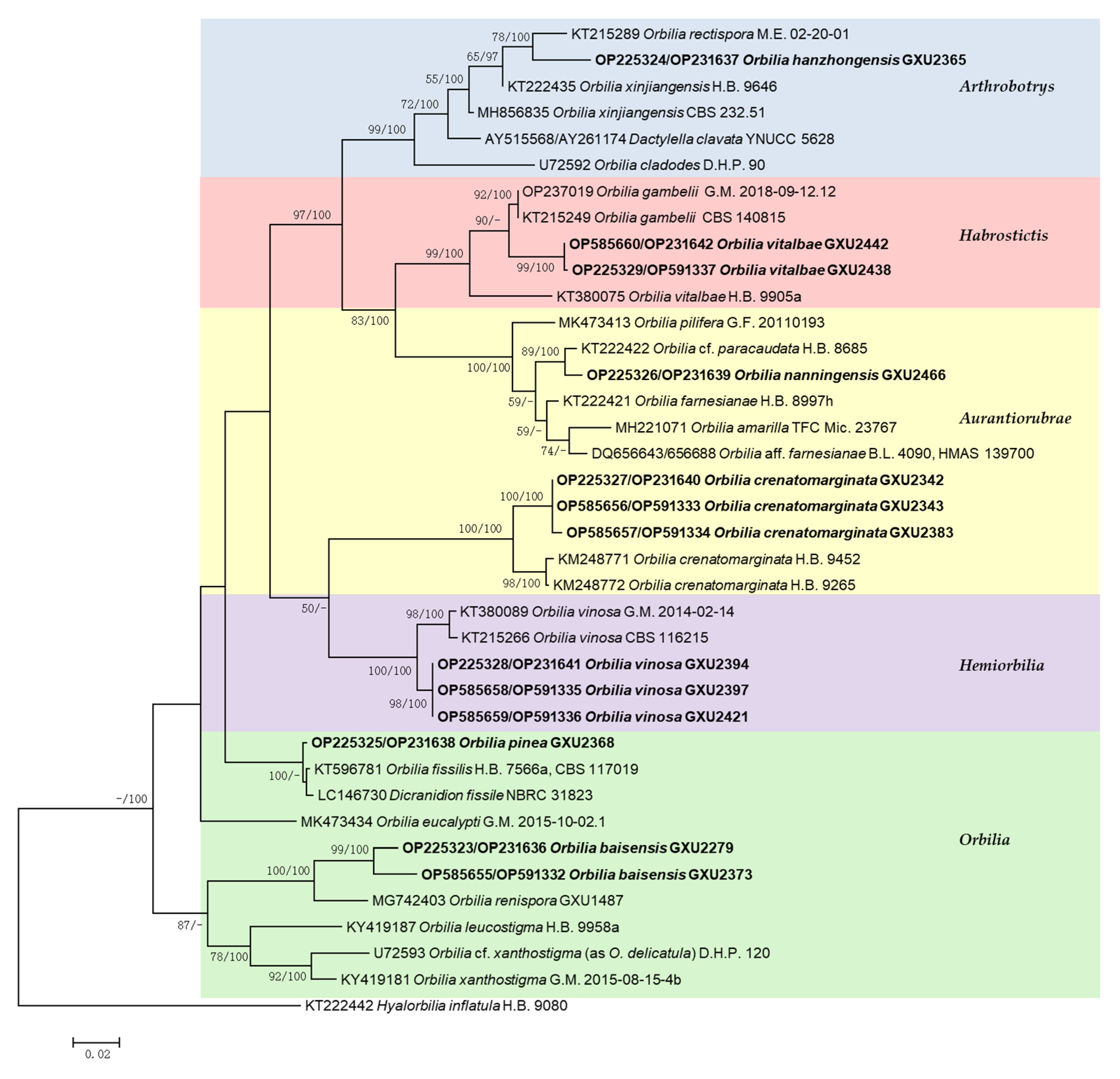
3.2. Phylogenetic Analysis

The phylogenetic tree (Figure 8) was inferred from maximum likelihood analyses and Bayesian inference analyses with the combined ITS and LSU (528 bp from ITS and 561 bp from LSU) sequences. The analysis involved 38 nucleotide sequences that belonged to 25 species, 13 sequences were recognized in this study. The tree was composed of 37 strains as ingroup. Hyalorbilia inflatula (P. Karst.) Baral and G. Marson was used as the outgroup taxon. Maximum likelihood and Bayesian inference analyses generated semblable tree topologies. In the phylogenetic tree, five clades corresponding to sections of Orbilia, including Arthrobotrys, Aurantiorubrae, Habrostictis, Hemiorbilia and Orbilia, were revealed (Figure 8).
In the phylogenetic tree inferred from combined sequences, our 13 samples were considered as four new species and three new record species in Orbilia. The new species Orbilia baisensis were located in a clade with high statistical support (MLBP/BIPP = 100%/100%) with O. renispora. The two specimens (GXU2279, GXU2373) formed a subclade and designated as O. baisensis (MLBP/BIPP = 99%/100%). Orbilia hanzhongensis and O. rectispora received medium statistical support (MLBP/BIPP = 78%/100%). Orbilia nanningensis was related to O. cf. paracaudata, O. nanningensis and O. cf. paracaudata clustered together in a high supported subclade (MLBP/BIPP = 89%/100%). Orbilia pinea and O. fissilis, Dicranidion fissile were located in a clade with high support (MLBP = 100%, BIPP < 95%). O. pinea clustered with O. fissilis in a high support (MLBP = 100%, BIPP < 95%).
New records species Orbilia crenatomarginata and O. vinosa clustered in a clade, which was divided into two strong supported monophyletic subclades. Our three specimens (GXU2342, GXU2343, GXU2383) formed a subclade corresponded to O. crenatomarginata (MLBP/BIPP = 100%/100%) and the three specimens (GXU2394, GXU2397, GXU2421) formed a subclade corresponding to O. vinosa (MLBP/BIPP = 100%/100%). Orbilia vitalbae and O. gambelii were located in a clade with high statistical support (MLBP/BIPP = 99%/100%). Our two specimens (GXU2438, GXU2442) formed a subclade corresponding to O. vitalbae (MLBP/BIPP = 99%/100%).

4. Discussion

The genus Orbilia is diversely and widely distributed in China. The morphological characteristic of the specific ascus and the polymorphic ascospores, especially the strongly refractive spore body, makes Orbilia distinctly unique to the other discomycetes. So far, only several species of Orbilia have been reported from Guangxi and Shaanxi province, China. In this study, sixteen specimens were collected from Guangxi and Shaanxi province, China. Seven species of Orbilia were identified based on morphological characters and phylogenetic analyses, containing four new species, viz. Orbilia baisensis, O. hanzhongensis, O. nanningensis, O. pinea, and three newly recorded species to China, viz. O. crenatomarginata, O. vinosa and O. vitalbae. Asexual states of the four new species are confirmed by obtaining pure cultures from the fresh apothecium, which connected to the anamorphic genus of Dicranidion, Dactylella and Trinacrium.
Orbilia baisensis is clustered with O. renispora, O. leucostigma and O. xanthostigma by having reniform to nephroid or C-shaped ascospores. O. baisensis features on 8-spored asci, flexuous base and forked to L-, T- or Y-shaped at the base, pronounced reniform ascospores, round and larger at the one end, small pointed base at the other end, the middle section being the widest and capitate paraphyses. Orbilia renispora differs from O. baisensis by the smaller and lower curl ascospores [18]. It can be confused with species of O. xanthostigma-leucostigma complex. However, O. leucostigma and O. xanthostigma differ from O. baisensis by the equant end of ascospores. Ascospores of xanthostigma-leucostigma complex are smaller, and with verrucose granule on the dorsal side [11,16]. The distinct warts on the dorsal side of ascospores were reported for the first time by Spooner [7], whereas Baral treated O. delicatula as the synonymy of O. cf. xanthostigma [3]. It was problematic to identify as O. xanthostigma and O. leucostigma only drawing on different color of apothecia by previous research, actually they contained different species, so they were arranged into xanthostigma-leucostigma complex. Baral revealed the high genovariation and represented multiple invariable genotypes in Orbilia xanthostigma and Orbilia leucostigma [23].
Orbilia hanzhongensis is characterized by fusoid to ellipsoid, to ovoid, to lemon-shaped ascospores with subacute to acute at the one end, round or often acute at the other end, globose SBs. It was related to O. rectispora, but differed in having ovoid-fusoid and smaller ascospores. Meanwhile, O. hanzhongensis deviated from O. rectispora [46] by a 9.47% distance in the ITS region. The sequences taken from the pure culture of Orbilia nanningensis comprised ITS and LSU regions, it was closed to O. cf. paracaudata, whereas O. nanningensis was deviated from O. cf. paracaudata [3] by a 5.8% distance in the ITS region and it had smooth margin of apothecia and capitate paraphyses. Orbilia pinea was related to O. fissilis and D. fissile, it differed from O. fissilis by smooth and pronounced reniform ascospores. Orbilia crenatomarginata was described and illustrated in detailed under the name of Orbilia crystallina [47]. The species is distinguished by white to cream apothecia and margin with the crystalline tooth, flexuous and forked to L-, or Y-shaped asci, helicoid or S-shaped to sickle-shaped ascospores.
Orbilia vinosa has been reported in Africa, America, Asia and Europe, growing on gymnosperms and angiosperms [16], but was first reported in China. Peziza vinosa is the primitive name of O. vinosa and described poorly [48], Spooner supplemented descriptions in detail and solved some problems with the type of Peziza vinosa [7]. O. vinosa clustered in a clade with O. crenatomarginata, but the former one differs by clavate ascospores.
Orbilia vitalbae can grow on rotten branches of various trees (Clematis et al.), decayed wood or herbaceous plants (Sideritis et al.) [49], and it is illustrated by asci †(27–)30–50(–54) × (3.5–)4–5.3(–5.5) μm and ascospores †5–8(–10) × 1.4–1.6 or 1.8–2.5(–2.7) μm [3]. In this study, the sizes of asci (†20.0–51.0 × 3.1–5.2 μm) and ascospores (†5.1–7.7 × 1.8–2.5 μm), the shape of ascospores, are well in agreement with the previous findings of Rehm.
Members of Orbilia are often found on dead twigs and branches hanging on trees, distributed in tropical, subtropical and temperate regions. There are 470 species currently known in the family Orbiliaceae [3], of which more than 100 species have been reported in China. Surveys of fungal resources in various regions with different climates and geographic structures will improve our understanding of the species diversity of orbiliaceous fungi in the country. It is necessary to investigate fungal resources in various regions in the future.

Author Contributions

B.L. supervised the project, conceived and designed the study, edited and revised the manuscript; X.-Y.O. collected samples, extracted DNA and PCR and wrote the manuscript; Y.-Y.S. conducted the experiments and phylogenetic analyses; H.-F.Z. conducted morphological observation. All authors have read and agreed to the published version of the manuscript.

Funding

This research was supported by the National Natural Science Foundation of China (No. 30860006) and Ministry of Science and Technology of China for Fundamental Research (No. 2013FY110400).

Institutional Review Board Statement

Not applicable.

Informed Consent Statement

Not applicable.

Data Availability Statement

The sequencing data were submitted to GenBank.

Acknowledgments

The authors are thankful to the project for Fundamental Research on Science and Technology that provided funding, and we thank Usman Rasheed for corrections of the language, and also thanks for the anonymous reviewers for their valuable comments and suggestions.

Conflicts of Interest

The authors declare no conflict of interest.

References

  1. Ekanayaka, A.H.; Hyde, K.D.; Jones, E.B.G.; Zhao, Q. Orbiliaceae from Thailand. Mycosphere 2018, 9, 155–168. [Google Scholar] [CrossRef]
  2. Marchal, E. Champignons coprophiles de Belgique. Bull. Soc. R. Bot. Belg. 1885, 24, 56–77. [Google Scholar]
  3. Baral, H.O.; Weber, E.; Marson, G. Monograph of Orbiliomycetes (Ascomycota) Based on Vital Taxonomy; National Museum of Natural History: Luxembourg, 2020. [Google Scholar]
  4. Baral, H.O.; Weber, E.; Gams, W.; Hagedorn, G.; Liu, B.; Liu, X.Z.; Marson, G.; Marvanová, L.; Stadler, M.; Weiβ, M. Generic names in the Orbiliaceae (Orbiliomycetes) and recommendations on which names should be protected or suppressed. Mycol. Prog. 2018, 17, 5–31. [Google Scholar] [CrossRef] [Green Version]
  5. Fires, E.M. Corpus florarum Provincialium Sueciae. I. Floram scanicum. XXIV; Sebell & C: Upasla, Sweden, 1835; 394p. [Google Scholar]
  6. Nannfeldt, J.Z. Studien über die Morphologie und Systematik der nich-lichenisierten inoperculaten Discomyceten. Nova Acta Regiae Soc. Sci. Ups. Ser. 1932, 48, 368. [Google Scholar]
  7. Spooner, M.P. Helotiales of Australasia: Geoglossaceae, Orbiliaceae, Sclerotiniaceae, Hyaloscyphaceae. Biblioth Mycol. 1987, 116, 1–711. [Google Scholar]
  8. Eriksson, O.E.; Baral, H.O.; Currah, R.S.; Hansen, K.; Kurtzman, C.P.; Læssøe, T.; Rambold, G. Notes on ascomycete systematics Nos 3580–3623. Myconet 2003, 9, 91–103. [Google Scholar]
  9. Zhang, Y.; Yu, Z.F.; Baral, H.O.; Qiao, M.; Zhang, K.Q. Pseudorbilia gen. nov. (Orbiliaceae) from Yunnan, China. Fungal Divers. 2007, 26, 305–312. [Google Scholar]
  10. Kumar, T.A.; Healy, R.; Spatafora, J.W.; Blackwell, M.; Mclanughlin, D.J. Orbilia ultrastructure, character evolution and phylogeny of Pezizomycotina. Mycologia 2012, 104, 462–476. [Google Scholar] [CrossRef] [Green Version]
  11. Guo, J.W.; Li, S.F.; Yang, L.F.; Yang, J.; Ye, T.Z.; Yang, L. New records and new distribution of known species in the family Orbiliaceae from China. Afr. J. Microbiol. Res. 2014, 8, 3178–3190. [Google Scholar]
  12. Qiao, M.; Li, J.Y.; Baral, H.O.; Zhang, Y.; Qiao, W.Y.; Su, H.Y.; Yu, Z.F. Orbilia yuanensis sp. nov. and its anamorph. Mycol. Prog. 2015, 14, 1. [Google Scholar] [CrossRef]
  13. Quijada, L.; Baral, H.O.; Beltrán-Tejera, E. New species of Orbilia (Orbiliales) from arid ecosystems of the Canary Islands (Spain). Nova Hedwig. 2012, 96, 237–248. [Google Scholar] [CrossRef]
  14. Quijada, L.; Baral, H.O.; Jaen-Molina, R.; Weiss, M.; Caujapé-Castelis, J.; Beltrán-Tejera, E. Phylogenetic and morphological circumscription of the Orbilia aurantiorubra group. Phytotaxa 2014, 175, 30. [Google Scholar] [CrossRef]
  15. Quijada, L.; Baral, H.O.; Beltrán-Tejera, E. Diversity of Hyalorbilia (Orbiliales) in the Macaronesian Region. Nova Hedwig. 2015, 100, 1–14. [Google Scholar] [CrossRef]
  16. Quijada, L.; Baral, H.O.; Beltrán-Tejera, E. A revision of the genus Orbilia in the Canary Islands. Phytotaxa 2016, 284, 231–262. [Google Scholar] [CrossRef]
  17. Quijada, L.; Baral, H.O. Orbilia beltraniae, a new succulenticolous species from the Canary Islands. MycoKeys 2017, 25, 1–12. [Google Scholar] [CrossRef] [Green Version]
  18. Shao, Y.Y.; Baral, H.O.; Ou, X.Y.; Wu, H.; Huang, F.C.; Zheng, H.F.; Liu, B. New species and records of orbiliaceous fungi from Georgia, USB. Mycol. Prog. 2018, 17, 1225–1235. [Google Scholar] [CrossRef]
  19. Deng, C.; Yu, Z.F. The complete mitochondrial genomes of Dactylellina leptospora (Orbiliales, Orbiliaceae). Mitochondrial DNA Part B Resour. 2019, 4, 1615–1616. [Google Scholar] [CrossRef] [Green Version]
  20. Wang, S.J.; Fang, M.L.; Xu, J.P.; Jiang, L.L.; Zhou, D.Y.; Zhang, K.Q.; Zhang, Y. Complete mitochondrial genome and phylogenetic analysis of Orbilia dorsalia, a species producing mature sexual structures on culture. Mitochondrial DNA B 2019, 4, 573–574. [Google Scholar] [CrossRef] [Green Version]
  21. White, T.J.; Bruns, T.; Lee, S.; Taylor, J. Amplification and Direct Sequencing of Fungal Ribosomal RNA Genes for Phylogenetics; Academic Press: San Diego, CA, USA, 1990; pp. 315–322. [Google Scholar]
  22. Vilgalys, R.; Hester, M. Rapid genetic identification and mapping of enzymatically amplified ribosomal DNA from several Cryptococcus species. J. Bacteriol. 1990, 172, 4239–4246. [Google Scholar] [CrossRef] [Green Version]
  23. Baral, H.O.; Johnston, P.; Quijada, L.; Healy, R.; Pfister, D.H.; Lobuglio, K.F.; Rodriguez, V.; Weber, E. Cryptic speciation in Orbilia xanthostigma and O. leucostigma (Orbiliomycetes): An aggregate with worldwide distribution. Mycol. Prog. 2021, 20, 1503–1537. [Google Scholar] [CrossRef]
  24. O’Donnell, K.; Kistler, H.C.; Cigelnik, E.; Ploetz, R.C. Multiple evolutionary origins of the fungus causing Panama disease of banana: Concordant evidence from nuclear and mitochondrial gene genealogies. Proc. Natl. Acad. Sci. USA 1998, 95, 2044–2049. [Google Scholar] [CrossRef] [PubMed] [Green Version]
  25. O’Donnell, K.; Cigelnik, E. Two divergent intragenomic rDNA ITS2 types within a monophyletic lineage of the fungus Fusarium are nonorthologous. Mol. Phylogenet. Evol. 1997, 7, 103–116. [Google Scholar] [CrossRef] [PubMed]
  26. Woudenberg, J.H.C.; Aveskamp, M.M.; De-Gruyter, J.; Spiers, A.G.; Crous, P.W. Multiple Didymella teleomorphs are linked to the Phoma clematidina morphotype. Persoonia 2009, 22, 56–62. [Google Scholar] [CrossRef] [Green Version]
  27. Liu, Y.J.; Whelen, S.; Hall, B.D. Phylogenetic relationships among ascomycetes: Evidence from an RNA polymerase II subunit. Mol. Biol. Evol. 1999, 16, 1799–1808. [Google Scholar] [CrossRef] [PubMed] [Green Version]
  28. Sung, G.H.; Sung, J.M.; Hywel-Jones, N.L.; Spatafora, J.W. A multigene phylogeny of Clavicipitaceae (Ascomycota, fungi): Identification of localized incongruence using a combinational bootstrap approach. Mol. Phylogenet. Evol. 2007, 44, 1204–1223. [Google Scholar] [CrossRef] [PubMed]
  29. Carbone, I.; Kohn, L.M. A method for designing primer sets for speciation studies in filamentous ascomycetes. Mycologia 1999, 91, 553–556. [Google Scholar] [CrossRef]
  30. Brefeld, O. AmericaUntersuchungen aus dem Gesammtgebiete der Mykologie, Ascomyceten II. Mycology 1891, 7, 50–51. [Google Scholar]
  31. Pfister, D.H. Castor, Pollux, and life histories of fungi. Micologia 1997, 89, 1–23. [Google Scholar] [CrossRef]
  32. Harkness, H.W. Fungi of the Pacific Coast. Bull. Calif. Acad. Sci. 1885, 1, 159–176. [Google Scholar]
  33. Hughes, S.J. Conidiophores, conidia, and classification. Can. J. Bot. 1953, 31, 577–659. [Google Scholar] [CrossRef]
  34. Peek, C.A.; Solheim, W.G. The hyphomycetous genera of H. W. Harkness and the ascomycetous genus Cleistosama Harkn. Mycologia 1958, 50, 844–861. [Google Scholar] [CrossRef]
  35. Berthet, P. Forrnes conidiennes de divers Discomycetes. Bull. Soc. Mycol. Fr. 1964, 80, 125–149. [Google Scholar]
  36. Liu, B.; Liu, X.Z.; Zhuang, W.Y. Orbilia querci sp. nov. and its knob-forming nematophagous anamorph. FEMS Microbiol. Lett. 2005, 245, 99–105. [Google Scholar] [CrossRef] [PubMed] [Green Version]
  37. Doyle, J.J. A rapid DNA isolation procedure for small quantities of fresh leaf tissue. Phytochem. Bull. 1987, 19, 11–15. [Google Scholar]
  38. Vitória, N.S.; Bezerra, J.L.; Gramacho, K.P. A simplified DNA Extraction Method for PCR Analysis of Camarotella spp. Braz. Arch. Biol. Technol. 2010, 55, 249–252. [Google Scholar] [CrossRef]
  39. Thompson, J.D.; Gibson, T.J.; Plewniak, F.; Jeanmougin, F.; Higgins, D.G. The Clustal_X windows interface: Flexible strategies for multiple sequence alignment aided by quality analysis tools. Nucleic Acids Res. 1997, 25, 4876–4882. [Google Scholar] [CrossRef] [Green Version]
  40. Tamura, K.; Stecher, G.; Peterson, D.; Filipski, A.; Kumar, S. MEGA6: Molecular Evolutionary Genetics Analysis version 6.0. Mol. Biol. Evol. 2013, 30, 2725–2729. [Google Scholar] [CrossRef] [Green Version]
  41. Zhang, D.; Gao, F.L.; Jakovlic, I.; Zhou, H.; Zhang, J.; Li, W.X.; Wang, G.T. Phylosuite: An integrated and scalable desktop platform for streamlined molecular sequence data management and evolutionary phylogenetics studies. Mol. Ecol. Resour. 2020, 20, 348–355. [Google Scholar] [CrossRef]
  42. Swofford, D.L. PAUP 4.0b10: Phylogenetic Analysis Using Parsimony (* and Other Methods); Sinauer Associates: Sunderland, MA, USA, 2002. [Google Scholar]
  43. Nylander, J.A.A. MrModeltest v2. Program Distributed by the Author; Evolutionary Biology Centre, Uppsala University: Uppsala, Sweden, 2004. [Google Scholar]
  44. Ronquist, F.; Huelsenbeck, J.P. MrBayes 3: Bayesian phylogenetic inference under mixed models. Bioinformatics 2003, 19, 1572–1574. [Google Scholar] [CrossRef]
  45. Rambaut, A. FigTree v1.4.3 2016. Available online: http://tree.bio.ed.ac.uk/software/figtree/ (accessed on 1 August 2022).
  46. Liu, B.; Liu, X.Z.; Zhuang, W.Y.; Baral, H.O. Orbiliaceous fungi from Tibet, China. Fungal Divers. 2006, 22, 107–120. [Google Scholar]
  47. Priou, J.P.; Poncelet, A. Orbilia crystallina et Tapesina griseovitellina, deux rare ascomycètes nouveaux pour la Bretagne. Cah. Mycol. Nantais 2006, 18, 3–8. [Google Scholar]
  48. Albertini, I.B.; Schweinitz, L.D. Conspectus Fungorum in Lusatiae Superioris Agro Niskiensi Crescentium e Methodo Persooniana; Sumtibus Kummerianis: Lipsiae, Germany, 1805; 376p. [Google Scholar]
  49. Ade, A. Mykologische Beiträge. Hedwigia 1923, 64, 286–320. [Google Scholar]
Figure 1. Orbilia baisensis and dicranidion-like asexual morph (strain DL17 was isolated from GXU2279). (a,b) apothecia; (c,d) basal excipular cells; (eh) vertical section of apothecium; (ik) ascus; (l,m) ascospores; (n) paraphyses; (o) asci and paraphyses; (ps) colony after 10 d at 25 °C, (p) on PDA, (q) on MEA, (r) on CMA, (s) on LY; (t,u) conidia; (vx) conidiophores with conidia. Scale bars: (a,b) = 0.5 mm; (c,d,f,g,io,tx) = 10 μm; (e,h) = 20 μm; (ps) = 10 mm.
Jof 08 01188 g001
Figure 2. Orbilia hanzhongensis and dactylella-like asexual morph (strain BY35 isolated from GXU2365). (a) apothecia; (b) vertical section of apothecium; (c) basal excipular cells; (d) ascus; (e) ascospores; (f) paraphyses; (hk) colony after 10 d at 25 °C, (h) on PDA, (i) on MEA, (j) on CMA, (k) on LY; (g,l) conidia; (mn) conidiophores with conidia. Scale bars: (a) = 0.5 mm; (b) = 20 μm; (cg,ln) = 10 μm; (hk) = 10 mm.
Jof 08 01188 g002
Figure 3. Orbilia nanningensis and trinacrium-like asexual morph (strain NN01 was isolated from GXU2466). (a,b) apothecia; (c) vertical section of apothecium; (d,f) basal excipular cells; (e) ascus; (g) ascospores; (h,i) paraphyses; (j,k) conidia; (l) conidiophores with conidia. Scale bars: (a,b) = 0.2 mm; (c) = 20 μm; (dl) = 10 μm.
Jof 08 01188 g003
Figure 4. Orbilia pinea and dicranidion-like asexual morph (strain BY38 was isolated from GXU2368). (a,b) apothecia; (c) vertical section of apothecium; (d,e) basal excipular cells; (f,g) ascus; (h) ascospores; (i) paraphyses; (j) conidia; (kn) colony at 25 °C, (h) on PDA after 10 d, (i) on MEA after 10 d, (j) on CMA after 30 d, (k) on LY after 30 d; Scale bars: (a,b) = 0.5 mm; (cj) = 10 μm; (kn) = 10 mm.
Jof 08 01188 g004
Figure 5. Orbilia crenatomarginata ((a,e,f,g,j,kl) from GXU2342; (b,c) from GXU2343; (d,h,i) from GXU2383). (ad) apothecia; (e,f) vertical section of apothecium; (g,h) ascus; (i,j) ascospores; (k,l) paraphyses. Scale bars: (a,b) = 0.2 mm; (e,gl) = 10 μm; (f) = 20 μm.
Jof 08 01188 g005
Figure 6. Orbilia vinosa ((a,df,h,j,k,n) from GXU2394; (b,m) from GXU2397; (g,i) from GXU2415; (c,l) from GXU2421). (ac) apothecia; (d,e) vertical section of apothecium; (f,g) ascus; (hj) ascospores; (km) paraphyses; (n) conidia (from apothecium). Scale bars: (ac) = 0.2 mm; (d) = 20 μm; (en) = 10 μm.
Jof 08 01188 g006
Figure 7. Orbilia vitalbae ((a,b,d,e,h,j) from GXU2438; (c,f,g,i,k) from GXU2442). (ac) apothecia; (d) vertical section of apothecium; (e,f) ascus; (gi) ascospores; (j,k) paraphyses. Scale bars: (ac) = 0.2 mm; (d) = 20 μm; (ek) = 10 μm.
Jof 08 01188 g007
Figure 8. Phylogenetic tree generated from maximum likelihood analyses based on the combined ITS and LSU sequences expressing relationship of Orbilia species. Maximum likelihood bootstrap support ≥ 50% (left) and Bayesian posterior probability values ≥ 95% (right) are indicated at nodes (BIBP/MLBP). Hyalorbilia inflatula H.B. 9080 was used as outgroup. Bold names represent new species and new Chinese record.
Jof 08 01188 g008
Table 1. GenBank accession numbers of taxa used in phylogenetic analyses.
SpeciesStrain NumberGenBank Accession Number
ITSLSU
Dactylella clavataYNUCC 5628AY515568AY261174
Dicranidion fissileNBRC 31823LC146730LC146730
Hyalorbilia inflatulaH.B. 9080KT222442KT222442
Orbilia amarillaTFC Mic. 23767MH221071MH221071
Orbilia baisensis *DL17 (GXU2279)OP225323OP231636
Orbilia baisensis *BY44 (GXU2373)OP585655OP591332
Orbilia cladodesD.H.P. 90U72592U72592
Orbilia crenatomarginata **(GXU2342)OP225327OP231640
Orbilia crenatomarginata **(GXU2343)OP585656OP591333
Orbilia crenatomarginata **(GXU2383)OP585657OP591334
Orbilia crenatomarginataH.B. 9452KM248771KM248771
Orbilia crenatomarginataH.B. 9265KM248772KM248772
Orbilia eucalyptiG.M. 2015-10-02.1MK473434MK473434
Orbilia farnesianaeH.B. 8997hKT222421KT222421
Orbilia aff. farnesianaeB.L. 4090 (HMAS 139700)DQ656643DQ656688
Orbilia fissilisCBS 117019KT596781KT596781
Orbilia gambeliiCBS 140815KT215249KT215249
Orbilia gambeliiG.M. 2018-09-12.12OP237019OP237019
Orbilia hanzhongensis *BY35 (GXU2365)OP225324OP231637
Orbilia leucostigmaH.B. 9958aKY419187KY419187
Orbilia nanningensis *NN01 (GXU2466)OP225326OP231639
Orbilia cf. paracaudataH.B. 8685KT222422KT222422
Orbilia piliferaG.F. 20110193MK473413MK473413
Orbilia pinea *BY38 (GXU2368)OP225325OP231638
Orbilia rectisporaM.E. 02-20-01KT215289KT215289
Orbilia renisporaGXU1487MG742403MG742404
Orbilia vinosa **(GXU2394)OP225328OP231641
Orbilia vinosa **(GXU2397)OP585658OP591335
Orbilia vinosa **(GXU2421)OP585659OP591336
Orbilia vinosaG.M. 2014-02-14KT380089KT380089
Orbilia vinosaCBS 116215KT215266KT215266
Orbilia vitalbae **(GXU2438)OP225329OP231642
Orbilia vitalbae **(GXU2442)OP585660OP591337
Orbilia vitalbaeH.B. 9905aKT380075KT380075
Orbilia xanthostigmaG.M. 2015-08-15-4bKY419181KY419181
Orbilia cf. xanthostigmaD.H.P. 120U72593U72593
Orbilia xinjiangensisCBS 232.51MH856835MH856835
Orbilia xinjiangensisH.B. 9646KT222435KT222435
Note: * new species, ** new Chinese record, specimen numbers are shown in parentheses, sequences newly generated in this study are in bold.
Publisher’s Note: MDPI stays neutral with regard to jurisdictional claims in published maps and institutional affiliations.

