Article

New Insights into Lichenization in Agaricomycetes Based on an Unusual New Basidiolichen Species of Omphalina s. str.

1
State Key Laboratory of Mycology, Institute of Microbiology, Chinese Academy of Sciences, Beijing 100101, China
2
College of Life Sciences, University of Chinese Academy of Sciences, Beijing 100049, China
3
Center of Excellence in Fungal Research, Mae Fah Luang University, Chiang Rai 57100, Thailand
4
Dipartimento di Scienze Della Vita e Biologia Dei Sistemi, Università di Torino, Viale P.A. Mattioli 25, 10125 Torino, Italy
5
College of Life Science and Technology, Xinjiang University, Urumqi 830000, China
6
The Wanan County Second Middle School, Wanan 343800, China
*
Authors to whom correspondence should be addressed.
These authors contributed equally to this work.
J. Fungi 2022, 8(10), 1033; https://doi.org/10.3390/jof8101033
Submission received: 23 August 2022 / Revised: 25 September 2022 / Accepted: 28 September 2022 / Published: 29 September 2022

Abstract

:
The genus Omphalina is an ideal genus for studying the evolutionary mechanism of lichenization. Based on molecular phylogeny using ITS and nuLSU sequences by means of Bayesian and maximum likelihood analyses and morphological examination, combining the existence of green algae in basidiomata stipe and a Botrydina-type vegetative thallus, we described a bryophilous new basidiolichen species, Omphalina licheniformis, from a residential area of Jiangxi Province, China. This finding of unusual new basidiolichen species updated our understanding of the delimitation of Omphalina, indicating that both non-lichen-forming and lichen-forming fungal species are included simultaneously. The presence of algal cells in the basidiomata should receive more attention, as this would be helpful to distinguish more potential basidiolichens and explore the cryptic species diversity. This work provides new insights and evidence for understanding the significance of lichenization during the evolution of Agaricomycetes.

1. Introduction

Lichens are symbionts of fungi (mycobionts) and algae and/or cyanobacteria (photobionts), among which only 0.9% species belong to the Basidiomycota [1]. Omphalina Quél. is an undisputedly important genus when talking about basidiolichen species, because it included both non-lichenized and lichenized species originally, and was subsequently separated into non-lichenized Omphalina s. str. and lichenized genera such as Lichenomphalia Redhead, Lutzoni, Moncalvo & Vilgalys [2] and Agonimia Zahlbr. (syn. Marchandiomphalina Diederich, Manfr. Binder & Lawrey [3]). In addition, Omphalina also includes saprophytic, parasitic, and bryophilous species [4]. Therefore, Omphalina has been regarded as an ideal genus for studying the evolutionary mechanisms associated with lichenization [4,5].
Recently, molecular phylogenetic analyses pointed out that the classical concept of Omphalina, mainly based on morphological features [6,7,8,9], includes several omphalinoid genera nested inside the order Agaricales [2,10,11,12,13,14], as well as Hymenochaetales Oberw. [15,16,17,18]. Inside the Agaricales there are omphalinoid taxa in the suborders Hygrophorineae (family Hygrophoraceae, subfamily Lichenomphaloideae [13]), Marasmiineae (family Porotheleaceae [19,20]) and Tricholomatineae (family Omphalinaceae [21]). Rickenellaceae is the family encompassing omphalinoid taxa in the Hymenochaetales [17,18,22]. Omphalina was restricted to the species phylogenetically related to O. pyxidata (the conserved lectotype of Omphalina [2,10,23,24]), which typically show reddish brown, rusty, or orange-brown tinges on the pileus and stipe, and a non-concolorous hymenophore [2]. Micromorphological features, such as non-amyloid spores, sub-regular to irregular hymenophoral trama and pileipellis with encrusting pigment, are shared by all members of the genus Omphalina s. stricto [2,9,25,26,27,28,29]. This genus is sister to Infundibulicybe [14,21,30,31,32], and together they form the family Omphalinaceae [33] in the suborder Tricholomatineae.
Basidiolichens are mainly distributed in five orders of Agaricomycetes, viz. Agaricales, Atheliales, Lepidostromatales, Cantharellales and Corticiales, among which Agaricales and the family Hygrophoraceae within this order accommodate most of the basidiolichen species [1]. In China, thirteen basidiolichen species, including five Dictyonema spp. (Hygrophoraceae, Agaricales), four Lichenomphalia spp. (Hygrophoraceae, Agaricales), and four Sulzbacheromyces spp. (Lepidostromataceae, Lepidostromatales), have been discovered [34,35,36].
A cluster of bryophilous Omphalina basidiomata was found in a residential area of Jiangxi Province, China. The phylogenetic analyses of nrDNA ITS and nuLSU sequences also confirmed it to be an unknown Omphalina species. However, interestingly, it was found in the stipe existence of green algae; moreover, far fewer vegetative thalli consisting of green tiny globules (Botrydina-type) were also seen near to the hairs at the base of stipe. Therefore, an unusual new basidiolichen species of Omphalina is described and reported here. This finding indicates that Omphalina s. str. still consists of both non-lichenized and lichenized species with very a close phylogenetic relationship, and further provides new insights into the lichenization in Agaricomycetes.

2. Materials and Methods

2.1. Taxon Sampling and Morphological Examination

Five basidiomata specimens were collected from a residential area of Wan’an County, Jiangxi Province of China (Figure 1), and are preserved in the Herbarium Mycologicum Academiae Sinicae, Beijing, China (HMAS). Morphology and anatomy were examined using a MOTIC SMZ-168 stereomicroscope and a LEICA M125 dissecting microscope equipped with a Leica DFC450 camera.
Photographs of fresh specimens were taken immediately in the residential area, and the basidiomes were gathered. The morphological characteristics, including cap, stipe, pileus, and odor, were recorded. Specimens were dried in an electrical food drier at 55 °C to ensure that no moisture was left, and then were sealed in plastic bags.
To observe anatomic characteristics, parts of dried specimens were cut and mounted in 5% KOH and stained with 1% Congo Red. Anatomic characteristics including basidiospores, basidia, cystidia and pileipellis were observed under an Olympus CX31RTSF microscope (Made in Philippines, Tokyo, Japan) with at least 20 records. Data were analyzed and recorded as X = the mean of length by width ± SD, Q = the quotient of basidiospore length to width, and Qm = the mean of Q values ± SD. All of the protocols of the morphological study followed Largent’s methodology [37].

2.2. DNA Extraction, PCR, and Sequencing

DNA was extracted from two fresh basidiomata (Table S1) by means of the modified CTAB method [38]. PCR was performed to amplify two gene loci: nuclear ribosomal DNA internal transcribed spacer (ITS) and large subunit (nuLSU), using primers ITS4 and ITS5 [39], and LR0R and LR5 [40], respectively. The PCR procedure followed Yang et al. [41].

2.3. Sequence Alignment and Phylogenetic Analysis

A total of 136 DNA sequences including four new sequences were used in this study (Table S1). Representative species of the lichenized genera of Hygrophoraceae (Agaricales), non-lichenized genera of Agaricales, and other related orders in the Agaricomycetes were chosen in the phylogenetic analyses. Multiclavula spp. (Clavulinaceae, Cantharellales) were taken as the outgroup.
Raw sequences were firstly assembled and edited with SeqMan [42], and then aligned using MAFFT v.7 [43]. We used the program Gblocks v.0.19b [44,45] to remove ambiguously aligned sites. The congruence of the two loci (ITS and nuLSU) was tested as described previously [46,47]. All maximum likelihood (ML) and Bayesian inference (BI) analyses were performed using the GTR + I + G model selected by jModelTest 2 [48]. The ML analysis involved 1000 pseudoreplicates with RAxML v.8.2.6 [49]. The BI analysis was performed using MrBayes v. 3.2.7 [50,51] with two parallel Markov chain Monte Carlo (MCMC), each using 5 million generations and sampling every 1000th generation. We used TRACER v.1.7.2 [52] to examine the standard deviation of split frequencies less than 0.01, reflecting the fact that the two trees differed very little, and the parameters converged. The 50% majority rule consensus tree was generated after discarding the first 25% as burn-in.
Phylogenetic analysis was run on the Cipres Science Gateway (http://www.phylo.org, (accessed on 18 July 2011)) and visualized using FigTree v.1.4.3 (http://tree.bio.ed.ac.uk/software/figtree, (accessed on 28 August 2014)). The clades with bootstrap (BP) values above 75% or posterior probability (PP) values above 0.95 were considered highly supportive.

3. Results

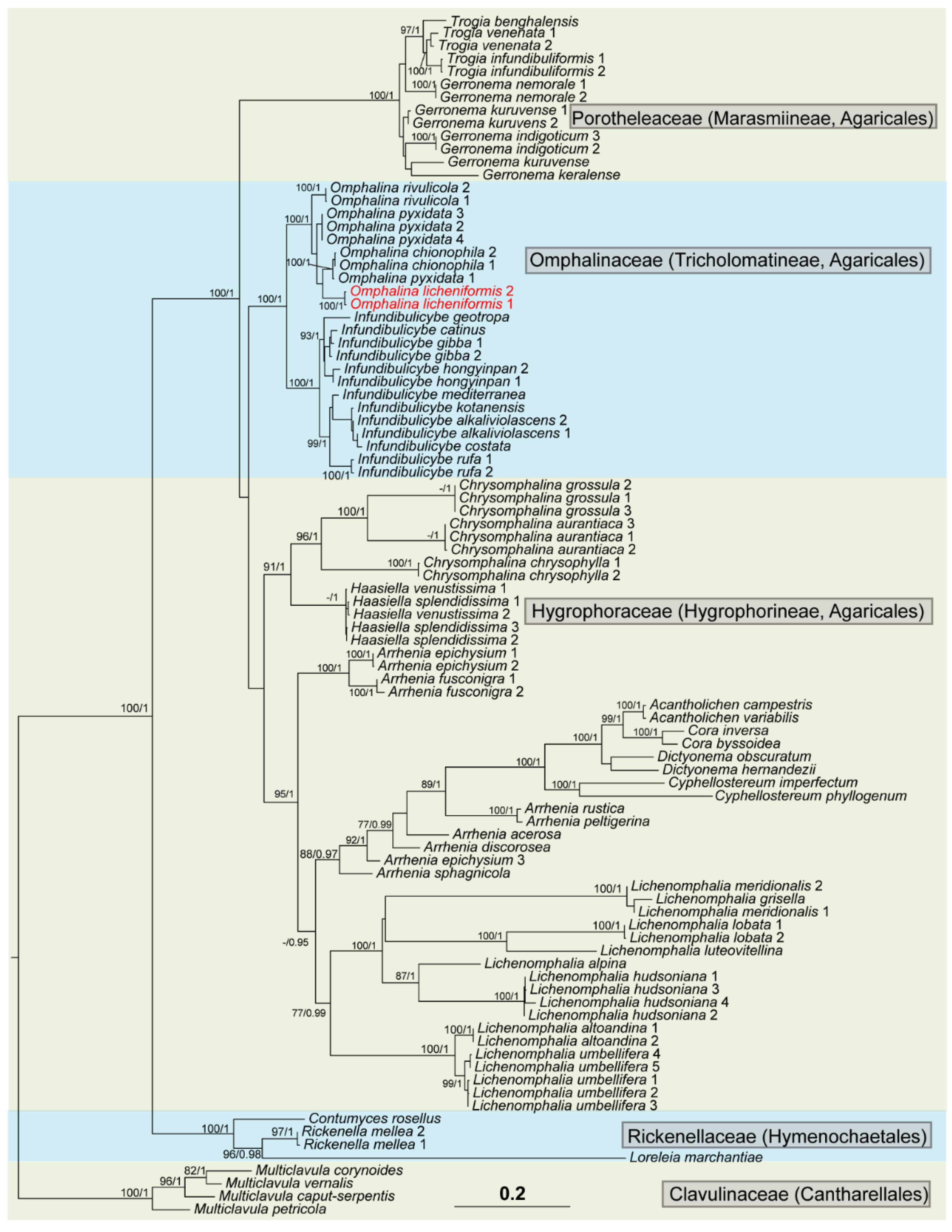
3.1. Phylogenetic Analysis
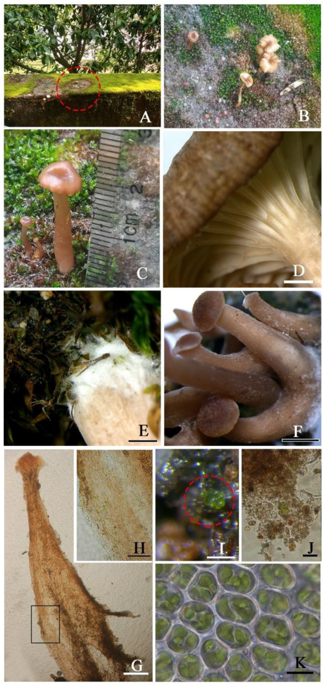
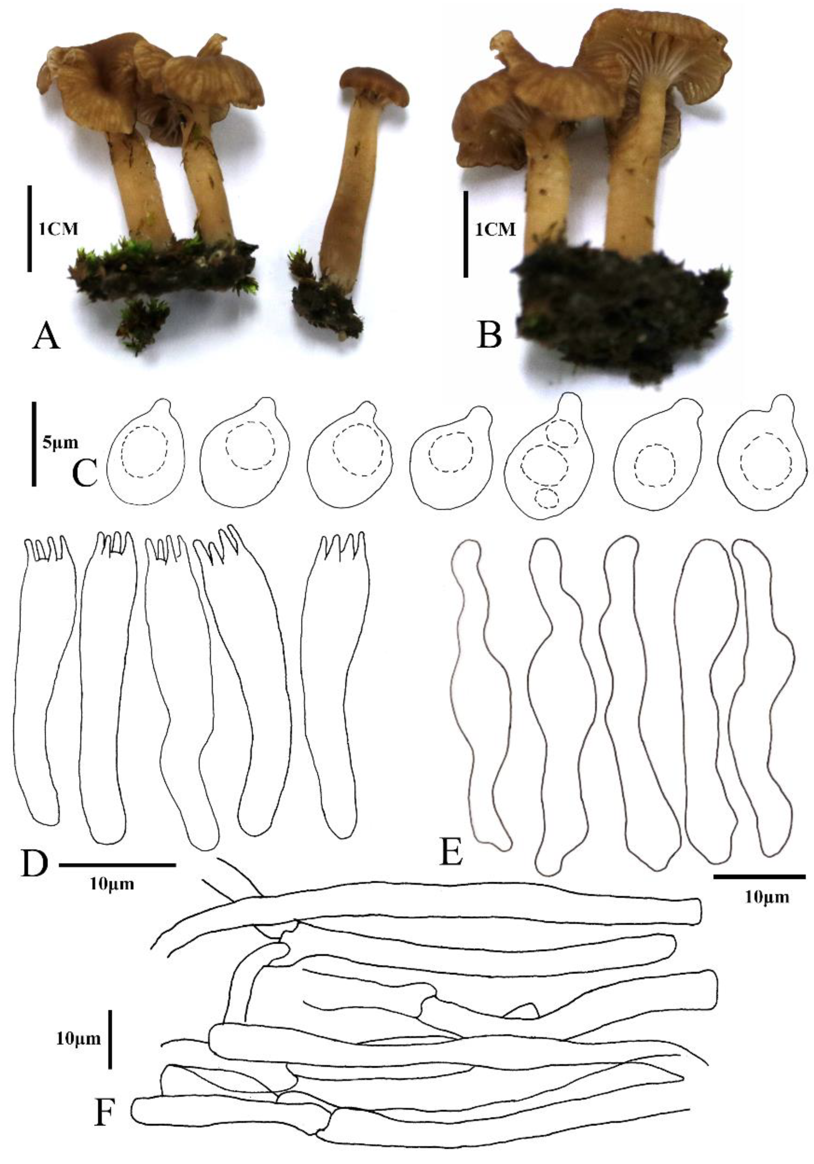
The aligned matrix contained 2011 unambiguous nucleotide (1129 ITS and 882 nuLSU) position characteristics for the full dataset of 93 members. BI and ML phylogenetic trees were constructed, and they had similar topological structures. The RAxML tree is shown in Figure 2 with both bootstrap support (BS) and posterior probability (PP) values of BI analysis. In the tree, all of the Omphalina species clustered into a well-supported monophyletic clade (BS 100/PP 1.00), obviously separated from other groups, in which the two samples (Coll. Nos. JX001 and ZRL20220005) are included and co-formed into a separate branch (BS 100/PP 0.99), indicating that this is a new species also supported by the morphological characteristics (see below). The BI phylogenetic tree and two single-gene-locus RAxML trees are shown in Figures S1–S3.

3.2. Taxonomy

The genus Omphalina is known to be a non-lichenized basidiolichen genus, because the original lichenized species contained in this genus have been separated out and formed totally different other genera such as Lichenomphalia [2]. However, an Omphalina new species was found to be lichenized in this study, and is described below. Therefore, the definition of the genus Omphalina also needs to be redefined as the genus with lichenized species in some cases.
Omphalina Quél., Enchir. fung. (Paris): 42 (1886)
Type species: O. pyxidata (Bull.) Que’l., incertae sedis, Agaricales, Agaricomycetes, worldwide. This genus is characterized by small basidiomes, pileus convex to umbilicate, reddish brown tinged, smooth, without scales; lamellae decurrent, paler and well-developed; stipe central, reddish brown tinged; hymenial and pileal cystidia absent or sparse, and presence of clamp-connections [2,53,54]. Sometimes lichenized.
Omphalina licheniformis X.L. Wei, Z.H. Cao & R.L. Zhao, sp. nov. (Figure 3 and Figure 4)
Fungal Names No.: FN571066
Etymology: The epithet ‘licheniformis’ indicates that this species is a lichen-forming fungus.
Typus: China. Jiangxi Province, Ji’an City, Wan’an County, 26.465265° N 114.798753° E, 65 m alt., on moss Hyophila involuta, 10 March 2022, Z.H. Cao & W.Q. Guo JX001 (HMAS–L 154705, holotype), ZRL20220005 (HMAS 281952, isotype).
Diagnosis: Omphalina licheniformis is distinguished from other species of this genus by having smaller basidiospores (4.9–5.5 × 3.8–4.6 μm) and presenting distinctive cheilocystidia. It is also characterized by the presence of far fewer vegetative thalli consisting of green tiny globules (Botrydina-type) near to the hairs at the base of the stipe, and green algae in the stipe.
Description: Thalli not obvious, Botrydina-type, globular, with globules clustered, very few and tiny, plump when wet, yellow-green to green, c. 80 µm in diam.; globules consist of unicellular green alga enveloped by the hyaline hyphae, hyphae 2.5–5 µm wide, algal cells 6–10 µm in diam.
Basidiomes small, omphalinoid. Pileus 7–15 mm in diam., subhemispherical when young and subfunnel when mature, distinctly depressed, hygrophanous, red brown to yellowish brown, edge paler, becoming alutaceous when drying, margin involute when young, wavy and striate when mature. Context thin, up to 1 mm, concolorous with pileus surface. Lamellae decurrent, distant, thick, forked and anastomosing, concolorous with pileus, but paler. Stipe 15–20 × 3–5 mm, cylindrical, hollow, smooth to white fibrillose, concolorous with the pileus, darker at the middle and lower parts with white mycelium-like cilia. Smell and taste indistinct.
Basidiospores 4.9–5.5 × 3.8–4.6 μm, [x = 5.2 ± 0.2 × 4.1 ± 0.2, Q = 1.2–1.3, Qm = 1.3, n = 20], broadly ellipsoid to ellipsoid, rough with granular contents, smooth, thick-walled, hyaline. Basidia 25.0–27.4 × 3.3–4.7 μm, clavate, hyaline, (2-)4-spored, smooth. Cheilocystidia 27.1–35.7 × 3.7–7.0 μm, cylindrical and flexuose, narrowly clavate or lageniform, thin-walled, hyaline. Hymenophoral trama irregular, consisting of 3.0–7.6 μm-wide hyphae. Pileipellis a cutis composed of hyphae of 3.3–5.3 μm in diam., smooth, cylindrical, hyaline; pigment epiparietal, minutely to strongly encrusting.
Habitat and distribution: This species is bryophilous, growing on the moss Hyophila involuta (Hook.) A. Jaeger mixing with soil located on a community balcony in a residential area of Wan’an County, Jiangxi Province of China, which is characterized by a subtropical monsoon climate, and is the only known distribution up to now.
Additional specimens examined: CHINA. Jiangxi Province, Ji’an City, Wan’an County, 26.465265° N 114.798753° E, 65 m alt., on moss, 30 March 2022, Z.H. Cao & W.Q. Guo JX002 (HMAS–L 154704), ZRL20220006 (HMAS 281953); 7 April 2022, Z.H. Cao & W.Q. Guo JX003 (HMAS–L 154706).
Notes: The vegetative thalli of this species are so tiny and few in number that they can very easily be ignored. The coexistence of algal cells in the base of the stipe near to the hairs is a very new finding, because previously, algal cells were only reported in the vegetative thallus of basidiolichens and known as green algae Coccomyxa [2,34,35]. The algal cells found in the new species are also unicellular and green, and unfortunately, this algal species has not been identified. However, the possibility that they are moss chloroplasts can be excluded, although moss chloroplasts are also unicellular and green, because chloroplasts are organelles within the cells of moss and need to live in the cytoplasm [55], and so it seems unlikely that the moss chloroplasts would escape from the moss cells and exist separately, trapped in the fungal hyphae. Furthermore, the moss chloroplasts are oval and 2.5–3 × 5–6 μm in size (Figure 3K), different from the algal cells (6–10 μm in diam., Figure 3H). Inconspicuous or absent thalli are common in the basidiolichens of Agaricales, for example in the bryophilous or phycophilous basidiolichen genus Lichenomphalia, and the thalli are not obvious in some species such as L. umbellifera (L.) Redhead, Lutzoni, Moncalvo & Vilgalys and L. velutina (Quél.) Redhead, Lutzoni, Moncalvo & Vilgalys [2,34]. The Botrydina-type globular thallus of the new species (Figure 3I) is also similar to L. meridionalis (Contu & La Rocca) P.A. Moreau & Courtec. [56], and the algal cells and hyphae are also observed (Figure 3J). However, the new species is distant from Lichenomphalia spp., but clusters within the genus Omphalina, close to O. pyxidata (Bull.) Quél. and O. chionophila Lamoure in phylogeny (Figure 2), which also have brown caps, small agarics, and decurrent lamellae [57], but are not known to have a lichenized form.

4. Discussion

Phylogenetic and morphological analyses support this new species as a member of Omphalina s. stricto. In the phylogenetic tree (Figure 2), O. chionophile is sister to O. licheniformis. However, in morphology, they can be distinguished by the size of the basidospores—that of O. chionophile is 8–10 × 5–6 μm [25,54,58], and that of O. licheniformis is 4.9–5.5 × 3.8–4.6 μm. Furthermore O. chionophile lacks cheilocystidia. The type species O. pyxidata is also phylogenetically close to O. licheniformis, sharing similar basidiome features which remain difficult to distinguish in situ. However, they can be distinctly separated under a microscope according to the basidiospore and cheilocystidia, as the basidiospores of O. pyxidata (7–8 × 5–6 μm) are larger than those of O. licheniformis (4.9–5.5 × 3.8–4.6 μm), and the cheilocystidia of O. pyxidata are often branched, while the cheilocystidia of O. licheniformis are cylindrical and flexuose, narrowly clavate or lageniform, and not branched [53,54].
The following species differ significantly from O. licheniformis (4.9–5.5 × 3.8–4.6 μm) in spore size: O. rivulicola (8–10.5 × 5.5–7 μm), O. mutila (6.5–10 × 4–6 μm), and O. subhepatica (6–8 × 4–5 μm) [54,59]. In terms of the presence or absence of cheilocystidia, O. licheniformis possesses cheilocystidia, but none of the following species have cheilocystidia: O. mutila, O. demissa, O. subhepatica, O. chionophile, O. galericolor, O. kuehneri, O.arctia, O. rivulicola [54,60]. In terms of habitat, O. licheniformis is found on mosses, but the following species are found in other habitats: O. mutila grows on humid soil in heathland and marshes with Calluna, Erica and Mokunia; O. chionophile grows on naked solifluction soil; and O. pyxidata and O. rivulicola grow on dry sandy soil [54]. In terms of the color of basidiomes, O. licheniformis is reddish brown to yellowish brown, but O. mutila forms white basidiomes [54,61]. Therefore, O. licheniformis is a distinguished species in both molecular composition and morphology.
Lichen-forming fungi are an important component of the kingdom Fungi, making up nearly 20% of the known fungal species, among which over 99% of species belong to Ascomycota [1]. Previous studies showed that more losses than gains of lichenization have occurred during the evolution of Ascomycota, resulting in lichen-forming fungi becoming the ancestors of major lineages of non-lichen-forming fungi in Ascomycota [62]. Compared with ascolichens, basidiolichens are much rarer, and comprise less than 1% of species of the known lichen-forming fungi [1]; however, lichenization in the evolution of Basidiomycota is also very important in related research, and is even treated as one of necessary models to study the evolution of lichens [63].
As early as 1995, Gargas et al. found three independent origins of lichenization in Basidiomycota, i.e., coral Multiclavula (Coker) R.H. Petersen as the basal origin, gilled mushroom Lichenomphalia umbellifera (syn. Omphalina umbellifera), and cyanolichen Dictyonema pavonium (Weber & D. Mohr) Parmasto. Basidiolichens are known to consist of far more than three genera nowadays, as mentioned above, including 5 orders, 5 families, 15 genera and 172 species, and six to seven independent lichenization events happened in the Basidiomycota, among which over 85% (147 species) belong to eight genera of Hygrophoraceae Lotsy in Agaricales, viz. Acantholichen P.M.Jørg., Arrhenia Fr., Cora Fr., Corella Vain., Cyphellostereum D.A.Reid, Dictyonema C.Agardh ex Kunth, Lichenomphalia, and Semiomphalina Redhead [1]. Except for Corella and Semiomphalina, all of the other six genera of Hygrophoraceae were included in our phylogenetic analyses (Figure 2), among which the genus Lichenomphalia was included in Omphalina [2].
The genus Omphalina has been taken as a model system to study lichenization since the late 1990s due to its variable nutritional modes [4]; however, after a series of species such as Omphalina umbellifera, etc., were transferred to other genera, no lichen-forming species were reported in Omphalina s.str., until the finding of Omphalina licheniformis in this study. Our study indicates that within the Agaricales, the lichenization process also occurred in the Omphalinaceae of the suborder Tricholomatineae. In the previous reports on basidiolichens, algal cells have never been found in the fruiting body structures [2,34,35,63], but indeed existed in the stipe of Omphalina licheniformis (Figure 3). This finding of unusual new species updated our understanding of the delimitation of Omphalina, indicating that both non-lichen-forming and lichen-forming fungal species are included simultaneously. Moreover, these results provide new insights and evidence for understanding the significance of lichenization during the evolution of Basidiomycota. Through this study, it should be noted that we need to pay more attention to the Basidiomycota fungi, especially to whether the algal cells are present in the fruiting bodies, which would be very helpful to distinguish more potential basidiolichens and explore the cryptic species diversity through these algal cell examinations.
The presence of algal cells as lichen photobionts is well-known to provide a carbon source for the mycobiont [64], which is relatively easy to understand in ascolichens, because fruiting bodies such as apothecia and pycnidia are closely connected parts of the lichen thallus, and the photobiont can be found both in the thallus and fruiting bodies, except the lecideine-type apothecia and pycnidia without hymenial algae. The algal cells in basidiolichens are assumed to be similar in function [5,65], but there is still an absence of strong evidence, especially due to the fruiting bodies of basidiolichens, which often look separable from the thallus in most cases, and algal cells have only been reported in the thallus previously [2,34,35,63]; moreover, sometimes the thallus is not obvious [2,34].

Supplementary Materials

The following supporting information can be downloaded at: https://www.mdpi.com/article/10.3390/jof8101033/s1, Table S1: Specimens used for DNA extraction and GenBank accession number of all samples used in this study, Figure S1: The Bayesian tree based on the concatenated ITS + nuLSU (two genes) dataset, Figure S2: The maximum likelihood tree based on the ITS dataset, Figure S3: The maximum likelihood tree based on the nuLSU dataset.

Author Contributions

X.W. and R.Z. conceived and designed the study. Z.C. and W.G. discovered and collected specimens. T.Z., X.Z. and B.L. generated the DNA sequence data, X.Z., S.Q., X.W. and R.Z. performed the phenotypic analysis, T.Z., X.Z., A.V., X.W. and R.Z. analyzed the DNA data and checked issues related to nomenclatural articles. X.W. wrote the manuscript draft. A.V. and R.Z. All authors have read and agreed to the published version of the manuscript.

Funding

This research was funded by the National Natural Science Foundation of China (32070096, 31961143010, 31970010), National Science and Technology Fundamental Resources Investigation Program of China (Grant No. 2019FY101808), Biological Resources Programme, Chinese Academy of Sciences (KFJ-BRP-009), Beijing Innovative Consortium of Agriculture Research System (Project ID: BAIC05-2022).

Data Availability Statement

Publicly available datasets were analyzed in this study. All newly generated sequences were deposited in GenBank (accessed on 9 June 2022, https://www.ncbi.nlm.nih.gov/genbank/; Table S1). All new taxa were deposited in Fungal names (accessed on 14 July 2022, https://www.fungalinfo.im.ac.cn).

Acknowledgments

The authors sincerely thank L. Zhang at the Fairy Lake Botanical Garden (Shenzhen), Chinese Academy of Sciences, for his great help in the identification of the moss in this study. Special thanks go to Q. X. Yang for her assistance during our studies in HMAS–L.

Conflicts of Interest

The authors declare no conflict of interest.

References

  1. Lücking, R.; Hodkinson, B.P.; Leavitt, S.D. The 2016 classification of lichenized fungi in the Ascomycota and Basidiomycota—Approaching one thousand genera. Bryologist 2017, 119, 361–416. [Google Scholar] [CrossRef]
  2. Redhead, S.; Lutzoni, F.; Moncalvo, J.M.; Vilgalys, R. Phylogeny of agarics: Partial systematics solutions for core Omphalinoid genera in the Agaricales (Euagarics). Mycotaxon 2002, 83, 19–57. [Google Scholar]
  3. Lücking, R.; Moncada, B. Dismantling Marchandiomphalina into Agonimia (Verrucariaceae) and Lawreymyces gen. nov. (Corticiaceae): Setting a precedent to the formal recognition of thousands of voucherless fungi based on type sequences. Fungal Divers. 2017, 84, 119–138. [Google Scholar] [CrossRef]
  4. Lutzoni, F.; Vilgalys, R. Omphalina (Basidiomycota, Agaricales) as a model system for the study of coevolution in lichens. Cryptogam. Bot. 1995, 5, 71–81. [Google Scholar]
  5. Lutzoni, F.M. Phylogeny of lichen- and non-lichen-forming omphalinoid mushrooms and the utility of testing for combinability among multiple data sets. Syst. Biol. 1997, 46, 373–406. [Google Scholar] [CrossRef] [PubMed]
  6. Bigelow, H.E. Omphalina in North America. Mycologia 1970, 62, 1–32. [Google Scholar] [CrossRef]
  7. Clémençon, H. Kompendium der Blätterpilze Europäische omphalinoide Tricholomataceae. Zeitschr. F. Mykol. 1982, 48, 195–237. [Google Scholar]
  8. Norvell, L.L.; Redhead, S.A.; Ammirati, J.F. Omphalina sensu lato in North America 1-2. 1: Omphalina wynniae and the genus Chrysomphalina. 2: Omphalina sensu Bigelow. Mycotaxon 1994, 50, 379–407. [Google Scholar]
  9. Bon, M. Flore mycologique d’Europe 4. Les Clitocybes, Omphales et ressemblants. Doc. Mycologia. Mém. Hors Sér 1997, 4, 1–181. [Google Scholar]
  10. Moncalvo, J.-M.; Vilgalys, R.; Redhead, S.; Johnson, J.; James, T.; Aime, M.; Valerie, H.; Verduin, S.; Larsson, E.; Baroni, T.; et al. One Hundred and Seventeen Clades of Eu-agarics. Mol. Phylogenet. Evol. 2002, 23, 357–400. [Google Scholar] [CrossRef]
  11. Matheny, P.; Curtis, J.; Valerie, H.; Aime, M.; Moncalvo, J.-M.; Ge, Z.-W.; Slot, J.; Ammirati, J.; Baroni, T.; Bougher, N.; et al. Major clades of Agaricales: A multilocus phylogenetic overview. Mycologia 2006, 98, 982–995. [Google Scholar] [CrossRef] [PubMed]
  12. Lawrey, J.; Lücking, R.; Sipman, H.; Chaves, J.; Redhead, S.; Bungartz, F.; Sikaroodi, M.; Gillevet, P. High concentration of basidiolichens in a single family of agaricoid mushrooms (Basidiomycota: Agaricales: Hygrophoraceae). Mycol. Res. 2009, 113, 1154–1171. [Google Scholar] [CrossRef] [PubMed]
  13. Lodge, D.; Padamsee, M.; Matheny, P.; Aime, M.; Cantrell, S.; Boertmann, D.; Kovalenko, A.; Vizzini, A.; Dentinger, B.; Kirk, P.; et al. Molecular phylogeny, morphology, pigment chemistry and ecology in Hygrophoraceae (Agaricales). Fungal Divers. 2013, 64, 1–99. [Google Scholar] [CrossRef]
  14. Varga, T.; Krizsan, K.; Földi, C.; Dima, B.; Sanchez-Garcia, M.; Sanchez-Ramirez, S.; Szollosi, G.; Szarkándi, J.; Papp, V.; Albert, L.; et al. Megaphylogeny resolves global patterns of mushroom evolution. Nat. Ecol. Evol. 2019, 3, 668–678. [Google Scholar] [CrossRef]
  15. Redhead, S.; Moncalvo, J.-M.; Vilgalys, R.; Lutzoni, F. Phylogeny of agarics: Partial systematics solutions for bryophilous omphalinoid agarics outside of the Agaricales (Euagarics). Mycotaxon 2002, 82, 151–168. [Google Scholar]
  16. Larsson, K.-H.; Parmasto, E.; Fischer, M.; Langer, E.; Nakasone, K.; Redhead, S. Hymenochaetales: A molecular phylogeny for the hymenochaetoid clade. Mycologia 2006, 98, 926–936. [Google Scholar] [CrossRef]
  17. Korotkin, H.; Swenie, R.; Miettinen, O.; Budke, J.; Chen, K.-H.; Lutzoni, F.; Smith, M.; Matheny, P. Stable isotope analyses reveal previously unknown trophic mode diversity in the Hymenochaetales. Am. J. Bot. 2018, 105, 1869–1887. [Google Scholar] [CrossRef]
  18. Olariaga, I.; Huhtinen, S.; Læssøe, T.; Petersen, J.; Hansen, K. Phylogenetic origins and family classification of typhuloid fungi, with emphasis on Ceratellopsis, Macrotyphula and Typhula (Basidiomycota). Stud. Mycol. 2020, 96, 155–184. [Google Scholar] [CrossRef]
  19. Vizzini, A.; Consiglio, G.; Marchetti, M.; Borovička, J.; Campo, E.; Cooper, J.; Lebeuf, R.; Ševčíková, H. New data in Porotheleaceae and Cyphellaceae: Epitypification of Prunulus scabripes Murrill, the status of Mycopan Redhead, Moncalvo & Vilgalys and a new combination in Pleurella Horak emend. Mycol. Prog. 2022, 21, 44. [Google Scholar] [CrossRef]
  20. Vizzini, A.; Picillo, B.; Luigi, P.; Dovana, F. Chrysomycena perplexa gen. et sp. nov. (Agaricales, Porotheleaceae), a new entity from the Lazio region (Italy). Riv. Micol. Romana 2019, 107, 96–107. [Google Scholar]
  21. Sanchez-Garcia, M.; Matheny, P. Is the switch to an ectomycorrhizal state an evolutionary key innovation in mushroom-forming fungi? A case study in the Tricholomatineae (Agaricales). Evolution 2016, 71, 51–65. [Google Scholar] [CrossRef] [PubMed]
  22. Vizzini, A. Segnalazioni di muscinputa laevis (Basidiomycota, Agaricomycetes) per il Nord Italia. Micol. Veg. Mediterr. 2010, 25, 141–148. [Google Scholar]
  23. Redhead, S.; Norvell, L.L. Notes on Bondarzewia, Heterobasidion and Pleurogala, new genus. Mycotaxon 1993, 48, 371–380. [Google Scholar]
  24. Moncalvo, J.-M.; Lutzoni, F.; Rehner, S.; Johnson, J.; Vilgalys, R. Phylogenetic Relationships of Agaric Fungi Based on Nuclear Large Subunit Ribosomal DNA Sequences. Syst. Biol. 2000, 49, 278–305. [Google Scholar] [CrossRef]
  25. Lamoure, D. Agaricales de la zone alpine. Genre Omphalina (1ère partie). Trav. Sci. Parc. Natl. Vanoise 1974, 5, 149–164. [Google Scholar]
  26. Lamoure, D. Alpine and circumpolar Omphalina species. In Arctic and Alpine Mycology 1; Laursen, G.A., Ammirati, J.F., Eds.; University of Washington Press: Seattle, WA, USA, 1982; pp. 201–215. [Google Scholar]
  27. Bigelow, H.E. The Clitocybe pyxidata group. In Travaux Mycologiques Dédiés à R. Kühner, Numéro spécial du Bulletin de la Société Linnéenne de Lyon; Société Linnéenne de Lyon: Lyon, France, 1974; pp. 39–46. [Google Scholar]
  28. Bigelow, H.E. North American species of Clitocybe. Part I. Beih. Nova Hedwigia 1982, 72, 1–280. [Google Scholar]
  29. Bigelow, H.E. North American species of Clitocybe. Part II. Beih. Nova Hedwigia 1985, 81, 281–471. [Google Scholar]
  30. Sanchez-Garcia, M.; Ryberg, M.; Khan, F.; Varga, T.G.; Nagy, L.; Hibbett, D. Fruiting body form, not nutritional mode, is the major driver of diversification in mushroom-forming fungi. Proc. Natl. Acad. Sci. USA 2020, 117, 32528–32534. [Google Scholar] [CrossRef]
  31. Vizzini, A.; Para, R.; Fontenla, R.; Ghignone, S.; Ercole, E. A preliminary ITS phylogeny of Melanoleuca (Agaricales), with special reference to European taxa. Mycotaxon 2012, 118, 361–381. [Google Scholar] [CrossRef]
  32. Vizzini, A.; Marco, C.; Musumeci, E.; Ercole, E. A new taxon in the Infundibulicybe gibba complex (Basidiomycota, Agaricales, Tricholomataceae) from Sardinia (Italy). Mycologia 2010, 103, 203–208. [Google Scholar] [CrossRef]
  33. Vizzini, A.; Consiglio, G.; Marchetti, M.; Alvarado, P. Insights into the Tricholomatineae (Agaricales, Agaricomycetes): A new arrangement of Biannulariaceae and Callistosporium, Callistosporiaceae fam. nov., Xerophorus stat. nov., and Pleurocollybia incorporated into Callistosporium. Fungal Divers. 2020, 101, 211–259. [Google Scholar] [CrossRef]
  34. Liu, D.; Goffinet, B.; Wang, X.Y.; Hur, J.S.; Shi, H.X.; Zhang, Y.Y.; Yang, M.X.; LI, l.J.; Yin, A.C.; Wang, L.S. Another lineage of basidiolichen in China, the genera Dictyonema and Lichenomphalia (Agaricales: Hygrophoraceae). Mycosystema 2018, 37, 849–864. [Google Scholar] [CrossRef]
  35. Liu, D.; Goffinet, B.; Ertz, D.; De Kesel, A.; Wang, X.; Hur, J.S.; Shi, H.; Zhang, Y.; Yang, M.; Wang, L. Circumscription and phylogeny of the Lepidostromatales (lichenized Basidiomycota) following discovery of new species from China and Africa. Mycologia 2017, 109, 730–748. [Google Scholar] [CrossRef] [PubMed]
  36. Wei, J.C. The Enumeration of Lichenized Fungi in China; China Forestry Publishing House: Beijing, China, 2020; pp. 1–606. [Google Scholar]
  37. Largent, D.L. How to Identify Mushrooms to Genus; Mad River Press: Eureka, CA, USA, 1986; Volume 1–5. [Google Scholar]
  38. Rogers, S.O.; Bendich, A.J. Extraction of DNA from plant tissues. In Plant Molecular Biology Manual A6; Gelvin, S.B., Schilperoort, R.A., Eds.; Kluwer Academic Publishers: Boston, MA, USA, 1988; pp. 1–10. [Google Scholar]
  39. White, T.; Bruns, T.; Lee, S.; Taylor, J.; Innis, M.; Gelfand, D.; Sninsky, J. Amplification and Direct Sequencing of Fungal Ribosomal RNA Genes for Phylogenetics. PCR Protoc. Guide Methods Appl. 1990, 31, 315–322. [Google Scholar]
  40. Vilgalys, R.; Hester, M. Rapid genetic identification and mapping of enzymatically amplified ribosomal DNA from several Cryptococcus species. J. Bacteriol. 1990, 172, 4238–4246. [Google Scholar] [CrossRef]
  41. Yang, Q.; Cheng, X.; Zhang, T.; Liu, X.; Wei, X. Five new species of the lichen-forming fungal genus Peltula from China. J. Fungi 2022, 8, 134. [Google Scholar] [CrossRef]
  42. Swindell, S.R.; Plasterer, T.N. SEQMAN. In Sequence Data Analysis Guidebook; Swindell, S.R., Ed.; Humana Press: Totowa, NJ, USA, 1997; pp. 75–89. [Google Scholar]
  43. Katoh, K.; Asimenos, G.; Toh, H. Multiple alignment of DNA sequences with MAFFT. Methods Mol. Biol. 2009, 537, 39–64. [Google Scholar] [CrossRef] [PubMed]
  44. Castresana, J. Selection of conserved blocks from multiple alignments for their use in phylogenetic analysis. Mol. Biol. Evol. 2000, 17, 540–552. [Google Scholar] [CrossRef]
  45. Talavera, G.; Castresana, J. Improvement of phylogenies after removing divergent and ambiguously aligned blocks from protein sequence alignments. Syst. Biol. 2007, 56, 564–577. [Google Scholar] [CrossRef]
  46. Swofford, D. PAUP*. Phylogenetic Analysis Using Parsimony (*and Other Methods); Version 4.0b10; Sinauer Associates, Inc.: Sunderland, UK.
  47. Zhang, T.; Zhang, X.; Yang, Q.; Wei, X. Hidden species diversity was explored in two genera of catapyrenioid lichens (Verrucariaceae, Ascomycota) from the deserts of China. J. Fungi 2022, 8, 729. [Google Scholar] [CrossRef]
  48. Darriba, D.; Taboada, G.L.; Doallo, R.; Posada, D. jModelTest 2: More models, new heuristics and parallel computing. Nat. Methods 2012, 9, 772. [Google Scholar] [CrossRef] [PubMed]
  49. Stamatakis, A. RAxML Version 8: A Tool for Phylogenetic Analysis and Post-Analysis of Large Phylogenies. Bioinformatics 2014, 30, 1312–1313. [Google Scholar] [CrossRef]
  50. Ronquist, F.; Teslenko, M.; van der Mark, P.; Ayres, D.L.; Darling, A.; Höhna, S.; Larget, B.; Liu, L.; Suchard, M.A.; Huelsenbeck, J.P. MrBayes 3.2: Efficient Bayesian phylogenetic inference and model choice across a large model space. Syst. Biol. 2012, 61, 539–542. [Google Scholar] [CrossRef] [Green Version]
  51. Huelsenbeck, J.P.; Ronquist, F. MRBAYES: Bayesian inference of phylogenetic trees. Bioinformatics 2001, 17, 754–755. [Google Scholar] [CrossRef] [PubMed]
  52. Rambaut, A.; Drummond, A.J.; Xie, D.; Baele, G.; Suchard, M.A. Posterior Summarization in Bayesian Phylogenetics Using Tracer 1.7. Syst. Biol. 2018, 67, 901–904. [Google Scholar] [CrossRef] [PubMed]
  53. Vizzini, A.; Curti, M.; Contu, M.; Ercole, E. A new cystidiate variety of Omphalina pyxidata (Basidiomycota, tricholomatoid clade) from Italy. Mycotaxon 2012, 120, 361–371. [Google Scholar] [CrossRef]
  54. Vesterholt, J. Funga Nordica. Agaricoid, boletoid, cyphelloid and gasteroid genera. In Funga Nordica; Knudsen, H., Vesterholt, J., Eds.; Nordsvamp: Copenhagen, Denmark, 2012; pp. 485–487. [Google Scholar]
  55. Kutschera, U.; Niklas, K.J. Endosymbiosis, cell evolution, and speciation. Theory Biosci. 2005, 124, 1–24. [Google Scholar] [CrossRef]
  56. Masumoto, H.; Ohmura, Y.; Degawa, Y. Lichenomphalia meridionalis (Hygrophoraceae, lichenized Basidiomycota) new to Asia. Opusc. Philolichenum 2019, 18, 379–389. [Google Scholar]
  57. Voitk, A.; Saar, I.; Moncada, B.; Lickey, E. Circumscription and typification of sphagnicolous omphalinoid species of Arrhenia (Hygrophoraceae) in Newfoundland and Labrador: Three obligate and one facultative species. Mycol. Prog. 2022, 21, 57. [Google Scholar] [CrossRef]
  58. Oberwinkler, F. Basidiolichens. In Fungal Associations; Hock, B., Ed.; Springer: Berlin/Heidelberg, Germany, 2001; pp. 211–225. [Google Scholar]
  59. Gulden, G.; Senn-Irlet, B.; Jenssen, K.M.; Stordal, J. Arctic and Alpine Fungi Volume 3: Agarics of the Swiss Alps; Soppkonsulenten: Drammen, Norway, 1990; pp. 1–58. [Google Scholar]
  60. Courtecuisse, R.; Duhem, B. Guide des Champignons de France et d’Europe; Delachaux et NiestIe: Paris, France, 1994; pp. 1–544. [Google Scholar]
  61. Courtecuisse, R.; Duhem, B. Les Champignons de France; Eclectis, La Maison d’édition de la CAMIF: Paris, France, 1994. [Google Scholar]
  62. Lutzoni, F.; Pagel, M.; Reeb, V. Major fungal lineages are derived from lichen symbiotic ancestors. Nature 2001, 411, 937–940. [Google Scholar] [CrossRef]
  63. Gargas, A.; DePriest, P.; Grube, M.; Tehler, A. Multiple origins of lichen symbioses in fungi suggested by SSU rDNA phylogeny. Science 1995, 268, 1492–1495. [Google Scholar] [CrossRef] [PubMed]
  64. Honegger, R. Functional aspects of the lichen symbiosis. Annu. Rev. Plant Physiol. Plant Mol. Biol. 1991, 42, 553–578. [Google Scholar] [CrossRef]
  65. Tschermak-Woess, E. The algal partner. In Handbook of Lichenology; Galum, M., Ed.; CRC Press: Boca Raton, FL, USA, 1988; pp. 39–92. [Google Scholar]
Figure 1. Collection site. (A). Jiangxi Province is colored with pink. (B). Ji’an City is circled with a red line and the collection site is marked with a red solid circle.
Jof 08 01033 g001
Figure 2. The RAxML tree of omphalinoid species based on the concatenated ITS + nuLSU dataset. The numbers in each node represent bootstrap support (BS) and posterior probability (PP) values. BS values ≥ 75% and PP values ≥ 0.95 were plotted on the branches of the tree. The samples corresponding to the new species are in red. Scale in 0.2 substitution per site.
Jof 08 01033 g002
Figure 3. The habitat and habits of Omphalina licheniformis sp. nov (holotype HMAS–L 154705). (A) Community balcony in a residential area—the habitat of basidiomata is marked by a red circle. (B,C) The basidiomata in situ. (D) Lamellae. (E) Hair. (F) Young basidiomata. (G) Microscopic observation of a young basidiomata with green algae cells in the stipe marked by the black box. (H) Zoom in on the black box. (I) Tiny and very few Botrydina-type vegetative thalli. (J) Green algae cells and hyphae in the thallus. (K) Leaf cells and chloroplasts inside of the moss Hyophila involute. Bars: D-F = 1 mm, G = 200 µm, H-I = 100 µm, J = 20 µm, K = 10 µm.
Jof 08 01033 g003
Figure 4. The basidiomes’ habits and the anatomic structure of Omphalina licheniformis sp. nov (ZRL 20220005, HMAS 281952, isotype). (A,B) Basidiomes. (C) Basidiospores. (D) Basidia. (E) Cheilocystidia. (F) Pileipellis hyphae.
Jof 08 01033 g004
Publisher’s Note: MDPI stays neutral with regard to jurisdictional claims in published maps and institutional affiliations.

