Article

A Molecular Systematics and Taxonomy Research on Trechispora (Hydnodontaceae, Trechisporales): Concentrating on Three New Trechispora Species from East Asia

1
Yunnan Key Laboratory of Plateau Wetland Conservation, Restoration and Ecological Services, Southwest Forestry University, Kunming 650224, China
2
College of Biodiversity Conservation, Southwest Forestry University, Kunming 650224, China
3
Key Laboratory for Forest Resources Conservation and Utilization in the Southwest Mountains of China, Ministry of Education, Southwest Forestry University, Kunming 650224, China
4
Yunnan Key Laboratory for Fungal Diversity and Green Development, Kunming Institute of Botany, Chinese Academy of Sciences, Kunming 650201, China
*
Author to whom correspondence should be addressed.
J. Fungi 2022, 8(10), 1020; https://doi.org/10.3390/jof8101020
Submission received: 20 August 2022 / Revised: 17 September 2022 / Accepted: 24 September 2022 / Published: 27 September 2022

Abstract

:
Trechispora are an important genus of wood-inhabiting fungi that have the ability to decompose rotten wood in the forest ecosystem. In this study, we reported three new species of Trechispora: T. murina, T. odontioidea, T. olivacea from a subtropical region of Yunnan Province, China. Species descriptions were based on a combination of morphological features and phylogenetic analyses of the ITS and LSU region of nuclear ribosomal DNA. Trechispora murina is characterized by the resupinate basidiomata, grandinioid hymenial surface with a greyish tint, monomitic hyphal system and ellipsoid, thick-walled, ornamented basidiospores; T. odontioidea has an odontioid hymenial surface with cylindrical to conical, blunt aculei and subglobose to globose, colorless, slightly thick-walled, ornamented basidiospores; T. olivacea has a farinaceous hymenial surface with olivaceous tint, basidia clavate and thick-walled, ornamented, broadly ellipsoid to globose basidiospores. Sequences of the ITS and nLSU rDNA markers of the studied samples were generated, and phylogenetic analyses were performed with maximum likelihood, maximum parsimony, and Bayesian inference methods. After a series of phylogenetic analyses, the 5.8S+nLSU dataset was constructed to test the phylogenetic relationship of Trechispora with other genera of Hydnodontaceae. The ITS dataset was used to evaluate the phylogenetic relationship of the three new species with other species of Trechispora. Using ITS phylogeny, the new species T. murina was retrieved as a sister to T. bambusicola with moderate supports; T. odontioidea formed a single lineage and then grouped with T. fimbriata and T. nivea; while T. olivacea formed a monophyletic lineage with T. farinacea, T. hondurensis, and T. mollis.

1. Introduction

Fungi form an essential branch of the tree of life, inferred from the important relationship with animals and plants [1], and it drives the carbon cycling in forest soils, mediate mineral nutrition of plants, and alleviates carbon limitations of other soil organisms as the decomposers and mutualists of plants and animals being the fundamental ecological roles [2]. Inferred from growing on a variety of the boreal, temperate, subtropical, and tropical divers vegetations, wood-inhabiting fungi have a rich diversity [3,4,5,6,7,8,9,10,11,12,13]. Trechispora P. Karst. belongs to Trechisporales, a small but strongly supported order in Agaricomycotina [14,15]. Trechispora (Hydnodontaceae Jülich) typified by T. onusta P. Karst., which is characterized by resupinate to effused basidiomata; a smooth to hydnoid to poroid hymenophore; ampullaceous septa; short cylindric basidia; and smooth to verrucose or aculeate basidiospores [5,16]. Currently, MycoBank and Index Fungorum have registered 121 specific and intraspecific names in Trechispora. About 60 species are currently accepted in Trechispora worldwide [5,17,18,19,20,21,22,23,24,25,26,27], of which 18 species of the genus have been found in China [28,29,30,31,32,33,34].
The high phylogenetic diversity on the corticioid Agaricomycetes based on two genes, 5.8S and 28S in which nine taxa of Trechispora nested into trechisporoid clade [35]. The molecular systematics suggested that Trechispora belonged to Hydnodontaceae and was related to genera Brevicellicium K.H. Larss. & Hjortstam, Porpomyces Jülich, Sistotremastrum J. Erikss., and Subulicystidium Parmasto [36], the similar morphological characters of Trechispora to these genera are basidiomata resupinate, hyphal system monomitic, cystidia absent [5,37]. The phylogeny of Trechisporales was inferred from a combined ITS-nLSU sequences, which revealed that several related genera Porpomyces, Scytinopogon Singer, and Trechispora grouped closely together and nested within Hydnodontaceae [38].
Based on the ITS and nLSU datasets, the phylogenetic study of Trechispora reports two new Trechispora species: T. cyatheae Ordynets, Langer & K.H. Larss. and T. echinocristallina Ordynets, Langer & K.H. Larss., which were found in La Réunion Island [24]. Recently, a new species of Trechispora has been reported from North America and China [26,33,34].
During the investigations of the corticioid fungi, Yunnan Province, China, we collected three fungal taxa, which could not be assigned to any described species within Hydnodontaceae. We present morphological and molecular phylogenetic evidence that support them as the three new species in Trechispora.

2. Materials and Methods

2.1. Sample Collection and Herbarium Specimen Preparation

Fresh fruiting bodies of the fungi growing on fallen angiosperm branches were collected in 2019 from the Honghe and Wenshan of Yunnan Province, China. The samples were photographed in situ and macroscopic details were recorded. Field photographs were taken by a Jianeng 80D camera (Tokyo, Japan). All photographs were focus-stacked and merged using Helicon Focus Pro 7.7.5 software. Once the macroscopic details were recorded, the specimens were transported to a field station where the specimens were dried on an electronic food dryer at 45 °C. Once dried, the specimens were labeled and sealed in envelopes and plastic bags. The dried specimens were deposited in the herbarium of the Southwest Forestry University (SWFC), Kunming, Yunnan Province, China.

2.2. Morphology

The macromorphological descriptions were based on field notes and photos captured in the field and laboratory. Color, texture, taste and odor of fruit bodies were mostly based on the authors’ field trip investigations. Color terminology follows Kornerup and Wanscher [39]. All materials were examined under a Nikon 80i microscope (Nikon Corporation, Tokyo, Japan). Drawings were made with the aid of a drawing tube. The measurements and drawings were made from slide preparations stained with cotton blue (0.1 mg aniline blue dissolved in 60 g pure lactic acid), Melzer’s reagent (1.5 g potassium iodide, 0.5 g crystalline iodine, 22 g chloral hydrate, aq. dest. 20 mL), and 5% potassium hydroxide. Spores were measured from the sections of the basidiomata and when presenting spore size data, 5% of the measurements excluded from each end of the range are shown in parentheses [40]. The following abbreviations were used: KOH = 5% potassium hydroxide water solution, CB = cotton clue, CB– = acyanophilous, IKI = Melzer’s reagent, IKI– = both inamyloid and indextrinoid, L = means spore length (arithmetic average for all spores), W = means spore width (arithmetic average for all spores), Q = variation in the L/W ratios between the specimens studied, and n = a/b ((a) number of spores were measured in total, coming from (b) number of specimen).

2.3. Molecular Phylogeny

The CTAB rapid plant genome extraction kit-DN14 (Aidlab Biotechnologies Co., Ltd., Beijing, China) was used to obtain genomic DNA from the dried specimens following the manufacturer’s instructions [41]. The nuclear ribosomal ITS region was amplified with the primers ITS5 and ITS4 [42]. The nuclear nLSU region was amplified with the primer pairs LR0R and LR7 (http://lutzonilab.org/nuclear-ribosomal-dna/, accessed on 7 June 2019). The PCR procedure used for ITS was as follows: initial denaturation at 95 °C for 3 min, followed by 35 cycles at 94 °C for 40 s, 58 °C for 45 s, and 72 °C for 1 min, and a final extension of 72 °C for 10 min. The PCR procedure used for nLSU was as follows: initial denaturation at 94 °C for 1 min, followed by 35 cycles at 94 °C for 30 s, 48 °C for 1 min, and 72 °C for 1.5 min, and a final extension of 72 °C for 10 min. The PCR products were purified and sequenced at Kunming Tsingke Biological Technology Limited Company (Kunming, Yunnan Province, China). All the newly generated sequences were deposited in NCBI GenBank (https://www.ncbi.nlm.nih.gov/genbank/, accessed on 28 November 2021) (Table 1).
The sequences and alignment were adjusted manually using AliView version 1.27 [52]. The datasets were aligned with Mesquite version 3.51. The 5.8S+nLSU sequences dataset was used to know the phylogenetic relationship of the three new species in Trechispora and related genera, and the ITS dataset was used to evaluate the phylogenetic relationships of the new species with known species of the genus. Sequences of Porpomyces mucidus (Pers.) that Jülich and P. submucidus F. Wu & C.L. Zhao retrieved from GenBank were used as the outgroup for the 5.8S+nLSU analysis (Figure 1) [34], and sequences of Fibrodontia alba that Yurchenko & Sheng H. Wu and F. brevidens (Pat.) Hjortstam & Ryvarden retrieved from GenBank were used as the outgroup for the ITS analysis (Figure 2) [24,34].
The three combined datasets were analyzed using maximum parsimony (MP), maximum likelihood (ML), and Bayesian inference (BI), according to Zhao and Wu [41]. Maximum parsimony analyses were constructed using PAUP* version 4.0b10 [53]. All characters were equally weighted and gaps were treated as missing data. Trees were inferred using the heuristic search option with TBR branch swapping and 1000 random sequence additions. Max trees were set to 5000, branches of zero length were collapsed, and all parsimonious trees were saved. Clade robustness was assessed using bootstrap (BT) analysis with 1000 replicates [54]. Descriptive tree statistics—tree length (TL), consistency index (CI), retention index (RI), rescaled consistency index (RC), and homoplasy index (HI)—were calculated for each maximum parsimonious tree generated. Multiple sequence alignment was also analyzed using ML in RAxML-HPC2 through the Cipres Science Gateway [55]. Branch support (BS) for ML analysis was determined by 1000 bootstrap replicates.
MrModeltest 2.3 [56] was used to determine the best-fit evolution model for each dataset for Bayesian inference (BI), which was performed using MrBayes 3.2.7a with a GTR+I+G model of DNA substitution and a gamma distribution rate variation across sites [57]. A total of 4 Markov chains were run, each consisting of 1.6 million generations, with random starting trees for 5.8S+nLSU (Figure 1) and 1.2 million generations for ITS (Figure 2) with trees and parameters sampled every 1000 generations. The first one-fourth of all generations were discarded as burn-in. The majority rule consensus tree of all remaining trees was calculated. Branches were considered as significantly supported if they received a maximum likelihood bootstrap value (BS) ≥ 70%, maximum parsimony bootstrap value (BT) ≥ 70%, or Bayesian posterior probabilities (BPP) ≥ 0.95.

3. Results

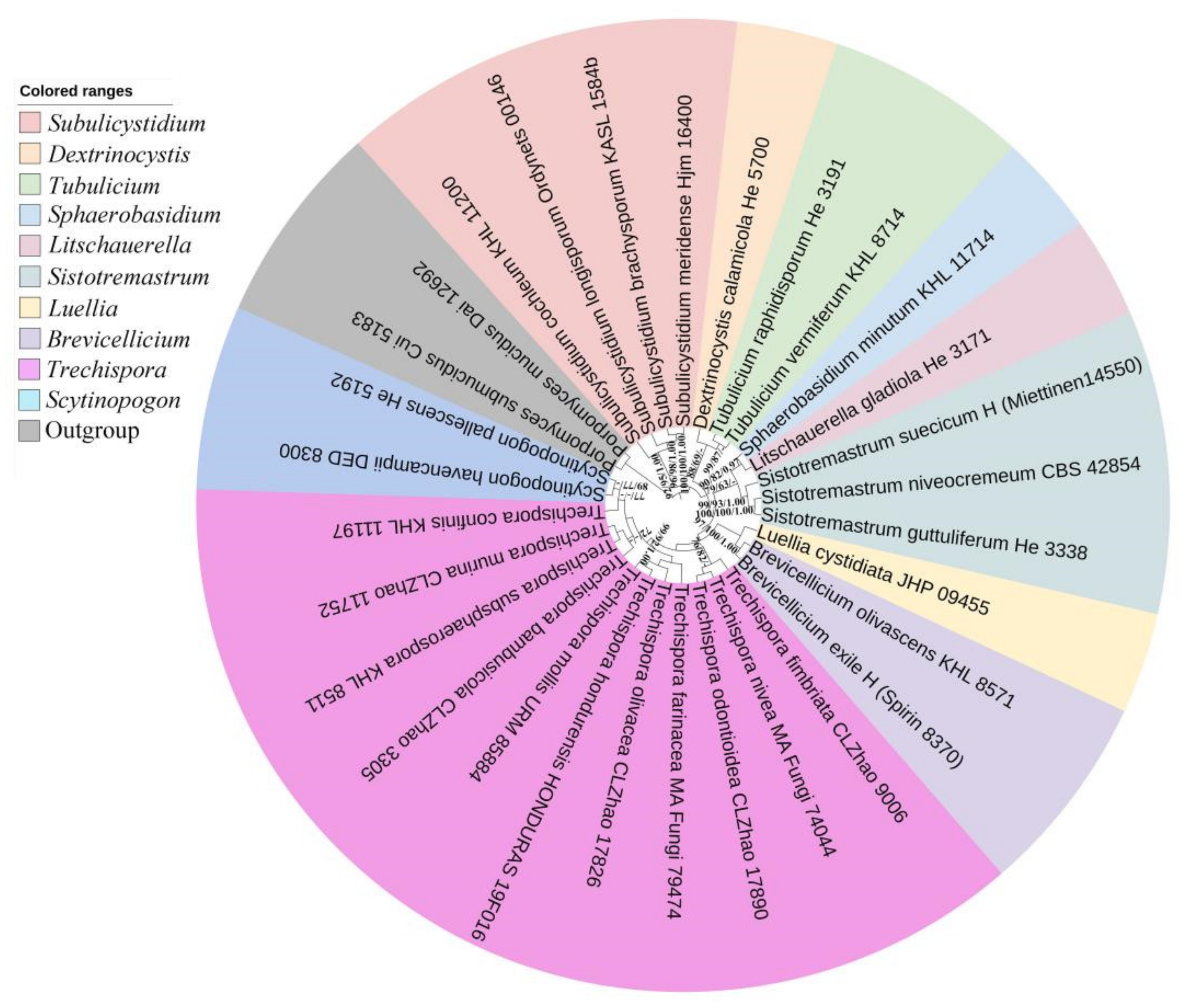
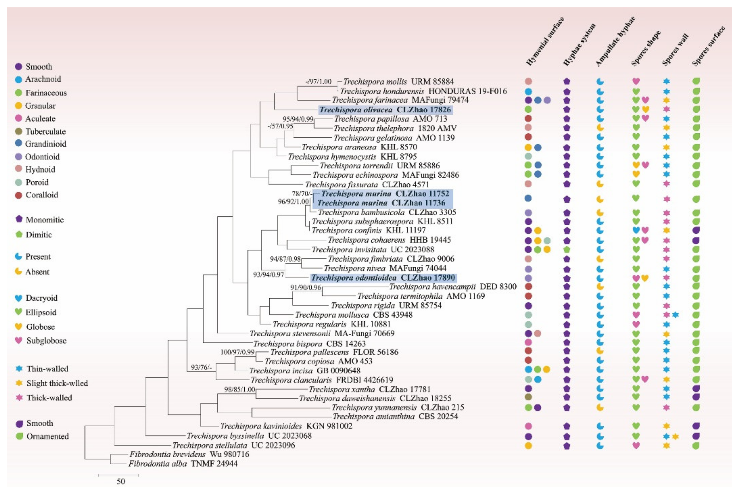
3.1. Molecular Phylogeny

The 5.8S+nLSU dataset (Figure 1) included sequences from 30 fungal samples representing 30 species. The dataset had an aligned length of 1508 characters, of which 1141 characters are constant, 104 are variable and parsimony uninformative, and 263 are parsimony informative. Maximum parsimony analysis yielded 54 equally parsimonious trees (TL = 986, CI = 0.5172, HI = 0.4828, RI = 0.5211, and RC = 0.2695). The best model was GTR+I+G (lset nst = 6, rates = invgamma; prset statefreqpr = dirichlet (1,1,1,1)). Bayesian and ML analyses showed a topology similar to that of MP analysis with split frequencies equal to 0.022581 (BI), and the effective sample size (ESS) across the two runs is double of the average ESS (avg ESS) = 869.5.
The 5.8S+nLSU rDNA gene regions (Figure 1) include ten genera within Trechisporales, Brevicellicium, Dextrinocystis Gilb. & M. Blackw., Litschauerella Oberw., Luellia K.H. Larss. & Hjortstam, Scytinopogon, Sistotremastrum J. Erikss., Sphaerobasidium Oberw., Subulicystidium Parmasto, Tubulicium Oberw., and Trechispora, shows that all related genera cluster into Trechisporales and the three new species grouped into Trechispora.
The ITS-alone dataset (Figure 2) included sequences from 42 fungal specimens representing 41 species. The dataset had an aligned length of 580 characters, of which 178 characters are constant, 61 are variable and parsimony-uninformative, and 341 are parsimony-informative. Maximum parsimony analysis yielded 584 equally parsimonious trees (TL = 2802, CI = 0.3123, HI = 0.6877, RI = 0.2519, and RC = 0.0787). Best model for the ITS dataset estimated and applied in the Bayesian analysis was GTR+I+G (lset nst = 6, rates = invgamma; prset statefreqpr = dirichlet (1,1,1,1). Bayesian and ML analyses resulted in a topology similar to that of MP analysis with split frequencies equal to 0.025000 (BI), and the effective sample size (ESS) across the two runs is double of the average ESS (avg ESS) = 621.5.
The phylogram inferred from the ITS dataset (Figure 2) indicated that the three new species grouped into Trechispora, in which the new species T. murina was sister to T. bambusicola with higher supports (96% BS, 92% BP, and 1.00 BPP); T. odontioidea formed a unique position within the clade of T. fimbriata C.L. Zhao and T. nivea (Pers.) K.H. Larss; while T. olivacea shared a clade formed by the members of T. farinacea (Pers.) Liberta, T. hondurensis Schoutteten & Haelew., and T. mollis.

3.2. Taxonomy

Trechispora murina K.Y. Luo & C.L. Zhao, sp. nov. Figure 3 and Figure 4.
MycoBank no.: 842491.
Holotype—China, Yunnan Province, Wenshan, Funing County, Guying Village, GPS coordinates 23°44′ N, 105°56′ E, altitude 750 m asl., on a fallen angiosperm branch, leg. C.L. Zhao, 20 January 2019, CLZhao 11752 (SWFC).
Etymologymurina (Lat.): Referring to the furry mouse-like hymenial surface.
Basidiomata—Annual, resupinate, thin, growing adnate but easily separable, up to 15 cm long, 3 cm wide, 100–500 µm thick. Hymenial surface grandinioid, pale greyish to grey when fresh, turn to greyish upon drying. Sterile margin concolorous with a hymenial surface, up to 2 mm wide.
Hyphal system—Monomitic; generative hyphae with clamp connections; colorless; thick-walled with a wide to lumen; richly branched; interwoven; encrusted; 2–3.5 µm in diameter; IKI–, CB–; tissues unchanged in KOH.
Hymenium—Cystidia and cystidioles absent; basidia more or less clavate, with four sterigmata and a basal clamp connection, 10.0–14.0 × 3.5–4.5 µm; basidioles dominant; basidioles in shape similar to basidia, but slightly smaller.
Spores—Basidiospores ellipsoid, colorless, thick-walled, ornamented, IKI–, CB–, (2.5–) 3–4 × (2–) 2.5–3 µm, L = 3.42 µm, W = 2.87 µm, Q = 1.17–1.20 (n = 60/2).
Additional specimen examined (paratype)—China, Yunnan Province, Wenshan, Funing County, Guying Village, GPS coordinates 23°39′ N, 105°59′ E, altitude 1400 m asl., on a fallen angiosperm branch, leg. C.L. Zhao, 20 January 2019, CLZhao 11736 (SWFC).
Trechispora odontioidea K.Y. Luo & C.L. Zhao, sp. nov. Figure 5 and Figure 6.
MycoBank no.: 844493.
Holotype—China, Yunnan Province, Honghe, Pingbian County, Daweishan National Nature Reserve. GPS coordinates: 23°420′ N, 103°300′ E; altitude: 1500 m asl., on fallen angiosperm branches, leg. C.L. Zhao, 1 August 2019, CLZhao 17890 (SWFC).
Etymology—odontioidea (Lat.): Referring to the odontioid hymenophore.
Basidiomata—Annual, adnate, thin, up to 11 cm long, 2.5 cm wide, 50–200 µm thick. Hymenial surface odontioid, aculei cylindrical to conical, blunt, 0.3–0.6 mm long, pale buff when fresh, turn to buff upon drying. Sterile margin indistinct, cream to buff, 0.5–1 mm wide.
Hyphal system—Monomitic; generative hyphae with clamp connections; colorless, thin- to thick-walled; frequently branched; interwoven; 2–3.5 µm in diameter; ampullate hyphae frequently present; IKI−, CB−; tissues unchanged in KOH.
Hymenium—Cystidia and cystidioles absent.; basidia clavate, with four sterigmata and a basal clamp connection, 8.0–12.0 × 2.5–4 µm; basidioles dominant, in shape similar to basidia, but smaller.
Spores—Basidiospores subglobose to globose, colorless, slightly thick-walled, ornamented, IKI−, CB−, 2–3 × 1.5–2.5 µm, L = 2.53 µm, W = 2.00 µm, Q = 1.27 (n = 30/1).
Trechispora olivacea K.Y. Luo & C.L. Zhao, sp. nov. Figure 7 and Figure 8.
MycoBank no.: 844494.
HolotypeChina, Yunnan Province, Honghe, Pingbian County, Daweishan National Nature Reserve. GPS coordinates: 23°420′ N, 103°300′ E; altitude: 1500 m asl., on fallen angiosperm branches, leg. C.L. Zhao, 1 August 2019, CLZhao 17826 (SWFC).
Etymologyolivacea (Lat.): Referring to the olivaceous hymenial surface.
Basidiomata—Annual, resupinate, thin, very hard to separate from substrate, up to 11 cm long, 2.5 cm wide, 30–80 µm thick. Hymenial surface farinaceous, pale white to slightly olivaceous when fresh, turn to olivaceous upon drying. Sterile margin indistinct, slightly olivaceous, 0.2–0.5 mm wide.
Hyphal systemMonomitic; generative hyphae with clamp connections; colorless; thin- to thick-walled; occasionally branched; interwoven; 1.5–3.0 µm in diameter; ampullate hyphae present; IKI–, CB–; tissues unchanged in KOH.
HymeniumCystidia and cystidioles absent; basidia clavate, with four sterigmata and a basal clamp connection, 10.0–12.0 × 4.5–5 µm; basidioles dominant, with the shape similar to basidia, but smaller.
SporesBasidiospores broadly ellipsoid to globose, colorless, thick-walled, ornamented, IKI–, CB–, 2.5–4 × 1.5–2.5 µm, L = 3.30 µm, W = 2.65 µm, Q = 1.25 (n = 30/1).

4. Discussion

The classification of corticioid fungi revealed that two taxa of Trechispora farinacea and T. hymenocystis nested into Trechispora located in Hydnodontaceae (Trechisporales) [15]. In the present study (Figure 2), Trechispora murina, T. odontioidea, and T. olivacea are nested into Trechispora, in which T. murina was sister to T. bambusicola; T. odontioidea formed a monophyletic lineage and then grouped with T. fimbriata and T. nivea; while T. olivacea formed a monophyletic lineage and then grouped with T. farinacea, T. hondurensis, and T. mollis. However, T. bambusicola is morphologically distinguishable from T. murina by having the odontioid hymenophore with cream to buff the hymenial surface [33]. Trechispora fimbriata is distinguishable from T. odontioidea by having the hydnoid hymenial surface and longer basidiospores (3–3.6 × 2.4–3.2 µm) [33]; T. nivea differs from T. odontioidea by its thin-walled, larger basidiospores (3.5–4 × 2.5–3 µm) [5]. Trechispora farinacea is distinguishable from T. olivacea by its smooth to grandinioid or odontioid hymenophore with whitish to ochraceous hymenial surface and larger basidiospores (4–5 × 3.5–4 µm) [5]; T. hondurensis is separated from T. olivacea by having a hydnoid to partly irpicoid hymenial surface and thin-walled, wider basidiospores (3.6–3.8 × 2.7–2.9 µm) [58]; T. mollis is distinguishable from T. olivacea because it has white-yellow to pale yellow hydnoid hymenial surface, and wider ampullate septa at generative hyphae (reaching 8 µm in width) [26].
Morphologically, Trechispora murina is similar to T. farinacea, T. rigida, T. subsphaerospora (Litsch.) Liberta, and T. torrendii Chikowski & K.H. Larss. Based on the character of the grandinioid hymenial surface. However, Trechispora farinacea is separated from T. murina by having a whitish to ochraceous hymenial surface and larger, subglobose to broadly elliposid basidiospores (4–5 × 3.5–4 µm) [5]. Trechispora rigida differs from T. murina due to the presence of its dirty white to buff hymenophore [59] and having larger basidiospores (4.5–5.5 × 4 µm) [27]. Trechispora subsphaerospora differs from T. murina by having smooth basidiospores [34]. Trechispora torrendii differs in its pale yellow to yellow hymenophore [26] and has globose to subglobose basidiospores (2.8–3.5 × 3–3.5 µm) [27].
Trechispora murina is similar to T. canariensis Ryvarden & Liberta, T. fastidiosa (Pers.) Liberta, T. praefocata (Bourdot & Galzin) Liberta, T. stevensonii (Berk. & Broome) K.H. Larss., and T. yunnanensis C.L. Zhao due to the presence of the ellipsoid, ornamented basidiospores. However, Trechispora canariensis differs from T. murina because it has arachonoid to pelliculose hymenial surface and thin-walled, larger basidiospores (5–7 × 3–3.5 µm) [5]. Trechispora fastidiosa is separated from T. murina by having a membranaceous, whitish to cream hymenial surface and larger basidiospores (6–7 × 4.5–5.5 µm) [5]. Trechispora praefocata differs by having the farinaceous to arachnoid hymenial surface and larger basidiospores (5–6.5 × 4–5.5 µm) [5]. Trechispora stevensonii differs from T. murina by its hydnoid hymenophore and larger basidiospores (4–4.5 × 3–3.5 µm) [5]. Trechispora yunnanensis is separated from T. murina by having the farinaceous hymenial surface and larger basidiospores (7–8.5 × 5–5.5 µm) [31].
Trechispora odontioidea is similar to T. bambusicola C.L. Zhao and T. nivea in having an odontioid hymenial surface. However, Trechispora bambusicola differs from T. odontioidea because it has granulose basidiomata, and the absence of the ampullaceous septa [33]. Trechispora nivea differs from T. odontioidea due to the presence of white to ochraceous basidiomata and broadly ellipsoid to subglobose, thin-walled, larger basidiospores (3.5–4 × 2.5–3 µm) [5].
Trechispora odontioidea resembles T. clancularis (Park.-Rhodes) K.H. Larss., T. cyatheae Ordynets, Langer & K.H. Larss., T. hymenocystis (Berk. & Broome) K.H. Larsson, T. invisitata (H.S. Jacks.) Liberta, and T. torrendii Chikowski & K.H. Larss. due to the presence of ornamented or aculeate basidiospores. However, Trechispora clancularis is distinguishable from T. odontioidea due to the presence of its poroid to irpicoid hymenial surface and subglobose to ovoid, larger basidiospores (6–6.5 × 5–6 µm) [5]. Trechispora cyatheae differs from T. odontioidea in having a farinaceous to grandinioid hymenial surface, and broadly elliptical to slightly lacrymiform and adaxial side convex or straight, longer basidiospores (3–3.5 × 2–3 µm) [24]. Trechispora hymenocystis is distinguishable from T. odontioidea by its poroid basidiomata and broadly ellipsoidal to ellipsoidal, larger basidiospores (4.5–5.5 × 3.5–4.5 µm) [19]. Trechispora invisitata differs from T. odontioidea because it has a smooth to porulose, farinaceous to granulose hymenial surface and ellipsoid to ovate, larger basidiospores (4.5–5.5 × 3–4 µm) [5]. Trechispora torrendii differs from T. odontioidea because it has a farinose to grandinioid hymenial surface and larger basidiospores (3.2–3.5 × 2.8–3.2 µm) [26].
Trechispora olivacea is similar to T. caucasica (Parmasto) Liberta, T. dimitica Hallenb., T. gelatinosa Meiras-Ottoni & Gibertoni, T. verruculosa (G. Cunn.) K.H. Larss., and T. yunnanensis C.L. Zhao due to the presence of a farinaceous hymenial surface. However, Trechispora caucasica differs from T. olivacea by having a white to greyish hymenial surface and narrowly ellipsoid to reniform with a median constriction, larger basidiospores (5–5.5 × 4–4.5 µm) [5]. Trechispora dimitica differs from T. olivacea in its white to pale greyish hymenial surface, dimitic hyphal system, and shorter basidia (7–10 × 4.5–5.5 µm) [5]. Trechispora gelatinosa is distinguishable from T. olivacea by its coralloid basidiomata and wider basidiospores (3.2–4.5 × 2.5–3.5 µm) [27]. Trechispora verruculosa differs from T. olivacea because it has granulose to hydnoid with small cylindrical aculei, white to yellowish to ochraceous hymenial surface and larger basidiospores (4.5–5.5 × 3.5–4.5 µm) [5]. Trechispora yunnanensis can be delimited from T. olivacea by its larger basidiospores (7–8.5 × 5–5.5 µm) [31].
Trechispora olivacea resembles T. hypogeton (Maas Geest.) Hjortstam & K.H. Larss., T. nivea, T. rigida, and T. thelephora (Lév.) Ryvarden in having broadly ellipsoid to globose, ornamented basidiospores. However, Trechispora hypogeton is distinguishable from T. olivacea by its stipitate basidiomata and wider basidiospores (3.8–4.3 × 2.7–3.1 µm) [26]. Trechispora nivea differs from T. olivacea by the presence of a odontioid hymenial surface with white to pale ochraceous and wider basidiospores (3.5–4 × 2.5–3 µm) [5]. Trechispora rigida differs from T. olivacea due to the presence of a colliculose hymenial surface and larger basidiospores (4.5–5.5 × 4 µm) [26]. Trechispora thelephora differs from T. olivacea because it has a stipitate basidiomata and larger basidiospores (4.0–5.0 × 3.4–4.5 µm) [26].
Wood-rotting fungi are an extensively studied group of Basidiomycota [12,13,60,61,62,63,64,65,66] and the three taxa of Trechispora are a typical example group of wood-rotting fungi [15,33,34,35,67]. Based on our present morphology and phylogeny focusing on Trechispora, all taxa in this genus can be separated from the three new species.
Key to 21 accepted species of Trechispora in China
  • 1. Basidiospores smooth-------------------------------------------------------------------------------------2
  • 1′ Basidiospores aculeate, verrucose or ornamented--------------------------------------------------5
  • 2. Ampullate hyphae > 5 μm in width, basidiospores angular----------------T. subsphaerospora
  • 2′ Ampullate hyphae < 5 μm in width, basidiospores ellipsoid------------------------------------3
  • 3. Basidiospores thick-walled-----------------------------------------------------------------T. cohaerens
  • 3′ Basidiospores thin-walled--------------------------------------------------------------------------------4
  • 4. Hymenial surface tuberculate-------------------------------------------------------T. daweishanensis
  • 4′ Hymenial surface smooth----------------------------------------------------------------------T. xantha
  • 5. Hyphal system dimitic------------------------------------------------------------------------T. dimitica
  • 5′ Hyphal system monomitic-------------------------------------------------------------------------------6
  • 6. Hyphae without ampullate septa----------------------------------------------------------------------7
  • 6′ Hyphae with ampullate septa-------------------------------------------------------------------------12
  • 7. Basidiospores thin-walled, ovoid to subglobose---------------------------------------T. suberosa
  • 7′ Basidiospores thick-walled, ellipsoid-----------------------------------------------------------------8
  • 8. Basidiospores > 7 μm in length--------------------------------------------------------T. yunnanensis
  • 8′ Basidiospores < 7 μm in length-------------------------------------------------------------------------9
  • 9. Basidiomata margin greyish----------------------------------------------------------------T. murina
  • 9′ Basidiomata margin white to cream----------------------------------------------------------------10
  • 10. Hymenial surface odontioid---------------------------------------------------------T. bambusicola
  • 10′ Hymenial surface hydnoid--------------------------------------------------------------------------11
  • 11. Hymenophore with blunt aculei--------------------------------------------------------T. fimbriata
  • 11′ Hymenophore with sharp aculei--------------------------------------------------------T. fissurata
  • 12. Sphaerocysts present, hyphae inflated-------------------------------------------T. hymenocystis
  • 12′ Sphaerocysts absent, hyphae uninflated---------------------------------------------------------13
  • 13. Ampullate septa > 6 μm in width------------------------------------------------------------------14
  • 13′ Ampullate septa < 6 μm in width------------------------------------------------------------------15
  • 14. Basidiospores sparsely verrucose-----------------------------------------------T. polygonospora
  • 14′ Basidiospores densely aculeate---------------------------------------------------------T. mollusca
  • 15. Subhymenium with short-celled hyphae--------------------------------------------------------16
  • 15′ Subhymenium with long-celled hyphae---------------------------------------------------------17
  • 16. Basidiome thin, ochraceous--------------------------------------------------------------T. farinacea
  • 16′ Basidiome thick, dirty white to buff-------------------------------------------------------T. rigida
  • 17. Basidiospores thin-walled---------------------------------------------------------------------------18
  • 17′ Basidiospores thick-walled--------------------------------------------------------------------------19
  • 18. Hymenophore with hydnoid-----------------------------------------------------------------T. nivea
  • 18′ Hymenophore without hydnoid------------------------------------------------------T. microspora
  • 19. Basidiospores > 5 μm in length---------------------------------------------------------T. praefocata
  • 19′ Basidiospores < 5 μm in length---------------------------------------------------------------------20
  • 20. Hymenial surface farinaceous with olivaceous--------------------------------------T. olivacea
  • 20′ Hymenial surface odontioid with buff--------------------------------------------T. odontioidea

Author Contributions

Conceptualization, C.Z.; methodology, C.Z. and K.L.; software, C.Z. and K.L.; validation, C.Z. and K.L.; formal analysis, C.Z. and K.L.; investigation, C.Z. and K.L.; resources, C.Z.; writing—original draft preparation, C.Z. and K.L.; writing—review and editing, C.Z. and K.L.; visualization, C.Z. and K.L.; supervision, C.Z.; project administration, C.Z.; funding acquisition, C.Z. All authors have read and agreed to the published version of the manuscript.

Funding

The research was supported by the National Natural Science Foundation of China (Project No. 32170004, U2102220), Yunnan Fundamental Research Project (Grant No. 202001AS070043), the High-level Talents Program of Yunnan Province (YNQR-QNRC-2018-111), and the Opening Foundation of Yunnan Key Laboratory of Plateau Wetland Conservation, Restoration and Ecological Services (202105AG070002).

Institutional Review Board Statement

Not applicable.

Informed Consent Statement

Not applicable.

Data Availability Statement

Publicly available datasets were analyzed in this study. This data can be found here: [https://www.ncbi.nlm.nih.gov/, accessed on 28 November 2021; https://www.mycobank.org/page/Simple%20names%20search].

Conflicts of Interest

The authors declare no conflict of interest.

References

  1. James, T.Y.; Stajich, J.E.; Hittinger, C.T.; Rokas, A. Toward a fully resolved fungal tree of life. Annu. Rev. Microbiol. 2020, 74, 291–313. [Google Scholar] [CrossRef] [PubMed]
  2. Tedersoo, L.; Bahram, M.; Põlme, S.; Koljalg, U.; Yorou, N.S.; Wijesundera, R.; Ruiz, L.V.; Vasco-Palacios, A.M.; Thu, P.Q.; Suija, A.; et al. Global diversity and geography of soil fungi. Science 2014, 346, 1256688. [Google Scholar] [CrossRef] [PubMed]
  3. Gilbertson, R.L.; Ryvarden, L. North American Polypores 1–2; Fungiflora: Oslo, Norway, 1987; pp. 1–433. [Google Scholar]
  4. Núñez, M.; Ryvarden, L. East Asian polypores 2. Synop. Fungorum 2001, 14, 165–522. [Google Scholar]
  5. Bernicchia, A.; Gorjón, S.P. Fungi Europaei 12: Corticiaceae s.l.; Edizioni Candusso: Alassio, Italy, 2010. [Google Scholar]
  6. Dai, Y.C. Polypore diversity in China with an annotated checklist of Chinese polypores. Mycoscience 2012, 53, 49–80. [Google Scholar] [CrossRef]
  7. Ryvarden, L.; Melo, I. Poroid fungi of Europe. Synop. Fungorum 2014, 31, 1–455. [Google Scholar]
  8. Dai, Y.C.; Cui, B.K.; Si, J.; He, S.H.; Hyde, K.D.; Yuan, H.S.; Liu, X.Y.; Zhou, L.W. Dynamics of the worldwide number of fungi with emphasis on fungal diversity in China. Mycol. Prog. 2015, 14, 62. [Google Scholar] [CrossRef]
  9. Dai, Y.C.; Yang, Z.L.; Cui, B.K.; Wu, G.; Yuan, H.S.; Zhou, L.W.; He, S.H.; Ge, Z.W.; Wu, F.; Wei, Y.L.; et al. Diversity and systematics of the important macrofungi in Chinese forests. Mycosystema 2021, 40, 770–805. [Google Scholar]
  10. Cui, B.K.; Yuan, H.S.; Zhou, L.W.; He, S.H.; Wei, Y.L. Diversity of wood-decaying fungi in conifer trees of the Greater and Lesser Khinggan Mountains. Biodivers. Sci. 2019, 27, 887–895. [Google Scholar] [CrossRef]
  11. Wu, F.; Yuan, H.S.; Zhou, L.W.; Yuan, Y.; Cui, B.K.; Dai, Y.C. Polypore diversity in South China. Mycosystema 2020, 39, 653–682. [Google Scholar]
  12. Luo, K.Y.; Chen, Z.Y.; Zhao, C.L. Phylogenetic and taxonomic analyses of three new wood-inhabiting fungi of Xylodon (Basidiomycota) in a forest ecological system. J. Fungi 2022, 8, 405. [Google Scholar] [CrossRef]
  13. Qu, M.H.; Wang, D.Q.; Zhao, C.L. A phylogenetic and taxonomic study on Xylodon (Hymenochaetales): Focusing on three new Xylodon species from southern China. J. Fungi 2022, 8, 35. [Google Scholar] [CrossRef]
  14. Hibbett, D.S.; Binder, M.; Bischoff, J.F.; Blackwell, M.; Cannon, P.F.; Eriksson, O.E.; Huhndorf, S.; James, T. A higher-level phylogenetic classification of the Fungi. Mycol. Res. 2007, 111, 509–547. [Google Scholar] [CrossRef]
  15. Larsson, K.H. Re-thinking the classification of corticioid fungi. Mycol. Res. 2007, 111, 1040–1063. [Google Scholar] [CrossRef]
  16. Karsten, P.A. Fragmenta mycologica XXIX. Nova Hedwig. 1890, 29, 147–149. [Google Scholar]
  17. Liberta, A.E. On Trechispora. Taxon 1966, 15, 317–319. [Google Scholar] [CrossRef]
  18. Liberta, A.E. The genus Trechispora (Basidiomycetes, Corticiaceae). Can. J. Bot. 1973, 51, 1871–1892. [Google Scholar] [CrossRef]
  19. Larsson, K.H. Poroid species in Trechispora and the use of calcium oxalate crystals for species identification. Mycol. Res. 1994, 98, 1153–1172. [Google Scholar] [CrossRef]
  20. Ryvarden, L. A note on the genus Hydnodon Banker. Synop. Fungorum 2002, 15, 31–33. [Google Scholar]
  21. Trichiès, G.; Schultheis, B. Trechispora antipus sp. nov., une seconde espèce bisporique du genre Trechispora (Basidiomycota, Stereales). Mycotaxon 2002, 82, 453–458. [Google Scholar]
  22. Miettinen, O.; Larsson, K.H. Trechispora elongata species nova from North Europe. Mycotaxon 2006, 96, 193–198. [Google Scholar]
  23. Kirk, P.M.; Cannon, P.F.; David, J.C.; Minter, D.W.; Stalpers, J.A. Ainsworth and Bisby’s Dictionary of the Fungi, 10th ed.; CAB International Press: Wallingford, UK, 2008; p. 783. [Google Scholar] [CrossRef]
  24. Ordynets, A.; Larsson, K.H.; Langer, E. Two new Trechispora species from La Réunion Island. Mycol. Prog. 2015, 14, 113. [Google Scholar] [CrossRef]
  25. Phookamsak, R.; Hyde, K.D.; Jeewon, R.; Bhat, D.J.; Jones, E.B.G.; Maharachchikumbura, S.S.N.; Raspe´, O.; Karunarathna, S.C.; Wanasinghe, D.N.; Hongsanan, S.; et al. Fungal diversity notes 929–1035: Taxonomic and phylogenetic contributions on genera and species of fungi. Fungal Divers. 2019, 95, 1–273. [Google Scholar] [CrossRef]
  26. Chikowski, R.S.; Larsson, K.H.; Gibertoni, T.B. Taxonomic novelties in Trechispora from Brazil. Mycol. Prog. 2020, 19, 1403–1414. [Google Scholar] [CrossRef]
  27. Meiras-Ottoni, A.; Larsson, K.H.; Gibertoni, T.B. Additions to Trechispora and the status of Scytinopogon (Trechisporales, Basidiomycota). Mycol. Prog. 2021, 20, 203–222. [Google Scholar] [CrossRef]
  28. Dai, Y.C. A checklist of polypores in China. Mycosystema 2009, 28, 315–327. [Google Scholar]
  29. Dai, Y.C. A revised checklist of corticioid and hydnoid fungi in China for 2010. Mycoscience 2011, 52, 69–79. [Google Scholar] [CrossRef]
  30. Yuan, H.S.; Dai, Y.C. Wood-inhabiting fungi in southern China. 6. Polypores from Guangxi Autonomous region. Ann. Bot. Fenn. 2012, 49, 341–351. [Google Scholar] [CrossRef]
  31. Xu, T.M.; Chen, Y.H.; Zhao, C.L. Trechispora yunnanensis sp. nov. from China. Phytotaxa 2019, 424, 253–261. [Google Scholar] [CrossRef] [Green Version]
  32. Wang, K.; Zhao, M.J.; Su, J.H.; Yang, L.; Deng, H.; Wang, Y.H.; Wu, H.J.; Li, Y.; Wu, H.M.; Wei, X.D.; et al. The use of checklist of fungi in China database in the red list assessment of macrofungi in China. Biodivers. Sci. 2020, 28, 74–98. [Google Scholar] [CrossRef]
  33. Zhao, W.; Zhao, C.L. The phylogenetic relationship revealed three new wood-inhabiting fungal species from genus Trechispora. Front. Microbiol. 2021, 12, 650195. [Google Scholar] [CrossRef] [PubMed]
  34. Zong, T.K.; Liu, C.M.; Wu, J.R.; Zhao, C.L. Trechispora daweishanensis and T. xantha spp. nov. (Hydnodontaceae, Trechisporales) found in Yunnan Province of China. Phytotaxa 2021, 479, 147–159. [Google Scholar] [CrossRef]
  35. Larsson, K.H.; Larsson, E.; Kõljalg, U. High phylogenetic diversity among corticioid homobasidiomycetes. Mycol. Res. 2004, 108, 983–1002. [Google Scholar] [CrossRef]
  36. Telleria, M.T.; Melo, I.; Dueñas, M.; Larsson, K.H.; Paz, M.M.P. Molecular analyses confifirm Brevicellicium in Trechisporales. IMA Fungus 2013, 4, 21–28. [Google Scholar] [CrossRef]
  37. Jülich, W. Notes on some Basidiomycetes (Aphyllophorales and Heterobasidiomycetes). Persoonia 1982, 11, 421–428. [Google Scholar]
  38. Liu, S.L.; Ma, H.X.; He, S.H.; Dai, Y.C. Four new corticioid species in Trechisporales (Basidiomycota) from East Asia and notes on phylogeny of the order. MycoKeys 2019, 48, 97–113. [Google Scholar] [CrossRef]
  39. Kornerup, A.; Wanscher, J. Metheun Hand Book of Colour, 3rd ed.; Metheun London Ltd.: London, UK, 1978; pp. 144–148. [Google Scholar]
  40. Wu, F.; Zhou, L.W.; Vlasák, J.; Dai, Y.C. Global diversity and systematics of Hymenochaetaceae with poroid hymenophore. Fungal Divers. 2022, 113, 1–192. [Google Scholar] [CrossRef]
  41. Zhao, C.L.; Wu, Z.Q. Ceriporiopsis kunmingensis sp. nov. (Polyporales, Basidiomycota) evidenced by morphological characters and phylogenetic analysis. Mycol. Prog. 2017, 16, 93–100. [Google Scholar] [CrossRef]
  42. White, T.J.; Bruns, T.; Lee, S.; Taylor, J. Amplification and direct sequencing of fungal ribosomal RNA genes for phylogenetics. PCR Protoc. A Guide Methods Appl. 1990, 18, 315–322. [Google Scholar] [CrossRef]
  43. Spirin, V.; Volobuev, S.; Viner, I.; Miettinen, O.; Vlasak, J.; Schoutteten, N.; Motato-Vásquez, V.; Kotiranta, H.; Hernawati; Larsson, K.H. On Sistotremastrum and similar-looking taxa (Trechisporales, Basidiomycota). Mycol. Prog. 2021, 20, 453–476. [Google Scholar] [CrossRef]
  44. Yurchenko, E.; Wu, S.H. Hyphoderma formosanum sp. nov. (Meruliaceae, Basidiomycota) from Taiwan. Sydowia 2014, 66, 19–23. [Google Scholar]
  45. Wu, F.; Yuan, Y.; Zhao, C.L. Porpomyces submucidus (Hydnodontaceae, Basidiomycota) from tropical China. Phytotaxa 2015, 230, 61–68. [Google Scholar] [CrossRef]
  46. Desjardin, D.E.; Perry, B.A. A new species of Scytinopogon from the island of Príncipe, Republic of São Tomé and Príncipe, West Africa. Mycosphere 2015, 6, 434–441. [Google Scholar] [CrossRef]
  47. Vu, D.; Groenewald, M.; de Vries, M.; Gehrmann, T.; Stielow, B.; Eberhardt, U.; Al-Hatmi, A.; Groenewald, J.Z.; Cardinali, G.; Houbraken, J.; et al. Large-scale generation and analysis of fifilamentous fungal DNA barcodes boosts coverage for kingdom fungi and reveals thresholds for fungal species and higher taxon delimitation. Stud. Mycol. 2019, 92, 135–154. [Google Scholar] [CrossRef]
  48. Larsson, K.H.; Parmasto, E.; Fischer, M.; Langer, E.; Nakasone, K.K.; Redhead, S.A. Hymenochaetales: A molecular phylogeny for the hymenochaetoid clade. Mycologia 2006, 98, 926–936. [Google Scholar] [CrossRef] [PubMed]
  49. Ordynets, A.; Scherf, D.; Pansegrau, F.; Denecke, J.; Langer, E. Short-spored Subulicystidium (Trechisporales, Basidiomycota): High morphological diversity and only partly clear species boundaries. MycoKeys 2018, 35, 41–99. [Google Scholar] [CrossRef]
  50. Ordynets, A.; Liebisch, R.; Lysenko, L.; Scherf, D.; Volobuev, S.; Saitta, A.; Larsson, K.H.; Yurchenko, E.; Buyck, B.; Bolshakov, S. Morphologically similar but not closely related: The long-spored species of Subulicystidium (Trechisporales, Basidiomycota). Mycol. Prog. 2020, 19, 691–703. [Google Scholar] [CrossRef]
  51. Vasco-Palacios, A.M.; Lopez-Quintero, C.; Franco-Molano, A.E.; Boekhout, T. Austroboletus amazonicus sp. nov. and Fistulinella campinaranae var. scrobiculata, two commonly occurring boletes from a forest dominated by Pseudomonotes tropenbosii (Dipterocarpaceae) in Colombian Amazonia. Mycologia 2014, 106, 1004–1014. [Google Scholar] [CrossRef] [PubMed]
  52. Larsson, A. AliView: A fast and lightweight alignment viewer and editor for large data sets. Bioinformatics 2014, 30, 3276–3278. [Google Scholar] [CrossRef] [PubMed]
  53. Swofford, D.L. PAUP *: Phylogenetic Analysis Using Parsimony (* and Other Methods); Version 4.0b10; Sinauer Associates: Sunderland, MA, USA, 2002. [Google Scholar]
  54. Felsenstein, J. Confidence intervals on phylogenetics: An approach using bootstrap. Evolution 1985, 39, 783–791. [Google Scholar] [CrossRef] [PubMed]
  55. Miller, M.A.; Pfeiffer, W.; Schwartz, T. The CIPRES science gateway: Enabling high-impact science for phylogenetics researchers with limited resources. Assoc. Comput. Mach. 2012, 39, 1–8. [Google Scholar] [CrossRef]
  56. Nylander, J.A.A. MrModeltest V2. Program Distributed by the Author; Evolutionary Biology Centre, Uppsala University: Uppsala, Sweden, 2004. [Google Scholar]
  57. Ronquist, F.; Teslenko, M.; van der Mark, P.; Ayres, D.L.; Darling, A.; Hohna, S.; Larget, B.; Liu, L.; Suchard, M.A.; Huelsenbeck, J.P. Mrbayes 3.2: Efficient bayesian phylogenetic inference and model choice across a large model space. Syst. Biol. 2012, 61, 539–542. [Google Scholar] [CrossRef]
  58. Haelewaters, D.; Dima, B.; Abdel-Hafiz, A.I.; Abdel-Wahab, M.A.; Abul-Ezz, S.R.; Acar, I.; Aguirre-Acosta, E.; Aime, M.C.; Aldemir, S.; Ali, M.; et al. Fungal systematics and evolution: Fuse 6. Sydowia 2020, 72, 231–356. [Google Scholar] [CrossRef]
  59. Larsson, K.H. New species and combinations in Trechispora (Corticiaceae, Basidiomycotina). Nord. J. Bot. 1996, 16, 83–98. [Google Scholar] [CrossRef]
  60. Zhang, M.; Wang, C.Q.; Gan, M.S.; Li, Y.; Shao, S.C.; Qin, W.Q.; Deng, W.Q.; Li, T.H. Diversity of Cantharellus (Cantharellales, Basidiomycota) in China with Description of Some New Species and New Records. J. Fungi 2022, 8, 483. [Google Scholar] [CrossRef]
  61. Song, C.G.; Chen, Y.Y.; Liu, S.; Xu, T.M.; He, X.L.; Wang, D.; Cui, B.K. A Phylogenetic and Taxonomic Study on Phellodon (Bankeraceae, Thelephorales) from China. J. Fungi 2022, 8, 429. [Google Scholar] [CrossRef]
  62. Hu, J.; Zhao, G.; Tuo, Y.; Rao, G.; Zhang, Z.; Qi, Z.; Yue, L.; Liu, Y.; Zhang, T.; Li, Y.; et al. Morphological and Molecular Evidence Reveal Eight New Species of Gymnopus from Northeast China. J. Fungi 2022, 8, 349. [Google Scholar] [CrossRef]
  63. Mao, N.; Xu, Y.Y.; Zhao, T.Y.; Lv, J.C.; Fan, L. New Species of Mallocybe and Pseudosperma from North China. J. Fungi 2022, 8, 256. [Google Scholar] [CrossRef]
  64. Wu, F.; Tohtirjap, A.; Fan, L.F.; Zhou, L.W.; Alvarenga, R.L.M.; Gibertoni, T.B.; Dai, Y.C. Global diversity and updated phylogeny of Auricularia (Auriculariales, Basidiomycota). J. Fungi 2021, 7, 933. [Google Scholar] [CrossRef]
  65. Guan, Q.X.; Zhao, C.L. Taxonomy and phylogeny of the wood-inhabiting fungal genus Hyphoderma with descriptions of three new species from East Asia. J. Fungi 2021, 7, 308. [Google Scholar] [CrossRef]
  66. Wang, D.Q.; Zhao, C.L. Morphological and Phylogenetic Evidence for Recognition of Two New Species of Phanerochaete from East Asia. J. Fungi 2021, 7, 1063. [Google Scholar] [CrossRef]
  67. He, M.Q.; Zhao, R.L.; Hyde, K.D.; Begerow, D.; Kemler, M.; Yurkov, A.; McKenzie, E.H.C.; Raspe, O.; Kakishima, M.; Sanchez-Ramırez, S.; et al. Notes, outline and divergence times of Basidiomycota. Fungal Divers. 2019, 99, 105–367. [Google Scholar] [CrossRef] [Green Version]
Figure 1. Maximum parsimony strict consensus tree illustrating the phylogeny of Trechispora and related genera in Trechisporales based on 5.8S+nLSU sequences. The genera represented by each color are indicated in the upper left of the phylogenetic tree. Branches are labelled with maximum likelihood bootstrap value ≧ 70%, parsimony bootstrap value ≧ 50%, and Bayesian posterior probabilities ≧ 0.95, respectively.
Jof 08 01020 g001
Figure 2. Maximum parsimony strict consensus tree illustrating the phylogeny of three new species and related species in Trechispora based on ITS sequences. Branches are labelled with maximum likelihood bootstrap value ≧ 70%, parsimony bootstrap value ≧ 50%, and Bayesian posterior probabilities ≧ 0.95, respectively. The new species are in bold.
Jof 08 01020 g002
Figure 3. Basidiomata of Trechispora murina (holotype CLZhao 11752): the front of the basidiomata (A), characteristic hymenophore (B). Bars: (A) = 5 mm and (B) = 1 mm.
Jof 08 01020 g003
Figure 4. Microscopic structures of Trechispora murina (holotype CLZhao 11752): basidiospores (A), a cross-section of basidiomata (B), basidia and basidioles (C). Bars: (A) = 5 μm, (B,C) = 10 µm.
Jof 08 01020 g004
Figure 5. Basidiomata of Trechispora odontioidea (holotype CLZhao 17890): the front of the basidiomata (A), characteristic hymenophore (B). Bars: (A) = 1 cm and (B) = 1 mm.
Jof 08 01020 g005
Figure 6. Microscopic structures of Trechispora odontioidea (holotype CLZhao 17890): basidiospores (A), basidia and basidioles (B), a cross section of basidiomata (C). Bars: (A) = 5 μm, (B,C) = 10 µm.
Jof 08 01020 g006
Figure 7. Basidiomata of Trechispora olivacea (holotype CLZhao 17826): the front of the basidiomata (A), characteristic hymenophore (B). Bars: (A) = 1 cm and (B) = 1 mm.
Jof 08 01020 g007
Figure 8. Microscopic structures of Trechispora olivacea (holotype CLZhao 17826): basidiospores (A), basidia and basidioles (B), a cross-section of basidiomata (C). Bars: (A) = 5 μm, (B,C) = 10 µm.
Jof 08 01020 g008
Table 1. List of species, specimens, and GenBank accession numbers of sequences used in this study, the newly generated sequences are in bold fonts.
Species NameSpecimen No.GenBank Accession No.References
ITSnLSU
Brevicellicium exileH (Spirin 8370)MT002322MT002338[43]
B. olivascensKHL 8571HE963792HE963793[36]
Dextrinocystis calamicolaHe 5700MK204534MK204547[38]
Fibrodontia albaTNMF 24944NR153983NG060401[24]
F. brevidensWu 9807-16KC928276KC928277[44]
Litschauerella gladiolaHe 3171MK204555MK204556[38]
Luellia cystidiataJHP 09455MW371211 Unpublished
Porpomyces mucidusDai 12692KT157833KT157838[45]
P. submucidusCui 5183KT152143KT152145[45]
Scytinopogon pallescensHe 5192 MK204553[38]
S. havencampiiDED 8300KT253946KT253947[46]
Sistotremastrum guttuliferumHe 3338MK204540MK204552[38]
S. niveocremeumCBS 42854MH857381MH868921[47]
S. suecicumH (Miettinen14550)MT075860MT002336[43]
Sphaerobasidium minutumKHL 11714DQ873652DQ873653[48]
Subulicystidium brachysporumKASL 1584bMH041544MH041610[49]
S. cochleumKHL 11200MN207036MN207024[50]
S. longisporumOrdynets 00146MN207039MN207032[50]
S. meridenseHjm 16400MH041538MH041604[49]
Trechispora amianthinaCBS 202.54MH857292 [47]
T. araneosaKHL 8570AF347084 [35]
T. bambusicolaCLZhao 3305MW544022MW520172[33]
T. bisporaCBS 142.63MH858241 [47]
T. byssinellaUC 2023068KP814481 Unpublished
T. clancularisFRDBI 4426619MW487976 Unpublished
T. cohaerensHHB 19445MW740327 Unpublished
T. copiosaAMO 453MN701018 [27]
T. confinisKHL 11197AY463473AY586719[35]
T. daweishanensisCLZhao 18255MW302338 [34]
T. echinosporaMA Fungi 82486JX392853 [36]
T. farinaceaMA Fungi 79474JX392855JX392856[36]
T. fimbriataCLZhao 9006MW544025MW520175[33]
T. fissurataCLZhao 4571MW544027 [33]
T. gelatinosaAMO 1139MN701021 [27]
T. havencampiiDED 8300NR154418 [46]
T. hondurensisHONDURAS 19-F016MT571523MT636540Unpublished
T. hymenocystisKHL 8795AF347090 [35]
T. incisaGB 0090648KU747095 Unpublished
T. invisitataUC 2023088KP814425 Unpublished
T. kavinioidesKGN 981002AF347086 [35]
T. mollisURM 85884MK514945 [26]
T. molluscaCBS 43948MH856428 [47]
T. murinaCLZhao 11736OL615003 Present study
T. murinaCLZhao 11752OL615004OL615009Present study
T. niveaMA Fungi 74044JX392832 [36]
T.odontioideaCLZhao 17890ON417458 Present study
T.olivaceaCLZhao 17826ON417457 Present study
T. pallescensFLOR 56186MK458766 Unpublished
T. papillosaAMO 713MN701022 [27]
T. regularisKHL 10881AF347087 [35]
T. rigidaURM 85754MT406381 [26]
T. stellulataUC 2023096KP814450 Unpublished
T. stevensoniiMA Fungi 70669JX392841 [36]
T. subsphaerosporaKHL 8511AF347080 [35]
T. termitophilaAMO 1169MN701028 [27]
T. thelephora1820 AMVKF937369 [51]
T. torrendiiURM 85886MK515148 [26]
T. xanthaCLZhao 17781MW302340 [34]
T. yunnanensisCLZhao 215MN654923 [31]
Tubulicium raphidisporumHe 3191MK204537MK204545[38]
T. vermiferumKHL 8714AY463477[35]
Publisher’s Note: MDPI stays neutral with regard to jurisdictional claims in published maps and institutional affiliations.

