Article

Diagnosing Fungal Keratitis and Simultaneously Identifying Fusarium and Aspergillus Keratitis with a Dot Hybridization Array

1
Department of Ophthalmology, Kaohsiung Chang Gung Memorial Hospital and Chang Gung University, College of Medicine, Kaohsiung City 83301, Taiwan
2
School of Medicine, Chang Gung University, Taoyuan City 33302, Taiwan
3
Department of Ophthalmology, Chung-Ho Memorial Hospital, Kaohsiung Medical University, Kaohsiung City 80708, Taiwan
4
Department of Laboratory Medicine, Kaohsiung Chang Gung Memorial Hospital and Chang Gung University, College of Medicine, Kaohsiung City 83301, Taiwan
5
Department of Medical Biotechnology and Laboratory Sciences, College of Medicine, Chang Gung University, Taoyuan City 333323, Taiwan
6
Department of Ophthalmology, Kaohsiung Veterans General Hospital, Kaohsiung City 81362, Taiwan
*
Authors to whom correspondence should be addressed.
J. Fungi 2022, 8(1), 64; https://doi.org/10.3390/jof8010064
Submission received: 10 December 2021 / Revised: 22 December 2021 / Accepted: 5 January 2022 / Published: 7 January 2022
(This article belongs to the Special Issue Molecular Tissue Diagnosis of Fungal Infections)

Abstract

:
Fungal keratitis (FK) is one of the most common microbial keratitis, which often leads to poor prognosis as a result of delayed diagnosis. Several studies implied that early differentiation of the two major FK, Fusarium and Aspergillus keratitis, could be helpful in selecting effective anti-fungal regimens. Therefore, a novel dot hybridization array (DHA) was developed to diagnose FK and differentiate Fusarium and Aspergillus keratitis in this study. One hundred forty-six corneal scrapes obtained from one hundred forty-six subjects impressed with clinically suspected FK were used to evaluate the performance of the DHA. Among these patients, 107 (73.3%) patients had actual FK confirmed by culture and DNA sequencing. We found that the DHA had 93.5% sensitivity and 97.4% specificity in diagnosing FK. In addition, this array had 93.2% sensitivity and 93.8% specificity in diagnosing Fusarium keratitis, as well as 83.3% sensitivity and 100% specificity in diagnosing Aspergillus keratitis. Furthermore, it had 83.9% sensitivity and 100% specificity in identifying Fusarium solani keratitis. Thus, this newly developed DHA will be beneficial to earlier diagnosis, more precise treatment, and improve prognosis of FK, by minimizing medical refractory events and surgical needs.

1. Introduction

Fungal keratitis (FK) is an opportunistic corneal infection of fungi predisposed by corneal surface trauma [1]. According to the Asia Cornea Society Infectious Keratitis Study (ACS IKS) [2], FK was one of the most common microbial keratitis (MK), which was secondary to bacterial keratitis (BK) (FK:BK = 33%:38%). In addition, they found that trauma was the most common risk factor for MK. However, FK is easily overlooked due to its relatively sluggish progressive course. In addition, less intense pain in the early phase [3] often leads to longer delay before seeking medical care, which results in a worse visual outcome than BK. Previous reports showed clinical diagnosis of FK is highly challenging [4,5]. The sensitivity and specificity of clinical diagnosis of FK were 38% and 45%, respectively [4]. Even for an experienced corneal physician, the diagnostic accuracy via slit lamp image was only about 76% [6]. Consequently, about 12 to 58% of FK patients needed therapeutic keratoplasty or other surgeries to quiet down their infection episodes [7,8,9], and the surgical cure rate of FK was the worst among various MKs [10]. Finally, up to 25% of FK patients might lose their vision [11].
Among the 2831 isolated microorganisms in the ACS IKS [2], the top 3 pathogens were Fusarium spp. (18%), Pseudomonas spp. (10%), and Aspergillus spp. (8%). A recent comprehensive review confirmed Fusarium and Aspergillus as FK’s most common fungal isolates globally [11]. FK responds poorly to anti-fungal agents once the deep invasion of fungi occurs, which is why early diagnosis of FK is crucial. If diagnosis and treatment are made early, polyenes and azoles were active against Fusarium spp. and Aspergillus spp., respectively [12]. Moreover, the Mycotic Ulcer Treatment Trial (MUTT) also found that Fusarium spp. were least susceptible to voriconazole, whereas Aspergillus spp. were least susceptible to natamycin [13,14]. For advanced FK shown in MUTT 2, by adjunctive oral voriconazole to topical natamycin, only Fusarium keratitis cases may get better visual outcome [15]. Furthermore, compared to other Fusarium spp., F. solani has been shown to have higher voriconazole resistance and a worse visual outcome [16]. The evidence above suggests that the overall prognosis of FK will be increased by prompt diagnosis of FK, differentiation of Fusarium and Aspergillus, and identification of critical fungal species such as F. solani.
We previously developed a dot hybridization array (DHA) for rapid diagnosis of FK, of which this assay provided much higher sensitivity than that of the culture [17]. This assay was accomplished by amplifying the internal transcribed spacer region (ITS) that contained the target gene (5.8 S rRNA gene) by polymerase chain reaction (PCR), followed by hybridization of the PCR amplicon to a fungus-specific oligonucleotide probe immobilized on a nylon membrane. It can detect fungi in the corneal scrapes within a shorter turnaround time (one working day) than that of the culture. Based on the superiority of this molecular technique, this study aimed to develop and verify a novel DHA for fulfilling the unmet clinical need by expanding its detection potential from not only diagnosing FK, but also differentiating Fusarium and Aspergillus keratitis, as well as identifying target fungal species.

2. Materials and Methods

2.1. Reference Strains and Clinical Isolates

Several reference strains and clinical isolates (Table 1) of fungi were used for the preclinical specificity test. A newly developed DHA (Figure 1) for detecting all fungi, Fusarium spp., F. solani, F. verticillioides (formerly F. moniliforme), Aspergillus spp., A. flavus, and A. fumigatus (Table 2) was developed for clinical verification via a prospective multi-center study after passing the preclinical test with target and non-target microorganisms.

2.2. Participants

All procedures involving human subjects adhered to the Declaration of Helsinki and were approved (approval period from 14 February 2018 to 6 February 2021) by the Committee of Medical Ethics and Human Experiments of Chang Gung Memorial Hospital (CGMH), Kaohsiung Veterans General Hospital (KVGH), and Kaohsiung Medical University Hospital (KMUH). The patients included in this study were suspected FK subjects or suspected MK subjects, of which FK could not be ruled out via clinical morphology. Patients who were less than 20 years old or more than 85 years old and unwilling to participate in this study were excluded.

2.3. Collection of Clinical Samples

The corneal scraping samples for the above subjects were collected from the enrolled subjects of CGMH, KVGH, and KMUH. A 15# sterilized knife was used to scrape the superficial cornea with infiltrates, especially at the margin of ulceration. One part of the corneal scrapes was sent to the section of microbiology or laboratory diagnostic department in CGMH, KVGH, and KMUH for conventional microbial examination. The remaining part was washed into a 3-mL sterile microcentrifuge tube with 2.5 mL of normal saline. The sample was then sent to our laboratory for microbial DNA extraction followed by molecular diagnostic tests. The tube was frozen in a −20 °C refrigerator up to 1 week before DNA extraction.

2.4. Oligonucleotide Probe Development and Fabrication of the DHA

The oligonucleotide probes were diluted 1:1 (final concentration, 10 mM) with a tracking dye solution, drawn into wells of 96-well microtiter plates, and spotted onto nylon membranes (Roche, Mannheim, Germany) as described previously [19]. Arrays were prepared with an automatic arrayer (Ezspot, Taipei, Taiwan) by using a solid pin of 400-μm diameter. A new-generation DHAs for FK was shown in Figure 1, which was designed to diagnose FK and identify two fungal genus and four fungal species via specially designed oligonucleotide probes (Table 2). The position markers were shown on the array after hybridization and helped to pinpoint the hybridized probes. After all probes had been applied, the membrane was exposed to shortwave UVs (Stratalinker 1800; Stratagene, La Jolla, CA, USA) for 30 s. For differential diagnosis, other DHAs previously established for diagnosing bacterial keratitis, herpes keratitis, acanthamoebic keratitis, and microsporidial keratitis were used on-demand [20,21,22].

2.5. DNA Extraction, PCR Amplification, and Hybridization with DNA Array

DNA was extracted using DNeasy® Blood and Tissue Kit (Qiagen, Valencia, CA, USA) according to the manufacturer’s instructions with some modifications. For isolated molds, mycelium (approximately 0.5 × 0.5 cm) was acquired from the culture medium via ultrasound oscillation to the frozen tube filled with ddH2O. After oscillation for 30 s, we reached the mixed fluid for centrifugation. The pretreated product was obtained after the supernatant was removed. Digoxigenin (dig)-labeled ITS (internal transcribed spacer) for array hybridization was amplified by PCR using universal primers [18,19]. Each primer was labeled with a digoxigenin molecule at its 5′ end and was synthesized by Bio Basic Inc. (Markham, ON, Canada). PCR reaction mixture was prepared from the KAPA HiFi PCR Kits (Kapa Biosystems, Inc., Cape Town, South Africa). PCR thermocycling followed the condition of Bouchara et al. [23]. A negative control was performed with each run by replacing the template DNA with sterile water in the PCR mixture. The procedures were the same as those described previously [18,19], except that the hybridization step was conducted at 50 °C for 90 min.

2.6. Fungal DNA Sequencing for Discrepant Analysis

The gold standard for diagnosing FK in this study was (1) culture positive for fungus, or (2) DNA sequencing positive for fungus. For a cornea scraping sample that demonstrated a positive result for fungus either by culture or the DHA, the extracted DNA was re-amplified with primers ITS1/ITS5 and ITS4 (without 5′ end labeling). Then, the PCR product was used to confirm the presence of fungal DNA in the sample. The amplified ITS fragment was then sequenced, and the determined sequence was used to search for homologous sequences in GenBank using the BLASTN program (http://blast.ncbi.nlm.nih.gov; accessed on 1 September 2021).

2.7. Statistical Analysis

Microsoft Excel and PowerPoint 2016 (Microsoft Corporation, Redmond, WA, USA) were used as graphic tools. GraphPad Prism version 9.3.0 for Windows (GraphPad Software, San Diego, CA, USA) was used for statistical analysis. The 95% Wilson/Brown binomial confidence intervals for these indices were estimated.

3. Results

3.1. Demographic Data of Participants

This multi-center study applied a novel fungal DHA (Figure 1 and Table 2) for simultaneously diagnosing FK, identifying two crucial fungal genera, Fusarium spp. and Aspergillus spp., and four commonly reported pathogenic fungal species, F. solani, F. moniliforme, A. flavus, and A. fumigatus (Figure 2). A total of 146 subjects, suspected FK patients or suspected MK patients, in which FK could not be ruled out via clinical manifestation, were included in this study (Table 3). The number of male subjects were significantly higher than that of the female subjects (p < 0.0001). There was no significant difference for the involved eye. Ocular trauma (65 patients, 44.5%) was the most common risk factor, while diabetes mellitus was the most common systemic risk factor. Among these patients, 107 (73.3%) patients were confirmed FK by culture and DNA sequencing. In addition, 39 non-FK patients, including 16 bacterial keratitis, two herpes keratitis, three microsporidial stromal keratitis, three acanthamoebic keratitis, and 15 non-infectious keratitis, were enrolled in this study.

3.2. Detection of Fungi in Corneal Scraping Samples

The FP probe in this fungal DHA chip was used to diagnose FK (Figure 1 and Table 2). Seven patients were fungal culture-positive but DHA-negative (Table 4). One of the seven patients was confirmed as FK by DNA sequencing, but the other six patients could not be confirmed by DNA sequencing. Twenty-seven subjects were culture-negative but DHA-positive (Table 4). According to the DNA sequencing results, 1 patient was false-positive, while the other 26 patients were true-positive. This DHA’s performance for diagnosing FK was estimated based on the diagnostic criteria of FK. The sensitivity, specificity, positive predictive rate (PPR), and negative predictive rate (NPR) were 93.5%, 97.4%, 99.0%, and 84.4%, respectively (Table 4). This result revealed that the novel DHA has good performance in the diagnosis of FK.

3.3. Identification of Fusarium sp. in Scrapes

Three genus probes, Fu1, Fu2, and Fu3, were used to diagnose Fusarium keratitis (Figure 1 and Table 2). Fusarium keratitis was diagnosed if any one of the three probes was positive. F. solani keratitis was diagnosed if the probe Fuso was positive. Similarly, F. verticillioides keratitis was diagnosed if the probe Fumo was positive. Based on the result of culture and DNA sequencing, the DHA’s performance in diagnosing Fusarium keratitis, F. solani keratitis, and F. verticillioides keratitis were estimated. The sensitivity, specificity, PPR, and NPR in diagnosing Fusarium keratitis were 93.2%, 93.8%, 87.2%, and 96.8%, respectively (Table 5). In addition, the sensitivity, specificity, PPR, and NPR in diagnosing F. solani keratitis were 83.9%, 100%, 100%, and 95.6%, respectively. The sensitivity, specificity, PPR, and NPR in diagnosing F. verticillioides keratitis were 100%, 99.3%, 50%, and 100%, respectively. However, only one subject had F. verticillioides keratitis.

3.4. Identification of Aspergillus sp. in Scrapes

Similarly, two genus probes, Asp2 and Asp3, were used to diagnose Aspergillus keratitis (Figure 1 and Table 2). Aspergillus keratitis was diagnosed if any one of the two probes was positive. A. flavus keratitis was diagnosed if the probe Asfla was positive. Similarly, A. fumigatus keratitis was diagnosed if the probe Asfum was positive. Similar to Fusarium keratitis, the DHA’s performance in diagnosing Aspergillus keratitis, A. flavus keratitis, and A. fumigatus keratitis were respectively estimated. The sensitivity, specificity, PPR, and NPR in diagnosing Aspergillus keratitis were 83.3%, 100%, 100%, and 99.3%, respectively (Table 6). Moreover, the accuracy in diagnosing both A. flavus keratitis and A. fumigatus keratitis was 100%. However, there were only two patients with A. flavus keratitis and two patients with A. fumigatus keratitis.

4. Discussion

FK is acknowledged as a catastrophic MK with a slow yet relentless clinical course. It had a changeable presentation during the progression of corneal infection. FK should be differentiated from herpetic and acanthamoebic keratitis in the early epithelitis dominant phase, while it should be distinguished from bacterial, necrotizing herpetic, and microsporidial stromal keratitis in the late stromal infiltration stage. Moreover, FK carried a higher failure rate of medical treatment than other MK, especially for patients with delayed diagnosis and erratic application of corticosteroids. Fusarium and Aspergillus are two major genera that cause FK. However, Fusarium spp. is more susceptible to natamycin than to voriconazole, whereas Aspergillus spp. is more sensitive to voriconazole than to natamycin [14,24,25]. Accordingly, the treatment outcome via topical voriconazole was inferior to that via natamycin for a FK cohort with a higher prevalence of Fusarium keratitis [26], and Aspergillus keratitis had a higher medical failure and surgical rate under natamycin treatment [27]. The novel DHA developed in this study had an excellent diagnostic performance in diagnosing FK. Moreover, it was capable of differentiating between Fusarium and Aspergillus keratitis and recognizing F. solani keratitis. Therefore, our DHA is helpful in providing an earlier diagnosis and the opportunity for a more precise treatment for FK.
DHA is a highly sensitive technique with the potential to develop an oligonucleotide array for identifying fungal pathogens to species level [18]. We previously applied this technique to diagnose FK [17] and assessed the bacterial bioburden for the orthokeratology lens care system [28]. However, FK has broad spectra of fungal pathogens, which are almost opportunistic by means of ocular trauma. A universal probe for detecting fungus is not enough to help physicians in determining a personalized anti-fungal strategy. In this study, the novel DHA showed promising results for this goal because it is capable of diagnosing FK and differentiating between Fusarium and Aspergillus keratitis.
Among seven patients with culture-positive but DHA-negative for fungi (Table 4), only one patient was confirmed by DNA sequencing. The possible cause of failed DHA detection for this patient was that simultaneous detection of several targets may have dispersed DNA amplicons to different probes, which increased the detection limit of the universal probe and would have needed more microorganisms in a scrapping sample. The other six patients could not be confirmed by DNA sequencing for fungi. We speculated that the result was caused by sampling failure, which led to no or insufficient microorganisms in the scrape for the DHA assessment. Among the 27 patients with culture negative but DHA positive (Table 4), 26 patients were confirmed positive but 1 patient was false-positive by discrepant analysis. DHA was more sensitive than culture, which cannot detect fastidious or nonviable microorganisms [17]. However, DHA is a susceptible molecular test, which may detect very few contaminated fungi or fungal amplicons and cause a false-positive result.
Too few 
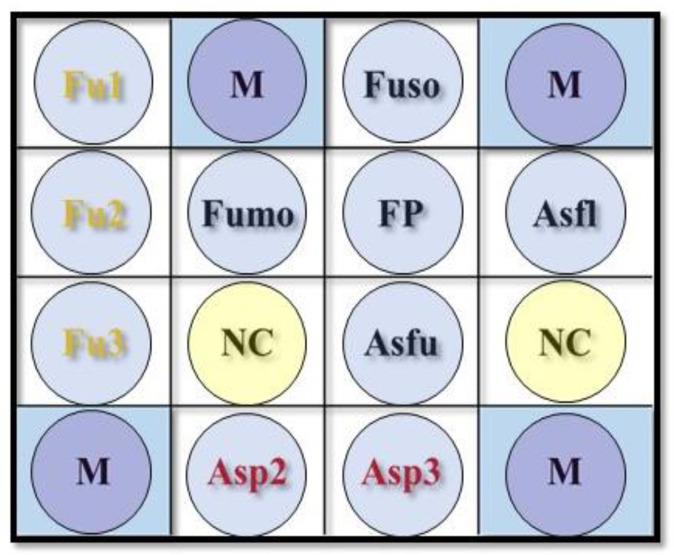
F. verticillioides keratitis led to failure to estimate sensitivity and PPR, and therefore, we could only conclude that probe Fumo had reasonable specificity and NPR (Table 5). Thus, the DHA’s performance for diagnosing F. verticillioides keratitis could not be sufficiently verified. However, the DHA’s performance was well-validated for diagnosing Fusarium keratitis and F. solani with high accuracies of 93.6% and 96.4%, respectively. Among the three false-negative scrapes via probes Fu1, Fu2, and Fu3 for detecting Fusarium spp., two samples with F. solani and one sample with Fusarium spp. were confirmed by DNA sequencing. Amplicons distributed to the universal probe FP and the species probe Fuso could be the false-negative reason. Among the six false-positive scrapes via probe Fuso for detecting F. solani, four samples were recognized as Colletotrichum spp. (two C. siamense, one C. fructicola, and one C. gloeosporioides), one sample was identified as Scedosporium apiospermum, and one sample was confirmed as Gjaerumia spp. One sample was a false-positive detection for F. verticillioides, where the DNA sequencing result was F. solani. Modifying the species’ probes Fuso and Fumo for specific detection of F. solani and F. verticillioides, or designing new probes for Colletotrichum, Scedosporium, and Gjaerumia will be considered.
There were only six scrapes with Aspergillus spp., including two A. flavus keratitis, two A. fumigatus keratitis, one A. niger, and one A. tamarii. Therefore, it was weak to estimate sensitivity and PPR for the probes for diagnosing Aspergillus genera, A. flavus, and A. fumigatus (Table 6). Only reasonable specificity and NPR could be claimed for these probes. For the sample with false-negative detection for Aspergillus spp. (by probes Asp2 and Asp3), the result of DNA sequencing was A. tamarii. The design of a new probe for detecting A. tamarii or the modification of current genus probes will be the solution for avoiding misdiagnosis for the species.
Due to the limitation in which some target pathogens were rare, the sensitivity and PPR of the probes for identifying Aspergillus keratitis and the species probes for recognizing F. verticillioides, A. flavus, and A. fumigatus could not be confidently validated. However, these probes undoubtedly had acceptable specificity and NPR. Moreover, they were speculated to have a similar performance to the probes for diagnosing Fusarium keratitis and F. solani keratitis because all probes had passed the preclinical challenge with target and non-target microorganisms (Table 1).
According to the comprehensive review of Hoffman et al. [11], filamentous FK, particularly Fusarium and Aspergillus keratitis, is a global treat. FK has an apparent geographical variation, and Fusarium or Aspergillus keratitis often accounted for the top two prevalent pathogens, even in North America. In some temperate areas such as Europe and North America, Candida spp. has a chance to be in the top two most common pathogens of FK. The severity of Candida keratitis is less than that of filamentous FK. Thus, the DHA can also be used in non-Asian countries to detect Candida spp. via probe FP and exclude vision-threatening Fusarium and Aspergillus keratitis via species and genus probes. The DHA can be used as an adjunctive clinical test of conventional culture for a mycology laboratory to increase the recovery rate and efficiency in diagnosing FK and differentiating Fusarium from Aspergillus keratitis for early precise anti-fungal treatment. However, the procedures need well-trained staff and sterile technique in the laboratory.
There are several novel antifungal regimens for FK [29,30,31]. Keratosept ophthalmic solution containing hexamidine diisethionate 0.05% is a potential candidate for the treatment of Candida and staphylococcal infections of the ocular surface [29]. Corneal collagen cross-linking treatment via Riboflavin/UVA (CXL) showed therapeutic efficacy against FK [30]. CXL combined with 0.02% chlorhexidine is also an effective therapy against FK, particularly for multi-resistant Fusarium keratitis [31]. Following the development of novel anti-fungal treatments, we believe the DHA, or a modified DHA targeting specific fungal species or genus, can provide a rapid and precise diagnosis helping physicians to choose a suitable anti-fungal treatment.

5. Conclusions

The novel DHA had an excellent diagnostic performance in diagnosing FK, differentiating Fusarium and Aspergillus keratitis, and identifying F. solani keratitis. Although, its performance in diagnosing Aspergillus spp., A. flavus, A. fumigatus, and F. verticillioides was not well-verified in this study due to limited cases, this DHA revealed a promising result toward rapid diagnostic precision medicine. We believe this DHA will improve the prognosis of FK via early diagnosis and more precise guidance of anti-fungal regimens.

Author Contributions

Conceptualization, M.-T.K. and J.-L.C.; methodology, M.-T.K., Y.-H.L. and H.-L.Y.; validation, P.-C.F. and H.-J.Y.; formal analysis, M.-T.K. and S.-L.H.; investigation, M.-T.K., J.-L.C., S.-L.H., P.-C.F., H.-J.Y., C.-Y.T. and Y.-H.L.; resources, M.-T.K., J.-L.C., S.-L.H., P.-C.F., H.-J.Y. and S.-F.K.; data curation, C.-Y.T. and Y.-H.L.; writing—original draft preparation, M.-T.K.; writing—review and editing, J.-L.C. and A.C.; visualization, M.-T.K. and A.C.; supervision, M.-T.K., J.-L.C. and H.-L.Y.; project administration, M.-T.K.; funding acquisition, M.-T.K. All authors have read and agreed to the published version of the manuscript.

Funding

This research was funded by the Ministry of Science and Technology, grant numbers MOST 107–2314-B-182A-091-MY2 and MOST 109-2314-B-182A-018-MY3.

Institutional Review Board Statement

The study was conducted according to the guidelines of the Declaration of Helsinki and approved by the Committee of Medical Ethics and Human Experiments of Chang Gung Memorial Hospital, the institutional review boards of Kaohsiung Veterans General Hospital and Kaohsiung Medical University Hospital (protocol code no. 201702184A3 and date of approval 14 February 2018).

Informed Consent Statement

Informed consent was obtained from all subjects involved in the study. Written informed consent has been obtained from all patients to publish this paper.

Data Availability Statement

Data is fully available upon reasonable request to corresponding author.

Acknowledgments

We thank Yu-Ting Huang for her assistance with connecting investigators from different medical centers and laboratories.

Conflicts of Interest

The authors declare no conflict of interest. The funders had no role in the design of the study; in the collection, analyses, or interpretation of data; in the writing of the manuscript, or in the decision to publish the results.

References

  1. Xie, L.; Zhong, W.; Shi, W.; Sun, S. Spectrum of Fungal Keratitis in North China. Ophthalmology 2006, 113, 1943–1948. [Google Scholar] [CrossRef]
  2. Khor, W.B.; Prajna, V.N.; Garg, P.; Mehta, J.S.; Xie, L.; Liu, Z.; Padilla, M.D.B.; Joo, C.K.; Inoue, Y.; Goseyarakwong, P.; et al. The Asia Cornea Society Infectious Keratitis Study: A Prospective Multicenter Study of Infectious Keratitis in Asia. Am. J. Ophthalmol. 2018, 195, 161–170. [Google Scholar] [CrossRef]
  3. Thomas, P.A.; Leck, A.K.; Myatt, M. Characteristic clinical features as an aid to the diagnosis of suppurative keratitis caused by filamentous fungi. Br. J. Ophthalmol. 2005, 89, 1554–1558. [Google Scholar] [CrossRef] [PubMed]
  4. Dahlgren, M.A.; Lingappan, A.; Wilhelmus, K.R. The clinical diagnosis of microbial keratitis. Am. J. Ophthalmol. 2007, 143, 940–944. [Google Scholar] [CrossRef] [PubMed] [Green Version]
  5. Dalmon, C.; Porco, T.C.; Lietman, T.M.; Prajna, N.V.; Prajna, L.; Das, M.R.; Kumar, J.A.; Mascarenhas, J.; Margolis, T.P.; Whitcher, J.P.; et al. The Clinical Differentiation of Bacterial and Fungal Keratitis: A Photographic SurveyDifferentiation of Bacterial and Fungal Keratitis. Investig. Ophthalmol. Vis. Sci. 2012, 53, 1787–1791. [Google Scholar] [CrossRef]
  6. Kuo, M.T.; Hsu, B.W.Y.; Yin, Y.K.; Fang, P.C.; Lai, H.Y.; Chen, A.; Yu, M.-S.; Tseng, V.S. A deep learning approach in diagnosing fungal keratitis based on corneal photographs. Sci. Rep. 2020, 10, 14424. [Google Scholar] [CrossRef]
  7. Mundra, J.; Dhakal, R.; Mohamed, A.; Jha, G.; Joseph, J.; Chaurasia, S.; Murthy, S. Outcomes of therapeutic penetrating keratoplasty in 198 eyes with fungal keratitis. Indian J. Ophthalmol. 2019, 67, 1599–1605. [Google Scholar] [CrossRef] [PubMed]
  8. Ho, J.W.; Fernandez, M.M.; Rebong, R.A.; Carlson, A.N.; Kim, T.; Afshari, N.A. Microbiological profiles of fungal keratitis: A 10-year study at a tertiary referral center. J. Ophthalmic Inflamm. Infect. 2016, 6, 5. [Google Scholar] [CrossRef] [Green Version]
  9. Cunha, A.M.; Loja, J.T.; Torrão, L.; Moreira, R.; Pinheiro, D.; Falcão-Reis, F.; Pinheiro-Costa, J. A 10-Year Retrospective Clinical Analysis of Fungal Keratitis in a Portuguese Tertiary Centre. Clin. Ophthalmol. 2020, 14, 3833–3839. [Google Scholar] [CrossRef] [PubMed]
  10. Tew, T.B.; Chu, H.S.; Hou, Y.C.; Chen, W.L.; Wang, I.J.; Hu, F.R. Therapeutic penetrating keratoplasty for microbial keratitis in Taiwan from 2001 to 2014. J. Formos. Med. Assoc. 2020, 119, 1061–1069. [Google Scholar] [CrossRef]
  11. Hoffman, J.J.; Burton, M.J.; Leck, A. Mycotic Keratitis—A Global Threat from the Filamentous Fungi. J. Fungi 2021, 7, 273. [Google Scholar] [CrossRef]
  12. Manikandan, P.; Abdel-Hadi, A.; Randhir Babu Singh, Y.; Revathi, R.; Anita, R.; Banawas, S.; Bin Dukhyil, A.A.; Alshehri, B.; Shobana, C.S.; Panneer Selvam, K.; et al. Fungal Keratitis: Epidemiology, Rapid Detection, and Antifungal Susceptibilities of Fusarium and Aspergillus Isolates from Corneal Scrapings. BioMed Res. Int. 2019, 2019, 6395840. [Google Scholar] [CrossRef] [Green Version]
  13. Prajna, N.V.; Lalitha, P.; Rajaraman, R.; Krishnan, T.; Raghavan, A.; Srinivasan, M.; O’Brien, K.S.; Zegans, M.; McLeod, S.D.; Acharya, N.R.; et al. Changing Azole Resistance: A Secondary Analysis of the MUTT I Randomized Clinical Trial. JAMA Ophthalmol. 2016, 134, 693–696. [Google Scholar] [CrossRef] [PubMed] [Green Version]
  14. Lalitha, P.; Sun, C.Q.; Prajna, N.V.; Karpagam, R.; Geetha, M.; O’Brien, K.S.; Cevallos, V.; McLeod, S.D.; Acharya, N.R.; Lietman, T.M. In vitro susceptibility of filamentous fungal isolates from a corneal ulcer clinical trial. Am. J. Ophthalmol. 2014, 157, 318–326. [Google Scholar] [CrossRef] [PubMed] [Green Version]
  15. Prajna, N.V.; Krishnan, T.; Rajaraman, R.; Patel, S.; Shah, R.; Srinivasan, M.; Devi, L.; Das, M.; Ray, K.J.; O’Brien, K.S.; et al. Adjunctive Oral Voriconazole Treatment of Fusarium Keratitis: A Secondary Analysis From the Mycotic Ulcer Treatment Trial II. JAMA Ophthalmol. 2017, 135, 520–525. [Google Scholar] [CrossRef] [Green Version]
  16. Oechsler, R.A.; Feilmeier, M.R.; Miller, D.; Shi, W.; Hofling-Lima, A.L.; Alfonso, E.C. Fusarium keratitis: Genotyping, in vitro susceptibility and clinical outcomes. Cornea 2013, 32, 667–673. [Google Scholar] [CrossRef] [Green Version]
  17. Kuo, M.T.; Chang, H.C.; Cheng, C.K.; Chien, C.C.; Fang, P.C.; Chang, T.C. A highly sensitive method for molecular diagnosis of fungal keratitis: A dot hybridization assay. Ophthalmology 2012, 119, 2434–2442. [Google Scholar] [CrossRef] [PubMed]
  18. Hsiao, C.R.; Huang, L.; Bouchara, J.-P.; Barton, R.; Li, H.C.; Chang, T.C. Identification of medically important molds by an oligonucleotide array. J. Clin. Microbiol. 2005, 43, 3760–3768. [Google Scholar] [CrossRef] [Green Version]
  19. Leaw, S.N.; Chang, H.C.; Barton, R.; Bouchara, J.-P.; Chang, T.C. Identification of medically important Candida and non-Candida yeast species by an oligonucleotide array. J. Clin. Microbiol. 2007, 45, 2220–2229. [Google Scholar] [CrossRef] [Green Version]
  20. Fang, P.C.; Chien, C.C.; Yu, H.J.; Ho, R.W.; Tseng, S.L.; Lai, Y.H.; Kuo, M.T. A dot hybridization assay for the diagnosis of bacterial keratitis. Mol. Vis. 2017, 23, 306–317. [Google Scholar]
  21. Kuo, M.T.; Fang, P.C.; Yu, H.J.; Chao, T.L.; Chien, C.C.; Chen, S.H.; Wang, J.R.; Tseng, S.L.; Lai, Y.H.; Hsiao, C.C.; et al. A Multiplex Dot Hybridization Assay For Detection and Differentiation of Acanthamoeba and Herpes Keratitis. Investig. Ophthalmol. Vis. Sci. 2016, 57, 2158–2163. [Google Scholar] [CrossRef] [PubMed] [Green Version]
  22. Huang, F.C.; Hsieh, H.Y.; Chang, T.C.; Su, S.L.; Tseng, S.L.; Lai, Y.H.; Kuo, M.T. A DNA dot hybridization model for molecular diagnosis of parasitic keratitis. Mol. Vis. 2017, 23, 614–623. [Google Scholar] [PubMed]
  23. Bouchara, J.-P.; Hsieh, H.Y.; Croquefer, S.; Barton, R.; Marchais, V.; Pihet, M.; Chang, T.C. Development of an Oligonucleotide Array for Direct Detection of Fungi in Sputum Samples from Patients with Cystic Fibrosis. J. Clin. Microbiol. 2009, 47, 142–152. [Google Scholar] [CrossRef] [Green Version]
  24. Lalitha, P.; Shapiro, B.L.; Srinivasan, M.; Prajna, N.V.; Acharya, N.R.; Fothergill, A.W.; Ruiz, J.; Chidambaram, J.D.; Maxey, K.J.; Hong, K.C.; et al. Antimicrobial susceptibility of Fusarium, Aspergillus, and other filamentous fungi isolated from keratitis. Arch. Ophthalmol. 2007, 125, 789–793. [Google Scholar] [CrossRef] [PubMed]
  25. Sun, C.Q.; Lalitha, P.; Prajna, N.V.; Karpagam, R.; Geetha, M.; O’Brien, K.S.; Oldenburg, C.E.; Ray, K.J.; McLeod, S.D.; Acharya, N.R.; et al. Association between in vitro susceptibility to natamycin and voriconazole and clinical outcomes in fungal keratitis. Ophthalmology 2014, 121, 1495–1500.e1. [Google Scholar] [CrossRef] [Green Version]
  26. Prajna, N.V.; Krishnan, T.; Mascarenhas, J.; Rajaraman, R.; Prajna, L.; Srinivasan, M.; Raghavan, A.; Oldenburg, C.E.; Ray, K.J.; Zegans, M.E.; et al. The mycotic ulcer treatment trial: A randomized trial comparing natamycin vs voriconazole. JAMA Ophthalmol. 2013, 131, 422–429. [Google Scholar] [CrossRef]
  27. Lalitha, P.; Prajna, N.V.; Kabra, A.; Mahadevan, K.; Srinivasan, M. Risk factors for treatment outcome in fungal keratitis. Ophthalmology 2006, 113, 526–530. [Google Scholar] [CrossRef]
  28. Kuo, M.T.; Chien, C.C.; Lo, J.; Hsiao, C.C.; Tseng, S.L.; Lai, Y.H.; Fang, P.C.; Chang, T.C. A DNA dot hybridization model for assessment of bacterial bioburden in orthokeratology lens storage cases. Investig. Ophthalmol. Vis. Sci. 2015, 56, 445–450. [Google Scholar] [CrossRef] [Green Version]
  29. Pinna, A.; Donadu, M.G.; Usai, D.; Dore, S.; Boscia, F.; Zanetti, S. In Vitro Antimicrobial Activity of a New Ophthalmic Solution Containing Hexamidine Diisethionate 0.05% (Keratosept). Cornea 2020, 39, 1415–1418. [Google Scholar] [CrossRef]
  30. Zhu, Z.; Zhang, H.; Yue, J.; Liu, S.; Li, Z.; Wang, L. Antimicrobial efficacy of corneal cross-linking in vitro and in vivo for Fusarium solani: A potential new treatment for fungal keratitis. BMC Ophthalmol. 2018, 18, 65. [Google Scholar] [CrossRef]
  31. Kunt, Z.; Yağmur, M.; Kandemir, H.; Harbiyeli, I.; Erdem, E.; Kalkancı, A.; De Hoog, G.S.; Ilkit, M. In Vitro Efficacy of Chlorhexidine and a riboflavin/UVA Combination on Fungal Agents of Keratitis. Curr. Eye Res. 2020, 45, 7–11. [Google Scholar] [CrossRef] [PubMed]
Figure 1. The layout of a novel dot hybridization array for detecting fungal keratitis. The universal fungal probe FP in the layout (0.6 × 0.6 cm) was designed for detecting all fungal species (Table 2). The genus probes “Fu1”, “Fu2”, and “Fu3” were designed to detect all Fusarium sp. The probes “Fuso” and “Fumo” were used to identify F. solani and F. verticillioides, respectively. The genus probes “Asp2” and “Asp3” were designed to detect all Aspergillus sp. The probes “Asfl” and “Asfu” were used to identify A. fumigatus and A. flavus, respectively. The dot “NC” is a negative control (tracking dye only). The probe “M” is a position marker, i.e., a digoxigenin-labeled oligonucleotide probe (digoxigenin-GCATATCAATAAGCGGAGGA).
Jof 08 00064 g001
Figure 2. Representative results of the dot hybridization array for detecting fungal keratitis. (ac) Fusarium keratitis patients with respective pathogens of F. solani, F. verticillioides, F. delphinoides; (df) Aspergillus keratitis patients with respective pathogens of A. flavus, A. fumigatus, and A. niger; (g,h) Fungal keratitis patients with respective pathogens of Curvularia geniculata and Candida tropicalis; (i) Pseudomonas aeruginosa keratitis; (j) herpes simplex keratitis; (k) Acanthamoeba palestinensis keratitis; (l) microsporidal (Vittaforma corneae) keratitis.
Jof 08 00064 g002
Table 1. Reference strains and clinical isolates for preclinical test of the fungal dot hybridization assay.
SpeciesReference Strain (s) aNo. of Clinical IsolatesTotal No. of Strains
Target fungal species for sensitivity test for species and genus probes
Fusarium solaniATCC 36031, CBS 109028, BCRC 3244869
Fusarium verticillioidesBCRC 31492, BCRC 31745, BCRC 35113, BCRC 3287804
Fusarium oxysporumATCC 26225, CBS 798.9502
Other Fusarium spp.BCRC3355445
Aspergillus flavusBCRC 30006, BCRC 30007, BCRC 30008, BCRC 30009, BCRC 3018727
Aspergillus fumigatusBCRC 30099, BCRC 30502, BCRC 32120, BCRC 32149, BCRC 3283616
Aspergillus nigerBCRC 30201, BCRC 30204, BCRC 3113003
Aspergillus nidulansATCC 11267, ATCC 13833, BCRC 3010003
Aspergillus terreusBCRC 30135, BCRC 31128, BCRC 3206803
Aspergillus clavatusBCRC 31116, BCRC 31486, BCRC 3173603
Aspergillus versicolorBCRC 30225, BCRC 31123, BCRC 3148803
Non-target fungal species for specificity test for species and genus probes
Curvularia spp.CBS 351.65, BCRC 30899, CBS 102694, CBS 149.71, CBS 148.6327
Candida albicansBCRC 20511, BCRC 20512, BCRC 2051303
Candida kruseiBCRC 20514, BCRC 21321, BCRC 2172003
Candida glabrataBCRC 20586, CBS 860, CBS 86103
Candida parapsilosisBCRC 20515, BCRC 21253, BCRC 2154403
Candida tropicalisBCRC 20520, BCRC 21436, BCRC 2156003
Candida guilliermondiiBCRC 20862, BCRC 21549, BCRC 2150003
Candida rugosaBCRC 21356, BCRC 2170902
Acremonium spp.BCRC 33315, BCRC 3223913
Bipolaris spp.CBS 274.5212
Pseudallescheria boydiiATCC 44329, ATCC 44331, ATCC 4433203
Cryptococcus neoformansBCRC 20528, BCRC 20532, BCRC 2287305
Non-target species from non-fungal pathogens for specificity test for all probes
Staphylococcus aureusBCRC 10451, BCRC 1528702
Staphylococcus epidermidisBCRC 10785, BCRC 1524502
Streptococcus pneumoniaeBCRC 14733, BCRC 1079402
Acinetobacter baumanniiBCRC 10591, BCRC 1588403
Moraxella catarhalisBCRC 10629, BCRC 1062802
Klebsiella pneumoniaeBCRC 11644, CCUG 1593804
Escherichia coliBCRC 15481, BCRC 1548404
Pseudomonas aeruginosaBCRC 10944, ATCC 2785368
Serratia marcescensBCRC 15326, BCRC 1157605
Burkholderia cepaciaBCRC 13208, BCRC 1390602
Stenotrophomonas maltophiliaBCRC 1073703
Mycobacterium chelonaeATCC 35749, CCUG 3782702
Mycobacterium fortuitumBCRC 15320, JCM 638702
Mycobacterium abscessusNCTC 1026901
Herpes simplex virus type 1 22
Herpes simplex virus type 2 22
Varicella zoster virusRod strain34
Encephalitozoon cuniculiATCC 5078901
Encephalitozoon hellemATCC 5050401
Encephalitozoon intestinalisATCC 5065101
Vittaforma corneae 11
Acanthamoeba castellaniiATCC 30010, ATCC 50374, ATCC 5037003
Acanthamoeba griffiniATCC 30731, ATCC 5070202
a ATCC, American Type Culture Collection, Manassas, Va., USA; CBS, Centraalbureau voor Schimmelcultures, Utrech, The Netherlands; BCRC: Bioresources Collection and Research Center, Hsinchu, Taiwan; CCUG, Culture Collection, University of Göteborg, Sweden; JCM: Japan Collection of Microorganisms, RIKEN BioResource Research Center, Ibaraki, Japan; NCTC: National Collection of Type Cultures, Central Public Laboratory Service, London, UK.
Table 2. Probes used in the dot hybridization array.
Target MicroorganismProbe Code aSequence (5′ to 3′)Length (bp)Tm b (°C)LocationGenBank Accession No.
All fungiFP [17]GCATCGATGAAGAACGCAGCttttttttt c2057.2228–247FR727118
Fusarium solaniFuso [18]AGTAGCTAACACCTCGCGACTGGAGA2656.0446–471AF129105
Fusarium verticillioidesFumo [18]CGAGTCAAATCGCGTTCCCCAAATTG2654.4395–420AY533376
Aspergillus flavusAsfl [18]CGAACGCAAATCAATCTTTTTCCAGGT2751.6512–538AY373848
Aspergillus fumigatusAsfu [18]GCCAGCCGACACCCAACTTTATTTTTCTAA3055.2213–242AY230140
Fusarium sp.Fu1 dGCGTCATTTCAACCCTCAAGCCCC2463.7340–363AM412639
Fu2 dCTTCTGAGTAAAACAAGCAAATAAAT2648.9164–189AM412639
Fu3 dAGCTTCCATAGCGTAGTAGYAA2253.8442–463AM412639
Aspergillus sp.Asp2 dGGACGGGCCCRAAAGGCAGCGGCGGC2677.8426–451AF138290
Asp3 dGGCAGCGGCGGCACCGYGTCCGGTCCT2779.7440–466AF138290
a Oligonucleotide probes are arranged on the dot hybridization array, as indicated in Figure 1. b Tm = melting temperature. c Multiple bases of thymine (t) were added to the 3′ end of the probe. d Newly designed probes used in this study.
Table 3. Demographic data of subjects.
Clinical ParametersValue
Number of patients146
Age (years; mean ± s.d.)59.3 ± 15.8
Sex (women/men; no./no.)52/94
Disease eye (OD/OS; no./no.)72/74
Final diagnosis (no.)
Fungal keratitis107
Bacterial keratitis16
Herpes keratitis2
Acanthamoebic keratitis3
Microsporidial stromal keratitis3
Noninfectious keratitis15
Major risk factors (no.)
Trauma65
Contact lens wear14
Dirty water exposure7
Ocular surface disease3
Neurotrophic keratopathy1
Lagophthalmos3
Facial palsy1
Hyperthyroidism1
Diabetes mellitus11
Chemotherapy1
Undetermined39
Table 4. The performance of the dot hybridization array for diagnosing fungal keratitis.
N = 146CultureCulture or DNA SequencingSensitivitySpecificityPPRNPR
PositiveNegativePositiveNegative(C.I.; %)(C.I.; %)(C.I.; %)(C.I.; %)
DHAPositive7427100193.597.499.084.4
Negative738738(87.1–96.8)(86.8–99.9)(94.6–100.0)(71.2–92.3)
DHA = dot hybridization array; PPR = positive predictive rate; NPR = negative predictive rate; C.I. = confidence interval.
Table 5. Diagnostic performance of Fusarium keratitis after discrepant analysis.
N = 140 aPost-DiscrepancySensitivitySpecificityPPRNPR
PositiveNegative(C.I.; %)(C.I.; %)(C.I.; %)(C.I.; %)
DHAFusarium sp.Positive41693.293.887.296.8
Negative390(81.8–97.7)(87.0–97.1)(74.8–94.0)(90.9–99.1)
F. solaniPositive26083.9100.0100.095.6
Negative5109(67.4–92.9)(96.6–100.0)(87.1–100.0)(90.1–98.1)
F. verticillioidesPositive11100.099.350.0100.0
Negative0138(5.1–100.0)(96.0–100.0)(2.6–97.4)(97.3–100.0)
a Diagnostic performance was estimated after excluding six cases with suspected sampling failure (positive culture but negative DNA sequencing results). DHA = dot hybridization array; PPR = positive predictive rate; NPR = negative predictive rate; C.I. = confidence interval.
Table 6. Diagnostic performance of Aspergillus keratitis after discrepant analysis.
N = 140 aPost-DiscrepancySensitivitySpecificityPPRNPR
PositiveNegative(C.I.; %)(C.I.; %)(C.I.; %)(C.I.; %)
DHAAspergillus sp.Positive5083.3100.0100.099.3
Negative1134(43.7–99.2)(97.2–100.0)(56.6–100.0)(95.9–100.0)
A. flavusPositive20100.0100.0100.0100.0
Negative0138(17.8–100.0)(97.3–100.0)(17.8–100.0)(97.3–100.0)
A. fumigatusPositive20100.0100.0100.0100.0
Negative0138(17.8–100.0)(97.3–100.0)(17.8–100.0)(97.3–100.0)
a Diagnostic performance was estimated after excluding six cases with suspected sampling failure (positive culture but negative DNA sequencing results). DHA = dot hybridization array; PPR = positive predictive rate; NPR = negative predictive rate; C.I. = confidence interval.
Publisher’s Note: MDPI stays neutral with regard to jurisdictional claims in published maps and institutional affiliations.

