Review

Enhancing Molecular Testing for Effective Delivery of Actionable Gene Diagnostics

2nd Department of Paediatrics, Semmelweis University, Üllői út 26, 1085 Budapest, Hungary
*
Author to whom correspondence should be addressed.
Bioengineering 2022, 9(12), 745; https://doi.org/10.3390/bioengineering9120745
Submission received: 12 September 2022 / Revised: 7 November 2022 / Accepted: 29 November 2022 / Published: 1 December 2022
(This article belongs to the Special Issue Novel Expansions and Trends in Gene Diagnostics and Gene Therapy)

Abstract

:
There is a deep need to navigate within our genomic data to find, understand and pave the way for disease-specific treatments, as the clinical diagnostic journey provides only limited guidance. The human genome is enclosed in every nucleated cell, and yet at the single-cell resolution many unanswered questions remain, as most of the sequencing techniques use a bulk approach. Therefore, heterogeneity, mosaicism and many complex structural variants remain partially uncovered. As a conceptual approach, nanopore-based sequencing holds the promise of being a single-molecule-based, long-read and high-resolution technique, with the ability of uncovering the nucleic acid sequence and methylation almost in real time. A key limiting factor of current clinical genetics is the deciphering of key disease-causing genomic sequences. As the technological revolution is expanding regarding genetic data, the interpretation of genotype–phenotype correlations should be made with fine caution, as more and more evidence points toward the presence of more than one pathogenic variant acting together as a result of intergenic interplay in the background of a certain phenotype observed in a patient. This is in conjunction with the observation that many inheritable disorders manifest in a phenotypic spectrum, even in an intra-familial way. In the present review, we summarized the relevant data on nanopore sequencing regarding clinical genomics as well as highlighted the importance and content of pre-test and post-test genetic counselling, yielding a complex approach to phenotype-driven molecular diagnosis. This should significantly lower the time-to-right diagnosis as well lower the time required to complete a currently incomplete genotype–phenotype axis, which will boost the chance of establishing a new actionable diagnosis followed by therapeutical approach.

Graphical Abstract

1. Introduction

A great proportion of genetic disorders manifest phenotypically by early adulthood, resulting in a cumulative incidence of observed rare diseases between 1.5–6.2% in the general population [1,2]. Due to the wide phenotypic heterogeneity and lack of robust molecular testing strategies, diagnosis is challenging and is often delayed by several years. According to the latest telomere-to-telomere human genome assembly, the size of the human genome is of the order of 3.055 Gbp [3]. Besides the relatively large size of the genome, dysfunctional methylation, histone modifications and RNA expression may also elicit a phenotypic burden; therefore, a systematic approach is needed to correctly address genotype–phenotype correlations [4]. Currently, diagnostically used cytogenetics and molecular biology techniques depict only certain types of human genetic alterations (e.g., due to DNA fragmentation during library preparation for next-generation sequencing (NGS), the detection of short tandem repeats, as trinucleotide repeat expansions are not feasible, and also the detection of structural variants is quite limited [5]. The resolution of G-band karyotyping is mostly limited to 3 Mbp, and the higher resolution providing an array-comparative genome hybridization (array-CGH) approach cannot detect low-level mosaicism, balanced translocations or uncover copy number variations where oligoprobes have not been designed in the array) [6]. A comprehensive meta-analysis has shown that the diagnostic utility of the array-CGH and NGS approaches (whole-exome sequencing, whole-genome sequencing) varies between 10–41%, but many cases it still remains unresolved [7]. Therefore, a new comprehensive cytogenomic approach is needed to improve the yield of molecular cytogenomic diagnostics. Long-read nanopore sequencing permits the detection of both nucleotide-level and structural-level variations and methylation pattern alterations, and it is the only method that allows direct RNA sequencing [8].
Here, we reviewed the role of nanopore sequencing within the current molecular testing strategies, its relevance to actionable genetic diagnosis and its impact on genetic counselling. The human genome has a highly complex structure and functions as the backbone of the cell by coordinating the fate of many cellular functions. If critical damage occurs at genomic level, critical cellular functions may occur, mostly in a cell-type-dependent manner, revealing itself in various phenotypic alterations: (1) Some inheritable disorders reveal themselves by the accumulation of a plethora of structural minor anomalies and organ dysfunctions, even at the prenatal, perinatal or early childhood period [9]. A clinical diagnosis and a targeted molecular testing approach should be performed. (2) Most inheritable disorders mask themselves by a subtle phenotype alteration by early childhood, showing unspecific signs and symptoms or age-dependent phenotype penetrance [10]. There is a high need for an effective and time-efficient diagnostic strategy for this group of patients. (3) In the third case, there are patients with underlying genetic disorders with age-dependent/incomplete phenotype penetrance, with mostly the first symptom being the sign of organ dysfunction/a detrimental clinical event. The diagnostic approach and journey for these patients also raises many unanswered ethical questions. The importance of phenotyping accompanied by a detailed genetic anamnesis and at least a four-generation extended pedigree should be utterly prioritized before targeted or high-throughput molecular testing, otherwise the clinical utility of these tests is limited [6,11,12].

2. Molecular Testing—Timing and Approach

The timing of molecular testing is of essential importance. The molecular testing of germ line cells is limited. In males, germline cells can be tested from a testis biopsy for (1) nucleotide-level variations in a targeted fashion with Sanger sequencing (SS)/NGS or a nanopore-based sequencing (ONT) approach, or (2) structural-level variations in a targeted fashion with fluorescence in situ hybridization (FISH) with ONT. For a whole-exome or whole-genome approach to detect nucleotide variations, NGS or ONT can be applied. To detect structural variations array-comparative genome hybridization (arrayCGH), ONT or optical genome mapping (OGM) can be applied [13]. For methylation-based analysis, a methylation-sensitive multiple-ligation-based assay (MS-MLPA), pyrosequencing or ONT can be applied. Therefore, ONT provides a comprehensive genome-testing method whilst preserving native nucleic acid modifications [14]. As for preconceptual testing, carrier screening, for most diseases, showing an autosomal recessive inheritance pattern and is available in many countries at a reasonable price. Preimplantation testing during in vitro fertilization and the application of preimplantation gene testing via the detection of pathogenic structural variants in embryos could be of key importance [15]. For targeted prenatal testing, ONT has been successfully applied for the testing of fetal DNA from maternal blood samples [16]. As for postnatal testing, for whole-genome sequencing and diagnosis, a world-record time of 7 h and 18 min has been achieved by the ONT approach [17].
High-throughput deep-sequencing enables the uncovering of variant frequencies and methylation CpG patterns in heterogenous samples. The recently developed nanopore Cas9-targeted sequencing (nCATS) method has proven to provide the aforementioned quality at a targeted level in a cost-effective way [14].
From a methodological approach, the following two major points need to be addressed to deliver an effective molecular testing strategy: (1) what type of molecular alteration should be detected, and (2) what are the functional consequences of the detected variants? At the DNA level, molecular alterations could be categorized by nucleotide variants and structural variants. Nucleotide variants are usually DNA sequence variations ranging in size between 1–100 bp that may be of critical etiological value in several inheritable disorders and somatic pathogenic variants. Structural variants, ranging in size from several hundred base pairs up to few Mbp, may also elicit inheritable disorders and tumor predisposition or may be of a benign polymorphism value (Figure 1).

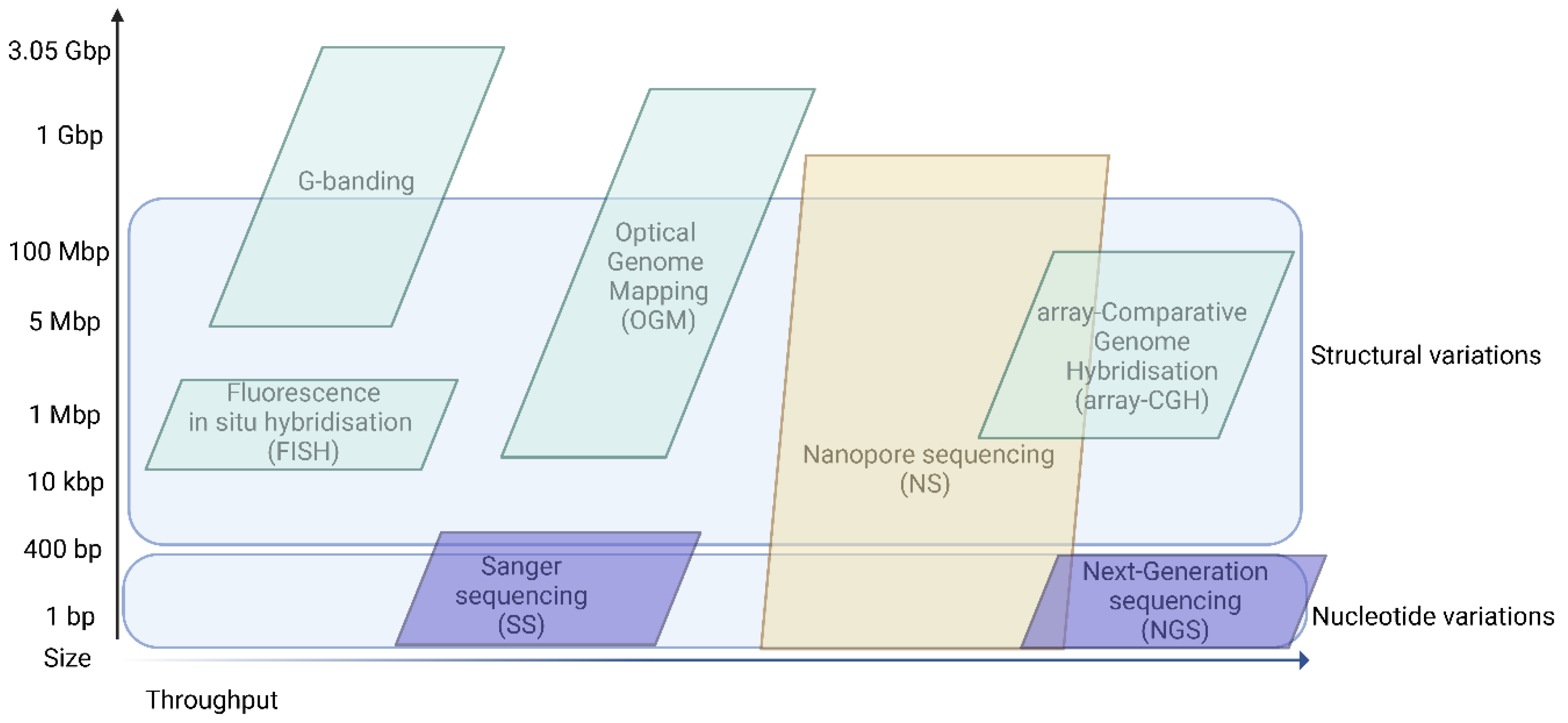
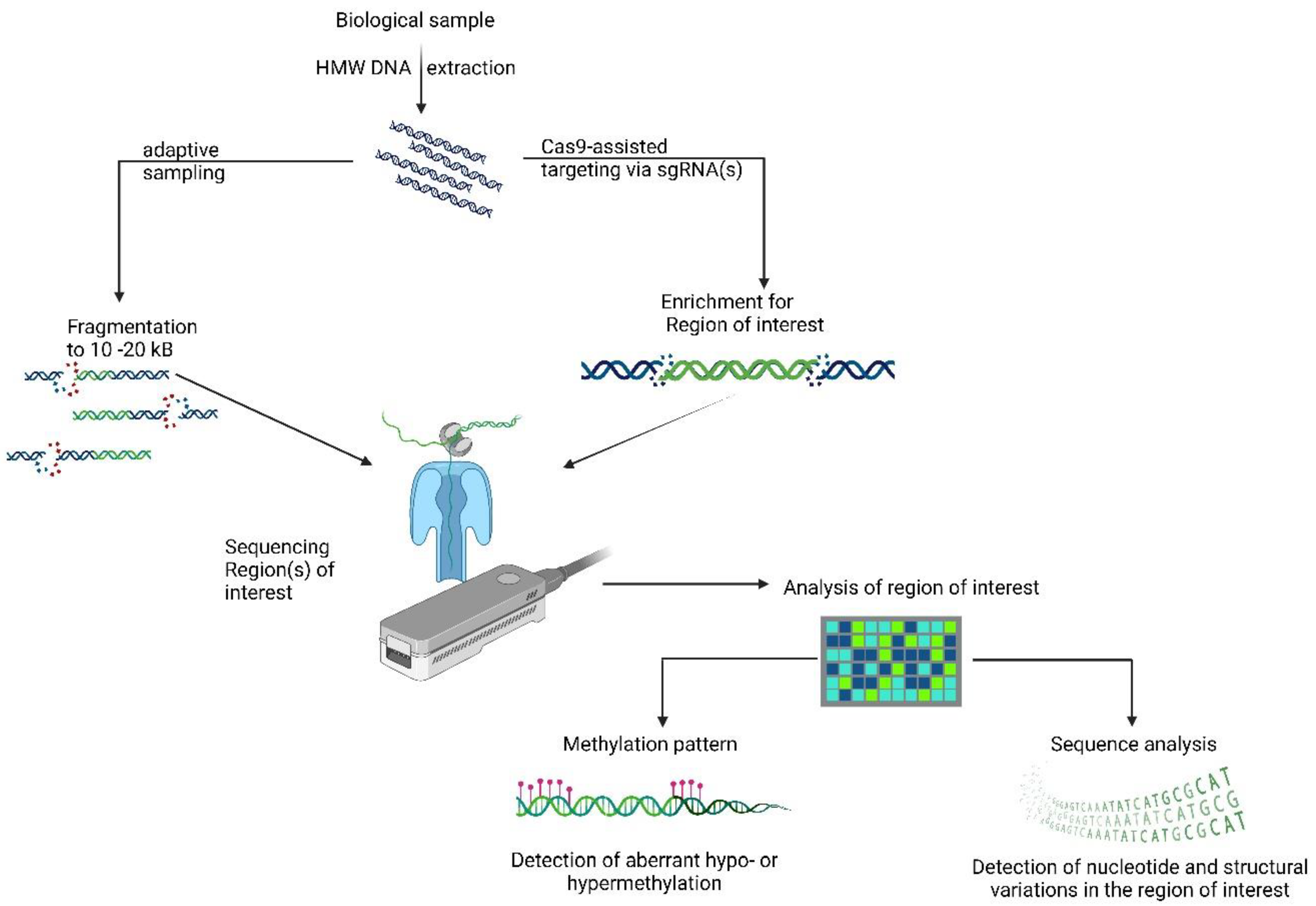
3. Nanopore Sequencing

Recent advances have led to the rapid and highly efficient deciphering of pathogenic variants using nanopore-based sequencing, allowing rapid clinical diagnosis [18]. ONT allows the uncovering of targeted nucleic acid sequences or whole-genome, epigenome (5-methylcytosine), transcriptome and epitranscriptome (N6-methyladenine) analysis [18]. Nanoscale-sized nanopores act as biosensors for detecting ionic current changes in real time during single-stranded DNA or RNA molecules (unwound by a motor protein possessing helicase activity) passing in a step-by-step manner [18]. A typical workflow starts with high-molecular-weight DNA extraction coupled with optional fragmentation or size selection (to remove overrepresented small DNA fragments). For library preparation, a relatively short DNA repair and adapter ligation strategy could be used, followed by a loading step on the nanopore-flow cells and real-time sequencing. By applying hybrid error correction tools, the long-read error rate is nowadays between 1-4% of that of short reads. Four main branches could use the advantages of ONT in clinical settings: (1) to identify the background of genetic diseases, (2) to molecularly diagnose cancer patients (e.g., acute leukaemias, solid tumors where certain molecular alterations may greatly influence the therapy of choice [19,20]), (3) rapid pathogen identification in an infectious disease scenario, and (4) to rapidly sequence the major histocompatibility of genes for recipient–donor tissues in transplantation medicine. The strength of nanopore sequencing relies in resolving long-range information, which is one of the main limitations of short-read sequencing technologies [21]. By coupling the unique molecular identifiers (UMI) used in single-cell transcriptomics and genomic long-read transcriptomes, transcriptomes may be sequenced at single-cell resolution with ONT [22]. In Figure 2 we present two methodological approaches for targeted nanopore-based sequencing.

4. Where to Fit Long-Read Sequencing in Clinical Genomics?

The relevant milestones that should shape the fitting of long-read sequencing are:
(1) The phenotype-driven molecular testing of both nucleotide variant (e.g., single nucleotide variants, SNV [23,24], short insertion–deletions, indels [25], short tandem repeats (STR) [26], trinucleotide-repeat expansions, etc.) and structural variant detection (e.g., deletions, duplications, cryptic microdeletions/duplications, both balanced and unbalanced translocations [27], gene fusions, complex rearrangements [28], marker chromosomes [29], etc.). Furthermore, methylation pattern detection allows the diagnosis of imprinting disorders as well as haplotyping/phasing [30,31,32] being available when relevant.
(2) Targeted molecular testing in a time-dependent manner for the establishment of an actionable diagnosis, e.g., gene therapy, a modified therapeutical approach and enzyme replacement therapy. Recently, a feasibility study designed to address targeted gene analysis from noninvasive prenatal testing (NIPT) samples using ONT has been described [16]. ONT provides a direct construction of haplotypes, through relative haplotype dosage analysis from maternal blood plasma, which is of importance in diseases such as ß-thalassaemia, spinal muscular atrophy, Duchenne’s and Becker’s muscular dystrophies and Hunter’s disease [16]. As is summarized in Table 1, the molecular diagnosis of actionable rare diseases and haematological malignancies is feasible in a rapid and reliable fashion with ONT. Identified pathogenic variants in the GLA (leading to Fabry’s disease), GBA (leading to Gaucher’s disease) or PAH (leading to phenolketonuria) genes involve a close follow-up, and the affected patients may be eligible for specific enzyme replacement therapy and/ or substrate-reduction therapy [8,33,34]. As for rare disease diagnostics, one of the most common causes of the intellectual disability, Fragile X syndrome, causing STR alterations in the FMR1 gene as well other genes of interest causing intellectual disability, skeletal or inborn heart disorders, has been successfully detected by the ONT platform. One of the key questions in tumor diagnostics is to identify the actionable gene alterations that may modify the therapeutic strategy in a rapid fashion as well to conclude whether the detected alteration is limited to the somatic tumor-associated tissue or is a germline alteration. The second part of Table 1 depicts such gene alterations that are actionable findings in tumoral settings.
(3) Multiomical and high-resolution-based testing for the depiction of heterogeneity and mosaicism. In several in vitro studies involving cell lines and primary cells, ONT has been successfully applied, providing further evidence of nanopore application for variant analysis (Supplementary Table S1).
The current limitations of ONT include the slightly different approach to DNA extraction and library preparation. The input and quality of the sample DNA may limit the sequencing throughput, and as for post-sequencing, the sequence and methylation pattern need different analysis pipelines. Also the calling of SNVs although has been significantly improved (varying between 1–4%) in recent years, it needs further enhancement to be below 1% of the error rate [5].
Actionable genetic diagnosis:
The definition of an actionable genetic diagnosis in conjunction with the ACMG’s newest guidelines can be defined as an available medical intervention for certain genetic disorders by reducing morbidity and mortality and enhancing the quality of life [47]. Possible medical interventions arise from a gene-therapeutic point, addressing pathogenic variants at the DNA level in a tissue-specific/tropic fashion. Next, acting at the RNA level, small interfering RNAs or anti-sense oligonucleotides may act at the cellular level to reduce the phenotypic burden. At the protein level, enzyme-replacement therapy is available for quite a few inborn error-of-metabolism disorders, and molecular chaperones enhancing enzymatic activity or substrate-reducing agents with disease-modifying effect are available for selected lysosomal storage disorders. Medical interventions of actionable genetic diagnoses are applied nowadays in the postnatal lifecycle; however, the implementation of disease-modifying therapies in the prenatal period [48] or as a new core concept a possible new era of in utero gene therapy may also arise [49] (Figure 3).

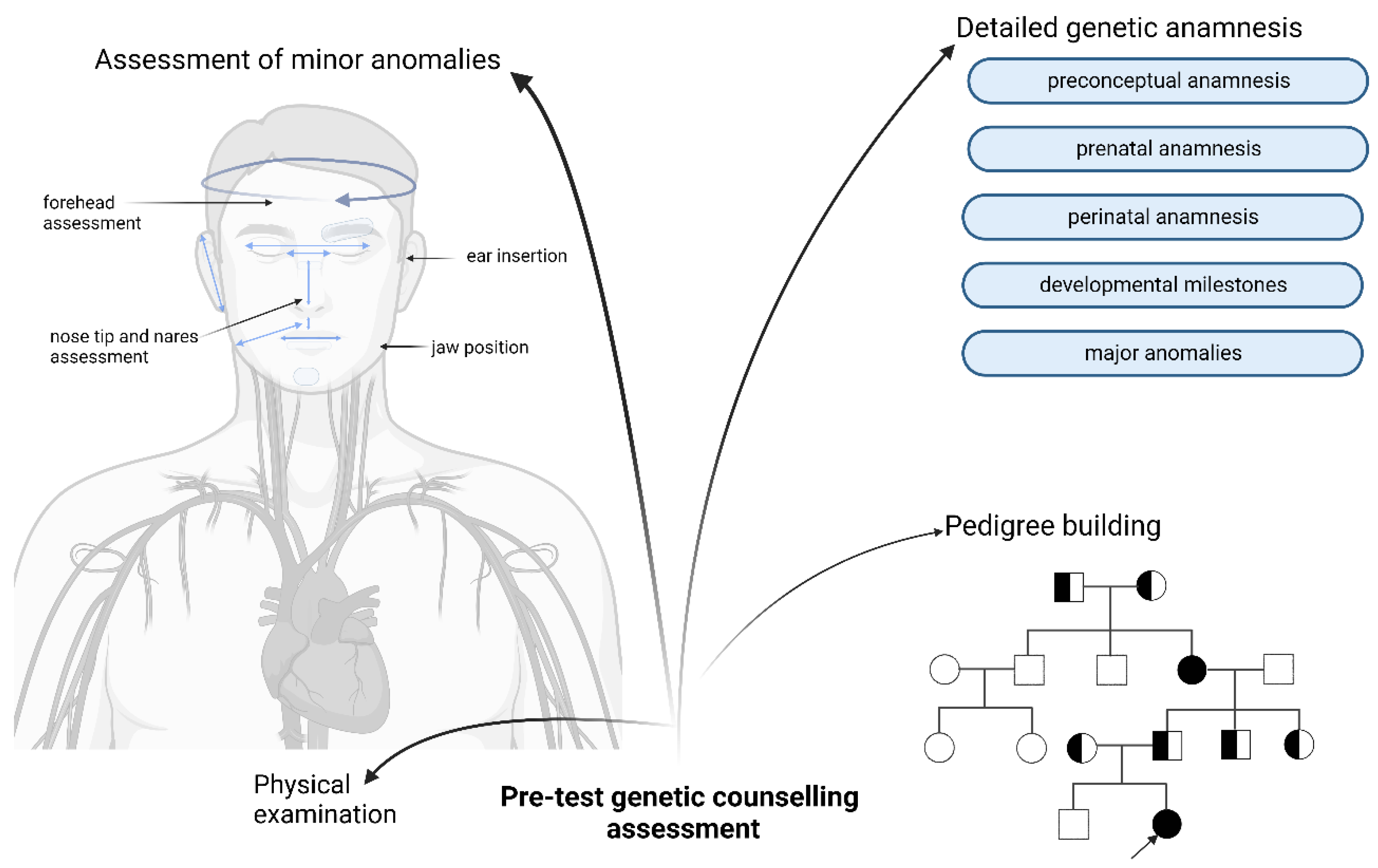
5. Genetic Counselling—State of the Art

Genetic counselling reflects the backbone of human genomics analysis by finding the answer to the following three fundamental questions: (1) Why is it recommended to opt for a genetic test? (2) What is our target nucleic acid sequence that should be analyzed by a suitable test available at that particular timepoint, highlighting the strengths and limitations of it? (3) How should we critically interpret and obtain insight about the data provided by the genetic analysis?
One of the most challenging tasks of genetic counselling is to maximize the clinical utility and, at the same time, minimize the uncertainty of information [50]. This could be enhanced by setting up multidisciplinary professional healthcare teams who can synthesize and collaborate to precisely define and follow up phenotypic spectra, which may maximize the uncovering of phenotype–genotype associations. Thus, the clinical pathway of patients can be significantly improved, often influencing the screening strategies and/or therapeutic approach.

6. Role of Pre-Test Genetic Counselling

To define the genotype–phenotype correlation as precisely as possible, a detailed phenotyping and pedigree building is essential, which can also be enhanced by artificial intelligence (AI). Phenotyping begins with a detailed genetic anamnesis. This should include a preconceptual anamnesis (age of the biological mother and biological father at conception, mode of conception and use of periconceptual vitamins) and a prenatal anamnesis (use of or contact with teratologic agents during the embryonal and/or fetal critical period. TORCH-screening, pathological ultrasound findings, the use of prenatal vitamins, drug use, and prenatal genetic tests such as NIPT, G-banding, chromosomal microarray or whole-exome sequencing could be conducted) [51]. The anamnesis of the perinatal period should cover the mode of delivery, possible hypoxic events, the age of gestation, the weight, height and head circumference at birth, the APGAR score, neonatal cardiorespiratory adaptation, the use of O2 therapy, newborn hearing screening, newborn feeding difficulties and breastfeeding. With a special emphasis on the advances of developmental milestones and the use of early childhood development therapies, language, social skills and learning ability development should be questioned. At the end of genetic anamnesis, the building of a four- or five-generation genetic pedigree is advised. For the digitalization of pedigrees, different software packages (e.g., Evagene Clinical—a free open-source software available at www.evagene.com (accessed on 6 September 2022) or GenoPro Waterloo, Ontario, Canada—a paid software option available at www.genopro.com (accessed on 6 September 2022)) can be used. Critical questions should be covered during the pedigree assessment such as (1) the spontaneous abortion history of the index patient’s mother and maternal grandmother; (2) any known perinatal death in the family; (3) the occurrence of sudden cardiac death in the family; (4) any malignant tumor development before the age of 45 in the family; (5) any recurring deep-vein thrombosis, any pulmonary emboly in the family; (6) any consanguineous marriage in the family; and (7) any infertility in the family.
Next, a genetic physical assessment should include the detailed phenotyping of minor anomalies. Detailed phenotyping is an important way to evaluate the impact of penetrance, the possibility of uncovering a second genetic alteration and of expanding the phenotypic variability of a molecular finding [52,53,54,55]. The use of prenatal data may also enhance the findings of a high-throughput molecular analysis [51]. If a targeted approach is used, the detailed phenotype will determine the test of choice [56]. Guidelines on the standard use of systematic phenotyping are available [57,58,59]. The use of artificial intelligence for the refinement of minor anomalies (e.g., Face2Gene—a free open-source online platform [60]) can also be useful. Systematic phenotyping includes the depiction of facial minor anomalies, which should include the size of the nasal bridge, a nasal tip and nares assessment, canthal folds, endocanthal lengths, interpupillary distance, exocanthal length, palpebral fissure slanting, eyelash length and density, eyebrow thickness and/or conjoined eyebrows, philtrum size, lower and upper lip thickness and slanting, intercommissural distance, enlarged interdental space, a high narrow palate, bifurcated uvula, tongue size and asymmetry, bilateral philtrum–mandibular angle distance, forehead size and protrusion, bitemporal distance, ear set height, ear asymmetry, rotation of the external ear, jaw position and mandibular size. Next, the shape and size of the skull as well as the hairline insertion and hair thickness anomalies should be assessed. In addition, skin alterations (e.g., café-au-lait spots, angiokeratomas, etc.) and hand and feet minor anomalies should be assessed in addition to a regular physical examination. A detailed depiction of minor anomalies and their annotation should be performed according to the human phenotype ontology (HPO) [61]. Next, the objectifying of minor anomalies by precise measurements during minor anomaly depiction should be calculated for age- and gender-specific percentile values. In addition, a detailed depiction of growth curve tendencies by a comparison of percentiles specific to age, gender and diagnosis (e.g., growth percentiles for girls between age 0–2 diagnosed with Down’s syndrome) should be conducted. Based on the evaluation of the detailed genetic anamnesis, physical examination and pedigree, the best-suited targeted or high-throughput analysis should be chosen. Detailed information about the usefulness, benefits and limitations of the proposed test should be provided. Then, informed consent should be signed, with special emphasis on the reporting of secondary findings according to the latest ACMG guidelines [47] (Figure 4).

7. Role of Post-Test Genetic Counselling

The role of post-test genetic counselling is a critical evaluation of the results of the applied genetic test in light of the phenotype. The evaluation of genotype alterations should be in line with the observed phenotype. It is recommendable, when possible, that the positive findings be confirmed by another technique. Of note, if variants of unknown significance have been identified or the methylation pattern has also been determined in addition to sequencing, it could be re-evaluated in the near future as more data on variants of unknown significance start to shed light on either their benign or pathogenic effect. As the genotype–phenotype correlation that has been established the genetic medical report should contain the precise genetic alteration both at the cytogenetic and molecular nomenclature (preferably according to the latest assembly, e.g., GRCh38), the accession number/link of the detected variant, the interpretation of the variant at the cellular level and the next detailed clinical significance should be provided. In the detailed clinical significance, the possible therapeutical approaches and follow-up strategy should be written and discussed with the patient and/or legal guardian by taking into account the national human genetic laws of a specific country.

8. Concluding Remarks

The precise uncovering of our genetic information of interest should be one of our highest priorities, which should be conducted by providing the most complete molecular landscape in order to establish next-generation genetic counselling and guidance. As technology advances, AI has become more prevalent in molecular medicine, and genetic data has become more prone to being sequenced at larger and larger scales. Timing is of the essence to molecular diagnosis, as the patient will have the highest opportunity to benefit from specific medical care and/or treatment the earlier the diagnosis is conducted. Precise and detailed phenotyping should be performed in order to conclude the correct genotype–phenotype correlation and medical care to be provided as soon as possible for every actionable genetic diagnosis.

Supplementary Materials

The following supporting information can be downloaded at: https://www.mdpi.com/article/10.3390/bioengineering9120745/s1, Table S1: Relevant data on human cell line samples processed by ONT. (References [62,63,64,65,66] are cited in the supplementary materials).

Author Contributions

Conceptualization, Á.F.K. and G.F.; writing—original draft preparation, Á.F.K. and Z.N.; writing—review and editing, Á.F.K. and T.A.; visualization, T.A.; supervision, G.T.K.; All authors have read and agreed to the published version of the manuscript.

Funding

Á.F.K. was supported by the Hungarian National Research, Development and Innovation Office—NKFIH, OTKA PD_21 138521 grant. G.T.K. was supported by Hungarian Academy of Sciences MTA PC-2022-9/2022.

Institutional Review Board Statement

Not applicable.

Informed Consent Statement

Not applicable.

Data Availability Statement

Not applicable.

Conflicts of Interest

The authors declare no conflict of interest.

Abbreviations

ACMGAmerican College of Medical Genetics and Genomics
AIartificial intelligence
arrayCGHarray comparative genome hybridization
CRISPRclustered regularly interspaced short palindromic repeats
FISHfluorescence in situ hybridization
HMW DNAhigh-molecular-weight DNA
HPOhuman phenome ontology
nCATSnanopore Cas9-targeted sequencing
MS-MLPAmethylation-sensitive multiple ligation-based assay
NGSnext-generation sequencing
NIPTnoninvasive prenatal testing
OGMoptical genome mapping
ONTnanopore-based sequencing
SNVsingle nucleotide variation
SSSanger sequencing
STRshort tandem repeats
TORCHtoxoplasmosis, others, rubella, cytomegalovirus and herpes simplex virus

References

  1. Ferreira, C.R. The burden of rare diseases. Am. J. Med. Genet. Part A 2019, 179, 885–892. [Google Scholar] [CrossRef]
  2. Nguengang Wakap, S.; Lambert, D.M.; Olry, A.; Rodwell, C.; Gueydan, C.; Lanneau, V.; Murphy, D.; Le Cam, Y.; Rath, A. Estimating cumulative point prevalence of rare diseases: Analysis of the Orphanet database. Eur. J. Hum. Genet. 2020, 28, 165–173. [Google Scholar] [CrossRef] [Green Version]
  3. Nurk, S.; Koren, S.; Rhie, A.; Rautiainen, M.; Bzikadze, A.V.; Mikheenko, A.; Vollger, M.R.; Altemose, N.; Uralsky, L.; Gershman, A.; et al. The complete sequence of a human genome. Science 2022, 376, 44–53. [Google Scholar] [CrossRef] [PubMed]
  4. Claussnitzer, M.; Cho, J.H.; Collins, R.; Cox, N.J.; Dermitzakis, E.T.; Hurles, M.E.; Kathiresan, S.; Kenny, E.E.; Lindgren, C.M.; MacArthur, D.G.; et al. A brief history of human disease genetics. Nature 2020, 577, 179–189. [Google Scholar] [CrossRef] [PubMed] [Green Version]
  5. Hu, T.; Chitnis, N.; Monos, D.; Dinh, A. Next-generation sequencing technologies: An overview. Hum. Immunol. 2021, 82, 801–811. [Google Scholar] [CrossRef]
  6. Cotton, C.M.; Murray, J.C. 17—The Human Genome and Neonatal Care. In Avery’s Diseases of the Newborn, 10th ed.; Gleason, C.A., Juul, S.E., Eds.; Elsevier: Amsterdam, The Netherlands, 2018; pp. 180–189.e2. Available online: https://www.sciencedirect.com/science/article/pii/B9780323401395000176 (accessed on 1 September 2022).
  7. Clark, M.M.; Stark, Z.; Farnaes, L.; Tan, T.Y.; White, S.M.; Dimmock, D.; Kingsmore, S.F. Meta-analysis of the diagnostic and clinical utility of genome and exome sequencing and chromosomal microarray in children with suspected genetic diseases. NPJ Genom. Med. 2018, 3, 16. [Google Scholar] [CrossRef] [PubMed] [Green Version]
  8. Miller, D.E.; Sulovari, A.; Wang, T.; Loucks, H.; Hoekzema, K.; Munson, K.M.; Lewis, A.P.; Fuerte, E.P.A.; Paschal, C.R.; Walsh, T.; et al. Targeted long-read sequencing identifies missing disease-causing variation. Am. J. Hum. Genet. 2021, 108, 1436–1449. [Google Scholar] [CrossRef]
  9. Campaña, H.; Rittler, M.; Poletta, F.A.; Gili, J.A.; Pawluk, M.S.; Scala, S.C.; Camelo, J.S.L. Minor Anomalies: Can They Predict Specific Major Defects? A Study Based on 23 Major and 14 Minor Anomalies in Over 25,000 Newborns with Birth Defects. Am. J. Perinatol. 2013, 31, 447–454. [Google Scholar] [CrossRef]
  10. Forrest, I.S.; Chaudhary, K.; Vy, H.M.T.; Petrazzini, B.O.; Bafna, S.; Jordan, D.M.; Rocheleau, G.; Loos, R.J.F.; Nadkarni, G.N.; Cho, J.H.; et al. Population-Based Penetrance of Deleterious Clinical Variants. JAMA 2022, 327, 350–359. [Google Scholar] [CrossRef]
  11. Boycott, K.M.; Hartley, T.; Adam, S.; Bernier, F.P.; Chong, K.; Fernandez, B.A.; Friedman, J.; Geraghty, M.T.; Hume, S.; Knoppers, B.; et al. The clinical application of genome-wide sequencing for monogenic diseases in Canada: Position Statement of the Canadian College of Medical Geneticists. J. Med. Genet. 2015, 52, 431–437. [Google Scholar] [CrossRef] [PubMed]
  12. Haldeman-Englert, C.R.; Saitta, S.C.; Zackai, E.H. 19—The Dysmorphic Infant. In Avery’s Diseases of the Newborn, 10th ed.; Gleason, C.A., Juul, S.E., Eds.; Elsevier: Amsterdam, The Netherlands, 2018; pp. 201–210.e1. Available online: https://www.sciencedirect.com/science/article/pii/B978032340139500019X (accessed on 1 September 2022).
  13. Dremsek, P.; Schwarz, T.; Weil, B.; Malashka, A.; Laccone, F.; Neesen, J. Optical Genome Mapping in Routine Human Genetic Diagnostics—Its Advantages and Limitations. Genes 2021, 12, 1958. [Google Scholar] [CrossRef] [PubMed]
  14. Gilpatrick, T.; Lee, I.; Graham, J.; Raimondeau, E.; Bowen, R.; Heron, A.; Downs, B.; Sukumar, S.; Sedlazeck, F.J.; Timp, W. Targeted nanopore sequencing with Cas9-guided adapter ligation. Nat. Biotechnol. 2020, 38, 433–438. [Google Scholar] [CrossRef] [PubMed]
  15. Madjunkova, S.; Sundaravadanam, Y.; Antes, R.; Abramov, R.; Chen, S.; Yin, Y.; Zuzarte, P.C.; Moskovtsev, S.I.; Jorgensen, L.G.; Baratz, A.; et al. Detection of Structural Rearrangements in Embryos. N. Engl. J. Med. 2020, 382, 2472–2474. [Google Scholar] [CrossRef] [PubMed]
  16. Jiang, F.; Liu, W.; Zhang, L.; Guo, Y.; Chen, M.; Zeng, X.; Wang, Y.; Li, Y.; Xian, J.; Du, B.; et al. Noninvasive prenatal testing for β-thalassemia by targeted nanopore sequencing combined with relative haplotype dosage (RHDO): A feasibility study. Sci. Rep. 2021, 11, 5714. [Google Scholar] [CrossRef] [PubMed]
  17. Gorzynski, J.E.; Goenka, S.D.; Shafin, K.; Jensen, T.D.; Fisk, D.G.; Grove, M.E.; Spiteri, E.; Pesout, T.; Monlong, J.; Baid, G.; et al. Ultrarapid Nanopore Genome Sequencing in a Critical Care Setting. N. Engl. J. Med. 2022, 386, 700–702. [Google Scholar] [CrossRef]
  18. Wang, Y.; Zhao, Y.; Bollas, A.; Wang, Y.; Au, K.F. Nanopore sequencing technology, bioinformatics and applications. Nat. Biotechnol. 2021, 39, 1348–1365. [Google Scholar] [CrossRef]
  19. Wang, J.; Bhakta, N.; Miller, V.A.; Revsine, M.; Litzow, M.R.; Paietta, E.; Fedoriw, Y.; Roberts, K.G.; Gu, Z.; Mullighan, C.G.; et al. Acute Leukemia Classification Using Transcriptional Profiles From Low-Cost Nanopore mRNA Sequencing. JCO Precis. Oncol. 2022, 6, e2100326. [Google Scholar] [CrossRef]
  20. Cumbo, C.; Minervini, C.F.; Orsini, P.; Anelli, L.; Zagaria, A.; Minervini, A.; Coccaro, N.; Impera, L.; Tota, G.; Parciante, E.; et al. Nanopore Targeted Sequencing for Rapid Gene Mutations Detection in Acute Myeloid Leukemia. Genes 2019, 10, 1026. [Google Scholar] [CrossRef] [Green Version]
  21. Karst, S.M.; Ziels, R.M.; Kirkegaard, R.H.; Sørensen, E.A.; McDonald, D.; Zhu, Q.; Knight, R.; Albertsen, M. High-accuracy long-read amplicon sequences using unique molecular identifiers with Nanopore or PacBio sequencing. Nat. Methods 2021, 18, 165–169. [Google Scholar] [CrossRef]
  22. Ma, Z.; Li, L.; Ye, C.; Peng, M.; Zhang, Y.-P. Hybrid assembly of ultra-long Nanopore reads augmented with 10x-Genomics contigs: Demonstrated with a human genome. Genomics 2018, 111, 1896–1901. [Google Scholar] [CrossRef]
  23. Gabrieli, T.; Sharim, H.; Fridman, D.; Arbib, N.; Michaeli, Y.; Ebenstein, Y. Selective nanopore sequencing of human BRCA1 by Cas9-assisted targeting of chromosome segments (CATCH). Nucleic Acids Res. 2018, 46, e87. [Google Scholar] [CrossRef] [PubMed] [Green Version]
  24. Leija-Salazar, M.; Sedlazeck, F.J.; Toffoli, M.; Mullin, S.; Mokretar, K.; Athanasopoulou, M.; Donald, A.; Sharma, R.; Hughes, D.; Schapira, A.H.V.; et al. Evaluation of the detection of GBA missense mutations and other variants using the Oxford Nanopore MinION. Mol. Genet. Genomic. Med. 2019, 7, e564. [Google Scholar] [CrossRef] [PubMed] [Green Version]
  25. Minervini, C.F.; Cumbo, C.; Orsini, P.; Brunetti, C.; Anelli, L.; Zagaria, A.; Minervini, A.; Casieri, P.; Coccaro, N.; Tota, G.; et al. TP53 gene mutation analysis in chronic lymphocytic leukemia by nanopore MinION sequencing. Diagn. Pathol. 2016, 11, 96. [Google Scholar] [CrossRef] [PubMed] [Green Version]
  26. De Roeck, A.; De Coster, W.; Bossaerts, L.; Cacace, R.; De Pooter, T.; Van Dongen, J.; D’Hert, S.; De Rijk, P.; Strazisar, M.; Van Broeckhoven, C.; et al. NanoSatellite: Accurate characterization of expanded tandem repeat length and sequence through whole genome long-read sequencing on PromethION. Genome Biol. 2019, 20, 229. [Google Scholar] [CrossRef] [Green Version]
  27. Au, C.H.; Ho, D.N.; Ip, B.B.; Wan, T.S.; Ng, M.H.; Chiu, E.K.; Chan, T.L.; Ma, E.S. Rapid detection of chromosomal translocation and precise breakpoint characterization in acute myeloid leukemia by nanopore long-read sequencing. Cancer Genet. 2019, 239, 22–25. [Google Scholar] [CrossRef] [Green Version]
  28. Tham, C.Y.; Tirado-Magallanes, R.; Goh, Y.; Fullwood, M.J.; Koh, B.T.; Wang, W.; Ng, C.H.; Chng, W.J.; Thiery, A.; Tenen, D.G.; et al. NanoVar: Accurate characterization of patients’ genomic structural variants using low-depth nanopore sequencing. Genome Biol. 2020, 21, 56. [Google Scholar] [CrossRef] [Green Version]
  29. Mariya, T.; Kato, T.; Sugimoto, T.; Miyai, S.; Inagaki, H.; Ohye, T.; Sugihara, E.; Muramatsu, Y.; Mizuno, S.; Kurahashi, H. Target enrichment long-read sequencing with adaptive sampling can determine the structure of the small supernumerary marker chromosomes. J. Hum. Genet. 2022, 67, 363–368. [Google Scholar] [CrossRef]
  30. Lang, K.; Surendranath, V.; Quenzel, P.; Schöfl, G.; Schmidt, A.H.; Lange, V. Full-Length HLA Class I Genotyping with the MinION Nanopore Sequencer. Methods Mol. Biol. 2018, 1802, 155–162. [Google Scholar] [CrossRef]
  31. Liu, C.; Xiao, F.; Hoisington-Lopez, J.; Lang, K.; Quenzel, P.; Duffy, B.; Mitra, R.D. Accurate Typing of Human Leukocyte Antigen Class I Genes by Oxford Nanopore Sequencing. J. Mol. Diagn. 2018, 20, 428–435. [Google Scholar] [CrossRef] [Green Version]
  32. Duke, J.L.; Mosbruger, T.L.; Ferriola, D.; Chitnis, N.; Hu, T.; Tairis, N.; Margolis, D.J.; Monos, D.S. Resolving MiSeq-Generated Ambiguities in HLA-DPB1 Typing by Using the Oxford Nanopore Technology. J. Mol. Diagn. 2019, 21, 852–861. [Google Scholar] [CrossRef]
  33. Nowak, A.; Murik, O.; Mann, T.; Zeevi, D.A.; Altarescu, G. Detection of single nucleotide and copy number variants in the Fabry disease-associated GLA gene using nanopore sequencing. Sci. Rep. 2021, 11, 22372. [Google Scholar] [CrossRef] [PubMed]
  34. Graham, O.; Pitcher, T.; Liau, Y.; Miller, A.; Dalrymple-Alford, J.; Anderson, T.; Kennedy, M. Nanopore sequencing of the glucocerebrosidase (GBA) gene in a New Zealand Parkinson’s disease cohort. Park. Relat. Disord. 2020, 70, 36–41. [Google Scholar] [CrossRef] [Green Version]
  35. Lüth, T.; Laβ, J.; Schaake, S.; Wohlers, I.; Pozojevic, J.; Jamora, R.D.G.; Rosales, R.L.; Brüggemann, N.; Saranza, G.; Diesta, C.C.E.; et al. Elucidating Hexanucleotide Repeat Number and Methylation within the X-Linked Dystonia-Parkinsonism (XDP)-Related SVA Retrotransposon in TAF1 with Nanopore Sequencing. Genes 2022, 13, 126. [Google Scholar] [CrossRef] [PubMed]
  36. Giesselmann, P.; Brändl, B.; Raimondeau, E.; Bowen, R.; Rohrandt, C.; Tandon, R.; Kretzmer, H.; Assum, G.; Galonska, C.; Siebert, R.; et al. Analysis of short tandem repeat expansions and their methylation state with nanopore sequencing. Nat. Biotechnol. 2019, 37, 1478–1481. [Google Scholar] [CrossRef] [PubMed] [Green Version]
  37. Stevanovski, I.; Chintalaphani, S.R.; Gamaarachchi, H.; Ferguson, J.M.; Pineda, S.S.; Scriba, C.K.; Tchan, M.; Fung, V.; Ng, K.; Cortese, A.; et al. Comprehensive genetic diagnosis of tandem repeat expansion disorders with programmable targeted nanopore sequencing. Sci. Adv. 2022, 8, eabm5386. [Google Scholar] [CrossRef]
  38. Bruels, C.C.; Littel, H.R.; Daugherty, A.L.; Stafki, S.; Estrella, E.A.; McGaughy, E.S.; Truong, D.; Badalamenti, J.P.; Pais, L.; Ganesh, V.S.; et al. Diagnostic capabilities of nanopore long-read sequencing in muscular dystrophy. Ann. Clin. Transl. Neurol. 2022, 9, 1302–1309. [Google Scholar] [CrossRef] [PubMed]
  39. Jeck, W.R.; Lee, J.; Robinson, H.; Le, L.P.; Iafrate, A.J.; Nardi, V. A Nanopore Sequencing–Based Assay for Rapid Detection of Gene Fusions. J. Mol. Diagn. 2018, 21, 58–69. [Google Scholar] [CrossRef] [Green Version]
  40. Minervini, C.F.; Cumbo, C.; Redavid, I.; Conserva, M.R.; Orsini, P.; Zagaria, A.; Anelli, L.; Coccaro, N.; Tota, G.; Impera, L.; et al. Nanopore sequencing approach for immunoglobulin gene analysis in chronic lymphocytic leukemia. Sci. Rep. 2021, 11, 17668. [Google Scholar] [CrossRef]
  41. Ton, K.N.T.; Cree, S.L.; Gronert-Sum, S.J.; Merriman, T.R.; Stamp, L.K.; Kennedy, M.A. Multiplexed Nanopore Sequencing of HLA-B Locus in Māori and Pacific Island Samples. Front. Genet. 2018, 9, 152. [Google Scholar] [CrossRef] [Green Version]
  42. Jeck, W.R.; Iafrate, A.J.; Nardi, V. Nanopore Flongle Sequencing as a Rapid, Single-Specimen Clinical Test for Fusion Detection. J. Mol. Diagn. 2021, 23, 630–636. [Google Scholar] [CrossRef]
  43. Martignano, F.; Munagala, U.; Crucitta, S.; Mingrino, A.; Semeraro, R.; Del Re, M.; Petrini, I.; Magi, A.; Conticello, S.G. Nanopore sequencing from liquid biopsy: Analysis of copy number variations from cell-free DNA of lung cancer patients. Mol. Cancer 2021, 20, 32. [Google Scholar] [CrossRef] [PubMed]
  44. Zhao, H.; Chen, Y.; Shen, C.; Li, L.; Li, Q.; Tan, K.; Huang, H.; Hu, G. Breakpoint mapping of a t(9;22;12) chronic myeloid leukaemia patient with e14a3 BCR-ABL1 transcript using Nanopore sequencing. J. Gene Med. 2020, 23, e3276. [Google Scholar] [CrossRef] [PubMed]
  45. Cumbo, C.; Orsini, P.; Anelli, L.; Zagaria, A.; Minervini, C.F.; Coccaro, N.; Tota, G.; Impera, L.; Parciante, E.; Conserva, M.R.; et al. Nanopore sequencing sheds a light on the FLT3 gene mutations complexity in acute promyelocytic leukemia. Leuk. Lymphoma 2020, 62, 1219–1225. [Google Scholar] [CrossRef]
  46. Zascavage, R.R.; Thorson, K.; Planz, J.V. Nanopore sequencing: An enrichment-free alternative to mitochondrial DNA sequencing. ELECTROPHORESIS 2018, 40, 272–280. [Google Scholar] [CrossRef] [Green Version]
  47. Miller, D.T.; Lee, K.; Abul-Husn, N.S.; Amendola, L.M.; Brothers, K.; Chung, W.K.; Gollob, M.H.; Gordon, A.S.; Harrison, S.M.; Hershberger, R.E.; et al. ACMG SF v3.1 list for reporting of secondary findings in clinical exome and genome sequencing: A policy statement of the American College of Medical Genetics and Genomics (ACMG). Genet. Med. Off. J. Am. Coll. Med. Genet. 2022, 24, 1407–1414. [Google Scholar] [CrossRef]
  48. Schneider, H.; Faschingbauer, F.; Schuepbach-Mallepell, S.; Körber, I.; Wohlfart, S.; Dick, A.; Wahlbuhl, M.; Kowalczyk-Quintas, C.; Vigolo, M.; Kirby, N.; et al. Prenatal Correction of X-Linked Hypohidrotic Ectodermal Dysplasia. N. Engl. J. Med. 2018, 378, 1604–1610. [Google Scholar] [CrossRef] [PubMed]
  49. Peranteau, W.H.; Flake, A.W. The Future of In Utero Gene Therapy. Mol. Diagn. Ther. 2020, 24, 135–142. [Google Scholar] [CrossRef]
  50. Patch, C.; Middleton, A. Genetic counselling in the era of genomic medicine. Br. Med. Bull. 2018, 126, 27–36. [Google Scholar] [CrossRef] [Green Version]
  51. Aoi, H.; Mizuguchi, T.; Suzuki, T.; Makino, S.; Yamamoto, Y.; Takeda, J.; Maruyama, Y.; Seyama, R.; Takeuchi, S.; Uchiyama, Y.; et al. Whole exome sequencing of fetal structural anomalies detected by ultrasonography. J. Hum. Genet. 2021, 66, 499–507. [Google Scholar] [CrossRef] [PubMed]
  52. Founti, P.; Topouzis, F.; Van Koolwijk, L.; Traverso, C.E.; Pfeiffer, N.; Viswanathan, A.C. Biobanks and the importance of detailed phenotyping: A case study--the European Glaucoma Society GlaucoGENE project. Br. J. Ophthalmol. 2009, 93, 577–581. [Google Scholar] [CrossRef]
  53. Corbin, L.J.; Tan, V.Y.; Hughes, D.A.; Wade, K.H.; Paul, D.S.; Tansey, K.E.; Butcher, F.; Dudbridge, F.; Howson, J.M.; Jallow, M.W.; et al. Formalising recall by genotype as an efficient approach to detailed phenotyping and causal inference. Nat. Commun. 2018, 9, 711. [Google Scholar] [CrossRef] [Green Version]
  54. Maddirevula, S.; Alsahli, S.; Alhabeeb, L.; Patel, N.; Alzahrani, F.; Shamseldin, H.E.; Anazi, S.; Ewida, N.; Alsaif, H.S.; Mohamed, J.Y.; et al. Expanding the phenome and variome of skeletal dysplasia. Genet. Med. 2018, 20, 1609–1616. [Google Scholar] [CrossRef]
  55. Hennekam, R.C.; Biesecker, L.G. Next-generation sequencing demands next-generation phenotyping. Hum. Mutat. 2012, 33, 884–886. [Google Scholar] [CrossRef] [PubMed] [Green Version]
  56. Snoek, R.; van Eerde, A.M.; Knoers, N.V.A.M. Importance of reliable variant calling and clear phenotyping when reporting on gene panel testing in renal disease. Kidney Int. 2017, 92, 1325–1327. [Google Scholar] [CrossRef]
  57. Allanson, J.E.; Cunniff, C.; Hoyme, H.; McGaughran, J.; Muenke, M.; Neri, G. Elements of morphology: Standard terminology for the head and face. Am. J. Med. Genet. Part A 2009, 149A, 6–28. [Google Scholar] [CrossRef] [Green Version]
  58. Gripp, K.W.; Slavotinek, A.M.; Hall, J.G.; Allanson, J.E. Handbook of Physical Measurements; Oxford University Press: Oxford, UK, 2013. [Google Scholar] [CrossRef]
  59. Biesecker, L.G.; Adam, M.P.; Chung, B.H.; Kosaki, K.; Menke, L.A.; White, S.M.; Carey, J.C.; Hennekam, R.C.M. Elements of morphology: Standard terminology for the trunk and limbs. Am. J. Med. Genet. Part A 2022, 188, 3191–3228. [Google Scholar] [CrossRef]
  60. Marwaha, A.; Chitayat, D.; Meyn, M.S.; Mendoza-Londono, R.; Chad, L. The point-of-care use of a facial phenotyping tool in the genetics clinic: Enhancing diagnosis and education with machine learning. Am. J. Med. Genet. Part A 2021, 185, 1151–1158. [Google Scholar] [CrossRef]
  61. Köhler, S.; Gargano, M.; Matentzoglu, N.; Carmody, L.C.; Lewis-Smith, D.; Vasilevsky, N.A.; Danis, D.; Balagura, G.; Baynam, G.; Brower, A.M.; et al. The Human Phenotype Ontology. Nucleic Acids Res. 2021, 49, D1207–D1217. [Google Scholar] [CrossRef]
  62. Williams, M.S.; Basma, N.J.; Amaral, F.M.R.; Williams, G.; Weightman, J.P.; Breitwieser, W.; Nelson, L.; Taylor, S.S.; Wiseman, D.H.; Somervaille, T.C.P. Targeted nanopore sequencing for the identification of ABCB1 promoter translocations in cancer. BMC Cancer 2020, 20, 1075. [Google Scholar] [CrossRef]
  63. Kuderna, L.F.K.; Solís-Moruno, M.; Batlle-Masó, L.; Julià, E.; Lizano, E.; Anglada, R.; Ramírez, E.; Bote, A.; Tormo, M.; Marquès-Bonet, T.; et al. Flow Sorting Enrichment and Nanopore Sequencing of Chromosome 1 From a Chinese Individual. Front. Genet. 2020, 10, 1315. [Google Scholar] [CrossRef] [Green Version]
  64. Karousis, E.D.; Gypas, F.; Zavolan, M.; Mühlemann, O. Nanopore sequencing reveals endogenous NMD-targeted isoforms in human cells. Genome Biol. 2021, 22, 223. [Google Scholar] [CrossRef]
  65. Lee, I.; Razaghi, R.; Gilpatrick, T.; Molnar, M.; Gershman, A.; Sadowski, N.; Sedlazeck, F.J.; Hansen, K.D.; Simpson, J.T.; Timp, W. Simultaneous profiling of chromatin accessibility and methylation on human cell lines with nanopore sequencing. Nat. Methods 2020, 17, 1191–1199. [Google Scholar] [CrossRef] [PubMed]
  66. Jain, M.; Koren, S.; Miga, K.H.; Quick, J.; Rand, A.C.; Sasani, T.A.; Tyson, J.R.; Beggs, A.D.; Dilthey, A.T.; Fiddes, I.T.; et al. Nanopore sequencing and assembly of a human genome with ultra-long reads. Nat. Biotechnol. 2018, 36, 338–345. [Google Scholar] [CrossRef] [PubMed]
Figure 1. Reliable detection of genetic variants by size and technique throughput. Different techniques allow the detection of certain nucleotides or structural variants. The throughput of the different techniques also varies. Of note, throughput is also instrument-dependent, and this is not reflected entirely in the figure, as in some cases the instrumentation may change the throughput order represented in the figure. Created with BioRender.com (accessed on 6 September 2022).
Bioengineering 09 00745 g001
Figure 2. Principles of targeted nanopore sequencing: adaptive sampling and Cas9-assisted methods. High-molecular-weight DNA (HMW DNA) is extracted from relevant biological samples. After quality checking for concentration and purity, two highly potent methods can be applied for a targeted sequencing approach. The adaptive sampling approach can be used for selective enrichment of regions of interest to be sequenced. To enrich prior sequencing during library preparation with designed sgRNAs, the region(s) of interest can be selectively enriched and loaded to the nanopore-based sequencing platform. After quality control assessment, two pipelines can be run, one for methylation pattern analysis, and another for detection of nucleotide and structural variations. This highlights the unique power of nanopore sequencing: parallel detection of both sequence and methylation pattern. Created with BioRender.com (accessed on 6 September 2022).
Bioengineering 09 00745 g002
Figure 3. Actionable genetic diagnosis—identification of certain genetic diagnoses opens the pathway for either disease-specific therapy or medical actions that may significantly lower the burden of phenotype and enhance the quality of patients’ life. Created with BioRender.com (accessed on 16 November 2022).
Bioengineering 09 00745 g003
Figure 4. Role of modern genetic counselling—during pre-test counselling, the goal is to elucidate as far as possible the phenotype alterations and to assess the family tree in order to choose the adequate genetic tier test. Created with BioRender.com (accessed on 8 September 2022).
Bioengineering 09 00745 g004
Table 1. Clinically actionable gene alterations successfully detected by ONT from processed human samples.
Region of InterestSampleDNA/RNA ExtractionLibrary PrepSequencing PlatformResultsValidationReference
Genetic regions of interest for inborn errors of metabolism
GLABloodFlexigene DNA kit (Qiagen)PBK004MinIONDetected GLA nucleotide variationsKnown GLA variants were sequenced[33]
GBABloodPhenol-chloroform salting outLSK-109GridIONDetected variants, SNVSS[34]
PAHBlood, saliva, fibroblastsNALSK-109GridIONSuccessful SV, complex rearrangements, SNV detection with adaptive samplingSS, Southern blot[8]
Genetic regions of interest for intellectual disability
TAF1BloodDNA Midi kit (Qiagen)LSK-109GridIONDetected repeat expansionsFluorescence-based PCR[35]
C9orf72, FMR1 hiPSC from patients Phenol-chloroform extractionsLSK-108 or LSK-109MinIONSTRSouthern blot[36]
RFC1, NOTCH2NLC, FXN, AR, DMPK Blood Qiagen Gentra PureGene blood kit (NSW) or QIAsymphony DSP DNA Midi KitLSK109 or LSK110MinION or GridIONSTR and methylation profilingRP-PCR and Southern blot[37]
Genetic regions of interest for skeletal and or heart disorders
DMDBlood and salivaDNA extractionLSK-109 or LSK-110MinION or GridIONDetected SVs and SNVsSS[38]
ALMS1, DMD, ABCA4, AGL, XYLT1 and other ROIBlood, saliva, fibroblastsNALSK-109GridIONSuccessful SV, complex rearrangements, SNV detection with adaptive samplingSS, Southern blot[8]
Genetic regions of interest tumour predisposition
PML-RARABlood and bone marrowPromega Maxwell InstrumentLSK-108MinIONGene fusion detectionNGS—Illumina[39]
IGHVBloodQIAamp DNA blood mini kitLSK-109MinIONDetection of IGHV small subclonesSS[40]
BRCA1, KRT19, BRAF, KRAS, TP53Breast tumorMasterPure kit (Lucigen)LSK-109MinIONSNV, SV, methylation by Cas9-approachIllumina WGBS[14]
HLAB2BloodGuanidium–HCl based chloroform extractionNSK007MinIONHLA-B genotypingSS[41]
BCR-ABL1, FGFR2 fusions,Hematologic and solid tissue specimensRNA extraction PromegaLSK-108MinION, FlongleGene fusion detectionNGS[42]
EGFR Blood plasma QIAamp Circulating Nucleic Acid KitLSK-109MinIONEGFR amplificationsNGS, SS[43]
NPM1, FLT3, CEBPA, TP53, IDH1 and IDH2 Bone marrow QIAamp DNA Blood mini kitLSK-108MinIONSNV, indel detectionSS[20]
BCR-ABL1 Blood Blood genomic DNA mini kitLSK-108MinIONSpecific positions of translocationSS[44]
FLT3 Blood NALSK-108MinIONFLT3 allelic mutationsNGS—Ion Torrent[45]
mtDNA
mtDNA Blood QIAamp Mini Blood Kit NSK007 or RAD001 MinIONSNV, homopolymer, dinucleotide repeat NIST-traceable mtDNA sequencing standard [46]
NA—no available data on the used kit, NGS—next-generation sequencing, SS—Sanger sequencing, SV—structural variation STR—short tandem repeats.
Publisher’s Note: MDPI stays neutral with regard to jurisdictional claims in published maps and institutional affiliations.

