

Acute Toxicity Assays with Adult Coral Fragments: A Method for Standardization
 ,
,  , , ,
, , ,
Abstract
:1. Introduction
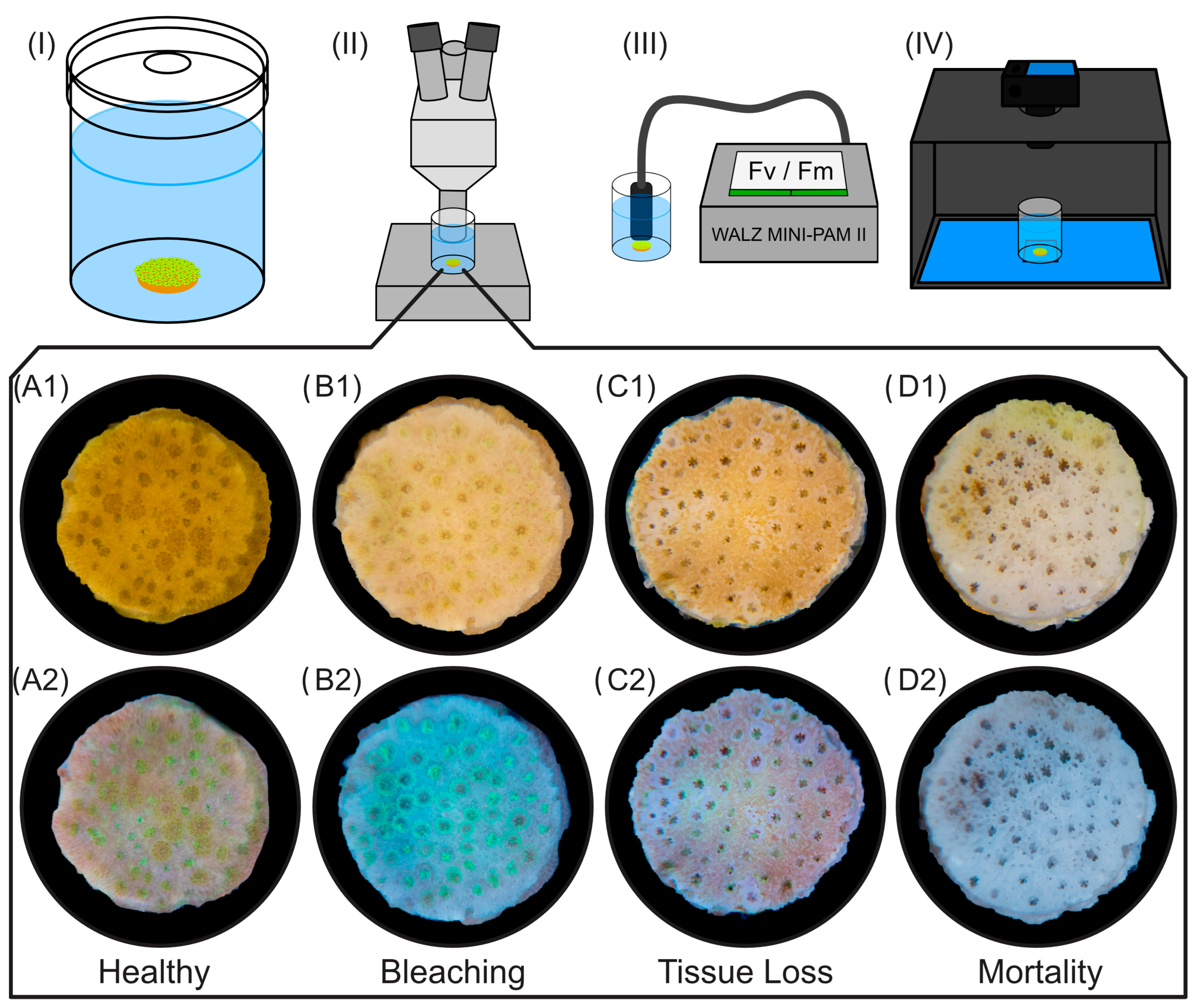
2. Materials and Methods
2.1. Preparation of M. digitata Fragments
2.2. Endpoint Assessments
2.3. Preparation of Stock Solutions and Test Concentrations
2.4. Experimental Setup and Procedure
2.4.1. Water Sampling
2.4.2. Validation Criteria
2.5. Chemical Control Analyses
2.5.1. Analyses of Organic Test Substances: BP-3 and DCMU
2.5.2. Analyses of Inorganic Test Substance: CuCl2
2.6. Data Analysis
3. Results
3.1. Recovery of the Test Substances
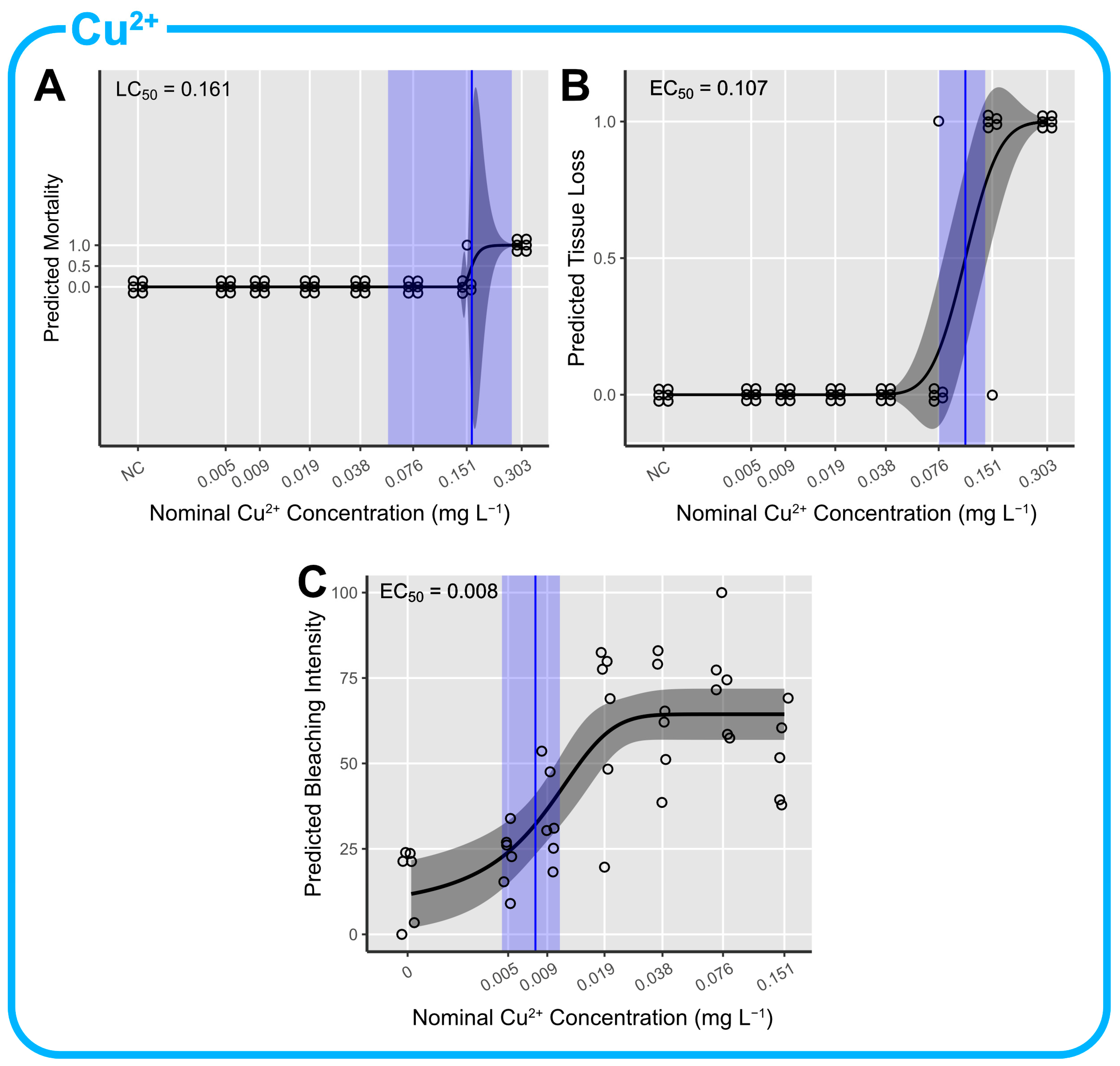
3.2. Cu2+ Toxicity
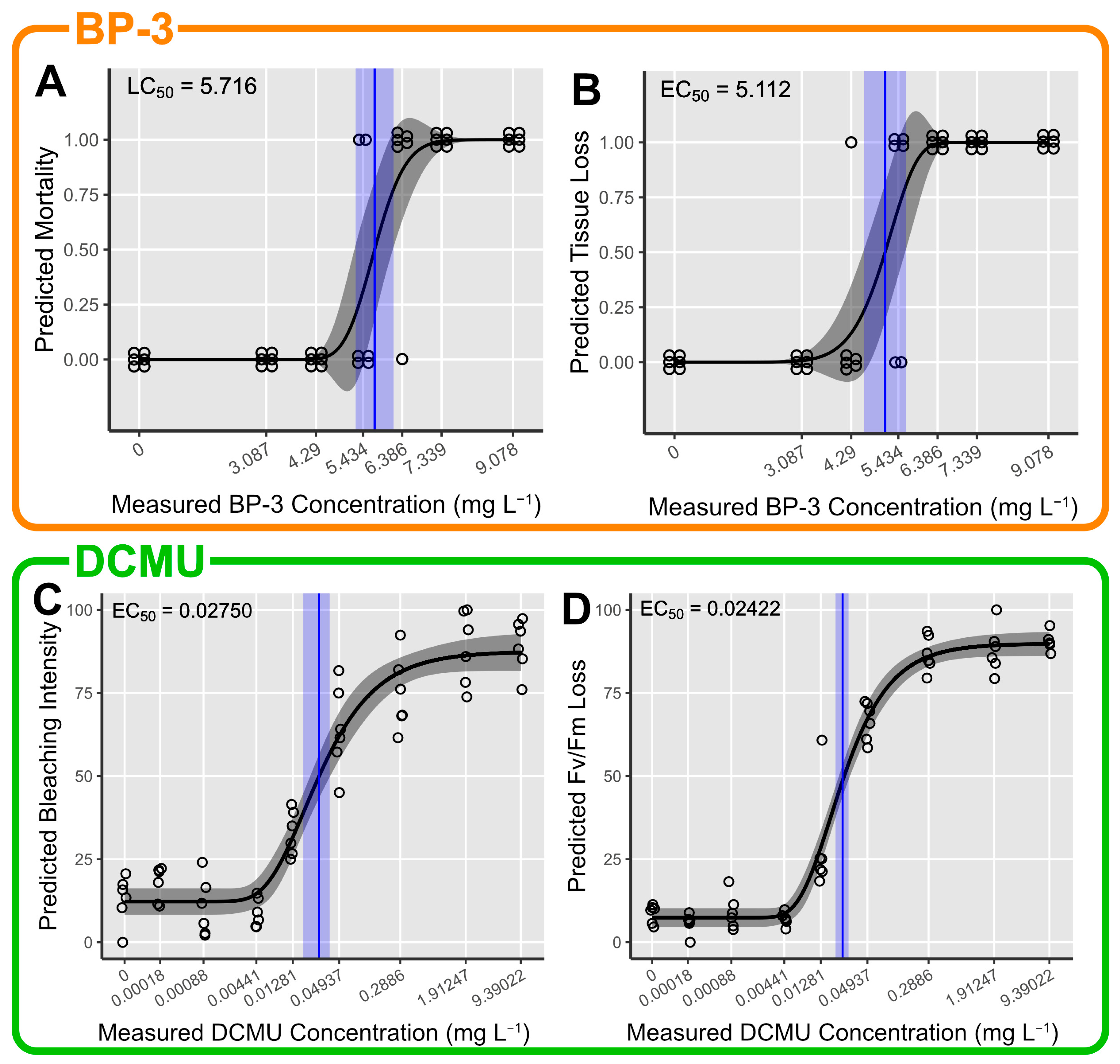
3.3. BP-3 Toxicity
3.4. DCMU Toxicity
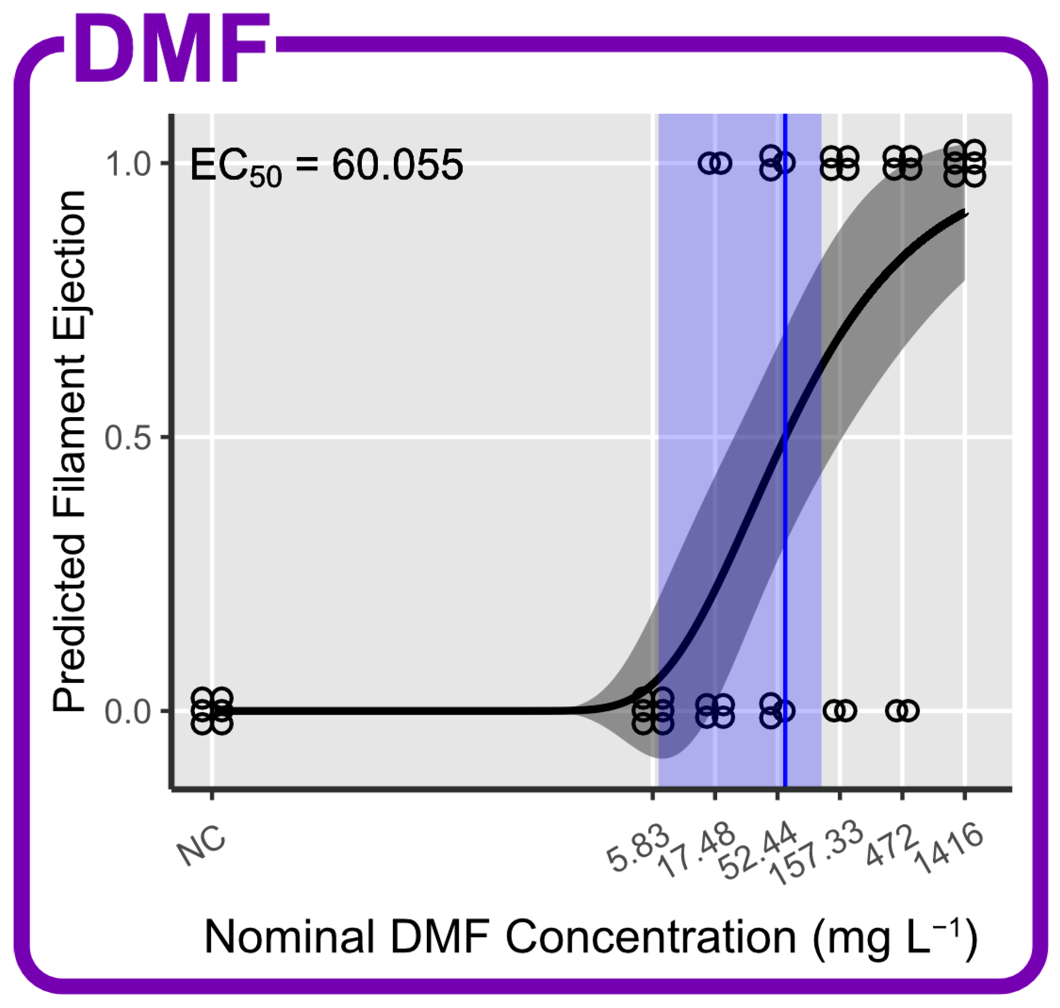
3.5. Effects of DMF as Carrier Solvent
4. Discussion
5. Conclusions
Supplementary Materials
Author Contributions
Funding
Institutional Review Board Statement
Informed Consent Statement
Data Availability Statement
Acknowledgments
Conflicts of Interest
References
- Hughes, T.P.; Barnes, M.L.; Bellwood, D.R.; Cinner, J.E.; Cumming, G.S.; Jackson, J.B.C.; Kleypas, J.; van de Leemput, I.A.; Lough, J.M.; Morrison, T.H.; et al. Coral Reefs in the Anthropocene. Nature 2017, 546, 82–90. [Google Scholar] [CrossRef]
- Eddy, T.D.; Lam, V.W.Y.; Reygondeau, G.; Cisneros-Montemayor, A.M.; Greer, K.; Palomares, M.L.D.; Bruno, J.F.; Ota, Y.; Cheung, W.W.L. Global Decline in Capacity of Coral Reefs to Provide Ecosystem Services. One Earth 2021, 4, 1278–1285. [Google Scholar] [CrossRef]
- Hoegh-Guldberg, O.; Pendleton, L.; Kaup, A. People and the Changing Nature of Coral Reefs. Reg. Stud. Mar. Sci. 2019, 30, 100699. [Google Scholar] [CrossRef]
- Woodhead, A.J.; Hicks, C.C.; Norström, A.V.; Williams, G.J.; Graham, N.A.J. Coral Reef Ecosystem Services in the Anthropocene. Funct. Ecol. 2019, 33, 1023–1034. [Google Scholar] [CrossRef]
- Álvarez-Muñoz, D.; Llorca, M.; Blasco, J.; Barceló, D. Contaminants in the Marine Environment. In Marine Ecotoxicology; Elsevier: Amsterdam, The Netherlands, 2016; pp. 1–34. ISBN 978-0-12-803371-5. [Google Scholar]
- Ouédraogo, D.-Y.; Mell, H.; Perceval, O.; Burga, K.; Domart-Coulon, I.; Hédouin, L.; Delaunay, M.; Guillaume, M.M.M.; Castelin, M.; Calvayrac, C.; et al. What Are the Toxicity Thresholds of Chemical Pollutants for Tropical Reef-Building Corals? A Systematic Review. Environ. Evid. 2023, 12, 4. [Google Scholar] [CrossRef]
- Mitchelmore, C.L.; Burns, E.E.; Conway, A.; Heyes, A.; Davies, I.A. A Critical Review of Organic Ultraviolet Filter Exposure, Hazard, and Risk to Corals. Environ. Toxicol. Chem. 2021, 40, 967–988. [Google Scholar] [CrossRef]
- Moeller, M.; Pawlowski, S.; Petersen-Thiery, M.; Miller, I.B.; Nietzer, S.; Heisel-Sure, Y.; Kellermann, M.Y.; Schupp, P.J. Challenges in Current Coral Reef Protection—Possible Impacts of UV Filters Used in Sunscreens, a Critical Review. Front. Mar. Sci. 2021, 8, 665548. [Google Scholar] [CrossRef]
- Miller, I.B.; Pawlowski, S.; Kellermann, M.Y.; Petersen-Thiery, M.; Moeller, M.; Nietzer, S.; Schupp, P.J. Toxic Effects of UV Filters from Sunscreens on Coral Reefs Revisited: Regulatory Aspects for “Reef Safe” Products. Environ. Sci. Eur. 2021, 33, 74. [Google Scholar] [CrossRef]
- Conway, A.J.; Gonsior, M.; Clark, C.; Heyes, A.; Mitchelmore, C.L. Acute Toxicity of the UV Filter Oxybenzone to the Coral Galaxea Fascicularis. Sci. Total Environ. 2021, 796, 148666. [Google Scholar] [CrossRef]
- Miller, I.B.; Moeller, M.; Kellermann, M.Y.; Nietzer, S.; Di Mauro, V.; Kamyab, E.; Pawlowski, S.; Petersen-Thiery, M.; Schupp, P.J. Towards the Development of Standardized Bioassays for Corals: Acute Toxicity of the UV Filter Benzophenone-3 to Scleractinian Coral Larvae. Toxics 2022, 10, 244. [Google Scholar] [CrossRef]
- Pawlowski, S.; Moeller, M.; Miller, I.B.; Kellermann, M.Y.; Schupp, P.J.; Petersen-Thiery, M. UV Filters Used in Sunscreens—A Lack in Current Coral Protection? Integr. Environ. Assess. Manag. 2021, 17, 926–939. [Google Scholar] [CrossRef] [PubMed]
- OECD. Test No. 202: Daphnia Sp. Acute Immobilisation Test. In OECD Guidelines for the Testing of Chemicals, Section 2; OECD Publishing: Paris, France, 2004. [Google Scholar]
- OECD. Test No. 203: Fish, Acute Toxicity Test. In OECD Guidelines for the Testing of Chemicals, Section 2; OECD Publishing: Paris, France, 2019. [Google Scholar]
- Burns, E.E.; Davies, I.A. Coral Ecotoxicological Data Evaluation for the Environmental Safety Assessment of Ultraviolet Filters. Environ. Toxicol. Chem. 2021, 40, 3441–3464. [Google Scholar] [CrossRef] [PubMed]
- Stubblefield, W.; Barron, M.; Bragin, G.; DeLorenzo, M.; De Jourdan, B.; Echols, B.; French-McCay, D.; Jackman, P.; Loughery, J.; Parkerton, T.; et al. Improving the Design and Conduct of Aquatic Toxicity Studies with Oils Based on 20 Years of CROSERF Experience. Aquat. Toxicol. 2023, 261, 106579. [Google Scholar] [CrossRef] [PubMed]
- Brinkman, D.L.; Flores, F.; Luter, H.M.; Nordborg, F.M.; Brooks, M.; Parkerton, T.F.; Negri, A.P. Sensitivity of the Indo-Pacific Coral Acropora Millepora to Aromatic Hydrocarbons. Environ. Pollut. 2023, 332, 121963. [Google Scholar] [CrossRef] [PubMed]
- He, T.; Tsui, M.M.P.; Tan, C.J.; Ng, K.Y.; Guo, F.W.; Wang, L.H.; Chen, T.H.; Fan, T.Y.; Lam, P.K.S.; Murphy, M.B. Comparative Toxicities of Four Benzophenone Ultraviolet Filters to Two Life Stages of Two Coral Species. Sci. Total Environ. 2019, 651, 2391–2399. [Google Scholar] [CrossRef] [PubMed]
- Turner, N.R.; Parkerton, T.F.; Renegar, D.A. Toxicity of Two Representative Petroleum Hydrocarbons, Toluene and Phenanthrene, to Five Atlantic Coral Species. Mar. Pollut. Bull. 2021, 169, 112560. [Google Scholar] [CrossRef]
- Binet, M.T.; Reichelt-Brushett, A.; McKnight, K.; Golding, L.A.; Humphrey, C.; Stauber, J.L. Adult Corals Are Uniquely More Sensitive to Manganese Than Coral Early-Life Stages. Environ. Toxicol. Chem. 2023, 42, 1359–1370. [Google Scholar] [CrossRef]
- Baker, A.C.; Cunning, R. Coral “Bleaching” as a Generalized Stress Response to Environmental Disturbance. In Diseases of Coral; John Wiley & Sons, Ltd.: Hoboken, NJ, USA, 2015; pp. 396–409. ISBN 978-1-118-82850-2. [Google Scholar]
- Van Oppen, M.J.H.; Lough, J.M. (Eds.) Coral Bleaching; Ecological Studies; Springer International Publishing: Cham, Switzerland, 2018; Volume 233, ISBN 978-3-319-75392-8. [Google Scholar]
- Bhagooli, R.; Mattan-Moorgawa, S.; Kaullysing, D.; Louis, Y.D.; Gopeechund, A.; Ramah, S.; Soondur, M.; Pilly, S.S.; Beesoo, R.; Wijayanti, D.P.; et al. Chlorophyll Fluorescence—A Tool to Assess Photosynthetic Performance and Stress Photophysiology in Symbiotic Marine Invertebrates and Seaplants. Mar. Pollut. Bull. 2021, 165, 112059. [Google Scholar] [CrossRef]
- Marzonie, M.; Flores, F.; Sadoun, N.; Thomas, M.C.; Valada-Mennuni, A.; Kaserzon, S.; Mueller, J.F.; Negri, A.P. Toxicity Thresholds of Nine Herbicides to Coral Symbionts (Symbiodiniaceae). Sci. Rep. 2021, 11, 21636. [Google Scholar] [CrossRef]
- Jones, R. The Ecotoxicological Effects of Photosystem II Herbicides on Corals. Mar. Pollut. Bull. 2005, 51, 495–506. [Google Scholar] [CrossRef]
- Roth, M.S. The Engine of the Reef: Photobiology of the Coral-Algal Symbiosis. Front. Microbiol. 2014, 5, 422. [Google Scholar] [CrossRef] [PubMed]
- Nalley, E.M.; Tuttle, L.J.; Barkman, A.L.; Conklin, E.E.; Wulstein, D.M.; Richmond, R.H.; Donahue, M.J. Water Quality Thresholds for Coastal Contaminant Impacts on Corals: A Systematic Review and Meta-Analysis. Sci. Total Environ. 2021, 794, 148632. [Google Scholar] [CrossRef] [PubMed]
- Veron, J.E.N. Montipora Digitata. In Corals of the World; Australian Institute of Marine Science: Townsville, Australia, 2000; Volume 1–3, p. 1410. [Google Scholar]
- Stobart, B. A Taxonomic Reappraisal of Montipora Digitata Based on Genetic and Morphometric Evidence. Zool. Stud. 2000, 39, 179–190. [Google Scholar]
- Johnson, C.E.; Goulet, T.L. A Comparison of Photographic Analyses Used to Quantify Zooxanthella Density and Pigment Concentrations in Cnidarians. J. Exp. Mar. Biol. Ecol. 2007, 353, 287–295. [Google Scholar] [CrossRef]
- Winters, G.; Holzman, R.; Blekhman, A.; Beer, S.; Loya, Y. Photographic Assessment of Coral Chlorophyll Contents: Implications for Ecophysiological Studies and Coral Monitoring. J. Exp. Mar. Biol. Ecol. 2009, 380, 25–35. [Google Scholar] [CrossRef]
- Akkaynak, D.; Treibitz, T.; Xiao, B.; Gürkan, U.A.; Allen, J.J.; Demirci, U.; Hanlon, R.T. Use of Commercial Off-the-Shelf Digital Cameras for Scientific Data Acquisition and Scene-Specific Color Calibration. JOSA A 2014, 31, 312–321. [Google Scholar] [CrossRef]
- Rueden, C.T.; Schindelin, J.; Hiner, M.C.; DeZonia, B.E.; Walter, A.E.; Arena, E.T.; Eliceiri, K.W. ImageJ2: ImageJ for the next Generation of Scientific Image Data. BMC Bioinform. 2017, 18, 529. [Google Scholar] [CrossRef]
- Schindelin, J.; Arganda-Carreras, I.; Frise, E.; Kaynig, V.; Longair, M.; Pietzsch, T.; Preibisch, S.; Rueden, C.; Saalfeld, S.; Schmid, B.; et al. Fiji: An Open-Source Platform for Biological-Image Analysis. Nat. Methods 2012, 9, 676–682. [Google Scholar] [CrossRef]
- Wickham, H.; Averick, M.; Bryan, J.; Chang, W.; McGowan, L.D.; François, R.; Grolemund, G.; Hayes, A.; Henry, L.; Hester, J.; et al. Welcome to the Tidyverse. J. Open Source Softw. 2019, 4, 1686. [Google Scholar] [CrossRef]
- Ho, J.; Tumkaya, T.; Aryal, S.; Choi, H.; Claridge-Chang, A. Moving beyond P Values: Data Analysis with Estimation Graphics. Nat. Methods 2019, 16, 565–566. [Google Scholar] [CrossRef]
- Hothorn, T.; Bretz, F.; Westfall, P. Simultaneous Inference in General Parametric Models. Biom. J. 2008, 50, 346–363. [Google Scholar] [CrossRef]
- Fox, J.; Weisberg, S. An R Companion to Applied Regression, 3rd ed.; Sage: Thousand Oaks, CA, USA, 2019. [Google Scholar]
- Zeileis, A. Econometric Computing with HC and HAC Covariance Matrix Estimators. J. Stat. Softw. 2004, 11, 1–17. [Google Scholar] [CrossRef]
- Zeileis, A.; Köll, S.; Graham, N. Various Versatile Variances: An Object-Oriented Implementation of Clustered Covariances in R. J. Stat. Softw. 2020, 95, 1–36. [Google Scholar] [CrossRef]
- Hothorn, T.; Hornik, K.; van de Wiel, M.A.; Zeileis, A. Implementing a Class of Permutation Tests: The Coin Package. J. Stat. Softw. 2008, 28, 1–23. [Google Scholar] [CrossRef]
- Holm, S. A Simple Sequentially Rejective Multiple Test Procedure. Scand. J. Stat. 1979, 6, 65–70. [Google Scholar]
- Ritz, C.; Baty, F.; Streibig, J.C.; Gerhard, D. Dose-Response Analysis Using R. PLoS ONE 2015, 10, e0146021. [Google Scholar] [CrossRef]
- Hirose, K. Chemical Speciation of Trace Metals in Seawater: A Review. Anal. Sci. 2006, 22, 1055–1063. [Google Scholar] [CrossRef]
- OECD. Test No. 201: Freshwater Alga and Cyanobacteria, Growth Inhibition Test. In OECD Guidelines for the Testing of Chemicals, Section 2; OECD Publishing: Paris, France, 2011. [Google Scholar]
- OECD. Guidance Document on Aquatic Toxicity Testing of Difficult Substances and Mixtures. In OECD Series on Testing and Assessment; OECD Publishing: Paris, France, 2019. [Google Scholar]
- European Chemicals Agency Brief Profile—Oxybenzone. Available online: https://echa.europa.eu/de/brief-profile/-/briefprofile/100.004.575 (accessed on 30 October 2023).
- European Chemicals Agency Brief Profile—Diuron (ISO); 3-(3,4-Dichlorophenyl)-1,1-dimethylurea. Available online: https://echa.europa.eu/de/brief-profile/-/briefprofile/100.005.778 (accessed on 30 October 2023).
- Lide, D.R. CRC Handbook of Chemistry and Physics (CD-ROM Version 2010), 90th ed.; CRC Press/Taylor and Francis: Boca Raton, FL, USA, 2010. [Google Scholar]
- Di Mauro, V.; Kamyab, E.; Kellermann, M.Y.; Moeller, M.; Nietzer, S.; Luetjens, L.H.; Pawlowski, S.; Petersen-Thiery, M.; Schupp, P.J. Ecotoxicological Effects of Four Commonly Used Organic Solvents on the Scleractinian Coral Montipora Digitata. Toxics 2023, 11, 367. [Google Scholar] [CrossRef] [PubMed]
- Flores, F.; Marques, J.A.; Uthicke, S.; Fisher, R.; Patel, F.; Kaserzon, S.; Negri, A.P. Combined Effects of Climate Change and the Herbicide Diuron on the Coral Acropora Millepora. Mar. Pollut. Bull. 2021, 169, 112582. [Google Scholar] [CrossRef]
- Jones, R.; Muller, J.; Haynes, D.; Schreiber, U. Effects of Herbicides Diuron and Atrazine on Corals of the Great Barrier Reef, Australia. Mar. Ecol. Prog. Ser. 2003, 251, 153–167. [Google Scholar] [CrossRef]
- Råberg, S.; Nyström, M.; Erös, M.; Plantman, P. Impact of the Herbicides 2,4-D and Diuron on the Metabolism of the Coral Porites Cylindrica. Mar. Environ. Res. 2003, 56, 503–514. [Google Scholar] [CrossRef]
- Jones, R. Testing the Photoinhibition Model of Coral Bleaching Using Chemical Inhibitors. Mar. Ecol. Prog. Ser. 2004, 284, 133–145. [Google Scholar] [CrossRef]
- Bielmyer, G.K.; Grosell, M.; Bhagooli, R.; Baker, A.C.; Langdon, C.; Gillette, P.; Capo, T.R. Differential Effects of Copper on Three Species of Scleractinian Corals and Their Algal Symbionts (Symbiodinium spp.). Aquat. Toxicol. 2010, 97, 125–133. [Google Scholar] [CrossRef]


| Validation Criteria | Target |
|---|---|
| Temperature | 24–28 °C, but constant within ±1.5 °C of cultivation temperature |
| Salinity | 33–36 PSU |
| pH | 7.7–8.5 |
| O2 saturation | ≥75% * |
| Alkalinity | 5–9 °dKH |
| Bleaching intensity of control | ≤30% |
| Fv/Fm of control | Fv/Fm ≥ 0.450 |
| Tissue loss of control | ≤20% (1 fragment) |
| Mortality of control | 0% |
| Chem. | Nominal Conc. | Mean Measured Conc. of t0 to t96 | Recovery of Nominal | Mean Measured Conc. 1st Phase | Mean Measured Conc. 2nd Phase | Recovery t0–t48 | Recovery t48–t96 | Mean Recovery | ||
|---|---|---|---|---|---|---|---|---|---|---|
| t0 | t48 | t48 | t96 | Semi -static test method | ||||||
| fresh | aged | fresh | aged | |||||||
| mg L−1 | mg L−1 | % | mg L−1 | % | % | % | ||||
| Control | 0 | <LoD | 100.0 | <LoD | <LoD | <LoD | <LoD | 100.0 | 100.0 | 100.0 |
| BP-3 | 7.000 | 3.08700 | 44.1 | 3.37900 | 2.64100 | 3.91800 | 2.97300 | 78.2 | 75.9 | 77.1 |
| 8.000 | 4.29000 | 53.6 | 4.65600 | 3.70200 | 5.32800 | 4.17500 | 79.5 | 78.4 | 79.0 | |
| 9.000 | 5.43400 | 60.4 | 6.13600 | 4.62100 | 6.56200 | 5.33300 | 75.3 | 81.3 | 78.3 | |
| 10.000 | 6.38600 | 63.9 | 7.12000 | 5.37200 | 7.81600 | 6.31800 | 75.4 | 80.8 | 78.1 | |
| 11.000 | 7.33900 | 66.7 | 8.10800 | 5.92800 | 9.45100 | 7.31000 | 73.1 | 77.3 | 75.2 | |
| 12.000 | 9.07800 | 75.6 | 10.04900 | 7.39500 | 10.54600 | 9.54300 | 73.6 | 90.5 | 82.0 | |
| Control | 0 | 0 | 100.0 | 0 | 0 | 0 | 0 | 100.0 | 100.0 | 100.0 |
| 0.00013 | 0.00018 | 139.1 | 0.00016 | 0.00018 | 0.00018 | 0.00017 | 111.5 | 93.0 | 102.2 | |
| 0.00064 | 0.00088 | 137.0 | 0.00081 | 0.00089 | 0.00085 | 0.00091 | 110.2 | 106.6 | 108.4 | |
| 0.00320 | 0.00441 | 137.7 | 0.00410 | 0.00442 | 0.00446 | 0.00452 | 107.6 | 101.3 | 104.4 | |
| 0.01600 | 0.01281 | 80.1 | 0.01150 | 0.00995 | 0.01607 | 0.01470 | 86.5 | 91.5 | 89.0 | |
| DCMU | 0.08000 | 0.04937 | 61.7 | 0.04125 | 0.03875 | 0.05913 | 0.05918 | 93.9 | 100.1 | 97.0 |
| 0.40000 | 0.28860 | 72.2 | 0.22682 | 0.21678 | 0.36728 | 0.35198 | 95.6 | 95.8 | 95.7 | |
| 2.00000 | 1.91247 | 95.6 | 1.53252 | 1.53191 | 2.28243 | 2.29802 | 100.0 | 100.7 | 100.3 | |
| 10.00000 | 9.39022 | 93.9 | 7.06999 | 7.16035 | 11.55859 | 11.69601 | 101.3 | 101.2 | 101.2 | |
| 50.00000 | 40.93958 | 81.9 | 37.01764 | 34.42754 | 50.58926 | 44.58775 | 93.0 | 88.1 | 90.6 | |
| Control | 0 | 0.00125 | 100.1 | 0 | 0 | 0 | 0.005 | 100.0 | 100.0 | 100.0 |
| Cu2+ | 0.005 | 0.00200 | 40.0 | n.d. | 0 | 0.003 | 0.003 | n.d. | 100.0 | 100.0 |
| 0.009 | 0.00433 | 48.1 | n.d. | 0.003 | 0.005 | 0.005 | n.d. | 100.0 | 100.0 | |
| 0.019 | 0.00675 | 35.5 | 0.006 | 0.006 | 0.007 | 0.008 | 100.0 | 114.3 | 107.1 | |
| 0.038 | 0.01425 | 37.5 | 0.009 | 0.016 | 0.018 | 0.014 | 177.8 | 77.8 | 127.8 | |
| 0.075 | 0.01700 | 22.7 | 0.015 | n.d. | n.d. | 0.019 | n.d. | n.d. | n.d. | |
| 0.151 | 0.03150 | 20.9 | 0.023 | 0.023 | 0.042 | 0.038 | 100.0 | 90.5 | 95.2 | |
| 0.303 | n.d. | n.d. | n.d. | n.d. | n.d. | n.d. | n.d. | n.d. | n.d. | |
| Endpoint | Cu2+ | BP-3 | DCMU | DMF | |
|---|---|---|---|---|---|
| Mortality | LC50 | 0.161 (0.055–0.267) | 5.716 (5.256–6.176) | >9.390 | >1416 |
| Tissue Loss | EC50 | 0.107 (0.076–0.138) | 5.112 (4.604–5.619) | >9.390 | >1416 |
| Bleaching | EC50 | 0.008 (0.005–0.011) | >9.078 | 0.0275 (0.01750–0.03751) | >1416 |
| Fv/Fm Loss | EC50 | >0.303 | >9.078 | 0.0242 (0.01972–0.02872) | >1416 |
| Filament Ejection | EC50 | >0.303 | >9.078 | >40.939 | 60.055 (6.46–113.65) |
| Polyp Debilitation | EC50 | >0.303 | <3.087 | 0.0258 (−0.00013–0.05176) | >1416 |
Disclaimer/Publisher’s Note: The statements, opinions and data contained in all publications are solely those of the individual author(s) and contributor(s) and not of MDPI and/or the editor(s). MDPI and/or the editor(s) disclaim responsibility for any injury to people or property resulting from any ideas, methods, instructions or products referred to in the content. |
© 2023 by the authors. Licensee MDPI, Basel, Switzerland. This article is an open access article distributed under the terms and conditions of the Creative Commons Attribution (CC BY) license (https://creativecommons.org/licenses/by/4.0/).
Share and Cite
Brefeld, D.; Di Mauro, V.; Kellermann, M.Y.; Nietzer, S.; Moeller, M.; Lütjens, L.H.; Pawlowski, S.; Petersen-Thiery, M.; Schupp, P.J. Acute Toxicity Assays with Adult Coral Fragments: A Method for Standardization. Toxics 2024, 12, 1. https://doi.org/10.3390/toxics12010001
Brefeld D, Di Mauro V, Kellermann MY, Nietzer S, Moeller M, Lütjens LH, Pawlowski S, Petersen-Thiery M, Schupp PJ. Acute Toxicity Assays with Adult Coral Fragments: A Method for Standardization. Toxics. 2024; 12(1):1. https://doi.org/10.3390/toxics12010001
Chicago/Turabian StyleBrefeld, David, Valentina Di Mauro, Matthias Y. Kellermann, Samuel Nietzer, Mareen Moeller, Laura H. Lütjens, Sascha Pawlowski, Mechtild Petersen-Thiery, and Peter J. Schupp. 2024. "Acute Toxicity Assays with Adult Coral Fragments: A Method for Standardization" Toxics 12, no. 1: 1. https://doi.org/10.3390/toxics12010001