Article

Acute and Transgenerational Effects of Non-Steroidal Anti-Inflammatory Drugs on Daphnia magna

by
Anna Michalaki
and
Konstantinos Grintzalis
*
School of Biotechnology, Dublin City University, D09 N920 Dublin, Ireland
*
Author to whom correspondence should be addressed.
Toxics 2023, 11(4), 320; https://doi.org/10.3390/toxics11040320
Submission received: 28 January 2023 / Revised: 23 March 2023 / Accepted: 27 March 2023 / Published: 29 March 2023
(This article belongs to the Special Issue Toxicity of Contaminants on Aquatic Organisms II)

Abstract

:
Pharmaceuticals pose a great threat to organisms inhabiting the aquatic environment. Non-steroidal anti-inflammatory drugs (NSAIDs) are major pharmaceutical pollutants with a significant presence in freshwater ecosystems. In this study, the impact of indomethacin and ibuprofen, two of the most commonly prescribed NSAIDs, was assessed on Daphnia magna. Toxicity was assessed as the immobilization of animals and used to determine non-lethal exposure concentrations. Feeding was assessed as a phenotypic endpoint and key enzymes were used as molecular endpoints of physiology. Feeding was decreased in mixture exposures for five-day-old daphnids and neonates. Furthermore, animals were exposed to NSAIDs and their mixture in chronic and transgenerational scenarios revealing changes in key enzyme activities. Alkaline and acid phosphatases, lipase, peptidase, β-galactosidase, and glutathione-S-transferase were shown to have significant changes in the first generation at the first and third week of exposure, and these were enhanced in the second generation. On the other hand, the third recovery generation did not exhibit these changes, and animals were able to recover from the induced changes and revert back to the control levels. Overall, our study points towards transgenerational exposures as more impactful laboratory studies to understand pharmaceutical stressors with a combination of molecular and phenotypic markers of physiology.

1. Introduction

Pharmaceutical compounds pose a greater risk to the aquatic environment and human health due to bioaccumulation and human activities [1,2,3]. Because of the global increase in human population and, as a result, the increase in elderly people, the use of pharmaceuticals is becoming highly significant. Pharmaceuticals end up in the environment as a result of their widespread consumption, where they are extremely difficult to biodegrade [3,4]. Pharmaceutical compounds have been observed in potable and groundwater, surface water, sewage treatment plants, Waste Water Treatment Plants (WWTPs), soils, and sediments [5,6]. Pharmaceuticals are primarily derived from industrial or domestic wastewater, improper disposal of expired drugs, and products used in farming and aquaculture [3]. Furthermore, pharmaceutical metabolites and their subsequent transformation products may enter the environment via the WWTP process [2,3,4]. Besides that, biotic or abiotic processes such as hydrolysis or photolysis can degrade pharmaceuticals further [4,7]. These procedures may result in the production of additional pharmaceutical products that are potentially more toxic than the parent compound [4,8]. As a result, it is critical to develop new, more efficient methods for removing pharmaceutical compounds from the environment [2].
Non-steroidal anti-inflammatory drugs (NSAIDs) are a pharmaceutical class that is commonly found in aquatic ecosystems worldwide, accounting for more than 15% of all pharmaceuticals found in the environment [9,10]. As an outcome, NSAIDs can be detected in concentrations greater than 1 μg/L in wastewater treatment plant (WWTP) influx and outflow and in lower concentrations (ng/L) in surface waters [9]. Even though the concentrations of NSAIDs in freshwater ecosystems are low, their high biological activity may cause significant harm to non-target organisms [9,10]. The constant inflow of NSAIDs from WWTPs poses a serious threat to the aquatic environment and its organisms, which are constantly exposed to these emerging contaminants [10]. NSAIDs can be categorized into two groups based on their COX selectivity: nonselective NSAIDs, which block both COX isoforms (indomethacin and ibuprofen) and COX-2-selective inhibitors [11].
Indomethacin is one of the most potent nonselective NSAIDs available, and it was among the first NSAID medications used to treat migraines and headaches [11]. It is an indole-derivative and one of the most commonly detected NSAIDs due to its widespread use [12]. Additionally, indomethacin belongs to the class of intestinal toxicants that can cause gastrointestinal lesions through direct mucosal irritation and inhibition of prostaglandin synthesis [13]. Despite its therapeutic properties, indomethacin is known for its adverse effects in a percentage range of 30–60% of patients receiving it. These adverse effects can be classified as cardiovascular, gastrointestinal, nervous system, hematologic, ocular and otic, renal and electrolyte, dermatologic and sensitivity reaction, and hepatic [11]. Indomethacin can enter the ecosystem through both direct and indirect routes by being improperly disposed of in the toilet or as an excretion product. Furthermore, traces of indomethacin have been found in wastewater samples from Peterborough, Canada, as well as raw water samples from Bosnia and Herzegovina, Croatia, Serbia, and Romania [3,14]. It has been reported that indomethacin is one of the NSAIDs with the lowest detection levels (<2 μg/L) in Romanian WWTPs influents [3]. This observation is in line with a previous publication, which states that indomethacin poses a less serious risk to the environment due to its low concentrations [15].
Ibuprofen is the third most popular of the NSAIDs used globally and has been included in the WHO (World Health Organization) Essential Drug list 2021 [16,17]. Ibuprofen inhibits cyclooxygenase-1 (COX-1) and cyclooxygenase-2 (COX-2) and has a significant analgesic and antipyretic role [18,19]. Ibuprofen is a derivative of propanoic acid and can be detected in several freshwater ecosystems [17,20,21]. Despite the fact that ibuprofen has significant analgesic and antipyretic properties, excessive or inappropriate use may have negative effects on the gastrointestinal tract, the kidney, and the coagulation system [19]. Ibuprofen can enter freshwater ecosystems indirectly or directly [20]. Due to its inability to be fully metabolized by humans, it enters the ecosystem as an excretion product. The direct way for ibuprofen to end up in the environment is by being improperly disposed of in the toilet [20]. Ibuprofen has been detected at concentrations of 45 μg/L, 1.38 μg/L, and 5.78 μg/L in wastewater in Canada, South Africa, and Belgium, respectively. However, even concentrations of 1.673 mg/L and 1.417 mg/L have been detected in wastewater in Pakistan and in surface waters in China [17,22].
To improve our understanding of water pollution, the use of key species as bioindicators are currently highlighted. The combination of phenotypic and sensitive molecular endpoints is a game changer in ecotoxicology and risk assessment. Such approaches fall under the category of New Approach Methodologies (NAMs), which can supplement current analytical tools with sophisticated molecular endpoints [23]. Daphnids are important freshwater ecosystem components that are commonly used in freshwater ecology and ecotoxicology due to their geographical distribution, central role in freshwater food webs, adaptation to a variety of habitats, and, last but not least, sensitivity to anthropogenic chemicals. Daphnids are simple to culture in the laboratory and serve as a useful model organism for molecular ecotoxicology and mechanistic insight into novel pollutants [24,25,26].
This study aimed to investigate the effects of indomethacin, ibuprofen, and their combined mixture on D. magna, specifically through chronic and transgenerational exposure at a non-lethal concentration that is not environmentally relevant, using feeding assay as a phenotypic endpoint and molecular markers of physiology. It should be noted that most studies usually focus on phenotypic endpoints such as mortality in daphnids and usually in acute or chronic exposures but only in one generation. This is the first study to look at the effects of indomethacin, ibuprofen, and their combined mixture on daphnids using molecular markers rather than toxicity or phenotypic markers alone and in-depth in more generations. Moving toward transgenerational studies provide more insight into the relation to imprinted stress and its mechanisms. In this study, the effects of two NSAIDs, indomethacin and ibuprofen, were assessed on D. magna. Furthermore, in the actual environment, chemicals are not encountered alone; therefore, for a realistic scenario, their 1:1 mixture was assessed in non-lethal concentrations. The toxicity of these pharmaceuticals and their mixture on daphnids was evaluated using acute toxicity curves. These toxicity curves were constructed based on the mortality of daphnids, which was assessed as immobilization [27]. Following this, their impact on a phenotypic endpoint, feeding rate, was evaluated with a novel approach based on the ingestion of fluorescent microparticles. The impact of indomethacin, ibuprofen, and their mixture on the physiology of daphnids was evaluated using biochemical markers in chronic exposures. The main goal of this study was to assess, even at higher than environmental concentrations, if daphnids are affected and to what extent and if they can actually recover from the stress of exposure when they are subsequently cultured in clean media.

2. Materials and Methods

2.1. Reagents

All chemicals used in this study were of the highest purity >99.9% and quality. Indomethacin, ibuprofen, KCl, Na2SeO3, latex beads, carboxylate-modified polystyrene, fluorescent red, bovine serum albumin, brilliant blue G, p-nitrophenyl butyrate, 2-nitrophenyl-B-D-galactopyranoside, 1-chloro-2,4-dinitrobenzene, L-glutathione reduced, sodium phosphate dibasic were purchased from Sigma-Aldrich. CaCl2·2H2O, MgSO4·7H2O, NaHCO3, HCl, p-nitrophenyl phosphate, boric acid, ammonium acetate, NaOH, methanol, and DMSO were purchased from Fisher Scientific.

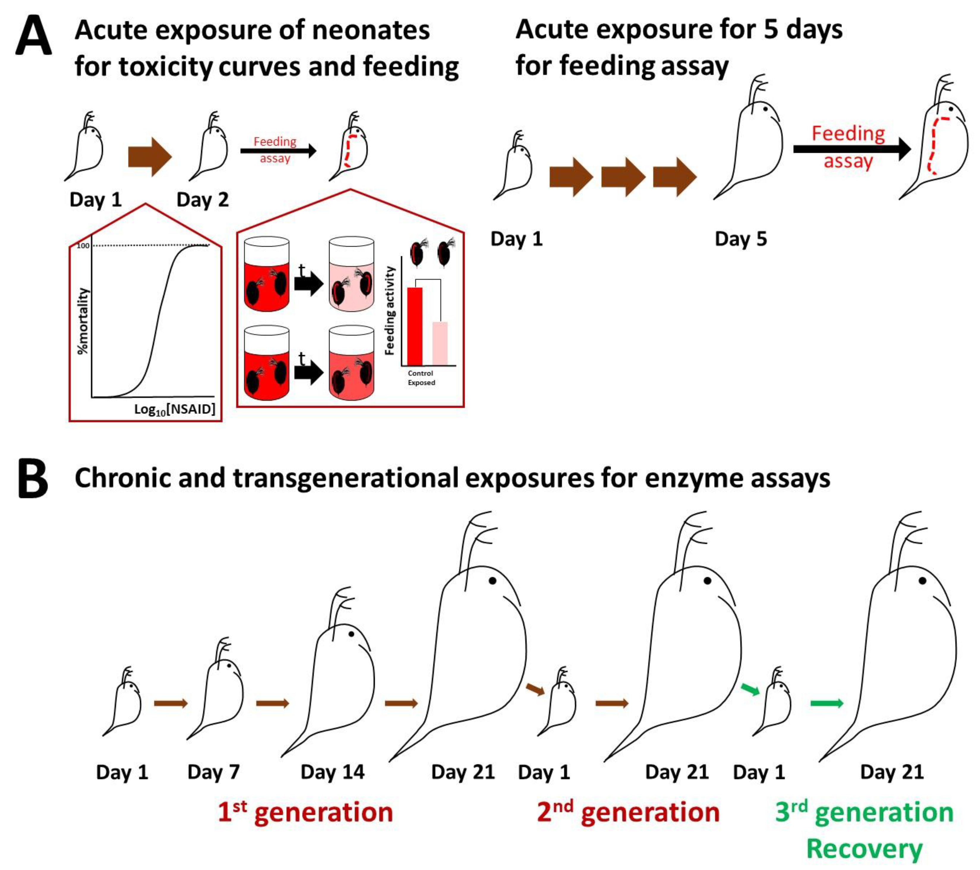
2.2. Culturing of Daphnids and Acute Toxicity

Daphnids were cultured under a 16 h:8 h of light:dark photoperiod at 20 °C in OECD media (final concentrations 0.29 g CaCl2·2H2O/L, 0.123 g MgSO4·7H2O/L, 0.065 g NaHCO3/L, 0.0058 g KCl/L, 2 μg Na2SeO3/L, pH 7.7) [28]. Indomethacin and ibuprofen were dissolved in DMSO, which had a final concentration of 0.005% v/v in the exposure, which is within the concentration range used in previous studies [27,29,30]. Neonates (<24 h) were collected from the third brood of their mothers and used for experiments. To assess toxicity, 15 neonates were exposed to each NSAID separately, and their 1:1 mixture in a final volume of 50 mL OECD media with four replicates per concentration tested. Based on similar studies [21,28,30,31,32,33,34], all assays were conducted with four (toxicity curves and biochemical assays) or five (feeding assay) replicates per condition tested. For toxicity, the OECD guidelines state that at least 40 animals (preferably in four groups of 10 animals should be used at each test concentration); however, in our study, we exceeded that number to 60 animals. Toxicity curves were plotted for 24 h exposures, and EC values were calculated using the four-parameter logistic (4PL) model, following the equation Span = Top − Bottom and Y = Bottom + (Top − Bottom)/(1 + 10^((LogIC50 − X) × HillSlope)), using the GraphPad software. The parameters top and bottom were commonly fixed to 100 and 0, accordingly. Mortality in daphnids was assessed as their immobilization [27].

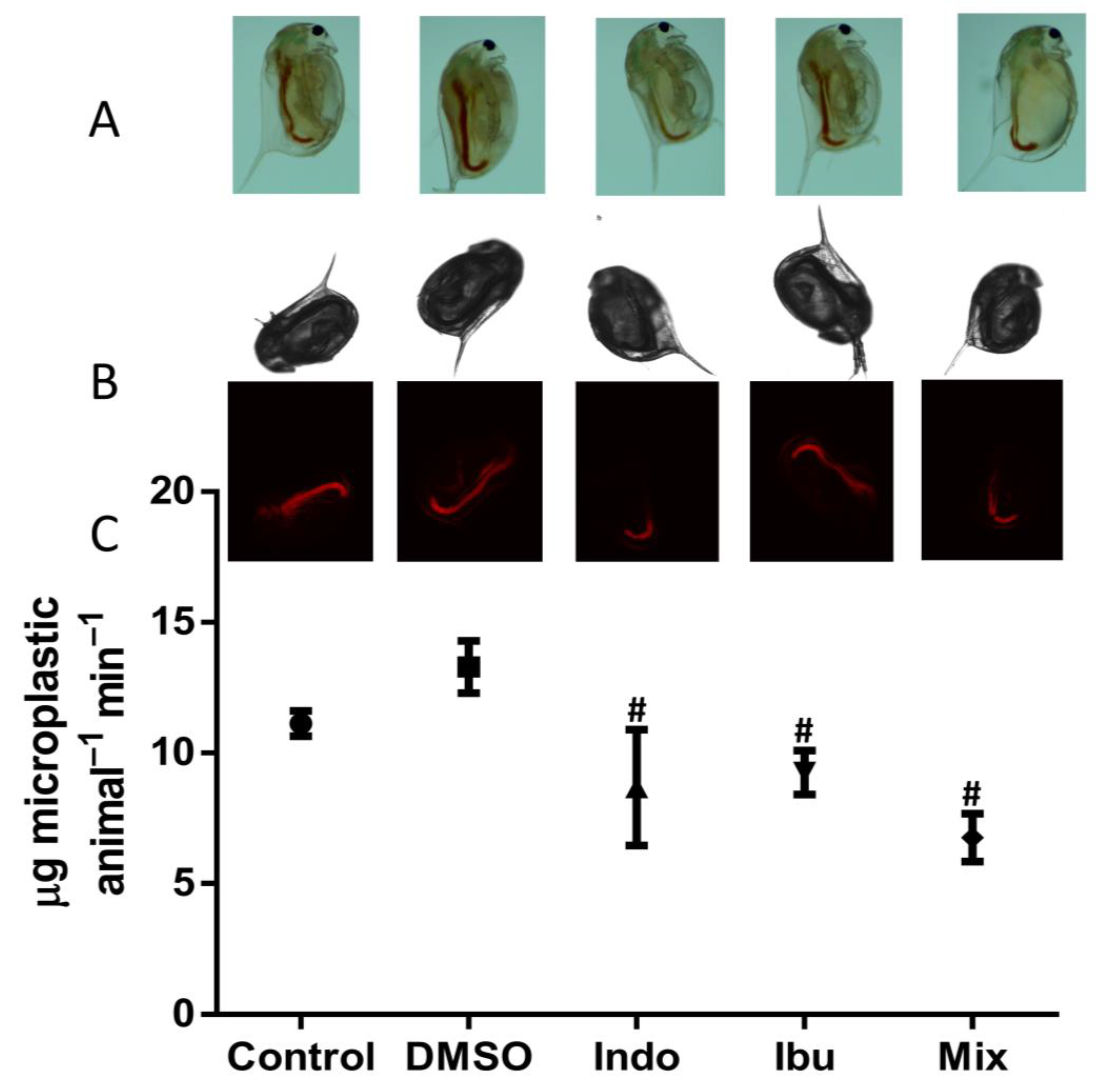
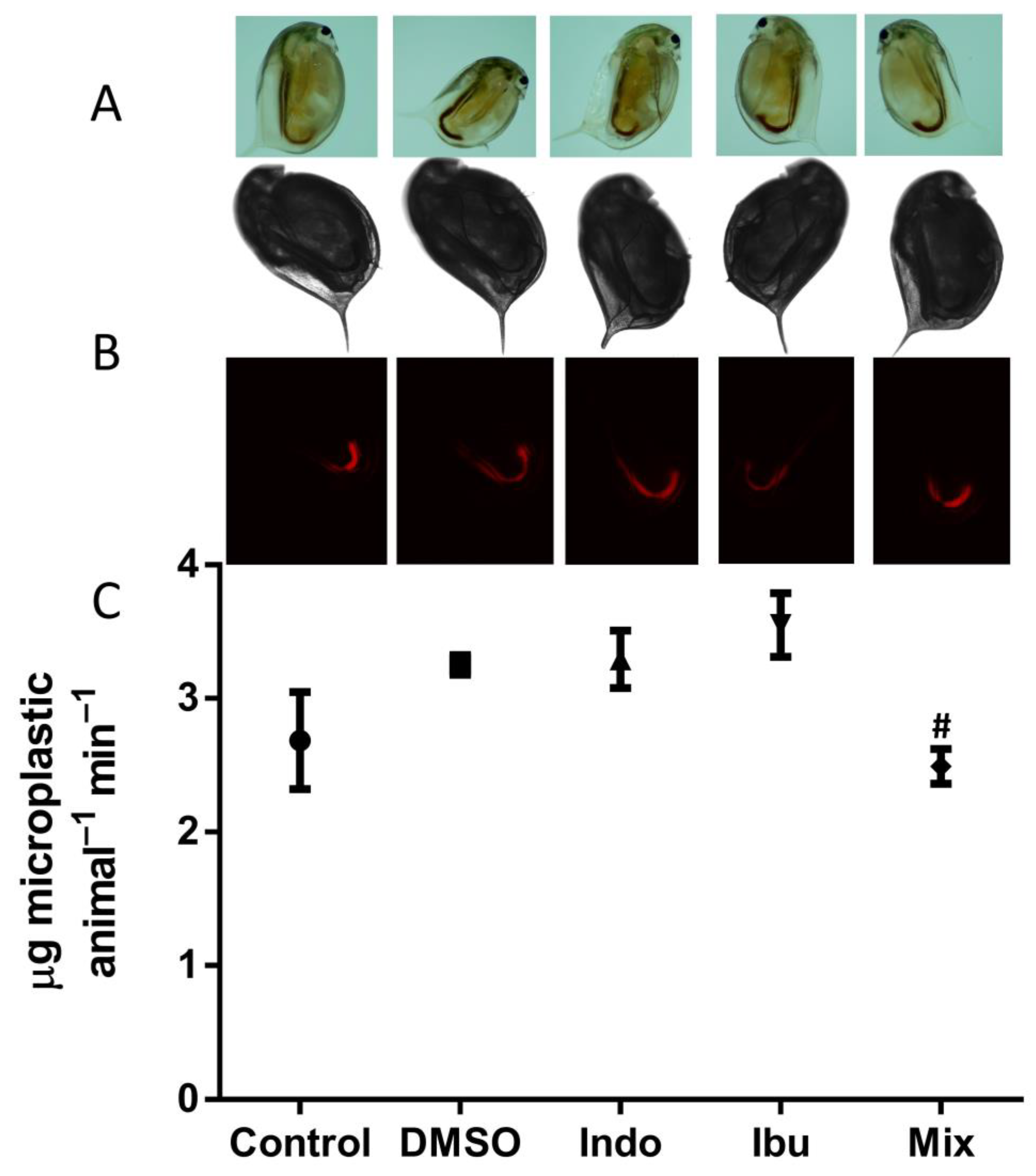
2.3. Feeding Assay and Imaging

The feeding rate was assessed in neonates, which were exposed to NSAIDs at a non-lethal concentration of 1 mg/L for 24 h or cultured until five days old (Figure 1A). To assess the feeding rate of neonates, D1 daphnids were exposed for 24 h to indomethacin, ibuprofen, and their mixture at 1 mg/L. After 24 h, twenty daphnids were transferred in a 12-well plate with 6 mL OECD containing the carboxylate-modified polystyrene, fluorescent red microparticles (2.0 μm mean particle size) at a concentration of 13 mg/L. The animals were exposed to the microplastic for up to 90 min, and media was collected every 15 min to estimate the removed microparticles by fluorescence at Ex 560 Em 590 nm using a TECAN plate reader. The concentration of microparticles in the media was optimized to ensure an excess of microparticles for the accurate quantification of ingestion. Furthermore, we have extensively tested these particles, which show no toxicity to the daphnids for the short exposure periods used. We performed control experiments with neonates, as they would be more sensitive, for even up to 10 h to microplastics at even higher concentrations (up to 52 mg/L), and all animals from all experiments following the feeding assay were alive and healthy. Fluorescence was expressed to the amount of ingested microparticles using a standard curve. To confirm the ingestion of microparticles in the gut of daphnids, animals were imaged with a stereoscope and fluorescence microscopy using the TRITC filter for carboxyl functionalized particles. For D5 daphnids, neonates were exposed for 5 days to indomethacin, ibuprofen, and their mixture at 1 mg/L in 600 mL OECD. The animals were fed daily with 2 mL of fresh algae (Chlamydomonas reinhardtii) with a concentration of 6 million cells/mL. On the 5th day of exposure, five daphnids were brought to a 12-well plate with 6 mL OECD containing the fluorescent red microparticles at a concentration of 13 mg/L. Daphnids were allowed to ingest the microparticles for 60 min, and then the same procedure as described above was followed (Figure 1A).

2.4. Biochemical Assays

Chronic exposure was performed at 1 mg/L of each NSAID and their 1:1 mixture separately, which was selected as a non-lethal concentration. For enzyme activities in chronic and transgenerational exposures, neonates were cultured for two consecutive generations and following a 21-day recovery period (Figure 1B). Twenty neonates (<24 h) were exposed for 7, 14, and 21 days to indomethacin, ibuprofen, and their mixture at 1 mg/L in 600 mL OECD media. Media and NSAIDs were renewed twice a week. Although the concentration of NSAIDs was not measured throughout the exposure, their renewal twice a week was sufficient to exert their action. Furthermore, indomethacin has been shown to be stable for up to 12 days at room temperature [35,36]. There are many reports stating that ibuprofen is stable for 15 days at room temperature [37,38]. Ibuprofen can maintain >90% average strength for 5 months, according to [39], and half-life (t1/2) for more than a year (t1/2 > 1 year) at 25 °C [40]. Daphnids were fed daily with 2 mL fresh algae (C. reinhardtii) with a concentration of 6 million cells/mL, and a seaweed extract (Ascophylum nodosum) was supplemented only on the days of media change. Neonates from the D21 daphnids (of the first generation) were exposed to the same conditions for 21 days as the second-generation exposure. For recovery, neonates from the second generation were cultured only in OECD media in the absence of NSAIDs for 21 days as a recovery generation.
Five 7, 14, and 21-day-old animals were used per biological replicate and homogenized in 0.5 mL ddH2O using a pestle homogenizer. The homogenate was cleared by centrifugation (12,000× g for 5 min at 5 °C), and the clear supernatant was collected and used to assess the enzyme activity as described elsewhere [28,41]. 200 μL appropriately diluted sample in buffer was assessed for the activity of phosphatases (in 100 mM acetic acid pH 4.5 for acid phosphatase or 100 mM boric acid pH 9.8 for alkaline phosphatase) using 50 μL of the substrate p-nitrophenyl phosphate (8 mM) and monitoring the production of p-nitrophenol at 405 nm after its alkalinization (with 50 μL 4M NaOH). In addition, the activities of β-galactosidase and lipase were measured with the same experimental conditions by the generation of nitrophenol from the catalysis of o-nitrophenyl-β-galactoside or p-nitrophenyl butyrate, respectively, but in phosphate buffer pH 7.2. Glutathione-S-transferase (GST) activity was assessed by the reaction of reduced glutathione with 1-chloro-2,4-dinitrobenzene. 200 μL appropriately diluted sample in phosphate buffer pH 7.2 was mixed with 50 μL 2 mM CDNB, and 6 mM reduced glutathione, and the formation of the complex was measured continuously at 340 nm and converted to units of activity with the extinction coefficient [42,43]. Protein was quantified by an ultrasensitive Bradford protocol to normalize the results [44].

2.5. Statistical Analysis

The biochemical data were presented as mean ± standard deviation and analyzed with the GraphPad Prism software. Statistically significant differences were compared by Student’s t-test over unexposed control with a p value of 0.05 for chemical exposures.

3. Results

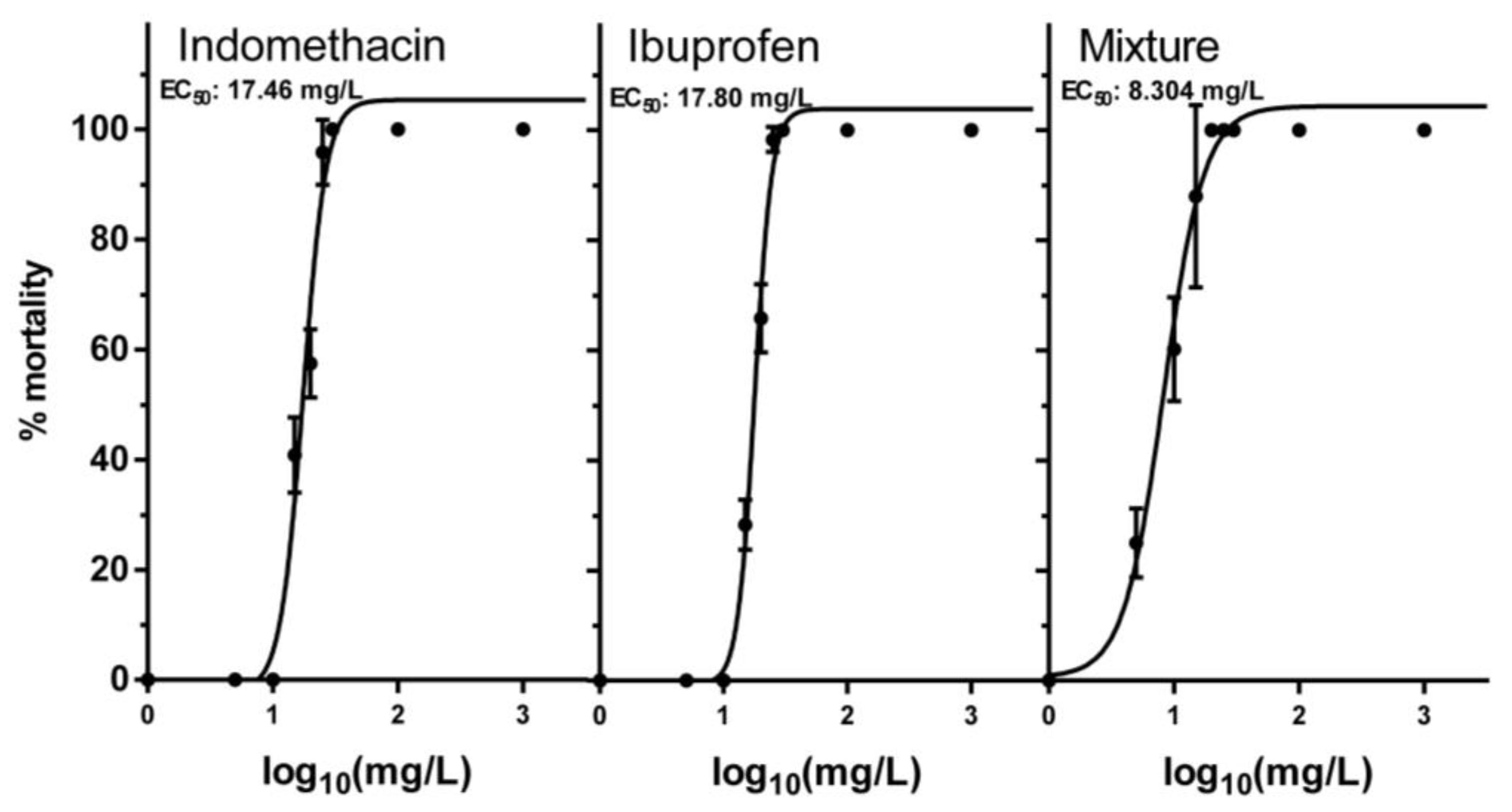
3.1. Acute Toxicity of NSAIDs and Their Mixture

Acute exposures of neonates to NSAIDs, indomethacin, ibuprofen, and their mixture were assessed via toxicity curves (Figure 2), and the effective concentration (EC) values were calculated (Table 1). The EC50 values for indomethacin and ibuprofen were very close to each other, 17.46 mg/L and 17.80 mg/L, respectively. A 1:1 mixture for both NSAIDs was also tested for mortality and showed a lower EC50 of 8.304 mg/L, indicating a potential synergy of the two NSAIDs. A non-lethal concentration of 1 mg/L was chosen for both NSAIDs and their mixture for chronic exposures.

3.2. Feeding Assay and Imaging

Feeding is a phenotypic endpoint used to evaluate the physiology of daphnids as a non-invasive test [34]. We used particles with a mean particle size of 2.0 μm because D. magna feeds non-selectively on a wide range of particles with sizes ranging from 1 to 50 μm [45]. These particles were selected as they would allow tracking with fluorescence microscopy and were not toxic to daphnids. The feeding rate was determined by the ingestion of fluorescent microparticles in neonates and five-day-old daphnids (Figure 3 and Figure 4). The ingestion of microplastics was confirmed using stereoscopy, bright field, and fluorescent microscopy. Exposure of neonates to indomethacin, ibuprofen, and their composite mixture reduced the feeding rate by 35%, 30%, and 49%, respectively, when compared to DMSO (Figure 3). However, when five-days-old animals were exposed, a different pattern was observed, and only their mixture decreased the feeding rate by 23% (Figure 4). This can be potentially explained as the animals are older and more resistant to the stress of NSAIDs when compared to neonates.

3.3. Enzymatic Activity following Chronic and Transgenerational Exposure of Daphnids to NSAIDs

Chronic exposure to 1 mg/L of indomethacin, ibuprofen, and their mixture resulted in significant changes in the physiology of daphnids during their growth (Table 2). Following the first 7 days of exposure, indomethacin significantly reduced the activities of alkaline phosphatase (ALP), β-galactosidase (β-gal), and lipase (LIP), while ibuprofen and their mixture decreased the activities of only the latter two. After 14 days of exposure, the only enzyme which showed a change in activity was β-gal by 28% in the NSAID mixture. Exposure for one more week resulted in a decrease in the activity of acid phosphatase (ACP) for indomethacin and the NSAID mixture. In contrast, indomethacin increased the activities of β-gal and peptidase (PEP), whereas ibuprofen and their mixture increased the activity of glutathione-S-transferase (GST). The first generation of exposures was continued for additional 21 days in daphnids for the second generation. Indomethacin inhibited the activities of LIP, PEP, and GST, whilst ibuprofen inhibited the activities of ALP, β-gal, LIP, and PEP. Ibuprofen also increased the activity of GST. Apart from GST, the NSAID mixture had an impact on all enzymes. Except for ACP, the activities of ALP, β-gal, LIP, and PEP decreased. Finally, daphnids following their second generation of exposure were transferred for 21 days in OECD media as a third generation of a 21-day recovery. PEP and GST were the only enzymes with increased activity during the recovery period. Indomethacin and ibuprofen increased the activity of GST by 19% and 6%, respectively, and their mixture increased the activity of PEP by 16%, thus showing that the recovery period in clean media allowed the animals to return to a control condition for the majority of the enzymes. The general conclusion is that initial exposure had an impact on markers of physiology, while during the period the animals prepare for fertility (14 days), this was alleviated and followed more changes in the first generation over 21 days, which were even stronger in the second generation. Finally, for the recovery, it was observed a return to the control condition.

4. Discussion

The acute and chronic effects of indomethacin, ibuprofen, and their mixture were assessed using a combination of physiological indicators, such as mortality and feeding, as well as biochemical markers. As biochemical markers, we assessed the activity of phosphatases, β-galactosidase, lipase, peptidase, and glutathione-S-transferase. NSAIDs, such as ibuprofen, diclofenac, and naproxen, can form reactive oxygen species (ROS). The disruption of the balance between ROS and the antioxidant systems in the organism is referred to as oxidative stress. When oxidative stress occurs, it causes lipid and protein peroxidation, damage to DNA structure, as well as inhibition of digestive enzymes (trypsin, β-galactosidase) [27,46]. According to these reports, ibuprofen increases lipid peroxidation, protein carbonyl content (protein oxidation), and enzymes of antioxidant defense SOD and CAT. As a result, lipid peroxidation affects differently different types of enzymes, such as phosphatases and lipases. Additionally, the oxidation of proteins affects the activity of proteins such as peptidases, β-galactosidase, lipases, and phosphatases [47]. Glutathione-S-transferase belongs to a group of enzymes involved in the detoxification processes [48]. An increase in this enzyme might indicate that the daphnids use these detoxification processes in order to adapt and survive the stress that the chemicals cause [49]. Most NSAIDs are toxic to organisms due to their bioaccumulation in the ecosystems. There are a few studies that have been published about the impact of NSAIDs, such as indomethacin and ibuprofen, on D. magna, but the majority of them rely on mortality, growth, and reproduction rate to determine toxicity [21,29,31,50,51]. Extending to other aquatic species, it has been noted that indomethacin and ibuprofen can harm other marine and freshwater organisms.
Indomethacin has been shown to significantly increase the biomass of Chironomous riparius, while it had no effect on the survival rate or biomass of Physella acuta [52]. After 24 h of exposure, lethal concentrations for Thamnocephalus platyurus, Oryzias latipes, and D. magna were 16.14 mg/L, 81.92 mg/L, and 22.38 mg/L, respectively [3,5]. There are not sufficient data on indomethacin toxicity in daphnids, although the findings of the two aforementioned studies [3,5] are very close to our lethal concentration. The toxicity of indomethacin in other species, such as male zebrafish (Danio rerio) for 96 h, was in a similar order of magnitude with an EC50 of 76.30 mg/L. However, indomethacin has been shown to cause significant changes in the transcriptome of zebrafish in marker genes such as superoxide dismutase 1, glutathione peroxidase 1, interleukin-1, tumor necrosis factor-alpha, and others [13].
Ibuprofen has been described in the literature as a substance with high mobility in aquatic ecosystems. Despite the fact that up to 90% of ibuprofen can be removed from the environment efficiently, it is present in high concentrations in raw sewage [20,21]. It has been reported that chronic exposure of neonates D. magna to concentrations of 20, 40, and 80 mg/L for 14 days affected somatic growth, reproduction, and survival [21]. Somatic growth, in particular, increased with increasing concentrations. There was a significant delay in fecundity and a total decrease in reproduction in daphnids exposed to 40 mg/L and 80 mg/L, respectively. Finally, survival of daphnids exposed to 20 mg/L and 40 mg/L was unaffected, yet it was reduced by 19% in daphnids exposed to 80 mg/L [21]. These findings are consistent with previous reports, which tested the same concentrations of ibuprofen to D. magna for a 10-day period [50]. Ibuprofen had a similar dose-dependent effect on reproduction and survival [50]. Exposure of adult daphnids (14 days old) for 8 days to 20, 40, and 80 mg/L ibuprofen showed a decrease in fecundity at concentrations greater than 20 mg/L. However, the brood release was not postponed. The broods produced after exposure to ibuprofen at 80 mg/L consisted of almost dead and/or undeveloped embryos [31]. Grzesiuk exposed D. magna to ibuprofen over six generations and found that exposed daphnids had higher growth rates than unexposed daphnids in the 6th generation, which agrees with the findings of Heckmann and Hayashi. In addition, 20%, 70%, and 20% of the animals in the first generation of exposed daphnids produced abnormal offspring. These percentages increased significantly to 80%, 90%, and 50% of individuals in the fifth generation, respectively. They also discovered undeveloped embryos, neonates missing an eye, and daphnids with malformed antennules and carapace. All these abnormalities are considered lethal, reinforcing the claim that even low concentrations of ibuprofen in the environment might have significant impacts on D. magna [20]. The toxicity of ibuprofen has been studied in other crustaceans, including T. platyurus. T. platyurus was exposed for 24 h to a range of concentrations ranging from 0.1 to 66.7 mg/L. The lethal concentration after 24 h of exposure was calculated to be 19.59 mg/L. This EC closely matches our findings, while the EC for ibuprofen, according to our study, is 17.80 mg/L [5]. It has also been reported in the literature that chronic exposure to ibuprofen at concentrations of 0.0001, 0.05, 1, 8, and 25 mg/L affects some phenotypic markers of zebrafish (Danio rerio), such as spontaneous movement, free swimming distance, duration and speed under dark condition, as well as enzymatic markers, such as the activity of GST [53,54]. Specifically, 28-day exposure of zebrafish to 25 mg/L ibuprofen caused an increase in the activity of GST [53]. Furthermore, ibuprofen caused toxic effects on the mussel Mytilus galloprovincialis [55,56], the crayfish Procambarus clarkii [57], the marine clam Ruditapes philippinarum [58], the clam Corbicula fluminea [59] and to the mussel Dreissena polymorpha [60].
Given the scarcity of research on the effects of indomethacin and ibuprofen on freshwater organisms, particularly D. magna, our study aimed to highlight their significance while also introducing additional markers of physiology other than mortality, growth, and reproduction rate as common endpoints. We recently demonstrated that markers related to physiology and enzymatic activity, in addition to metabolic perturbations, are useful endpoints for pollution assessment [28,30]. It is worth noting that this is the first reference of indomethacin, ibuprofen, and daphnids using additional molecular markers rather than using only toxicity results.
According to our findings, both indomethacin and ibuprofen, as well as their mixture, were toxic to daphnids. Even though 1 mg/L is not an environmentally relevant concentration (ng/L to μg/L), there are many reports that tested concentrations of NSAIDs significantly higher than 1 mg/L [21,27,29,30,31,50,51,61]. In our setup, the concentration of NSAIDs was not measured during chronic exposures. Monitoring the concentration could provide valuable insight regarding their stability in water. Nevertheless, there are reports indicating that indomethacin and ibuprofen can be stable for 12 and 15 days at room temperature, respectively [35,36,37,38]. A novel approach to assessing the feeding rate was used as an alternative approach when compared to other feeding assays [62,63]. To date, the feeding assays that are being used rely on algae counts, although these approaches employ large volumes of media or longer feeding periods (4 to 24 h) [62,63,64,65]. Microparticles, on the other hand, are uniformly defined by the manufacturer, and thus they offer flexibility in fluorescence, whereas algae are only fluorescent due to chlorophyll. Moreover, because microparticles differ from chlorophyll or other potentially fluorescent compounds, they can be easily identified and visualized inside the animals using microscopy. Therefore, this approach allows the measurement of the microparticles in the media missing and in the animal present. Another benefit of this test is that it is immediate and fast, allowing the process of a large number of samples in a high throughput manner. For these reasons, as well as the high reproducibility, our previously published method [34] has been optimized to a microparticle approach. In terms of the toxicity of microparticles, while particles, in general, are toxic to daphnids, this is accurate for long periods of exposure and at higher concentrations [66]. The toxicity of the microplastics used in our study was assessed using a toxicity curve (part of another research paper), which revealed that these particles are toxic only at extremely high concentrations and over a longer exposure period. Polystyrene microplastics were toxic to D. magna at concentrations higher than 30 mg/L for neonates and 100 mg/L for adults, as well as during longer exposure periods (>96 h) [45].
Indomethacin, ibuprofen, and their mixture appear to affect feeding rates differently in neonates and D5 daphnids. When compared to DMSO, the two NSAIDs and their mixture reduced the feeding rate in neonates by 35%, 30%, and 49%, respectively. Diclofenac is also known to reduce the feeding rate in neonates of D. magna [67,68]. The assessment of the feeding rate in D5 daphnids, on the other hand, revealed that only the mixture affected, in particular, decreased the feeding rate by 23%. Exposure of D5 daphnids to ketoprofen did not affect the feeding [67]. In chronic exposures of D7 daphnids, indomethacin, ibuprofen, and their combination reduced enzyme activities, although activities of all enzymes except β-gal were not affected after 14 days of exposure. One plausible explanation is that D14 daphnids are older and thus more resilient to indomethacin and ibuprofen. Another possibility is that D14 daphnids are entering their reproductive stage and preparing to release their first large brood [69]. After 21 days of exposure, indomethacin, ibuprofen, and their mixture impacted a number of key enzymes. It has been reported that ibuprofen caused an upregulation in the activity of triacylglycerol lipase [31]. On the contrary, no increase in the activity of lipase was observed in our study. Other NSAIDs, such as diclofenac or acetylsalicylic acid, did not affect the daphnids in the same way. For example, chronic exposure of daphnids to diclofenac reduced the activity of ALP and ACP, while increasing the activity of PEP [30]. Acute exposure of daphnids to acetylsalicylic acid only decreased the activity of PEP [28]. In our study, we observed a reduction of ALP caused by indomethacin on D7 daphnids and by ibuprofen and their mixture on D21 daphnids of the second generation. The activity of ACP was decreased by indomethacin and their mixture on D21 daphnids. The activity of PEP was increased by indomethacin on D21 daphnids and decreased by indomethacin and their mixture on D21 daphnids of the second generation. Finally, the activity of PEP increased by their mixture on D21 daphnids of recovery generation. Ibuprofen and their mixture increased the activity of GST by 25% on D21 daphnids. These results are in line with the previous publication [70]. However, it has been observed that exposure of M. galloprovincialis to ibuprofen for 14 days reduced the activity of GST [71]. Additionally, another NSAID, ketoprofen, increased the activity of GST in D. magna when exposed to intermediate concentrations (1.2 and 6 μg/L) [67]. Daphnids may have used this detoxification enzyme as a way to survive the stress that the chemicals caused [49]. However, a more significant effect was observed in the second generation of D21 daphnids. This effect was mostly reversed by the third recovery generation in clean media, with the exception of GST activity. In a number of transgenerational studies in daphnids, stress has been shown to be more prominent with the increase of the generation applied, and this transgenerational inheritance has been shown to be environmentally induced by epigenetic marks [72].

5. Conclusions

In conclusion, the acute, chronic, and transgenerational effects of indomethacin, ibuprofen, and their mixture were investigated on D. magna using a combination of physiological indicators and biochemical markers. The results revealed that these NSAIDs and their combined mixture impacted the feeding rate, as well as the activity of enzymes associated with digestion and detoxification processes in two generations of D. magna. Despite the fact that the chosen concentration of 1 mg/L was higher than environmentally relevant levels, the 14-day exposure of daphnids to these NSAIDs and their mixture did not impact the activity of several enzymes of metabolism in these animals. A plausible explanation for this outcome is that D14 daphnids are transitioning into their reproductive phase and preparing to release their first large brood [69]. As a result, the effect of these chemicals cannot be detected on D14 daphnids even though the pollutants are still present. Furthermore, the third generation of daphnids was able to recover from the stress induced by these pharmaceuticals and their mixture in the previous generations. Therefore, even if the previous generations were exposed to the pollutants, the impact is not reflected in the recovery generation, despite the fact that the concentration used was greater than environmentally relevant. Consequently, our findings indicate that some pollutants may become undetectable even by bioindicator species once they enter the aquatic environment. Moreover, our research aimed to highlight the importance of species with plasticity and responsive mechanisms, with the aim to develop new metrics for pollution assessment using daphnids as an equivalent to the canary in the coal mine [73]. This study is a preliminary pilot work for a coming work on environmentally relevant levels but in the depth of exposures to more generations, thereby showing that we need to emphasize long-term exposures in this species.

Author Contributions

Conceptualization, K.G.; methodology, K.G. and A.M.; investigation, A.M.; writing—original draft preparation, A.M.; writing—review and editing; supervision, K.G.; funding acquisition, K.G. All authors have read and agreed to the published version of the manuscript.

Funding

This research was funded by SCIENCE FOUNDATION IRELAND under grant number [18/SIRG/5563 Metabolomic approaches in mechanistic toxicology] and the ALEXANDER VON HUMBOLDT FOUNDATION to support K.G. with a research visit fellowship to the UFZ. The APC was free of charge as this was an invited article with KG being the guest editor of the special issue “Toxicity of Contaminants on Aquatic Organisms II”.

Institutional Review Board Statement

Ethical review and approval were waived for this study, due to the fact that daphnids are regarded as “animals” in terms of being members of the kingdom Animalia, however, they are not “animals” as defined in regulation SI543 of 2012 on the protection of animals used for scientific purposes. Therefore, the study does not require authorization from the Health Products Regulatory Authority (HPRA), while is also in line with the aim of working under the 3Rs (reduce, refine, replacement) strategy, since daphnids are commonly used in ecology and ecotoxicology as replacements of more evolutionary advanced species (i.e., fishes), posing no ethical implications.

Informed Consent Statement

Not applicable.

Data Availability Statement

Not applicable.

Acknowledgments

The authors acknowledge the administrative support from the Science Foundation Ireland, the UFZ, and the Alexander von Humboldt Foundation to enable traveling and connection between Dublin City University and the UFZ.

Conflicts of Interest

The authors declare no conflict of interest.

References

  1. Aydin, S.; Aydin, M.E.; Ulvi, A. Monitoring the release of anti-inflammatory and analgesic pharmaceuticals in the receiving environment. Environ. Sci. Pollut. Res. Int. 2019, 26, 36887–36902. [Google Scholar] [CrossRef]
  2. Fernandes, J.P.; Almeida, C.M.R. Pharmaceutical Compounds in Aquatic Environments-Occurrence, Fate and Bioremediation Prospective. Toxics 2021, 9, 257. [Google Scholar] [CrossRef]
  3. Gheorghe, S.; Petre, J.; Lucaciu, I.; Stoica, C.; Nita-Lazar, M. Risk screening of pharmaceutical compounds in Romanian aquatic environment. Environ. Monit. Assess. 2016, 188, 379. [Google Scholar] [CrossRef]
  4. Nikolaou, A.; Meric, S.; Fatta, D. Occurrence patterns of pharmaceuticals in water and wastewater environments. Anal. Bioanal. Chem. 2007, 387, 1225–1234. [Google Scholar] [CrossRef]
  5. Kim, J.W.; Ishibashi, H.; Yamauchi, R.; Ichikawa, N.; Takao, Y.; Hirano, M.; Koga, M.; Arizono, K. Acute toxicity of pharmaceutical and personal care products on freshwater crustacean (Thamnocephalus platyurus) and fish (Oryzias latipes). J. Toxicol. Sci. 2009, 34, 227–232. [Google Scholar] [CrossRef] [Green Version]
  6. Vieno, N.M.; Tuhkanen, T.; Kronberg, L. Seasonal variation in the occurrence of pharmaceuticals in effluents from a sewage treatment plant and in the recipient water. Environ. Sci. Technol. 2005, 39, 8220–8226. [Google Scholar] [CrossRef]
  7. Tixier, C.; Singer, H.P.; Oellers, S.; Müller, S.R. Occurrence and fate of carbamazepine, clofibric acid, diclofenac, ibuprofen, ketoprofen, and naproxen in surface waters. Environ. Sci. Technol. 2003, 37, 1061–1068. [Google Scholar] [CrossRef]
  8. Andreozzi, R.; Raffaele, M.; Nicklas, P. Pharmaceuticals in STP effluents and their solar photodegradation in aquatic environment. Chemosphere 2003, 50, 1319–1330. [Google Scholar] [CrossRef]
  9. Parolini, M. Toxicity of the Non-Steroidal Anti-Inflammatory Drugs (NSAIDs) acetylsalicylic acid, paracetamol, diclofenac, ibuprofen and naproxen towards freshwater invertebrates: A review. Sci. Total Environ. 2020, 740, 140043. [Google Scholar] [CrossRef]
  10. Swiacka, K.; Michnowska, A.; Maculewicz, J.; Caban, M.; Smolarz, K. Toxic effects of NSAIDs in non-target species: A review from the perspective of the aquatic environment. Environ. Pollut. 2020, 273, 115891. [Google Scholar] [CrossRef]
  11. Lucas, S. The Pharmacology of Indomethacin. Headache 2016, 56, 436–446. [Google Scholar] [CrossRef]
  12. Qiu, J.; Huang, Y.; Wu, Y.; Shi, P.; Xu, B.; Chu, W.; Pan, Y. Detection, transformation, and toxicity of indole-derivative nonsteroidal anti-inflammatory drugs during chlorine disinfection. Chemosphere 2020, 260, 127579. [Google Scholar] [CrossRef]
  13. Ryu, B.; Kim, C.Y. Development of an alternative zebrafish model for drug-induced intestinal toxicity. J. Appl. Toxicol. 2018, 38, 259–273. [Google Scholar] [CrossRef]
  14. Kudłak, B.; Wieczerzak, M.; Namieśnik, J. Determination of Toxicological Parameters of Selected Bioactive Organic Chemicals Using the Ostracodtoxkit FTM. Chem. Didact. Ecol. Metrol. 2018, 23, 113–126. [Google Scholar] [CrossRef] [Green Version]
  15. Marsik, P.; Rezek, J.; Zidkova, M.; Kramulova, B.; Tauchen, J.; Vanek, T. Non-steroidal anti-inflammatory drugs in the watercourses of Elbe basin in Czech Republic. Chemosphere 2017, 171, 97–105. [Google Scholar] [CrossRef]
  16. World Health Organization. World Health Organization Model List of Essential Medicines—22nd List; 2021 (WHO/MHP/HPS/EML/2021.02); World Health Organization: Geneva, Switzerland, 2021.
  17. Chopra, S.; Kumar, D. Ibuprofen as an emerging organic contaminant in environment, distribution and remediation. Heliyon 2020, 6, e04087. [Google Scholar] [CrossRef]
  18. Diaz-Gonzalez, F.; Sanchez-Madrid, F. NSAIDs: Learning new tricks from old drugs. Eur. J. Immunol. 2015, 45, 679–686. [Google Scholar] [CrossRef] [Green Version]
  19. Bushra, R.; Aslam, N. An overview of clinical pharmacology of Ibuprofen. Oman Med. J. 2010, 25, 155–1661. [Google Scholar] [CrossRef]
  20. Grzesiuk, M.; Pijanowska, J.; Markowska, M.; Bednarska, A. Morphological deformation of Daphnia magna embryos caused by prolonged exposure to ibuprofen. Environ. Pollut. 2020, 261, 114135. [Google Scholar] [CrossRef]
  21. Heckmann, L.H.; Callaghan, A.; Hooper, H.L.; Connon, R.; Hutchinson, T.H.; Maund, S.J.; Sibly, R.M. Chronic toxicity of ibuprofen to Daphnia magna: Effects on life history traits and population dynamics. Toxicol. Lett. 2007, 172, 137–145. [Google Scholar] [CrossRef]
  22. Ashfaq, M.; Nawaz Khan, K.; Saif Ur Rehman, M.; Mustafa, G.; Faizan Nazar, M.; Sun, Q.; Iqbal, J.; Mulla, S.I.; Yu, C.-P. Ecological risk assessment of pharmaceuticals in the receiving environment of pharmaceutical wastewater in Pakistan. Ecotoxicol. Environ. Saf. 2017, 136, 31–39. [Google Scholar] [CrossRef]
  23. Westmoreland, C.; Bender, H.J.; Doe, J.E.; Jacobs, M.N.; Kass, G.E.N.; Madia, F.; Mahony, C.; Manou, I.; Maxwell, G.; Prieto, P.; et al. Use of New Approach Methodologies (NAMs) in regulatory decisions for chemical safety: Report from an EPAA Deep Dive Workshop. Regul. Toxicol. Pharmacol. RTP 2022, 135, 105261. [Google Scholar] [CrossRef]
  24. Dodson, S.I.; Hanazato, T. Commentary on effects of anthropogenic and natural organic chemicals on development, swimming behavior, and reproduction of Daphnia, a key member of aquatic ecosystems. Environ. Health Perspect. 1995, 103 (Suppl. S4), 7–11. [Google Scholar] [CrossRef]
  25. Shaw, J.R.; Pfrender, M.E.; Eads, B.D.; Klaper, R.; Callaghan, A.; Sibly, R.M.; Colson, I.; Jansen, B.; Gilbert, D.; Colbourne, J.K. Daphnia as an emerging model for toxicological genomics. In Advances in Experimental Biology; Hogstrand, C., Kille, P., Eds.; Elsevier: Amsterdam, The Netherlands, 2008; Volume 2, pp. 165–328. [Google Scholar]
  26. Lampert, W. Daphnia: Development of a Model Organism in Ecology and Evolution. Excell. Ecol. 2011, 21, 1–275. [Google Scholar]
  27. Gomez-Olivan, L.M.; Galar-Martinez, M.; Garcia-Medina, S.; Valdes-Alanis, A.; Islas-Flores, H.; Neri-Cruz, N. Genotoxic response and oxidative stress induced by diclofenac, ibuprofen and naproxen in Daphnia magna. Drug Chem. Toxicol. 2014, 37, 391–399. [Google Scholar] [CrossRef]
  28. Michalaki, A.; McGivern, A.R.; Poschet, G.; Büttner, M.; Altenburger, R.; Grintzalis, K. The Effects of Single and Combined Stressors on Daphnids—Enzyme Markers of Physiology and Metabolomics Validate the Impact of Pollution. Toxics 2022, 10, 604. [Google Scholar] [CrossRef]
  29. Han, S.; Choi, K.; Kim, J.; Ji, K.; Kim, S.; Ahn, B.; Yun, J.; Choi, K.; Khim, J.S.; Zhang, X.; et al. Endocrine disruption and consequences of chronic exposure to ibuprofen in Japanese medaka (Oryzias latipes) and freshwater cladocerans Daphnia magna and Moina macrocopa. Aquat. Toxicol. 2010, 98, 256–264. [Google Scholar] [CrossRef]
  30. O’Rourke, K.; Engelmann, B.; Altenburger, R.; Rolle-Kampczyk, U.; Grintzalis, K. Molecular Responses of Daphnids to Chronic Exposures to Pharmaceuticals. Int. J. Mol. Sci. 2023, 24, 4100. [Google Scholar] [CrossRef]
  31. Heckmann, L.-H.; Sibly, R.M.; Connon, R.; Hooper, H.L.; Hutchinson, T.H.; Maund, S.J.; Hill, C.J.; Bouetard, A.; Callaghan, A. Systems biology meets stress ecology: Linking molecular and organismal stress responses in Daphnia magna. Genome Biol. 2008, 9, R40. [Google Scholar] [CrossRef] [Green Version]
  32. Tsarpali, V.; Belavgeni, A.; Dailianis, S. Investigation of toxic effects of imidazolium ionic liquids, [bmim][BF4] and [omim][BF4], on marine mussel Mytilus galloprovincialis with or without the presence of conventional solvents, such as acetone. Aquat. Toxicol. 2015, 164, 72–80. [Google Scholar] [CrossRef]
  33. Tsarpali, V.; Dailianis, S. Toxicity of two imidazolium ionic liquids, [bmim][BF4] and [omim][BF4], to standard aquatic test organisms: Role of acetone in the induced toxicity. Ecotoxicol. Environ. Saf. 2015, 117, 62–71. [Google Scholar] [CrossRef]
  34. Grintzalis, K.; Dai, W.; Panagiotidis, K.; Belavgeni, A.; Viant, M.R. Miniaturising acute toxicity and feeding rate measurements in Daphnia magna. Ecotoxicol. Environ. Saf. 2017, 139, 352–357. [Google Scholar] [CrossRef] [Green Version]
  35. Moudry, R.; Hornstein, S.; Fehr-Bigger, M.; Guyer, S. TCH-024 Long-Term Stability of Indomethacin 0.2 mg/ml Ready-To-Use Solution For Intravenous Use. Eur. J. Hosp. Pharm. Sci. Pract. 2013, 20, A77. [Google Scholar] [CrossRef] [Green Version]
  36. Walker, S.E.; Gray, S.; Schmidt, B. Stability of reconstituted indomethacin sodium trihydrate in original vials and polypropylene syringes. Am. J. Health-Syst. Pharm. AJHP Off. J. Am. Soc. Health-Syst. Pharm. 1998, 55, 154–158. [Google Scholar] [CrossRef]
  37. Volonté, M.a.G.; Valora, P.D.; Cingolani, A.; Ferrara, M. Stability of ibuprofen in injection solutions. Am. J. Health-Syst. Pharm. 2005, 62, 630–633. [Google Scholar] [CrossRef]
  38. Walker, S.E.; Choudhury, J.; Law, S.; Iazzetta, J. Stability of Ibuprofen solutions in normal saline or 5% dextrose in water. Can. J. Hosp. Pharm. 2011, 64, 354–361. [Google Scholar] [CrossRef] [Green Version]
  39. Archibald, T.; Brown, S. Monitoring Commercial Ibuprofen Potency Changes Over 1 Year When Stored in a Household Setting. J. Pharm. Technol. 2020, 36, 16–21. [Google Scholar] [CrossRef]
  40. Toński, M.; Dołżonek, J.; Stepnowski, P.; Białk-Bielińska, A. Hydrolytic stability of selected pharmaceuticals and their transformation products. Chemosphere 2019, 236, 124236. [Google Scholar] [CrossRef]
  41. Worthington, K.; Worthington, V. Worthington Enzyme Manual. Available online: https://www.worthington-biochem.com/index/manual.html (accessed on 10 January 2023).
  42. Tang, S.S.; Lin, C.C.; Chang, G.G. Metal-catalyzed oxidation and cleavage of octopus glutathione transferase by the Cu(II)-ascorbate system. Free Radic. Biol. Med. 1996, 21, 955–964. [Google Scholar] [CrossRef]
  43. Warholm, M.; Guthenberg, C.; Mannervik, B.; Pacifici, G.M.; Rane, A. Glutathione S-transferases in human fetal liver. Acta Chem. Scandinavica. Ser. B Org. Chem. Biochem. 1981, 35, 225–227. [Google Scholar] [CrossRef] [Green Version]
  44. Grintzalis, K.; Georgiou, C.D.; Schneider, Y.J. An accurate and sensitive Coomassie Brilliant Blue G-250-based assay for protein determination. Anal. Biochem. 2015, 480, 28–30. [Google Scholar] [CrossRef]
  45. Eltemsah, Y.S.; Bohn, T. Acute and chronic effects of polystyrene microplastics on juvenile and adult Daphnia magna. Environ. Pollut. 2019, 254, 112919. [Google Scholar] [CrossRef]
  46. Lv, X.; Huang, B.; Zhu, X.; Jiang, Y.; Chen, B.; Tao, Y.; Zhou, J.; Cai, Z. Mechanisms underlying the acute toxicity of fullerene to Daphnia magna: Energy acquisition restriction and oxidative stress. Water Res. 2017, 123, 696–703. [Google Scholar] [CrossRef]
  47. Stadtman, E.R.; Levine, R.L. Protein Oxidation. Ann. N. Y. Acad. Sci. 2000, 899, 191–208. [Google Scholar] [CrossRef]
  48. Dasari, S.; Ganjayi, M.S.; Oruganti, L.; Hari, B.; Meriga, B. Glutathione S-transferases Detoxify Endogenous and Exogenous Toxic Agents-Minireview. J. Dairy Vet. Anim. Res. 2017, 5, 00154. [Google Scholar] [CrossRef] [Green Version]
  49. Galhano, V.; Zeumer, R.; Monteiro, M.S.; Knopf, B.; Meisterjahn, B.; Soares, A.M.V.M.; Loureiro, S.; Schlechtriem, C.; Lopes, I. Effects of wastewater-spiked nanoparticles of silver and titanium dioxide on survival, growth, reproduction and biochemical markers of Daphnia magna. Sci. Total Environ. 2022, 839, 156079. [Google Scholar] [CrossRef]
  50. Hayashi, Y.; Heckmann, L.H.; Callaghan, A.; Sibly, R.M. Reproduction recovery of the crustacean Daphnia magna after chronic exposure to ibuprofen. Ecotoxicology 2008, 17, 246–251. [Google Scholar] [CrossRef]
  51. Kwak, K.; Ji, K.; Kho, Y.; Kim, P.; Lee, J.; Ryu, J.; Choi, K. Chronic toxicity and endocrine disruption of naproxen in freshwater waterfleas and fish, and steroidogenic alteration using H295R cell assay. Chemosphere 2018, 204, 156–162. [Google Scholar] [CrossRef]
  52. Lopez-Doval, J.C.; Kukkonen, J.V.; Rodrigo, P.; Munoz, I. Effects of indomethacin and propranolol on Chironomus riparius and Physella (Costatella) acuta. Ecotoxicol. Environ. Saf. 2012, 78, 110–115. [Google Scholar] [CrossRef]
  53. Bartoskova, M.; Dobsikova, R.; Stancova, V.; Zivna, D.; Blahova, J.; Marsalek, P.; Zelníckova, L.; Bartos, M.; di Tocco, F.C.; Faggio, C. Evaluation of ibuprofen toxicity for zebrafish (Danio rerio) targeting on selected biomarkers of oxidative stress. Neuro Endocrinol. Lett. 2013, 34 (Suppl. S2), 102–108. [Google Scholar]
  54. Xia, L.; Zheng, L.; Zhou, J.L. Effects of ibuprofen, diclofenac and paracetamol on hatch and motor behavior in developing zebrafish (Danio rerio). Chemosphere 2017, 182, 416–425. [Google Scholar] [CrossRef]
  55. Gonzalez-Rey, M.; Bebianno, M.J. Non-steroidal anti-inflammatory drug (NSAID) ibuprofen distresses antioxidant defense system in mussel Mytilus galloprovincialis gills. Aquat. Toxicol. 2011, 105, 264–269. [Google Scholar] [CrossRef]
  56. Gonzalez-Rey, M.; Bebianno, M.J. Does non-steroidal anti-inflammatory (NSAID) ibuprofen induce antioxidant stress and endocrine disruption in mussel Mytilus galloprovincialis? Environ. Toxicol. Pharmacol. 2012, 33, 361–371. [Google Scholar] [CrossRef]
  57. Trombini, C.; Kazakova, J.; Montilla-Lopez, A.; Fernandez-Cisnal, R.; Hampel, M.; Fernandez-Torres, R.; Bello-Lopez, M.A.; Abril, N.; Blasco, J. Assessment of pharmaceutical mixture (ibuprofen, ciprofloxacin and flumequine) effects to the crayfish Procambarus clarkii: A multilevel analysis (biochemical, transcriptional and proteomic approaches). Environ. Res. 2021, 200, 111396. [Google Scholar] [CrossRef]
  58. Trombini, C.; Hampel, M.; Blasco, J. Assessing the effect of human pharmaceuticals (carbamazepine, diclofenac and ibuprofen) on the marine clam Ruditapes philippinarum: An integrative and multibiomarker approach. Aquat. Toxicol. 2019, 208, 146–156. [Google Scholar] [CrossRef]
  59. Aguirre-Martinez, G.V.; DelValls, A.T.; Laura Martin-Diaz, M. Yes, caffeine, ibuprofen, carbamazepine, novobiocin and tamoxifen have an effect on Corbicula fluminea (Muller, 1774). Ecotoxicol. Environ. Saf. 2015, 120, 142–154. [Google Scholar] [CrossRef]
  60. Contardo-Jara, V.; Lorenz, C.; Pflugmacher, S.; Nutzmann, G.; Kloas, W.; Wiegand, C. Exposure to human pharmaceuticals Carbamazepine, Ibuprofen and Bezafibrate causes molecular effects in Dreissena polymorpha. Aquat. Toxicol. 2011, 105, 428–437. [Google Scholar] [CrossRef]
  61. Bang, S.H.; Ahn, J.-Y.; Hong, N.-H.; Sekhon, S.S.; Kim, Y.-H.; Min, J. Acute and chronic toxicity assessment and the gene expression of Dhb, Vtg, Arnt, CYP4, and CYP314 in Daphnia magna exposed to pharmaceuticals. Mol. Cell. Toxicol. 2015, 11, 153–160. [Google Scholar] [CrossRef]
  62. Barata, C.; Alanon, P.; Gutierrez-Alonso, S.; Riva, M.C.; Fernandez, C.; Tarazona, J.V. A Daphnia magna feeding bioassay as a cost effective and ecological relevant sublethal toxicity test for Environmental Risk Assessment of toxic effluents. Sci. Total Environ. 2008, 405, 78–86. [Google Scholar] [CrossRef]
  63. Hite, J.L.; Pfenning-Butterworth, A.C.; Vetter, R.E.; Cressler, C.E. A high-throughput method to quantify feeding rates in aquatic organisms: A case study with Daphnia. Ecol. Evol. 2020, 10, 6239–6245. [Google Scholar] [CrossRef]
  64. Pan, Y.; Yan, S.-w.; Li, R.-z.; Hu, Y.-w.; Chang, X.-X. Lethal/sublethal responses of Daphnia magna to acute norfloxacin contamination and changes in phytoplankton-zooplankton interactions induced by this antibiotic. Sci. Rep. 2017, 7, 40385. [Google Scholar] [CrossRef] [Green Version]
  65. Pablos, M.V.; Rodríguez, J.A.; García-Hortigüela, P.; Fernández, A.; Beltrán, E.M.; Torrijos, M.; Fernández, C. Sublethal and chronic effects of reclaimed water on aquatic organisms. Looking for relationships between physico-chemical characterisation and toxic effects. Sci. Total Environ. 2018, 640–641, 1537–1547. [Google Scholar] [CrossRef]
  66. Fadare, O.O.; Wan, B.; Guo, L.-H.; Xin, Y.; Qin, W.; Yang, Y. Humic acid alleviates the toxicity of polystyrene nanoplastic particles to Daphnia Magna. Environ. Sci. Nano 2019, 6, 1466–1477. [Google Scholar] [CrossRef]
  67. Alkimin, G.D.; Soares, A.M.V.M.; Barata, C.; Nunes, B. Evaluation of ketoprofen toxicity in two freshwater species: Effects on biochemical, physiological and population endpoints. Environ. Pollut. 2020, 265, 114993. [Google Scholar] [CrossRef]
  68. Nkoom, M.; Lu, G.; Liu, J.; Dong, H.; Yang, H. Bioconcentration, behavioral, and biochemical effects of the non-steroidal anti-inflammatory drug diclofenac in Daphnia magna. Environ. Sci. Pollut. Res. 2019, 26, 5704–5712. [Google Scholar] [CrossRef]
  69. Ebert, D. Epidemiology, and Evolution of Parasitism in Daphnia; National Center for Biotechnology Information: Bethesda, MD, USA, 2005. [Google Scholar]
  70. Wang, L.; Peng, Y.; Nie, X.; Pan, B.; Ku, P.; Bao, S. Gene response of CYP360A, CYP314, and GST and whole-organism changes in Daphnia magna exposed to ibuprofen. Comp. Biochem. Physiol. Part C Toxicol. Pharmacol. 2016, 179, 49–56. [Google Scholar] [CrossRef]
  71. Almeida, Â.; Solé, M.; Soares, A.M.V.M.; Freitas, R. Anti-inflammatory drugs in the marine environment: Bioconcentration, metabolism and sub-lethal effects in marine bivalves. Environ. Pollut. 2020, 263, 114442. [Google Scholar] [CrossRef] [Green Version]
  72. Feiner, N.; Radersma, R.; Vasquez, L.; Ringner, M.; Nystedt, B.; Raine, A.; Tobi, E.W.; Heijmans, B.T.; Uller, T. Environmentally induced DNA methylation is inherited across generations in an aquatic keystone species. iScience 2022, 25, 104303. [Google Scholar] [CrossRef]
  73. Abdullahi, M.; Li, X.; Abdallah, M.A.; Stubbings, W.; Yan, N.; Barnard, M.; Guo, L.H.; Colbourne, J.K.; Orsini, L. Daphnia as a Sentinel Species for Environmental Health Protection: A Perspective on Biomonitoring and Bioremediation of Chemical Pollution. Environ. Sci. Technol. 2022, 56, 14237–14248. [Google Scholar] [CrossRef]
Figure 1. Experimental design. (A) Acute exposures in neonates and five-day-old daphnids to assess mortality and feeding. (B) Chronic and transgenerational exposures followed by a recovery generation for the quantification of enzyme activities. Figure created with BioRender.com. Brown arrows indicate exposure to NSAIDs and their combined mixture, while green arrows show recovery exposure in the absence of NSAIDs.
Toxics 11 00320 g001
Figure 2. Acute toxicity curves in neonates for indomethacin, ibuprofen, and their mixture. Data represent average ± standard deviation (N = 4 replicates).
Toxics 11 00320 g002
Figure 3. The impact of NSAIDs on feeding rate to neonates. The ingested microparticles were visualized by optical stereoscopy (Panel A), bright field, and fluorescence microscopy (Panel B). Feeding rate was quantified by the ingestion of microparticles based on their fluorescence in the incubation media (Panel C). Data represent average ± standard deviation (N = 5 replicates). # Statistically significant by Student’s t-test denotes significant difference in comparison to the DMSO carrier solvent.
Toxics 11 00320 g003
Figure 4. The impact of NSAIDs on feeding rate to D5 daphnids. The ingested microparticles were visualized by optical stereoscopy (Panel A), bright field, and fluorescence microscopy (Panel B). Feeding rate was quantified by the ingestion of microparticles based on their fluorescence in the incubation media (Panel C). Data represent average ± standard deviation (N = 5 replicates). # Statistically significant by Student’s t-test denotes significant difference in comparison to the DMSO carrier solvent.
Toxics 11 00320 g004
Table 1. EC values (in mg/L) from toxicity curves.
ChemicalEC50(Min–Max)Hill Slope EC1EC5EC10EC20
Indomethacin17.4616.38–18.614.7316.619.3710.9713.02
Ibuprofen17.8017.30–18.316.3418.6211.1912.5914.3
Mixture 1:1 8.3047.48–9.212.6961.512.783.674.97
Table 2. Biochemical markers of daphnid physiology following chronic and transgenerational exposure to NSAIDs. Data represent mean ± standard deviation (N = 4 replicates) of enzyme activity. Enzyme activity was expressed as units/mg protein for phosphatases, β-gal, LIP, and PEP, and as munits/mg protein for GST. Symbols (#) and (*) indicate statistically significant differences by Student’s t-test compared with DMSO and control, respectively.
Age (Days) ControlDMSO IndomethacinIbuprofen Mixture
7ALP 5.45 ± 0.347.16 ± 0.4 *6.22 ± 0.21 #* (−13%)6.55 ± 0.26 *7.25 ± 0.34 *
ACP 5.61 ± 0.447.74 ± 1.156.82 ± 0.51 *8.14 ± 0.33 *9.07 ± 0.72 *
β-gal1.66 ± 0.181.51 ± 0.040.91 ± 0.14 #* (−40%)1.26 ± 0.06 #* (−17%)1.41 ± 0.03 # (−7%)
LIP131.01 ± 19.67137.72 ± 2.7172.94 ± 11.09 #* (−47%)123.76 ± 2.26 # (−10%)93.14 ± 6.58 # (−32%)
PEP24.57 ± 1.0423.62 ± 2.5327.26 ± 5.0129.54 ± 7.3421.73 ± 4.54
GST91.2 ± 397.6 ± 12.3107.9 ± 11.695.6 ± 1076.7 ± 12.7
14ALP2.61 ± 0.023.24 ± 0.353.05 ± 0.292.63 ± 0.143.13 ± 0.16 *
ACP 4.57 ± 0.285.35 ± 0.916.02 ± 0.714.55 ± 0.24.39 ± 0.19
β-gal2.08 ± 0.082.46 ± 0.192.23 ± 0.392.08 ± 0.751.77 ± 0.16 # (−28%)
LIP159.59 ± 16.43141.47 ± 8.09193.84 ± 25.81148.12 ± 37.62140.13 ± 10.69
PEP 1.99 ± 0.12.32 ± 0.291.94 ± 0.061.93 ± 0.271.9 ± 0.12
GST132.3 ± 2.7162.1 ± 12.4 *144.7 ± 5.4 *151.7 ± 11.7151.3 ± 19.2
21ALP 5.32 ± 0.374.86 ± 0.25.05 ± 0.174.89 ± 0.374.69 ± 0.18 *
ACP 2.96 ± 0.073.17 ± 0.09 *2.84 ± 0.13 # (−10%)3.09 ± 0.042.87 ± 0.11 # (−9%)
β-gal6.04 ± 0.315.36 ± 0.24 *5.93 ± 0.19 # (+11%)5.13 ± 0.25 *5.05 ± 0.29 *
LIP95.93 ± 5.8581.04 ± 7.29 *90.86 ± 0.4278.43 ± 4.27 *77.29 ± 7.29 *
PEP 11.41 ± 0.69.35 ± 0.43 *10.82 ± 0.6 # (+16%)10.52 ± 1.399.75 ± 0.59 *
GST44.2 ± 3.454.2 ± 3.7 *52.3 ± 1.5 *67.6 ± 4.4 #* (+25%)67.7 ± 5.8 #* (+25%)
21
2nd generation
ALP3.3 ± 0.263.64 ± 0.193.8 ± 0.18 *3.27 ± 0.15 # (−10%)3.09 ± 0.16 # (−15%)
ACP3.09 ± 0.092.75 ± 0.16 *2.67 ± 0.15 *2.78 ± 0.213.29 ± 0.19 # (+20%)
β-gal4.48 ± 0.244.76 ± 0.24.7 ± 0.233.93 ± 0.22 #* (−17%)3.8 ± 0.09 #* (−20%)
LIP70.46 ± 3.8766.3 ± 5.6556.41 ± 3.09 #* (−15%)47.53 ± 1.66 #* (−28%)56.53 ± 4.31 #* (−15%)
PEP7.43 ± 0.67.89 ± 0.566.81 ± 0.26 # (−14%)7.15 ± 0.136.44 ± 0.33 # (−18%)
GST98.4 ± 4.2104.3 ± 0.694.6 ± 2.9 # (−9%)123.5 ± 2.1 #* (+18%)111 ± 5.8 *
21
3rd generation
recovery
ALP4.83 ± 0.364.2 ± 0.11 *4.16 ± 0.24 *4.55 ± 0.444.32 ± 0.28
ACP 3.42 ± 0.282.83 ± 0.08 *3.21 ± 0.283.24 ± 0.473.06 ± 0.14
β-gal 6 ± 0.544.75 ± 0.17 *5.03 ± 0.44 *4.54 ± 0.24 *4.97 ± 0.36 *
LIP55.14 ± 1.7953.39 ± 2.0851.19 ± 2.04 *53.32 ± 6.5153.56 ± 3.73
PEP 6.21 ± 0.454.47 ± 0.27 *5.33 ± 0.494.61 ± 0.35 *5.18 ± 0.41 #* (+16%)
GST124.4 ± 11.8130.3 ± 3.5154.7 ± 4.2 #* (+19%)138.3 ± 3.5 # (+6%)141.8 ± 9.3
Disclaimer/Publisher’s Note: The statements, opinions and data contained in all publications are solely those of the individual author(s) and contributor(s) and not of MDPI and/or the editor(s). MDPI and/or the editor(s) disclaim responsibility for any injury to people or property resulting from any ideas, methods, instructions or products referred to in the content.

