Article

Serotypes, Antimicrobial Resistance Profiles, and Virulence Factors of Salmonella Isolates in Chinese Edible Frogs (Hoplobatrachus rugulosus) Collected from Wet Markets in Hong Kong

by
Sara Boss
1,
Roger Stephan
1,
Jule Anna Horlbog
1,
Ioannis Magouras
2,3,
Violaine Albane Colon
2,
Kittitat Lugsomya
2,
Marc J. A. Stevens
1 and
Magdalena Nüesch-Inderbinen
1,*
1
Institute for Food Safety and Hygiene, Vetsuisse Faculty, University of Zurich, 8057 Zurich, Switzerland
2
Department of Infectious Diseases and Public Health, Jockey Club College of Veterinary Medicine and Life Sciences, City University of Hong Kong, Kowloon, Hong Kong SAR, China
3
Centre for Applied One Health Research and Policy Advice, Jockey Club College of Veterinary Medicine and Life Sciences, City University of Hong Kong, Kowloon, Hong Kong SAR, China
*
Author to whom correspondence should be addressed.
Foods 2023, 12(11), 2245; https://doi.org/10.3390/foods12112245
Submission received: 4 May 2023 / Revised: 27 May 2023 / Accepted: 30 May 2023 / Published: 1 June 2023
(This article belongs to the Section Food Microbiology)

Abstract

:
Salmonella is an important agent of gastrointestinal disease in humans. While livestock, such as cattle, poultry, and pigs, are well-recognised animal reservoirs of Salmonella, there is a lack of data on Salmonella in edible frogs, even though frog meat is a popular food worldwide. In this study, 103 live edible Chinese frogs (Hoplobatrachus rugulosus) were collected from wet markets throughout Hong Kong. After euthanasia, faeces or cloacal swabs were examined for Salmonella. Overall, Salmonella spp. were isolated from 67 (65%, CI: 0.554–0.736) of the samples. The serotypes included S. Saintpaul (33%), S. Newport (24%), S. Bareilly (7%), S. Braenderup (4%), S. Hvittingfoss (4%), S. Stanley (10%), and S. Wandsworth (16%). Many isolates were phylogenetically related. A high number of genes encoding for resistance to clinically relevant antimicrobials, and a high number of virulence determinants, were identified. Antimicrobial susceptibility testing (AST) identified multidrug resistance (MDR) in 21% of the isolates. Resistance to ampicillin, ciprofloxacin, nalidixic acid, and tetracycline was common. These results demonstrate that a high percentage of live frogs sold for human consumption in wet markets are carriers of multidrug-resistant Salmonella. Public health recommendations for handling edible frogs should be considered, to mitigate the risk of Salmonella transmission to humans.

Graphical Abstract

1. Introduction

Non-typhoidal Salmonella (NTS) species are among the most important etiological agents of gastrointestinal diseases in humans worldwide, causing an estimated 180 million diarrhoeal illnesses each year [1]. Most human infections involve the Salmonella enterica subspecies enterica, which contains more than 2600 serotypes (serovars) [2]. Salmonella serotypes are assigned using the traditional antibody-based serological method, according to the Kauffmann–White–Le Minor scheme [3], or by applying DNA-sequenced-based typing methods, such as multilocus sequence typing (MLST) and genetic antigen prediction using whole genome sequencing (WGS) data [4,5].
The five most prevalent Salmonella serotypes involved in culture-confirmed human salmonellosis cases in the European Union in 2021 were S. Enteritidis, S. Typhimurium, monophasic S. Typhimurium 4,[5],12:i:−, S. Infantis, and S. Derby [6]. The five most prevalent Salmonella serotypes involved in culture-confirmed human salmonellosis cases in the United States in 2016 were S. Enteritidis, S. Newport, S. Typhimurium, S. Javiana, and monophasic S. Typhimurium 4,[5],12:i:− [7,8]. The five most prevalent Salmonella serotypes involved in culture-confirmed human salmonellosis cases in China, between 2014 and 2021 were S. Typhimurium, S. Enteritidis, monophasic S. Typhimurium 4,[5],12:i:-, S. London, and S. Stanley [9].
Typically, salmonelloses present as self-limiting episodes; however, severe cases of infection—including bacteraemia and meningitis—require antimicrobial treatment [10]. Ciprofloxacin is a common first-line antimicrobial for treating salmonellosis, but because fluoroquinolones are not used for treating children, β-lactams (ampicillin or third-generation cephalosporins) and azithromycin are of equal importance [11]. Antimicrobial resistance (AMR) in Salmonella is associated with higher morbidity and mortality, compared to susceptible strains, and is therefore a major concern for public health [12]. As a consequence of the global increase of AMR in NTS, fluoroquinolone-resistant Salmonella feature on the WHO high priority list of resistant pathogens for which research and development of new antimicrobials are urgently required [13].
The most common reservoir for Salmonella spp. is the intestinal tract of domestic and wild animals, including cattle, pigs, and poultry [7]: accordingly, a variety of food matrices, such as beef, pork, poultry meat, raw eggs, and seafood may serve as vehicles for the transmission of susceptible and resistant Salmonella to humans [14].
Frog meat is an important source of protein in many African and Asian countries [15]. The Chinese edible bullfrog (Hoplobatrachus rugulosus) is a large, amphibian species, mainly found in Asian countries, such as Cambodia, China, the Philippines, Taiwan, Thailand, and Vietnam [15]. H. rugulosus is widely farmed or harvested in the wild in several Asian countries, and is sold for human consumption in the markets of these countries, or is traded internationally as frogs’ legs, in large numbers [16]. Between 2010 and 2019, the EU imported an estimated 40,700 tonnes of frogs’ legs, corresponding to about 814–2000 million individual frogs, with a large amount of these animals imported from Asia [15,17].
Despite the popularity of frogs among consumers worldwide, and the economic importance to many countries of the global trade in frogs’ legs, data on edible frogs as possible reservoirs for foodborne pathogens such as Salmonella are currently lacking.
Therefore, this pilot study aimed to assess the occurrence of Salmonella in H. rugulosus sold for human consumption in wet markets in Hong Kong, and to characterise the isolates, using phenotypic and genotypic methods, including serotyping, antimicrobial susceptibility testing, virulence profiling, and whole genome analysis.

2. Materials and Methods

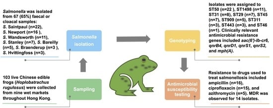
2.1. Sample Collection

All officially registered wet markets in Hong Kong (a total of 94 markets distributed across Hong Kong, including Hong Kong Island, Kowloon, and the New Territories) [18] were visited twice by a research assistant within a period of three months, and markets that sold edible frogs were noted. Of these wet markets, a total of nine (designated A–I) were selected randomly, and a total of 103 live Chinese edible frogs (H. rugulosus) were collected during March and April 2022 (Table 1). The country of origin of the frogs from each market was recorded from the vendors.
The collected frogs were taken to the Department of Infectious Diseases and Public Health, City University of Hong Kong. Physical examinations of the frogs were performed by a certified veterinarian, and age, sex, and weight were documented. For this study, all frogs were considered, regardless of their age and sex.
Anaesthesia was induced by MS-222 (Tricaine methanesulfonate, Syndel, Ferndale, WA, USA) bathing at a concentration of 2 g/L. The MS-222 was buffered by an equal concentration of sodium bicarbonate (Sigma-Aldrich, St. Louis, MO, USA). Once anaesthesia was confirmed, humane euthanasia was induced by an intravenous injection of pentobarbital (Dorminal 20%, Alfasan, Woerden, The Netherlands) at a dose of 100 mg/kg in the ventral abdominal vein. Decapitation was performed as a secondary method of euthanasia, following the guidelines of the American Veterinary Medical Association [19]. During post-mortem examination, faeces or cloacal swabs were aseptically collected, and placed into sterile tubes containing Amies agar gel (Thermo Fisher Scientific, Melbourne, Australia), until further processing.

2.2. Salmonella Isolation and Identification

Salmonella spp. detection was performed, using the ISO 6579-1: 2017 method for isolation and identification of Salmonella [20]. Each swab was placed in 10 mL buffered peptone water (BPW; Thermo Fisher Scientific), and incubated at 37 °C for 18 h. Following incubation, 0.1 mL of cultured BPW was used to inoculate 10 mL Rappaport-Vassiliadis Soya Peptone broth (RVS broth; bioMérieux, Marcy-l’Étoile, France), with incubation at 41.5°C for 24 h. Cultured RVS broth was streaked on Xylose Lysine Deoxycholate agar (XLD agar; Thermo Fisher Scientific), and incubated at 37 °C for 24 h. Salmonella Typhimurium ATCC 14028 was used as a positive control. No plating media, other than XLD, were used, and no other biochemical characterisations were done. Putative Salmonella colonies with black morphologies were subjected to species identification by Matrix-assisted laser desorption/ionisation time-of-flight mass spectrometry (MALDITOF; Bruker, MA, US), and analysed using the MALDI Biotyper (Bruker, MA, US). For calibration and internal quality control of the MALDI Biotyper System, the Bruker Bacterial Test Standard (BTS) containing extract of Escherichia coli DH5 alpha was used, according to the manufacturer’s instructions (Bruker). Salmonella Typhimurium ATCC 14028 was used for internal evaluation.

2.3. Salmonella Serotyping

All isolates were serotyped at the Swiss National Reference Centre for Enteropathogenic Bacteria and Listeria (NENT), Switzerland. Serotyping was performed according to the Kauffmann–White–Le Minor scheme [3], using commercially available antisera (Sifin Diagnostics, Berlin, Germany).

2.4. DNA Extraction and Whole Genome Sequencing

Isolates were grown on sheep blood agar at 37 °C overnight, prior to DNA isolation using the DNeasy Blood & Tissue Kit (Qiagen, Hombrechtikon, Switzerland). DNA libraries were prepared, using a Nextera DNA Flex Sample Preparation Kit (Illumina, San Diego, CA, USA). Whole genome sequencing was performed on an Illumina MiniSeq Sequencer (Illumina). The Illumina reads files passed the standard quality checks, using the software package FastQC 0.11.7 (Babraham Bioinformatics, Cambridge, UK), and were assembled using the Spades 3.14.1-based software Shovill 1.0.4 [21], using default settings. The assembly was filtered, retaining contigs >500 bp, and annotated using the NCBI prokaryotic genome annotation pipeline [22].
Whole-genome-based Salmonella serotyping was performed, using SeqSero with standard settings [23]. Sequence types (STs) were determined using a 7-house-keeping gene-based in silico MLST scheme and the Ridom SeqSphereC + software version 8.5 (Ridom GmbH, Münster, Germany). The genetic relatedness of the isolates was assessed by core genome MLST (cgMLST) analyses based on a 3002-locus cgMLST scheme using Ridom SeqSphere+. A minimum spanning tree (MST) was generated for visualisation, with the threshold for cluster identification set to ≤10 alleles between a pair of neighbouring isolates, according to the Ridom SeqSphere+ software. Antimicrobial resistance genes were determined by the Resistance Gene Identifier (RGI) 4.2.2, using database version 3.2.6 [24]. Virulence factors (VFs) were identified by a bi-directional best-hit approach, using Diamond with standard setting [25]⁠. The Prokka-predicted proteomes were compared to the representative-proteins data set A of the virulence factor database VFDB, downloaded in May 2023 [26]⁠. The aggregate VF score was defined as the number of unique VF detected for each isolate.

2.5. Antimicrobial Susceptibility Testing

Antimicrobial susceptibility tests were performed, using the disk-diffusion method, according to the guidelines of the CLSI [27]. Antimicrobial agents included beta-lactams including penicillins and cephalosporins (ampicillin (AM), amoxicillin/clavulanic acid (AMC), cefazolin (CZ), cefotaxime (CTX), cefepime (FEP)), quinolones including fluoroquinolones (nalidixic acid (NA), ciprofloxacin (CIP)), aminoglycosides (gentamicin (G), kanamycin (K), streptomycin (S)), a sulfonamide (sulfamethoxazole–trimethoprim (SXT)), a phosphonic antibiotic (fosfomycin (FOS)), a macrolide (azithromycin (AZM)), a nitrofuran (nitrofurantoin (F/M)), a tetracycline antibiotic (tetracycline (TE)), and an amphenicol (chloramphenicol (C)) (Becton, Dickinson, Heidelberg, Germany). The MICs of the carbapenems ertapenem (ETP), imipenem (IP), and meropenem (MP) for the blaIND-6-carrying S. Wandsworth ST1498 (isolate F33) were determined using ETest® strips (bioMérieux, Marcy-l’Étoile, France). The results were interpreted according to CLSI breakpoints for human clinical isolates, and the isolates were classified as susceptible (S), intermediate (I), or resistant (R) [27]. In the absence of clinical breakpoints for AZM resistance for Salmonella other than S. enterica serotype Typhi, a zone diameter of ≤ 12 mm was interpreted as resistant [27]. Multidrug resistance (MDR) was defined as resistance to at least three antimicrobials from different classes [28].

2.6. Statistical Analysis

Comparisons of the proportions of samples containing Salmonella from frogs of different age categories (adult or juvenile) and of different sex (male or female) were performed by Fisher’s exact test. The significance criterion was set at p ≤ 0.05. Calculations were performed using GraphPad (https://www.graphpad.com, accessed on 8 March 2023).

2.7. Ethics Statement

This study was approved of by the Animal Research Ethics Sub-Committee of the City University of Hong Kong (Internal Ref: A- 0698).

3. Results

3.1. Demographic Data of H. rugulosus Collected from Different Wet Markets

Of the 103 collected frogs, 76 (74%) were adult, and 27 (26%) were juvenile animals. A total of 81 (79%) were male, 21 (20%) were female, and 1 (1%) was a hermaphrodite. The median weight was 164 g (range 91–292 g). Frogs purchased in wet markets A–H were imported from mainland China, whereas those from market I were imported from Thailand.

3.2. Prevalence of Salmonella among H. rugulosus

Salmonella enterica subsp. Enterica was isolated from 67 (65%, 95% CI: 0.554–0.736) of the frogs (Table 1). Frogs harbouring Salmonella were collected from all nine wet markets, and the proportions of Salmonella-positive animals varied, from 17% of the animals collected from wet market C, to 100% of those from markets D, E, and H (Table 1).
The proportions of adult and of juvenile frogs that tested positive for Salmonella were 48/76 (63%) and 19/27 (70%), respectively—a difference that was not statistically significant (p = 0.8600). The proportions of male and female frogs harbouring Salmonella were 50/81 (62%) and 16/21 (76%), respectively: again, the difference was not significant (p = 0.5742).

3.3. Serotypes

A total of seven different Salmonella enterica subsp. Enterica serotypes were identified, using the Kauffmann–White–Le Minor scheme, and were later confirmed by WGS and serotyping by the SeqSero scheme (Table 1). Among the 67 isolates, S. Saintpaul (n = 22) and S. Newport (n = 16) were the predominant serotypes, accounting for 33% and 24% of all Salmonella isolates, respectively. Other serotypes included S. Bareilly (n = 5; 7%), S. Braenderup (n = 3; 4%), S. Hvittingfoss (n = 3; 4%), S. Stanley (n = 7; 10%), and S. Wandsworth (n = 11; 16%) (Table 1).

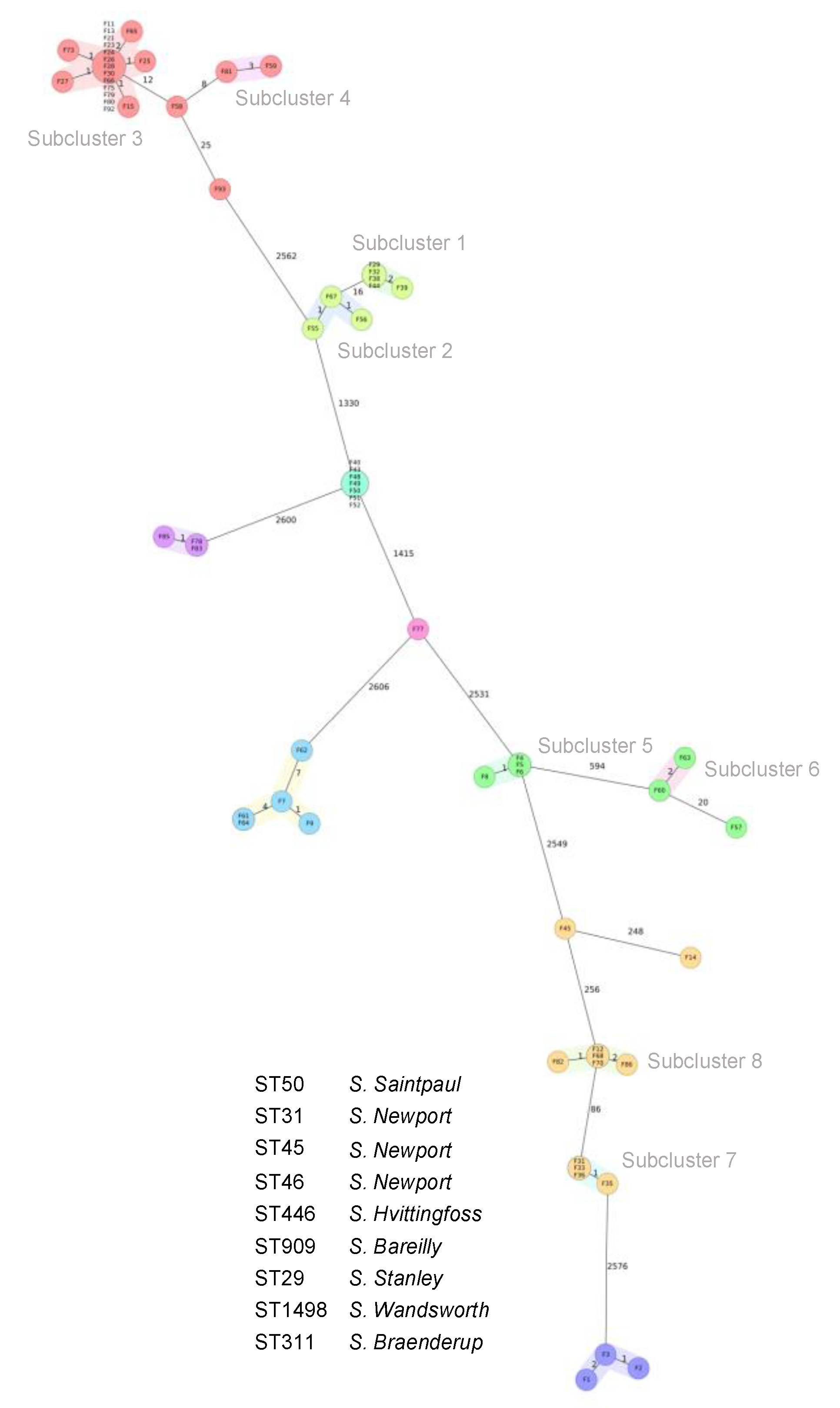
3.4. Core Genome Multilocus Sequence Types (cgMLST) and Phylogenetic Relatedness

Based on WGS data, the 67 isolates were assigned to nine different STs, whereby ST50 (n = 22; 33%) and ST1498 (n = 11;16%) were predominant (Table 2). Isolates from the same serotype were assigned to the same ST, except for the S. Newport isolates, which showed three different MLST patterns corresponding to ST31 (n = 8), ST45 (n = 7), and ST46 (n = 1), respectively (Table 2). The genetic relatedness of the isolates was visualised by constructing a cgMLST-based phylogenetic tree. The isolates grouped according to serotypes and STs, which were separated by clear allelic distances (Figure 1). Isolates belonging to S. Bareilly, S. Braenderup, and S. Hvittingfoss clustered tightly within their respective serotypes and STs. By contrast, the S. Newport ST31 isolates formed two different subclusters (subcluster 1 and subcluster 2 in Figure 1), which consisted of five and three isolates, respectively, while the S. Newport ST45 isolates were indistinguishable by cgMLST (Figure 1). Isolates belonging to S. Saintpaul ST50 appeared in two subclusters, with one major subcluster containing 18 isolates, and one minor subcluster formed by two isolates (subcluster 3 and subcluster 4 in Figure 1). Two further S. Saintpaul isolates were observed at distinct positions in the tree (Figure 1). Likewise, the S. Stanley ST29 isolates formed two clusters consisting of four and two isolates, respectively (subcluster 5 and subcluster 6 in Figure 1). Finally, the S. Wandsworth ST1438 isolates comprised two subclusters containing five and four isolates, respectively (subcluster 7 and subcluster 8), and two distinctly separated isolates (Figure 1).

3.5. Antimicrobial Resistance Genotypes

Analysis of WGS data identified a number of antimicrobial resistance genes, including genes conferring resistance to aminoglycosides (aadA16, ant(3″)-Ib, aph(3″)-Ib, aph(3′)-Ia, ant(3″)-IIa, aac(6′)-Ib-cr6, aph(6′)-Id), beta-lactams (blaDHA-1, blaIND-6, blaOXA-1, blaOXA-10, blaTEM-1, blaTEM-60), chloramphenicol (catB3, catI, catII), diaminopyrimidines (dfrA14, dfrA27, dfrB4), fluoroquinolones (aac(6′)-Ib-cr6, qnrB4, qnrD1, qnrS1, qnrS2), lincosamides (linG), macrolides (mphA), rifamycin (arr-2, arr-3), sulfonamides (sul1, sul2), tetracycline (tet(A), tet(D), and vancomycin (vanX). The distribution of antimicrobial resistance genes among the isolates is shown in Table 2.
The dfrA27 gene was identified in S. Wandsworth ST1498, while blaOXA-1, blaDHA-1, catB3, and qnrB4 were found exclusively among S. Newport ST45 (Table 2).
The metallo-beta-lactamase gene blaIND-6 was found in one S. Wandsworth ST1498 (isolate F33) (Table 2). Notably, vanX was identified in one S. Wandsworth ST1498 (isolate F86), but was not related to vancomycin resistance, because Gram-negative bacteria are insensitive to vancomycin [31]. All isolates carried either aac(6′)-Iy or aac(6′)-Iaa, which are cryptic genes that do not confer phenotypic aminoglycoside resistance [29,30]. Furthermore, all isolates contained a large number of genes encoding MDR efflux pumps, and genes encoding for antibiotic target alterations (Supplemental Table S1): among the latter, the gyrA (S83F) mutation was detected in one S. Newport ST46 (isolate F77) (Supplemental Table S1). Additional data are provided in supplemental File S1.

3.6. Antimicrobial Susceptibility Profiles

Among the 67 Salmonella isolates, resistance to ampicillin (n = 16; 24%), amoxycillin/clavulanic acid (n = 6; 9%), cefazolin (n = 6; 9%), nalidixic acid (n = 24; 36%), ciprofloxacin (n = 15; 22%), gentamicin (n = 1; 1.5%), kanamycin (n = 7; 10%), streptomycin (n = 9; 13%), sulfamethoxazole–trimethoprim (n = 8; 12%), azithromycin (n = 5; 7%), tetracycline (n = 38; 57%), and chloramphenicol (n = 7;10%) was observed (Table 2). MDR was observed for 14 (21%) of the isolates (Table 2). The antimicrobial susceptibility profiles are detailed in supplemental Table S2. Notably, intermediate resistance to cefotaxime (n = 1; 1.5%), cefepime (n = 1; 1.5%), nalidixic acid (n = 17; 25%), ciprofloxacin (n = 52; 78%), streptomycin (n = 2; 3%), nitrofurantoin (n = 1; 1.5%), and chloramphenicol (n = 2; 3%) was found (Supplemental Table S2). The MIC values for ertapenem, imipenem, and meropenem in Wandsworth ST1498 (isolate F33) were 0.002 µg/mL, 0.094 µg/mL, and 0.08 µg/mL, respectively.
None of the isolates were resistant to cefotaxime, cefepime, fosfomycin, or nitrofurantoin (Table 2 and Supplemental Table S2). There was a correlation between phenotypic antimicrobial resistance and the presence of at least one ARG encoding resistance to aminoglycosides, amoxicillin/clavulanic acid, folate pathway inhibitors, phenicols, macrolides, and tetracycline, but no concordance between phenotypic and genotypic resistance to extended-spectrum beta-lactams and nalidixic acid (Table 2).

3.7. Virulence Factors

The results of the VFDB-based virulence profiling for the 67 Salmonella isolates are shown in Supplemental Table S3. The majority of the genes (352/496) were present universally in all isolates. The median aggregate VF scores (and ranges) for isolates belonging to different serotypes were the following: S. Bareilly VF 394 (393–395); S. Braenderup VF 409 (409–409); S. Hvittingfoss VF 419 (419–419); S. Newport VF 415 (403–433); S. Saintpaul VF 429 (427–434); S. Stanley VF 401(401–404); and S. Wandsworth VF 413 (408–418). Among the fimbrial adherence determinants, the majority (19/32) were common to all Salmonella isolates, except cfaA, cfaB, and cfaC, which were additionally present in S. Bareilly (n = 5), S. Stanley (n = 4), S. Wandsworth (n = 2), and S. Newport ST46 (n = 1). Likewise, the genes belonging to the Salmonella enterica type III secretion system, sopD, sopE2, slrP, sifA, sseL, sptP, sipA/sspA, steC, steA, and sopD, were common to all isolates (Supplemental Table S3). By contrast, VFs belonging to the Aeromonas hydrophila type VI secretion system were present solely in S. Wandsworth (n = 11) [32,33] (Supplemental Table S3). Another set of VFs that were present in all isolates were the toxin genes cyaB, cyaD, senB, cylA, and plcD [34].

4. Discussion

Although frog meat is a popular food worldwide [35], there is a lack of information regarding the occurrence of Salmonella in frogs sold for human consumption. In the current study, the overall prevalence of Salmonella carriage among H. rugulosus obtained from wet markets in Hong Kong was 65%, and included a variety of Salmonella serotypes, with many isolates featuring genotypic and phenotypic resistance to clinically relevant antimicrobials.
Among the isolates, several serovars, including S. Hvittingfoss, S. Newport, S. Stanley, and S. Wandsworth, had been described previously in H. rugulosus farmed in Thailand [36]. Furthermore, S. Saintpaul and S. Wandsworth had been isolated from H. rugulosus used as feeder frogs for captive reptiles in Thailand [37]. These reports are suggestive of frogs as natural hosts for these serotypes. Some serotypes in this study are infrequently reported, but have nevertheless been associated with human cases of salmonellosis. For example, the rare serotype S. Hvittingfoss ST446, found in three frogs in this study, was recently linked to an outbreak of salmonellosis in travellers returning from Hong Kong [38]. Likewise, S. Wandsworth, identified in 11 frogs, was associated with infections in small children in the USA in 2009, and with peritonitis in a patient in Hong Kong in 2022 [39,40]. The identification of this rare serotype among 16% of the frogs indicates that S. Wandsworth may be widespread among frogs, and that frogs may represent an important reservoir for this serotype. Other serotypes found in this study are more frequent among human infections, and have also been implicated in recent disease outbreaks in the EU (S. Bareilly, S. Braenderup, S. Newport, S. Stanley) [6], or are endemic in Asia but less frequent in Europe, such as S. Stanley [41].
Phylogenetic analysis showed that many of the Salmonella STs in this study were closely related by cgMLST, with subclusters observed among isolates from frogs obtained from different markets. Despite the lack of details on the acquisition of the frogs sold at the wet markets, these subclusters may be considered to reflect a common origin of the frogs, e.g., farm or location of capture. In particular, the clonality of the MDR S. Newport ST45 isolates is suggestive of a lineage deriving from a common source.
Multiple genes conferring resistance to antimicrobials that are listed as critically important, or very important to human health, were detected [42]. One of the clinically relevant AMR genes included blaDHA-1, identified among six MDR S. Newport ST45. DHA-1 belongs to the family of plasmid-mediated AmpC beta-lactamases that confer resistance to the beta-lactamase inhibitor clavulanic acid [43]. Accordingly, all DHA-1-producing S. Newport in this study were phenotypically resistant to ampicillin and to amoxycillin/clavulanic acid. The blaDHA-1 gene is infrequently reported in Salmonella and other Enterobacterales [44]. To the best of our knowledge, this is the first report of blaDHA-1 in S. Newport isolated from amphibians, although one DHA-1-producing human clinical S. Newport was described in Spain between 2004 and 2009 [45]. One of the most prevalent AMR genes in this study was blaTEM-60, which encodes an extended-spectrum beta-lactamase (ESBL); however, in this study, blaTEM-60-carriers failed to exhibit a detectable ESBL phenotype. Likewise, the blaIND-6-harbouring S. Wandsworth did not exhibit resistance to carbapenems. By contrast, the abundance of the plasmid-mediated quinolone resistance (PMQR) genes aac(6′)-Ib-cr6, qnrB4, qnrS1, and qnrS2 among the isolates was reflected in a high proportion of nalidixic-acid- and ciprofloxacin-resistant isolates. Markedly, none of the isolates in this study were fully susceptible to ciprofloxacin. PMQR among Salmonella is of concern, because it has the potential to spread horizontally, and, although PMQR genes only confer low-level resistance to quinolones, they promote step-wise development to high-level fluoroquinolone resistance [46,47]. The presence of PMQR genes in human pathogens isolated from animals destined for human consumption is of concern, because Salmonella with decreased ciprofloxacin resistance carrying PMQR genes have been linked to outbreaks, as previously described, in the UK with S. Virchow [48], in the USA with S. Newport [49], and in Spain with S. Corvallis [50].
With resistance to fluoroquinolones increasing worldwide, azithromycin is currently considered an important alternative for the treatment of invasive salmonellosis [42]. Azithromycin resistance in human NTS is currently rare in the USA (0.5%) and Europe (0.8%) [51,52]. Moreover, resistance to azithromycin was either not detected, or was identified only at very low levels in food-producing animals, including calves (0%), broilers (1.9%), pigs (0.3%), and turkeys (1.2%), in the EU, between 2019 and 2020 [52]. By contrast, azithromycin resistance, mainly associated with the presence of mphA, has been increasing over time, and seems to be more prevalent in human NTS in Europe than in Taiwan (3.1%) [51], and in Salmonella from humans, animals, and food in China (3.9%) [53]. The observation, in our study, that azithromycin resistance and the mph gene occur more frequently in Salmonella of amphibian origin (5.7%) than in Salmonella of livestock or human origin may suggest the potential of H. rugulosus as a source of dissemination of azithromycin-resistant Salmonella.
Animals including amphibians may acquire antimicrobial-resistant bacteria from other animals, their environment, or their feed. Little information is available regarding frog farming and the usage of antimicrobial agents in frog farms in Asia; however, a recent report from Vietnam indicates that antimicrobials, such as amoxicillin, enrofloxacin, ciprofloxacin, sulfamethoxazole–trimethoprim, doxycycline, oxytetracycline, tetracycline, florfenicol, and rifampicin, are commonly applied empirically by frog farmers to treat frog diseases [16,54]: thus, the use and misuse of large amounts of different antimicrobials may select for resistant strains in farmed frogs. Although information on the origins of the frogs analysed in this study was available at country level, data on the aquacultural or natural settings were lacking, and it cannot be excluded that many of the frogs may have been farmed under comparable conditions. Similarly to our study, a recent investigation on wet markets in Hong Kong recently reported a high prevalence (42%) of Salmonella, and a high proportion of AMR isolates, among edible freshwater turtles, further underlining the potential risk of Salmonella and AMR in non-traditional aquaculture [55].
The presence of genotypic and phenotypic resistance characteristics in Salmonella in food animals is worrying, and indicates that first-line antibiotic ciprofloxacin, as well as ampicillin, amoxycillin/clavulanic acid, and azithromycin, used against such strains, may fail, should infectious transmissions to humans occur. Likewise, isolates showing intermediate resistance, i.e., reduced susceptibility, are of concern for public health. Reduced susceptibility is important, because it facilitates the selection of isolates with higher-level resistance, and contributes to the development and spread of resistance to critically important antimicrobial agents, such as ciprofloxacin [46,47].
The in silico screening of the VFs revealed an abundance of VFs in all the isolates whilst, concurrently, the differences in VF content among the various serotypes was rather unremarkable: these findings are in agreement with previous studies, in which VFs were reportedly conserved among NT Salmonella serotypes [56]. Based on the distribution of the VFs, and the lack of variation of median aggregate VF scores among the serotypes, there was no specific Salmonella serotype in this study that showed a distinct virulence profile; however, the universal presence of fimbrial genes, genes encoding secretion systems, and toxins among the isolates was an indication that these Salmonella serotypes have the potential to cause disease in humans.
This study had some limitations. Firstly, the interpretation of the results should take into consideration that sampling was restricted to H. rugulosus because this is the only frog species sold in wet markets in Hong Kong: thus, the results cannot be generalised to other frog species that may be available for human consumption elsewhere. Secondly, there were unequal sample sizes among the different wet markets from which the frogs were obtained; therefore, there remains the possibility of unintended overrepresentation of the proportion of positive samples from individual markets, which were randomly selected. Thirdly, the study was challenged by the lack of any previous data on the prevalence of Salmonella in frogs, and lack of data on frog import and retail volumes in Hong Kong; therefore, essentially, this study was a pilot study, to collect some baseline data. Finally, the sampling period was short: it should be considered that the prevalence of Salmonella among the frogs may have been subject to unknown factors—for example, fluctuations among the suppliers of the wet markets—and that such differences would have remained undetected.

5. Conclusions

This study demonstrates that a high proportion of live frogs sold for human consumption in wet markets are carriers of Salmonella, including serotypes that are frequently linked to cases of human disease, and Salmonella that contain clinically relevant AMR genes. To the best of the authors’ knowledge, no official advice or guidelines exist regarding the handling of live edible frogs. To this end, the results presented in this study offer useful information for specialists in the public health and food safety sectors. Raised awareness and public health recommendations for hygiene requirements at the stages of handling and processing of edible frogs and their meat should be considered, to mitigate the risk of transmission of susceptible and resistant Salmonella to humans. The role of edible frogs and derived meat products, e.g., frog legs, as sources of antimicrobial-resistant and susceptible Salmonella and other foodborne pathogens, needs to be further investigated in future.

Supplementary Materials

The following supporting information can be downloaded at https://www.mdpi.com/article/10.3390/foods12112245/s1. Table S1: Antibiotic efflux pump genes and target alteration resistance mechanisms detected by RGI software in 67 Salmonella from Chinese edible frogs (Hoplobatrachus rugulosus) collected from wet markets in Hong Kong. Table S2: Antimicrobial susceptibility determined by disk diffusion of 67 Salmonella isolated from Chinese edible frogs (Hoplobatrachus rugulosus) from wet markets in Hong Kong. Table S3: Distribution of virulence genes detected via VFDB software in 67 Salmonella from Chinese edible frogs (Hoplobatrachus rugulosus) collected from wet markets in Hong Kong. File S1: Metadata of antimicrobial resistance genes collected by whole genome sequencing of 67 Salmonella isolates.

Author Contributions

Conceptualization, I.M. and V.A.C.; data curation, M.J.A.S.; formal analysis, S.B., M.J.A.S., J.A.H., M.N.-I., K.L. and V.A.C.; funding acquisition, I.M., R.S.; investigation, V.A.C. and K.L.; methodology, K.L., S.B., M.J.A.S., J.A.H. and V.A.C.; project administration, I.M. and R.S.; resources, I.M. and R.S.; supervision, I.M. and R.S.; visualization: M.J.A.S. and M.N.-I.; writing—original draft, S.B. and M.N.-I.; writing—review and editing, S.B., M.J.A.S., M.N.-I., V.A.C., K.L., I.M. and R.S. All authors have read and agreed to the published version of the manuscript.

Funding

This research was funded by the City University of Hong Kong, start-up grant for new Faculty (project no. 9610449) and the teaching development grant (project no. 6000701). This work was supported in part by the Swiss Federal Office of Public Health, Division of Communicable Diseases.

Data Availability Statement

Sequencing read data and genome assemblies have been deposited under BioProject accession number PRJNA966132. Accession numbers for the individual isolates from this study are listed in Table 2.

Acknowledgments

We thank Nicole Cernela for technical support with Illumina sequencing, and Sandra Schoch for assistance with serotyping.

Conflicts of Interest

The authors declare no conflict of interest.

References

  1. Besser, J.M. Salmonella epidemiology: A whirlwind of change. Food Microbiol. 2018, 71, 55–59. [Google Scholar] [CrossRef] [PubMed]
  2. Issenhuth-Jeanjean, S.; Roggentin, P.; Mikoleit, M.; Guibourdenche, M.; De Pinna, E.; Nair, S.; Fields, P.I.; Weill, F.-X. Supplement 2008–2010 (no. 48) to the White–Kauffmann–Le Minor scheme. Res. Microbiol. 2014, 165, 526–530. [Google Scholar] [CrossRef] [Green Version]
  3. Grimont, P.A.D.; Weill, F.-X. Antigenic formulae of the Salmonella serovars. WHO Collab. Cent. Ref. Res. Salmonella 2007, 9, 1–166. [Google Scholar]
  4. Achtman, M.; Wain, J.; Weill, F.-X.; Nair, S.; Zhou, Z.; Sangal, V.; Krauland, M.G.; Hale, J.L.; Harbottle, H.; Uesbeck, A.; et al. Multilocus sequence typing as a replacement for serotyping in Salmonella enterica. PLoS Pathog. 2012, 8, e1002776. [Google Scholar] [CrossRef] [Green Version]
  5. Chattaway, M.A.; Langridge, G.C.; Wain, J. Salmonella nomenclature in the genomic era: A time for change. Sci. Rep. 2021, 11, 7494. [Google Scholar] [CrossRef] [PubMed]
  6. European Food Safety Authority and European Centre for Disease Prevention and Control. The European Union One Health 2021 zoonoses report. EFSA J. 2022, 20, e07666. [Google Scholar] [CrossRef]
  7. Ferrari, R.G.; Rosario, D.K.A.; Cunha-Neto, A.; Mano, S.B.; Figueiredo, E.E.S.; Conte-Junior, C.A. Worldwide epidemiology of Salmonella serovars in animal-based foods: A meta-analysis. Appl. Environ. Microbiol. 2019, 85, e00591-19. [Google Scholar] [CrossRef] [Green Version]
  8. Centers for Disease Control and Prevention. National Enteric Disease Surveillance: Salmonella Annual Report. Available online: https://www.cdc.gov/nationalsurveillance/pdfs/2016-Salmonella-report-508.pdf (accessed on 14 April 2023).
  9. Li, W.; Han, H.; Liu, J.; Ke, B.; Zhan, L.; Yang, X.; Tan, D.; Yu, B.; Huo, X.; Ma, X.; et al. Antimicrobial resistance profiles of Salmonella isolates from human diarrhea cases in China: An eight-year surveillance study. One Health Adv. 2023, 1, 1–8. [Google Scholar] [CrossRef]
  10. Zollner-Schwetz, I.; Krause, R. Therapy of acute gastroenteritis: Role of antibiotics. Clin. Microbiol. Infect. 2015, 21, 744–749. [Google Scholar] [CrossRef] [PubMed] [Green Version]
  11. Medalla, F.; Gu, W.; Mahon, B.E.; Judd, M.; Folster, J.; Griffin, P.M.; Hoekstra, R.M. Estimated incidence of antimicrobial drug-resistant nontyphoidal Salmonella infections, United States, 2004–2012. Emerg. Infect. Dis. 2016, 23, 29–37. [Google Scholar] [CrossRef] [Green Version]
  12. Mølbak, K. Human health consequences of antimicrobial drug-resistant Salmonella and other foodborne pathogens. Clin. Infect. Dis. 2005, 41, 1613–1620. [Google Scholar] [CrossRef] [PubMed]
  13. Pulcini, C.; Beovic, B.; Béraud, G.; Carlet, J.; Cars, O.; Howard, P.; Levy-Hara, G.; Li, G.; Nathwani, D.; Roblot, F.; et al. Ensuring universal access to old antibiotics: A critical but neglected priority. Clin. Microbiol. Infect. 2017, 23, 590–592. [Google Scholar] [CrossRef] [PubMed] [Green Version]
  14. European Food Safety Authority and European Centre for Disease Prevention and Control. The European Union One Health 2019 zoonoses report. EFSA J. 2021, 19, e06406. [Google Scholar] [CrossRef]
  15. Altherr, S.; Auliya, M.; Nithart, C. Deadly dish—Role and Responsibility of the European Union in the International Frogs’ Legs Trade. Report by Pro Wildlife & Robin des Bois. 2022. Available online: https://www.prowildlife.de/wp-content/uploads/2022/06/DEADLY (accessed on 21 March 2023).
  16. Auliya, M.; Altherr, S.; Nithart, C.; Hughes, A.; Bickford, D. Numerous uncertainties in the multifaceted global trade in frogs’ legs with the EU as the major consumer. Nat. Conserv. 2023, 51, 71–135. [Google Scholar] [CrossRef]
  17. Grano, M. The Asian market of frogs as food for humans during COVID-19. Risk and consequences for public health. Med. Pap. 2020, 6, 77–87. [Google Scholar]
  18. Food and Environmental Hygiene Department. List of FEHD Public Markets/Cooked Food Markets. Available online: https://www.fehd.gov.hk/english/pleasant_environment/tidy_market/Markets_CFC_list.html (accessed on 3 May 2023).
  19. Underwood, W.; Raymond, A. American Veterinary Medical Association AVMA Guidelines for the Euthanasia of Animals: 2020 Edition. Available online: https://www.spandidos-publications.com/var/AVMA_euthanasia_guidelines_2020.pdf (accessed on 1 March 2023).
  20. Mooijman, K.A.; Pielaat, A.; Kuijpers, A.F.A. Validation of EN ISO 6579-1-Microbiology of the food chain-Horizontal method for the detection, enumeration and serotyping of Salmonella-Part 1 detection of Salmonella spp. Internat. J. Food Microbiol. 2019, 288, 3–12. [Google Scholar] [CrossRef]
  21. Seemann, T.S. Available online: https://githubcom/tseemann/shovill (accessed on 13 February 2023).
  22. Tatusova, T.; DiCuccio, M.; Badretdin, A.; Chetvernin, V.; Nawrocki, E.P.; Zaslavsky, L.; Lomsadze, A.; Pruitt, K.D.; Borodovsky, M.; Ostell, J. NCBI prokaryotic genome annotation pipeline. Nucleic. Acids Res. 2016, 44, 6614–6624. [Google Scholar] [CrossRef]
  23. Zhang, S.; Yin, Y.; Jones, M.B.; Zhang, Z.; Deatherage Kaiser, B.L.; Dinsmore, B.A.; Fitzgerald, C.; Fields, P.I.; Deng, X. Salmonella serotype determination utilizing high-throughput genome sequencing data. J. Clin. Microbiol. 2015, 53, 1685–1692. [Google Scholar] [CrossRef] [Green Version]
  24. Bortolaia, V.; Kaas, R.S.; Ruppe, E.; Roberts, M.C.; Schwarz, S.; Cattoir, V.; Philippon, A.; Allesoe, R.L.; Rebelo, A.R.; Florensa, A.F.; et al. ResFinder 4.0 for predictions of phenotypes from genotypes. J. Antimicrob. Chemother. 2020, 75, 3491–3500. [Google Scholar] [CrossRef]
  25. Buchfink, B.; Reuter, K.; Drost, H.-G. Sensitive protein alignments at tree-of-life scale using DIAMOND. Nat. Methods 2021, 18, 366–368. [Google Scholar] [CrossRef]
  26. Liu, B.; Zheng, D.; Zhou, S.; Chen, L.; Yang, J. VFDB 2022: A general classification scheme for bacterial virulence factors. Nucleic. Acids Res. 2022, 50, D912–D917. [Google Scholar] [CrossRef] [PubMed]
  27. CLSI. Performance Standards for Antimicrobial Susceptibility Testing, 33rd ed.; CLSI Supplement M100; Clinical and Laboratory Standards Institute: Wayne, PA, USA, 2023. [Google Scholar]
  28. Magiorakos, A.-P.; Srinivasan, A.; Carey, R.B.; Carmeli, Y.; Falagas, M.E.; Giske, C.G.; Harbarth, S.; Hindler, J.F.; Kahlmeter, G.; Olsson-Liljequist, B.; et al. Multidrug-resistant, extensively drug-resistant and pandrug-resistant bacteria: An international expert proposal for interim standard definitions for acquired resistance. Clin. Microbiol. Infect. 2012, 18, 268–281. [Google Scholar] [CrossRef] [PubMed] [Green Version]
  29. Salipante, S.J.; Hall, B.G. Determining the limits of the evolutionary potential of an antibiotic resistance gene. Mol. Biol. Evol. 2003, 20, 653–659. [Google Scholar] [CrossRef] [PubMed]
  30. Magnet, S.; Lambert, T.; Courvalin, P.; Blanchard, J.S. Kinetic and mutagenic characterization of the chromosomally encoded Salmonella enterica AAC(6’)-Iy aminoglycoside N-acetyltransferase. Biochemistry 2001, 40, 3700–3709. [Google Scholar] [CrossRef]
  31. Rowe-Magnus, D.A.; Mazel, D. Resistance gene capture. Curr. Opin. Microbiol. 1999, 2, 483–488. [Google Scholar] [CrossRef]
  32. Dos Santos, A.M.P.; Ferrari, R.G.; Conte-Junior, C.A. Virulence factors in Salmonella Typhimurium: The sagacity of a bacterium. Curr. Microbiol. 2019, 76, 762–773. [Google Scholar] [CrossRef]
  33. Ma, S.; Dong, Y.; Wang, N.; Liu, J.; Lu, C.; Liu, Y. Identification of a new effector-immunity pair of Aeromonas hydrophila type VI secretion system. Vet. Res. 2020, 51, 1–14. [Google Scholar] [CrossRef]
  34. Banerji, R.; Karkee, A.; Kanojiya, P.; Saroj, S.D. Pore-forming toxins of foodborne pathogens. Compr. Rev. Food Sci. Food Saf. 2021, 20, 2265–2285. [Google Scholar] [CrossRef]
  35. Auliya, M.; Altherr, S.; Hughes, A.; Nithart, C.; Ohler, A.; Bickford, D. The European market remains the largest consumer of frogs’ legs from wild species. Conservation 2023, 3, 53–58. [Google Scholar] [CrossRef]
  36. Ribas, A.; Poonlaphdecha, S. Wild-caught and farm-reared amphibians are important reservoirs of Salmonella, a study in North-East Thailand. Zoonoses Public Health 2017, 64, 106–110. [Google Scholar] [CrossRef]
  37. Prapasarakul, N.; Pulsrikarn, C.; Vasaruchapong, T.; Lekcharoen, P.; Chanchaithong, P.; Lugsomya, K.; Keschumras, N.; Thanomsuksinchai, N.; Tanchiangsai, K.; Tummaruk, P. Salmonella serovar distribution in cobras (Naja kaouthia), snake-food species, and farm workers at Queen Saovabha Snake Park, Thailand. J. Vet. Diagn. Investig. 2012, 24, 288–294. [Google Scholar] [CrossRef] [Green Version]
  38. Fang, Y.; Huang, H.; Li, B.; Ruan, F.; Li, Z.; Huang, W.; Wei, Q.; Huang, K. An outbreak of Salmonella Hvittingfoss infection in a tourist group back from Hong Kong, Southeast China. J. Infect. 2022, 84, e28–e30. [Google Scholar] [CrossRef] [PubMed]
  39. Pak, W.L.W.; Chan, K.L.; Chan, Z.; Law, W.P.; Wong, Y.H.; Lam, C.K.; Wong, S.H.S. Salmonella Wandsworth peritoneal dialysis-related peritonitis following persistent infection: A case report with a review of literature. Nephrology 2022, 27, 468–470. [Google Scholar] [CrossRef]
  40. Sotir, M.J.; Ewald, G.; Kimura, A.C.; Higa, J.I.; Sheth, A.; Troppy, S.; Meyer, S.; Hoekstra, R.M.; Austin, J.; Archer, J.; et al. Outbreak of Salmonella Wandsworth and Typhimurium infections in infants and toddlers traced to a commercial vegetable-coated snack food. Pediatr. Infect. Dis. J. 2009, 28, 1041–1046. [Google Scholar] [CrossRef] [PubMed]
  41. Aung, K.T.; Khor, W.C.; Octavia, S.; Ye, A.; Leo, J.; Chan, P.P.; Lim, G.; Wong, W.K.; Tan, B.Z.Y.; Schlundt, J. Distribution of Salmonella serovars in humans, foods, farm animals and environment, companion and wildlife animals in Singapore. Int. J. Environ. Res. Public Health 2020, 17, 5774. [Google Scholar] [CrossRef] [PubMed]
  42. World Health Organization. Critically Important Antimicrobials for Human Medicine, 6th ed.; WHO: Geneva, Switzerland, 2019. [Google Scholar]
  43. Bush, K. A resurgence of β-lactamase inhibitor combinations effective against multidrug-resistant Gram-negative pathogens. Int. J. Antimicrob. Agents. 2015, 46, 483–493. [Google Scholar] [CrossRef]
  44. Hennequin, C.; Ravet, V.; Robin, F. Plasmids carrying DHA-1 β-lactamases. Eur. J. Clin. Microbiol. Infect. Dis. 2018, 37, 1197–1209. [Google Scholar] [CrossRef] [PubMed]
  45. Pérez-Moreno, M.O.; Picó-Plana, E.; de Toro, M.; Grande-Armas, J.; Quiles-Fortuny, V.; Pons, M.J.; Gomes, C.; Sáenz, Y.; Torres, C.; Ruiz, J. β-Lactamases, transferable quinolone resistance determinants, and class 1 integron-mediated antimicrobial resistance in human clinical Salmonella enterica isolates of non-Typhimurium serotypes. Int. J. Med. Microbiol. 2013, 303, 25–31. [Google Scholar] [CrossRef]
  46. Robicsek, A.; Strahilevitz, J.; Sahm, D.F.; Jacoby, G.A.; Hooper, D.C. qnr prevalence in ceftazidime-resistant Enterobacteriaceae isolates from the United States. Antimicrob. Agents. Chemother. 2006, 50, 2872–2874. [Google Scholar] [CrossRef] [Green Version]
  47. Robicsek, A.; Strahilevitz, J.; Jacoby, G.A.; Macielag, M.; Abbanat, D.; Park, C.H.; Bush, K.; Hooper, D.C. Fluoroquinolone-modifying enzyme: A new adaptation of a common aminoglycoside acetyltransferase. Nat. Med. 2006, 12, 83–88. [Google Scholar] [CrossRef] [PubMed]
  48. Hopkins, K.L.; Wootton, L.; Day, M.R.; Threlfall, E.J. Plasmid-mediated quinolone resistance determinant qnrS1 found in Salmonella enterica strains isolated in the UK. J. Antimicrob. Chemother. 2007, 59, 1071–1075. [Google Scholar] [CrossRef] [PubMed] [Green Version]
  49. Lin, D.; Chen, K.; Wai-Chi Chan, E.; Chen, S. Increasing prevalence of ciprofloxacin-resistant food-borne Salmonella strains harboring multiple PMQR elements but not target gene mutations. Sci. Rep. 2015, 5, 14754. [Google Scholar] [CrossRef] [PubMed] [Green Version]
  50. Vázquez, X.; Fernández, J.; Hernáez, S.; Rodicio, R.; Rodicio, M.R. Plasmid-mediated quinolone resistance (PMQR) in two clinical strains of Salmonella enterica serovar Corvallis. Microorganisms 2022, 10, 579. [Google Scholar] [CrossRef] [PubMed]
  51. Chiou, C.S.; Hong, Y.P.; Wang, Y.W.; Chen, B.H.; Teng, R.H.; Song, H.Y.; Liao, Y.S. Antimicrobial resistance and mechanisms of azithromycin resistance in nontyphoidal Salmonella isolates in Taiwan, 2017 to 2018. Microbiol. Spectr. 2023, 11, e0336422. [Google Scholar] [CrossRef]
  52. European Food Safety Authority and European Centre for Disease Prevention and Control. The European Union summary report on antimicrobial resistance in zoonotic and indicator bacteria from humans, animals and food in 2019–2020. EFSA J. 2022, 20, 1–197. [Google Scholar] [CrossRef]
  53. Zhang, Z.; Yang, J.; Xu, X.; Zhou, X.; Shi, C.; Zhao, X.; Liu, Y.; Shi, X. Co-existence of mphA, oqxAB and blaCTX-M-65 on the IncHI2 plasmid in highly drug-resistant Salmonella enterica serovar Indiana ST17 isolated from retail foods and humans in China. Food Control 2020, 118, 107269. [Google Scholar] [CrossRef]
  54. Nguyen, Q.T.; Tran, M.P. Drugs and chemicals use in frog farming in Dong Thap province. Can. Tho. Univ. J. Sc. 2021, 13, 73–78. [Google Scholar] [CrossRef]
  55. Colon, V.A.; Lugsomya, K.; Lam, H.K.; Wahl, L.C.; Parkes, R.S.V.; Cormack, C.A.; Horlbog, J.A.; Stevens, M.; Stephan, R.; Magouras, I. Serotype diversity and antimicrobial resistance profile of Salmonella enterica isolates from freshwater turtles sold for human consumption in wet markets in Hong Kong. Front Vet. Sci. 2022, 9, 912693. [Google Scholar] [CrossRef]
  56. Hassena, A.B.; Haendiges, J.; Zormati, S.; Guermazi, S.; Gdoura, R.; Gonzalez-Escalona, N.; Siala, M. Virulence and resistance genes profiles and clonal relationships of non-typhoidal food-borne Salmonella strains isolated in Tunisia by whole genome sequencing. Intern. J. Food. Microbiol. 2021, 337, 108941. [Google Scholar] [CrossRef]
Figure 1. Phylogenetic relationship of 67 Salmonella serotypes isolated from Chinese edible frogs (Hoplobatrachus rugulosus), based on their core genome multilocus sequence type (cgMLST) allelic profiles. The minimum spanning tree was generated using SeqSphere+ (Ridom GmbH). The colours of the circles represent STs according to in silico MLST. Salmonella serotypes and subclusters 1–8 are indicated. Clusters are displayed by pastel background colours. Numbers on connecting lines indicate the number of allelic differences between two strains. Each circle contains the strain ID(s), as shown in Table 2.
Foods 12 02245 g001
Table 1. Origin, number of samples, and proportion of Salmonella-positive samples from Chinese edible frogs (Hoplobatrachus rugulosus) from nine different wet markets in Hong Kong.
Wet MarketSamples (n)Positive Samples (n)Proportion of Positive Samples (%)Salmonella Serotypes Identified (n)
A201680S. Newport (4), S. Saintpaul (8), S. Wandsworth (4)
B211571S. Hvittingfoss (3), S. Newport (2), S. Saintpaul (7), S. Wandsworth (3)
C18317S. Saintpaul (2), S. Wandsworth (1)
D66100S. Bareilly (2), S. Stanley (4)
E1010100S. Bareilly (3), S. Newport (2), S. Saintpaul (2), S. Stanley (3)
F10550S. Saintpaul (3), S. Wandsworth (2)
G10660S. Newport (5), S. Wandsworth (1)
H33100S. Braenderup (3)
I5360S. Newport (3)
Table 2. Genotypic and phenotypic characteristics of Salmonella isolated from Chinese edible frogs (Hoplobatrachus rugulosus).
Isolate IDSerotypeSTAntimicrobial Resistance Genes aResistance Profile bGenBank Accession No.
F7S. Bareilly909aac(6′)-Iy, ant(3″)-Ib, ant(3″)-Iia, blaTEM-1, blaTEM-60, linG, mphA, mrX, qnrS1, tet(A)Beta-lactams (AM), macrolides (AZM), aminoglycosides (S), tetracyclines (TE)JASBEW000000000
F9S. Bareilly909aac(6′)-Iy, ant(3″)-Ib, ant(3″)-Iia, blaTEM-1, blaTEM-60, linG, mphA, mrX, qnrS1, tet(A)Beta-lactams (AM), macrolides (AZM), aminoglycosides (S), tetracyclines (TE)JASBEI000000000
F61S. Bareilly909aac(6′)-Iy, ant(3″)-Ib, ant(3″)-Iia, blaTEM-1, blaTEM-60, linG, mphA, mrX, qnrS1, tet(A)Beta-lactams (AM), macrolides (AZM), aminoglycosides (S), tetracyclines (TE)JASBFI000000000
F62S. Bareilly909aac(6′)-Iy, blaTEM-60JASBFD000000000
F64S. Bareilly909aac(6′)-Iy, ant(3″)-Ib, ant(3″)-Iia, blaTEM-1, blaTEM-60, linG, mphA, mrX, qnrS1, tet(A)Beta-lactams (AM), macrolides (AZM), aminoglycosides (S), tetracyclines (TE)JASBFG000000000
F1S. Braenderup311aac(6′)-Iaa, blaTEM-60, qnrS2JASBGW000000000
F2S. Braenderup311aac(6′)-Iaa, blaTEM-60Aminoglycosides (S)JASBGV000000000
F3S. Braenderup311aac(6′)-Iaa, blaTEM-60, qnrS2Quinolones (NA)JASBGH000000000
F78S. Hvittingfoss446aac(6′)-Iy, blaTEM-60, qnrS2, tet(A)Fluoroquinolones (CIP), tetracyclines (TE)JASBET000000000
F83S. Hvittingfoss446aac(6′)-Iy, blaTEM-60, qnrS2, tet(A)tetracyclines (TE)JASBEL000000000
F85S. Hvittingfoss446aac(6′)-Iy, blaTEM-60, qnrS2, tet(A)tetracyclines (TE)JASBEN000000000
F29S. Newport31aac(6′)-Iy, blaTEM-1, blaTEM-60, qnrS1, tet(A)Beta-lactams (AM), tetracyclines (TE)JASBGL000000000
F32S. Newport31aac(6′)-Iy, blaTEM-1, blaTEM-60, qnrS1, tet(A)Beta-lactams (AM), tetracyclines (TE)JASBGC000000000
F38S. Newport31aac(6′)-Iy, blaTEM-60JASBGA000000000
F39S. Newport31aac(6′)-Iy, blaTEM-60, blaTEM-1, qnrS1, tet(A)Beta-lactams (AM), tetracyclines (TE)JASBFZ000000000
F44S. Newport31aac(6′)-Iy, blaTEM-1, blaTEM-60, qnrS1, tet(A)Beta-lactams (AM), tetracyclines (TE)JASBFW000000000
F55S. Newport31aac(6′)-Iy, blaTEM-60, dfrA14, qnrS1, tet(A)Sulfonamides (SXT), tetracyclines (TE)JASBFO000000000
F56S. Newport31aac(6′)-Iy, blaTEM-60, dfrA14, qnrS1, tet(A)Sulfonamides (SXT), tetracyclines (TE)JASBFN000000000
F67S. Newport31aac(6′)-Iy, blaTEM-60, dfrA14, qnrS1, tet(A)Fluoroquinolones (CIP), sulfonamides (SXT), tetracyclines (TE)JASBFB000000000
F40S. Newport45aac(6′)-Ib-cr6, aac(6′)-Iy, aph(3″)-Ib, aph(3′)-Ia, aph(6)-Id, arr-3, blaDHA-1, blaOXA-1, catB3, catII, qnrB4, sul1, sul2, tet(A), tet(D)Beta-lactams (AM, AMC, CZ), amphenicols (C), fluoroquinolones (CIP), aminoglycosides (K, S), tetracyclines (TE)JASBFX000000000
F43S. Newport45aac(6′)-Ib-cr6, aac(6′)-Iy, aph(3″)-Ib, aph(3′)-Ia, aph(6)-Id, arr-3, blaDHA-1, blaOXA-1, catB3, catII, qnrB4, sul1, sul2, tet(D)Beta-lactams (AM, AMC, CZ), amphenicols (C), aminoglycosides (K, S), tetracyclines (TE)JASBFV000000000
F48S. Newport45aac(6′)-Ib-cr6, aac(6′)-Iy, aph(3″)-Ib, aph(3′)-Ia, aph(6)-Id, arr-3, blaOXA-1, catB3, catII, sul1, sul2, tet(A), tet(D)Beta-lactams (AM), amphenicols (C), fluoroquinolones (CIP), aminoglycosides (K), tetracyclines (TE)JASBFU000000000
F49S. Newport45aac(6′)-Ib-cr6, aac(6′)-Iy, aph(3″)-Ib, aph(6)-Id, arr-3, blaDHA-1, blaOXA-1, catB3, catII, qnrB4, sul1, sul2, tet(A), tet(D)Beta-lactams (AM, AMC, CZ), amphenicols (C), aminoglycosides (K, S), tetracyclines (TE)JASBFR000000000
F50S. Newport45aac(6′)-Ib-cr6, aac(6′)-Iy, aph(3″)-Ib, aph(3′)-Ia, aph(6)-Id, arr-3, blaDHA-1, blaOXA-1, catB3, catII, qnrB4, sul1, sul2, tet(A), tet(D)Beta-lactams (AM, AMC, CZ), amphenicols (C), aminoglycosides (K, S), tetracyclines (TE)JASBFQ000000000
F51S. Newport45aac(6′)-Ib-cr6, aac(6′)-Iy, aph(3″)-Ib, aph(3′)-Ia, aph(6)-Id, arr-3, blaDHA-1, blaOXA-1, catB3, catII, qnrB4, sul1, sul2, tet(A), tet(D)Beta-lactams (AM, AMC, CZ), amphenicols (C), fluoroquinolones (CIP), aminoglycosides (GM, K, S), tetracyclines (TE)JASBFL000000000
F52S. Newport45aac(6′)-Ib-cr6, aac(6′)-Iy, aph(3″)-Ib, aph(6)-Id, arr-3, blaDHA-1, blaOXA-1, catB3, catII, qnrB4, sul1, sul2, tet(A), tet(D)Beta-lactams (AM, AMC, CZ), amphenicols (C), fluoroquinolones (CIP), aminoglycosides (K, S), tetracyclines (TE)JASBFP000000000
F77S. Newport46aac(6′)-Iy, tet(A)Quinolones (NA), tetracyclines (TE)JASBEQ000000000
F11S. Saintpaul50aac(6′)-Iy, blaTEM-60, qnrS1, tet(A)(Fluoro)quinolones (CIP, NA), tetracyclines (TE)JASBGS000000000
F13S. Saintpaul50aac(6′)-Iy, blaTEM-60Quinolones (NA)JASBGR000000000
F15S. Saintpaul50aac(6′)-Iy, blaTEM-60, qnrS1, tet(A)Quinolones (NA), tetracyclines (TE)JASBGT000000000
F21S. Saintpaul50aac(6′)-Iy, blaTEM-60Quinolones (NA)JASBGP000000000
F23S. Saintpaul50aac(6′)-Iy, blaTEM-60Quinolones (NA)JASBGO000000000
F24S. Saintpaul50aac(6′)-Iy, blaTEM-60Quinolones (NA)JASBGJ000000000
F25S. Saintpaul50aac(6′)-Iy, blaTEM-60Quinolones (NA)JASBGN000000000
F26S. Saintpaul50aac(6′)-Iy, blaTEM-60Quinolones (NA)JASBGI000000000
F27S. Saintpaul50aac(6′)-Iy, blaTEM-60Quinolones (NA)JASBGK000000000
F28S. Saintpaul50aac(6′)-Iy, blaTEM-60, qnrS1, tet(A)(Fluoro)quinolones (CIP, NA), tetracyclines (TE)JASBGM000000000
F30S. Saintpaul50aac(6′)-Iy, blaTEM-60Quinolones (NA)JASBGD000000000
F65S. Saintpaul50aac(6′)-Iy, blaTEM-60, qnrS1, tet(A)(Fluoro)quinolones (CIP, NA), tetracyclines (TE)JASBFC000000000
F66S. Saintpaul50aac(6′)-Iy, blaTEM-60Quinolones (NA)JASBEZ000000000
F73S. Saintpaul50aac(6′)-Iy, blaTEM-60, qnrS1, tet(A)(Fluoro)quinolones (CIP, NA), tetracyclines (TE)JASBEY000000000
F75S. Saintpaul50aac(6′)-Iy, blaTEM-60, qnrS1, tet(A)(Fluoro)quinolones (CIP, NA), tetracyclines (TE)JASBEP000000000
F79S. Saintpaul50aac(6′)-Iy, blaTEM-60, qnrS1, tet(A)(Fluoro)quinolones (CIP, NA), tetracyclines (TE)JASBEU000000000
F80S. Saintpaul50aac(6′)-Iy, blaTEM-60Quinolones (NA)JASBEV000000000
F92S. Saintpaul50aac(6′)-Iy, blaTEM-60, qnrS1, tet(A)(Fluoro)quinolones (CIP, NA), tetracyclines (TE)JASBEJ000000000
F59S. Saintpaul50aac(6′)-Iy, blaTEM-60, qnrS1, tet(A)Quinolones (NA), tetracyclines (TE)JASBFE000000000
F81S. Saintpaul50aac(6′)-Iy, blaTEM-60Quinolones (NA)JASBES000000000
F58S. Saintpaul50aac(6′)-Iy, blaTEM-60Quinolones (NA)JASBFK000000000
F93S. Saintpaul50aac(6′)-Iy, ant(3″)-IIa, ant(3″)-IIa, arr-2, blaOXA-10, blaTEM-60, catI, dfrA14, dfrB4, mphA, mrX, qnrS1, sul1, sul2, tet(A)Beta-lactams (AM), macrolides (AZM), (fluoro)quinolones (CIP, NA), tetracyclines (TE)JASBEK000000000
F4S. Stanley29aac(6′)-Iy, blaTEM-60JASBFT000000000
F5S. Stanley29aac(6′)-Iy, blaTEM-60JASBFS000000000
F6S. Stanley29aac(6′)-Iy, blaTEM-60JASBFF000000000
F8S. Stanley29aac(6′)-Iy, blaTEM-60JASBEO000000000
F60S. Stanley29aac(6′)-Iy, blaTEM-60JASBFH000000000
F63S. Stanley29aac(6′)-Iy, blaTEM-60JASBFJ000000000
F57S. Stanley29aac(6′)-Iy, blaTEM-60JASBFM000000000
F12S. Wandsworth1498aac(6′)-Iy, blaTEM-60, qnrS2JASBGQ000000000
F68S. Wandsworth1498aac(6′)-Iy, blaTEM-60, qnrS2JASBFA000000000
F70S. Wandsworth1498aac(6′)-Iy, blaTEM-60, qnrS2JASBEX000000000
F82S. Wandsworth1498aac(6′)-Ib-cr6, aac(6′)-Iy, aadA16, arr-3, blaTEM-60, dfrA27, qnrS2, sul1, tet(A)Fluoroquinolones (CIP), sulfonamides (SXT), tetracyclines (TE)JASBER000000000
F86S. Wandsworth1498aac(6′)-Iy, blaTEM-60, qnrS2, vanXJASBEM000000000
F31S. Wandsworth1498aac(6′)-Ib-cr6, aac(6′)-Iy, aadA16, ant(3″)-IIa, arr-3, blaTEM-60, dfrA27, linG, qnrS1, sul1, tet(A)Sulfonamides (SXT), tetracyclines (TE)JASBGB000000000
F33S. Wandsworth1498aac(6′)-Ib-cr6, aac(6′)-Iy, aadA16, ant(3″)-IIa, arr-3, blaIND-6, blaTEM-60, dfrA27, linG, qnrS1, sul1, tet(A)Sulfonamides (SXT), tetracyclines (TE)JASBGG000000000
F35S. Wandsworth1498aac(6′)-Ib-cr6, aac(6′)-Iy, aadA16, ant(3″)-IIa, arr-3, blaTEM-60, dfrA27, linG, qnrS1, sul1, tet(A)Sulfonamides (SXT), tetracyclines (TE)JASBGE000000000
F36S. Wandsworth1498aac(6′)-Ib-cr6, aac(6′)-Iy, aadA16, ant(3″)-IIa, arr-3, blaTEM-60, dfrA27, linG, qnrS1, sul1, tet(A)Sulfonamides (SXT), tetracyclines (TE)JASBGF000000000
F14S. Wandsworth1498aac(6′)-Iy, blaTEM-60, qnrS1, tet(A)Tetracyclines (TE)JASBGU000000000
F45S. Wandsworth1498aac(6′)-Iy, blaTEM-60, qnrD1JASBFY000000000
a aac(6′)-Iy and aac(6′)-Iaa are cryptic, and do not confer phenotypic aminoglycoside resistance [29,30]. b Resistance profiles show the antimicrobial class and, in brackets, the individual antimicrobials. AM: ampicillin; AMC: amoxicillin/clavulanic acid; AZM: azithromycin; CIP: ciprofloxacin; CZ: cefazoline; GM: gentamicin; K: kanamycin; NA: nalidixic acid; ST: sequence type; SXT: sulfamethoxazole-trimethoprim; TE: tetracycline.
Disclaimer/Publisher’s Note: The statements, opinions and data contained in all publications are solely those of the individual author(s) and contributor(s) and not of MDPI and/or the editor(s). MDPI and/or the editor(s) disclaim responsibility for any injury to people or property resulting from any ideas, methods, instructions or products referred to in the content.

