Article

Ganoderma lucidum Ethanol Extraction Promotes Dextran Sulphate Sodium Induced Colitis Recovery and Modulation in Microbiota

1
State Key Laboratory of Food Science and Technology, Jiangnan University, Wuxi 214122, China
2
School of Food Science and Technology, Jiangnan University, Wuxi 214122, China
3
National Engineering Research Center for Functional Food, Jiangnan University, Wuxi 214122, China
*
Author to whom correspondence should be addressed.
Foods 2022, 11(24), 4023; https://doi.org/10.3390/foods11244023
Submission received: 5 September 2022 / Revised: 3 December 2022 / Accepted: 9 December 2022 / Published: 13 December 2022

Abstract

:
Popular edible mushrooms Ganoderma lucidum and Gloeostereum incarnatum can improve physical health as a prebiotic and positively alter intestinal microbiota. Our research investigated the prebiotic effects of Ganoderma lucidum and Gloeostereum incarnatum on colon inflammation through G. lucidum water extraction polysaccharides (GLP), G. incarnatum water extraction polysaccharides (GIP), G. lucidum ethanol extraction (GLE), and G. incarnatum ethanol extraction (GIE) administered in mice after 7 days of dextran sulphate sodium (DSS) administration. Among the extracts, GLE showed reduced mortality rates, prevention of weight loss, mitigated colon length shortening, and decreased disease activity indices and histological scores. COX-2, MPO, and iNOS activities and the inflammatory cytokines’ expressions were determined to demonstrate the inhibition inflammation by GLE. Meanwhile, GLE upregulated the levels of MUC2, ZO-1, claudin-3, and occluding to protect the intestinal barrier. Furthermore, GLE modulated the composition of gut microbiota disturbed by DSS, as it decreased the abundance of Bacteroides, Staphylococcus, and Escherichia_Shigella, and increased Turicibacter and Bifidobacterium. Through cell experiment, GLE had a positive influence on adherens junction, tight junction, and TRAF6/MyD88/NF-κB signaling pathways. In conclusion, GLE supplementation promotes DSS-induced colitis recovery by regulating inflammatory cytokines, preserving the intestinal mucosal barrier, positively modulating microbiota changes, and positively influences immune response in TRAF6/MyD88/NF-κB signaling pathways.

Graphical Abstract

1. Introduction

Inflammatory bowel disease (IBD), characterized by chronic gut inflammation, affects approximately 11.2 million people worldwide, with the highest incidence in North America and Europe [1]. The clinical manifestations of IBD include abdominal pain, diarrhea, rectal bleeding, constipation, pressing bowel movements, abdominal cramps, tenesmus, vomiting, and nausea [2]. Although studies have revealed the etiology and pathogenesis of IBD, the underlying molecular mechanism is still unknown. Despite such insufficient understanding, some treatments have been proven to be effective against IBD. Effective recovering methods include immunosuppressive drugs, prebiotics, polyphenols, cellulose, and fatty acids [3,4,5,6,7,8,9].
Prebiotics showed alleviative effect on IBD by maintaining the balanced intestinal flora, enhancing the intestinal barrier function, promoting intestinal immune tolerance, interfering with intestinal inflammation, and inhibiting apoptosis of intestinal epithelial cells [10]. Edible mushrooms Ganoderma lucidum and Gloeostereum incarnatum as prebiotics are used in traditional Chinese medicines to treat various diseases and to promote physical well-being [11,12,13,14]. G. incarnatum extracts possess antioxidant, immunomodulatory, anti-inflammatory, antibacterial, and antitumor (anticancer) activities [15,16,17]. G. lucidum polysaccharides (GLPs) include (13), (16)-a/β-glucans, and glycoproteins showed antihypoglycemic, antitumor (anticancer), anti-fatigue, immunomodulatory, antioxidant, antihypolipidemic, anti-inflammatory, and anti-decrepitude (prolonging life) activities [18,19,20,21]. Ganoderma lucidum ethanol extracts, including ganoderic acids and saponins, ameliorated lipid metabolic disorders, are hypoglycemic, antioxidant, and modulate the gut microbiota [22,23,24,25].
Edible mushrooms such as G. lucidum and G. incarnatum are always used in water extraction and ethanol extraction. In our research, we investigated two application methods of G. lucidum and G. incarnatum as water extraction polysaccharides and ethanol extraction, by whether they had prebiotic impacts in dextran sulfate sodium (DSS)-induced colitis. First, iNOS, MPO and COX-2 activities, integrity of the intestinal barrier, and protection by pro-inflammatory and inflammatory cytokines were evaluated to determine the effects of G. lucidum and G. incarnatum extracts on DSS-induced colitis. Next, we investigated whether the useful extracts could regulate the composition of the gut microbiota. Finally, through cell experiment, we explored the mechanism by useful extracts in DSS-induced colitis.

2. Materials and Methods

2.1. Preparation of the G. lucidum Extracts

G. lucidum and G. incarnatum were obtained in October from Jilin, China and Yunnan, China, respectively. Obtained G. lucidum and G. incarnatum were dried in 50 °C. G. lucidum water extraction polysaccharides (GLP) and G. incarnatum water extraction polysaccharides (GIP) were extracted through a 95 °C hot water bath for 30 min three times and then 75% ethanol precipitation. GLE and GIE were extracted through 75% ethanol at 40 °C as previously described [26]. Water extraction polysaccharides of G. lucidum and G. incarnatum needed removal of residual proteins through the Sevage method, and removed the small molecules through a 3 kDa molecular weight cut-off membrane [27]. All extracts were followed by freeze-drying to obtain GLP, GIP, GLE, and GIE.

2.2. DSS-Induced Animal Study

7-week-old male C57BL6/J mice (18 to 22 g) were raised at standard barrier conditions. 80 mice were divided into eight groups and fed sterile water and standard feed. After 7 days, the mice received 3.0% w/v DSS drinking water for 7 days, which then was followed by 7 days of normal water. The study protocols were approved by the Ethics Committee of Jiangnan University (JN. No20200710c1720920(186)). An amount of 500 mg/kg GLP, 50 and 100 mg/kg GLE, 500 mg/kg GIP and 100 mg/kg GIE were suspended in a phosphate buffer solution and fed to mice from days 8–14 until the end of the experiments. From day 8 to day 14, morbidity rates, body weight, and pathological characteristics were recorded daily. Disease activity index (DAI) is shown in Table 1 [28], as well as weight changes (%) performance as measured body weight (day 8–14)/body weight (day 0).

2.3. Histopathology

The appropriate length colon samples were resolved in 4% paraformaldehyde, embedded in paraffin, sectioned into 4μm thickness, and followed by standard hematoxylin and eosin (H&E) procedures. Histological pathology as crypt damage, severity of inflammation, epithelial erosion, mucosal edema, and goblet cell depletion were recorded as previously described [26].

2.4. ELISA Measurement

Frozen colon tissues were suspended with PBS at a 1:9 ratio. After centrifugation at 12,000 rpm for 15 min, the supernatant for activity measurements was collected. MUC2, tight junction (ZO-1 and Occludin), COX-2, iNOS, and MPO were measured using ELISA kits (Nanjing SenBeiJia Biotechnology Co., Ltd., Nanjing, Jiangsu, China) according to the manufacturer’s instructions and as previously reported [29].

2.5. Fecal Genomic DNA Extraction and 16S rRNA Sequencing

The FastDNA Spin Kit for Feces (MP Biomedicals, LLC., Irvine, CA, USA) was used to extract Fecal genomic DNA from 0.1 g frozen fecal samples. DNA concentration was measured by a NanoDrop 2000 spectrophotometer (Thermo Scientific, Wilmington, NC, USA). The V3-V4 regions of the bacterial primers were amplified as described [30]. The DNA Gel Extraction Kit (Biomiga, California, USA) was used to purify PCR products, and the Qubit™ dsDNA BR assay kit (Life Invitrogen, Los Angeles, USA) was used to quantify the PCR products. The Illumina MiSeq platform was used to pair end sequenced purified and pooled amplicon libraries.

2.6. Caco-2 Cell Experiment

Caco-2 cells were cultured in a DMEM medium (90% MEM basic culture medium, 10% fetal bovine serum, 100 U/mL penicillin, and 100 mg/mL streptomycin), and kept in an incubator at 5% CO2, 37 °C, and 95% relative humidity. The concentration of DSS and GLE were obtained through MTT assay [30]. According to the MTT assay results’ selected suitable concentration of DSS and GLE, GLE concentration was at maximum concentration without cytotoxicity and DSS concentration was selected near 50%. Then, seeded cells in 6-well plates at 5.0×10⁵ cells per well were discarded of the supernatant after formation of the epithelial monolayer, washed 3 times by PBS, added to the suitable concentration of DSS and GLE, and cultured for 18 h to collect the cells for qRT-PCR analysis.

2.7. Measurement of Cytokines using Reverse-Transcription Quantitative Polymerase Chain Reaction (RT-qPCR)

Total colon tissues (10–20 mg) and cells (2–5 × 10⁶) RNA were extracted using the FastPure® Cell/Tissue Total RNA Isolation Kit (Vazyme Biotech Co., Ltd, Nanjing, China), transcribed to cDNA using the HiScript® Ⅲ All-in-one RT SuperMix Perfect for qPCR (Vazyme Biotech Co., Ltd, Nanjing, China), and qRT-PCR analysis was carried out using the Taq Pro Universal SYBR qPCR Master Mix (Vazyme Biotech Co., Ltd, Nanjing, China) [31]. The 2−∆∆Ct method was used to perform quantification, and the quantification was a standardized expression of GAPDH or β-actin and expressed as a fold change compared to the control group. The sequences of all primers used for RT-qPCR are listed in Table 2.

2.8. Statistical Analysis

SPSS software 20 and GraphPad Prism 7 were carried out for statistical analysis. QIIME 2 was used for Microbiota-relevant analysis; the linear discriminant analysis (LDA) and LDA effect size (LEfSe) method were applied to analyze the predominance of bacterial communities between groups, based on the Kruskal–Wallis rank-sum test (p < 0.05) to determine significantly different abundances and the LDA score(log10) = 4.0 as the cut-off value.

3. Results

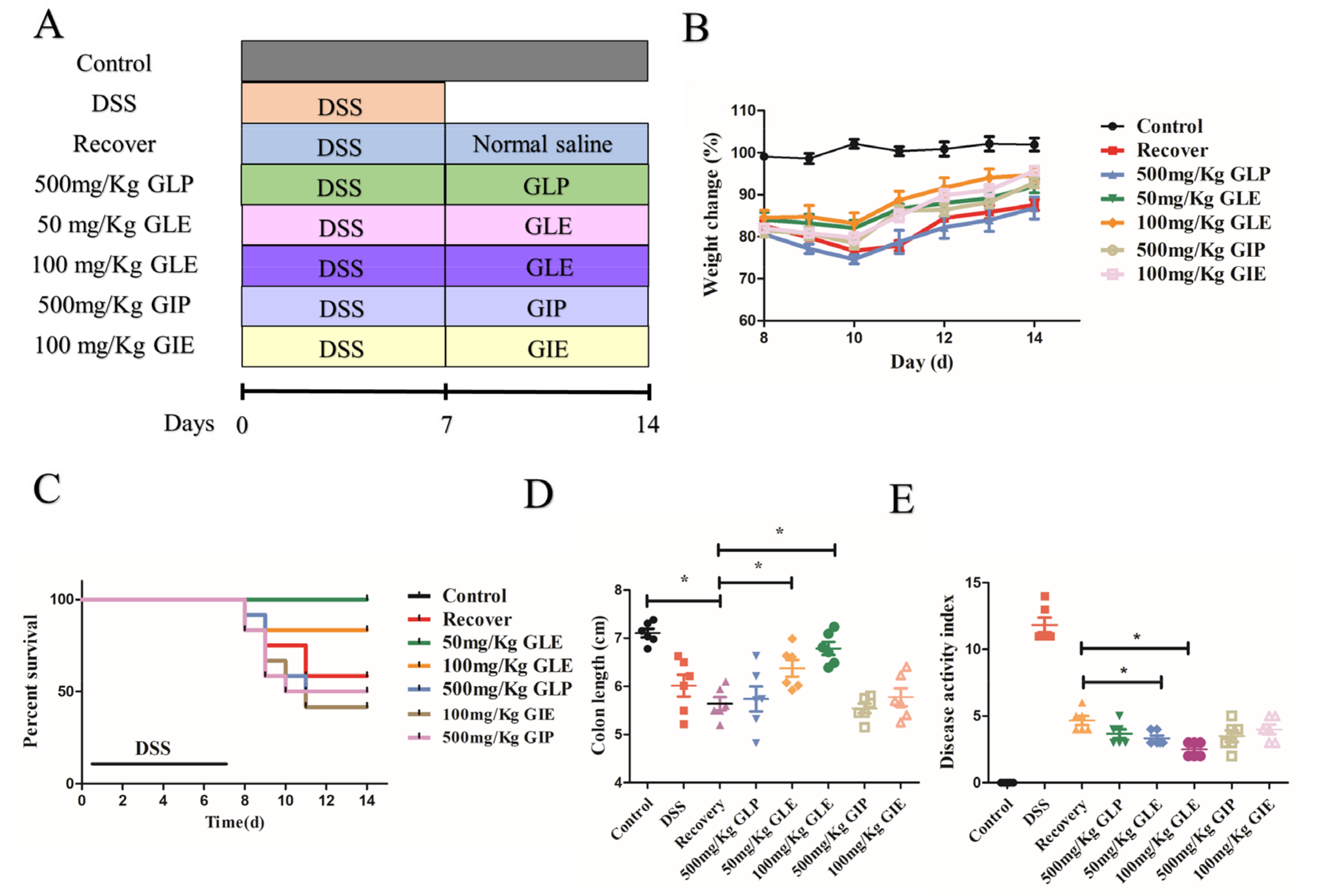
3.1. Influence of G. lucidum and G. incarnatum Extract Administration on the Recovery of DSS-Induced Colitis

DSS-induced colitis in the mouse model was used to evaluate whether G. lucidum and G. incarnatum extracts had an obviously prebiotic effect on IBD. DSS-induced colitis solution is shown in Figure 1A. After analysis, we found that GLE administration at 50 and 100 mg/kg showed better prebiotic effects in promoting recovery from colitis, displayed as significantly decreased weight loss (Figure 1B), reduced mortality (Figure 1C), reduced weight loss (Figure 1B), significantly decreased colon shortening (Figure 1D), and lower DAI scores were observed (Figure 1E). Compared with the recovery group, GLP administration in 500 mg/kg didn’t show effectiveness in promoting recovery in decreased weight loss (Figure 1B), reduced mortality (Figure 1C), reduced weight loss (Figure 1B), decreased colon shortening (Figure 1D), and lower DAI scores (Figure 1E). Compared with GLP administration in 500 mg/kg, 500 mg/kg GIP, and 100 mg/kg, GIE administration promoted weight gain while it showed no difference in reduced mortality (Figure 1C), reduced weight loss (Figure 1B), decreased colon shortening (Figure 1D), and lower DAI scores (Figure 1E).

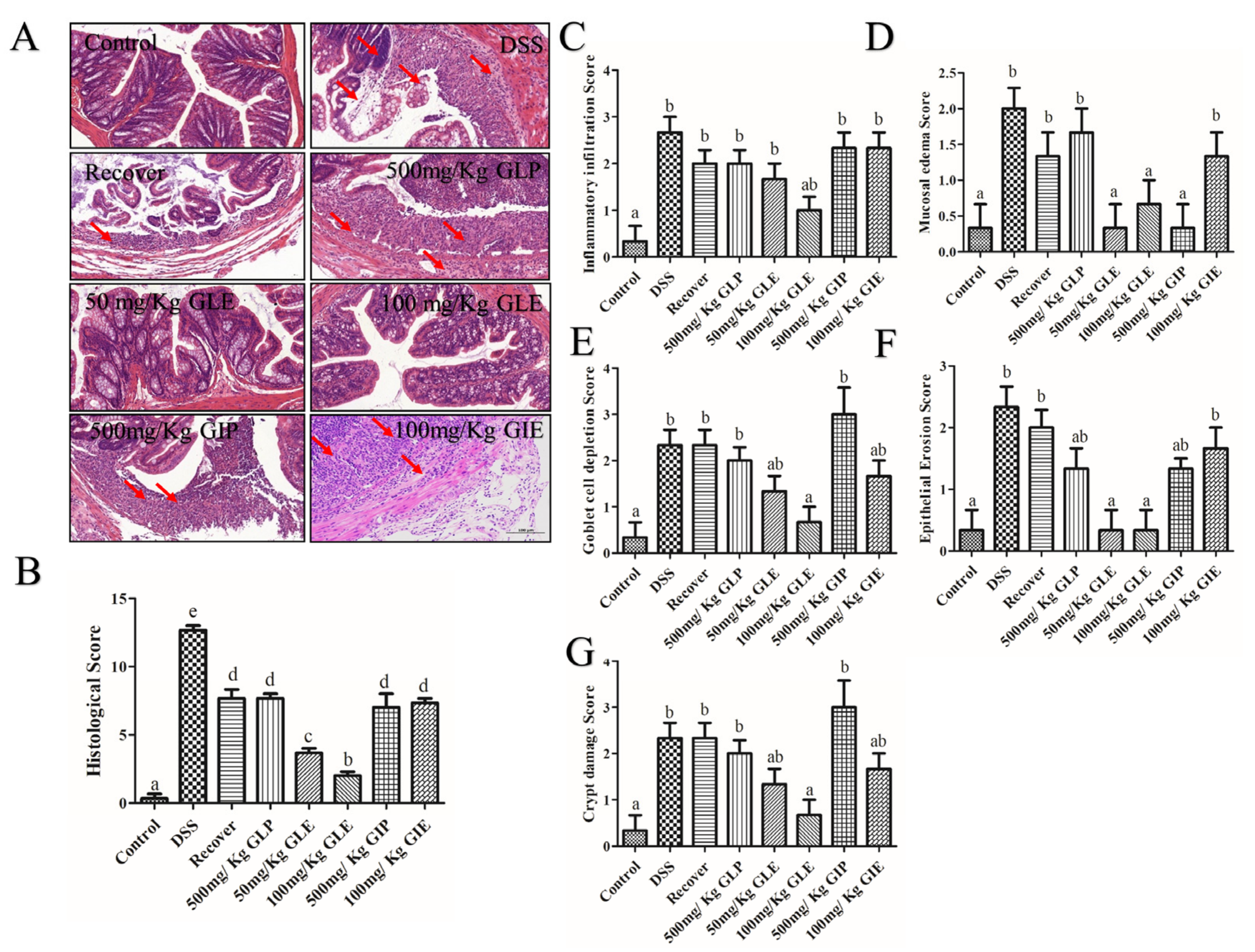
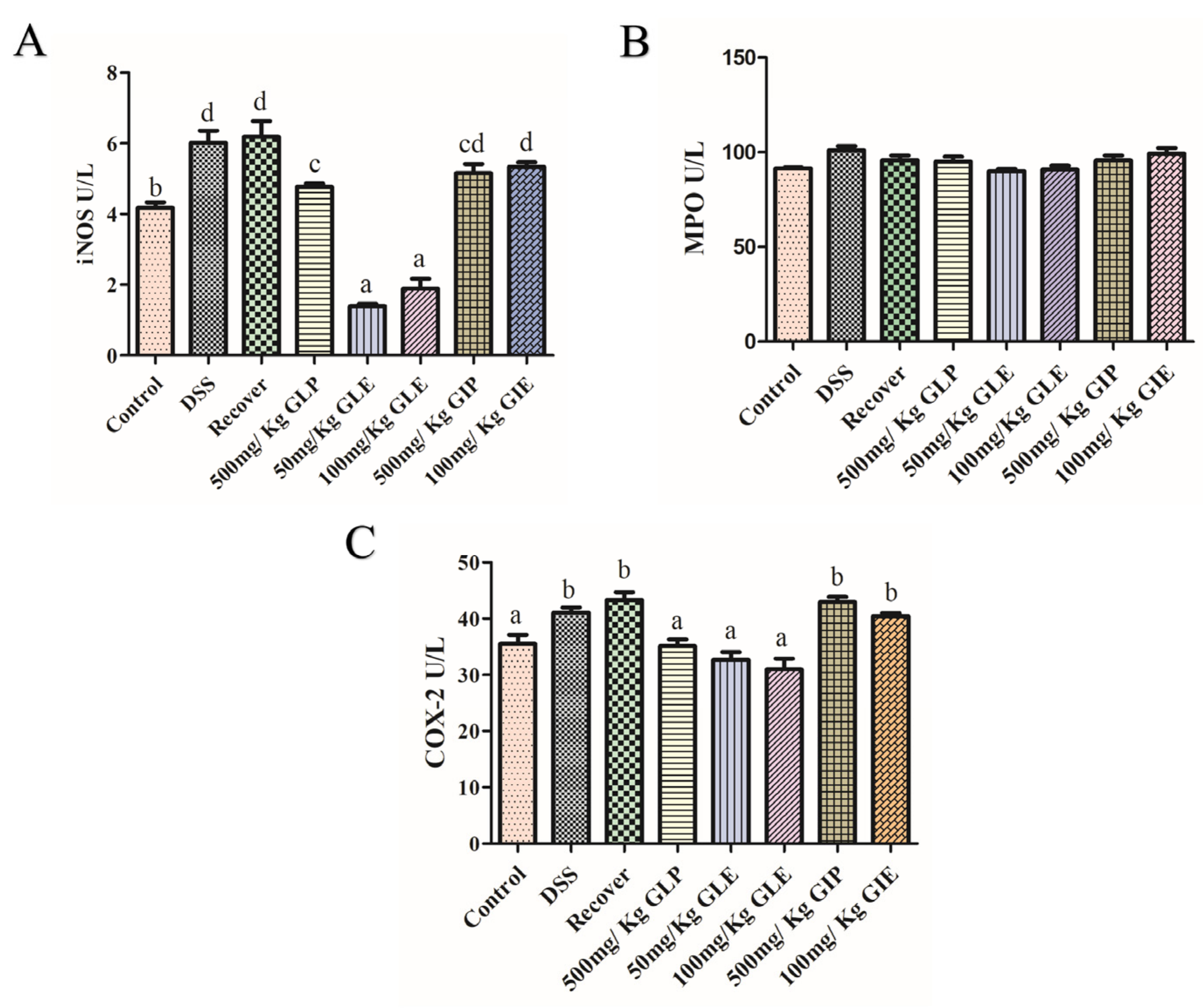
3.2. Influence of G. lucidum and G. incarnatum Extract Administration on DSS-Induced Colonic Tissue Damage and Regulation of Inflammatory Enzymes

To further evaluate its prebiotic effect of GLP, GLE, GIP, and GIE against DSS-induced colitis in mice, histological analyses were performed by staining with hematoxylin and eosin (H&E). In the recovery, GLP, GIP, and GIE groups, locally saw more epithelial cells and incomplete structures of the mucosal layer, connective tissue hyperplasia with a small amount of lymphocytic infiltration, and more focal lymphocytic infiltration in the lamina propria (Figure 2A). Low histological scores, epithelial erosion scores, goblet cell depletion scores, crypt damage scores, mucosal edema scores, and inflammatory infiltration scores were shown in the 50 and 100 mg/kg GLE groups, and the lower scores were in 100 mg/kg GLE (p < 0.05) (Figure 2B–G). In the 50 and 100 mg/kg GLE groups, the mucosal layer was structurally intact, with closely arranged epithelial cells and abundant glands in the lamina propria, and a small local focal infiltration of lymphocytes (Figure 2A). Compared with the recovery group, the GLP and GIE groups showed no significant difference in the histological score, and the epithelial erosion, goblet cell depletion, crypt damage scores, mucosal edema, and inflammatory infiltration showed no significant difference (Figure 2B–G). The GIP-treated exhibited a lower mucosal edema score than the recovery group, and other scores revealed a consistent trend (Figure 2B–G).
Activities of COX-2, iNOS, and MPO were analyzed to evaluate the effects of G. lucidum and G. incarnatum extracts on colonic inflammatory enzymes. No significant difference was seen in MPO concentrations on control, DSS, recover, GLP, GLE, GIP, and GIE groups (Figure 3B). Compared with the recovery group, GLP administration at 500 mg/Kg, GLE administration at 50 and 100 mg/kg significantly decreased the concentrations of iNOS and COX-2 (Figure 3A,C). The concentrations of iNOS and COX-2 in GLE administration were lower than in GLP administration (Figure 3A,C). Compared with the recovery group, GIE groups showed no difference in the concentrations of iNOS and COX-2 (Figure 3A,C).

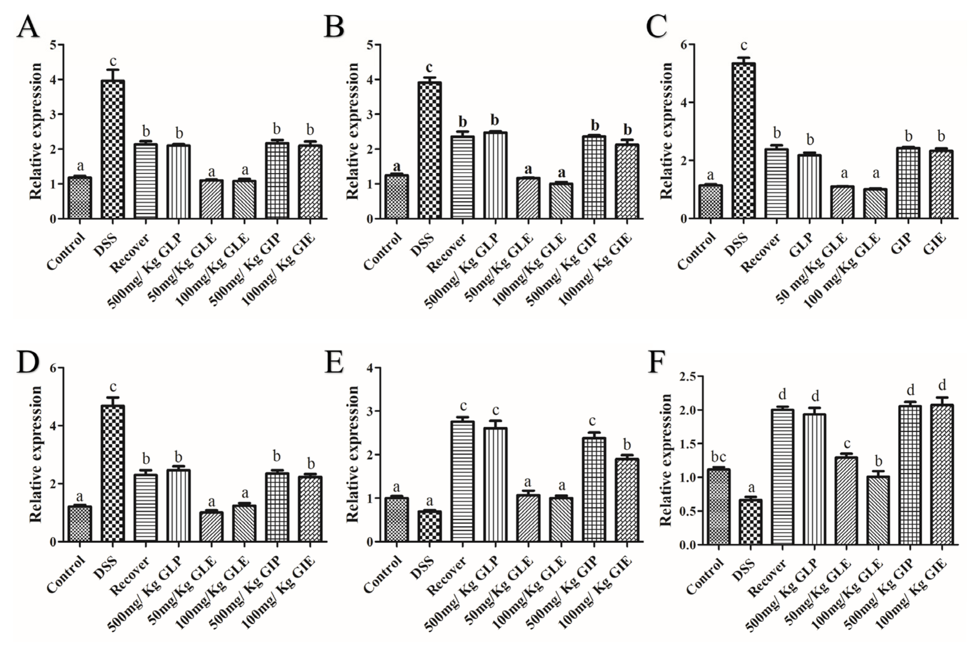
3.3. Effects of G. lucidum Extract Administration on the Regulation of Inflammatory Cytokines

The expression of TNF-α, IL-1β, IL-17, IL-6, PPAR-γ, and IL-10 were assayed to assess the effects of G. lucidum and G. incarnatum extracts on the modulation of inflammatory cytokines. Demonstrated in Figure 4, compared with the recovery group, 500 mg/kg GLP, 500 mg/kg GIP, and 100 mg/kg GIE administration showed no significant difference in the expression of pro-inflammatory cytokines (including IL-6, IL-1β, IL-17, and TNF-α) and anti-inflammatory cytokines (including IL-10 and PPAR-γ). GLE at 50 and 100 mg/kg displayed a significant decrease expression in pro-inflammatory cytokines, including IL-6, IL-1β, IL-17, and TNF-α, and a significant increase expression in anti-inflammatory cytokines, including IL-10 and PPAR-γ (p < 0.05).

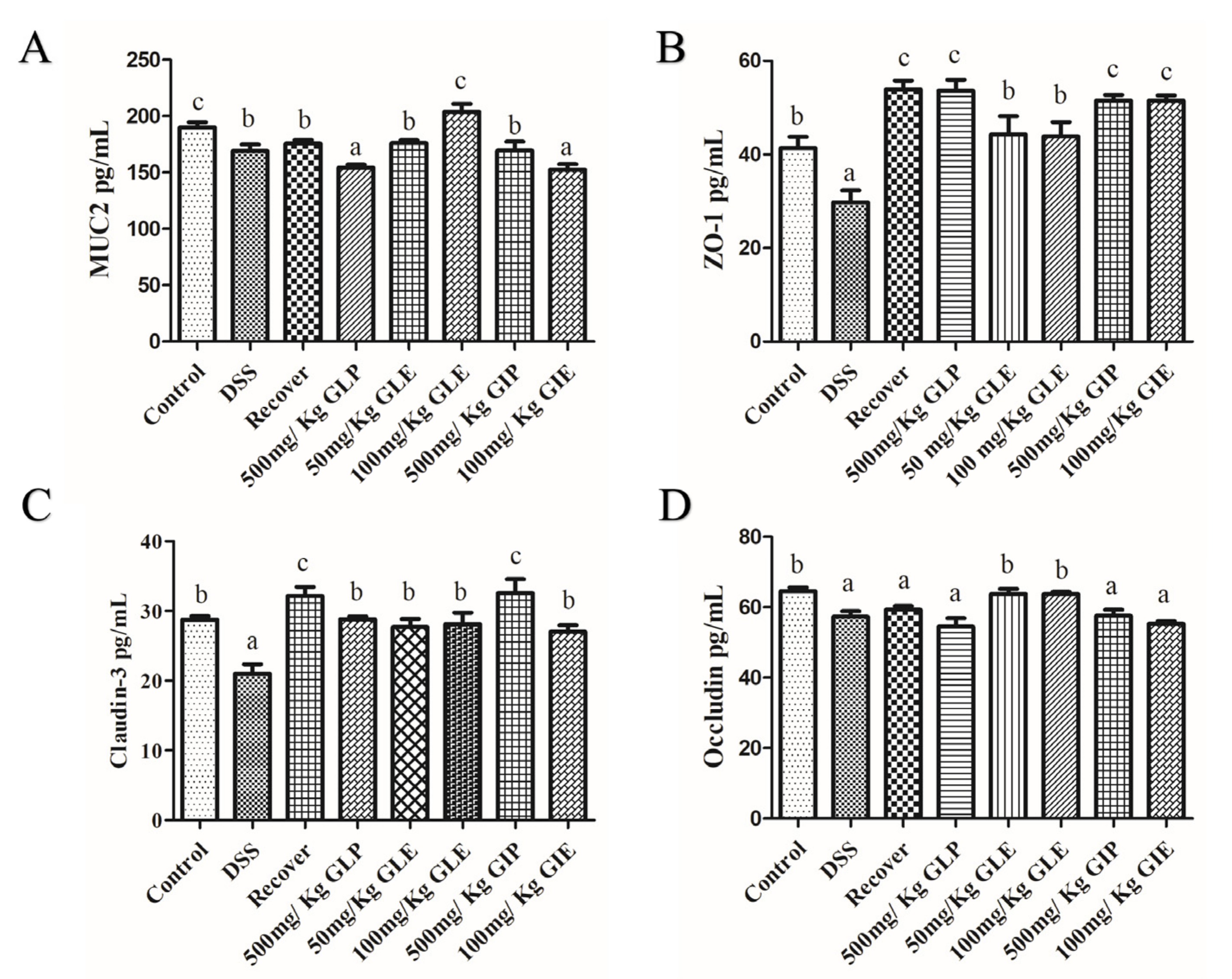
3.4. Effects of G. lucidum Extract Administration on the Protection of Intestinal Barrier

The concentration of mucin 2 (MUC2), claudin-3, and occludin ZO-1 were measured to analyze the effects of G. lucidum and G. incarnatum extracts for intestinal barrier protection. Firstly, DSS indeed destroyed the intestinal barrier and significantly decreased the concentrations of MUC2, ZO-1, Claudin-3, and Occludin (Figure 5). During the recovery period, the intestinal barrier showed repairing effects with increased concentrations of ZO-1 and Claudin-3, while there were no differences in concentrations of MUC2 and Occludin (Figure 5). The concentrations of MUC2, ZO-1, Claudin-3, and Occludin in GLE administration showed no significant difference with the control group, indicating that GLE administration promoted the reparation of the intestinal barrier and showed no difference with the control group (Figure 5). The concentrations of MUC2, ZO-1, Claudin-3, and Occludin in GLP, GIP, and GIE administration showed no different in recovery, and demonstrated that GLP, GIP, and GIE could not promote the recovery in the intestinal barrier (Figure 5).

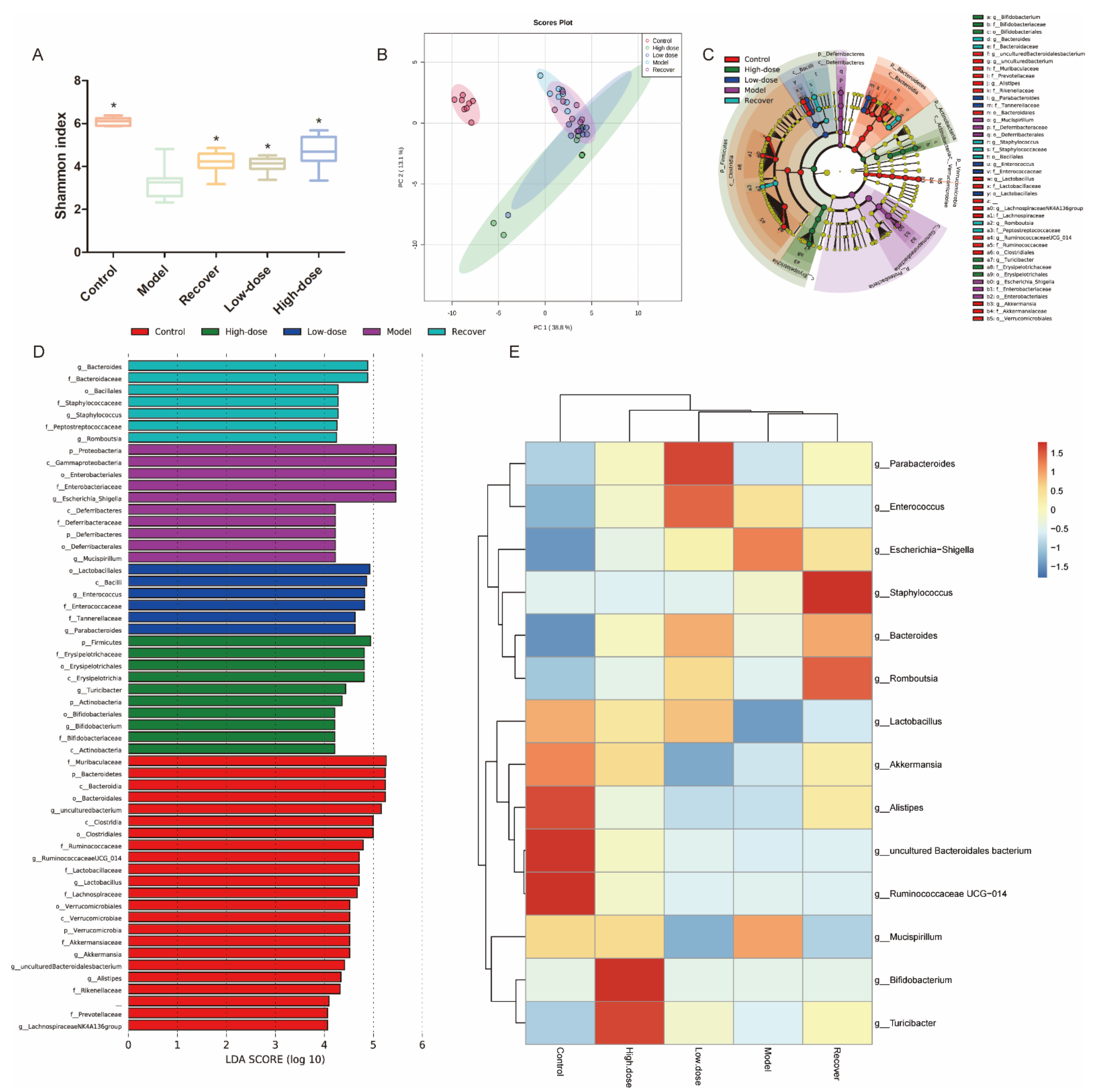
3.5. Intestinal Microbiota Modulation by GLE Administration

We analyzed the gut microbiota structure at different levels by GLE administration. Deferribacteres, Bacteroidetes, Actinobacteria, Verrucomicrobia, Proteobacteria, and Firmicutes represented dominant bacteria of all groups at the phylum level (Figure 6). Shown from the Shannon index, the GLE-treated group had a higher alpha multiplicity compared with the recovery group (p < 0.05), while there was no significant difference in alpha diversity between the recovery group and GLE-treated group (Figure 6A). From the Principal coordinate analysis (PCoA), there was community structures between the recovery group and GLE-treated groups, indicating a separation in compositional structures (Figure 6B). LEfSe was used to analyze the gut microbiota diversity; with LDA set as 4.0, we could see dominant communities of ten, seven, six, and ten taxa were found in the model, recovery, low-dose GLE-treated, and high-dose GLE-treated groups, respectively. Additionally, compared with the model group, the Firmicutes/Bacteroidetes (F/B) ratio was significantly decreased, while there were no differences in the group of control, recovery, low-dose-GLE, and high-dose-GLE (Figure 6D). The heat map at the genus level is displayed, compared with the recovery group. 100 mg/kg GLE significantly increased the abundance of Turicibacter and Bifidobacterium and decreased the abundance of Bacteroides, Staphylococcus, and Romboutsia; 50 mg/kg GLE significantly increased the abundance of Enterococcus and Parabacteroides and decreased the abundance of Staphylococcus and Romboutsia (Figure 6E).

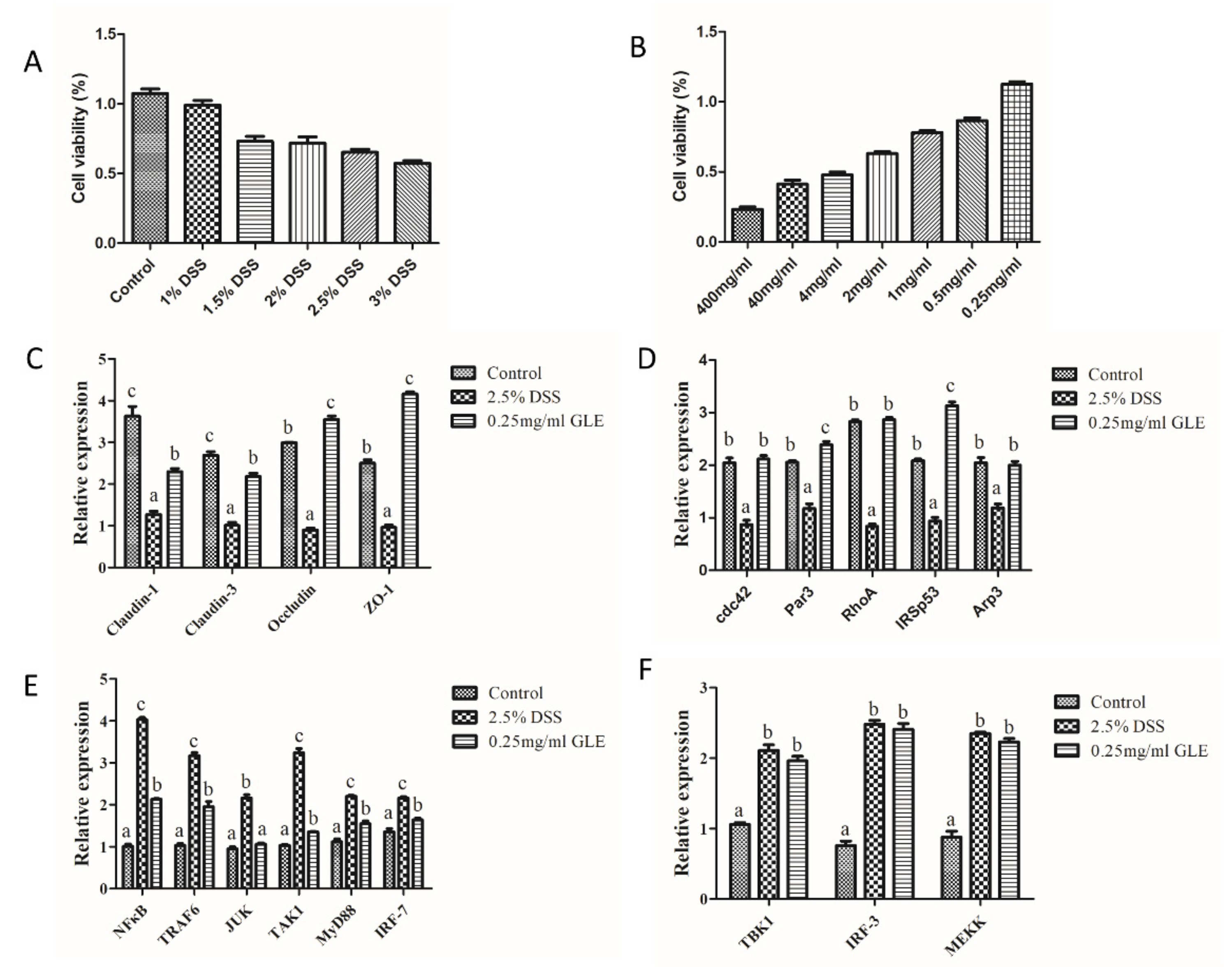
3.6. GLE Improved Intestinal Barrier Function and Inhibited TRAF6/MyD88/NF-κB Signaling

To analyze the regulation mechanism of GLE by DSS induced colitis, we used Caco-2 cells to investigate the signaling of the intestinal barrier function and inflammation. From the MTT analysis, the appropriate concentration of DSS was 3% DSS, where cell viability was near 50%, and the maximum safe concentration of GLE was 0.25 mg/mL, where cell viability was nearly 100% (Figure 7A,B). From the analysis of the results, with 3% DSS induced Caco-2 cells regulated by 0.25 mg/mL GLE, GLE had a positive effect on the intestinal barrier function including the tight junction and adherens junction, as well as inhibited the TRAF6/MyD88/NF-κB signaling pathway (Figure 7C–F). Specifically, 0.25 mg/mL GLE could significantly increase the expression of Claudin-3, Claudin-1, ZO-1, Occludin, cdc42, Par3, RhoA, IRSp53, and Arp3, significantly decrease the mRNA expression levels of NFκB, TRAF6, JNK, MyD88, and IRF-7, and had no significant difference in the expression of TBK1, IRF-3, and MEKK.

4. Discussion

G. lucidum ethanol extract was enriched with triterpenoids, including ganoderic acids and saponins, which has wide pharmacological effects, such as anti-obesity, increased immunity, and anti-inflammatory activities [23,32,33]. In addition, our study certificated G. lucidum ethanol extract, showing a prebiotic effect on DSS-induced colitis. Although Guo et al. demonstrated that the application of GLP prevented DSS-induced colitis in mice by enhancing the intestinal barrier function, increasing the production of SCFAs, and regulating the intestinal microbiota [34], our research of GLP did not display promoting effects in the recovery from colitis.
G. lucidum ethanol extract increased immunity during the recovery period. G. lucidum ethanol extract administration significantly reduced iNOS and COX-2 activities. High levels of COX-2 are associated with gastroenteritis and unregulated iNOS is frequently noted in chronic inflammation, and these molecules are classical biomarkers for assessing the level of inflammatory response in the colon [35,36,37]. Also, G. lucidum ethanol extract administration decreased the expression of inflammatory mediators IL-1β, IL-6, IL-17, and TNF-α in DSS-induced colitis. The levels of inflammatory factors contribute to the initiation, development, and progression of DSS-induced colitis [38]. Overproduction of pro-inflammatory cytokines, including TNF-α, IL-1β, IL-17, and IL-6, causes severe tissue damage and enhances the inflammatory response [39,40,41,42]. Meanwhile, after GLE administration, the levels of anti-inflammatory cytokines (IL-10 and PPAR-γ) were consistent with those in the control group. IL-10 and PPAR-γ are immune-modulating cytokines with anti-inflammatory activity, and they have shown the capability to reduce clinical symptoms in patients with IBD [43]. Edible mushrooms could enhance immune responses via MAPK and NFκB signal pathways [44], and GLE administration immune responses were in TRAF6/MyD88/NF-κB signaling pathways.
Intestinal inflammation in IBD can disturb the epithelial barrier integrity, causing increased permeability and infiltration of pathogens [45]. GLE administration promoted the recovery of the intestinal barrier through promoting the expression of the tight junction and adherens junction. Therefore, after GLE administration, the concentrations of MUC2, ZO-1, claudin-3, and occludin were similar with the control group. The mucosal barrier can protect the intestinal mucosa from damage and stop exogenous substances from destroying the intestinal tissues [46]. Tight junction proteins (ZO-1, claudins, and occludin) governed the intestinal barrier and play a critical role in IBD conservation [47].
The gut microbiome characterized by IBD patients exhibits decreased profitable metabolites (as butyric acid), enrichment of the phylum Aspergillus (as Escherichia coli), and depletion of strictly anaerobic bacteria [48]. In our study, 16S rRNA sequencing was used to examine the prospective alterations in microbial composition and diversity. GLE-treated mice showed significant clustering separation from DSS-induced mice using PCoA, which suggests that GLE treatment noticeably altered the biological structure. Through LEfSe analysis, Escherichia_Shigella showed relative enrichment in DSS-induced mice. Gram-negative bacterium Escherichia_Shigella leads to diarrheal diseases worldwide [49]. Its levels significantly increase in Crohn’s disease remission [21]. In our results, the relative abundance of Escherichia_Shigella was significantly higher in DSS-induced colitis mice, and during the recovery period, it was reduced. After GLE administration, the relative abundance of Escherichia_Shigella was lower compared to that in the recovery group, and the lowering was correlated with the dose of GLE. Bacteroides and Staphylococcus displayed a relative enrichment in the recovery group, and the heatmap showed that Staphylococcus (a gram-positive bacterium) was more abundant during the recovery period; after GLE administration, the abundance of Staphylococcus decreased. The abundance of Bacteroides is negatively correlated with the extent of tight junction proteins (ZO-1, occluding, and claudin-1), and positively associated with levels of pro-inflammatory cytokines (IL-1β, TNF-α, and IL-6) [49]. There was a significant increase of Bacteroides abundance during the recovery period, and high-dose GLE administration could reduce the abundance of Bacteroides. Parabacteroides displayed relative enrichment in low-dose GLE-treated mice, which was associated with amino acid metabolism [50]. Turicibacter and Bifidobacterium showed relative enrichment in high-dose GLE-treated mice. Turicibacter is a genus of anaerobic gram-positive bacteria, and reduced Turicibacter abundance is shown in obesity and irritable bowel syndrome [51,52,53]. Moreover, with Turicibacter and Bifidobacteria as probiotics, the former can influence gastrointestinal motor patterns and improve the production of SCFAs, while the latter can interact with the host and play a positive regulation role on the immune system [54,55]. Bifidobacteria can alleviate DSS-induced colitis by regulation of the intestinal microbiota and maintain the mucosal barrier [35,56]. In addition, the abundance of Lactobacillus increased significantly following GLE administration, whatever the dose. The Lactobacillus genera have been previously studied to mediate host immunity by regulating T lymphocytes, macrophages, and natural killer (NK) cells [57,58]. In addition, many studies certificated that Lactobacillus could relieve DSS-induced colitis by modulating gut microbiota composition and immune response [30,59,60].

5. Conclusions

In conclusion, we evaluated the prebiotic effects of G. lucidum and G. incarnatum’s different consumption ways (GLP, GLE, GIP, and GIE) on DSS-induced colitis. GLE demonstrated effective effects in promoting recovery from colitis as it prevented weight loss, reduced mortality, relieved colonic shortening, and reduced DAI and histological scores. In contrast, GLP, GIP, and GIE did not have a positive effect on relieving colitis. Recovery-promoting effects of GLE are mainly achieved by increasing immunity and protecting the intestinal barrier. In addition, GLE administration altered the gut microbiota composition, significantly increased the abundance of Turicibacter, Bifidobacterium, and Parabacteroides, and decreased the abundance of Escherichia_Shigella, Bacteroides, and Staphylococcus.

Author Contributions

Writing—original draft, M.L.; writing reviewing and editing—L.Y. and Q.Z.; data curation, formal analysis, methodology—M.L., L.Y., B.L. and F.T.; project administration—M.L., L.Y. and F.T., funding acquisition—L.Y., J.Z., W.C. and F.T. All authors have read and agreed to the published version of the manuscript.

Funding

This research received no external funding.

Institutional Review Board Statement

Not applicable.

Data Availability Statement

The data showed in this study are contained within the article.

Acknowledgments

This work was supported by the National Key Research and Development Project (2022YFF1100200), the National Natural Science Foundation of China Key Program (U1903205, 32001665), the Natural Science Foundation of Jiangsu Province (BE2021623, BK20220155), the Key Scientific and Technological Research Projects in the Key Areas of the Xinjiang Production and Construction Corps (2018AB010), the Talent Project for young and middle-aged people (HB2020031), and the Precision Medicine Project (J202109) of Wuxi Health Committee.

Conflicts of Interest

The authors declare no conflict of interest.

References

  1. Malinowski, B.; Wiciński, M.; Sokołowska, M.M.; Hill, N.A.; Szambelan, M. The rundown of dietary supplements and their effects on inflammatory bowel disease—A review. Nutrients 2020, 12, 1423. [Google Scholar] [CrossRef] [PubMed]
  2. Yangyang, R.Y.; Rodriguez, J.R. Clinical presentation of Crohn’s, ulcerative colitis, and indeterminate colitis: Symptoms, extraintestinal manifestations, and disease phenotypes. Semin. Pediatr. Surg. 2017, 26, 349–355. [Google Scholar]
  3. Barbalho, S.M.; de Alvares Goulart, R.; Quesada, K.; Bechara, M.D.; de Carvalho, A.d.C.A. Inflammatory bowel disease: Can omega-3 fatty acids really help? Ann. Gastroenterol. 2016, 29, 37. [Google Scholar] [PubMed]
  4. Farrokhyar, F.; Marshall, J.K.; Easterbrook, B.; Irvine, J.E. Functional gastrointestinal disorders and mood disorders in patients with inactive inflammatory bowel disease: Prevalence and impact on health. Inflamm. Bowel Dis. 2006, 12, 38–46. [Google Scholar] [CrossRef] [PubMed]
  5. Halpin, S.J.; Ford, A.C. Prevalence of symptoms meeting criteria for irritable bowel syndrome in inflammatory bowel disease: Systematic review and meta-analysis. Am. J. Gastroenterol. 2012, 107, 1474–1482. [Google Scholar] [CrossRef]
  6. Lupp, C.; Robertson, M.L.; Wickham, M.E.; Sekirov, I.; Champion, O.L.; Gaynor, E.C.; Finlay, B.B. Host-mediated inflammation disrupts the intestinal microbiota and promotes the overgrowth of Enterobacteriaceae. Cell Host Microbe 2007, 2, 119–129. [Google Scholar] [CrossRef] [Green Version]
  7. Peterson, D.A.; Frank, D.N.; Pace, N.R.; Gordon, J.I. Metagenomic approaches for defining the pathogenesis of inflammatory bowel diseases. Cell Host Microbe 2008, 3, 417–427. [Google Scholar] [CrossRef] [Green Version]
  8. Scarano, A.; Butelli, E.; De Santis, S.; Cavalcanti, E.; Hill, L.; De Angelis, M.; Giovinazzo, G.; Chieppa, M.; Martin, C.; Santino, A. Combined dietary anthocyanins, flavonols, and stilbenoids alleviate inflammatory bowel disease symptoms in mice. Front. Nutr. 2018, 4, 75. [Google Scholar] [CrossRef] [Green Version]
  9. Wong, C.; Harris, P.J.; Ferguson, L.R. Potential benefits of dietary fibre intervention in inflammatory bowel disease. Int. J. Mol. Sci. 2016, 17, 919. [Google Scholar] [CrossRef] [Green Version]
  10. Rapozo, D.C.; Bernardazzi, C.; de Souza, H.S.P. Diet and microbiota in inflammatory bowel disease: The gut in disharmony. World J. Gastroenterol. 2017, 23, 2124. [Google Scholar] [CrossRef]
  11. Kanwal, S.; Joseph, T.P.; Aliya, S.; Song, S.; Saleem, M.Z.; Nisar, M.A.; Wang, Y.; Meyiah, A.; Ma, Y.; Xin, Y. Attenuation of DSS induced colitis by Dictyophora indusiata polysaccharide (DIP) via modulation of gut microbiota and inflammatory related signaling pathways. J. Funct. Foods 2020, 64, 103641. [Google Scholar] [CrossRef]
  12. Ren, Y.; Geng, Y.; Du, Y.; Li, W.; Lu, Z.-M.; Xu, H.-Y.; Xu, G.-H.; Shi, J.-S.; Xu, Z.-H. Polysaccharide of Hericium erinaceus attenuates colitis in C57BL/6 mice via regulation of oxidative stress, inflammation-related signaling pathways and modulating the composition of the gut microbiota. J. Nutr. Biochem. 2018, 57, 67–76. [Google Scholar] [CrossRef]
  13. Bunbamrung, N.; Intaraudom, C.; Dramae, A.; Boonyuen, N.; Veeranondha, S.; Rachtawee, P.; Pittayakhajonwut, P. Antimicrobial activity of illudalane and alliacane sesquiterpenes from the mushroom Gloeostereum incarnatum BCC41461. Phytochem. Lett. 2017, 20, 274–281. [Google Scholar] [CrossRef]
  14. Li, M.; Yu, L.; Zhao, J.; Zhang, H.; Chen, W.; Zhai, Q.; Tian, F. Role of dietary edible mushrooms in the modulation of gut microbiota. Phytochem. Lett. 2021, 83, 104538. [Google Scholar] [CrossRef]
  15. Lull, C.; Wichers, H.J.; Savelkoul, H.F. Antiinflammatory and immunomodulating properties of fungal metabolites. Mediat. Inflamm. 2005, 2005, 63–80. [Google Scholar] [CrossRef] [Green Version]
  16. Wang, D.; Li, Q.; Qu, Y.; Wang, M.; Li, L.; Liu, Y.; Li, Y. The investigation of immunomodulatory activities of Gloeostereum incaratum polysaccharides in cyclophosphamide-induced immunosuppression mice. Exp. Ther. Med. 2018, 15, 3633–3638. [Google Scholar]
  17. Zhang, Z.; Taylor, L.; Shommu, N.; Ghosh, S.; Reimer, R.; Panaccione, R.; Kaur, S.; Hyun, J.E.; Cai, C.; Deehan, E.C. A diversified dietary pattern is associated with a balanced gut microbial composition of Faecalibacterium and Escherichia/Shigella in patients with Crohn’s disease in remission. Crohns Colitis 2020, 14, 1547–1557. [Google Scholar] [CrossRef]
  18. Cör, D.; Knez, Ž.; Knez Hrnčič, M. Antitumour, antimicrobial, antioxidant and antiacetylcholinesterase effect of Ganoderma lucidum terpenoids and polysaccharides: A review. Molecules 2018, 23, 649. [Google Scholar] [CrossRef] [Green Version]
  19. Jakobsdottir, G.; Xu, J.; Molin, G.; Ahrne, S.; Nyman, M. High-fat diet reduces the formation of butyrate, but increases succinate, inflammation, liver fat and cholesterol in rats, while dietary fibre counteracts these effects. PLoS ONE 2013, 8, e80476. [Google Scholar]
  20. Asai, R.; Mitsuhashi, S.; Shigetomi, K.; Miyamoto, T.; Ubukata, M. Absolute configurations of (−)-hirsutanol A and (−)-hirsutanol C produced by Gloeostereum incarnatum. J. Antibiot. 2011, 64, 693–696. [Google Scholar]
  21. Wang, X.; Peng, J.; Sun, L.; Bonito, G.; Wang, J.; Cui, W.; Fu, Y.; Li, Y. Genome sequencing illustrates the genetic basis of the pharmacological properties of Gloeostereum incarnatum. Genes 2019, 10, 188. [Google Scholar] [CrossRef] [PubMed] [Green Version]
  22. Zhao, C.; Fan, J.; Liu, Y.; Guo, W.; Cao, H.; Xiao, J.; Liu, B. Hepatoprotective activity of Ganoderma lucidum triterpenoids in alcohol-induced liver injury in mice, an iTRAQ-based proteomic analysis. Food Chem. 2019, 271, 148–156. [Google Scholar] [CrossRef] [PubMed]
  23. Guo, W.L.; Pan, Y.Y.; Li, L.; Li, T.T.; Liu, B.; Lv, X.C. Ethanol extract of Ganoderma lucidum ameliorates lipid metabolic disorders and modulates the gut microbiota composition in high-fat diet fed rats. Food Funct. 2018, 9, 3419–3431. [Google Scholar] [CrossRef] [PubMed]
  24. Adeyi, A.O.; Awosanya, S.A.; Adeyi, O.E.; James, A.S.; Adenipekun, C.O. Ganoderma lucidum ethanol extract abrogates metabolic syndrome in rats: In vivo evaluation of hypoglycemic, hypolipidemic, hypotensive and antioxidant properties. Obes. Med. 2021, 22, 100320. [Google Scholar] [CrossRef]
  25. Hu, R.; Guo, W.; Huang, Z.; Li, L.; Liu, B.; Lv, X. Extracts of Ganoderma lucidum attenuate lipid metabolism and modulate gut microbiota in high-fat diet fed rats. J. Funct. Foods 2018, 46, 403–412. [Google Scholar] [CrossRef]
  26. Li, M.; Yu, L.; Zhai, Q.; Liu, B.; Zhao, J.; Zhang, H.; Chen, W.; Tian, F. Ganoderma applanatum polysaccharides and ethanol extracts promote the recovery of colitis through intestinal barrier protection and gut microbiota modulations. Food Funct. 2022, 13, 688–701. [Google Scholar] [CrossRef]
  27. Zhou, S.; Huang, G. Extraction, structural analysis and antioxidant activity of aloe polysaccharide. J. Mol. Struct. 2023, 1273, 134379. [Google Scholar] [CrossRef]
  28. Liu, Y.-J.; Tang, B.; Wang, F.-C.; Tang, L.; Lei, Y.-Y.; Luo, Y.; Huang, S.-J.; Yang, M.; Wu, L.-Y.; Wang, W. Parthenolide ameliorates colon inflammation through regulating Treg/Th17 balance in a gut microbiota-dependent manner. Theranostics 2020, 10, 5225. [Google Scholar] [CrossRef]
  29. Cui, G.; Martin, R.C.; Jin, H.; Liu, X.; Pandit, H.; Zhao, H.; Cai, L.; Zhang, P.; Li, W.; Li, Y. Up-regulation of FGF15/19 signaling promotes hepatocellular carcinoma in the background of fatty liver. J. Exp. Clin. Cancer Res. 2018, 37, 136. [Google Scholar] [CrossRef] [Green Version]
  30. Liu, M.; Ding, J.; Zhang, H.; Shen, J.; Hao, Y.; Zhang, X.; Qi, W.; Luo, X.; Zhang, T.; Wang, N. Lactobacillus casei LH23 modulates the immune response and ameliorates DSS-induced colitis via suppressing JNK/p-38 signal pathways and enhancing histone H3K9 acetylation. Food Funct. 2020, 11, 5473–5485. [Google Scholar] [CrossRef]
  31. Romero-Nava, R.; Alarcón-Aguilar, F.J.; Giacoman-Martínez, A.; Blancas-Flores, G.; Aguayo-Cerón, K.A.; Ballinas-Verdugo, M.A.; Sánchez-Muñoz, F.; Huang, F.; Villafaña-Rauda, S.; Almanza-Pérez, J.C. Glycine is a competitive antagonist of the TNF receptor mediating the expression of inflammatory cytokines in 3T3-L1 adipocytes. Inflamm. Res. 2021, 70, 605–618. [Google Scholar] [CrossRef]
  32. Baby, S.; Johnson, A.J.; Govindan, B. Secondary metabolites from Ganoderma. Phytochemistry 2015, 114, 66–101. [Google Scholar] [CrossRef]
  33. Nie, S.; Zhang, H.; Li, W.; Xie, M. Current development of polysaccharides from Ganoderma: Isolation, structure and bioactivities. Bioact. Carbohydr. Diet. Fibre 2013, 1, 10–20. [Google Scholar] [CrossRef]
  34. Guo, C.; Guo, D.; Fang, L.; Sang, T.; Wu, J.; Guo, C.; Wang, Y.; Wang, Y.; Chen, C.; Chen, J. Ganoderma lucidum polysaccharide modulates gut microbiota and immune cell function to inhibit inflammation and tumorigenesis in colon. Carbohydr. Polym. 2021, 267, 118231. [Google Scholar] [CrossRef]
  35. Chen, Y.; Jin, Y.; Stanton, C.; Paul Ross, R.; Zhao, J.; Zhang, H.; Yang, B.; Chen, W. Alleviation effects of Bifidobacterium breve on DSS-induced colitis depends on intestinal tract barrier maintenance and gut microbiota modulation. Eur. J. Nutr. 2021, 60, 369–387. [Google Scholar] [CrossRef]
  36. Jin, B.-R.; Chung, K.-S.; Cheon, S.-Y.; Lee, M.; Hwang, S.; Noh Hwang, S.; Rhee, K.-J.; An, H.-J. Rosmarinic acid suppresses colonic inflammation in dextran sulphate sodium (DSS)-induced mice via dual inhibition of NF-κB and STAT3 activation. Sci. Rep. 2017, 7, 46252. [Google Scholar] [CrossRef] [Green Version]
  37. Sharma, V.; Bhatia, P.; Alam, O.; Naim, M.J.; Nawaz, F.; Sheikh, A.A.; Jha, M. Recent advancement in the discovery and development of COX-2 inhibitors: Insight into biological activities and SAR studies (2008–2019). Bioorg. Chem. 2019, 89, 103007. [Google Scholar] [CrossRef]
  38. Wang, R.; Li, Y.; Tsung, A.; Huang, H.; Du, Q.; Yang, M.; Deng, M.; Xiong, S.; Wang, X.; Zhang, L. iNOS promotes CD24+ CD133+ liver cancer stem cell phenotype through a TACE/ADAM17-dependent Notch signaling pathway. Proc. Natl. Acad. Sci. USA 2018, 115, E10127–E10136. [Google Scholar] [CrossRef] [Green Version]
  39. Qian, B.; Wang, C.; Zeng, Z.; Ren, Y.; Li, D.; Song, J.-L. Ameliorative effect of sinapic acid on dextran sodium sulfate-(DSS-) induced ulcerative colitis in Kunming (KM) mice. Oxid. Med. Cell. Longev. 2020, 2020, 8393504. [Google Scholar] [CrossRef]
  40. Moschen, A.R.; Tilg, H.; Raine, T. IL-12, IL-23 and IL-17 in IBD: Immunobiology and therapeutic targeting. Nat. Rev. Gastroenterol. Hepatol. 2019, 16, 185–196. [Google Scholar] [CrossRef]
  41. Al-Sadi, R.; Guo, S.; Dokladny, K.; Smith, M.A.; Ye, D.; Kaza, A.; Watterson, D.M.; Ma, T.Y. Mechanism of interleukin-1β induced-increase in mouse intestinal permeability in vivo. J. Interferon Cytokine Res. 2012, 32, 474–484. [Google Scholar] [CrossRef] [PubMed] [Green Version]
  42. Papadakis, K.A.; Targan, S.R. Role of cytokines in the pathogenesis of inflammatory bowel disease. Annu. Rev. Med. 2000, 51, 289–298. [Google Scholar] [CrossRef] [PubMed]
  43. Ebrahimi Daryani, N.; Saghazadeh, A.; Moossavi, S.; Sadr, M.; Shahkarami, S.; Soltani, S.; Farhadi, E.; Rezaei, N. Interleukin-4 and interleukin-10 gene polymorphisms in patients with inflammatory bowel disease. Immunol. Investig. 2017, 46, 714–729. [Google Scholar] [CrossRef] [PubMed]
  44. Yin, Z.; Liang, Z.; Li, C.; Wang, J.; Ma, C.; Kang, W. Immunomodulatory effects of polysaccharides from edible fungus: A review. Food Sci. Hum. Wellness 2021, 10, 393–400. [Google Scholar] [CrossRef]
  45. Landy, J.; Ronde, E.; English, N.; Clark, S.K.; Hart, A.L.; Knight, S.C.; Ciclitira, P.J.; Al-Hassi, H.O. Tight junctions in inflammatory bowel diseases and inflammatory bowel disease associated colorectal cancer. World J. Gastroenterol. 2016, 22, 3117. [Google Scholar] [CrossRef]
  46. Pawłowska, B.; Sobieszczańska, B.M. Intestinal epithelial barrier: The target for pathogenic Escherichia coli. Adv. Clin. Exp. Med. 2017, 26, 1437–1445. [Google Scholar] [CrossRef] [Green Version]
  47. Chen, Y.; Yang, B.; Ross, R.P.; Jin, Y.; Stanton, C.; Zhao, J.; Zhang, H.; Chen, W. Orally administered CLA ameliorates DSS-induced colitis in mice via intestinal barrier improvement, oxidative stress reduction, and inflammatory cytokine and gut microbiota modulation. J. Agric. Food Chem. 2019, 67, 13282–13298. [Google Scholar] [CrossRef]
  48. Ni, J.; Shen, T.-C.D.; Chen, E.Z.; Bittinger, K.; Bailey, A.; Roggiani, M.; Sirota-Madi, A.; Friedman, E.S.; Chau, L.; Lin, A. A role for bacterial urease in gut dysbiosis and Crohn’s disease. Sci. Transl. Med. 2017, 9, eaah6888. [Google Scholar] [CrossRef] [Green Version]
  49. Lagerqvist, N.; Löf, E.; Enkirch, T.; Nilsson, P.; Roth, A.; Jernberg, C. Outbreak of gastroenteritis highlighting the diagnostic and epidemiological challenges of enteroinvasive Escherichia coli, County of Halland, Sweden, November 2017. Eurosurveillance 2020, 25, 1900466. [Google Scholar] [CrossRef] [Green Version]
  50. Wang, M.; Chen, G.; Chen, D.; Ye, H.; Sun, Y.; Zeng, X.; Liu, Z. Purified fraction of polysaccharides from Fuzhuan brick tea modulates the composition and metabolism of gut microbiota in anaerobic fermentation in vitro. Int. J. Biol. Macromol. 2019, 140, 858–870. [Google Scholar] [CrossRef]
  51. Bosshard, P.P.; Zbinden, R.; Altwegg, M. Turicibacter sanguinis gen. nov., sp. nov., a novel anaerobic, Gram-positive bacterium. Int. J. Syst. Evol. Microbiol. 2002, 52, 1263–1266. [Google Scholar]
  52. Jung, M.-J.; Lee, J.; Shin, N.-R.; Kim, M.-S.; Hyun, D.-W.; Yun, J.-H.; Kim, P.S.; Whon, T.W.; Bae, J.-W. Chronic repression of mTOR complex 2 induces changes in the gut microbiota of diet-induced obese mice. Sci. Rep. 2016, 6, 30887. [Google Scholar] [CrossRef] [Green Version]
  53. Zhuang, X.; Tian, Z.; Li, L.; Zeng, Z.; Chen, M.; Xiong, L. Fecal microbiota alterations associated with diarrhea-predominant irritable bowel syndrome. Front. Microbiol. 2018, 9, 1600. [Google Scholar] [CrossRef] [Green Version]
  54. Hoffman, J.M.; Margolis, K.G. Building community in the gut: A role for mucosal serotonin. Nat. Rev. Gastroenterol. Hepatol. 2020, 17, 6–8. [Google Scholar] [CrossRef]
  55. Zhong, Y.; Nyman, M.; Fåk, F. Modulation of gut microbiota in rats fed high-fat diets by processing whole-grain barley to barley malt. Mol. Nutr. Food Res. 2015, 59, 2066–2076. [Google Scholar] [CrossRef]
  56. Yan, S.; Yang, B.; Zhao, J.; Zhao, J.; Stanton, C.; Ross, R.P.; Zhang, H.; Chen, W. A ropy exopolysaccharide producing strain Bifidobacterium longum subsp. longum YS108R alleviates DSS-induced colitis by maintenance of the mucosal barrier and gut microbiota modulation. Food Funct. 2019, 10, 1595–1608. [Google Scholar] [CrossRef]
  57. Kim, D.H.; Kim, S.; Lee, J.H.; Kim, J.H.; Che, X.; Ma, H.W.; Seo, D.H.; Kim, T.I.; Kim, W.H.; Kim, S.W. Lactobacillus acidophilus suppresses intestinal inflammation by inhibiting endoplasmic reticulum stress. J. Gastroenterol. Hepatol. 2019, 34, 178–185. [Google Scholar] [CrossRef] [Green Version]
  58. Park, J.-S.; Choi, J.W.; Jhun, J.; Kwon, J.Y.; Lee, B.-I.; Yang, C.W.; Park, S.-H.; Cho, M.-L. Lactobacillus acidophilus improves intestinal inflammation in an acute colitis mouse model by regulation of Th17 and Treg cell balance and fibrosis development. J. Med. Food 2018, 21, 215–224. [Google Scholar] [CrossRef] [Green Version]
  59. Sun, M.; Liu, Y.; Song, Y.; Gao, Y.; Zhao, F.; Luo, Y.; Qian, F.; Mu, G.; Tuo, Y. The ameliorative effect of Lactobacillus plantarum-12 on DSS-induced murine colitis. Food Funct. 2020, 11, 5205–5222. [Google Scholar] [CrossRef]
  60. Xia, Y.; Chen, Y.; Wang, G.; Yang, Y.; Song, X.; Xiong, Z.; Zhang, H.; Lai, P.; Wang, S.; Ai, L. Lactobacillus plantarum AR113 alleviates DSS-induced colitis by regulating the TLR4/MyD88/NF-κB pathway and gut microbiota composition. J. Funct. Foods 2020, 67, 103854. [Google Scholar] [CrossRef]
Figure 1. The prebiotic effect of G. lucidum and G. incarnatum extract in DSS-induced colitis. (A) Schematic diagram of the experimental schedule. (B) Body weight change (%). (C) Survival rate. (D) Colon length (cm). (E) Disease activity index. n = 8 mice per group, the mean values ± SD are presented, * p < 0.05.
Foods 11 04023 g001
Figure 2. Recovery of histological injury in DSS-induced colitis. (A) H&E staining (20×). (B) Histology score. (C) Epithelial erosion score. (D) Crypt damage score. (E) Inflammatory infiltration score. (F) Goblet cell depletion score. (G) Mucosal edema score. n = 6 mice per group, the mean values ± SD are presented. Bars with different letters were considered significant at p < 0.05.
Foods 11 04023 g002
Figure 3. Prebiotic effect of G. lucidum and G. incarnatum extract on enzymes activities in DSS-induced colitis. (A) iNOS, (B) MPO, and (C) COX-2. n = 6 mice per group, the mean values ± SD are presented. Bars with different letters were considered significant at p < 0.05.
Foods 11 04023 g003
Figure 4. Effects of G. lucidum extract administration on the regulation of inflammatory cytokines. (A) TNF-α, (B) IL-1β, (C) IL-17, (D) IL-6, (E) PPAR-γ, (F) IL-10. n = 6 mice per group, the mean values ± SD are presented. Bars with different letters were considered significant at p < 0.05.
Foods 11 04023 g004
Figure 5. Protection of the intestinal barrier by GLP, GLE, GIP, and GIE administration. (A) ZO-1, (B) claudin-3, (C) occludin, and (D) MUC2. n = 6 mice per group, the mean values ± SD are presented. Bars with different letters were considered significant at p < 0.05.
Foods 11 04023 g005
Figure 6. Influence of microbiota modulation by GLE added in DSS-induced colitis. (A) Alpha diversity boxplot (Shannon) in colon. (B) PCoA using unweighted-UniFrac of beta diversity in colon of GLE group. (C) Taxonomic cladogram from LEfSe in colon intestine. (D) Differences among the control, high-dose, low-dose, model, and recovery groups in colon intestine, LDA score > 4. (E) Heatmap at the genus level. * p < 0.05.
Foods 11 04023 g006
Figure 7. GLE inhibited DSS-induced intestinal barrier function and inflammation. (A) MTT in DSS, (B) MTT in GLE, (C) Tight junction signaling, (D) Adherens junction signaling, (E) NFκB, MyD88 and JUK signaling, and (F) TBK1 and MEKK signaling. mean values ± SEM are presented (n = 6), Bars with different letters were considered significant at p<0.05.
Foods 11 04023 g007
Table 1. Scoring system for Disease Activity Index (DAI).
ScoreWeight LossStool ConsistencyBlood Stool
0No lossNormalNo blood
11–5%Loose stool
25–10%Watery diarrheaPresence of blood
310–20%Slimy diarrhea, little blood
4>20%Severe watery diarrhea with bloodGross bleeding
Table 2. Primer sequences used for qPCR.
GeneSequence (5’ to 3’)
ForwardReverse
GAPDHGACAAGCTTCCCGTTCTCAGGAGTCAACGGATTTGGTCGT
IL-6ACCAGAGGAAATTTTCAATAGGCTGATGCACTTGCAGAAAACA
TNF-αTGCCTATGTCTCAGCCTCTTCGGTCTGGGCCATAGAACTGA
IL-1βACCTTCCAGGATGAGGACATGACTAATGGGAACGTCACAC ACCA
IL-17CTCCAGAAGGCCCTCAGACTACGGGTCTTCATTGCGGTGG
PPAR-γCTGCTCAAGTATGGTGTCCATGATGAGATGAGGACTCCATCTTT ATTCA
IL-10GCTCTTACTGACTGGCATGAGCGCAGCTCTAGGAGCATGTG
β-actinCCTTCCCTCCTCAGATCATTGCATACTCCTGCTTGCTGATCCAC
Claudin-1TCTATGACCCTATGACCCCAGTTCTGGGAAATGATGGCACTAGC
Claudin-3CGAGAAGAAGTACACGGCCACGTCTGTCCCTTAGACGTAGTCC
OccludinCATTAACTTCGCCTGTGGATGACTCTCTTTGACCTTCCTGCTCTTC
ZO-1AGTACCAGAAATACCTGACGGTGCTTGGCTGACACTAGAAGTAGCA
MYD88TCGAAAAGAGGTTGGCTAGAAGGCTTGCTCTGCAGGTAATCATCAG
IRF-7CCCATCTTCGACTTCAGAGTCTTCGAAGCCCAGGTAGATGGTATAG
NFκBAGCTTCAGAATGGCAGAAGATGACAGTGCCATCTGTGGTTGAAATA
TRAF6TTGCTCTTATGGATTGTCCCCAAGACAGTTCTGGTCATGGATCTCT
TAK1GAGATCAAGAGGGTGATGCAGATCGAGTGATAAGCACATTAGCAGC
JUKGCCACAAAATCCTCTTTCCAGGAGGACATCAGGGAAGAGTTTCTC
MEKKTCACGAAGGAATCAAGAGAGCAA AAAATAAGCAGCCAACGAGTTCC
TBK1ACAGATTTTGGTGCAGCTAGAGATACCCCAATGCTCCAAAGATCAA
IRF-3CAAAGAAGGGTTGCGTTTAGCAACTCCAGATATTGCACCAGAAGG
Arp3GCTGCATGAAAATTCAGTTCGTCGCCACAGAGAAGATTCTTAGCCT
RhoATCCGGAAGAAACTGGTGATTGTTTCAGGCGATCATAATCTTCCACA
IRSP53CGACTCCTACTCCAACACACTCCAGAGTCTTGTTCTCGGTGGTG
cdc42CAGAAGCCTATCACTCCAGAGACGCAGCCAATATTGCTTCGTCAAA
Par3CTAATTGGCCTCTCCACTTCTGTTCCCATCCTCATCCTTCCTGTC
Publisher’s Note: MDPI stays neutral with regard to jurisdictional claims in published maps and institutional affiliations.

