Article

Shelf Life Extension of Chilled Pork by Optimal Ultrasonicated Ceylon Spinach (Basella alba) Extracts: Physicochemical and Microbial Properties

by
Yuthana Phimolsiripol
1,2,*,
Srirana Buadoktoom
1,
Pimporn Leelapornpisid
3,
Kittisak Jantanasakulwong
1,2,
Phisit Seesuriyachan
1,2,
Thanongsak Chaiyaso
1,2,
Noppol Leksawasdi
1,2,
Pornchai Rachtanapun
1,2,
Nareekan Chaiwong
1,2,
Sarana Rose Sommano
2,4,
Charles S. Brennan
5 and
Joe M. Regenstein
6
1
Faculty of Agro-Industry, Chiang Mai University, Chiang Mai 50100, Thailand
2
Cluster of Agro Bio-Circular-Green Industry, Chiang Mai University, Chiang Mai 50100, Thailand
3
Faculty of Pharmacy, Chiang Mai University, Chiang Mai 50200, Thailand
4
Faculty of Agriculture, Chiang Mai University, Chiang Mai 50200, Thailand
5
School of Science, STEM College, RMIT University, Melbourne 3000, Australia
6
Department of Food Science, Cornell University, Ithaca, NY 14853-7201, USA
*
Author to whom correspondence should be addressed.
Foods 2021, 10(6), 1241; https://doi.org/10.3390/foods10061241
Submission received: 4 April 2021 / Revised: 25 May 2021 / Accepted: 26 May 2021 / Published: 29 May 2021

Abstract

:
The effect of ultrasonication on the antioxidant and antibacterial properties of Ceylon spinach (Basella alba) extracts (CE) and the shelf life of chilled pork with CE were studied. The CE were ultrasonicated at different power levels (60–100%) for 10–40 min in an ultrasonic bath with the rise of antioxidant activities (p ≤ 0.05) proportional to the ultrasonication time. The additional investigation of antibacterial activities showed that the ultrasonicated extracts (100 mg/mL) could inhibit and inactivate Staphylococcus aureus and Escherichia coli with the optimal condition of 80% power for 40 min. For shelf life testing, fresh pork treated with the ultrasonicated extracts at 100 and 120 mg/mL had lower values of thiobarbituric acid reactive substances (TBARS) than the control (without dipping). For food safety as measured by the total microbial count, the fresh pork dipped with 100–120 mg/mL CE extract could be kept at 0 °C for 7 days, 2 to 3 days longer than control meat at 0 and 4 °C, respectively. A sensory evaluation using a nine-point hedonic scale showed that fresh pork dipped with 100-mg/mL CE extracts was accepted by consumers. It is suggested that CE extracts can be applied in the food industry to enhance the quality and extend the shelf life of meat products.

1. Introduction

Pig meat (pork) is one of the most eaten meats in the world, and pork is a human food cooked or processed. The Food and Agriculture Organization (FAO) forecasted global pork production, the direction of pork production is predicted to rise to 131 million tons in 2028 from 121 million tons in 2018 [1]. The meat industry is focused on consumer awareness of meat production for food safety to prevent foodborne diseases, and microbial growth can lead to food spoilage [2]. Antioxidants have been applied in meat and meat products to reduce oxidation [3]. The interaction of natural antibacterial-active extracts and packaging or storage methods appears to be the most economically appropriate technology known as bio-preservation strategies [4,5]. Moreover, lipid and protein oxidation cause the loss of meat quality and a shorter shelf life. Lipid oxidation can produce effects in meat by changing the sensory properties [6]. In addition, de Souza de Azevedo et al. [7] also applied nisin by dipping or spraying for the shelf life extension of pork meat. The utilization of plant extracts as alternatives for meat preservation, including burgers during storage, will be beneficial for both the industry and consumers [8].
Ultrasonication is a green extraction technology that is cost-effective, adaptable, efficient, and effective for extracting natural food ingredients [9]. The extraction times and high temperatures can be mitigated with improved yields ensuring the preservation of the active ingredients [10]. Ultrasound-assisted extracts have been shown to possess greater antioxidant and antimicrobial properties than conventional extraction samples [11]. The acoustic cavitation of an ultrasound facilitates the cell permeability of solvents through damaged cells walls [12]. For example, Thai propolis, which was extracted using an ultrasound for 30 and 60 min, showed increased antibacterial activities against Micrococcus luteus, Listeria monocytogenes, and Escherichia coli [13], with the lowest IC50 (50% inhibitory concentration) for the scavenging DPPH radicals when ultrasonicated for 15 min. The ultrasonication method could also produce extracts with higher antibacterial activities against S. aureus and Bacillus subtilis than the extraction using maceration [14]. Moreover, combined Soxhlet and ultrasonication has also been used for oleaginous seed extraction, which can improve the conventional Soxhlet extraction, resulting in higher yields and shorter extraction times [15].
Ceylon spinach (Basella alba) is a popular local vegetable in Thailand rich in vitamins A and C, phenolic compounds, and several other antioxidants. It is low in calories (by volume) and high in protein [16]. Basella alba has a long history of use as an additive for food preservation [17], as well as medicinal compounds that are used in astringents, demulcents, laxatives, and soothing agents [18]. Kumar et al. [19] reported in vitro assays in preclinical and clinical studies that have shown that Basella has antibacterial, antihyperglycemic, anti-inflammatory, and antiproliferative activity and is cytotoxic. Maran et al. [20] investigated the extraction of Basella rubra L. pigments using an ultrasound. They confirmed that an extraction with 94-W ultrasound power at 54 °C for 32 min with a solid:liquid ratio of 1:17 g/mL resulted in the maximum yield of betacyanin (1.43 mg/g) and betaxanthin (5.37 mg/g). Furthermore, Adesina et al. [21] found that the levels of the phospholipids of Basella alba and Basella rubra, using a Soxhlet extraction, were 1680 and 1920 mg/100 g, respectively. However, the antioxidant and antibacterial activities of Ceylon spinach extracts using Soxhlet combined with ultrasonication and its application in the shelf life of pork have not been investigated. Therefore, the objectives of this study were to investigate the effects of ultrasonic power and time on the antioxidant and antibacterial properties of Basella alba stems and to investigate the shelf life extensions of chilled, fresh pork mixed with stem extracts.

2. Materials and Methods

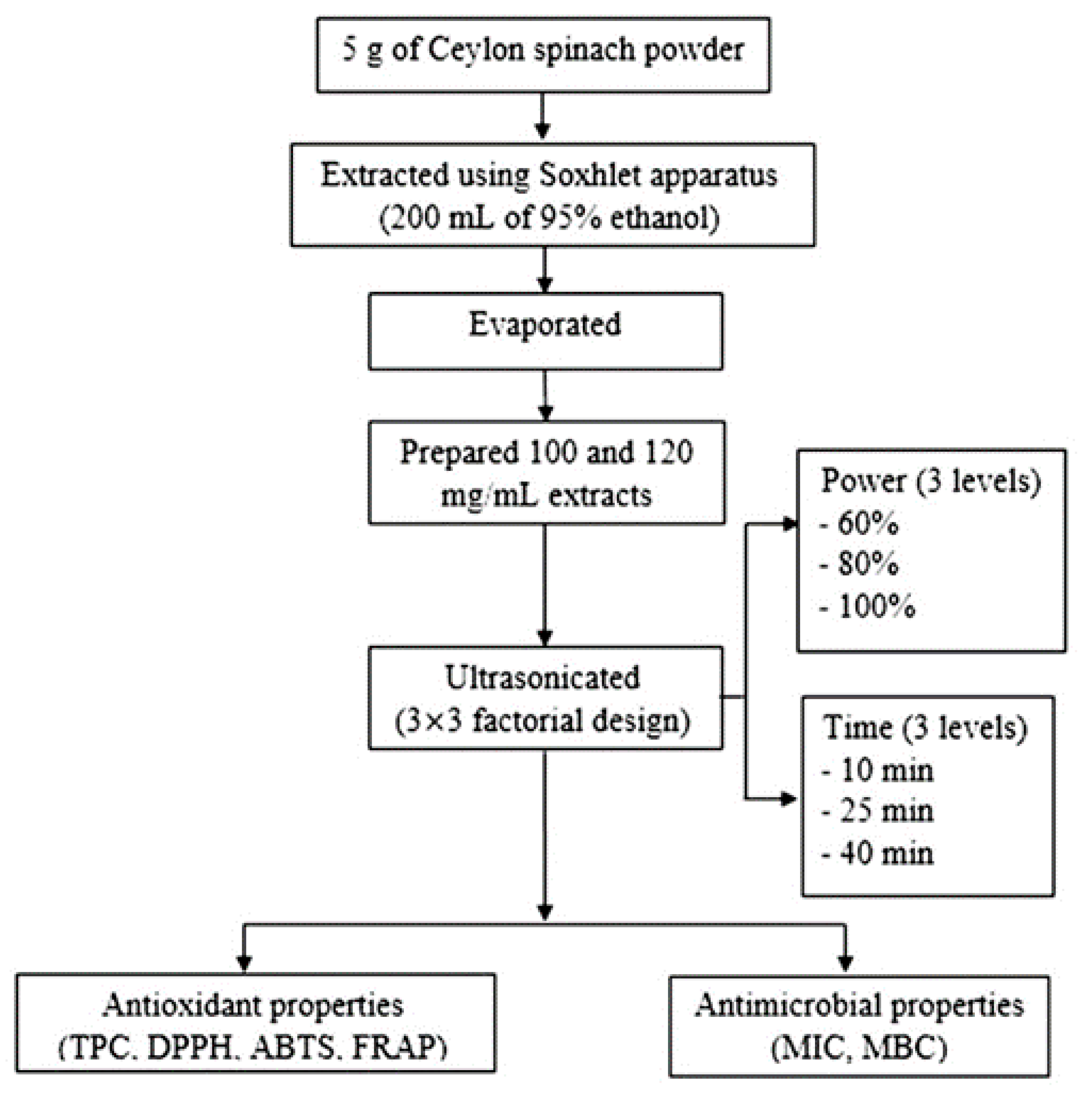
2.1. Plant Materials

Fresh Ceylon spinach (Basella alba) stems at 1.5–2 months after planting were collected from the College of Agriculture and Technology field plots (Mueang, Chiang Mai, Thailand) during February 2019. The samples were washed with tap water and dried in a hot-air oven (UNB 400, Memmert, Eagles, WI, USA) at 50 °C for 24 h. The dried plants were ground using an electric grinder (BL3071AD, Tefal, Bangkok, Thailand) and stored at −20 °C before extracting within 3 months.

2.2. Chemicals and Reagents

2,2-Diphenyl-1-picrylhydrazyl (DPPH), 2,2′-azino-bis (3-ethylbenzothiazoline-6-sulfonic acid (ABTS), 2,4,6-tris(2-pyridyl)-s-triazine (TPTZ), gallic acid (GA), and 2-thiobarbituric acid were purchased from Sigma–Aldrich (Singapore, Singapore). Ferrous sulfate, Na2CO3, and the Folin–Ciocalteu reagent were bought from Loba Chemie (Mumbai, India). Nutrient broth, Mueller–Hinton broth (MHB), and Mueller–Hinton agar (MHA) were purchased from Himedia (Mumbai, India). Plate count agar (PCA) and peptone water were purchased from Difco (Cockeysville, MD, USA). Other chemicals were analytical grade and obtained from RCI Labscan (Bangkok, Thailand).

2.3. Preparation of Extracts

Ceylon spinach powder (5 g) were extracted using a Soxhlet apparatus (Quicklet, Northern Ireland, UK) with 200 mL of 95% (v/v) ethanol at 80 °C for 4 h [16]. The extract was concentrated at 175-mbar reduced pressure in a water bath (B-490, Buchi, Saint Gallen, Switzerland) at 60 °C using a rotary evaporator (CH-9230, Buchi) to evaporate the ethanol, and then, the evaporated sample was further dried using a vacuum oven at 50 °C (VD53, Binder, Tuttlingen, Germany) to obtain dry Ceylon spinach extracts (CE) following the method of Sulaiman et al. [22]. The CE (100-mg of dry, crude extract after the evaporation/mL of distilled water), which corresponded to 1:10 (w/v), were ultrasonicated following the method adapted from Hashemi et al. [23]. The experiment had two factors: the extraction time (10–40 min) and power (60–100%) and were arranged as a 3 × 3 factorial in a completely randomized design (CRD) with duplicates, as shown in Figure 1. The CE samples in the test tube were ultrasonicated using an ultrasonic bath (40 kHz, 150 W, SB25-12DTD, Drawell, Jacksonville, FL, USA) at 25 °C. The untreated ultrasonication sample was used as the control.

2.4. Antioxidant Properties of Ultrasonicated CE

2.4.1. Total Phenolic Compounds (TPC)

TPC were determined as described by Oluwakemi et al. [24]. Briefly, 500 µL of the sample (100 mg/mL) was mixed with 2.5 mL of 10% Folin–Ciocalteu reagent and 2 mL of 7.5% Na2CO3. After incubation in the dark at 30 °C for 30 min, the absorption was measured at 765 nm using a multi-mode microplate reader (SpectraMax® i3x, Molecular Devices, San Jose, CA, USA). Aqueous solutions of GA were used to prepare the calibration curve. Results (n = 3) were expressed as GA equivalents (E).

2.4.2. DPPH and ABTS Radical Scavenging Activity and Ferric-Reducing Antioxidant Power (FRAP)

Free radical scavenging activity was determined using the DPPH radical assay of Surin et al. [25]. The sample solution (2 mL) was mixed with 2 mL of 0.2-mmol/L DPPH solution. After incubation in the dark at 30 °C for 30 min, the absorbance was measured at 517 nm. The percentage of inhibition of the DPPH radical (n = 3) was calculated. The results were expressed as IC50 values, the lowest concentration of the sample required to inhibit 50% of the radicals.
For the ABTS radical scavenging assay of Chaiwong et al. [26], 100 µL of the sample from 0.5–5.0 mg/mL was mixed with 900 µL of 7-mM ABTS reagent. After incubation in the dark at 30 °C for 6 min, the absorbance was measured at 734 nm. The percentage of inhibition of the ABTS radical was calculated. The results (n = 3) were expressed as the IC50 values.
The reducing power was determined using the FRAP assay of Surin et al. [27], with some modifications. Briefly, 100 µL of the sample (100 mg/mL) was mixed with 1900 µL of FRAP reagent, which consisted of 2.5 mL of 10-mM TPTZ in 40-mM HCl, 2.5 mL of 20-mM FeCl3 and 25 mL of 0.3-M acetate buffer (pH 3.6). The absorption was measured at 595 nm, and aqueous FeSO4 solutions were used to prepare the calibration curve. The measurements were done in triplicate (n = 3). The untreated ultrasonication sample was used as the control.

2.5. Antibacterial Activities of Ultrasonicated CE Using the Minimum Inhibitory Concentration (MIC) and Minimum Bactericidal Concentration (MBC)

The MIC is the concentration of CE required to inhibit the growth of the tested microorganism. The ultrasonicated samples were prepared at 100 mg/mL of crude extract after evaporation, and serial dilutions were done to obtain solutions at 50, 25, 12.5, 6.25, and 3.125 mg/mL using the modified method of Kumar et al. [28]. The tested microorganisms—namely, S. aureus TISTR 2320, E. coli TISTR 527, Salmonella Typhimurium TISTR 1469, and Pseudomonas aeruginosa TISTR 2370—were obtained from the Thailand Institute of Scientific and Technological Research (TISTR, Bangkok, Thailand). The initial microbial samples were, at ~1 × 10⁸ CFU/mL, obtained by adjusting the turbidity to match a 0.5 McFarland standard. Each sample solution (250 µL) was diluted with 250 µL of sterile MHB. The solution was inoculated with 250 µL of microbial suspension and then incubated at 37 °C for 24 h. Changes in the turbidity were measured at 600 nm for comparison with the control.
The MBC is the lowest concentration of extract with the ability to inactivate the tested microorganisms [28]. Six different concentrations of ultrasonicated CE were tested by streaking on MHA plates that were then incubated at 37 °C for 24 h. The lowest concentration of the plant extract required to inactivate the test microorganism was designated as the MBC value.

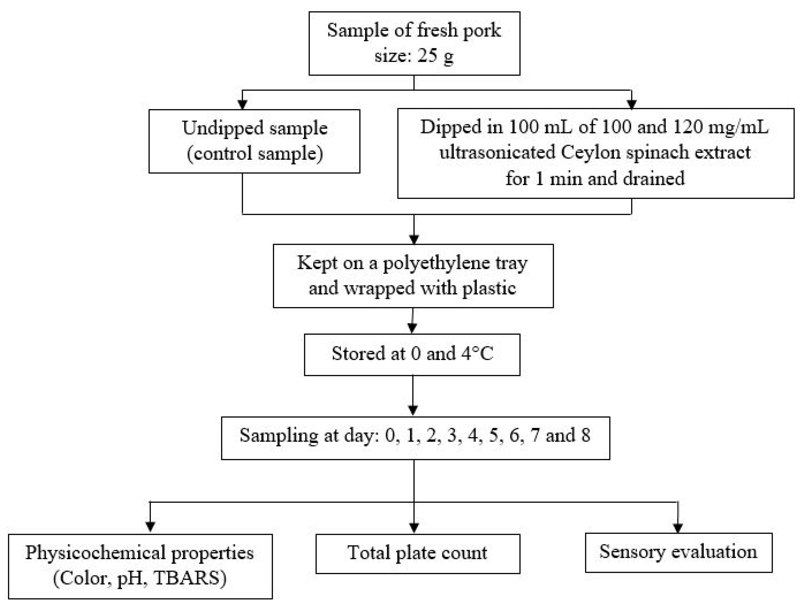
2.6. Shelf-Life Evaluation of Fresh, Chilled Pork

2.6.1. Preparation of Fresh, Chilled Pork

Fresh center-cut pork sirloins (Longissimus thoracis et lumborum, LTL) were selected and purchased from Charoen Pokphand Foods (Chiang Mai, Thailand) 1 day after slaughter and transported to the laboratory at 0–4 °C within 2 h. A Certificate of Analysis as a standard control for the fresh pork quality was provided by the company to ensure the safety of the product from the aerobic plate count, coliform bacteria, E. coli, S. aureus, Clostridium perfringens, Salmonella spp., Enterococcus spp., L. monooxygenase, Campylobacter jejuni, and yeast and molds following the standard US Food and Drug Administration protocols. The pork was sliced perpendicular to the long axis of the muscle with a knife into pieces of ~25 g with 2-cm thickness. Pork samples were dipped into two CE solutions with different concentrations (100 or 120 mg/mL) for 1 min. The preparation of the 100 or 120 mg/mL CE solution was done by dissolving 100 or 120 mg of dry ultrasonicated CE with 1 mL of distilled water. The optimum condition for ultrasonication was 80% power for 40 min, which was selected for the CE preparation. After dipping, the excess surface liquid on the samples were drained away and air-dried on a wire mesh at 20 °C for 10 min in a clean room before packing. The samples were kept in polyethylene (PE) trays (one/tray) and wrapped with food-grade polyvinyl chloride wrapping films (MMP, Bangkok, Thailand) with the experimental design shown in Figure 2. All samples were stored in two refrigerators (KB400, Binder, Bohemia, NY, USA) at 0 ± 1 and at 4 ± 1 °C and kept away from light for a period of 8 days and collected (10 pieces/treatment each time for different analyses) every day prior to comparison with the undipped control. Three pieces were used for the microbial analysis; another 3 pieces for the physicochemical analysis (color, pH, and TBARS); and the remaining 4 pieces were used for the sensory analysis.

2.6.2. Color, pH, and TBARS Measurements

The color prior to blooming of the packed and chilled fresh pork after being unwrapped (n = 3), as mentioned by Sen et al. [29], was measured using the CIELAB system using a Chroma meter (CR-410, Konica-Minolta, Tokyo, Japan) and illuminant D65 observer angle of 2°; aperture size of 50 mm; and expressed as L*, a*, and b*. The lightness (L*) value indicates black at 0 and white at 100, the a* value represents the red (+60)–green (−60) color, and the b* value describes the yellow (+60)–blue (−60) color. A 25-g meat sample with 225-mL distilled water was homogenized with a hand blender (MSM64110, Bosch, Bangkok, Thailand) for 30 s at 30 °C before pH measurements (n = 3) using a pH meter (FiveEasy F20, Mettler Toledo, Greifensee, Switzerland). The pH meter was calibrated at 30 °C using pH buffers at 4.0 and 7.0 (RCI Labscan, Ltd., Bangkok, Thailand). The TBARS in chilled pork was determined using the method of Lekjing and Venkatachalam [30]. The meat (1 g) was homogenized using a Vortex mixer (VTX-3000L, LMS, Tokyo, Japan) in a 10-mL mixture of 0.375% (w/v) thiobarbituric acid, 15% (w/v) trichloroacetic acid, and 0.875% (w/v) of 0.25-M HCl. The mixture was heated at 100 °C for 10 min to develop a pink color and cooled down with tap water. The mixture was centrifuged (Rotina 380R, Hettich, Tuttlingen, Germany) at 1520× g at 25 °C for 15 min. The supernatant was measured at 532 nm. The results were determined from a standard curve (0–3000-µM malondialdehyde—MDA) and expressed as the mg MDA/kg of chilled pork (n = 3).

2.6.3. Total Plate Count Analysis

Pork samples (25 g) were added to 225 mL of 0.1% peptone water and blended in a Stomacher (IUL-Instruments, Barcelona, Spain) for 2 min (n = 3). Subsequent dilutions were prepared using 9 mL of 0.1% peptone water and 1 mL of sample prior to the application of the total plate count technique by pouring on PCA plates and incubated at 37 °C for 24 h [31,32]. The results were expressed as log CFU/g.

2.6.4. Sensory Evaluation

The sensory evaluation was carried out using 50 untrained panelists (male and female, 50:50) following the American Meat Science Association [33] and Vilar et al. [34] recommendations. The ages of the consumers ranged from 19–50 years old. The consumer preferences were evaluated using a 9-point hedonic scale (from 1 = extremely dislikely to 9 = extremely likely), as described by Phimolsiripol et al. [35] and Chokumnoyporn et al. [36]. Consumer sensory panels were done in the sensory normalized testing room at the Chiang Mai University Sensory Research Unit, which conformed to the international standards (ISO) [37]. The analysis was done for 6 samples of fresh, chilled pork, evaluated in two sensory sessions within the same day, which were kept at 0 and 4 °C for up to 8 days of storage. Samples were served on polyethylene trays at 25 °C in a random order. Each sample was coded with a three-digit random number. Water was available for use between samples. The attributes of the unwrapped pork samples were appearance, color, odor, and overall liking.

2.7. Statistical Analysis

The statistical analysis was applied following the method of Biffin et al. [38], with a slight modification. The antioxidant properties, including TPC, DPPH IC50, ABTS IC50, and FRAP, were analyzed and compared to determine the effects of the power and time of the ultrasonication compared to the non-ultrasonicated sample using an analysis of variance (ANOVA) at the 95% confidence level (p ≤ 0.05). The experimental design was a factorial in the CRD model using the Statistical Package for the Social Sciences (SPSS version 17.0, SPSS, IBM Corp., Armonk, NY, USA). Mean comparisons were done using Duncan’s post-hoc test at p ≤ 0.05. Fixed effects in the full models included the power and time of the ultrasonication (treatments). The relationship or interaction terms between the responses as a function of the power and time of the ultrasonication and optimized optimal conditions were calculated using Design-Expert (Version 6.0.2, Stat-Ease, Inc., Minneapolis, MN, USA). Random terms for all the models included the extraction processing day and replication. Mean values and standard errors of the data were then reported.
For shelf life testing, the physical properties, including the color (L*, a*, and b*); pH; TBARS; and sensory data (appearance, color, odor, and overall liking), were calculated and compared using the ANOVA at the 95% confidence level (p ≤ 0.05) using SPSS. The fixed variables for the full models included the concentration of ultrasonicated CE, storage temperature, and storage time treatments. Random terms were grouped according to their relations to the sensory panel (test day, session number, testing order, and panelist). The microbial analysis was regressed to a linear equation showing the kinetic reaction rate (k) to delineate the trends of the microbial populations after storage at varied ultrasonicated CE concentration and storage temperature levels, as described by Phimolsiripol et al. [39].

3. Results and Discussion

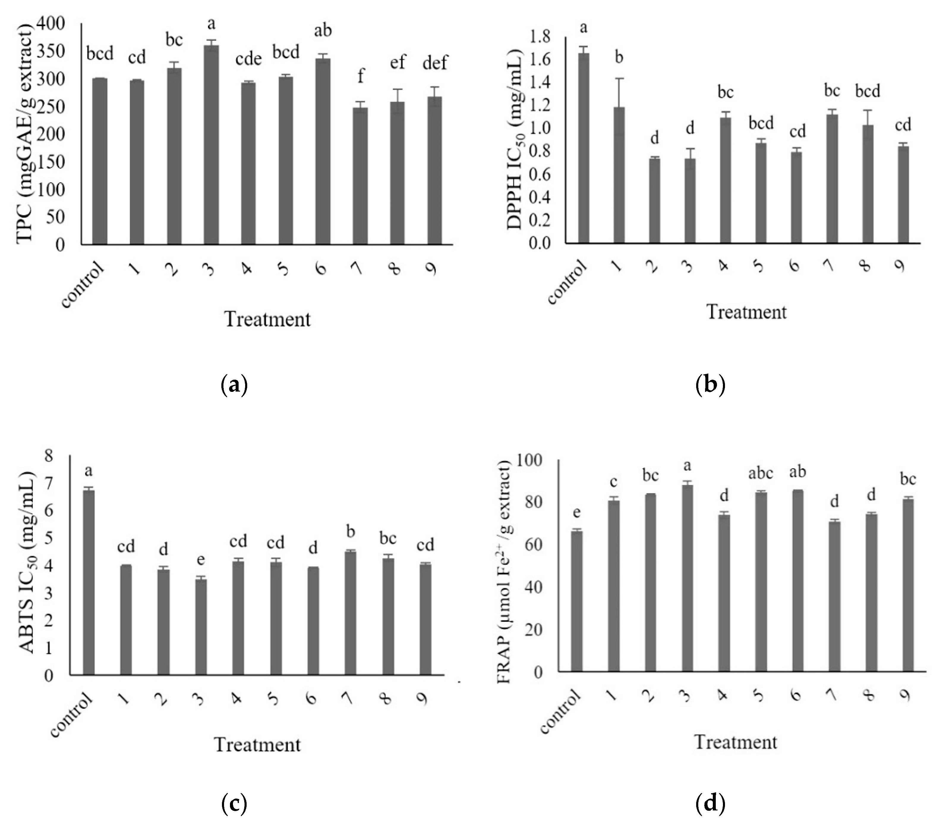
3.1. Antioxidant Properties of the Ultrasonicated Extracts

3.1.1. Total Phenolic Compounds (TPC)

The TPC of the CE ultrasonicated at 60% power for 40 min significantly increased (p ≤ 0.05) when compared to the control, as shown in Figure 3a. Increasing the time of the ultrasonication increased the TPC, which is consistent with previous studies [10]. The results indicated disruption of the plant cell walls by ultrasound waves. The phenolic compounds were released from within the solid matrix. The 100% ultrasonication power resulted in a lower TPC when compared to the control and 60% or 80% ultrasonication power. An increase in the power of the ultrasonication decreased the TPC, presumably due to the destruction of some of the extracted phenolic compounds [40], resulting in a reduction in the TPC when applied with too strong an ultrasonic power.

3.1.2. DPPH and ABTS Radical Scavenging Activity and Ferric-Reducing Antioxidant Power (FRAP)

There was a significant variation in the DPPH IC50 values of the ultrasonicated extracts, as shown in Figure 3b. All ultrasonicated extracts showed higher DPPH radical scavenging activities than the non-ultrasonicated extracts (control). The extract with the highest DPPH radical scavenging activity was obtained by ultrasonication at 60% power for 25 and 40 min. Ultrasonication at 60% power for 10 min resulted in the extract with the lowest (p ≤ 0.05) DPPH antioxidant capacity. Increasing the time of the ultrasonication led to increasing the DPPH antioxidant activity for 60% power. These results were consistent with an earlier report by Altemimi et al. [41] that showed the degradation of phenolic compounds when using high powers and high temperatures, thereby producing a cavitation bubble collapse.
The ABTS radical scavenging activity was quantified as the reduction in ABTS+ radicals and expressed as IC50 values (Figure 3c). The extracts with the highest and lowest potentials to inhibit the ABTS radicals were obtained by ultrasonication at 60% power for 40 min and 100% power for 10 min, respectively. The data showed that increasing the time of the ultrasonication increased the ABTS radical scavenging activity for the extracts ultrasonicated at 60% and 100% power. Higher ultrasonication times have been associated with higher flavonoid yields at the same ultrasonic power [10]. A longer extraction time permits more contact time for the cavitation bubbles to rupture more plant cells, in turn increasing the TPC extraction [40]. Therefore, the antioxidant capacities will increase. Ultrasound water baths produce enough cavitation to create shear forces to break the cell walls. Furthermore, ultrasonication increases the diffusion of cell contents into the extraction solution [42].
The FRAP of the extracts are shown in Figure 3d. The extract ultrasonicated at 60% power for 40 min showed the highest (p ≤ 0.05) potential to decrease the ferric ions (Fe3+), while the lowest potential to decrease the Fe3+ ions was observed with the extract ultrasonicated at 100% power for 10 min, suggesting that increasing the time of ultrasonication also increased the FRAP. Compared with the control (non-ultrasonicated sample), there was a significant increase (7–33%, p ≤ 0.05) in the FRAP values after ultrasonication. This trend was consistent with Ilghami et al. [43]. It might be due to an increase in the ultrasonic times, which can increase the diffusivity of the solvent into cells and enhance the desorption and solubility of the target compounds from the cells, thereby improving the antioxidant efficacy [44].

3.2. Antibacterial Activities of Ultrasonicated Extracts Using MIC and MBC

The antibacterial properties of the ultrasonicated extracts were measured by determining the MIC values (Table 1). The lowest MIC value of all nine extracts able to inhibit the growth of S. aureus and E. coli was 100-mg/mL. When using 60% power, the extracts inhibited S. typhimurium and P. aeruginosa at a MIC of 100 mg/mL. When the power was increased, the MIC values against S. typhimurium and P. aeruginosa were 50 mg/mL. It was evident that increasing the power resulted in increasing the antibacterial activities against S. typhimurium and P. aeruginosa. The MBC defined the lowest concentration of the extract that could inhibit the tested microorganisms. The MBC values showed the results of an in vitro test in which the fixed concentration of the extracts was being tested against an initially fixed concentration of microorganism [45]. All extracts had an MBC of a 100 mg/mL against S. aureus and E. coli. When ultrasonicated at 60% power for 10, 25, and 40 min, the MBC values against S. typhimurium and P. aeruginosa were 100 mg/mL. At 80% and 100% power for 10, 25, and 40 min, the MBC values against S. typhimurium and P. aeruginosa decreased to 50 mg/mL (Table 1).
A comparison between the ultrasonicated and non-ultrasonicated (control) extracts showed that the antibacterial activities against S. aureus and E. coli were similar. Whereas the antibacterial activities against S. typhimurium and P. aeruginosa increased when the ultrasonication power was increased to 80% and 100% for 10, 25, and 40 min (Table 1). Ultrasonic waves cause pressure and cavitation with the disruption of cell walls, so that the components of interest can be released [46] and the extracts are more easily released [47]. The Gram-positive and Gram-negative bacteria showed different sensitivities due to their different cell wall structures. The Gram-negative cell envelope is a thin structure that is covered by an outer membrane. On the other hand, Gram-positive bacteria lack the outer cell membrane, and the cell wall is typically much thicker, with multiple peptidoglycan layers [48]. Due to these distinct differences, it may be easier to inhibit Gram-negative bacteria than Gram-positive bacteria. Therefore, the MBC values of the extracts against S. typhimurium and P. aeruginosa were lower than the MBC value against S. aureus, which is a Gram-positive bacterium. However, the Gram-negative bacteria E. coli could tolerate a higher concentration of extracts than the other Gram-negative bacteria, because E. coli has the potential to form a dense biofilm around its cells, thus giving them protection against antibacterial agents [49]. As a result, the MBC values of the extracts against E. coli were higher than those for the other Gram-negative bacteria. Annatto dye has also been shown to have a greater antibacterial effect on Gram-positive bacteria (lower MIC and MBC) compared with Gram-negative bacteria [50]. These results were probably due to the presence of lipopolysaccharide in the cell wall of Gram-negative bacteria, which can prevent the influx of active compounds into the cytoplasmic membrane of these bacteria [51].

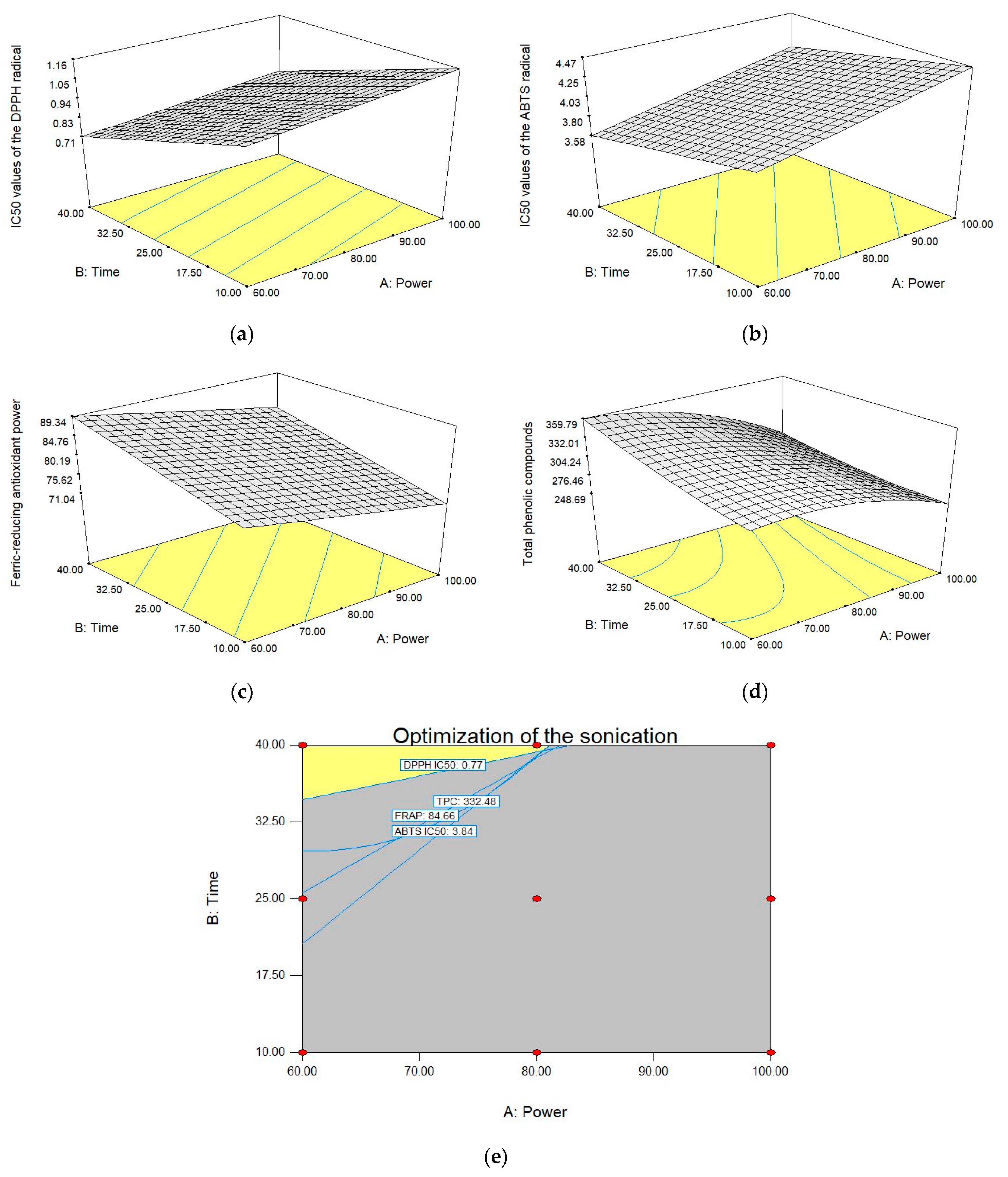
3.3. Optimization of Ultrasonication

The response surface plots (Figure 4) were used to visually observe the relationship between the responses and the various power and times of ultrasonication. The responses studied were TPC, IC50 values for the DPPH and ABTS radical scavenging capacities, and FRAP. The response surfaces (Figure 4a–d) were evaluated to predict the optimum power and time of ultrasonication. The optimum condition of ultrasonication was 80% power for 40 min, as shown in Figure 4e. With these conditions, the MIC and MBC values for S. typhimurium and P. aeruginosa were 50 mg/mL. The content of the TPC was 332-mg GAE/g extract. The IC50 values for DPPH and ABTS were 0.77 and 3.84 mg/mL, respectively, and FRAP was 84.7-µmol Fe2+/g extract.

3.4. Shelf Life Evaluation of Fresh Pork

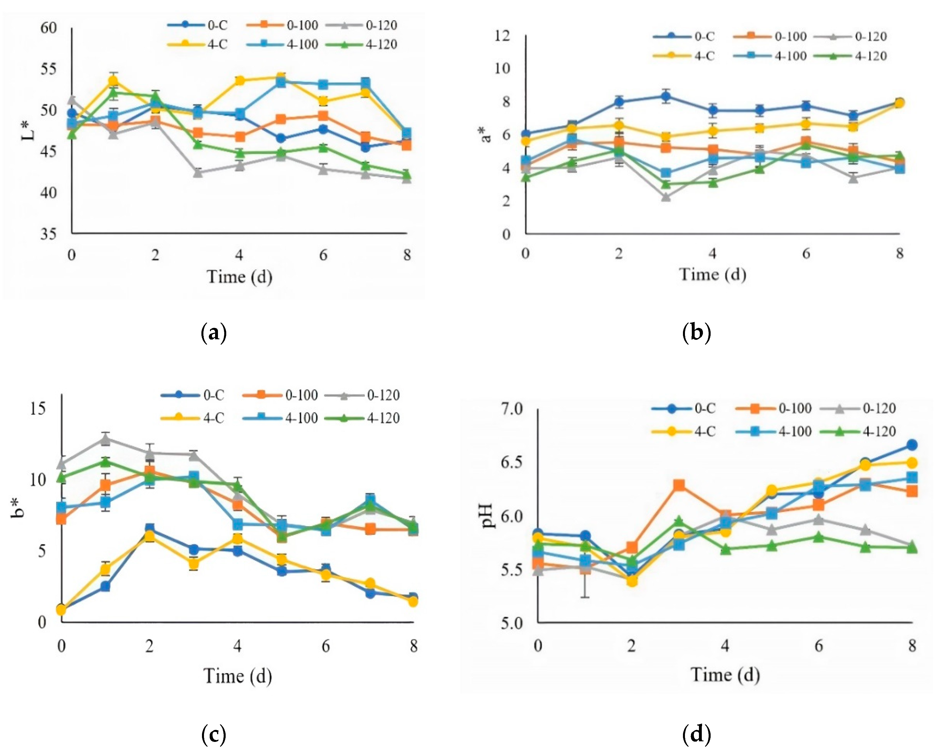
3.4.1. Color and pH Measurements

The L* values of all the chilled, fresh pork decreased significantly (p ≤ 0.05, Figure 5a) with the increasing storage time. Fresh pork dipped in 120-mg/mL ultrasonicated extract showed lower L* values when compared to fresh pork dipped with 100-mg/mL ultrasonicated extract and the control (nontreated). In addition, the L* value tended to decrease during the increased storage time. Reduction of the L* values reflected pigment materials in the extracts, including chlorophylls [52]. Polyphenol oxidases could oxidize the phenolic compounds to quinones and quinones, which are likely to be condensed to form darker compounds [53]. The control samples (nontreated) showed greater a* values than fresh pork dipped in 100 and 120-mg/mL ultrasonicated extracts at 0–8 day of storage (p ≤ 0.05, Figure 5b). Increasing the storage time resulted in an increasing redness (a*) of the control sample at day 8 of storage. The control samples showed lower b* values when compared to fresh pork dipped in the ultrasonicated extract. The values of b* were significantly decreased (p ≤ 0.05) with the increasing storage time, as seen in Figure 5c. Increasing the lipid oxidation could lead to a decrease in the a* and b* values [54]. The changes of the pH values of pork when kept at 0 and 4 °C for 8 days are shown in Figure 5d. The pH values of pork significantly increased (p ≤ 0.05) with the increased storage time. A similar pH trend was also reported by Lu et al. [31]. The increase of the pH values is associated with bacterial spoilage, related to the action of microbial enzymes, e.g., proteases and lipases, which increase the breakdown of nitrogenous compounds [55]. Changes in the pH may also be related to the spoilage of meat products. The pH usually decreases consequently with the bacterial growth and production of acid from lactic acid bacteria [56].

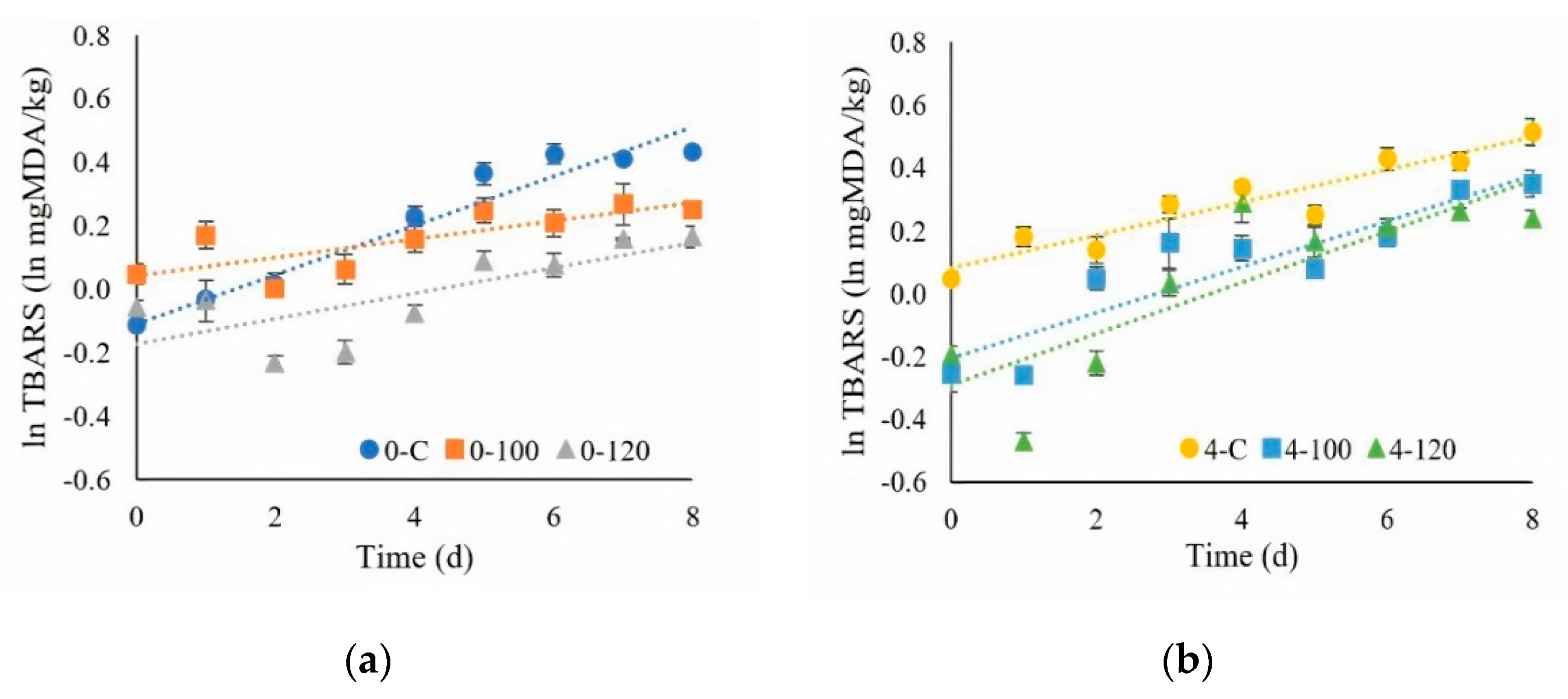
3.4.2. TBARS Measurement

The TBARS values of all the chilled, fresh pork increased significantly as the storage time increased (p ≤ 0.05, Figure 6a,b). The changes in TBARS in Figure 6a,b show the natural logarithm plot, which indicated a first order-type reaction. The elevation of TBARS with the storage time was observed. Lu et al. [31] mentioned that increasing the storage time resulted in increasing the TBARS values. Fresh pork dipped in 100 and 120-mg/mL ultrasonicated extract showed lower TBARS values compared to the control sample. The results showed that the antioxidative properties of the ultrasonicated extract had the potential to retard the lipid oxidation in pork. Lekjing and Venkatachalam [30] investigated the effects of a chitosan-based coating at 2% incorporated with 1.5% clove oil (CS + CO) on cooked pork sausage samples. They showed that the TBARS values of the CS + CO-treated samples were lower than those of the 2% chitosan-treated samples at 10 and 15 days of storage. Lorenzo et al. [57] also reported that the TBARS values of refrigerated pork patties treated with BHT, green tea extract, seaweed extract, or grape seed extract were lower than in the control samples. The antioxidant properties of the plant extracts could be used to retard the lipid oxidation [58]. Chilled, fresh pork stored at 0 °C had lower TBARS values when compared to fresh pork stored at 4 °C. The lower temperature was effective in decreasing the lipid oxidation [31]

3.4.3. Total Plate Count Analysis

As mentioned by Kim and Jang [32], the total plate count is an important parameter for fresh pork shelf life testing. The total plate counts of chilled, fresh pork with the ultrasonicated extract dipping and nontreated fresh pork (control) are shown in Table 2. The initial microbial loads of the nontreated and treated samples were similar. The total plate counts of all the chilled, fresh pork samples increased linearly with the increased storage time (p ≤ 0.05). For storage at 0 °C, the kinetic reaction rate (k) values for the control, 100, and 120 mg/mL were 0.698, 0.636, and 0.497 log CFU/g.day, respectively. While, for pork storage at 4 °C, the k values for the control, 100, and 120 mg/mL were 0.774, 0.700, and 0.681 log CFU/g.day, respectively. It showed that the 120-mg/mL CE had significantly delayed the growth of the total bacteria. For fresh, chilled pork, the total plate count standard [59] was 6.7 log CFU/g. The total plate counts of the control samples and fresh pork dipped in 100 and 120-mg/mL CE stored at 0 °C reached 6.7 log CFU/g by the 5th, 6th, and 7th days of storage, respectively, with visible signs of spoilage. Meanwhile, the control samples and fresh pork dipped in 100 and 120-mg/mL CE stored at 4 °C had total plate count values above the standard limit on the 4th, 5th, and 6th days of storage, respectively. A lower temperature could extend the storage time, as reported by Akoğlu et al. [60]. It was confirmed that the shelf life of chilled pork was prolonged by dipping with either 100 or 120-mg/mL of CE at a lower temperature (0 °C). This was probably due to the phenolic compounds in the ultrasonicated extracts. The phenolic compounds showed that the antimicrobial activity can increasingly against many Gram-positive bacteria. The Gram-positive bacteria showed a better susceptibility to antimicrobial activity, because the outer membrane of the Gram-negative bacteria represented a reduced absorption of the phenolic compounds and barrier of permeability [61]. They can denature the proteins of microbial cell membranes, leading to inactivation or death [62]. The microbial activities were consistent with the reports for different natural additives and their extracts in sausages [63] and bison meat [64]. The authors de Souza de Azevedo et al. [7] found that nisin can be used as an antimicrobial agent for shelf life extension in pork meat. Ranucci et al. [65] also reported sausage made from pork meat with a mix of Punica granatum and Citrus spp. extracts. They found that the extracts could extend the shelf life of pork by controlling the microbial growth and oxidation during refrigerated storage at 4 ± 1 °C.

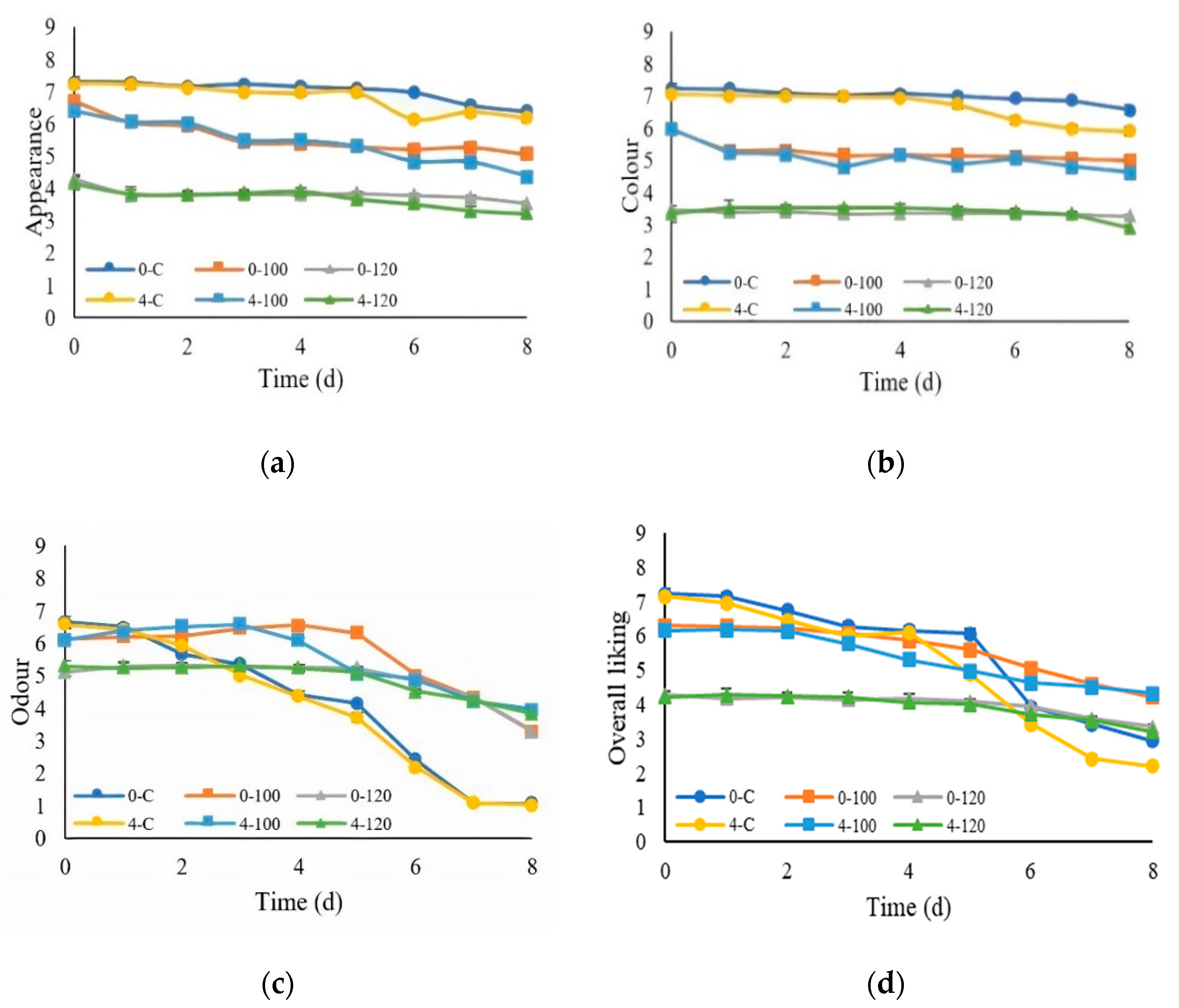
3.4.4. Sensory Evaluation

The sensory evaluation showed that the scores for the appearance, color, odor, and overall liking from all the chilled pork samples showed significant decreases (p ≤ 0.05) with the increasing storage time, as shown in Figure 7. The scores for the appearance and color from the control were higher than fresh pork dipped in the ultrasonicated extracts. The result showed that the fresh pork dipped in 120-mg/mL at 0 and 4 °C had lower consumer acceptance (<5 hedonic score) due to too strong a greenish appearance and color, affected by too much CE extract. The control samples at 0 and 4 °C had a spoiled odor after a storage period of 4–8 days, which resulted in lower scores of the overall liking. The consumers preferred fresh pork dipped in ultrasonicated extracts that received higher odor and overall liking scores than the control samples. This was probably due to the antimicrobial effect of the CE, as confirmed by the increased microbial population found in the experiments reported in Section 3.4.3. In addition, Ramírez-Rojo et al. [66] showed that pork patties treated with an ethanol extract of Mesquite leaves could increase the shelf life with acceptable sensory properties. Due to the sensory acceptance limitations of 120-mg/mL CE, 100-mg/mL CE should be selected to apply for shelf life extensions instead of 120-mg/mL CE with better acceptance by consumers.

4. Conclusions

Increasing the time of ultrasonication resulted in increasing the antioxidant activities (DPPH, ABTS, and FRAP) of the CE extracts. Increasing the ultrasonication power increased the capacity of the extracts to inhibit the growth of S. typhimurium and P. aeruginosa. The optimum ultrasonication condition was determined as ultrasonication at 80% power for 40 min. The fresh pork dipped in 100-mg/mL ultrasonicated extract could be kept for 7 days at 0 °C when compared to the control, which could be kept for only 5 days. The CE extracts by ultrasonication showed greater antioxidant and antimicrobial properties for improving the shelf life of fresh pork. In addition, the sensory evaluation of the fresh pork dipped in 100-mg/mL CE at 0 °C was also acceptable to consumers. Accordingly, these can be safety applied by the food industry to enhance the quality and extend the shelf life of meat products. More work is required to investigate the kinetic study of extraction using ultrasonication and the flavor profile of ultrasonicated CE alone and its impact on treated foods. More mechanisms of the shelf life studies of CE extracts are also needed.

Author Contributions

Conceptualization: Y.P. and P.L.; methodology, S.B.; formal analysis, S.B.; data curation, S.B.; writing—original draft preparation, Y.P. and S.B.; writing—review and editing, K.J., P.S., T.C., N.L., S.R.S., N.C., C.S.B., and J.M.R.; supervision, Y.P., C.S.B., and J.M.R.; and funding acquisition, Y.P. and P.R. All authors have read and agreed to the published version of the manuscript.

Funding

This research and the APC was funded by the Program Management Unit for Human Resources & Institutional Development, Research and Invitation, NXPO [Frontier Global Partnership for Strengthening Cutting-edge Technology and Innovations in Materials Science], grant number B16F640001. This project was also supported by Chiang Mai University.

Data Availability Statement

Not applicable.

Conflicts of Interest

All the authors declare that they have no conflict of interest.

References

  1. Djekic, I.; Bozickovic, I.; Djordjevic, V.; Smetana, S.; Terjung, N.; Ilic, J.; Doroski, A.; Tomasevic, I. Can we associate environmental footprints with production and consumption using Monte Carlo simulation? Case study with pork meat. J. Sci. Food Agric. 2021, 101, 960–969. [Google Scholar] [CrossRef]
  2. Perlo, F.; Fabre, R.; Bonato, P.; Jenko, C.; Tisocco, O.; Teira, G. Refrigerated storage of pork meat sprayed with rosemary extract and ascorbic acid. Cienc. Rural 2018, 48, e20170238. [Google Scholar] [CrossRef]
  3. Shah, M.A.; Bosco, S.J.D.; Mir, S.A. Plant extracts as natural antioxidants in meat and meat products. Meat Sci. 2014, 98, 21–33. [Google Scholar] [CrossRef]
  4. Castro, S.; Kolomeytseva, M.; Casquete, R.; Silva, J.; Queirós, R.; Saraiva, J.; Teixeira, P. Biopreservation strategies in combination with mild high pressure treatments in traditional Portuguese ready-to-eat meat sausage. Food Biosci. 2017, 19, 65–72. [Google Scholar] [CrossRef]
  5. Sahraee, S.; Milani, J.M.; Regenstein, J.M.; Kafil, H.S. Protection of foods against oxidative deterioration using edible films and coatings: A review. Food Biosci. 2019, 32, 100451. [Google Scholar] [CrossRef]
  6. Rocchetti, G.; Bernardo, L.; Pateiro, M.; Barba, F.J.; Munekata, P.E.; Trevisan, M.; Lorenzo, J.M.; Lucini, L. Impact of a Pitanga leaf extract to prevent lipid oxidation processes during shelf life of packaged pork burgers: An untargeted metabolomic approach. Foods 2020, 9, 1668. [Google Scholar] [CrossRef] [PubMed]
  7. de Souza de Azevedo, P.O.; Converti, A.; Gierus, M.; de Souza Oliveira, R.P. Application of nisin as biopreservative of pork meat by dipping and spraying methods. Braz. J. Microbiol. 2019, 50, 523–526. [Google Scholar] [CrossRef] [PubMed]
  8. Lorenzo, J.M.; Vargas, F.C.; Strozzi, I.; Pateiro, M.; Furtado, M.M.; Sant’Ana, A.S.; Rocchetti, G.; Barba, F.J.; Dominguez, R.; Lucini, L. Influence of pitanga leaf extracts on lipid and protein oxidation of pork burger during shelf life. Food Res. Int. 2018, 114, 47–54. [Google Scholar] [CrossRef]
  9. Chaisuwan, W.; Manassa, A.; Phimolsiripol, Y.; Jantanasakulwong, K.; Chaiyaso, T.; Pathom-aree, W.; You, S.G.; Seesuriyachan, P. Integrated ultrasonication and microbubble-assisted enzymatic synthesis of fructooligosaccharides from brown sugar. Foods 2020, 9, 1833. [Google Scholar] [CrossRef] [PubMed]
  10. Liu, Y.; Luo, X.; Lan, Z.; Tang, J.; Zhao, P.; Kan, H. Ultrasonic-assisted extraction and antioxidant capacities of flavonoids from Camellia fascicularis leaves. CYTA J. Food 2018, 16, 105–112. [Google Scholar] [CrossRef] [Green Version]
  11. Machado, A.P.D.F.; Sumere, B.R.; Mekaru, C.; Martinez, J.; Bezerra, R.M.N.; Rostagno, M.A. Extraction of polyphenols and antioxidants from pomegranate peel using ultrasound: Influence of temperature, frequency and operation mode. Int. J. Food Sci. Technol. 2019, 54, 2792–2801. [Google Scholar] [CrossRef]
  12. Tiwari, B.K. Ultrasound: A clean, green extraction technology. Trends Analyt. Chem. 2015, 71, 100–109. [Google Scholar] [CrossRef]
  13. Sanpa, S.; Sutjarittangtham, K.; Tunkasiri, T.; Eitssayeam, S.; Chantawannakul, P. Ultrasonic extraction of Thai propolis for antimicrobial and antioxidant properties. Adv. Mater. Res. 2012, 506, 371–374. [Google Scholar] [CrossRef]
  14. Balouiri, M.; Sadiki, M.; Ouedehiri, W.; Farah, A.; Abed, S.; Koraichi, S.I. Antibacterial activity of extracts from Salvia officinalis and Rosmarinus officinalis obtained by sonication and maceration methods. Int. J. Pharm. Pharm. Sci. 2014, 6, 167–170. [Google Scholar]
  15. Luque-García, J.L.; Luque de Castro, M.D. Ultrasound-assisted Soxhlet extraction: An expeditive approach for solid sample treatment: Application to the extraction of total fat from oleaginous seeds. J. Chromatogr. A 2004, 1034, 237–242. [Google Scholar] [CrossRef] [PubMed]
  16. Oyewole, O.; Kalejaiye, O. The antimicrobial activities of ethanolic extracts of Basella alba on selected microorganisms. Sci. J. Microbiol. 2012, 1, 113–118. [Google Scholar]
  17. Vongsak, B.; Sithisarn, P.; Mangmool, S.; Thongpraditchote, S.; Wongkrajang, Y.; Gritsanapan, W. Maximizing total phenolics, total flavonoids contents and antioxidant activity of Moringa oleifera leaf extract by the appropriate extraction method. Ind. Crops Prod. 2013, 44, 566–571. [Google Scholar] [CrossRef]
  18. Kumar, S.; Prasad, A.; Iyer, S.; Vaidya, S. Systematic pharmacognostical, phytochemical and pharmacological review on an ethno medicinal plant, Basella alba L. J. Pharmacogn. Phytotherapy 2013, 5, 53–58. [Google Scholar]
  19. Kumar, B.R.; Anupam, A.; Manchikanti, P.; Rameshbabu, A.P.; Dasgupta, S.; Dhara, S. Identification and characterization of bioactive phenolic constituents, anti-proliferative, and anti-angiogenic activity of stem extracts of Basella alba and rubra. J. Food Sci. Technol. 2018, 55, 1675–1684. [Google Scholar] [CrossRef]
  20. Maran, J.P.; Priya, B. Natural pigments extraction from Basella rubra L. fruits by ultrasound-assisted extraction combined with Box-Behnken response surface design. Sep. Sci. Technol. 2015, 50, 1532–1540. [Google Scholar] [CrossRef]
  21. Adesina, A.; Adefemi, S. Lipid composition of Basella alba and Basella rubra leaves consumed in South-Western Nigeria: Nutritional implications. Bangladesh J. Sci. Ind. Res. 2017, 52, 125–134. [Google Scholar] [CrossRef] [Green Version]
  22. Sulaiman, I.S.C.; Basri, M.; Masoumi, H.R.F.; Chee, W.J.; Ashari, S.E.; Ismail, M. Effects of temperature, time, and solvent ratio on the extraction of phenolic compounds and the anti-radical activity of Clinacanthus nutans Lindau leaves by response surface methodology. Chem. Cent. J. 2017, 11, 1–11. [Google Scholar] [CrossRef] [PubMed]
  23. Hashemi, S.M.B.; Khaneghah, A.M.; Akbarirad, H. The effects of amplitudes ultrasound-assisted solvent extraction and pretreatment time on the yield and quality of Pistacia khinjuk hull oil. J. Oleo Sci. 2016, 65, 733–738. [Google Scholar] [CrossRef] [PubMed] [Green Version]
  24. Oluwakemi, O.A.; Akindele, A.J.; Bosede, F.S. Ascorbic acid, total phenolic, flavonoid and antioxidant activity of two cultivars of Basella alba. Food Sci. Technol. 2017, 5, 92–96. [Google Scholar] [CrossRef]
  25. Surin, S.; Surayot, U.; Seesuriyachan, P.; You, S.G.; Phimolsiripol, Y. Antioxidant and immunomodulatory activities of sulphated polysaccharides from purple glutinous rice bran (Oryza sativa L.). Int. J. Food Sci. 2018, 53, 994–1004. [Google Scholar] [CrossRef] [Green Version]
  26. Chaiwong, N.; Leelapornpisid, P.; Jantanasakulwong, K.; Rachtanapun, P.; Seesuriyachan, P.; Sakdatorn, V.; Leksawasdi, N.; Phimolsiripol, Y. Antioxidant and moisturizing properties of carboxymethyl chitosan with different molecular weights. Polymers 2020, 12, 1445. [Google Scholar] [CrossRef]
  27. Surin, S.; You, S.G.; Seesuriyachan, P.; Muangrat, R.; Wangtueai, S.; Jambrak, A.R.; Phongthai, S.; Jantanasakulwong, K.; Chaiyaso, T.; Phimolsiripol, Y. Optimization of ultrasonic-assisted extraction of polysaccharides from purple glutinous rice bran (Oryza sativa L.) and their antioxidant activities. Sci. Rep. 2020, 10, 1–10. [Google Scholar]
  28. Kumar, D.; Jagarwal, P.; Shrama, R. Phytochemical screening and antimicrobial activity of Basella alba Linn. Int. J. Pharm. Chem. Sci. 2018, 8, 502–507. [Google Scholar]
  29. Sen, A.R.; Muthukumar, M.; Naveena, B.M.; Ramanna, D.B.V. Effects on colour characteristics of buffalo meat during blooming, retail display and using vitamin C during refrigerated storage. J. Food Sci. Technol. 2014, 51, 3515–3519. [Google Scholar] [CrossRef] [Green Version]
  30. Lekjing, S.; Venkatachalam, K. Influences of storage time and temperature on sensory and measured quality of green gram savory crackers. LWT-Food Sci. Technol. 2019, 113, 108310. [Google Scholar] [CrossRef]
  31. Lu, X.; Zhang, Y.; Zhu, L.; Luo, X.; Hopkins, D.L. Effect of superchilled storage on shelf life and quality characteristics of M. longissimus lumborum from Chinese yellow cattle. Meat Sci. 2019, 149, 79–84. [Google Scholar] [CrossRef]
  32. Kim, H.J.; Jang, A. Evaluation of the microbiological status of raw pork meat in Korea: Modification of the microbial guideline levels for meat. Food Sci. Biotechnol. 2018, 27, 1219–1225. [Google Scholar] [CrossRef] [PubMed]
  33. American Meat Science Association. Research Guidelines for Cookery, Sensory Evaluation, and Instrumental Tenderness Measurements of Meat, 2nd ed.; American Meat Science Association: Chicago, IL, USA, 2016. [Google Scholar]
  34. Vilar, E.G.; Ouyang, H.; O’Sullivan, M.G.; Kerry, J.P.; Hamill, R.M.; O’Grady, M.N.; Mohammed, H.O.; Kilcawley, K.N. Effect of salt reduction and inclusion of 1% edible seaweeds on the chemical, sensory and volatile component profile of reformulated frankfurters. Meat Sci. 2020, 161, 108001. [Google Scholar] [CrossRef] [PubMed]
  35. Phimolsiripol, Y.; Siripatrawan, U.; Teekachunhatean, S.; Wangtueai, S.; Seesuriyachan, P.; Surawang, S.; Laokuldilok, T.; Regenstein, J.M.; Henry, C.J.K. Technological properties, in vitro starch digestibility and in vivo glycaemic index of bread containing crude malva nut gum. Int. J. Food Sci. Technol. 2017, 52, 1035–1041. [Google Scholar] [CrossRef]
  36. Chokumnoyporn, N.; Sriwattana, S.; Phimolsiripol, Y.; Torrico, D.D.; Prinyawiwatkul, W. Soy sauce odour induces and enhances saltiness perception. Int. J. Food Sci. Technol. 2015, 50, 2215–2221. [Google Scholar] [CrossRef]
  37. International Standard Organization. Sensory Analysis: General Guidance for the Design of Test Rooms; ISO Central Secretariat: Geneva, Switzerland, 2007. [Google Scholar]
  38. Biffin, T.E.; Smith, M.A.; Bush, R.D.; Collins, D.; Hopkins, D.L. The effect of electrical stimulation and tenderstretching on colour and oxidation traits of alpaca (Vicunga pacos) meat. Meat Sci. 2019, 156, 125–130. [Google Scholar] [CrossRef] [PubMed]
  39. Phimolsiripol, Y.; Siripatrawan, U.; Cleland, D.J. Weight loss of frozen bread dough under isothermal and fluctuating temperature storage conditions. J. Food Eng. 2011, 106, 134–143. [Google Scholar] [CrossRef]
  40. Wang, X.; Wu, Q.; Wu, Y.; Chen, G.; Yue, W.; Liang, Q. Response surface optimized ultrasonic-assisted extraction of flavonoids from Sparganii rhizoma and evaluation of their in vitro antioxidant activities. Molecules 2012, 17, 6769–6783. [Google Scholar] [CrossRef]
  41. Altemimi, A.; Watson, D.G.; Choudhary, R.; Dasari, M.R.; Lightfoot, D.A. Ultrasound assisted extraction of phenolic compounds from peaches and pumpkins. PLoS ONE 2016, 11, e0148758. [Google Scholar] [CrossRef] [Green Version]
  42. Jerman, T.; Trebše, P.; Vodopivec, B.M. Ultrasound-assisted solid liquid extraction (USLE) of olive fruit (Olea europaea) phenolic compounds. Food Chem. 2010, 123, 175–182. [Google Scholar] [CrossRef]
  43. Ilghami, A.; Ghanbarzadeh, S.; Hamishehkar, H. Optimization of the ultrasonic-assisted extraction of phenolic compounds, ferric reducing activity and antioxidant activity of the Beta vulgaris using response surface methodology. Pharm. Sci. 2015, 21, 46–50. [Google Scholar] [CrossRef]
  44. Pankey, G.A.; Sabath, L. Clinical relevance of bacteriostatic versus bactericidal mechanisms of action in the treatment of Gram-positive bacterial infections. Clin. Infect. Dis. 2004, 38, 864–870. [Google Scholar] [CrossRef] [PubMed] [Green Version]
  45. Jahouach-Rabai, W.; Trabelsi, M.; Van Hoed, V.; Adams, A.; Verhé, R.; De Kimpe, N.; Frikha, M. Influence of bleaching by ultrasound on fatty acids and minor compounds of olive oil. Qualitative and quantitative analysis of volatile compounds (by SPME coupled to GC/MS). Ultrason. Sonochem. 2008, 15, 590–597. [Google Scholar] [CrossRef]
  46. Giacometti, J.; Žauhar, G.; Žuvić, M. Optimization of ultrasonic-assisted extraction of major phenolic compounds from olive leaves (Olea europaea L.) using response surface methodology. Foods 2018, 7, 149. [Google Scholar] [CrossRef] [Green Version]
  47. Donsì, F.; Annunziata, M.; Vincensi, M.; Ferrari, G. Design of nanoemulsion-based delivery systems of natural antimicrobials: Effect of the emulsifier. J. Biotechnol. 2012, 159, 342–350. [Google Scholar] [CrossRef] [PubMed]
  48. Weidenmaier, C.; Peschel, A. Teichoic acids and related cell-wall glycopolymers in Gram-positive physiology and host interactions. Nat. Rev. Microbiol. 2008, 6, 276–287. [Google Scholar] [CrossRef] [PubMed]
  49. Sharma, G.; Sharma, S.; Sharma, P.; Chandola, D.; Dang, S.; Gupta, S.; Gabrani, R. Escherichia coli biofilm: Development and therapeutic strategies. J. Appl. Microbiol. 2016, 121, 309–319. [Google Scholar] [CrossRef] [PubMed] [Green Version]
  50. Yolmeh, M.; Habibi-Najafi, M.B.; Shakouri, S.; Hosseini, F. Comparing antibacterial and antioxidant activity of annatto dye extracted by conventional and ultrasound-assisted methods. Zahedan J. Res. Med. Sci. 2015, 17, e1020. [Google Scholar] [CrossRef] [Green Version]
  51. McKeegan, K.S.; Borges-Walmsley, M.I.; Walmsley, A.R. Microbial and viral drug resistance mechanisms. Trends Microbiol. 2002, 10, s8–s14. [Google Scholar] [CrossRef]
  52. Dini, A.; Khanamani Falahati-Pour, S.; Behmaram, K.; Sedaghat, N. The kinetics of colour degradation, chlorophylls and xanthophylls loss in pistachio nuts during roasting process. J. Food Saf. 2019, 3, 251–263. [Google Scholar] [CrossRef]
  53. Zhang, H.; Wu, J.; Guo, X. Effects of antimicrobial and antioxidant activities of spice extracts on raw chicken meat quality. Food Sci. Hum. Wellness 2016, 5, 39–48. [Google Scholar] [CrossRef] [Green Version]
  54. Domínguez, R.; Pateiro, M.; Gagaoua, M.; Barba, F.J.; Zhang, W.; Lorenzo, J.M. A comprehensive review on lipid oxidation in meat and meat products. Antioxidants 2019, 8, 429. [Google Scholar] [CrossRef] [PubMed] [Green Version]
  55. Daniloski, D.; Petkoska, A.T.; Galić, K.; Ščetar, M.; Kurek, M.; Vaskoska, R.; Kalevska, T.; Nedelkoska, D.N. The effect of barrier properties of polymeric films on the shelf life of vacuum packaged fresh pork meat. Meat Sci. 2019, 158, 107880. [Google Scholar] [CrossRef]
  56. Silva, A.P.R.d.; Longhi, D.A.; Dalcanton, F.; Aragão, G.M.F.d. Modelling the growth of lactic acid bacteria at different temperatures. Braz. Arch. Biol. Technol. 2018, 61, e18160159. [Google Scholar] [CrossRef]
  57. Lorenzo, J.M.; Sineiro, J.; Amado, I.R.; Franco, D. Influence of natural extracts on the shelf life of modified atmosphere-packaged pork patties. Meat Sci. 2014, 96, 526–534. [Google Scholar] [CrossRef]
  58. Stefanello, F.S.; Cavalheiro, C.P.; Ludtke, F.L.; Silva, M.d.S.d.; Fries, L.L.M.; Kubota, E.H. Oxidative and microbiological stability of fresh pork sausage with added sun mushroom powder. Ciênc. Agrotechnol. 2015, 39, 381–389. [Google Scholar] [CrossRef] [Green Version]
  59. Teuteberg, V.; Kluth, I.-K.; Ploetz, M.; Krischek, C. Effects of duration and temperature of frozen storage on the quality and food safety characteristics of pork after thawing and after storage under modified atmosphere. Meat Sci. 2021, 174, 108419. [Google Scholar] [CrossRef]
  60. Akoğlu, I.; Bıyıklı, M.; Akoğlu, A.; Kurhan, Ş. Determination of the quality and shelf life of sous vide cooked Turkey cutlet stored at 4 and 12 °C. Rev. Bras. Cienc. Avic. 2018, 20, 1–8. [Google Scholar] [CrossRef]
  61. Álvarez-Martínez, F.J.; Barrajón-Catalán, E.; Encinar, J.A.; Rodríguez-Díaz, J.C.; Micol, V. Antimicrobial capacity of plant polyphenols against gram-positive bacteria: A comprehensive review. Curr. Med. Chem. 2020, 27, 2576–2606. [Google Scholar] [CrossRef]
  62. Viuda Martos, M.; Ciro Gómez, G.L.; Ruiz Navajas, Y.; Zapata Montoya, J.E.; Sendra, E.; Pérez-Álvarez, J.A.; Fernández-López, J. In vitro antioxidant and antibacterial activities of extracts from annatto (Bixa orellana L.) leaves and seeds. J. Food Saf. 2012, 32, 399–406. [Google Scholar] [CrossRef]
  63. Ali, F.; Abdel-Atty, N.; Helmy, E. Improving the quality and extending the shelf life of chilled fresh sausage using natural additives and their extracts. J. Microbiol. Biotechnol. Food Sci. 2018, 7, 580–585. [Google Scholar] [CrossRef]
  64. Sood, V.; Tian, W.; Narvaez-Bravo, C.; Arntfield, S.D.; González, A.R. Plant extracts effectiveness to extend bison meat shelf life. J. Food Sci. 2020, 85, 936–946. [Google Scholar] [CrossRef]
  65. Ranucci, D.; Roila, R.; Andoni, E.; Braconi, P.; Branciari, R. Punica granatum and Citrus spp. extract mix affects spoilage microorganisms growth rate in vacuum-packaged cooked sausages made from pork meat, emmer wheat (Triticum dicoccum Schübler), almond (Prunus dulcis Mill.) and hazelnut (Corylus avellana L.). Foods 2019, 8, 664. [Google Scholar] [CrossRef] [PubMed] [Green Version]
  66. Ramírez-Rojo, M.I.; Vargas-Sánchez, R.D.; Torres-Martínez, B.d.M.; Torrescano-Urrutia, G.R.; Lorenzo, J.M.; Sánchez-Escalante, A. Inclusion of ethanol extract of Mesquite leaves to enhance the oxidative stability of pork patties. Foods 2019, 8, 631. [Google Scholar] [CrossRef] [PubMed] [Green Version]
Figure 1. Design of the ultrasonic experiment.
Foods 10 01241 g001
Figure 2. Design of the experiments for the shelf life study.
Foods 10 01241 g002
Figure 3. Means and standard errors of the antioxidant properties of the sonicated extracts’ (a) Total phenolic compounds (TPC), (b) IC50 values of the 2,2-Diphenyl-1-picrylhydrazyl (DPPH) radical, (c) IC50 values of the 2,2′-azino-bis (3-ethylbenzothiazoline-6-sulfonic acid (ABTS) radical, and (d) FRAP—ferric-reducing antioxidant power (control = non-sonicated extract, 1 = 60% 10 min, 2 = 60% 25 min, 3 = 60% 40 min, 4 = 80% 10 min, 5 = 80% 25 min, 6 = 80% 40 min, 7 = 100% 10 min, 8 = 100% 25 min, and 9 = 100% 40 min). Different letters indicate significant differences between the treatments (p ≤ 0.05).
Foods 10 01241 g003
Figure 4. Response surfaces. (a) IC50 values of the DPPH radical, (b) IC50 values of the ABTS radical, (c) ferric-reducing antioxidant power, (d) total phenolic compounds, and (e) optimization of the sonication.
Foods 10 01241 g004
Figure 5. Changes in the means and standard errors of the colors (a) Lightness (L*), (b) Redness-greenness (a*), and (c) Yellowness-blueness (b*) and (d) pH when stored at 0 and 4 °C. (0-C = nondipped sample at 0 °C, 0-100 = 100-mg/mL dipped sample at 0 °C, 0-120 = 120-mg/mL dipped sample at 0 °C, 4-C = nondipped sample at 4 °C, 4-100 = 100-mg/mL dipped sample at 4 °C, and 4-120 = 120-mg/mL dipped sample at 4 °C.)
Foods 10 01241 g005
Figure 6. Means and standard errors of the TBARS values of pork sample when stored at (a) 0 °C and (b) 4 °C. (0-C = nondipped sample at 0 °C, 0-100 = 100-mg/mL dipped sample at 0 °C, 0-120 = 120-mg/mL dipped sample at 0 °C, 4-C = nondipped sample at 4 °C, 4-100 = 100-mg/mL dipped sample at 4 °C, and 4-120 = 120-mg/mL dipped sample at 4 °C.)
Foods 10 01241 g006
Figure 7. Means and standard errors of the sensory scores from the 9-point hedonic scores of the pork samples: (a) appearance, (b) color, (c) odor, and (d) overall liking. (0-C = nondipped sample at 0 °C, 0-100 = 100-mg/mL dipped sample at 0 °C, 0-120 = 120-mg/mL dipped sample at 0 °C, 4-C = nondipped sample at 4 °C, 4-100 = 100-mg/mL dipped sample at 4 °C, and 4-120 = 120-mg/mL dipped sample at 4 °C.)
Foods 10 01241 g007
Table 1. Minimum inhibitory concentrations (MIC) and minimum bactericidal concentrations (MBC) of the ultrasonicated extracts.
Power (%)Time (min)MIC (mg/mL)MBC (mg/mL)
S. aureusE. coliS. typhimuriumP. aeruginosaS. aureusE. coliS. typhimuriumP. aeruginosa
6010100100100100100100100100
6025100100100100100100100100
6040100100100100100100100100
801010010050501001005050
802510010050501001005050
804010010050501001005050
1001010010050501001005050
1002510010050501001005050
1004010010050501001005050
Control (Non-ultrasonicated extract)100100100100100100100100
Table 2. Total plate count (log CFU/g) of the pork samples dipped with 100 and 120-mg/mL CE in comparison with the undipped samples (control) during storage at 0 and 4 °C.
Storage Day0 °C4 °C
Control100 mg/mL120 mg/mLControl100 mg/mL120 mg/mL
03.33 ± 0.11 aG3.21 ± 0.26 aG2.99 ± 0.07 aE3.09 ± 0.39 aF3.05 ± 0.18 aG3.17 ± 0.13 aG
13.67 ± 0.06 bF2.79 ± 0.01 dH3.08 ± 0.15 cE3.64 ± 0.03 bE4.09 ± 0.04 aE2.74 ± 0.09 dH
23.21 ± 0.08 cG3.57 ± 0.04 bF2.88 ± 0.11 dEF4.03 ± 0.01 aD3.66 ± 0.06 bF3.34 ± 0.06 cG
34.41 ± 0.02 bE3.86 ± 0.07 dE2.71 ± 0.03 fF5.35 ± 0.10 aC4.27 ± 0.05 cE3.58 ± 0.03 eF
44.36 ± 0.03 bE4.95 ± 0.02 aC4.07 ± 0.23 cD5.15 ± 0.06 aC4.10 ± 0.10 bcE3.83 ± 0.02 cE
55.93 ± 0.22 cD4.68 ± 0.08 eD4.05 ± 0.04 fD7.21 ± 0.03 aB6.30 ± 0.03 bD5.36 ± 0.07 dD
66.70 ± 0.08 cC6.13 ± 0.01 eB5.21 ± 0.03 fC8.48 ± 0.01 aA7.08 ± 0.03 bC6.24 ± 0.02 dC
78.04 ± 0.02 bB6.21 ± 0.01 eB6.44 ± 0.05 dB8.48 ± 0.01 aA7.88 ± 0.09 bB7.42 ± 0.16 cB
88.48 ± 0.01 aA7.15 ± 0.10 bA7.22 ± 0.06 bA8.48 ± 0.01 aA8.48 ± 0.01 aA8.48 ± 0.01 aA
Different lowercase letters (a–f) indicate significant differences between treatments (p ≤ 0.05), and different uppercase letters (A–H) indicate significant differences between the times (p ≤ 0.05). A green color indicates the end of the shelf life (day). A red color indicates the measured values of the rejected samples after the end of the shelf life.
Publisher’s Note: MDPI stays neutral with regard to jurisdictional claims in published maps and institutional affiliations.

