

Mouth Breathing and Its Impact on Atypical Swallowing: A Systematic Review and Meta-Analysis
 and
and
Abstract
:1. Introduction
2. Materials and Methods
2.1. Protocol
- -
- P = General population without syndromes;
- -
- I = Patients with a diagnosis of mouth breathing;
- -
- C = Presence or not of tongue thrust;
- -
- O = Prevalence of tongue thrust or atypical swallowing.
- -
- MEDLINE and CENTRAL:
- Exposition: (mouth breathing [Mesh] OR mouth breathing [Title/Abstract] OR oral breathing [Title/Abstract]).
- Comparation: (tongue habits [Title/Abstract] OR tongue habits [Mesh] OR atypical swallowing [Title/Abstract] OR tongue thrust [Title/Abstract]).
- -
- WOS and SCOPUS:
- Exposition: (mouth breathing OR oral breathing).
- Comparation (tongue habits OR atypical swallowing OR tongue thrust).
2.2. Selection of Studies
- -
- Studies of clinical trials and cross-sectional and longitudinal descriptive studies that evaluate the appearance of tongue thrust in patients with mouth breathing;
- -
- Healthy subjects of any age, race or sex;
- -
- Studies with a minimum sample group of 5 cases.
- -
- The exclusion criteria were the following:
- -
- Studies with syndromic patients;
- -
- Articles from case reports, letters to the editor and/or publisher.
2.3. Data Extraction
2.4. Risk of Bias and Methodological Quality of the Studies
2.5. Data Register
2.6. Statistical Analysis
2.7. Updated Searches
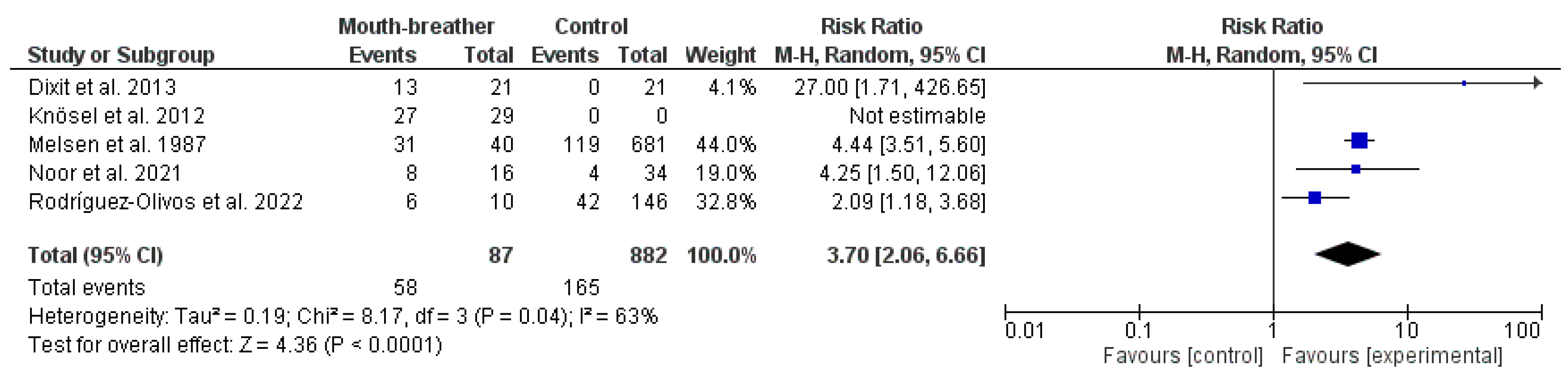
3. Results
3.1. Literature Search and Quality of the Papers
3.2. Characteristics of the Selected Studies
4. Discussion
5. Conclusions
Author Contributions
Funding
Institutional Review Board Statement
Informed Consent Statement
Data Availability Statement
Conflicts of Interest
Appendix A

| The Newcastle–Ottawa Scale (NOS) for Case–Control Study | |||
|---|---|---|---|
| Author | Dixit et al. 2013 [15] | Zegan et al. 2015 [22] | |
| Selection: (Maximum 4 stars) | 1. Is the case definition adequate? | ☆ | ☆ |
| 2. Representativeness of the cases | ☆ | ☆ | |
| 3. Selection of Controls | ☆ | / | |
| 4. Definition of Controls | ☆ | ☆ | |
| Comparability: (Maximum 2 stars) | 5. Comparability of cases and controls on the basis of the design or analysis | ☆ | ☆ |
| Outcome: (Maximum 3 stars) | 6. Ascertainment of exposure | ☆ | / |
| 7. Same method of ascertainment for cases and controls | ☆ | ☆ | |
| 8. Non-response rate | / | / | |
| Total score = | 7 | 5 | |
| The Joanna Briggs Institute (JBI) Critical Appraisal Checklist for Analytical Cross-Sectional Studies | |||||
|---|---|---|---|---|---|
| Authors | Castelo et al. 2005 [26] | Knosel et al. 2012 [16] | González et al. 2020 [25] | Noor et al. 2021 [23] | Rodriguez-Olivos et al. 2022 [24] |
| 1. Were the criteria for inclusion in the sample clearly defined? | Yes | Yes | No | Yes | Yes |
| 2. Were the study subjects and the setting described in detail? | Yes | Yes | Yes | Yes | Yes |
| 3. Was the exposure measured in a valid and reliable way? | Yes | Yes | Yes | Unclear | Yes |
| 4. Were objective, standard criteria used for measurement of the condition? | No | No | Yes | Unclear | Yes |
| 5. Were confounding factors identified? | No | No | No | No | No |
| 6. Were strategies to deal with confounding factors stated? | No | No | No | No | No |
| 7. Were the outcomes measured in a valid and reliable way? | Yes | Yes | Yes | Unclear | Yes |
| 8. Was appropriate statistical analysis used? | Yes | Yes | Yes | Yes | Yes |
| Overall appraisal: | Include (Seek further info) | Include (Seek further info) | Include (Seek further info) | Include (Seek further info) | Include (Seek further info) |
| The Joanna Briggs Institute (JBI) Critical Appraisal Checklist for Studies Reporting Prevalence Data | |||||
|---|---|---|---|---|---|
| Author | Melsen et al. 1987 [17] | Lagana et al. 2013 [27] | Shetty et al. 2013 [28] | Kasparaviciene et al. 2014 [19] | Caruso et al. 2019 [29] |
| 1. Was the sample frame appropriate to address the target population? | Yes | Yes | Yes | Yes | Yes |
| 2. Were study participants sampled in an appropriate way? | Yes | Yes | Yes | Yes | Yes |
| 3. Was the sample size adequate? | Yes | Yes | Yes | Yes | Yes |
| 4. Were the study subjects and the setting described in detail? | Yes | Yes | Yes | Yes | Yes |
| 5. Was the data analysis conducted with sufficient coverage of the identified sample? | Yes | Yes | Yes | Yes | Yes |
| 6. Were valid methods used for the identification of the condition? | Yes | Yes | Yes | Yes | Yes |
| 7. Was the condition measured in a standard, reliable way for all participants? | Yes | Yes | Yes | Yes | Yes |
| 8. Was there appropriate statistical analysis? | Yes | Yes | Yes | Yes | Yes |
| 9. Was the response rate adequate, and if not, was the low response rate managed appropriately? | Yes | Yes | Yes | Yes | Yes |
| Overall appraisal: | Include | Include | Include | Include | Include |
References
- Cattoni, D.M.; Fernandes, F.D.M.; Di Francesco, R.C.; De Oliveira Latorre, M.D.R.D. Quantitative evaluation of the orofacial morphology: Anthropometric measurements in healthy and mouth-breathing children. Int. J. Orofac. Myol. 2009, 35, 44–54. [Google Scholar] [CrossRef]
- Juliano, M.L.; Machado, M.A.C.; de Carvalho, L.B.C.; Zancanella, E.; Santos, G.M.S.; do Prado, L.B.F.; do Prado, G.F. Polysomnographic findings are associated with cephalometric measurements in mouth-breathing children. J. Clin. Sleep. Med. 2009, 5, 554–561. [Google Scholar] [CrossRef] [PubMed]
- Izu, S.C.; Itamoto, C.H.; Pradella-Hallinan, M.; Pizarro, G.U.; Tufik, S.; Pignatari, S.; Fujita, R.R. Obstructive sleep apnea syndrome (OSAS) in mouth breathing children. Braz J. Otorhinolaryngol. 2010, 76, 552–556. [Google Scholar] [CrossRef] [PubMed]
- Bandyopadhyay, A.; Slaven, J.E. Health outcomes associated with improvement in mouth breathing in children with OSA. Sleep. Breath. 2021, 25, 1635–1639. [Google Scholar] [CrossRef] [PubMed]
- Ribeiro, G.C.A.; dos Santos, I.D.; Santos, A.C.N.; Paranhos, L.R.; César, C.P.H.A.R. Influence of the breathing pattern on the learning process: A systematic review of literature. Braz. J. Otorhinolaryngol. 2016, 82, 466–478. [Google Scholar] [CrossRef]
- Leal, R.B.; Gomes, M.C.; Granville-Garcia, A.F.; Goes, P.S.A.; de Menezes, V.A. Impact of breathing patterns on the quality of life of 9- to 10-year-old schoolchildren. Am. J. Rhinol. Allergy 2016, 30, e147–e152. [Google Scholar] [CrossRef]
- Basheer, B.; Sundeep Hegde, K.; Bhat, S.S.; Umar, D.; Baroudi, K. Influence of mouth breathing on the dentofacial growth of children … Basheer B et al Original Research Conflict of Interest: None Source of Support: Nil Influence of Mouth Breathing on the Dentofacial Growth of Children: A Cephalometric Study. J. Int. Oral. Health 2014, 6, 50. [Google Scholar]
- Lee, S.; Guilleminault, C.; Chiu, H.; Sullivan, S.S. Mouth breathing, “nasal disuse”, and pediatric sleep-disordered breathing. Sleep. Breath. 2015, 19, 1257–1264. [Google Scholar] [CrossRef]
- Scarano, E.; Ottaviani, F.; Di Girolamo, S.; Galli, A.; Deli, R.; Paludetti, G. Relationship between chronic nasal obstruction and craniofacial growth: An experimental model. Int. J. Pediatr. Otorhinolaryngol. 1998, 45, 125. [Google Scholar] [CrossRef]
- Harari, D.; Redlich, M.; Miri, S.; Hamud, T.; Gross, M. The effect of mouth breathing versus nasal breathing on dentofacial and craniofacial development in orthodontic patients. Laryngoscope 2010, 120, 2089–2093. [Google Scholar] [CrossRef]
- Facciolli Hebling, S.R.; Cortellazzi, K.L.; da Silva Tagilaferro, E.P.; Hebling, E.; Bovi Ambrosano, G.M.; Meneghim, M.d.C.; Pereira, A.C. Relationship between malocclusion and behavioral, demographic and socioeconomic variables: A cross-sectional study of 5-year-olds. J. Clin. Pediatr. Dent. 2008, 33, 75–79. [Google Scholar] [CrossRef] [PubMed]
- Van Dyck, C.; Dekeyser, A.; Vantricht, E.; Manders, E.; Goeleven, A.; Fieuws, S.; Willems, G. The effect of orofacial myofunctional treatment in children with anterior open bite and tongue dysfunction: A pilot study. Eur. J. Orthod. 2016, 38, 227–234. [Google Scholar] [CrossRef] [PubMed]
- Paolantonio, E.G.; Ludovici, N.; Saccomanno, S.; La Torre, G.; Grippaudo, C. Association between oral habits, mouth breathing and malocclusion in Italian preschoolers. Eur. J. Paediatr. Dent. 2019, 20, 204–208. [Google Scholar] [CrossRef]
- Grippaudo, C.; Paolantonio, E.G.; Antonini, G.; Saulle, R.; La Torre, G.; Deli, R. Association between oral habits, mouth breathing and malocclusion. Acta Otorhinolaryngol. Ital. 2016, 36, 386–394. [Google Scholar] [CrossRef] [PubMed]
- Dixit, U.; Shetty, R. Comparison of soft-tissue, dental, and skeletal characteristics in children with and without tongue thrusting habit. Contemp. Clin. Dent. 2013, 4, 2–6. [Google Scholar] [CrossRef]
- Knösel, M.; Klein, S.; Bleckmann, A.; Engelke, W. Coordination of Tongue Activity During Swallowing in Mouth-breathing Children. Dysphagia 2012, 27, 401–407. [Google Scholar] [CrossRef]
- Melsen, B.; Attina, L.; Santuari, M.; Attina, A. Relationships between swallowing pattern, mode of respiration, and development of malocclusion. Angle Orthod. 1987, 57, 113–120. [Google Scholar]
- Brauer, J.S.; Holt, T.V. Tongue Thrust Classification. Angle Orthod. 1965, 35, 106–112. [Google Scholar]
- Kasparaviciene, K.; Sidlauskas, A.; Zasciurinskiene, E.; Vasiliauskas, A.; Juodzbalys, G.; Sidlauskas, M.; Marmaite, U. The Prevalence of Malocclusion and Oral Habits among 5–7-Year-Old Children. Med. Sci. Monit. Int. Med. J. Exp. Clin. Res. 2014, 20, 2036–2042. [Google Scholar] [CrossRef]
- Page, M.J.; McKenzie, J.E.; Bossuyt, P.M.; Boutron, I.; Hoffmann, T.C.; Mulrow, C.D.; Shamseer, L.; Tetzlaff, J.M.; Akl, E.A.; Brennan, S.E.; et al. The PRISMA 2020 statement: An updated guideline for reporting systematic reviews. PLoS Med. 2021, 18, e1003583. [Google Scholar] [CrossRef]
- Ma, L.; Wang, Y.; Yang, Z.; Huang, D.; Weng, H.; Zeng, X. Methodological quality (risk of bias) assessment tools for primary and secondary medical studies: What are they and which is better? Mil. Med. Res. 2020, 7, 7–8. [Google Scholar] [CrossRef] [PubMed]
- Zegan, G.; Dascalu, C.G.; Mavru, R.B.; Golovcencu, L. Risk Factors and Predictors of Crossbite at Children. Rev. Med. Chir. Soc. Med. Nat. Iasi 2015, 119, 564–571. [Google Scholar]
- Noor, N.; Zubair, A.; Ijaz, W. A Study Correlating Breathing Pattern with Different Malocclusions among Patients Reporting At Department of Orthodontics Ayub Medical College, Abbottabad, Pakistan. J. Ayub Med. Coll. Abbottabad 2021, 33, 664–667. [Google Scholar] [PubMed]
- Rodríguez-Olivos, L.H.G.; Chacón-Uscamaita, P.R.; Quinto-Argote, A.G.; Pumahualcca, G.; Pérez-Vargas, L.F. Deleterious oral habits related to vertical, transverse and sagittal dental malocclusion in pediatric patients. BMC Oral. Health 2022, 22, 88. [Google Scholar] [CrossRef] [PubMed]
- González Campoverde, L.; Rodríguez Soto, A.; Soto Cantero, L. Risk factors for malocclusion. Medicentro 2020, 24, 753–766. [Google Scholar]
- Castelo, P.M.; Gaviao, M.B.D.; Pereira, L.J.; Bonjardim, L.R. Relationship between oral parafunctional/nutritive sucking habits and temporomandibular joint dysfunction in primary dentition. Int. J. Paediatr. Dent. 2005, 15, 29–36. [Google Scholar] [CrossRef] [PubMed]
- Laganà, G.; Fabi, F.; Abazi, Y.; Beshiri Nastasi, E.; Vinjolli, F.; Cozza, P. Oral habits in a population of Albanian growing subjects. Eur. J. Paediatr. Dent. 2013, 14, 309–313. [Google Scholar] [PubMed]
- Shetty, R.M.; Shetty, M.; Shetty, N.S.; Reddy, H.; Shetty, S.; Agrawal, A. Oral habits in children of Rajnandgaon, Chhattisgarh, India—A prevalence study. Int. J. Public. Health Dent. 2013, 4, 1. [Google Scholar]
- Caruso, S.; Nota, A.; Darvizeh, A.; Severino, M.; Gatto, R.; Tecco, S. Poor oral habits and malocclusions after usage of orthodontic pacifiers: An observational study on 3–5 years old children. BMC Pediatr. 2019, 19, 294. [Google Scholar] [CrossRef]
- Weiss, C.E.; van Houten, J.T. A remedial program for tongue-thrust. Am. J. Orthod. 1972, 62, 499–506. [Google Scholar] [CrossRef]
- Ziliotto, K.N.; dos Santos, M.F.C.; Monteiro, V.G.; Pradella-Hallinan, M.; Moreira, G.A.; Pereira, L.D.; Weckx, L.L.M.; Fujita, R.R.; Pizarro, G.U. Auditory processing assessment in children with obstructive sleep apnea syndrome. Braz. J. Otorhinolaryngol. 2006, 72, 321–327. [Google Scholar] [CrossRef] [PubMed]
- Kuroishi, R.C.S.; Garcia, R.B.; Valera, F.C.P.; Anselmo-Lima, W.T.; Fukuda, M.T.H. Deficits in working memory, reading comprehension and arithmetic skills in children with mouth breathing syndrome: Analytical cross-sectional study. São Paulo Med. J. 2015, 133, 78–83. [Google Scholar] [CrossRef] [PubMed]
- Milanesi, J.M.; Borin, G.; Corrêa, E.C.R.; da Silva, A.M.T.; Bortoluzzi, D.C.; Souza, J.A. Impact of the mouth breathing occurred during childhood in the adult age: Biophotogrammetric postural analysis. Int. J. Pediatr. Otorhinolaryngol. 2011, 75, 999–1004. [Google Scholar] [CrossRef] [PubMed]
- da Silveira, W.; de Queiroz Mello, F.C.; Guimarães, F.S.; de Menezes, S.L.S. Postural alterations and pulmonary function of mouth-breathing children. Braz. J. Otorhinolaryngol. 2010, 76, 683–686. [Google Scholar] [CrossRef]
- Sanders, A.E.; Akinkugbe, A.A.; Slade, G.D.; Essick, G.K. Tooth loss and obstructive sleep apnea signs and symptoms in the US population. Sleep. Breath. 2016, 20, 1095–1102. [Google Scholar] [CrossRef]
- Zelano, C.; Jiang, H.; Zhou, G.; Arora, N.; Schuele, S.; Rosenow, J.; Gottfried, J.A. Nasal Respiration Entrains Human Limbic Oscillations and Modulates Cognitive Function. J. Neurosci. 2016, 36, 12448–12467. [Google Scholar] [CrossRef]
- Fraga, W.S.; Seixas, V.M.; Santos, J.C.; Paranhos, L.R.; César, C.P. Mouth breathing in children and its impact in dental malocclusion: A systematic review of observational studies. Minerva Stomatol. 2018, 67, 129–138. [Google Scholar] [CrossRef]
- Ovsenik, M. Incorrect orofacial functions until 5 years of age and their association with posterior crossbite. Am. J. Orthod. Dentofac. Orthop. 2009, 136, 375–381. [Google Scholar] [CrossRef]
- Sano, M.; Sano, S.; Kato, H.; Arakawa, K.; Arai, M. Proposal for a screening questionnaire for detecting habitual mouth breathing, based on a mouth-breathing habit score. BMC Oral. Health 2018, 18, 216. [Google Scholar] [CrossRef]
- Maspero, C.; Prevedello, C.; Giannini, L.; Galbiati, G.; Farronato, G. Atypical swallowing: A review. Minerva Stomatol. 2014, 63, 217–227. [Google Scholar]
- Begnoni, G.; Cadenas de Llano-Pérula, M.; Willems, G.; Pellegrini, G.; Musto, F.; Dellavia, C. Electromyographic analysis of the oral phase of swallowing in subjects with and without atypical swallowing: A case-control study. J. Oral. Rehabil. 2019, 46, 927–935. [Google Scholar] [CrossRef] [PubMed]
- Kurihara, K.; Fukui, T.; Sakaue, K.; Hori, K.; Ono, T.; Saito, I. The effect of tongue thrusting on tongue pressure production during swallowing in adult anterior open bite cases. J. Oral. Rehabil. 2019, 46, 895–902. [Google Scholar] [CrossRef] [PubMed]
- Di Vecchio, S.; Manzini, P.; Candida, E.; Gargari, M. Froggy mouth: A new myofunctional approach to atypical swallowing. Eur. J. Paediatr. Dent. 2019, 20, 33–37. [Google Scholar] [CrossRef] [PubMed]
- Matsuo, K.; Palmer, J.B. Coordination of mastication, swallowing and breathing. Jpn. Dent. Sci. Rev. 2009, 45, 31–40. [Google Scholar] [CrossRef]


| Author, Year, Location, Language of Publication | Place | Sample | Male | Female | Age | Race |
|---|---|---|---|---|---|---|
| Melsen et al. (1987) [17], Italy (English). | Trento village school (Italy) | 824 children | 424 male | 400 female | 13–14 years | Not specified |
| Castelo et al. (2005) [26], Brasil (English). | Piracicaba (Brasil) | 99 children | 58 male | 41 female | 3–5 years | Not specified |
| Knösel et al. (2012) [16], Argentina (English). | Two orthodontic centers in Santa Fé (Argentina) | 29 children | 16 male | 13 female | 6–16 years | Not specified |
| Dixit et al. (2013) [15], India (English). | City of Bagalkot (India) | - Initial sample: 864 children - Study sample: 42 children | 27 male - Control group: 21 children: 17 male - Tongue thrust group: 21 children: 10 male | 15 female - Control group: 21 children: 4 female - Tongue thrust group: 21 children: 11 female | 8–14 years | Not specified |
| Laganà et al. (2013) [27], Albania (English). | 15 public schools in Tirana (Albania) | 2617 children | 1257 male (48.4%) | 1360 female (51.6%) | 7–15 years | Exclusion criteria: non-Albanian people |
| Shetty et al. (2013) [28], India (English). | Department of Pediatrics in Rajnandgaon, (India) | 1891 children | 1043 male | 848 female | 6–11 years | Not specified |
| Kasparaviciene et al. (2014) [19], Lithuania (English). | 17 day care centers (Lithuania) | 503 children | 260 male | 243 female | 5–7 years | Not specified |
| Zegan et al. (2015) [22], Romania (English). | Orthodontic Clinic of “St. Spiridon” University Emergency Hospital Iasi (Romania) | 525 children | 217 male | 308 female | 6–18 years | Not specified |
| Caruso et al. (2019) [29], Italy (English). | University of l’Aquila (Italia) | 198 children | 96 male | 102 female | 3–5 years | Not specified |
| González et al. (2020) [25], Ecuador (Spanish). | Cuenca city school, (Ecuador) | 53 children | 22 male | 31 female | 5–12 years | Not specified |
| Noor et al. (2021) [23], Pakistan (English). | Department of Orthodontics, Ayub Medical College, Abbottabad, (Pakistan) | 62 children and adults | 29 male | 33 female | 6–20 years | Not specified |
| Rodríguez-Olivos et al. (2022) [24], Peru (English). | Undergraduate Clinic of the Faculty of Dentistry of the National University of San Marcos, (Peru) | 155 children | 75 male | 80 female | 6–12 years | Not specified |
| Author, Year | n | Mouth Breathing Evaluation Method | Results |
|---|---|---|---|
| Melsen et al. (1987) [17] | 824 children | Observational: Two operators observed whether the patient had a lip seal in a relaxed position. If this was not the case, the child was asked to close their lips and breathe deeply through their nose. If there was a contraction in the perioral muscles or the patient had difficulty breathing, they were asked where they usually breathed, through the mouth or through the nose. The breathing pattern was only collected if the patient’s version coincided with what was observed by the operators. | 40 presented mouth breathing |
| Castelo et al. (2005) [26] | 99 children | Questionnaire for parents: presence of qualitative (yes/no) and quantitative (frequent/occasional/never) mouth breathing. | 37 presented mouth breathing |
| Knösel et al. (2012) [16] | 29 children (who had an open mouth habit during the day) | Direct clinical observation (not specified). | 29 presented mouth breathing |
| Dixit et al. (2013) [15] | - Initial sample: 864 children - Study sample: 42 children | Direct clinical observation (not specified). | Of the 21 children with the tongue thrusting habit, 38% presented mouth breathing |
| Laganà et al. (2013) [27] | 2617 children | Direct clinical observation (not specified) + questionnaire administered to children. | 613 presented mouth breathing (303 male, 310 female) |
| Shetty et al. (2013) [28] | 1891 children | A calibrated examiner. Tried using a mirror. | 246 presented mouth breathing |
| Kasparaviciene et al. (2014) [19] | 503 children | Questionnaire for parents + extraoral examination of the face (a single examiner). The mouth breathing diagnostic test was only performed when the general clinical examination indicated mouth breathing and the parents confirmed the presence of the habit in the questionnaires. | 51 presented mouth breathing (32 male, 19 female) |
| Zegan et al. (2015) [22] | 525 children | Not described. | 34 presented mouth breathing |
| Castelo et al. (2019) [29] | 198 children | Questionnaire for parents + clinical examination by an orthodontist with more than 5 years of experience, calibrated. They used a protocol that they do not describe. | 71 presented mouth breathing |
| González et al. (2020) [25] | 53 children | Interview + facial and dental examination + Glatzel mirror. | 18 presented mouth breathing |
| Noor et al. (2021) [23] | 62 children and adults; 29 male 33 female | Clinical examination and medical history. Not specified. | Of the total sample: 51.50% of the women and 24.10% of the men presented mouth breathing; mixed breathing (mouth and nasal) 15.20% of women and 51.70% of men |
| Rodríguez-Olivos et al. (2022) [24] | 155 children | Observational: nasal breathing: tape was attached to the nasal septum that had two cotton pads, one in each nostril, and the movement was observed. Mouth breathing: observed napkin movement in a cut mask. | 10 presented mouth breathing |
| Author, Year | n | Tongue Thrust Evaluation Method | Results |
|---|---|---|---|
| Melsen et al. (1987) [17] | 824 children | Observational: Two operators observed mandibular movement and perioral muscle contraction when swallowing saliva or small sips of water. They then palpated the temporalis and masseter muscles while the patient repeated the process. If they had any doubt, the test was repeated. | 60 children presented simple tongue thrust and 90 complex tongue thrust. A total of 150 presented lingual interposition. |
| Castelo et al. (2005) [26] | 99 children | Observational: Two operators. Atypical swallowing was considered to occur when the activity of the lips produced strong tension in the perioral musculature and/or the tip of the tongue placed or pushed against the anterior teeth during swallowing. | 29 presented tongue thrust. |
| Knösel et al. (2012) [16] | 29 children (who had an open mouth habit during the day) | 1. Observational: patient swallowed saliva with open lips. 2. Polysensography: intraoral sensors in individualized splints were placed on the palate to perform simultaneous measurements of optical distance between the tongue and the palate. | 27 presented tongue thrust. |
| Dixit et al. (2013) [15] | - Initial sample: 864 children - Study sample: 42 children | For 864 patients in the initial sample: The child was asked to first swallow saliva and then 10 mL of water. The position of the tongue during swallowing was assessed by pressing the infant’s lower lip with the operator’s thumbs and at the same time feeling the activity of the masseter muscle with the index fingers. The child was diagnosed with tongue protrusion if they met any of the criteria established by Weiss and Van Houten [30]. For the tongue thrust group (21): The position of the tip of the tongue during swallowing was determined by covering the tip of the tongue with a developer solution with a brush and asking the child to swallow their own saliva. The area of the palate or teeth that was stained was noted. The presence or absence of clefts in the tongue was also recorded. | 46 presented tongue thrust. |
| Laganà et al. (2013) [27] | 2617 children | Direct clinical observation (not specified) + questionnaire administered to children. | 424 presented tongue thrust (189 male, 235 female). |
| Shetty et al. (2013) [28] | 1891 children | A calibrated examiner. The child was asked to first swallow saliva, and then 10 mL of water. The position of the tongue during swallowing was assessed by pressing the infant’s lower lip with the operator’s thumbs and at the same time feeling the activity of the masseter muscle with the index fingers. The child was diagnosed with tongue thrust if he met any of the following criteria established by Weiss and Van Houten [30]. | 329 presented tongue thrust. |
| Kasparaviciene et al. (2014) [19] | 503 children | Questionnaire for parents + extraoral examination of the face (a single examiner). The presence of tongue thrust was considered when there was hyperactivity of the perioral muscles and protrusion of the tongue between the upper and lower incisors or canines, without molar contact. Children were asked to swallow 3 times during the same visit. When in doubt, another drink was requested until the observer was satisfied with the judgement. | 27 presented tongue thrust (7 male, 20 female). |
| Zegan et al. (2015) [22] | 525 children | Not described. | 10 presented tongue thrust. |
| Caruso et al. (2019) [29] | 198 children | Questionnaire for parents + clinical examination by an orthodontist with more than 5 years of experience, calibrated + protocol that is not described. | 32 presented tongue thrust. |
| González et al. (2020) [25] | 53 children | Interview + facial examination + Payne’s test. | 19 presented tongue thrust. |
| Noor et al. (2021) [23] | 62 children and adults; 29 male, 33 female | Clinical examination and medical history. Not specified. | 12 presented tongue thrust. |
| Rodríguez-Olivos et al. (2022) [24] | 155 children | Glass of water + observe muscle contraction + see if water comes out of the mouth or if tongue is in interposition when swallowing. The swallowing process was also observed using oral retractors and introducing a little water with an injector. | 51 presented tongue thrust. |
Disclaimer/Publisher’s Note: The statements, opinions and data contained in all publications are solely those of the individual author(s) and contributor(s) and not of MDPI and/or the editor(s). MDPI and/or the editor(s) disclaim responsibility for any injury to people or property resulting from any ideas, methods, instructions or products referred to in the content. |
© 2024 by the authors. Licensee MDPI, Basel, Switzerland. This article is an open access article distributed under the terms and conditions of the Creative Commons Attribution (CC BY) license (https://creativecommons.org/licenses/by/4.0/).
Share and Cite
Gómez-González, C.; González-Mosquera, A.; Alkhraisat, M.H.; Anitua, E. Mouth Breathing and Its Impact on Atypical Swallowing: A Systematic Review and Meta-Analysis. Dent. J. 2024, 12, 21. https://doi.org/10.3390/dj12020021
Gómez-González C, González-Mosquera A, Alkhraisat MH, Anitua E. Mouth Breathing and Its Impact on Atypical Swallowing: A Systematic Review and Meta-Analysis. Dentistry Journal. 2024; 12(2):21. https://doi.org/10.3390/dj12020021
Chicago/Turabian StyleGómez-González, Carmen, Antonio González-Mosquera, Mohammad Hamdan Alkhraisat, and Eduardo Anitua. 2024. "Mouth Breathing and Its Impact on Atypical Swallowing: A Systematic Review and Meta-Analysis" Dentistry Journal 12, no. 2: 21. https://doi.org/10.3390/dj12020021