Article

PRF Lysates Enhance the Proliferation and Migration of Oral Squamous Carcinoma Cell Lines

1
Department of Oral Biology, University Clinic of Dentistry, Medical University of Vienna, 1090 Vienna, Austria
2
Department of Periodontology, School of Dental Medicine, University of Bern, 3010 Bern, Switzerland
3
Austrian Cluster for Tissue Regeneration, 1200 Vienna, Austria
*
Author to whom correspondence should be addressed.
Dent. J. 2023, 11(10), 242; https://doi.org/10.3390/dj11100242
Submission received: 20 July 2023 / Revised: 20 September 2023 / Accepted: 16 October 2023 / Published: 19 October 2023
(This article belongs to the Special Issue Regenerative Approaches in Dental Sciences)

Abstract

:
Platelet-rich fibrin (PRF) is an autologous fibrin-rich matrix where activated platelets and leucocytes accumulate. PRF has a wide spectrum of clinical indications with the overall aim of supporting tissue regeneration which in dentistry includes the healing of healthy oral mucosa with epithelial cells. In oral squamous cell carcinoma lesions, however, epithelial cells undergo malignant transformation, indicated by their unrestricted proliferation and migration potential, which should not be further enhanced by a wound-healing formula. Yet, little is known about how oral squamous cell carcinomas respond to PRF lysates. The aim of the present study was, therefore, to test the capacity of PRF lysates to change the transcriptome of HSC2 oral squamous carcinoma cells and perform bioassays to support the findings. Based on the RNAseq analysis, PRF lysates caused an increase in the genes functionally linked to cell replication and migration. In support of this screening approach, PRF lysates enhanced the proliferation of HSC2 oral squamous carcinoma cells, as indicated by 3[H]-thymidine incorporation, cell counting, and the expression of proliferation-related genes. Moreover, PRF lysates sped up cell migration in a scratch assay requiring actin polymerization. Taken together, our data showing that PRF lysates are mitogenic and stimulate motility of oral squamous carcinoma cell lines could be an indication that treatment with PRF in cases of oral carcinoma should be carefully considered.

1. Introduction

Platelet-rich fibrin is a blood-derived preparation of coagulated blood after centrifugation [1]. Blood is harvested by venous puncture and immediately centrifuged so that coagulation takes place after the separation of the plasma fraction from the bulk of the erythrocytes. The plasma fraction containing the platelets and some leukocytes then undergoes coagulation and can be separated from the red clot. To produce PRF membranes, the plasma clot is squeezed between two metal plates prior to its clinical use, for instance, to fill extraction sockets [2,3] and to prepare PRF blocks for bone augmentation [4]. Recent reviews provide an overview of PRF uses in dentistry, including the treatment of periodontal intrabony defects [5] and gingival recessions [6]. Under these conditions, PRF, similar to a blood clot, provides a surface for epithelial cells to grow from the edges toward the center of the defect [7]. Re-epithelialization of the oral mucosa is controlled by growth factors [8] and clinical evidence supports that PRF helps the re-epithelialization of the healthy oral mucosa [9].
Skin wound healing research underpins the impact of platelet-derived growth factors and other bioactive molecules on dermal keratinocytes; for instance, platelet-derived concentrates stimulated keratinocyte proliferation in vitro [10], and human platelet lysate enhanced the migration and proliferation of keratinocytes [11]. Other findings, however, suggest that platelet-released growth factors and platelet-rich plasma can inhibit the proliferation of keratinocytes [12,13]. In the context of dentistry, platelet-rich plasma was seen to enhance the proliferation of SCC25 epithelial-like cells originally isolated from the tongue of a patient with squamous cell carcinoma [14]; however, these cells are transformed cell lines representing an oral pathology. This study is an indication of the tumor-supporting action of platelet concentrates and, thus, metastatic potential.
One can imagine an in vivo scenario when the subject has a clinically undetectable oral tumor or a premalignant lesion [15] where the use of PRF might initiate a full-blown oral squamous cell carcinoma (OSCC), an entity that accounts for about 92–95% of all cancer in the oral cavity [16]. It takes years for OSCC to progress, and risk factors including smoking, alcohol, and human papillomavirus (HPV) infection are obvious [17,18]. Other related risk factors are the precancerous conditions of oral leukoplakia [19] and oral lichen planus [20]. Presumably, platelets also play a role in OSCC pathology. Platelet microthrombi were detected in OSCC and linked to lymph node metastasis [21]. Overall, platelets enhance the invasiveness of human and murine cancer cell lines [22,23] and platelet injection promotes the formation of metastatic nodules in murine cancer metastasis models [24]. In general, platelets alter the epithelial tumor cell attributes to drive metastasis [25].
In vitro, platelet–tumor interactions have been studied with Ca9.22 and HSC3 cells originating from OSCC [26]. We have previously implemented our established oral squamous carcinoma cell line HSC2 as a bioassay for testing the responsiveness to lysates prepared from PRF membranes, however, in the context of inflammation research [27,28]. Today’s attempts to assess the response of HSC2 and other cell types to PRF lysates are based on RNA sequencing, an omics technology that provides insight into how the genetic signature of cells changes [29]. Similar to the understanding of the response of gingival fibroblasts to milk, it is now time to identify how HSC2 cells respond to PRF lysates based on RNAseq, followed by an analysis of genes regulating the cell cycle such as cyclin D1, Ki67, and PCNA [30]. Also, histones providing structural support for chromosomal DNA are required when cells duplicate and are considered proliferation markers [31]. Heterochromatin involving the histones is also required for cell migration [32]. In addition, we use TR146 cells originating from the human neck metastasis of a buccal carcinoma and primary oral epithelial cells as a control. Thus, the primary aim of the study is to understand the overall response of HSC2 oral squamous carcinoma cells to PRF lysates and perform the respective proliferation and migration bioassays, to understand if PRF potentially supports the malignancy of oral squamous carcinoma cells.

2. Material and Methods

2.1. Oral Squamous Cell Carcinoma Cell Lines HSC2, TR146, and Primary Cells

HSC2 and TR146 are cell lines originating from oral squamous cell carcinomas (Health Science Research Resources Bank, Sennan, Japan, and European Collection of Authenticated Cell Cultures, respectively). Both cell lines were grown in DMEM serum-containing medium and antibiotic-antimycotic solution (Sigma-Aldrich, St. Louis, MO, USA). Primary human oral epithelial cells were isolated from gingiva obtained during wisdom tooth removal from healthy donors who gave their informed consent (Ethical Committee of the Medical University of Vienna, Nr. 631/2007). Epithelial cells were seeded at 2.5 × 10⁵ cells/cm2 onto culture dishes one day prior to overnight exposure to 30% PRF lysates in serum-free media.

2.2. PRF Lysates, Buffy Coat, and Platelet-Poor Plasma

The preparation of PRF was approved by the Ethical Committee of the Medical University of Vienna (Nr. 1644/2018). Glass tubes (Bio-PRF, Venice, FL, USA) supporting blood coagulation were subjected to 700 g for 8 min (Z306 Hermle, Universal Centrifuge, Wehingen, Germany). The yellow PRF clot was removed and squeezed between layers of gauze. One cm of PRF membrane was used per mL of serum-free medium. Liquid PRF fractions were prepared with polyethylenterephthalat tubes (Greiner Bio-One GmbH, Kremsmünster, Austria) after blood centrifugation at 2000× g for 8 min (Z306 Hermle). The uppermost 2 mL platelet-poor plasma (PPP) and the 1 mL buffy coat (BC) were then collected. All preparations underwent two cycles of freeze–thawing at −80 °C, followed by 30 s sonication at room temperature (Sonopuls 2000.2, Bandelin electronic, Berlin, Germany). The lysates underwent centrifugation at 15,000× g for 10 min before 1 mL aliquots of the supernatant were stored frozen for less than one month.

2.3. RNA Sequencing

Following RNA extraction (ExtractMe, Blirt S.A., Gdańsk, Poland), the quality was assessed with an Agilent 2100 Bioanalyzer (Agilent Technologies, Santa Clara, CA, USA). Sequencing libraries were prepared at the Core Facility Genomics at the Medical University of Vienna using the NEBNext Poly (A) mRNA Magnetic Isolation Module and the NEBNext Ultra™ II Directional RNA Library Prep Kit for Illumina, according to manufacturer’s protocols (New England Biolabs, Ipswich, MA, USA). Libraries were QC checked on a Bioanalyzer 2100 (Agilent Technologies) using a High Sensitivity DNA Kit and quantified using a Qubit dsDNA HS Assay (Invitrogen, Waltham, MA, USA). Pooled libraries were sequenced on a NextSeq500 device (Illumina, San Diego, CA, USA) with a 1 × 75 bp single-end sequencing method. Roughly 25 million reads were created per sample. Reads in fastq format were aligned to the human reference genome version GRCh38 (www.ncbi.nlm.nih.gov/grc/human, accessed on 25 January 2022) with Gencode 29 annotations (www.gencodegenes.org/human/release_29.html, accessed on 25 January 2022) using the STAR aligner 55 version 2.6.1a in the two-pass mode. Reads per gene were counted by STAR and differential gene expression was calculated using DESeq2 56 version 1.22.2. DESeq2. The STRING database was used to show protein–protein interactions (string-db.org, accessed on 25 January 2022).

2.4. Reverse Transcription Quantitative Real-Time PCR

The total RNA was reverse transcribed (LabQ, Labconsulting, Vienna, Austria) and cDNA was amplified on a CFX Connect™ RealTime PCR Detection System (Bio-Rad Laboratories, Hercules, CA, USA). The transcription levels of target genes indicated in Table 1 were normalized to GAPDH using the ΔΔCt method.

2.5. Proliferation Assay

Cells were incubated with 30% PRF lysate or left untreated before being pulse-labeled with 3[H]thymidine (0.5 mCi/well, Hartmann Analytic, Braunschweig, Germany) exposure for at least 16 h. The plates then underwent liquid scintillation counting (Packard, Meriden, CT, USA). Thymidine incorporation was indicated as counts per minute. In addition, cells were seeded at 3 × 10⁴ cells/cm2 and counted after 72 h of exposure with a phase contrast microscope.

2.6. Scratch Assay

Cells seeded at 2.5 × 10⁵ cells/cm2 were scratched with a sterile plastic micropipette tip to generate a regular gap width. Cells were washed with serum-free medium to remove debris and exposed overnight to 30% PRF lysate with and without the actin polymerization inhibitor cytochalasin D at 1 µM (Sigma-Aldrich).

2.7. Statistical Analysis

Single experiments were repeated at least four times. In the figures, the data points represent independent experiments with blood obtained from independent donors. Ordinary one-way ANOVA and paired t-tests were used for statistical analysis (Prism v9, GraphPad Software, La Jolla, CA, USA). p-values are indicated in the figures.

3. Results

3.1. RNAseq Analysis

To understand the overall response of the HSC2 cells exposed to the PRF lysate, an RNAseq analysis was performed, and the resulting data are shown in Table 2. Gene ontology revealed an enrichment of the 48 (>10-fold) upregulated genes related to GO:0006270 DNA replication initiation with 4 of 32 (biological process) and GO:0005149 interleukin-1 receptor binding with 3 of 17 (molecular function). Upregulated genes were also enriched for the GO:0000786 nucleosome with 6 of 106, GO:0032993 protein-DNA complex with 7 of 195, and GO:0000228 nuclear chromosome with 12 of 1256 (cellular component). No enrichment was identified for the 34 (>10-fold) down-regulated genes. Data from RNAseq and GO analysis suggest that the most relevant changes caused by the PRF lysates relate to the proliferation but also the expression of IL1 in HSC2 cells.

3.2. RT-PCR Analysis

To support the observation from RNAseq analysis, we performed an expression analysis, starting with confirming the up-regulation of histone H1-linking DNA and nucleosomes, as well as the core histone HIST1H2AJ (Figure 1A). Consistent with the overall picture obtained from RNAseq, genes that are linked to the cell cycle were increased in the HSC2 cells exposed to PRF lysates, e.g., Ki67, PCNA, and Cyclin D1 (Figure 1B). This was also true for fractions of liquid PRF, namely, buffy coat (BC) and platelet-poor plasma (PPP), as reported in Figure S1. The RNAseq findings are further supported by the increased expression of E2F1 and other genes based on RT-PCR (Figure 2). Finally, we confirmed that PRF lysates, but also BC and PPP, provoke a moderate but significant increase in IL1β expression in HSC2 cells (Figure 2, Figure S2). Taken together, the RT-PCR analysis confirmed the overall impression that PRF lysates exert a mitogenic activity and also enhance IL1 expression in HSC2 cells.

3.3. 3[H]-Thymidine Incorporations and Cell Counting
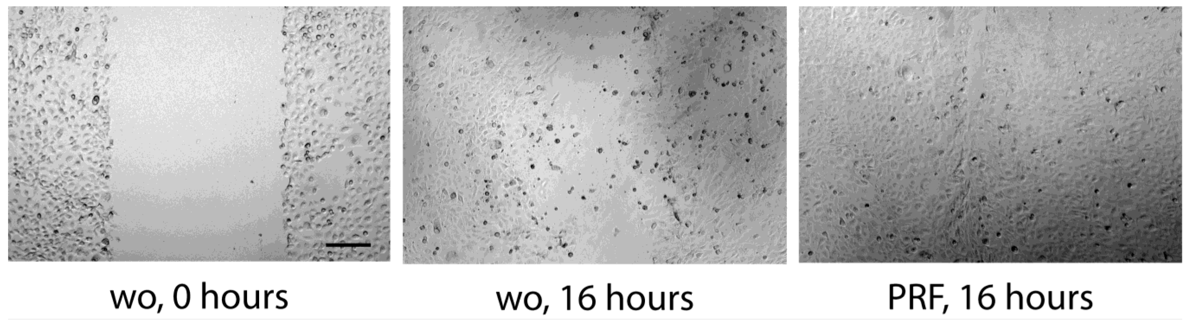
To support the observation from RNAseq, we performed a 3[H]-thymidine incorporation assay and counted the number of HSC2 cells incubated with PRF lysates. When HSC2 cells were exposed to PRF lysates, the incorporation of 3[H]-thymidine increased significantly by approximately two-fold (Figure 3A). TR146 also showed a moderate increase (Figure 3B) while, in contrast, the PRF lysates significantly reduced the 3[H]-thymidine uptake in primary oral epithelial cells (Figure 3C). This observation was further supported by the counting of HSC2 cells, showing that after 72 h of incubation with lysates from PRF, the cells more than doubled (Figure 4). Further support for the findings comes from data showing that the liquid fractions of blood, namely, the buffy coat and platelet-poor plasma, possess mitogenic activity in HSC2 cells (Figure S3). These findings further support the claim that the PRF lysates exert a mitogenic effect on the transformed cell lines cells, while the opposite was true for the primary oral epithelial cells.

3.4. Scratch Assay

To simulate a wound-like situation where the proliferation and migration of epithelial cells are necessary, we performed a classical scratch assay. We can report that the presence of PRF lysates resulted in complete coverage of the injured area, whereas in the presence of serum-free medium, HSC2 failed to fully recover the scratched area (Figure 5). Wound closure was suppressed by inhibition of actin polymerization by cytochalasin D (Figure 5). Consistently, lysates prepared from BC and PPP could support the wound closure of HSC2 scratches. In TR146 cells, wound closure was enhanced by PRF lysates but less impressive (Figure 6). These observations show that PRF lysates support the motility of the two oral squamous carcinoma cell lines. In primary epithelial cells, however, there was no obvious migration support by PRF lysates, but more of a reduced motility of the cells (Figure 7).

4. Discussion

PRF applications became a globally recognized therapy to support oral tissue healing, a process that requires oral epithelial cells to proliferate and migrate. However, in the context of oral oncology, platelet-rich plasma was reported to promote the proliferation of squamous cell carcinoma SCC25 cells [14], raising the possibility that PRF might exert a tumor-promoting activity. This assumption is supported by the fundamental understanding that platelets can change epithelial tumor cell characteristics to drive metastasis [25]. Moreover, platelets enhance the invasiveness of cancer cell lines [22,23], platelet injection promotes the in vivo formation of metastatic nodules [24], and platelet aggregates are found in OSCC [21]. This is also the reason why Ca9.22 and HSC3 cells originating from OSCC were used to study platelet–tumor interactions [26]. The present research was based on the fundamental question of how HSC2 cells respond to lysates prepared from PRF membranes on the molecular and cellular level.
Our main finding was based on an RNAseq approach. We identified a genetic signature that highlights two aspects; namely, the increased expression of genes related to cell proliferation and migration apart from the activation of IL1 expression in HSC2 cells. Likewise, the PRF lysates weakly supported TR146 cell proliferation while, in contrast, reducing primary oral epithelial cell proliferation. If we relate our findings to those of others, we have to accept the inconsistency of the present knowledge on how platelet-containing blood fractions modulate the proliferation of epithelial cells; for instance, while platelet-derived concentrates and lysate stimulate keratinocyte proliferation in vitro [10,11], platelet-released growth factors and platelet-rich plasma can also inhibit the proliferation of keratinocytes [12,13]. RNAseq further identified the downregulated genes related to cell replication [33]. In contrast, some found that platelet-rich plasma increased the proliferation of human HaCaT keratinocytes [34], while others found no relevant change in HaCaT proliferation [35]. Considering dentistry, platelet-rich plasma stimulated the proliferation of SCC25 epithelial-like cells originally isolated from the tongue of a patient with squamous cell carcinoma [14], while PRF decreased the viable cell number using an MTT assay of an oral epithelial cell line derived from a gingival carcinoma and metastasis to the cervical lymph node [36]. In light of existing knowledge, our observations provide another piece of information on how PRF membranes differentially affect the proliferation of oral carcinoma and healthy epithelial cells.
Considering the migration of epithelial cells, we have to recognize research on how platelet-containing blood fractions change the healing, and thus the migration, of healthy epithelial cells but also take the oncological properties of OSCC into account. In support of our findings, platelet-rich plasma supported the healing of scratches in HaCaT cells, a spontaneously immortalized keratinocyte cell line from adult human skin [34,37]. This is also true for PRF exosomes enhancing HaCaT cell migration [38] and microvesicles supporting the migration of human keratinocytes [39]. Consistently, PRP improved keratinocyte migration in scratch and transwell migration experiments [40]. PRP further supported the closure of gingival fibroblast scratches in vitro [41]. The proliferation and migration of HaCaT cells might be attributed to PAF (1-O-alkyl-2-acetyl-sn-glycero-3-phosphocholine), a phospholipid-derived regulator released from activated platelets [42]. Interestingly, PAF increased the expression of COX2, a master regulator of inflammatory prostaglandin synthesis [42], supporting our observation that PRF lysates increase IL1 expression in HSC2 cells. IL1 accelerates the migration of normal differentiated airway epithelial cells [43], corneal epithelial cell migration [44], and renal tubular epithelial HK-2 cells [45]—overall suggesting that IL1 might be related to the migration of HSC2 cells.
If we interpret our findings considering the heterogenicity of the current knowledge, we have to restrict our conclusions to the special conditions of our in vitro system and, in particular, state that the HSC2 cells are from oral squamous carcinoma established in the late 1980s [46,47]. The human oral squamous cell carcinoma cells are, therefore, not the oral epithelial cells of a healthy person being treated with PRF membranes. We thus have to be critical and discuss PRF as pro-tumorigenic. Moreover, caution is needed when asserting that wound healing and tumorigenesis have common characteristics, as one is a controlled, regulated physiological process while the other lacks these characteristics. The interpretation of our findings must be further expanded towards oncology, where oral mucositis in the context of radiation and chemotherapy is a heavy burden for patients and PRF might support wound healing under these conditions [48]. In addition, PRF might target oral epithelial cells in precancerous lesions such as oral leukoplakia [19] and oral lichen planus [20]. Thus, even though wound healing and cancer progression share regulation commonalities including proliferation and migration, our data should not be extrapolated towards physiological wound healing but used more as a basis to critically evaluate PRF therapy for oral cancerous lesions [49]. Importantly, we show here that PRF reduces the proliferation of oral epithelial cells, presumably because of its TGF-β content [50]. Thus, our findings do not provide a direct explanation of how PRF membranes support oral wound healing and prevent gingival recessions, a process that presumably involves the proliferation and migration of oral epithelial cells.

5. Conclusions and Future Research

Our data showing that PRF membranes support the proliferation and migration of HSC2 oral squamous carcinoma cells should be considered as a primer for upcoming research that is more sensitive to treatment with PRF in cases of oral carcinoma. Future research could build upon our RNAseq approach combined with the respective bioassays and develop a more clinically relevant assay for PRF. For instance, by embedding a tissue explant from OSCC and healthy gingiva into a PRP membrane, we can study the outgrowth and proliferation of the respective epithelial cells. The staining of proliferation markers such as Ki67 or PCNA in histological samples of OSCC and gingiva biopsies is also possible. Not only PRF lysates but also the respective lysates from buffy coat and platelet-poor plasma (PPP) are mitogenic and support the cell motility of HSC2 cells. This is interesting as PPP is, in particular, considered cell-free, suggesting that plasma components affect cell fate; for instance, the lipid fraction of PPP has anti-inflammatory activity [51]. Thus, the preclinical translation of our in vitro findings is worth consideration in future research.

Supplementary Materials

The following supporting information can be downloaded at: https://www.mdpi.com/article/10.3390/dj11100242/s1, Figure S1: RT-PCR analysis of proliferation markers; Figure S2: RT-PCR analysis of IL1; Figure S3: 3H-thymidine incorporations and cell counting.

Author Contributions

Conceptualization, L.P. and R.G.; data curation, L.P.; formal analysis, L.P. and R.G.; methodology, L.P., R.C., and S.G.; project administration, L.P., R.C., and S.G.; supervision, L.P. and R.G.; validation, L.P. and R.G.; visualization, L.P.; writing—original draft, L.P. and R.G.; writing—review and editing, L.P., R.C., S.G. and R.G. All authors have read and agreed to the published version of the manuscript.

Funding

L.P. and R.G. receive funds from the ITI Projects 1729-2023 and 1709-2022.

Institutional Review Board Statement

The preparation of PRF was approved by the Ethical Committee of the Medical University of Vienna (Nr. 1644/2018).

Informed Consent Statement

Informed and written consent was obtained from all subjects involved in the study.

Data Availability Statement

The original contributions presented in the study are included in the article. Further inquiries can be directed to the corresponding author.

Acknowledgments

We thank the Core Facility Genomics of the Medical University of Vienna for performing the RNAseq.

Conflicts of Interest

The authors declare that they have no conflicts of interest.

References

  1. Dohan, D.M.; Choukroun, J.; Diss, A.; Dohan, S.L.; Dohan, A.J.; Mouhyi, J.; Gogly, B. Platelet-rich fibrin (PRF): A second-generation platelet concentrate. Part I: Technological concepts and evolution. Oral Surg. Oral Med. Oral Pathol. Oral Radiol. Endod. 2006, 101, e37–e44. [Google Scholar] [CrossRef]
  2. Castro, A.B.; Van Dessel, J.; Temmerman, A.; Jacobs, R.; Quirynen, M. Effect of different platelet-rich fibrin matrices for ridge preservation in multiple tooth extractions: A split-mouth randomized controlled clinical trial. J. Clin. Periodontol. 2021, 48, 984–995. [Google Scholar] [CrossRef] [PubMed]
  3. Temmerman, A.; Vandessel, J.; Castro, A.; Jacobs, R.; Teughels, W.; Pinto, N.; Quirynen, M. The use of leucocyte and platelet-rich fibrin in socket management and ridge preservation: A split-mouth, randomized, controlled clinical trial. J. Clin. Periodontol. 2016, 43, 990–999. [Google Scholar] [CrossRef] [PubMed]
  4. Cortellini, S.; Castro, A.B.; Temmerman, A.; Van Dessel, J.; Pinto, N.; Jacobs, R.; Quirynen, M. Leucocyte- and platelet-rich fibrin block for bone augmentation procedure: A proof-of-concept study. J. Clin. Periodontol. 2018, 45, 624–634. [Google Scholar] [CrossRef] [PubMed]
  5. Miron, R.J.; Moraschini, V.; Fujioka-Kobayashi, M.; Zhang, Y.; Kawase, T.; Cosgarea, R.; Jepsen, S.; Bishara, M.; Canullo, L.; Shirakata, Y.; et al. Use of platelet-rich fibrin for the treatment of periodontal intrabony defects: A systematic review and meta-analysis. Clin. Oral Investig. 2021, 25, 2461–2478. [Google Scholar] [CrossRef] [PubMed]
  6. Miron, R.J.; Moraschini, V.; Del Fabbro, M.; Piattelli, A.; Fujioka-Kobayashi, M.; Zhang, Y.; Saulacic, N.; Schaller, B.; Kawase, T.; Cosgarea, R.; et al. Use of platelet-rich fibrin for the treatment of gingival recessions: A systematic review and meta-analysis. Clin. Oral Investig. 2020, 24, 2543–2557. [Google Scholar] [CrossRef] [PubMed]
  7. Singer, A.J.; Clark, R.A. Cutaneous wound healing. N. Engl. J. Med. 1999, 341, 738–746. [Google Scholar] [CrossRef]
  8. Tomikawa, K.; Yamamoto, T.; Shiomi, N.; Shimoe, M.; Hongo, S.; Yamashiro, K.; Yamaguchi, T.; Maeda, H.; Takashiba, S. Smad2 decelerates re-epithelialization during gingival wound healing. J. Dent. Res. 2012, 91, 764–770. [Google Scholar] [CrossRef]
  9. Mohanty, S.; Pathak, H.; Dabas, J. Platelet rich fibrin: A new covering material for oral mucosal defects. J. Oral Biol. Craniofac. Res. 2014, 4, 144–146. [Google Scholar] [CrossRef]
  10. Paulmann, A.; Strauss, S.; Limbourg, A.; Vogt, P.M. Platelet-derived concentrates influence human keratinocyte proliferation in vitro and induce wound healing in a prospective case series of chronic wounds of different entities in vivo. Innov. Surg. Sci. 2022, 7, 45–58. [Google Scholar] [CrossRef]
  11. Jafar, H.; Hasan, M.; Al-Hattab, D.; Saleh, M.; Ameereh, L.A.; Khraisha, S.; Younes, N.; Awidi, A. Platelet lysate promotes the healing of long-standing diabetic foot ulcers: A report of two cases and in vitro study. Heliyon 2020, 6, e03929. [Google Scholar] [CrossRef] [PubMed]
  12. Bayer, A.; Tohidnezhad, M.; Berndt, R.; Lippross, S.; Behrendt, P.; Kluter, T.; Pufe, T.; Jahr, H.; Cremer, J.; Rademacher, F.; et al. Platelet-released growth factors inhibit proliferation of primary keratinocytes in vitro. Ann. Anat. 2018, 215, 1–7. [Google Scholar] [CrossRef] [PubMed]
  13. Annunziata, M.; Oliva, A.; Buonaiuto, C.; Di Feo, A.; Di Pasquale, R.; Passaro, I.; Guida, L. In vitro cell-type specific biological response of human periodontally related cells to platelet-rich plasma. J. Periodontal Res. 2005, 40, 489–495. [Google Scholar] [CrossRef]
  14. Okuda, K.; Kawase, T.; Momose, M.; Murata, M.; Saito, Y.; Suzuki, H.; Wolff, L.F.; Yoshie, H. Platelet-rich plasma contains high levels of platelet-derived growth factor and transforming growth factor-beta and modulates the proliferation of periodontally related cells in vitro. J. Periodontol. 2003, 74, 849–857. [Google Scholar] [CrossRef] [PubMed]
  15. Feller, L.L.; Khammissa, R.R.; Kramer, B.B.; Lemmer, J.J. Oral squamous cell carcinoma in relation to field precancerisation: Pathobiology. Cancer Cell Int. 2013, 13, 31. [Google Scholar] [CrossRef]
  16. Ganesh, D.; Sreenivasan, P.; Ohman, J.; Wallstrom, M.; Braz-Silva, P.H.; Giglio, D.; Kjeller, G.; Hasseus, B. Potentially Malignant Oral Disorders and Cancer Transformation. Anticancer Res. 2018, 38, 3223–3229. [Google Scholar] [CrossRef]
  17. Rivera, C. Essentials of oral cancer. Int. J. Clin. Exp. Pathol. 2015, 8, 11884–11894. [Google Scholar]
  18. Jiang, S.; Dong, Y. Human papillomavirus and oral squamous cell carcinoma: A review of HPV-positive oral squamous cell carcinoma and possible strategies for future. Curr. Probl. Cancer 2017, 41, 323–327. [Google Scholar] [CrossRef]
  19. Villa, A.; Sonis, S. Oral leukoplakia remains a challenging condition. Oral Dis. 2018, 24, 179–183. [Google Scholar] [CrossRef]
  20. Kurago, Z.B. Etiology and pathogenesis of oral lichen planus: An overview. Oral Surg. Oral Med. Oral Pathol. Oral Radiol. 2016, 122, 72–80. [Google Scholar] [CrossRef]
  21. Duan, W.; Wu, Z.; Mei, X.; Li, J.; Lu, H.; Liang, Y.; Lao, X.; Liao, G. Intratumoral platelet microthrombi in oral squamous cell carcinoma: A marker of lymph node metastasis. Oral Dis. 2023. [Google Scholar] [CrossRef] [PubMed]
  22. Guo, Y.; Cui, W.; Pei, Y.; Xu, D. Platelets promote invasion and induce epithelial to mesenchymal transition in ovarian cancer cells by TGF-beta signaling pathway. Gynecol. Oncol. 2019, 153, 639–650. [Google Scholar] [CrossRef] [PubMed]
  23. Wang, Y.; Sun, Y.; Li, D.; Zhang, L.; Wang, K.; Zuo, Y.; Gartner, T.K.; Liu, J. Platelet P2Y12 is involved in murine pulmonary metastasis. PLoS ONE 2013, 8, e80780. [Google Scholar] [CrossRef]
  24. Haemmerle, M.; Taylor, M.L.; Gutschner, T.; Pradeep, S.; Cho, M.S.; Sheng, J.; Lyons, Y.M.; Nagaraja, A.S.; Dood, R.L.; Wen, Y.; et al. Platelets reduce anoikis and promote metastasis by activating YAP1 signaling. Nat. Commun. 2017, 8, 310. [Google Scholar] [CrossRef] [PubMed]
  25. Gay, L.J.; Felding-Habermann, B. Platelets alter tumor cell attributes to propel metastasis: Programming in transit. Cancer Cell 2011, 20, 553–554. [Google Scholar] [CrossRef]
  26. Park, S.Y.; Lee, S.K.; Lim, M.; Kim, B.; Hwang, B.O.; Cho, E.S.; Zhang, X.; Chun, K.S.; Chung, W.Y.; Song, N.Y. Direct Contact with Platelets Induces Podoplanin Expression and Invasion in Human Oral Squamous Cell Carcinoma Cells. Biomol. Ther. 2022, 30, 284–290. [Google Scholar] [CrossRef] [PubMed]
  27. Panahipour, L.; Oladzad Abbasabadi, A.; Gruber, R. Oral Cell Lysates Reduce the Inflammatory Response of Activated Macrophages. J. Clin. Med. 2023, 12, 1701. [Google Scholar] [CrossRef] [PubMed]
  28. Panahipour, L.; Nasserzare, S.; Amer, Z.; Brucke, F.; Stahli, A.; Kreissl, A.; Haiden, N.; Gruber, R. The anti-inflammatory effect of milk and dairy products on periodontal cells: An in vitro approach. Clin. Oral Investig. 2019, 23, 1959–1966. [Google Scholar] [CrossRef]
  29. Panahipour, L.; Moghaddam, D.M.; Nasirzade, J.; Kargarpour, Z.; Gruber, R. RNAseq of TGF-beta receptor type I kinase-dependent genes in oral fibroblast exposed to milk. BMC Oral Health 2021, 21, 581. [Google Scholar] [CrossRef] [PubMed]
  30. Goodlad, R.A. Quantification of epithelial cell proliferation, cell dynamics, and cell kinetics in vivo. Wiley Interdiscip. Rev. Dev. Biol. 2017, 6, e274. [Google Scholar] [CrossRef]
  31. Polo, S.E.; Almouzni, G. Histone metabolic pathways and chromatin assembly factors as proliferation markers. Cancer Lett. 2005, 220, 1–9. [Google Scholar] [CrossRef] [PubMed]
  32. Gerlitz, G. The Emerging Roles of Heterochromatin in Cell Migration. Front. Cell Dev. Biol. 2020, 8, 394. [Google Scholar] [CrossRef] [PubMed]
  33. Bayer, A.; Wijaya, B.; Mobus, L.; Rademacher, F.; Rodewald, M.; Tohidnezhad, M.; Pufe, T.; Drucke, D.; Glaser, R.; Harder, J. Platelet-Released Growth Factors and Platelet-Rich Fibrin Induce Expression of Factors Involved in Extracellular Matrix Organization in Human Keratinocytes. Int. J. Mol. Sci. 2020, 21, 4404. [Google Scholar] [CrossRef] [PubMed]
  34. Misiura, M.; Guszczyn, T.; Oscilowska, I.; Baszanowska, W.; Palka, J.; Miltyk, W. Platelet-Rich Plasma Promotes the Proliferation of Human Keratinocytes via a Progression of the Cell Cycle. A Role of Prolidase. Int. J. Mol. Sci. 2021, 22, 936. [Google Scholar] [CrossRef] [PubMed]
  35. Duan, G.H.; Ren, Z.Q.; Du, B.; Shao, W.; Dong, H.J.; Du, A.C. Platelet-rich plasma protects human keratinocytes from UVB-induced apoptosis by attenuating inflammatory responses and endoplasmic reticulum stress. J. Cosmet. Dermatol. 2023, 22, 1327–1333. [Google Scholar] [CrossRef] [PubMed]
  36. Tsai, C.-H.; Shen, S.-Y.; Zhao, J.-H.; Chang, Y.-C. Platelet-rich fibrin modulates cell proliferation of human periodontally related cells in vitro. J. Dental. Sci. 2009, 4, 130–135. [Google Scholar] [CrossRef]
  37. Carney, B.C.; Simbulan-Rosenthal, C.M.; Gaur, A.; Browne, B.J.; Moghe, M.; Crooke, E.; Moffatt, L.T.; Shupp, J.W.; Rosenthal, D.S. Inorganic polyphosphate in platelet rich plasma accelerates re-epithelialization in vitro and in vivo. Regen. Ther. 2020, 15, 138–148. [Google Scholar] [CrossRef]
  38. Xu, Y.; Lin, Z.; He, L.; Qu, Y.; Ouyang, L.; Han, Y.; Xu, C.; Duan, D. Platelet-Rich Plasma-Derived Exosomal USP15 Promotes Cutaneous Wound Healing via Deubiquitinating EIF4A1. Oxid. Med. Cell. Longev. 2021, 2021, 9674809. [Google Scholar] [CrossRef]
  39. Lovisolo, F.; Carton, F.; Gino, S.; Migliario, M.; Reno, F. Platelet rich plasma-derived microvesicles increased in vitro wound healing. Eur. Rev. Med. Pharmacol. Sci. 2020, 24, 9658–9664. [Google Scholar] [CrossRef]
  40. Yang, S.C.; Lin, C.F.; Alshetaili, A.; Aljuffali, I.A.; Chien, M.Y.; Fang, J.Y. Combining the dual antibacterial and regenerative activities of platelet-rich plasma with beta-lactams to mitigate MRSA-infected skin wounds. Biomed. Pharmacother. 2023, 165, 115017. [Google Scholar] [CrossRef]
  41. Leisi, S.; Farahpour, M.R. Effectiveness of topical administration of platelet-rich plasma on the healing of methicillin-resistant Staphylococcus aureus-infected full-thickness wound model. J. Plast. Reconstr. Aesthet. Surg. 2023, 77, 416–429. [Google Scholar] [CrossRef]
  42. Feuerherm, A.J.; Jorgensen, K.M.; Sommerfelt, R.M.; Eidem, L.E.; Laegreid, A.; Johansen, B. Platelet-activating factor induces proliferation in differentiated keratinocytes. Mol. Cell. Biochem. 2013, 384, 83–94. [Google Scholar] [CrossRef]
  43. White, S.R.; Fischer, B.M.; Marroquin, B.A.; Stern, R. Interleukin-1beta mediates human airway epithelial cell migration via NF-kappaB. Am. J. Physiol. Lung Cell. Mol. Physiol. 2008, 295, L1018–L1027. [Google Scholar] [CrossRef] [PubMed]
  44. Tseng, H.C.; Lee, I.T.; Lin, C.C.; Chi, P.L.; Cheng, S.E.; Shih, R.H.; Hsiao, L.D.; Yang, C.M. IL-1beta promotes corneal epithelial cell migration by increasing MMP-9 expression through NF-kappaB- and AP-1-dependent pathways. PLoS ONE 2013, 8, e57955. [Google Scholar] [CrossRef]
  45. Masola, V.; Carraro, A.; Granata, S.; Signorini, L.; Bellin, G.; Violi, P.; Lupo, A.; Tedeschi, U.; Onisto, M.; Gambaro, G.; et al. In vitro effects of interleukin (IL)-1 beta inhibition on the epithelial-to-mesenchymal transition (EMT) of renal tubular and hepatic stellate cells. J. Transl. Med. 2019, 17, 12. [Google Scholar] [CrossRef] [PubMed]
  46. Momose, F.; Araida, T.; Negishi, A.; Ichijo, H.; Shioda, S.; Sasaki, S. Variant sublines with different metastatic potentials selected in nude mice from human oral squamous cell carcinomas. J. Oral Pathol. Med. 1989, 18, 391–395. [Google Scholar] [CrossRef] [PubMed]
  47. Ichijo, H.; Momose, F.; Miyazono, K. Biological effects and binding properties of transforming growth factor-beta on human oral squamous cell carcinoma cells. Exp. Cell Res. 1990, 187, 263–269. [Google Scholar] [CrossRef]
  48. Lalla, R.V.; Saunders, D.P.; Peterson, D.E. Chemotherapy or radiation-induced oral mucositis. Dent. Clin. N. Am. 2014, 58, 341–349. [Google Scholar] [CrossRef]
  49. Sundaram, G.M.; Quah, S.; Sampath, P. Cancer: The dark side of wound healing. FEBS J. 2018, 285, 4516–4534. [Google Scholar] [CrossRef]
  50. Boyd, F.T.; Massague, J. Transforming growth factor-beta inhibition of epithelial cell proliferation linked to the expression of a 53-kDa membrane receptor. J. Biol. Chem. 1989, 264, 2272–2278. [Google Scholar] [CrossRef]
  51. Kargarpour, Z.; Panahipour, L.; Mildner, M.; Miron, R.J.; Gruber, R. Lipids of Platelet-Rich Fibrin Reduce the Inflammatory Response in Mesenchymal Cells and Macrophages. Cells 2023, 12, 634. [Google Scholar] [CrossRef] [PubMed]
Figure 1. RT-PCR analysis of histones and proliferation markers: HSC2 were exposed to PRF lysates, and the expression changes of histones (A) and proliferation marker genes (B) identified by RNAseq were evaluated. Data are expressed as the x-fold changes in the PRF-stimulated HSC2 cells compared to the respective untreated controls. Data points represent independent experiments. p-values originate from ordinary one-way ANOVA tests.
Dentistry 11 00242 g001
Figure 2. RT-PCR analysis of genes identified by RNAseq: HSC2 were exposed to PRF lysates, and the expression of genes identified by RNAseq was assessed. Data are expressed as the x-fold changes in the PRF-stimulated HSC2 cells over the respective untreated controls. Data points represent independent experiments. p-values originate from ordinary one-way ANOVA tests.
Dentistry 11 00242 g002
Figure 3. 3[H]-thymidine incorporations. (A) HSC2, (B) TR146, and (C) primary oral epithelial cells were incubated with and without PRF lysates for 24 h. The cells were pulse-labeled with 3[H]-thymidine for the last 6 h of culture and subjected to liquid scintillation counting. Data were expressed as counts. Data points represent independent experiments. p-values originate from paired t-tests.
Dentistry 11 00242 g003
Figure 4. Cell counting: HSC2 cells were incubated with and without PRF lysates for 24 h. Cell counting was performed after 72 h; data are expressed as cells per mL. (A) RT-PCR analysis. Data points represent independent experiments. p-values originate from paired t-tests. (B) counted with a phase contrast microscope. The scale bar represents 100 µm.
Dentistry 11 00242 g004
Figure 5. Scratch assay: HSC2 cells grow to confluence before creating a scratch. After removing debris, cells were treated overnight with 30% PRF lysate in the presence or absence of cytochalasin D and inhibition of actin polymerization. wo stands for without and is a serum-free medium. All pictures are taken after 16 h of incubation. The scale bar represents 100 µm.
Dentistry 11 00242 g005
Figure 6. Scratch assay: TR146 cells grow to confluence before creating a scratch. After removing debris, cells were treated overnight with 30% PRF lysate. wo stands for without and is a serum-free medium. Pictures are taken immediately after the scratch (0 h) and after 16 h of incubation. The scale bar represents 100 µm.
Dentistry 11 00242 g006
Figure 7. Scratch assay: Primary epithelial cells grow to confluence before creating a scratch. After removing debris, cells were treated overnight with 30% PRF lysate. wo stands for without and is a serum-free medium. Pictures are taken immediately after the scratch (0 h) and after 16 h of incubation. The scale bar represents 100 µm.
Dentistry 11 00242 g007
Table 1. Primer sequences.
GenesForward SequenceReverse Sequence
CDC45GAAGCGCACACGGTTAGAAGTTCACTCCCAGAGCCACTC
CyclinD1TCGGTGTCCTACTTCAAATGTGTGGGATGGTCTCCTTCATCTTAG
E2F1GGACCTGGAAACTGACCATCAGCAGTGAGGTCTCATAGCGTGAC
FAM111BGGTTCTGGGGAAATCCAGTCAAGATGGAGAAACAAGGATTGAA
HIST1H1AGGCAAAGAAACCTGCTAAGGCTGTAAGAGCTGCCAACGACACACC
HIST1H1DGCTTATCACCAAGGCAGTGGCACCAGAGTACCTTTGCTCACCAAG
HIST1H2AJCGACAACAAGAAGACTCGCATCATGTGCGATGGTGACTTTGCCCA
IL1bATGATGGCTTATTACAGTGGCAAGTCGGAGATTCGTAGCTGGA
Ki-67ATAAACACCCCAACACACACAAGCCACTTCTTCATTCCAGTTACA
MCM10CATGAAGCCCAAGGATGGGACCTTCTGAGGATGATCGATAG
PCNATGGAGAACTTGGAAATGGAAACGAACTGGTTCATTCATCTCTATGG
SULT1B1CCAACTACAGTGATGGATCATAGCGCGGAATTGAAGTGCAGTTTTGG
Table 2. RNAseq. HSC2 cells were incubated with and without PRF lysates for 24 h. The RNA was then subjected to RNAseq analysis. Genes are ranked by expression changes, with a cut-off of 10-fold.
Upregulated GenesDownregulated Genes
HIST1H1A83.0FOXA121.4
IL1B59.5KCNH419.6
E2F156.1SAMD1118.9
HIST1H1D43.3DPP718.7
SULT1B135.9TCHHL118.4
HIST1H2AJ33.7IFI44L16.9
XYLT124.7RGS1616.5
MMP924.7ROM116.0
NAV323.6SH2D3C14.9
OTUB222.4PIFO14.3
ZNF36721.6BLNK14.3
ORC121.3CACNA1A14.3
LRRC75B20.2KRTAP19-113.4
EXO119.3ATOH812.5
CAMP19.1S1PR112.5
CDCA718.2CAMK2A12.0
CLCF116.8C10orf9911.8
CCNE216.8TMPRSS11E11.6
F2R16.5TTLL311.6
SLCO4A116.3PDE3A11.3
ETV516.3TMEM86A11.1
FAM72D15.7SMAD910.9
IL1R214.9ZNF69110.7
MCM1014.8RCAN210.7
RAET1L14.6ACAA210.7
HIST1H2BF14.6CCL2610.7
HIST2H2AC14.6WDR9310.7
FAM111B13.6RBBP8NL10.7
IL1A13.5SLC41A210.7
GPSM313.5TM7SF210.6
HIST1H4C12.9NUPR110.6
PLXNA412.8GBP210.4
MAMDC212.3PAPLN10.1
CDH1112.3SERPINI110.1
SPRED112.3
AMIGO212.2
AREG11.7
VEPH111.5
VAX111.2
MYBPHL11.2
KLF1411.2
GLI211.2
ANKRD4411.2
IL1RN10.9
CLMP10.7
APCDD1L10.4
CDC4510.3
GPR6810.1
Disclaimer/Publisher’s Note: The statements, opinions and data contained in all publications are solely those of the individual author(s) and contributor(s) and not of MDPI and/or the editor(s). MDPI and/or the editor(s) disclaim responsibility for any injury to people or property resulting from any ideas, methods, instructions or products referred to in the content.

