Review

Oral Microbiome and Dental Caries Development

Faculty of Dentistry, The University of Hong Kong, Hong Kong SAR, China
*
Author to whom correspondence should be addressed.
Dent. J. 2022, 10(10), 184; https://doi.org/10.3390/dj10100184
Submission received: 13 September 2022 / Revised: 25 September 2022 / Accepted: 27 September 2022 / Published: 30 September 2022
(This article belongs to the Special Issue Updates and Highlights in Cariology)

Abstract

:
Dental caries remains the most prevalent oral disease worldwide. The development of dental caries is highly associated with the microbiota in the oral cavity. Microbiological research of dental caries has been conducted for over a century, with conventional culture-based methods and targeted molecular methods being used in order to identify the microorganisms related to dental caries. These methods’ major limitation is that they can identify only part of the culturable microorganisms in the oral cavity. Introducing sequencing-based technology and bioinformatics analysis has boosted oral microbiome research and greatly expanded the understanding of complex oral microbiology. With the continuing revolution of molecular technologies and the accumulated sequence data of the oral microbiome, researchers have realized that microbial composition alone may be insufficient to uncover the relationship between caries and the microbiome. Most updated evidence has coupled metagenomics with transcriptomics and metabolomics techniques in order to comprehensively understand the microbial contribution to dental caries. Therefore, the objective of this article is to give an overview of the research of the oral microbiome and the development of dental caries. This article reviews the classical concepts of the microbiological aspect of dental caries and updates the knowledge of caries microbiology with the results of current studies on the oral microbiome. This paper also provides an update on the caries etiological theory, the microorganisms related to caries development, and the shifts in the microbiome in dental caries development.

1. Introduction

Dental caries is the most common oral disease worldwide. Untreated caries affects 2.5 billion adults and 573 million children all over the world [1], placing a heavy health burden on health care systems and society. Over the past 25 years, the prevalence of dental caries has remained at a similarly high level despite oral health care providers’ efforts [2]. The high prevalence of dental caries indicates the effect of the research on dental caries. Dental caries is a multifactorial disease that involves microbial, behavioral, genetic, and environmental factors [3]. Although these factors are important in caries development, the role of microbial factors cannot be ignored. Because the development of dental caries is closely correlated to oral microorganisms, a comprehensive understanding of caries microbiology is essential.
The microbiology of dental caries has been investigated for over a century, with a revolutionized advance in study approaches. Previous studies have used traditional culture-based methods in order to identify the bacteria related to dental caries [4]. The culture-based method has allowed for a basic understanding of the dental plaque microbiota composition in dental caries to be established. Microorganisms have been isolated from carious lesions or dental plaque samples collected from a cross-sectional or longitudinal study using culture-based techniques [5,6]. However, bacteria can only be successfully cultured when provided with their own special growth requirements. At present, because artificial media cannot exactly mimic the natural environment within which oral bacteria reside, most fastidious bacteria remain uncultivable in vitro [7].
Targeted molecular methods for identifying and quantifying caries-associated bacteria were introduced in the early 1990s [8,9]. Targeted molecular methods use DNA probes obtained from cultured bacterial species to enumerate the bacteria [10]. This allows for identifying and quantifying multiple species in dental plaque samples from more individuals compared to culture-based methods. This also facilitates the analysis of species that are difficult to culture [4]. However, targeted molecular methods can only detect the preselected bacteria that culture-based methods have already confirmed. Unknown bacteria sets in the dental plaque cannot be detected.
The introduction of sequencing-based technology and bioinformatics analysis has greatly expanded the understanding of complex oral microbiology. Unlike the targeted molecular methods, next-generation sequencing (NGS) techniques identify sequences by comparing them to the curated microbiome database, enabling the detection of novel species that have historically been uncultivable [11]. Since the development of the Sanger sequencing technique in 1977, great efforts have been made to conquer the high cost and low efficiency of “reading” genes. NGS, also known as massively parallel sequencing technology, stands out for allowing ultra-high throughput with a deeper coverage of the microbial community at a lower cost. During the last decade, targeted 16S rRNA amplicon sequencing, which relies on the amplification of one to three selected hypervariable regions of 16S rDNA, has been widely adopted to study the oral microbiome’s compositional changes in dental caries. However, sequencing fragments of the 16S rRNA gene can miss variants that discriminate different species or strains, which compromises this technique’s taxonomic resolution [12].
The improvement in sequence length in metagenomics overcomes the limitation of 16S rRNA gene sequencing. It is promising in identifying more members of the microbial community [13]. Studies that are more recent coupled metagenomics with transcriptomics and metabolomics to investigate how the active microbial community collaborates in the initiation and progression of dental caries [14,15,16]. They investigated what the microorganisms in the studied niches were, as well as how the microorganisms actively worked together to cause disease. The understanding of caries microbiology has been greatly enriched with the advancement of sequencing-based technology and bioinformatics analysis in recent years. Therefore, this article aims to review the classical concepts on the microbiological aspect of dental caries and to update the knowledge of caries microbiology with the results of studies on the oral microbiome conducted over the past few years.

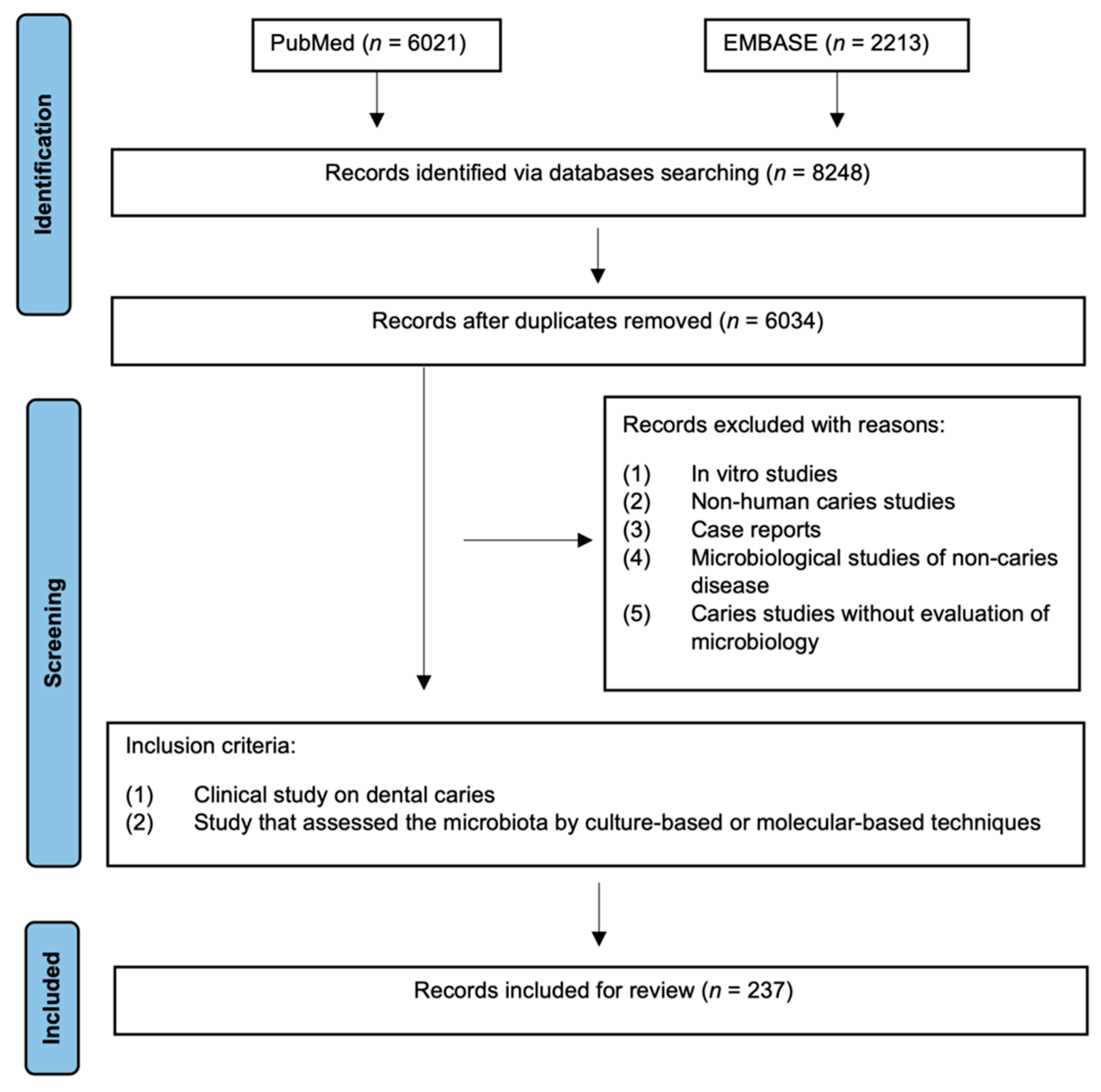
2. Materials and Methods

We searched the two most relevant electronic databases, PUBMED and EMBASE, for published evidence with the combination of the following key words: (caries OR “dental decay” OR “tooth decay” OR “carious lesion” OR “white spot”) AND (microbiome OR microbiota OR microbial OR biofilm OR microorganism OR mycobiome OR virome). Studies were limited to English publications published on or before 1 August 2022. Duplicate studies were discarded. Studies were included for review if they met the following criteria: (1) a study on human dental caries or (2) a study that assessed the microbiota of caries using culture-based or molecular-based techniques. The study selection process is presented in the flow diagram below (Figure 1).

3. Results

3.1. Oral Microbial Communities

The oral cavity is a complex ecology with various niches. Not only bacteria but also archaea, fungi, and viruses reside in these niches [17,18]. Among them, bacteria make up the main proportion of this diverse community. Bacteria are also the most extensively studied subtype of the oral microbiome. According to the expanded Human Oral Microbiome Database (eHOMD) (https://homd.org/ (accessed on 1 August 2022)), 774 oral bacterial species have been detected and studied. Around 58% of the species have been cultivated and officially named, 16% have been cultivated but remain unnamed, and 26% remain uncultivated. The majority of these species belong to six broad phyla: Firmicutes, Actinobacteria, Proteobacteria, Fusobacteria, Bacteroidetes, and Spirochaetes [19].
For several decades, cultivation studies have reported fungi to be oral inhabitants [20]. Their biodiversity and potential role in the oral ecosystem could not be extensively studied until the last decade because of the uncultivable features of most fungi and the relatively low proportion of the biomass [21]. A collection of studies has found more than 100 genus-level taxa of fungi as significant constituents of the oral mycobiome, of which only Candida and Malassezia have been well demarcated [22,23,24]. Moreover, only Candida has been extensively investigated and claimed to be involved in various oral diseases, including caries [25,26]. Although studies have reported hundreds of fungal taxa, only a few of them have been indicated to be true oral colonizers [22]. The further isolation and characterization of the other fungal taxa with low abundances are desirable for understanding the diversity and functionality of the fungi in the oral microbiome.
The Archaeal microorganism was first isolated from an oral subgingival sample in 1987 [27]. Later studies found the presence of Archaea at various oral sites, most frequently at periodontal sites [28,29,30,31,32,33], followed by endodontic sites [34,35], but rarely from dental caries [36], saliva [37], and the tongue [38]. Currently, Archaea detected in the human oral cavity is confined to a few phylotypes, including the most abundant Methanogenic archaea and Thermoplasmata [36,39,40]. The low abundance and the fastidious cultivation process can hamper the identification of Archaea, which can lead to an underestimation of the diversity of oral archaea. Furthermore, the information related to the role of oral archaea in dental caries is sparse.
Viruses have been identified from oral cavities, including a few eukaryotic viruses and various bacteriophages [41]. Interpreted from the limited evidence, the oral virome is highly individual-specific but temporally stable [42,43]. Bacteriophages, which are viruses targeting bacteria, are primarily lysogenic. In a previous study, the bacteriophages of lytic styles, which were predominant in the dental plaque of periodontitis, could eradicate their susceptible bacteria hosts or convey new functions to their bacteria host [44]. Thus, viruses may have a considerable capacity in shaping the oral microbial community’s structure and pathogenesis [45]. Despite the significance of virus–bacteria interactions to the whole oral microbiome, the role of oral viruses is understudied [45]. The interspecies and inter-kingdom interactions of the oral microorganisms are the key to maintaining oral health. The pathogenic shifts in the oral microbial communities contribute to the pathogenesis of polymicrobial diseases, including caries [33,44,46,47]. Therefore, the development of caries is highly associated with oral microbiological changes [2,14].

3.2. The Microbiological Hypothesis of Dental Caries Etiology

The continuous development of microbiological research methods has led to a shift in the microbiological theory of dental caries etiology. In the late nineteenth century, bacteria isolation and identification techniques were far from developed. The etiology of caries was postulated to be determined by the quantity of dental plaque, referred to as the “Traditional Non-specific Plaque Hypothesis”, which Miller proposed in 1890 [48]. With advances in microscopes and microorganism cultivation techniques, specific bacterial species, mainly Streptococcus mutans and Lactobacillus, were frequently found to be associated with initiating caries, characterizing them as cariogenic species [49,50,51,52]. Antibiotic treatment that targeted these species reduced caries formation [53]. Based on this evidence, Loesche proposed the “Specific Plaque Hypothesis” in 1976 [54]. The “Specific Plaque Hypothesis” states that specific cariogenic bacteria in dental plaque, such as Streptococcus mutans and Lactobacillus, are responsible for dental caries. However, the “Specific Plaque Hypothesis” cannot explain the fact that Streptococcus mutans is absent from some carious sites. In addition, Streptococcus mutans is constantly detected on sound tooth surfaces, indicating that the presence of this species is neither sufficient nor necessary for initiating caries [55].
With other bacteria species isolated from caries, Marsh proposed the “Ecological Plaque Hypothesis” in 1994 [56,57]. In this hypothesis, it is stated that caries develops along with the disruption of microbial homeostasis under ecological stress, in which some pathogenic species outnumber health-related microorganisms. The ecological plaque hypothesis stresses the critical role of the interaction between the environment and bacteria. The development of molecular identification techniques has led to a continuous revision of the etiology of dental caries. Hundreds of not-yet cultivable oral phylotypes have been identified using gene-sequencing methods, suggesting that an increasingly diverse microflora might play a critical role in the ecological changes resulting in caries onset [58]. Furthermore, a metatranscriptomic analysis revealed a discrete gene expression profile of oral microflora from which genomics are disclosed, directing the focus to the microbes that are actively involved in caries development [59]. In 2008, Takahashi further extended the “Ecological Plaque Hypothesis” by incorporating the metabolism of plaque microorganisms. In this extended version, it is stated that the neutral microenvironment tilts to an acidic condition when acid production outweighs the base metabolites’ buffering capacity, followed by acid-induced selection and adaptation within the microflora. This process disrupts a healthy microbial community’s stability and leads to dental caries [60].

3.3. Microorganisms Associated with Caries Development

Table 1 presents the microbial species that are potentially related to caries based on the currently available literature. Streptococcus mutans has been considered a major pathogen of dental caries since 1971 [49,61]. Many other bacteria have been isolated from carious sites or have been found to feature distinctly throughout the process of caries development, and they have been roughly proposed to be related to caries. With the development of culture-independent identification technology, some fastidious fungi have also been identified to be caries-related (Table 1).
It is worth noting that, even though various species of Lactobacillus and Bifidobacteria were reported to be strongly correlated with caries progression, other species of these two genera were demonstrated to be effective probiotics in the context of caries prevention [90]. These probiotics may exert caries prevention effects by regulating the microflora dysbiosis induced by environment stress [91]. Considering that different species of the same genus may play opposite roles in the caries development process, a species-level resolution analysis is required [92].
With the evolutional sequencing-based technology and bioinformatics approaches, more unknown bacterial species were identified, and their roles in the acid-producing process were gradually disclosed [93]. A comprehensive understanding of caries microbiology is under development.

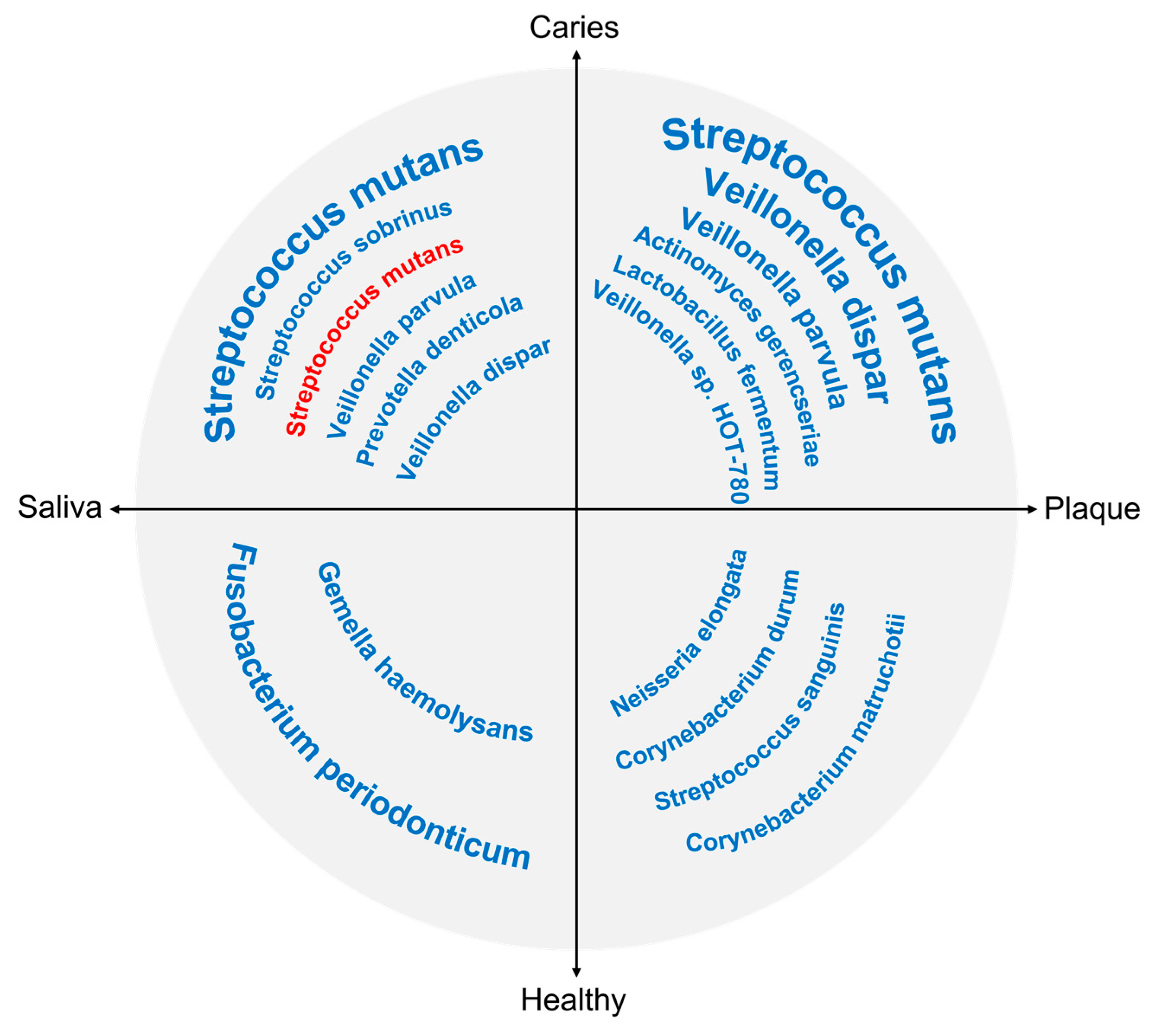
3.4. Shifts in the Oral Microbiome in Dental Caries

Evidence on the association between the oral microbial profile and caries has been surging in recent decades [94]. Generally, the oral bacterial community was less diverse in caries-affected than in caries-free subjects [95,96,97]. Specifically, the relative abundance of caries-related species rather than the taxonomic diversity changed along with caries development [83,94,98,99,100,101]. The changes in the fungal microbiome, or the mycobiome, were similar to those of the bacteriome, with the relative abundance of several taxa that mainly belong to candida increasing significantly in the dental plaque on caries surfaces [92]. The microbiome research of dental caries has not detected any specific microbial species uniquely associated with caries [102].
Different microbial interaction profiles between caries-affected and caries-free communities were found in an operational taxonomic unit (OTU) network analysis, although the results are discordant among studies. In a study on early childhood caries (ECC), the interconnection between OTUs was reported to be intensified in the caries-affected community compared to the caries-free community, suggesting the contribution of the intensive species interactions to the development of ECC [94]. In another study, the intercorrelation among predominant genera was increasingly complex and robust in caries-free sites in an adult population [101]. The research subjects’ different age groups may explain the inconsistency between the studies. Further explorations are required to obtain a valid conclusion on the changes in microbial interactions in patients with caries.
Because microbiome studies on dental caries are carried out separately in children and adults, there is no direct evidence on comparing the difference in the caries-related microbial shift between different age groups. However, evidence has shown that the microbial composition changes with dentition development [103]. Thus, the microflora that contributes to the caries process may differ from childhood to adulthood. We extracted the bacterial species with a significantly higher abundance in caries-affected and caries-free subjects from previous articles, and they are presented in Figure 2. Considering repeatability, only species that were reported in at least two studies were included as being caries-related or health-related. As shown in Figure 1, only Streptococcus mutans from saliva was shown to be significantly associated with caries in adults, while a variety of taxa from both saliva and plaque were reported to be caries-related or health-related in children [102].
Apart from the overall changes in the oral microbiome in patients with dental caries, the shifts in the caries-associated microorganisms at the different statuses of caries progression were also investigated.

3.4.1. Microbiome Shifts in Caries of Different Stages

The organic and mineral components, as well as the micro-environment, are diverse throughout the different parts of the tooth structure, indicating that the destruction of the different tooth parts may involve distinct microbial-related factors. An early metagenome study on caries reported that both the compositions and the functional profiles of the microbial communities differed greatly between enamel caries and dentin caries [97]. Genes that encode the functions of sugar fermentation, cell surface adhesion, and acid stress responses were overrepresented in enamel caries while presenting the opposite trend in dentine caries [97,104]. Interestingly, genes overexpressed in the deep dentin microflora correlated to the host’s immune response [97], indicating the potential significance of the interaction between microbes and their host. Additionally, Lactobacillus species were only detected in dentin caries, and a higher abundance of Prevotella was found deep in the dentin [97]. According to Richards et al., four species (S. mutans, Scardovia wiggsiae, Parascardovia denticolens, and Lactobacillus salivarius) exclusively exist in dentine caries [90]. On the contrary, some species with a higher abundance in enamel caries decreased or disappeared in dentine caries [97]. A metatranscriptomic study also showed that the active microbial community differed significantly between carious enamel and dentine, with lactobacilli expressing a much higher level in dentine caries [104]. In this study, a large number of species expressed at an extremely low abundance existed exclusively in either enamel caries or dentine caries, which indicates that minority species might play an essential role in caries occurrence [104].

3.4.2. Microbiome Shifts in Caries with Different Activities

Recently, the microbial contribution to dental caries activity was investigated. It was concluded from the evidence that existed that the microbial communities residing on active caries saw a reduction in richness compared to those residing on arrested caries [105], but they shared a similar beta diversity [105,106]. Additionally, the relative abundance of some caries-associated bacteria was increased in resistant active caries after silver diamine fluoride (SDF) treatment but was decreased in arrested caries [105]. An in vivo study found that Streptococcus and Veillonella were more evenly distributed with other taxa in arrested caries, while they were predominant in induced active caries [107]. Further evidence is needed to investigate the microbiome’s role in the shift in caries activity.

3.4.3. Microbiome Shifts in Caries in Different Locations

The biofilm composition of the dental root surface may differ from that of the coronal surface because of the influence of gingival crevicular fluid [108]. Patients with coronal caries may be free from root caries and vice versa. Thus, there arises the question of whether the bacterial community involved in root caries might differ from that involved in coronal caries. An early culture-based study showed that the major bacteria recovered from the plaque of root caries were distinct from those of enamel caries [109]. However, no molecular study has investigated the dis/similarity of the microbiome involved in coronal caries and root caries. Most of the up-to-date oral microbiome studies targeted the relationship between the microbial community and coronal caries. A recent study focusing on root caries found that Prevotella dominated the microflora of caries lesions that extended to the subgingival margin, while Streptococcus dominated the microbial community from root caries lesions that were confined within supragingival sites [110]. However, the periodontal microflora probably confounded the results of this study. The lack of evidence on this subject indicates the urgency of exploring this phenomenon.

4. Discussion and Future Perspectives

The recent advancements in oral microbiology studies have expanded our perspectives on caries etiology. Nevertheless, there are limitations in the caries microbiology studies discussed in the present literature review. Due to the high cost of the new-born omics techniques and the infancy of methodological and analytical protocols, most of the recent studies used 16S rRNA gene sequencing to investigate the microbiological contribution to caries development. However, 16S sequencing only provides information regarding bacterial composition at genus- or species-level resolution, failing to characterize the strain-level diversity of the microbial community and its functional capabilities [111]. Another non-negligible limitation is that current studies rarely study the diversity and the potential roles of nonbacterial microorganisms, regardless of the enormous diversity of the fungi identified in the oral cavity. As any other infectious disease, the host factor plays a vital role in caries development. Saliva, being the first line of defense in the oral cavity, helps shape the microbial profile of pioneer colonizers, which later “trains” the host immune system to defend against pathological invaders [112]. The dynamic balance between the host immune system and the microbial commensals maintains the oral health status [113]. Despite the convincing relationship between immunology and microbiology in the context of caries, there is a lack of studies examining how the microbiota interacts with the host immune system in the course of microbial dysbiosis. Lastly, the lack of consistency in the study protocol hampers the comparisons between studies, compromising the reproducibility of the results.
Future work should be directed toward resolving the limitations in the current studies. As demonstrated, the taxa detected with a high abundance in dental caries do not necessarily actively function and vice versa. Therefore, associating the microorganisms with their functional contributions to dental caries is highly desirable in future studies. Complementary methodological approaches are expected to be employed to uncover the diversity and contribution of the relatively unexplored domains of the microorganisms and their interactions with their bacteria counterparts in the oral microbiome community. Furthermore, the interaction between the microbial community and their host’s immune system should also be explored. Regarding the significant heterogeneity among the results of different studies, consistency in the study protocol, including sampling site selection, sampling methods, sample storage condition, sequencing technique, reference database, and bioinformatics pipelines, is required to achieve comparable results across the extensive number of studies.

5. Conclusions

Research using sequencing-based technology and bioinformatics analysis has revolutionized the classical microbiological concept of dental caries, which is based on culture-based methods. Contemporary evidence validates and extends the “Ecological Plaque Hypothesis”. The inter-species and inter-kingdom interactions of diverse microorganisms contribute to the development of dental caries. Both the predominance and the relative abundance of microbiota change along with the caries development stages. Researchers are further exploring the functionalities of caries-contributing species and the interactions between microbiota and their hosts.

Author Contributions

Conceptualization, O.Y.Y.; writing—original draft preparation, J.S.Z. and O.Y.Y.; visualization, J.S.Z.; analysis, J.S.Z.; writing—review and editing, C.-H.C. and O.Y.Y. All authors have read and agreed to the published version of the manuscript.

Funding

This research received no external funding.

Institutional Review Board Statement

Not applicable.

Informed Consent Statement

Not applicable.

Data Availability Statement

Not applicable.

Conflicts of Interest

The authors declare no conflict of interest.

References

  1. Kassebaum, N.; Smith, A. Global, Regional, and National Prevalence, Incidence, and Disability-Adjusted Life Years for Oral Conditions for 195 Countries, 1990–2015: A Systematic Analysis for the Global Burden of Diseases, Injuries, and Risk Factors. J. Dent. Res. 2017, 96, 380–387. [Google Scholar] [CrossRef] [PubMed]
  2. Yu, O.Y.; Lam, W.Y.; Wong, A.W.; Duangthip, D.; Chu, C.H. Nonrestorative Management of Dental Caries. Dent. J. 2021, 9, 121. [Google Scholar] [CrossRef] [PubMed]
  3. De Soet, J.J.; van Gemert-Schriks, M.C. Host and microbiological factors related to dental caries development. Caries Res. 2008, 42, 340–347. [Google Scholar] [CrossRef] [PubMed]
  4. Tanner, A.; Kressirer, C. The Caries Microbiome: Implications for Reversing Dysbiosis. Adv. Dent. Res. 2018, 29, 78–85. [Google Scholar] [CrossRef] [PubMed]
  5. Schüpbach, P.; Osterwalder, V. Human root caries: Microbiota in plaque covering sound, carious and arrested carious root surfaces. Caries Res. 1995, 29, 382–395. [Google Scholar] [CrossRef] [PubMed]
  6. Percival, R.; Challacombe, S. Age-related microbiological changes in the salivary and plaque microflora of healthy adults. J. Med. Microbiol. 1991, 35, 5–11. [Google Scholar] [CrossRef]
  7. Vartoukian, S.R.; Palmer, R.M.; Wade, W.G. Strategies for culture of ‘unculturable’ bacteria. FEMS Microbiol. Lett. 2010, 309, 1–7. [Google Scholar] [CrossRef]
  8. Bleiweis, A.S.; Oyston, P.C.; Brady, L.J. Molecular, immunological and functional characterization of the major surface adhesin of Streptococcus mutans. Adv. Exp. Med. Biol. 1992, 327, 229–241. [Google Scholar]
  9. Colby, S.; Harrington, D. Identification and genetic characterisation of melibiose-negative isolates of Streptococcus mutans. Caries Res. 1995, 29, 407–412. [Google Scholar] [CrossRef]
  10. Socransky, S.; Smith, C. “Checkerboard” DNA-DNA hybridization. BioTechniques 1994, 17, 788–792. [Google Scholar]
  11. Woo, P.C.; Lau, S.K. Then and now: Use of 16S rDNA gene sequencing for bacterial identification and discovery of novel bacteria in clinical microbiology laboratories. Clin. Microbiol. Infect. 2008, 14, 908–934. [Google Scholar] [CrossRef]
  12. Johnson, J.S.; Spakowicz, D.J. Evaluation of 16S rRNA gene sequencing for species and strain-level microbiome analysis. Nat. Commun. 2019, 10, 5029. [Google Scholar] [CrossRef] [Green Version]
  13. Van Rossum, T.; Ferretti, P.; Maistrenko, O.M.; Bork, P. Diversity within species: Interpreting strains in microbiomes. Nat. Rev. Microbiol. 2020, 18, 491–506. [Google Scholar] [CrossRef]
  14. Simón-Soro, A.; Mira, A. Solving the etiology of dental caries. Trends Microbiol. 2015, 23, 76–82. [Google Scholar] [CrossRef]
  15. Tong, X.; Hou, S. The integration of transcriptome-wide association study and mRNA expression profiling data to identify candidate genes and gene sets associated with dental caries. Arch. Oral Biol. 2020, 118, 104863. [Google Scholar] [CrossRef]
  16. Edlund, A.; Yang, Y. Meta-omics uncover temporal regulation of pathways across oral microbiome genera during in vitro sugar metabolism. ISME J. 2015, 9, 2605–2619. [Google Scholar] [CrossRef]
  17. Verma, D.; Garg, P.K.; Dubey, A.K. Insights into the human oral microbiome. Arch. Microbiol. 2018, 200, 525–540. [Google Scholar] [CrossRef]
  18. Horz, H. Archaeal Lineages within the Human Microbiome: Absent, Rare or Elusive? Life 2015, 5, 1333–1345. [Google Scholar] [CrossRef]
  19. Zhang, Y.; Wang, X. Human oral microbiota and its modulation for oral health. Biomed. Pharmacother. 2018, 99, 883–893. [Google Scholar] [CrossRef]
  20. Beighton, D.; Lynch, E. Relationships between yeasts and primary root-caries lesions. Gerodontology 1993, 10, 105–108. [Google Scholar] [CrossRef]
  21. Bandara, H.; Panduwawala, C. Biodiversity of the human oral mycobiome in health and disease. Oral Dis. 2019, 25, 363–371. [Google Scholar] [CrossRef] [PubMed]
  22. Diaz, P.I.; Dongari-Bagtzoglou, A. Critically Appraising the Significance of the Oral Mycobiome. J. Dent. Res. 2021, 100, 133–140. [Google Scholar] [CrossRef] [PubMed]
  23. Hong, B.; Hoare, A. The Salivary Mycobiome Contains 2 Ecologically Distinct Mycotypes. J. Dent. Res. 2020, 99, 730–738. [Google Scholar] [CrossRef] [PubMed]
  24. Ghannoum, M.; Jurevic, R. Characterization of the oral fungal microbiome (mycobiome) in healthy individuals. PLoS Pathog. 2010, 6, e1000713. [Google Scholar] [CrossRef]
  25. Pereira, D.; Seneviratne, C. Is the oral fungal pathogen Candida albicans a cariogen? Oral Dis. 2018, 24, 518–526. [Google Scholar] [CrossRef] [PubMed]
  26. Canabarro, A.; Valle, C. Association of subgingival colonization of Candida albicans and other yeasts with severity of chronic periodontitis. J. Periodontal Res. 2013, 48, 428–432. [Google Scholar] [CrossRef]
  27. Brusa, T.; Conca, R. The presence of methanobacteria in human subgingival plaque. J. Clin. Periodontol. 1987, 14, 470–471. [Google Scholar] [CrossRef]
  28. Belay, N.; Johnson, R. Methanogenic bacteria from human dental plaque. Appl. Environ. Microbiol. 1988, 54, 600–603. [Google Scholar] [CrossRef]
  29. Lepp, P.; Brinig, M. Methanogenic Archaea and human periodontal disease. Proc. Natl. Acad. Sci. USA 2004, 101, 6176–6181. [Google Scholar] [CrossRef]
  30. Faveri, M.; Gonçalves, L. Prevalence and microbiological diversity of Archaea in peri-implantitis subjects by 16S ribosomal RNA clonal analysis. J. Periodontal Res. 2011, 46, 338–344. [Google Scholar] [CrossRef]
  31. Horz, H.; Robertz, N. Relationship between methanogenic archaea and subgingival microbial complexes in human periodontitis. Anaerobe 2015, 35, 10–12. [Google Scholar] [CrossRef]
  32. Horz, H.; Seyfarth, I. McrA and 16S rRNA gene analysis suggests a novel lineage of Archaea phylogenetically affiliated with Thermoplasmatales in human subgingival plaque. Anaerobe 2012, 18, 373–377. [Google Scholar] [CrossRef]
  33. Kulik, E.; Sandmeier, H. Identification of archaeal rDNA from subgingival dental plaque by PCR amplification and sequence analysis. FEMS Microbiol. Lett. 2001, 196, 129–133. [Google Scholar] [CrossRef]
  34. Brzezińska-Błaszczyk, E.; Pawłowska, E. Presence of archaea and selected bacteria in infected root canal systems. Can. J. Microbiol. 2018, 64, 317–326. [Google Scholar] [CrossRef]
  35. Efenberger, M.; Agier, J. Archaea prevalence in inflamed pulp tissues. Cent. Eur. J. Immunol. 2015, 40, 194–200. [Google Scholar] [CrossRef]
  36. Dame-Teixeira, N.; de Cena, J. Presence of Archaea in dental caries biofilms. Arch. Oral Biol. 2020, 110, 104606. [Google Scholar] [CrossRef]
  37. Guindo, C.; Terrer, E. Culture of salivary methanogens assisted by chemically produced hydrogen. Anaerobe 2020, 61, 102128. [Google Scholar] [CrossRef]
  38. Göhler, A.; Samietz, S. Comparison of Oral Microbe Quantities from Tongue Samples and Subgingival Pockets. Int. J. Dent. 2018, 2018, 2048390. [Google Scholar]
  39. Belmok, A.; de Cena, J. The Oral Archaeome: A Scoping Review. J. Dent. Res. 2020, 99, 630–643. [Google Scholar] [CrossRef]
  40. Vianna, M.; Conrads, G. Identification and quantification of archaea involved in primary endodontic infections. J. Clin. Microbiol. 2006, 44, 1274–1282. [Google Scholar] [CrossRef]
  41. Baker, J.; Bor, B. Ecology of the Oral Microbiome: Beyond Bacteria. Trends Microbiol. 2017, 25, 362–374. [Google Scholar] [CrossRef]
  42. Abeles, S.; Robles-Sikisaka, R. Human oral viruses are personal, persistent and gender-consistent. ISME J. 2014, 8, 1753–1767. [Google Scholar] [CrossRef]
  43. Naidu, M.; Robles-Sikisaka, R. Characterization of bacteriophage communities and CRISPR profiles from dental plaque. BMC Microbiol. 2014, 14, 175. [Google Scholar] [CrossRef] [Green Version]
  44. Ly, M.; Abeles, S. Altered oral viral ecology in association with periodontal disease. mBio 2014, 5, e01133-14. [Google Scholar] [CrossRef]
  45. Pride, D.; Salzman, J. Evidence of a robust resident bacteriophage population revealed through analysis of the human salivary virome. ISME J. 2012, 6, 915–926. [Google Scholar] [CrossRef]
  46. Doel, J.; Benjamin, N. Evaluation of bacterial nitrate reduction in the human oral cavity. Eur. J. Oral Sci. 2005, 113, 14–19. [Google Scholar] [CrossRef]
  47. Harriott, M.; Noverr, M. Importance of Candida-bacterial polymicrobial biofilms in disease. Trends Microbiol. 2011, 19, 557–563. [Google Scholar] [CrossRef]
  48. Miller, W.D. The Micro-Organisms of the Human Mouth; The S.S. White Dental MFG. Co.: Philadelphia, PA, USA, 1890. [Google Scholar]
  49. Gibbons, R.; van Houte, J. Selective bacterial adherence to oral epithelial surfaces and its role as an ecological determinant. Infect. Immun. 1971, 3, 567–573. [Google Scholar] [CrossRef]
  50. JAY, P. The reduction of oral Lactobacillus acidophilus counts by the periodic restriction of carbohydrate. Am. J. Orthod. 1947, 33, 162–184. [Google Scholar] [CrossRef]
  51. Loesche, W.J. Role of Streptococcus mutans in human dental decay. Microbiol. Rev. 1986, 50, 353–380. [Google Scholar] [CrossRef]
  52. Zickert, I.; Emilson, C.G. Streptococcus mutans, lactobacilli and dental health in 13–14-year-old Swedish children. Community Dent. Oral Epidemiol. 1982, 10, 77–81. [Google Scholar] [CrossRef] [PubMed]
  53. Loesche, W.; Bradbury, D. Reduction of dental decay in rampant caries individuals following short-term kanamycin treatment. J. Dent. Res. 1977, 56, 254–265. [Google Scholar] [CrossRef] [PubMed]
  54. Loesche, W. Chemotherapy of dental plaque infections. Oral Sci. Rev. 1976, 9, 65–107. [Google Scholar] [PubMed]
  55. Lindquist, B.; Emilson, C. Dental location of Streptococcus mutans and Streptococcus sobrinus in humans harboring both species. Caries Res. 1991, 25, 146–152. [Google Scholar] [CrossRef]
  56. Marsh, P. Sugar, fluoride, pH and microbial homeostasis in dental plaque. Proc. Finn. Dent. Soc. Suom. Hammaslaak. Toim. 1991, 87, 515–525. [Google Scholar]
  57. Marsh, P. Microbial ecology of dental plaque and its significance in health and disease. Adv. Dent. Res. 1994, 8, 263–271. [Google Scholar] [CrossRef]
  58. Aas, J.; Paster, B. Defining the normal bacterial flora of the oral cavity. J. Clin. Microbiol. 2005, 43, 5721–5732. [Google Scholar] [CrossRef]
  59. Benítez-Páez, A.; Belda-Ferre, P.; Simón-Soro, A.; Mira, A. Microbiota diversity and gene expression dynamics in human oral biofilms. BMC Genom. 2014, 15, 311. [Google Scholar] [CrossRef]
  60. Takahashi, N.; Nyvad, B. Caries ecology revisited: Microbial dynamics and the caries process. Caries Res. 2008, 42, 409–418. [Google Scholar] [CrossRef]
  61. Zickert, I.; Emilson, C.G. Correlation of level and duration of Streptococcus mutans infection with incidence of dental caries. Infect. Immun. 1983, 39, 982–985. [Google Scholar] [CrossRef]
  62. Kressirer, C.A.; Chen, T. Functional profiles of coronal and dentin caries in children. J. Oral Microbiol. 2018, 10, 1495976. [Google Scholar] [CrossRef]
  63. Krzyściak, W.; Jurczak, A. The virulence of Streptococcus mutans and the ability to form biofilms. Eur. J. Clin. Microbiol. Infect. Dis. 2014, 33, 499–515. [Google Scholar] [CrossRef]
  64. Loesche, W.; Eklund, S. Longitudinal investigation of bacteriology of human fissure decay: Epidemiological studies in molars shortly after eruption. Infect. Immun. 1984, 46, 765–772. [Google Scholar] [CrossRef]
  65. Marchant, S.; Brailsford, S.R. The predominant microflora of nursing caries lesions. Caries Res. 2001, 35, 397–406. [Google Scholar] [CrossRef]
  66. Choi, E.J.; Lee, S.H. Quantitative real-time polymerase chain reaction for Streptococcus mutans and Streptococcus sobrinus in dental plaque samples and its association with early childhood caries. Int. J. Paediatr. Dent. 2009, 19, 141–147. [Google Scholar] [CrossRef]
  67. Byun, R.; Nadkarni, M.A. Quantitative analysis of diverse Lactobacillus species present in advanced dental caries. J. Clin. Microbiol. 2004, 42, 3128–3136. [Google Scholar] [CrossRef]
  68. Kianoush, N.; Adler, C. Bacterial profile of dentine caries and the impact of pH on bacterial population diversity. PLoS ONE 2014, 9, e92940. [Google Scholar] [CrossRef]
  69. Richards, V.P.; Alvarez, A.J.; Luce, A.R.; Bedenbaugh, M.; Mitchell, M.L.; Burne, R.A.; Nascimento, M.M. Microbiomes of Site-Specific Dental Plaques from Children with Different Caries Status. Infect. Immun. 2017, 85, e00106-17. [Google Scholar] [CrossRef] [PubMed]
  70. Caufield, P.W.; Schön, C.N. Oral Lactobacilli and Dental Caries: A Model for Niche Adaptation in Humans. J. Dent. Res. 2015, 94 (Suppl. 9), 110S–118S. [Google Scholar] [CrossRef] [PubMed]
  71. Aas, J.A.; Griffen, A.L. Bacteria of dental caries in primary and permanent teeth in children and young adults. J. Clin. Microbiol. 2008, 46, 1407–1417. [Google Scholar] [CrossRef] [PubMed]
  72. Brailsford, S.R.; Shah, B. The predominant aciduric microflora of root-caries lesions. J. Dent. Res. 2001, 80, 1828–1833. [Google Scholar] [CrossRef]
  73. Brailsford, S.R.; Tregaskis, R.B. The predominant Actinomyces spisolated from infected dentin of active root caries lesions. J. Dent. Res. 1999, 78, 1525–1534. [Google Scholar] [CrossRef]
  74. Piwat, S.; Teanpaisan, R. Lactobacillus species and genotypes associated with dental caries in Thai preschool children. Mol. Oral Microbiol. 2010, 25, 157–164. [Google Scholar] [CrossRef]
  75. Mantzourani, M.; Fenlon, M. Association between Bifidobacteriaceae and the clinical severity of root caries lesions. Oral Microbiol. Immunol. 2009, 24, 32–37. [Google Scholar] [CrossRef]
  76. Tanner, A.; Mathney, J. Cultivable anaerobic microbiota of severe early childhood caries. J. Clin. Microbiol. 2011, 49, 1464–1474. [Google Scholar] [CrossRef]
  77. Nakajo, K.; Takahashi, N. Resistance to acidic environments of caries-associated bacteria: Bifidobacterium dentium and Bifidobacterium longum. Caries Res. 2010, 44, 431–437. [Google Scholar] [CrossRef]
  78. Kaur, R.; Gilbert, S. Salivary levels of Bifidobacteria in caries-free and caries-active children. Int. J. Paediatr. Dent. 2013, 23, 32–38. [Google Scholar] [CrossRef]
  79. Qudeimat, M.A.; Alyahya, A. Dental plaque microbiota profiles of children with caries-free and caries-active dentition. J. Dent. 2021, 104, 103539. [Google Scholar] [CrossRef]
  80. Dame-Teixeira, N.; de Lima, A.K.A. Meta-Analysis Using NGS Data: The Veillonella Species in Dental Caries. Front. Oral Health 2021, 2, 770917. [Google Scholar] [CrossRef]
  81. Corralo, D.J.; Ev, L.D. Functionally Active Microbiome in Supragingival Biofilms in Health and Caries. Caries Res. 2021, 55, 603–616. [Google Scholar] [CrossRef]
  82. Zhou, P.; Manoil, D. Veillonellae: Beyond Bridging Species in Oral Biofilm Ecology. Front. Oral Health 2021, 2, 774115. [Google Scholar] [CrossRef]
  83. Arif, N.; Sheehy, E. Diversity of Veillonella spfrom sound and carious sites in children. J. Dent. Res. 2008, 87, 278–282. [Google Scholar] [CrossRef]
  84. Falsetta, M.L.; Klein, M.I. Symbiotic relationship between Streptococcus mutans and Candida albicans synergizes virulence of plaque biofilms in vivo. Infect. Immun. 2014, 82, 1968–1981. [Google Scholar] [CrossRef]
  85. Ellepola, K.; Truong, T.; Liu, Y.; Lin, Q.; Lim, T.K.; Lee, Y.M.; Cao, T.; Koo, H.; Seneviratne, C.J. Multi-omics Analyses Reveal Synergistic Carbohydrate Metabolism in Streptococcus mutans-Candida albicans Mixed-Species Biofilms. Infect. Immun. 2019, 87, e00339-19. [Google Scholar] [CrossRef] [PubMed] [Green Version]
  86. Xiao, J.; Huang, X. Candida albicans and Early Childhood Caries: A Systematic Review and Meta-Analysis. Caries Res. 2018, 52, 102–112. [Google Scholar] [CrossRef] [PubMed]
  87. Duangthip, D. Early childhood caries and candida albicans. Evid. Based Dent. 2018, 19, 100–101. [Google Scholar] [PubMed]
  88. Fechney, J.M.; Browne, G.V. Preliminary study of the oral mycobiome of children with and without dental caries. J. Oral Microbiol. 2019, 11, 1536182. [Google Scholar] [CrossRef]
  89. O’Connell, L.M.; Santos, R. Site-Specific Profiling of the Dental Mycobiome Reveals Strong Taxonomic Shifts during Progression of Early-Childhood Caries. Appl. Environ. Microbiol. 2020, 86, e02825-19. [Google Scholar] [CrossRef]
  90. Bustamante, M.; Oomah, B.D. Probiotics as an Adjunct Therapy for the Treatment of Halitosis, Dental Caries and Periodontitis. Probiotics Antimicrob. Proteins 2020, 12, 325–334. [Google Scholar] [CrossRef]
  91. Zaura, E.; Twetman, S. Critical Appraisal of Oral Pre- and Probiotics for Caries Prevention and Care. Caries Res. 2019, 53, 514–526. [Google Scholar] [CrossRef]
  92. Havsed, K.; Stensson, M. Bacterial Composition and Metabolomics of Dental Plaque From Adolescents. Front. Cell Infect. Microbiol. 2021, 11, 716493. [Google Scholar] [CrossRef]
  93. Camelo-Castillo, A.; Benítez-Páez, A. Streptococcus dentisani sp. nov., a novel member of the mitis group. Int. J. Syst. Evol. Microbiol. 2014, 64, 60–65. [Google Scholar] [CrossRef]
  94. Xu, H.; Tian, J. Oral Microbiome Shifts From Caries-Free to Caries-Affected Status in 3-Year-Old Chinese Children: A Longitudinal Study. Front. Microbiol. 2018, 9, 2009. [Google Scholar] [CrossRef]
  95. Kim, B.; Han, D. Association of Salivary Microbiota with Dental Caries Incidence with Dentine Involvement after 4 Years. J. Microbiol. Biotechnol. 2018, 28, 454–464. [Google Scholar] [CrossRef] [Green Version]
  96. Hurley, E.; Barrett, M.P.J. Comparison of the salivary and dentinal microbiome of children with severe-early childhood caries to the salivary microbiome of caries-free children. BMC Oral Health 2019, 19, 13. [Google Scholar] [CrossRef]
  97. Simón-Soro, A.; Belda-Ferre, P.; Cabrera-Rubio, R.; Alcaraz, L.D.; Mira, A. A tissue-dependent hypothesis of dental caries. Caries Res. 2013, 47, 591–600. [Google Scholar] [CrossRef]
  98. Jiang, S.; Gao, X.; Jin, L.; Lo, E.C. Salivary Microbiome Diversity in Caries-Free and Caries-Affected Children. Int. J. Mol. Sci. 2016, 17, 1978. [Google Scholar] [CrossRef]
  99. Kalpana, B.; Prabhu, P.; Bhat, A.H.; Senthilkumar, A.; Arun, R.P.; Asokan, S.; Gunthe, S.S.; Verma, R.S. Bacterial diversity and functional analysis of severe early childhood caries and recurrence in India. Sci. Rep. 2020, 10, 21248. [Google Scholar] [CrossRef]
  100. Pang, L.; Wang, Y. Metagenomic Analysis of Dental Plaque on Pit and Fissure Sites With and Without Caries Among Adolescents. Front. Cell Infect. Microbiol. 2021, 11, 740981. [Google Scholar] [CrossRef]
  101. Jiang, Q.; Liu, J. The Oral Microbiome in the Elderly With Dental Caries and Health. Front. Cell Infect. Microbiol. 2018, 8, 442. [Google Scholar] [CrossRef]
  102. Bhaumik, D.; Manikandan, D. Cariogenic and oral health taxa in the oral cavity among children and adults: A scoping review. Arch. Oral Biol. 2021, 129, 105204. [Google Scholar] [CrossRef] [PubMed]
  103. Xu, X.; He, J. Oral cavity contains distinct niches with dynamic microbial communities. Environ. Microbiol. 2015, 17, 699–710. [Google Scholar] [CrossRef] [PubMed]
  104. Simón-Soro, A.; Guillen-Navarro, M. Metatranscriptomics reveals overall active bacterial composition in caries lesions. J. Oral Microbiol. 2014, 6, 25443. [Google Scholar] [CrossRef] [PubMed]
  105. Mei, M.L.; Yan, Z. Effect of silver diamine fluoride on plaque microbiome in children. J. Dent. 2020, 102, 103479. [Google Scholar] [CrossRef] [PubMed]
  106. Mitwalli, H.; Mourao, M.D.A. Effect of Silver Diamine Fluoride Treatment on Microbial Profiles of Plaque Biofilms from Root/Cervical Caries Lesions. Caries Res. 2019, 53, 555–566. [Google Scholar] [CrossRef] [PubMed]
  107. Da Costa Rosa, T.; de Almeida Neves, A. The bacterial microbiome and metabolome in caries progression and arrest. J. Oral Microbiol. 2021, 13, 1886748. [Google Scholar] [CrossRef]
  108. Do, T.; Damé-Teixeira, N. Root Surface Biofilms and Caries. Monogr. Oral Sci. 2017, 26, 26–34. [Google Scholar]
  109. Syed, S.A.; Loesche, W.J. Predominant cultivable flora isolated from human root surface caries plaque. Infect. Immun. 1975, 11, 727–731. [Google Scholar] [CrossRef]
  110. Akenaka, S.; Edanami, N.; Komatsu, Y.; Nagata, R.; Naksagoon, T.; Sotozono, M.; Ida, T.; Noiri, Y. Periodontal Pathogens Inhabit Root Caries Lesions Extending beyond the Gingival Margin: A Next-Generation Sequencing Analysis. Microorganisms 2021, 9, 2349. [Google Scholar] [CrossRef]
  111. Baker, J.L.; Morton, J.T. Deep metagenomics examines the oral microbiome during dental caries, revealing novel taxa and co-occurrences with host molecules. Genome Res. 2021, 31, 64–74. [Google Scholar] [CrossRef]
  112. Chawhuaveang, D.D.; Yu, O.Y. Acquired salivary pellicle and oral diseases: A literature review. J. Dent. Sci. 2021, 16, 523–529. [Google Scholar] [CrossRef]
  113. Cugini, C.; Ramasubbu, N. Dysbiosis From a Microbial and Host Perspective Relative to Oral Health and Disease. Front. Microbiol. 2021, 12, 617485. [Google Scholar] [CrossRef] [PubMed]
Figure 1. Flow diagram of literature search and study selection.
Dentistry 10 00184 g001
Figure 2. Common bacteria affecting development of dental caries. Bacterial species with significantly higher abundances in caries-affected status or in healthy status from supragingival plaque or saliva samples. Each quadrant presents a different sample type; for example, the top right quadrant denotes plaque samples from caries-affected subjects. Font size is positively proportional to the frequency of detection. Font color is to distinguish age groups: blue for children and red for adults.
Dentistry 10 00184 g002
Table 1. Microbial species related to caries.
Microbial SpeciesDentitionLocationInfected
Tissue
Role in CariesReferences
Bacteria
S. mutansPrimaryCoronalEnamel and dentineBiofilm formation
Caries initiation and progression
[62,63,64,65,66]
S. sobrinusPrimaryCoronalN/AN/A[64,67,68,69,70,71]
L. salivariusPrimary and permanentCoronal and rootDentineCaries progression
L. gasseriPrimary and permanentCoronal and rootDentineCaries progression
L. fermentumPrimary and permanentCoronal and rootDentineCaries progression
L. caseiPrimary and permanentCoronal and rootDentineCaries progression
A. israeliiPermanentRootN/ACaries initiation and progression[5,72,73]
A. gerencseriaePrimary and permanentCoronal and rootN/ACaries initiation and progression
A. naeslundiiPermanentRootN/ACaries initiation and progression
S. wiggsiaePrimaryCoronalDentineCaries progression[67,69,74,75,76]
P. denticolensPrimary and permanentCoronal and rootDentineCaries progression[69,75]
B. dentiumPermanentCoronal and rootDentine Caries progression[74,75,76,77,78]
B. longumPermanentCoronal and rootDentine Caries progression
B. brevePermanentRootDentine Caries progression
L. shahiiPrimaryCoronalN/ACaries progression[71,77,78,79,80,81]
L. HOT 498PrimaryCoronalN/ACaries progression
P. melaninogenicaPrimaryCoronalEnamel and dentineCaries progression [77,78,79]
V. disparPrimaryCoronalEnamel and dentineBiofilm formation
Caries initiation and progression
[71,79,82,83]
V. parvulaPrimary and permanentCoronal and rootEnamel and dentineCaries initiation and progression
V. denticariosiPrimaryCoronalEnamel and dentine Caries initiation and progression
Fungi
C. albicansPrimaryCoronalEnamelSymbiotic with S. mutans
Caries initiation
[79,84,85,86,87,88,89]
C. dubliniensisPrimaryCoronalN/ACaries progression
N. oryzaePrimaryCoronalN/AN/A[89]
Publisher’s Note: MDPI stays neutral with regard to jurisdictional claims in published maps and institutional affiliations.

