


Peripheral Osteoma of the Mandibular Condyle—Case Series
Abstract
:1. Introduction
2. Case Series
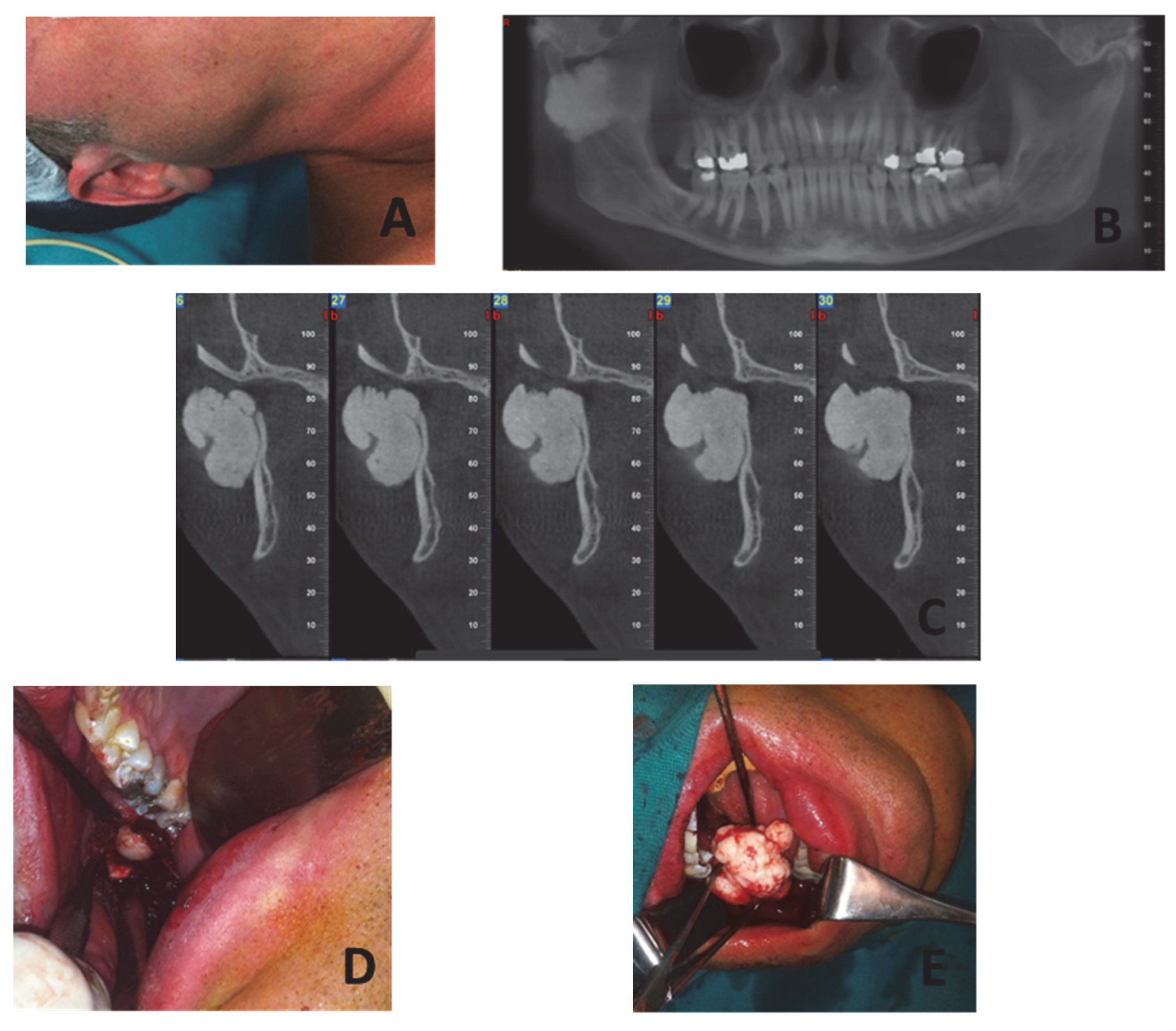
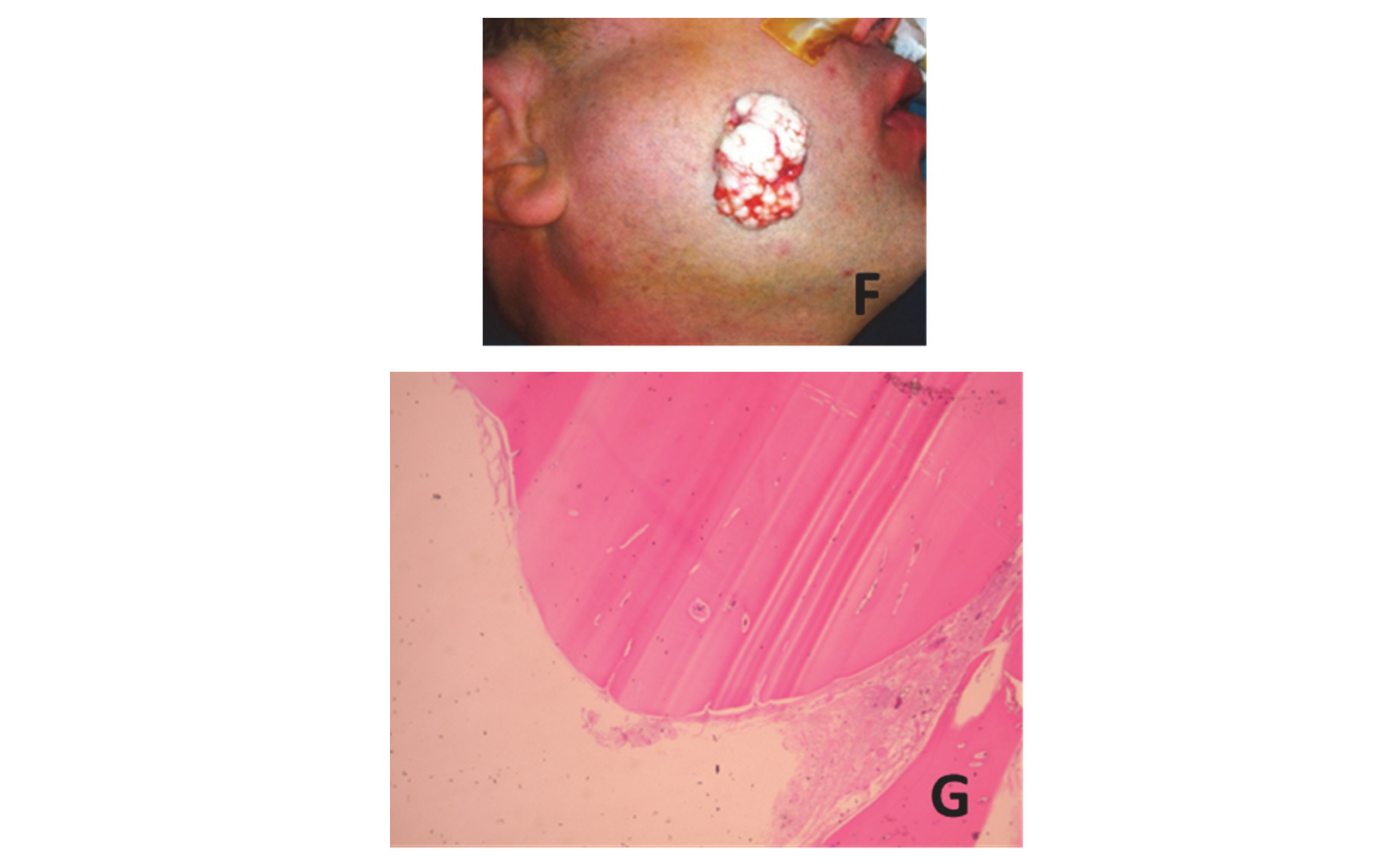
2.1. Case 1
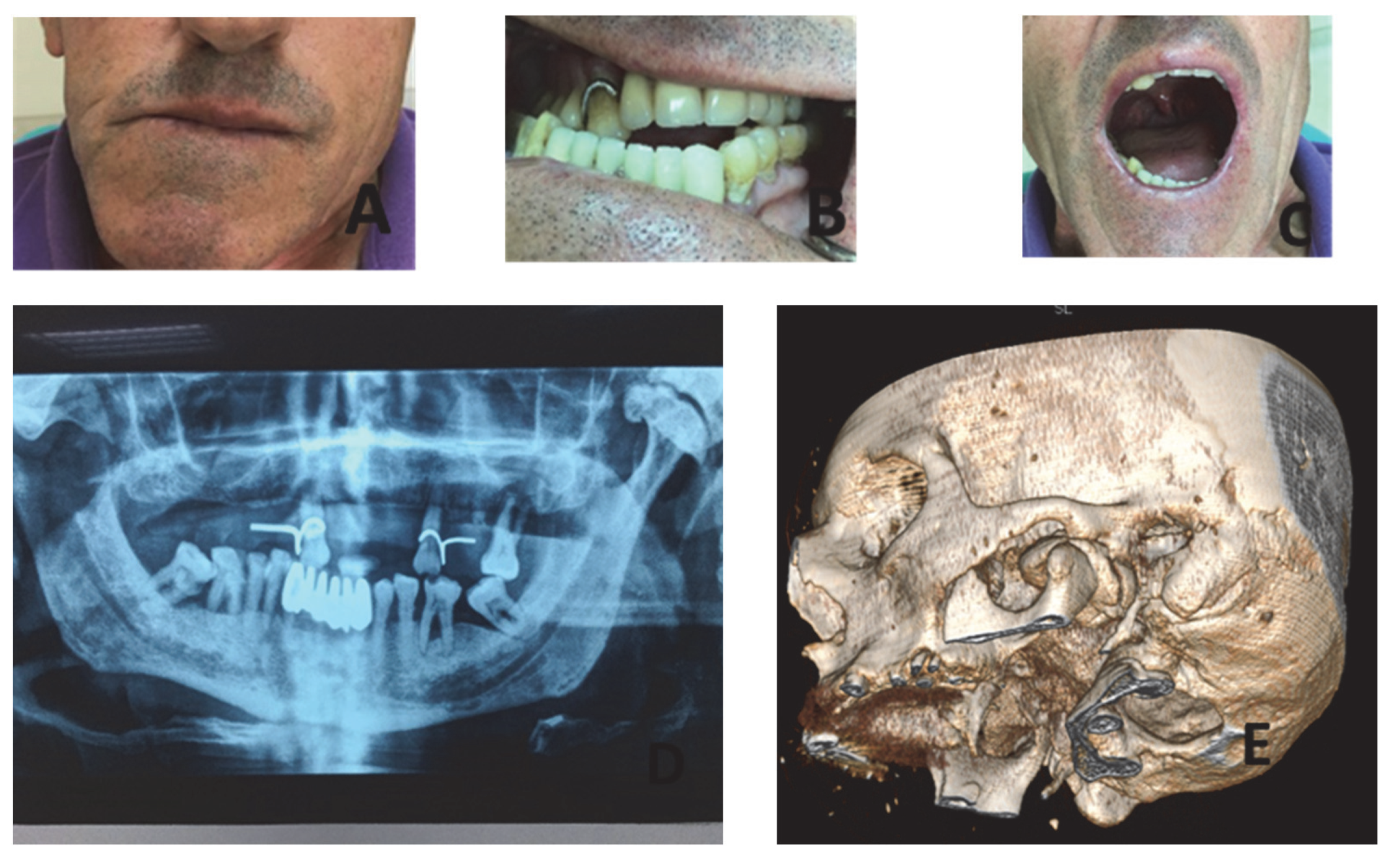
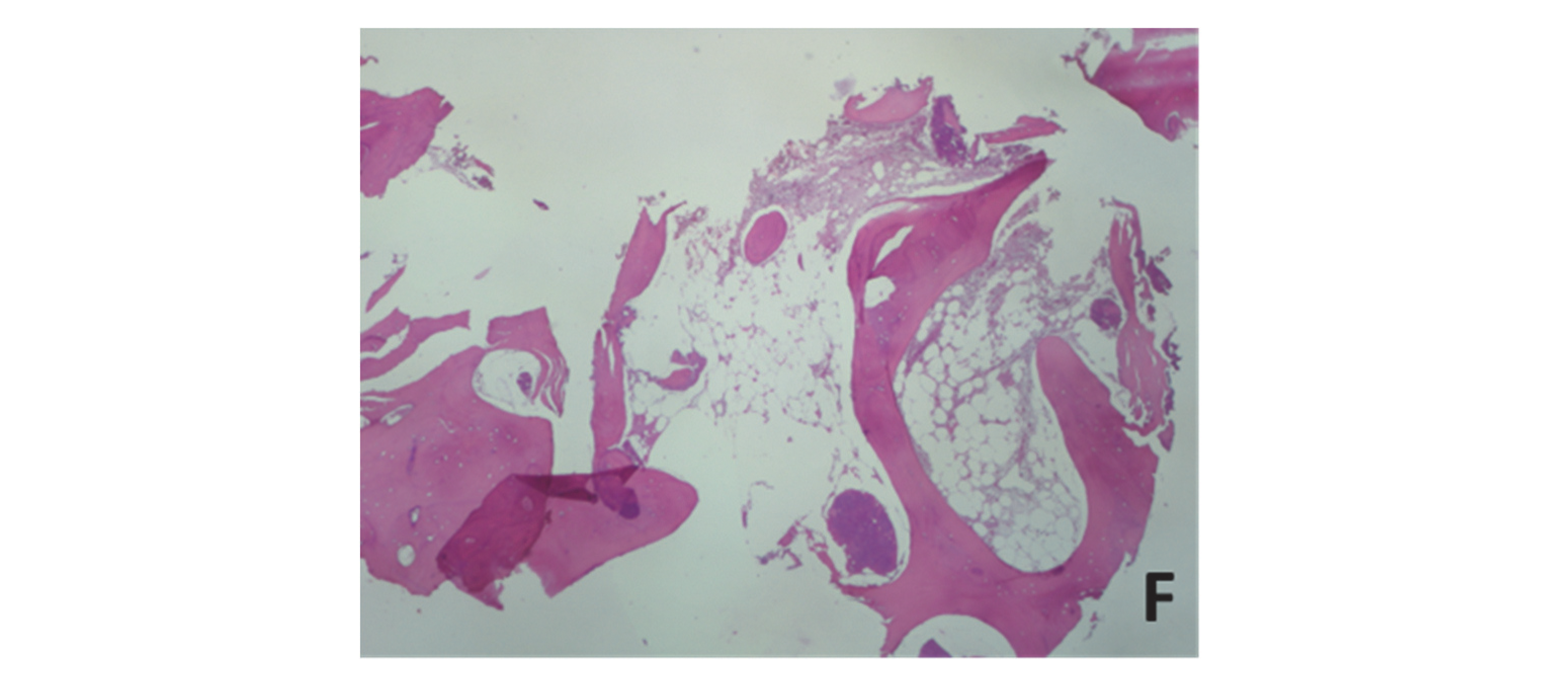
2.2. Case 2
2.3. Case 3
2.4. Case 4
3. Discussion
4. Conclusions
Author Contributions
Funding
Institutional Review Board Statement
Informed Consent Statement
Conflicts of Interest
References
- Kruger, G. Textbook of Oral and Maxillofacial Surgery, 6th ed.; Mosby: Maryland, MO, USA, 1984; p. 648. [Google Scholar]
- Longo, F.; Califano, L.; De Maria, G.; Ciccarille, R. Solitary osteoma of the mandibular ramus: Report of case. J. Oral Maxillofac. Surg. 2001, 59, 698–700. [Google Scholar] [CrossRef]
- Kaplan, I.; Nicolaou, Z.; Hatuel, D.; Calderon, S. Solitary central osteoma of the jaws: A diagnostic dilemma. Oral Surg. Oral Med. Oral Pathol. Oral Radiol. Endod. 2008, 106, e22–e29. [Google Scholar] [CrossRef] [PubMed]
- Deliverska, E. Peripheral osteoma of mandible—A case report and analysis of literature. J. IMAB 2016, 22, 1274–1278. [Google Scholar] [CrossRef]
- Kaplan, I.; Calderon, S.; Buchner, A. Peripheral osteoma of the mandible: A study of 10 new cases and analysis of the literature. J. Oral Maxillofac. Surg. 1994, 52, 467–470. [Google Scholar] [CrossRef]
- Al-Yahya, S.N.S.H.; Hamizan, A.K.W.; Zainuddin, N.; Arshad, A.I.; Ismail, F. Mastoid osteoma: Report of a rare case. Egypt. J. Ear Nose Throat Allied Sci. 2015, 16, 189–191. [Google Scholar] [CrossRef]
- Sondergaard, J.O.; Rusmussen, M.S.; Videbak, H.; Bernstein, I.; Myrhoj, T.; Kristiansen, K.; Sommer, P.; Bulow, S. Mandibular osteomas in sporadic colorectal carcinoma. A genetic marker. Scand. J. Gastroenterol. 1993, 28, 23–24. [Google Scholar] [CrossRef]
- Smrithi, D.V.; Jayanthi, K.; Dayananda, B.C. Solitary Peripheral Osteoma at a Curious Site with an Ambiguous Etiopathogenesis: A Case Report and Review of Literature. Int. J. Oral Maxillofac. Pathol. 2012, 3, 50. [Google Scholar]
- Sayan, N.B.; Ucok, C.; Karasu, H.A.; Gunhan, O. Peripheral osteoma of the oral and maxillofacial region: A study of 35 new cases. J. Oral Maxillofac. Surg. 2002, 60, 1299–1301. [Google Scholar] [CrossRef]
- Woldenberg, Y.; Nash, M.; Bodner, L. Peripheral osteoma of the maxillofacial region. Diagnosis and management: A study of 14 cases. Med. Oral Pathol. Oral Cir. Bucal 2005, 10, e139–e142. [Google Scholar]
- Shanavas, M.; Chatra, L.; Shenai, P.; Veena, K.; Rao, P.; Prabhu, R. Multiple Peripheral Osteomas of Forehead: Report of a Rare Case. Ann. Med. Health Sci. Res. 2013, 3, 105–107. [Google Scholar] [CrossRef]
- Larrea-Oyarbide, N.; Valmaseda-Castellón, E.; Berini-Aytés, L.; Escoda, C.G. Osteomas of the craniofacial region. Review of 106 cases. J. Oral Pathol. Med. 2008, 37, 38–42. [Google Scholar] [CrossRef] [PubMed]
- Schneider, L.C.; Dolinsky, H.B.; Grodkesk, J.E. Solitary peripheral osteoma of the jaws: Report of case and review of literature. J. Oral Surg. 1980, 38, 452–455. [Google Scholar] [PubMed]
- Dalabiras, S.; Boutsioukis, C.; Tilaveridis, I. Peripheral osteoma of the maxilla: Report of an unusual case. Oral Surg. Oral Med. Oral Pathol. Oral Radiol. Endod. 2005, 100, 19–24. [Google Scholar] [CrossRef]
- Chen, Y.K.; Lin, L.M.; Lin, C.C.; Hsue, S.S.; Huang, E.; Chuong, S.Y.; Chou, S.H. Peripheral osteoma of the mandibular condyle. J. Chin. Med. Assoc. 2003, 66, 123–126. [Google Scholar] [PubMed]
- Siar, C.H.; Jalil, A.A.; Ram, S.; Ng, K.H. Osteoma of the condyle as the cause of limited-mouth opening: A case report. J. Oral Sci. Mar. 2004, 46, 51–53. [Google Scholar] [CrossRef] [PubMed]
- Mancini, J.; Woltmann, M.; Felix, V.; Freitas, R. Peripheral osteoma of the mandibular condyle. Int. J. Oral Maxillofac. Surg. 2005, 34, 92–93. [Google Scholar] [CrossRef]
- Ortakoğlu, K.; Gunaydin, Y.; Aydintug, Y.S.; Safali, M. Osteoma of the mandibular condyle: Report of a case with 5-year follow-up. Mil. Med. 2005, 170, 117–120. [Google Scholar]
- Yonezu, H.; Wakoh, M.; Otonari, T.; Sano, T.; Hashimoto, S.; Uchiyama, T. Osteoma of mandibular condyle as cause of acute pain and limited-mouth-opening: Case report. Bull. Tokyo Dent. Coll. 2007, 48, 193–197. [Google Scholar] [CrossRef]
- Cogburn, A.C.; Hales, N.; Krempl, G.A. Endoscopic resection of a mandibular condyle osteoma: Report of a case. Laryngoscope 2008, 118, 1214–1216. [Google Scholar] [CrossRef]
- Bjornland, T.; Berstad, J.R.; Store, G. Peripheral osteoma of the mandible mimicking an ectopic condyle: A case report. Oral Surg. 2009, 2, 178–181. [Google Scholar] [CrossRef]
- Chaurasia, A.; Balan, A. Osteoid osteoma of jaws: An overview. J. Indian Acad. Oral Med. Radiol. 2008, 20, 6–8. [Google Scholar] [CrossRef]
- Almeida, L.E.; de Oliveira, F.M.A. Giant mandibular condyle osteoma. J. Craniofac. Surg. 2011, 22, 1147–1149. [Google Scholar] [CrossRef]
- Misra, N.; Srivastava, S.; Bodade, P.; Rastogi, V. Osteoma of temporomandibular joint: A rarity. BMJ Case Rep. 2013, 6, bcr2013200268. [Google Scholar] [CrossRef] [PubMed] [Green Version]
- De Souza, P.D.; Leonhardt, F.D.; Ahumada, N.G.; Abrahão, M.; Cervantes, O. Giant osteoma of the mandible. Braz. J. Otorhinolaryngol. 2015, 81, 107–108. [Google Scholar] [CrossRef] [PubMed]
- Rajshekar, V.M.; Basetty, N.R.; Govindaraju, R.; David, M.P. “Out of the ordinary”: A case report of osteoma of mandibular condyle. J. Indian Acad. Oral Med. Radiol. 2015, 27, 441–444. [Google Scholar] [CrossRef]
- Motlagh, M.F.; Janbaz, Y.; Abaszade, A. Hyge peripheral osteoma of the mandibular condyle: A case report. J. Oral Maxillofac. Surg. Med. Pathol. 2015, 27, 808–811. [Google Scholar] [CrossRef]
- Zafar, E.; Akbar, Z.; Niazi, K.; Pasha, B. Osteoma of mandibular condyle-a case report. Pak. Oral Dental J. 2016, 36, 383–386. [Google Scholar]
- De Souza, N.T.; Cavalcante, R.C.L.; Cavalcante, M.A.D.A.; Hespanhol, W.; Jr., M.R.D.O.; Ferreira, D.D.C.; Coutinho, T.M.D.C.; Gonçalves, L.S. An unusual osteoma in the mandibular condyle and the successful replacement of the temporomandibular joint with a custom-made prosthesis: A case report. BMC Res. Notes 2017, 10, 727. [Google Scholar] [CrossRef]
- Heitz, C.; Conci, R.A.; Tomazi, F.H.S.; Louzada, G.P.; Guarenti, M.M.; Fritscher, G.G. Giant Peripheral Temporomandibular Osteoma With Immediate Reconstruction of Mandible. J. Craniofac. Surg. 2018, 29, e146–e149. [Google Scholar] [CrossRef]
- Valente, L.; Tieghi, R.; Mandrioli, S.; Galiè, M. Mandibular condyle osteoma. Ann. Maxillofac. Surg. 2019, 9, 434–438. [Google Scholar] [CrossRef]
- Ostrofsky, M.; Morkel, J.A.; Titinchi, F. Osteoma of the mandibular condyle: A rare case report and review of the literature. J. Stomatol. Oral Maxillofac. Surg. 2019, 120, 584–587. [Google Scholar] [CrossRef] [PubMed]
- More, C.B.; Gupta, S. Osteochondroma of mandibular condyle: A clinic-radiographic correlation. J. Nat. Sci. Biol. Med. 2013, 4, 465–468. [Google Scholar] [CrossRef] [PubMed]
- Iatrou, I.A.; Leventis, M.D.; Dais, P.E.; Tosios, K.I. Peripheral osteoma of the maxillary alveolar process. J. Craniofac. Surg. 2007, 18, 1169–1173. [Google Scholar] [CrossRef] [PubMed]
- Norman, J.E.; Bramley, P. A Textbook and Colour Atlas of the Temporomandibular Joint; Wolfe Medical Publications Ltd.: London, UK, 1990; p. 26. [Google Scholar]
- Rodriguez, Y.; Baena, R.; Rizzo, S.; Fiandrino, G.; Lupi, S.; Galioto, S. Mandibular traumatic peripheral osteoma: A case report. Oral Surg. Oral Med. Oral Pathol. Oral Radiol. Endod. 2011, 112, e44–e48. [Google Scholar] [CrossRef]
- Pereira, C.U.; de Carvalho, R.; de Almeida, A.; Danta, R. Mastoid Osteoma. Consideration on Two Cases and Literature Review. Intl. Arch. Otorhinolaryngol. 2009, 13, 350–353. [Google Scholar]
- Singh, R.; Bhure, S.; Krishna, B.P.; Mazhar, H.; Thomas, A.; Soni, S.K. Cancellous osteoma of temporomandibular joint. J. Dent. Res. Rev. 2020, 7, 67–69. [Google Scholar]


| No | Authors/Year | Age/Gender | Location | Clinical Symptoms | Treatment |
|---|---|---|---|---|---|
| 1 | Chen et al., 2003 [15] | 27/M | Asymmetry-Malocclusion | Condylectomy | |
| 2 | Siar et al., 2004 [16] | 32/F | L-C | Trismus | Condylectomy |
| 3 | Mancini et al., 2005 [17] | 19/F | L-C | Asymmetry, Malocclusion | Condylectomy |
| 4 | Ortacoglu et al., 2005 [18] | 22/M | R-C | Malocclusion | Condylectomy |
| 5 | Yonezu et al., 2007 [19] | 50/M | L-C | Pain, trismus | Excision |
| 6 | Cogburn et al., 2008 [20] | 55/M | L-C | Trismus, Malocclusion | Endoscopic excision |
| 7 | Bjornland et al., 2009 [21] | 46/M | Swelling, Asymmetry | Excision | |
| 8 | Chaurasia 2009 [22] | 45/F | Swelling | None | |
| 9 | Almeida, de Oliveira Filho 2011 [23] | 45/M | Swelling, Trismus, Malocclusion | Excision | |
| 10 | Misra et al., 2013 [24] | 22/M | Trismus | Condylectomy | |
| 11 | De Souza et al., 2015 [25] | 41/F | L-C | Swelling | Excision |
| 12 | Rajshekar et al., 2015 [26] | 35/M | Trismus, Asymmetry | Condylectomy | |
| 13 | Motlagh et al.2015 [27] | 78/M | L-C | Difficulty in swallowing | Excision |
| 14 | Zafar et al., 2016 [28] | 60/M | Pain, Asymmetry | Excision | |
| 15 | De Souza et al., 2017 [29] | 67/F | R-C | Trismus, Asymmetry | Condylectomy |
| 16 | Heitz et al., 2018 [30] | 49/F | R-C | Swelling, Trismus | Condylectomy |
| 17 | Valente et al., 2019 [31] | 52/F | R-C | Pain, Trismus, Swelling | Condylectomy |
| 18 | Ostrofsky et al., 2019 [32] | 60/M | R-C | Trismus, Malocclusion | Condylectomy |
Publisher’s Note: MDPI stays neutral with regard to jurisdictional claims in published maps and institutional affiliations. |
© 2022 by the authors. Licensee MDPI, Basel, Switzerland. This article is an open access article distributed under the terms and conditions of the Creative Commons Attribution (CC BY) license (https://creativecommons.org/licenses/by/4.0/).
Share and Cite
Tilaveridis, I.; Katopodi, T.; Karakostas, P.; Venetis, G.; Dimitrakopoulos, I.; Tilaveridis, S.; Tilaveridou, S.; Zarampouka, K. Peripheral Osteoma of the Mandibular Condyle—Case Series. Dent. J. 2022, 10, 182. https://doi.org/10.3390/dj10100182
Tilaveridis I, Katopodi T, Karakostas P, Venetis G, Dimitrakopoulos I, Tilaveridis S, Tilaveridou S, Zarampouka K. Peripheral Osteoma of the Mandibular Condyle—Case Series. Dentistry Journal. 2022; 10(10):182. https://doi.org/10.3390/dj10100182
Chicago/Turabian StyleTilaveridis, Ioannis, Theodora Katopodi, Panagiotis Karakostas, Gregory Venetis, Ioannis Dimitrakopoulos, Stavros Tilaveridis, Sofia Tilaveridou, and Katerina Zarampouka. 2022. "Peripheral Osteoma of the Mandibular Condyle—Case Series" Dentistry Journal 10, no. 10: 182. https://doi.org/10.3390/dj10100182