Article

Oxidative, Genotoxic and Cytotoxic Damage Potential of Novel Borenium and Borinium Compounds

1
Department of Medical Services and Techniques, Vocational School of Health Services, University of Gaziantep, Gaziantep 27410, Turkey
2
Department of Medical Biology, Faculty of Medicine, Atatürk University, Erzurum 25040, Turkey
3
Department of Chemistry, Arts and Science Faculty, Düzce University, Düzce 81620, Turkey
4
Department of Otorhinolaryngology Diseases, Faculty of Medicine, Atatürk University, Erzurum 25040, Turkey
5
Science for Life Laboratory, KTH-Royal Institute of Technology, 11428 Stockholm, Sweden
6
Centre for Host-Microbiome Interactions, Faculty of Dentistry, Oral & Craniofacial Sciences, King’s College London, London WC2R 2LS, UK
*
Author to whom correspondence should be addressed.
Inorganics 2023, 11(8), 324; https://doi.org/10.3390/inorganics11080324
Submission received: 5 June 2023 / Revised: 20 July 2023 / Accepted: 27 July 2023 / Published: 31 July 2023

Abstract

:
In this study, the biological properties of novel borenium and borinium compounds in terms of their oxidative, genotoxic, and cytotoxic effects were assessed on cultured human peripheral blood cells, as well as several types of cancer cells. Our results revealed that the borinium compounds yielded the best results in terms of supporting total antioxidant capacity (TAC). In fact, borenium 1, borenium 2, borenium 3, borinium 4, and borinium 5 compounds elevated TAC levels of cultured human blood cells at rates of 42.8%, 101.5%, 69.8%, 33.3%, and 49.2%, respectively. There were no statistically significant differences (p > 0.05) between the negative control and the groups treated with all borinium and borenium concentrations from the micronucleus (MN) and chromosome aberration (CA) assays, demonstrating the non-genotoxic effects. Moreover, borenium 1 (60.7% and 50.7%), borenium 2 (70.4% and 57.2%), borenium 3 (53.1% and 45.2%), borinium 4 (55.1% and 48.1%), and borinium 5 (51.0% and 36.1%) minimized the mitomycin C(MMC)-induced genotoxic damages at different rates as determined using CA and MN assays, respectively. Again, it was found that the borinium compounds exhibited higher cytotoxic activity on cancer cells when compared to borenium compounds. Consequently, in light of our in vitro findings, it was suggested that the novel borinium and borenium compounds could be used safely in pharmacology, cosmetics, and various medical fields due to their antioxidant and non-genotoxic features, as well as their cytotoxicity potential on cancer cells.

1. Introduction

Around 400 different industrial domains involving the production of ceramics and fertilizers, glass and glass fibers, pharmaceuticals, chemicals, nuclear power, automobiles, and spacecrafts employ boron (B) in different chemical structures. In light of recent scientific findings based on the biological and physicochemical properties of boron-containing compounds (BCCs), B is now regarded as a strategic element whose usage and application domains are expanding constantly [1]. Due to its superior physicochemical properties and its preference in many industrial fields, new boron derivatives are synthesized by scientists and new BCCs are offered for use. B is considered to be a crucial microelement for plants. Despite the vast body of scientific evidence, it has not been conclusively reported that B is necessary for both humans and animals. In fact, several BCCs, such as borates, boronates, and boronic acids, exhibited interesting biological activities, including antiviral [2], antibacterial [3,4], antifungal and antiparasitic [5,6], antioxidant [7,8], wound healing [9], anti-inflammatory [10,11], antimutagenic [12], anticancerogenic [13,14], radiobiological [15], and neuroprotective properties [16,17]. Interestingly it was found that people living in B-rich regions had less cancer incidence than the people living in B-poor regions [18]. The functions of B in the human body are not known clearly but previously suggested health benefits of boron included protection of the liver, enhancement of fetal development, regulation of enzymatic activity associated with the immune system, and improvement of brain functions in humans and animals [19]. Hence, researchers and industries have recently given extensive effort in studying BCCs with the goal of understanding their physiological process and locating novel health technologies endowed with clinical safety.
A few studies on boron compounds that have been previously synthesized are the ionic liquids based on boronium cations [20]. These cations were considered to be a highly electrophilic species that was elusive and was reported to have a key role in the chemistry of B. A commercially available ionic fluid boron tetra-fluoro borate (BF4) anion has this cation. Compounds with a BF4- anion have a hypersensitivity to the reactivity of the pipe to air and water [21]. Given this context, the main objective of the current investigation was to synthetize novel cationic B-based compounds that exhibit high resistance properties due to a B cation and introduce novel boron compounds with potential to be used in the biomedical field. In accordance with this goal, we aimed to obtain useful electrolyte compounds indicating higher thermal stability and more resistance to heat treatment.
Understanding the behavior of the novel borenium and borinium compounds can guide researchers or applicators in choosing the relevant biomedical materials for different clinical and anatomical purposes. Along with the proper thermal stability and heat-resistance features of novel biomedical materials, their toxicity potential should be evaluated before safe clinical use. The chemical and physical features of biomedical materials influence their biological and toxic potential [22]. In this regard, the chemical, physical, and biological features of B provide medicinal chemists a unique chance to research and develop brand new fields of biomedical sciences, especially in drug discovery [23,24,25]. Recent investigations indicated that BCCs could affect the crucial cellular machineries implicating cell survival, tissue regeneration, and immunogenic responses [26]. Although the toxicity potentials by borenium and borinium compounds are not well known, their antibacterial and antiviral properties have been well documented [27,28,29]. Thus, these limited studies indicate that borenium and borinium compounds can be used safely in medical, cosmetics, and green chemistry domains [28,30]. Hence, the second aim of this investigation was to assess the oxidative, genotoxic, and cytotoxic effects of novel borenium and borinium compounds on human peripheral blood cells, as well as several types of cancer cells. We assessed the in vitro effects of these compounds on cytotoxicity using an MTT assay, DNA damage response via MN and CA assays, and antioxidant capacity using a TAC assay.

2. Materials and Methods

2.1. Novel Ionic Liquids

Ionic liquids are salts that exist in liquid form at room temperature, and at lower temperatures, and generally have an organic cationic part. Ionic liquids have high polarity, low vapor pressure, and are resistant to high temperatures. Due to these properties, ionic liquids can be used repeatedly as both solvent and catalyst in many reactions, and they can also be easily removed from the reaction medium without leaving any waste. In this way, ionic liquids are superior to other classic solvents and cause them to be preferred in terms of environment.
In this study, an oxidative, genotoxic, and cytotoxic evaluation of new boron-containing molecules to the family of ionic liquids was performed. In the five novel compounds we synthesized, new ionic liquids with a boron cationic center—not BF4- anion—were synthesized. Cationic forms of boron are produced in two different forms, borinium and borenium, as shown in Figure 1. The original ionic liquids were synthesized with organic extension derivatives of these structures containing chiral structures.
NMR analyses were performed in DMSO, CDCl3, and MeOH with an Agilent Premium Contact NMR 600 MHz spectrophotometer, and the chemical shift values (δ) in the spectrum were expressed in ppm. IR measurements were performed with the Agilent Cary 630 FTIR device. The recovery of the silver amino acid salts was confirmed with the Agilent 7700 series ICP-MS instrument. A Heidolp brand rotary evaporator was used for the solvent removal system. Melting point was determined with Stuart SMP40 brand melting point determination device. Analytical TLCs were performed using aluminum plates coated with a layer of silica gel (SiO2, Merck 60 F254).

2.2. Experimental Design

We used human U87MG, SHSY-5Y, PC-3, and Detroit-562cell lines for screening of cytotoxicity of boronated compounds. The U87MG, SHSY-5Y, and Detroit-562 cell lines were obtained from Atatürk University, Faculty of Medicine, Erzurum, Turkey. PC-3 cells were provided from the American Type Culture Collection (Manassas, VA, USA). Cells were harvested using 0.25% trypsin–EDTA solution and suspended with RPMI 1640 medium (Sigma-Aldrich, St. Louis, MO, USA) containing 10% FBS, L-glutamine (1%), and penicillin–streptomycin mixture (1%). The different concentrations (from 1.56 to 400 mg/L or from 2.91 to 1316.01 μM) of borenium and borinium compounds were applied to the cultures (n = 5). All experiments were performed in accordance with the rules of the World Medical Association.

2.3. Cytotoxicity Testing

The cytotoxic potential of the boron compounds was determined by MTT analysis. In brief, compounds were added into the cell culture plates at a wide concentration range from 1.56 to 400 mg/L or from 2.91 to 1316.01 μM and incubated for 48 h (n = 5). Then, 10 µL of MTT solution was added to wells and incubated for an additional 3 h at 37 °C. After discarding cell mediums, DMSO (100 µL) was added to wells for dissolving formazan crystals. Finally, the color intensities were measured via using a microplate reader at 570 nm [31]. A podophyllotoxin derivative, etoposide (Merck), which is a chemotherapy medication, was dissolved in DMSO (<1%) and used as a positive control agent for comparing cytotoxic action of boron compounds.

2.4. Genotoxicity Testing

The in vitro genotoxic/antigenotoxic potential of boron compounds on human whole blood cell cultures using chromosomal aberration (CA) and micronucleus (MN) assays was tested. For CA assay, a 0.65 mL aliquot of heparinized blood sample was cultured in 7 mL of culture medium (Chromosome medium B; Biochrom, Berlin, Germany) containing phytohemagglutinin (5 mg/mL, Biochrom). Then, the cultures were incubated for 72 h at 37 °C. Around 2 h before harvesting, 0.1 mL of colchicine (0.2 mg/mL, Sigma; St Louis, Missouri, USA) was added into the culture tubes and hypotonic treatment/fixation steps were performed. The prepared slides were stained using Giemsa solution (3%). For each culture tube (n = 5), 30 well-spread metaphases were scored to detect CA frequencies. Chromatid or chromosome gaps and chromatid or chromosome breaks were scored as recommended by Environmental Health Criteria 46 for environmental monitoring of human populations [32].
For MN assay, cytochalasin B (at a final concentration of 6 μg/mL, Sigma) was added into the culture tubes (contained 2 × 10⁶ cells/mL) after 44 h of culture and incubated for 72 h (n = 5). After the incubation period of three days, the lymphocytes were fixed using treatment with ice-cold methanol:acetic acid (1:1) and stained Giemsa (5%) for 12 min. The slides were scored by using a bright-field microscope (at 400× magnification, Olympus). A total of 2000 binucleated lymphocytes were scored per treatment type for the presence of one, two, or more micronuclei according to previously reported standard criteria [33]. Mitomycin C (MMC, 5 × 10⁻⁶ M) was used as the positive control in CA and MN assays [34].

2.5. Determining of TAC Levels

The commercially available kit (Rel Assay Diagnostics, Gaziantep, Turkey) was used to determine the antioxidant capabilities of the borenium and borinium compounds on human whole blood cell cultures for 72 h (n = 5). The principle of this kit assay is to monitor antioxidant levels of cultures via inhibiting formation of free radical featured 2,2′-azino-bis(3-ethylbenzothiazoline-6-sulfonic acid) compound. Furthermore, the assay is calibrated with Trolox equivalent vitamin E analogue. The cultures without boronated compounds were studied as a negative control group. Ascorbic acid (10 μM) was also used as a positive control in total antioxidant capacity (TAC) analysis [35].

2.6. Statistical Analyses

Statistical analysis was performed using IBM program SPSS version 25. All tests were performed for at least five different repeats. The obtained data were analyzed using a variance (ANOVA) test followed by Duncan’s test and values with p < 0.05 were accepted as significantly different. Probit regression analyses were performed to estimate the concentrations required to reduce cell viability rates by 50% using SPSS [36].

3. Results

Examples of ionic liquids containing boron cations are very rare in the literature [37]. In previous research, a series of ionic liquids containing N-alkylimidazole-amine BH2+ structures were synthesized, and their electronic and spectroscopic properties were examined [38]. Some new boronium cation-based ionic liquids were also synthesized and their potential for use in lithium ion batteries was investigated. In this study, it is of great importance to test the usability of ionic liquids with boron cations in practice [39].

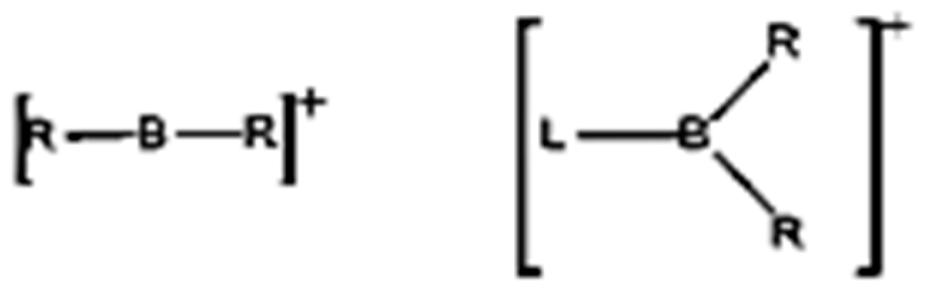
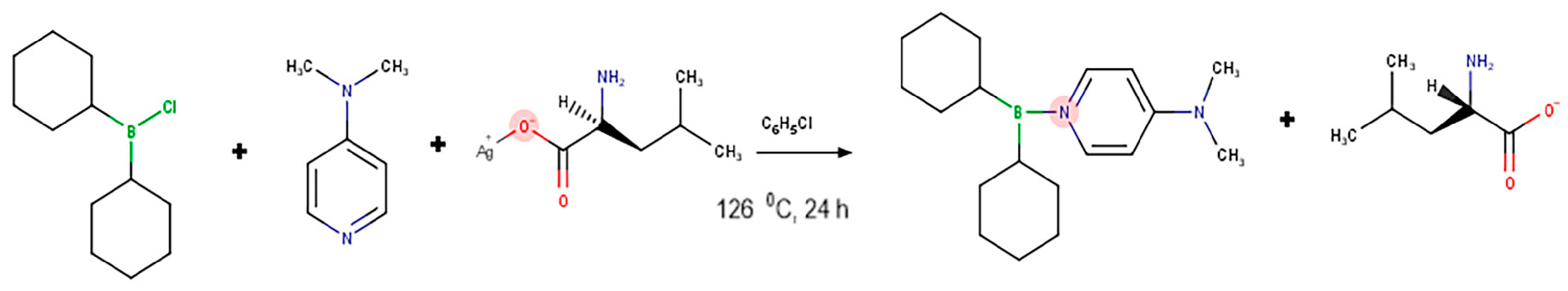
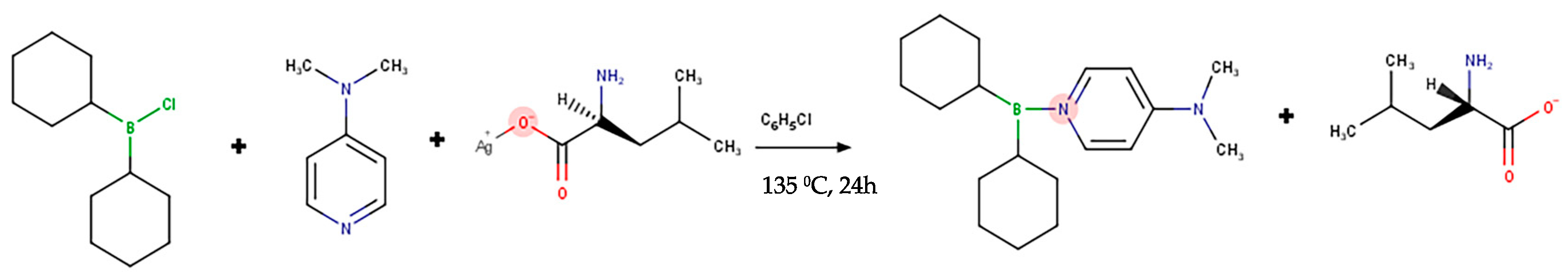
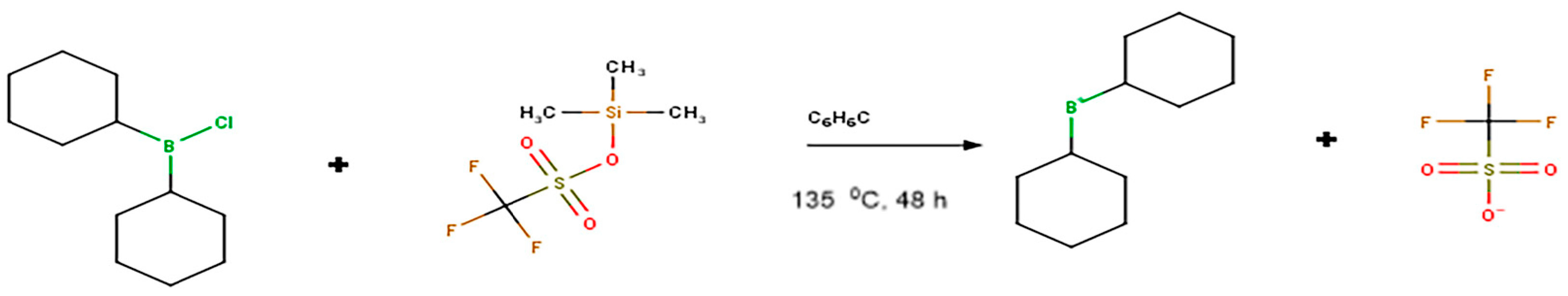
3.1. Synthesis of Borenium Ionic Liquids

The dialkyl aryl borane and the anion compound, chlorobenzene, were dissolved. After mixing, an aromatic amine compound was added to this reaction solution, and it was allowed to boil at a temperature >120 °C. Centrifugation was applied and the solvent component was removed. The synthesis reaction and code numbers of the synthesized borenium 1, 2, and 3 compounds are presented in Figure 2, Figure 3 and Figure 4. The IR, 1H-NMR, and 13C-NMR (Agilent, Premium Compact, 14.1 tesla, 600 MHz) spectra of three novel borenium compounds are presented in Supplementary Figures S1–S9.

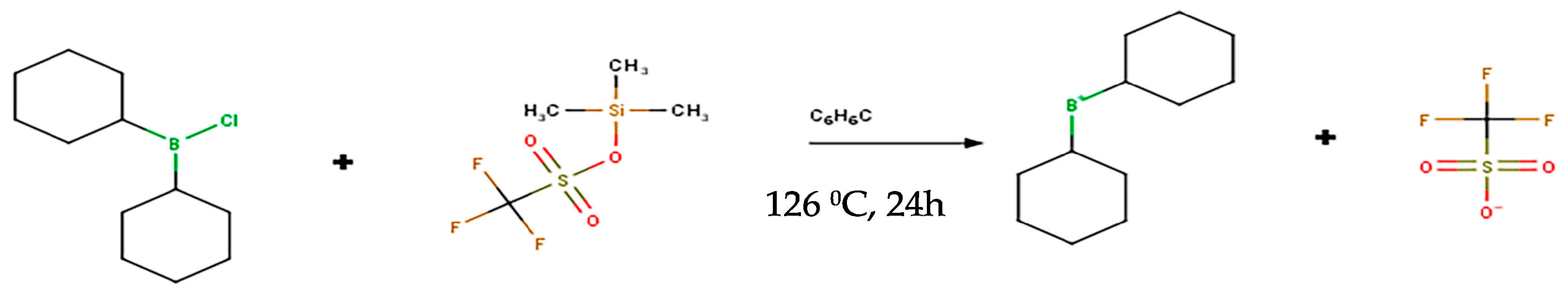
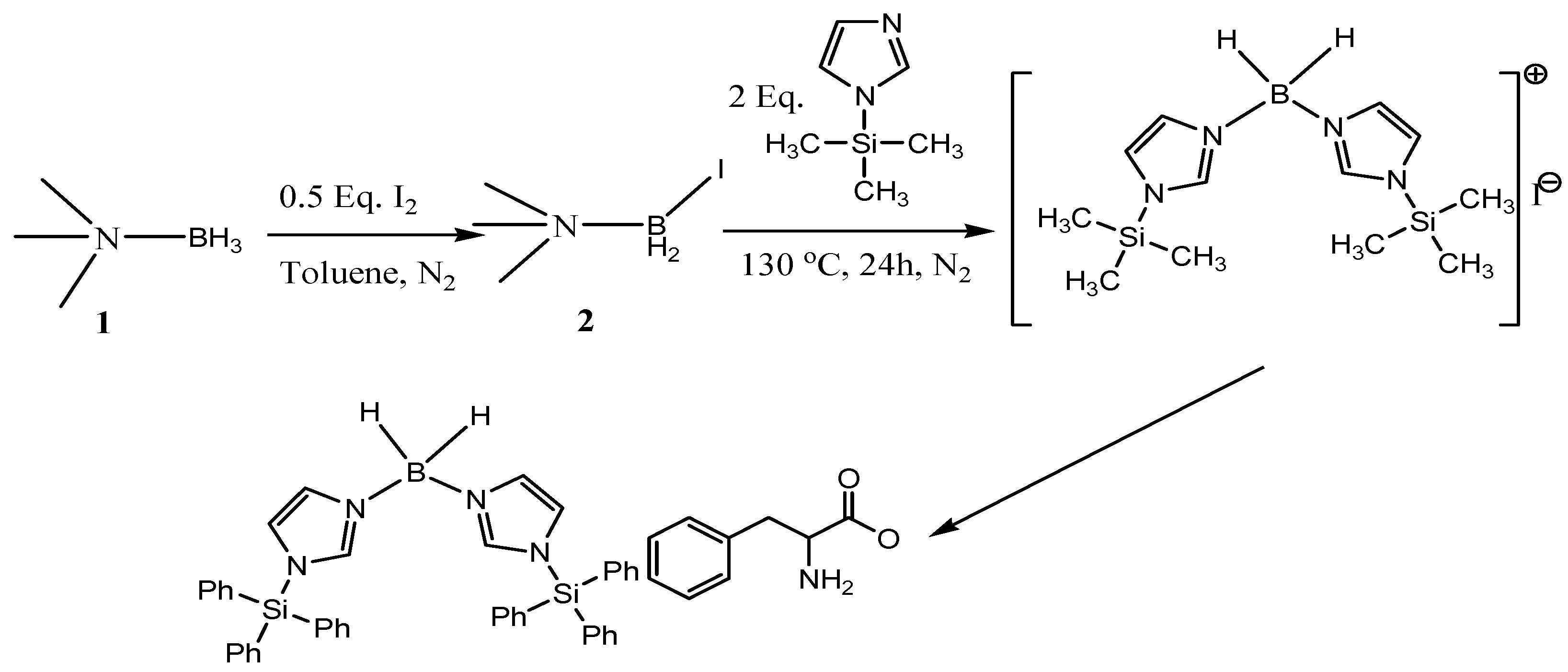
3.2. Synthesis of Borinium Ionic Liquids

The dialkyl aryl borane and the anion compound, chlorobenzene, were dissolved. After mixing, an aromatic amine compound was added to this reaction solution, and it was allowed to boil at a temperature of >130 °C. Centrifugation was applied and the solvent component was removed. After one night, after the reaction was cooled to room temperature, the first centrifugation was applied, and the solvent part was removed. The synthesis reactions of borinium 4 and borinium 5 are presented in Figure 5 and Figure 6. The IR (Agilent Cary 630), 1H-NMR, and 13C-NMR spectra of three novel borinium compounds are presented in Supplementary Figures S10–S15.
The cytotoxicity of the boron compounds on three different cancer cells, including human glioblastoma (U87MG), neuroblastoma (SHSY-5Y), prostate (PC-3) and pharyngeal (Detroit-562) cancer cell lines, as well as human whole blood cells, was determined. The determination of cytotoxicity was evaluated by calculating percent inhibition (IC50). Table 1 presents the IC50 values as estimated using the results of the MTT assay and Probit analysis. IC50 values are used to express the concentration corresponding to a survival rate of 50% under in vitro conditions. This value is commonly used for measuring antagonist drug potency using outputs from cell-based cytotoxicity tests; the lower the IC50 value, the more cytotoxic the compound, drug candidate, or drug is. The international authorities, such as the National Cancer Institute, debate IC50 values for classifying cytotoxicity potentials by compounds as high cytotoxic (IC50 < 20 mg/L), moderate cytotoxic (20 mg/L < IC50 < 200 mg/L), weak cytotoxic (200 mg/L < IC50 < 500 mg/L), and non-cytotoxic (IC50 > 500 mg/L) [40,41,42,43]. No statistical difference (p > 0.05) in cell viability was observed between the negative controls and vehicle (DMSO, <1%) controls. Borinium 5 was found to be the most promising compound as an anti-proliferative agent, but it seems necessary to take into consideration that concentration-dependent cytotoxicity on healthy cells might occur at excessive exposure. The PC-3 cells were found to be more sensitive to boronated compounds in comparison to other cancer cells, such as the U87MG, SHSY-5Y, and Detroit-562 lines. The concentration-dependent cell viability alterations are presented in Supplementary Figures S16–S20.
The results of the genotoxicity tests of five different borenium and borinium compounds, which were evaluated in cultured human lymphocytes by CA and MN assays, are shown in Table 2. The rates of chromosomal aberrations (abnormal cell, %) and abnormal cells (CAs/cell) were determined by blindly scoring at least 30 well-spread metaphases for each culture type (a total of at least 150 metaphases for each experimental group). Likewise, the rates of micronuclei (MNs) were monitored by blindly scoring at least 1000 binucleated cells (MN/1000 cells) for each culture type (a total of at least 4000 binucleated cells for each treatment). MMC treatment led to 6.13- and 6.69-fold change increases of CAs and MNs formations, respectively. On the contrary, the frequency of CAs and the rate of MNs in cells treated with different concentrations of the novel boron compounds were similar to those of the untreated control cells (p > 0.05). When assessed in terms of genotoxic damage potentials, the rates of CAs/cell and MN/1000 cells did not significantly (p > 0.05) increase at all tested concentrations of boron compounds. Hence, our findings reveal the non-genotoxic features of these novel boron compounds.
The results for the antigenotoxicity assays performed via MMC in combination with three different concentrations (25, 50, and 100 mg/L) of boronated compounds are also reflected in Table 3. MMC alone caused a statistically significant increase in CA and MN frequencies when compared to the control group (p < 0.05). On the contrary, co-treatment with boronated compounds significantly reduced the frequencies of CAs and MNs, which were elevated by MMC (p < 0.05). In fact, in lymphocytes treated with the combination of boronated compounds plus MMC at 72 h (except for 25 mg/L borenium 1), the frequencies of CAs and MNs were significantly reduced compared to the positive control (MMC) at 25, 50, and 100 mg/L. Borinium 4 (55.1% and 48.1%), borinium 5 (51.0% and 36.1%), borenium 1 (60.7% and 50.7%), borenium 2 (70.4% and 57.2%), and borenium 3 (53.1% and 45.2%) minimized the MMC-induced genotoxic damage at different rates, as determined using CA and MN assays, respectively. Furthermore, borenium 2 was found to be the most potent compound for the prevention of DNA damage induced by MMC.
In the TAC assay, the available antioxidants in the cultures reduced the colored free radical to its colorless form, and the alternation in absorbance at 660 nm refers to the total antioxidant level in samples from treated and untreated cultures. The presented values in Table 4 correspond to the mean value of at least four different absorbance readings from each culture type. The most significant contribution to the TAC level was observed after treatment with the borenium 2. In fact, borenium 2 elevated TAC levels at a rate of 10.6, as compared to those levels (6.3) in the negative control group. Furthermore, its highest concentration (400 mg/L) increased the TAC levels at a rate of 7.9%. Moreover, the decreasing order of effectiveness for enhancing TAC levels by the tested compounds was as follows: borenium 2 > borenium 1 > borinium 4 > borenium 3 > borinium 5. In fact, the borinium 4, borinium 5, borenium 1, borenium 2, and borenium 3 compounds elevated TAC levels at rates of 33.3%, 49.2%, 42.8%, 101.5%, and 69.8%, respectively. To the contrary, the increasing concentrations of borinium 4 (at 200 and 400 mg/L), borenium 1 (at 400 mg/L), borenium 3 (at 200 and 400 mg/L), and borinium 5 (at 100, 200 and 400 mg/L) caused statistical (p < 0.05) reductions of TAC levels (Table 4).

4. Discussion

Previous reports revealed that incorporation of cationic boron centers in organic heterocycles or transition-metal metallocenes could provide opportunities for the development of novel biomedical materials with superior redox activity and optical properties [21]. In our study, new boron-containing molecules were introduced into the family of ionic compounds, and the synthesis and characterization of the compounds were carried out. In the first step, the synthesis of boronium ionic liquids was performed. Briefly, boronium amino acid salts were obtained by anion exchange over the boronium iodide compounds obtained. At this stage, silver salts of various amino acid derivatives, which were dissolved in the appropriate solvent, were slowly added to the solution of boronium iodide salts dissolved in the appropriate solvent at room temperature in the dark, and the targeted compounds were obtained. In the second phase of our study, the synthesis of borenium ionic liquids was performed. When the 1H-NMR and 13C-NMR spectra of the synthesized compounds are examined, the presence of the peaks indicates that the synthesis of the targeted compounds has been successfully achieved.
The cytotoxic, genotoxic, and oxidative damage potentials of the newly synthesized borenium and borinium compounds were investigated in this study. The cytotoxic effects of these new ionic liquids of B have been demonstrated as a result of exposing U87MG, SHSY-5Y, PC-3, and Detroit-562 cancer cells to these liquids. Our findings revealed that the PC-3 cells were found to be more sensitive to boronated compounds in comparison to other cell lines. Furthermore, borinium 5 was found to be the most potent among them (Table 1). Similar to our findings, previous cellular and epidemiological studies revealed that B (as boric acid, BA) did not induce carcinogenicity. Moreover, supplementation with B might decrease the risk for prostate and brain cancers [44,45]. The underlying mechanisms of anticancer properties by BCCs are still unclear. It has been found that a Ca signal has a regulatory role in cell profiling and very little attention has been paid to cancer-preventive therapies. In a previous study, it was determined that supplementation with high amounts of boron (50 mg/L) inhibited the proliferation of human DU-145 prostate cancer cells via decreasing intracellular Ca signals and stores [46]. Another underlying anticancer mechanism due to B supplementation was associated with inhibition of serine proteases, such as prostate specific antigens and the affinity of BA to hydroxyl groups [47]. Again, BA was shown to contribute to proliferative inhibition via dose-dependent reductions in the expression of cyclins A-E and MAPK proteins in DU-145 prostate, HeLa cervical cancer, and DLD-1 colorectal adenocarcinoma cells [48,49,50]. In addition, ferroptosis (a new type of iron-dependent cell death) was characterized by intracellular iron ion accumulation. Moreover, B was reported to modulate the ferroptosis in HepG2 hepatocellular carcinoma cells, hence it could serve as a sensitizer to anticancer chemotherapeutics [51]. In this context, the induction of ferroptosis might be one of the possible underlying mechanisms for anticancerogenic action by introduced borenium or borinium compounds. In fact, in a recent investigation it was reported that BA (up to 1500 mg/L) was able to trigger both ferroptosis and apoptosis in C6 glioma cells and affected the emaphorin–neuropilin signaling pathway [52]. AKT phosphorylation by B could be proposed as another associated mechanism for explaining the anticancer properties of BCCs in hepatocellular carcinoma and glioma cases [53,54].
The determined IC50 values after exposure to boronated compounds in several cancer cell lines clearly revealed that the effects of boron compounds on different cancer types might be variable (Table 1). In a recent investigation, BA and borax (BX) enhanced the apoptosis in human DMS-114 lung cancer cells by upregulating pro-apoptotic genes, such as Bax and Casp-3. In addition, these borates modulated anti-apoptotic genes, such as BIRC2, BIRC5, and Bcl-2, and induced cell cycle arrest at the G2/M phase [55]. Similar to our findings, different concentrations (150–3000 mg/L) of BA exerted cytotoxic action on U87MG and T98G glioblastoma (GBM) cells with high IC50 values (1050 mg/L) [56]. In this regard, the novel boron compounds, especially borinium 5, might be novel sources for anti-GBM therapies with their moderate cytotoxic IC50 values without damaging healthy cells (Table 1). Alongside the cell cycle arresting and apoptosis modulating features, BCCs were found to interfere with other key tumorigenic pathways involving glycolysis, molybdenum Fe–S-containing flavin hydroxylases, and intratumoral IGF-I levels—as well as transient receptor potential in glioma cases [57].
In the present investigation, it was revealed that the tested borenium and borinium compounds exerted non-genotoxic features. All of the concentrations of these compounds did not induce the formations of CAs or MNs as compared to untreated cells (Table 2). In accordance with the present findings, the World Health Organization propounded that genotoxic damage cannot be associated with exposure to B in both animals and humans [58]. Likewise, previous multiplexed in vitro studies proved the non-genotoxic features of certain commercially important BCCs, including potassium tetraborate, BX, ulexite, colemanite, and B-ionic liquids [30]. In addition to these in vitro studies, the three orally administered BA doses (5, 10, and 20 mg/kg) did not cause DNA damage in the mononuclear leukocytes of rats [59]. Relatively high doses of BX (100 mg/kg) also did not induce DNA strand breaks in rat lymphocyte cells; hence, BX was reported to have a non-genotoxic nature [60]. Our findings also revealed that the genotoxic damages in the co-treated with MMC and borenium or borinium compounds monitored via CA and MN frequencies were lower than the solely MMC-applied group (Table 3).
Our findings indicated that both the borenium and borinium compounds also have in vitro antigenotoxic action potential. Cross-linking to DNA occurred in MMC-treated human cells, thus MMC application induced persistent DNA double-strand breaks [61]. BCCs, such as boric acid (BA), reduced the formation of DNA double-strand breaks and prevented chromosome loss of cells [62]. Moreover, ataxia–telangiectasia-mutated (ATM) protein kinase was reported to initiate DNA repair after formation of double-strand breaks by mutagens [63]. Furthermore, BA was able to lead to ATM activation and a DNA damage response of the cells [62]. The observed antigenotoxicity action by the borenium and borinium compounds (Table 3) could also be attributed to their direct chemical interaction before MMC caused genotoxic damage [64]. Previous evidence exerted that antioxidant-featured substances can eliminate ROS before these reactive chemicals interact with DNA and change in a DNA sequence [64]. Consistent with our findings, previous reports suggested that BCCs have antigenotoxic effects against several genotoxic agents, such as titanium, aluminum, aflatoxins, lead, bismuth, arsenic, and cadmium [65]. The observed antigenotoxic action by several boron compounds on animal or cell culture models could be primarily linked to their antioxidant properties. In fact, the antioxidative features of several BCCs were associated with their antigenotoxic action [66,67].
The results of our study put forward that the borinium compounds yielded the best results in terms of TAC values when compared to the untreated control treatment. Moreover, concentrations below 100 mg/L supported the TAC levels of human blood cultures (Table 4). Various experimental studies indicated that erythrocytes were especially susceptible to oxidative stress. Furthermore, B regulated the activity of cellular antioxidant enzymes, such as oxidoreductases, aldehyde dehydrogenase, xanthine oxidase, and cytochrome b5 oxidoreductase, and affected coagulation factors, such as glyceraldehyde-3-phosphate dehydrogenase and lactate dehydrogenase, by interacting with enzymes, such as serine proteases [61,68]. It was also observed that BA could alter the oxidative metabolism in animals. However, the eventual mechanisms of this change are still unclear [69]. The primer findings in this field manifested that several BCCs at relatively low doses (<80 mg/L) supported antioxidant enzyme activities in human whole blood cultures. Indeed, the activities of superoxide dismutase (SOD), catalase (CAT), glutathione peroxidase (GPX), glutathione-S-transferase (GST), glucose-6-phosphate dehydrogenase (G6PD), and the levels of total glutathione (TGSH), as well as TAC levels, were strengthened by application with certain BCCs, including BA, BX, and calcium borates [8,50,70].
In conclusion, the five novel borenium and borinium compounds exerted key biological functions involving (I) antioxidant (supporting TAC levels up to 101.5%), (II) non-genotoxic (having no clastogenic and eugenic effects), (III) antigenotoxic (minimizing MMC induced genotoxic damages in different rates up to 70.4%), and (IV) moderate cytotoxic (IC50 values < 200 mg/L on glioblastoma, neuroblastoma, prostate, and pharyngeal cancer cell lines and ranging from 41.941 to 179.662) properties. Our findings will contribute to further investigations on the biomedical evaluation of these borenium and borinium derivatives. They have great potential to be employed for nutritional, pharmacological, and medicinal purposes. These boron compounds deserve to be studied further in cancer treatment and nutrition due to their multi-biological functions. In the meantime, concentration adjustment should be undertaken while using these new boron compounds to promote health benefits.

Supplementary Materials

The following supporting information can be downloaded at: https://www.mdpi.com/article/10.3390/inorganics11080324/s1. Figure S1: IR spectrum of dicyclohexyl borenium dimethyl amino pyridine trifluoro methane sulphonate (Borenium 1); Figure S2: 1H-NMR spectrum of dicyclohexyl borenium dimethyl amino pyridine trifluoro methane sulphonate (Borenium 1); Figure S3: 13C-NMR spectrum of dicyclohexyl borenium dimethyl amino pyridine trifluoro methane sulphonate (Borenium 1); Figure S4: IR spectrum of dicyclohexyl borenium dimethyl amino pyridine 2-amino-4-methylpentanoate (Borenium 2); Figure S5: 1H-NMR spectrum of dicyclohexyl borenium dimethyl amino pyridine 2-amino-4-methylpentanoate (Borenium 2); Figure S6: 13C-NMR spectrum of dicyclohexyl borenium dimethyl amino pyridine 2-amino-4-methylpentanoate (Borenium 2); Figure S7: IR spectrum of bisdimethyl amino borenium dimethyl amino pyridine trifluoro acetate (Borenium 3); Figure S8: 1H-NMR spectrum of bisdimethyl amino borenium dimethyl amino pyridine trifluoro acetate (Borenium 3); Figure S9: 13C-NMR spectrum of bisdimethyl amino borenium dimethyl amino pyridine trifluoro acetate (Borenium 3); Figure S10: IR spectrum of dicyclohexyl borinium trifluoro methane sulphonate (Borinium 4); Figure S11: 1H-NMR spectrum of dicyclohexyl borinium trifluoro methane sulphonate (Borinium 4); Figure S12: 13C-NMR spectrum of dicyclohexyl borinium trifluoro methane sulphonate (Borinium 4); Figure S13: IR spectrum of Bis (2-methyl-1H-imidazol-3-yl) dihydroboronium iodide (Borinium 5); Figure S14: 1H-NMR spectrum of Bis (2-methyl-1H-imidazol-3-yl) dihydroboronium iodide (Borinium 5); Figure S15: 13C-NMR spectrum of bis(1,2-dimethyl-1H-imidazol-3-yl) dihydroboronium iodide (Borinium 5); Figure S16: Cytotoxic activity of compounds in SHSY-5Y cells; Figure S17: Cytotoxic activity of compounds in U87MG cells; Figure S18: Cytotoxic activity of compounds in PC-3 cells; Figure S19: Cytotoxic activity of compounds in Detroit-562 cells; Figure S20: Cytotoxic activity of compounds in cultured human whole blood cells.

Author Contributions

Conceptualization, S.B.O., A.M. and H.T.; methodology, S.B.O., A.T., H.I.U. and H.T.; software, S.B.O.; investigation, S.B.O., H.I.U. and H.T; writing-original draft preparation, S.B.O. and H.T.; writing-review and editing, S.B.O., H.I.U., A.T., A.M. and H.T. All authors have read and agreed to the published version of the manuscript.

Funding

This research received no external funding.

Institutional Review Board Statement

Not applicable.

Informed Consent Statement

Not applicable.

Data Availability Statement

The data presented in this study are available on request from the corresponding author.

Acknowledgments

The synthesis of the compounds was obtained with the results of the TUBITAK project with the code number 114M933.

Conflicts of Interest

The authors declare no conflict of interest.

References

  1. Turkez, H.; Yıldırım, S.; Sahin, E.; Arslan, M.E.; Emsen, B.; Tozlu, O.O.; Alak, G.; Ucar, A.; Tatar, A.; Hacimuftuoglu, A.; et al. Boron Compounds Exhibit Protective Effects against Aluminum-Induced Neurotoxicity and Genotoxicity: In Vitro and In Vivo Study. Toxics 2022, 10, 428. [Google Scholar] [CrossRef] [PubMed]
  2. Feng, D.; Lin, H.; Jiang, L.; Wang, Z.; Sun, Y.; Zhou, Z.; de Clercq, E.; Pannecouque, C.; Kang, D.; Zhan, P.; et al. Identification of Boronate-Containing Diarylpyrimidine Derivatives as Novel HIV-1 Non-Nucleoside Reverse Transcriptase Inhibitors. Molecules 2022, 27, 7538. [Google Scholar] [CrossRef]
  3. Banshoya, K.; Shirakawa, M.; Hieda, Y.; Ohnishi, M.; Sato, Y.; Inoue, A.; Tanaka, T.; Kaneo, Y. A Novel Amphotericin B Hydrogel Composed of Poly(Vinyl Alcohol)/Borate Complex for Ophthalmic Formulation. Chem. Pharm. Bull. 2023, 71, 70–73. [Google Scholar] [CrossRef] [PubMed]
  4. Ren, J.; Gao, Y.; Shi, W.; Xu, S.; Wang, Q.; Zhao, D.; Kong, L.; Song, W.; Wang, X.; Zhang, Y.; et al. Design and Synthesis of Boron-Containing ALK Inhibitor with Favorable In Vivo Efficacy. Bioorg. Med. Chem. 2022, 75, 117071. [Google Scholar] [CrossRef]
  5. Coghi, P.S.; Zhu, Y.; Xie, H.; Hosmane, N.S.; Zhang, Y. Organoboron Compounds: Effective Antibacterial and Antiparasitic Agents. Molecules 2021, 26, 3309. [Google Scholar] [CrossRef] [PubMed]
  6. Du, S.; Luo, X.; An, J.; Zhang, Z.; Zhang, S.; Wang, Y.; Ding, Y.; Jiang, W.; Zhang, B.; Ma, Y.; et al. Exploring Boron Applications in Modern Agriculture: Antifungal Activities and Mechanisms of Phenylboronic Acid Derivatives. Pest Manag. Sci. 2023, 79, 2748–2761. [Google Scholar] [CrossRef]
  7. Turkez, H.; Tozlu, O.O.; Arslan, M.E.; Mardinoglu, A. Safety and Efficacy Assessments to Take Antioxidants in Glioblastoma Therapy: From In Vitro Experiences to Animal and Clinical Studies. Neurochem. Int. 2021, 150, 105168. [Google Scholar] [CrossRef]
  8. Türkez, H.; Geyikoǧlu, F.; Tatar, A.; Keleş, S.; Özkan, A. Effects of Some Boron Compounds on Peripheral Human Blood. Z. Fur Naturforschung Sect. C J. Biosci. 2007, 62, 889–896. [Google Scholar] [CrossRef]
  9. Türkez, H.; Yıldırım, Ö.Ç.; Öner, S.; Kadı, A.; Mete, A.; Arslan, M.E.; Şahin, İ.O.; Yapça, Ö.E.; Mardinoğlu, A. Lipoic Acid Conjugated Boron Hybrids Enhance Wound Healing and Antimicrobial Processes. Pharmaceutics 2023, 15, 149. [Google Scholar] [CrossRef]
  10. Jalali, S.; Borumandnia, N.; Basiri, A.; Nagiee, M.; Amiri, F.B.; Tavasoli, S.; Kheirolahkhani, Y.; Taheri, M. A Comparison of Boron Supplement and Tamsulosin as Medical Expulsive Therapy for Urinary Stones After Extracorporeal Shock Wave Lithotripsy: A Randomized Controlled Clinical Trial. Biol. Trace Elem. Res. 2023. ahead of print. [Google Scholar] [CrossRef]
  11. Tekeli, H.; Asıcı, G.S.E.; Bildik, A. Anti- Inflammatory Effect of Boric Acid on Cytokines in Ovariectomy-Induced Rats. Cell. Mol. Biol. 2022, 67, 313–320. [Google Scholar] [CrossRef]
  12. Yamada, K.E.; Eckhert, C.D. Boric Acid Activation of EIF2α and Nrf2 Is PERK Dependent: A Mechanism That Explains How Boron Prevents DNA Damage and Enhances Antioxidant Status. Biol. Trace Elem. Res. 2019, 188, 2–10. [Google Scholar] [CrossRef] [PubMed]
  13. Wang, X.; Zhang, W.; Wen, T.; Miao, H.; Hu, W.; Liu, H.; Lei, M.; Zhu, Y. Design and Discovery of Novel Dipeptide Boronic Acid Ester Proteasome Inhibitors, an Oral Slowly-Released Prodrug for the Treatment of Multiple Myeloma. Eur. J. Med. Chem. 2023, 250, 115187. [Google Scholar] [CrossRef] [PubMed]
  14. Lei, M.; Feng, H.; Bai, E.; Zhou, H.; Wang, J.; Shi, J.; Wang, X.; Hu, S.; Liu, Z.; Zhu, Y. Design, Synthesis, In Vitro and In Vivo Evaluation, and Structure-Activity Relationship (SAR) Discussion of Novel Dipeptidyl Boronic Acid Proteasome Inhibitors as Orally Available Anti-Cancer Agents for the Treatment of Multiple Myeloma and Mechanism Studies. Bioorg. Med. Chem. 2018, 26, 3975–3981. [Google Scholar] [CrossRef]
  15. Hsu, C.-F.; Liu, H.-M.; Peir, J.-J.; Liao, J.-W.; Chen, K.-S.; Chen, Y.-W.; Chuang, Y.-J.; Chou, F.-I. Therapeutic Efficacy and Radiobiological Effects of Boric-Acid-Mediated BNCT in an Osteosarcoma-Bearing SD Rat Model. Life 2023, 13, 514. [Google Scholar] [CrossRef]
  16. Cacciatore, I.; Turkez, H.; di Rienzo, A.; Ciulla, M.; Mardinoglu, A.; di Stefano, A. Boron-Based Hybrids as Novel Scaffolds for the Development of Drugs with Neuroprotective Properties. RSC Med. Chem. 2021, 12, 1944–1949. [Google Scholar] [CrossRef]
  17. Ozdemir, H.S.; Yunusoglu, O.; Sagmanligil, V.; Yasar, S.; Colcimen, N.; Goceroglu, R.T.; Catalkaya, E. Investigation of the Pharmacological, Behavioral, and Biochemical Effects of Boron in Parkinson-Indicated Rats. Cell. Mol. Biol. 2022, 68, 13–21. [Google Scholar] [CrossRef] [PubMed]
  18. Korkmaz, M.; Uzgören, E.; Bakirdere, S.; Aydin, F.; Ataman, O.Y. Effects of Dietary Boron on Cervical Cytopathology and on Micronucleus Frequency in Exfoliated Buccal Cells. Environ. Toxicol. 2007, 22, 17–25. [Google Scholar] [CrossRef]
  19. Biţă, A.; Scorei, I.R.; Bălşeanu, T.A.; Ciocîlteu, M.V.; Bejenaru, C.; Radu, A.; Bejenaru, L.E.; Rău, G.; Mogoşanu, G.D.; Neamţu, J.; et al. New Insights into Boron Essentiality in Humans and Animals. Int. J. Mol. Sci. 2022, 23, 9147. [Google Scholar] [CrossRef]
  20. Prokofjevs, A.; Boussonnière, A.; Li, L.; Bonin, H.; Lacôte, E.; Curran, D.P.; Vedejs, E. Borenium Ion Catalyzed Hydroboration of Alkenes with N-Heterocyclic Carbene-Boranes. J. Am. Chem. Soc. 2012, 134, 12281–12288. [Google Scholar] [CrossRef] [Green Version]
  21. Piers, W.E.; Bourke, S.C.; Conroy, K.D. Borinium, Borenium, and Boronium Ions: Synthesis, Reactivity, and Applications. Angew Chem. Int. Ed. 2005, 44, 5016–5036. [Google Scholar] [CrossRef] [PubMed]
  22. Bapat, R.A.; Parolia, A.; Chaubal, T.; Dharamadhikari, S.; Abdulla, A.M.; Sakkir, N.; Arora, S.; Bapat, P.; Sindi, A.M.; Kesharwani, P. Recent Update on Potential Cytotoxicity, Biocompatibility and Preventive Measures of Biomaterials Used in Dentistry. Biomater. Sci. 2021, 9, 3244–3283. [Google Scholar] [CrossRef]
  23. Baker, S.J.; Ding, C.Z.; Akama, T.; Zhang, Y.K.; Hernandez, V.; Xia, Y. Therapeutic Potential of Boron-Containing Compounds. Future Med. Chem. 2009, 1, 1275–1288. [Google Scholar] [CrossRef] [Green Version]
  24. Marfavi, A.; Kavianpour, P.; Rendina, L.M. Carboranes in Drug Discovery, Chemical Biology and Molecular Imaging. Nat. Rev. Chem. 2022, 6, 486–504. [Google Scholar] [CrossRef] [PubMed]
  25. Ohta, K. Basic Organic and Inorganic Chemistry of Boron Clusters and Its Application to Drug Discovery. Yakugaku Zasshi 2023, 143, 421–428. [Google Scholar] [CrossRef] [PubMed]
  26. Türkez, H.; Arslan, M.E.; Tatar, A.; Özdemir, Ö.; Sönmez, E.; Çadirci, K.; Hacimüftüoğlu, A.; Ceylan, B.; Açikyildiz, M.; Kahraman, C.Y.; et al. Molecular Genetics and Cytotoxic Responses to Titanium Diboride and Zinc Borate Nanoparticles on Cultured Human Primary Alveolar Epithelial Cells. Materials 2022, 15, 2359. [Google Scholar] [CrossRef] [PubMed]
  27. Avdeeva, V.V.; Garaev, T.M.; Breslav, N.V.; Burtseva, E.I.; Grebennikova, T.V.; Zhdanov, A.P.; Zhizhin, K.Y.; Malinina, E.A.; Kuznetsov, N.T. New Type of RNA Virus Replication Inhibitor Based on Decahydro-Closo-Decaborate Anion Containing Amino Acid Ester Pendant Group. J. Biol. Inorg. Chem. 2022, 27, 421–429. [Google Scholar] [CrossRef]
  28. Hey-Hawkins, E.; Teixidor, C.V. Boron-Based Compounds: Potential and Emerging Applications in Medicine; John Wiley & Sons, Inc.: Hoboken, NJ, USA, 2018; pp. 1–470. [Google Scholar] [CrossRef]
  29. Fink, K.; Uchman, M. Boron Cluster Compounds as New Chemical Leads for Antimicrobial Therapy. Coord. Chem. Rev. 2021, 431, 213684. [Google Scholar] [CrossRef]
  30. Bayil Oguzkan, S.; Turkez, H.; Karagul, B.; Cakir, U.; Ugras, H.I. In Vitro Cytotoxic and Genotoxic Effects of Newly Synthesised Boron Ionic Liquids. Biotechnol. Biotechnol. Equip. 2019, 33, 86–92. [Google Scholar] [CrossRef] [Green Version]
  31. Lin, Y.P.; Hseu, Y.C.; Thiyagarajan, V.; Vadivalagan, C.; Pandey, S.; Lin, K.Y.; Hsu, Y.T.; Liao, J.W.; Lee, C.C.; Yang, H.L. The In Vitro and In Vivo Anticancer Activities of Antrodia Salmonea through Inhibition of Metastasis and Induction of ROS-Mediated Apoptotic and Autophagic Cell Death in Human Glioblastoma Cells. Biomed. Pharmacother. 2023, 158, 114178. [Google Scholar] [CrossRef]
  32. Environmental Health Criteria 46. Guidelines for the Study of Genetic Effects in Human Populations. 1985. Available online: https://apps.who.int/iris/handle/10665/41549 (accessed on 20 May 2023).
  33. Fenech, M.; Chang, W.P.; Kirsch-Volders, M.; Holland, N.; Bonassi, S.; Zeiger, E. HUMN Project: Detailed Description of the Scoring Criteria for the Cytokinesis-Block Micronucleus Assay Using Isolated Human Lymphocyte Cultures. Mutat. Res. 2003, 534, 65–75. [Google Scholar] [CrossRef]
  34. Yildiz, B.M.; Yuzbasioglu, D.; Yuksekdag, Z.; Cetin, D.; Unal, F.; Suludere, Z. In Vitro Genotoxic and Antigenotoxic Effects of an Exopolysaccharide Isolated from Lactobacillus Salivarius KC27L. Toxicol. Vitr. 2023, 86, 105507. [Google Scholar] [CrossRef] [PubMed]
  35. Bektur Aykanat, N.E.; Şahin, E.; Kaçar, S.; Bağcı, R.; Karakaya, Ş.; Dönmez, D.B.; Şahintürk, V. Cardiac Hypertrophy Caused by Hyperthyroidism in Rats: The Role of ATF-6 and TRPC1 Channels. Can. J. Physiol. Pharmacol. 2021, 99, 1226–1233. [Google Scholar] [CrossRef] [PubMed]
  36. Prabhavathy Das, G.; Pasha Shaik, A.; Jamil, K. Cytotoxicity and Genotoxicity Induced by the Pesticide Profenofos on Cultured Human Peripheral Blood Lymphocytes. Drug Chem. Toxicol. 2008, 29, 313–322. [Google Scholar] [CrossRef]
  37. Fox, P.A.; Griffin, S.T.; Reichert, W.M.; Salter, E.A.; Smith, A.B.; Tickell, M.D.; Wicker, B.F.; Cioffi, E.A.; Davis, J.H.; Rogers, R.D.; et al. Exploiting Isolobal Relationships to Create New Ionic Liquids: Novel Room-Temperature Ionic Liquids Based upon (N-Alkylimidazole)(Amine)BH2+”boronium” Ions. Chem. Commun. 2005, 29, 3679–3681. [Google Scholar] [CrossRef] [PubMed]
  38. Davis, J.H.; Ruether, T.; Dorman, S.C. (Keynote) Boronium Based Ionic Liquids: Salts of Boron Centered Cations as Promising Salts for Electrochemical Applications. ECS Trans. 2013, 50, 293–299. [Google Scholar] [CrossRef]
  39. Rüther, T.; Huynh, T.D.; Huang, J.; Hollenkamp, A.F.; Alan Salter, E.; Wierzbicki, A.; Mattson, K.; Lewis, A.; Davis, J.H. Stable Cycling of Lithium Batteries Using Novel Boronium-Cation-Based Ionic Liquid Electrolytes. Chem. Mater. 2010, 22, 1038–1045. [Google Scholar] [CrossRef]
  40. Beijnen, J.H.; Flora, K.P.; Halbert, G.W.; Henrar, R.E.; Slack, J.A. CRC/EORTC/NCI Joint Formulation Working Party: Experiences in the Formulation of Investigational Cytotoxic Drugs. Br. J. Cancer 1995, 72, 210–218. [Google Scholar] [CrossRef] [Green Version]
  41. Davignon, J.P.; Slack, J.A.; Beijnen, J.H.; Vezin, W.R.; Schoemaker, T.J. EORTC/CRC/NCI Guidelines for the Formulation of Investigational Cytotoxic Drugs. Eur. J. Cancer Clin. Oncol. 1988, 24, 1535–1538. [Google Scholar] [CrossRef]
  42. NCI Guidelines for Investigators: Adverse Event Reporting Requirements for DCTD (CTEP and CIP) and DCP INDS and IDES. 2013. Available online: https://www.hhs.gov/guidance/document/nci-guidelines-investigators-adverse-event-reporting-requirements-dctd-ctep-and-cip-and (accessed on 20 May 2023).
  43. Jažo, Z.; Glumac, M.; Paštar, V.; Bektić, S.; Radan, M.; Carev, I. Chemical Composition and Biological Activity of Salvia officinalis L. Essential Oil. Plants 2023, 12, 1794. [Google Scholar] [CrossRef]
  44. Hacioglu, C.; Kar, F.; Davran, F.; Tuncer, C. Borax Regulates Iron Chaperone- and Autophagy-Mediated Ferroptosis Pathway in Glioblastoma Cells. Environ. Toxicol. 2023, 38, 1690–1701. [Google Scholar] [CrossRef]
  45. Gallardo-Williams, M.T.; Chapin, R.E.; King, P.E.; Moser, G.J.; Goldsworthy, T.L.; Morrison, J.P.; Maronpot, R.R. Boron Supplementation Inhibits the Growth and Local Expression of IGF-1 in Human Prostate Adenocarcinoma (LNCaP) Tumors in Nude Mice. Toxicol. Pathol. 2004, 32, 73–78. [Google Scholar] [CrossRef] [PubMed]
  46. Henderson, K.; Stella, S.L.; Kobylewski, S.; Eckhert, C.D. Receptor Activated Ca(2+) Release Is Inhibited by Boric Acid in Prostate Cancer Cells. PLoS ONE 2009, 4, e6009. [Google Scholar] [CrossRef] [PubMed] [Green Version]
  47. Barranco, W.T.; Eckhert, C.D. Boric Acid Inhibits Human Prostate Cancer Cell Proliferation. Cancer Lett. 2004, 216, 21–29. [Google Scholar] [CrossRef]
  48. Park, M.; Li, Q.; Shcheynikov, N.; Zeng, W.; Muallem, S. NaBC1 Is a Ubiquitous Electrogenic Na+ -Coupled Borate Transporter Essential for Cellular Boron Homeostasis and Cell Growth and Proliferation. Mol. Cell 2004, 16, 331–341. [Google Scholar] [CrossRef]
  49. Barranco, W.T.; Eckhert, C.D. Cellular Changes in Boric Acid-Treated DU-145 Prostate Cancer Cells. Br. J. Cancer 2006, 94, 884–890. [Google Scholar] [CrossRef] [Green Version]
  50. Kirlangiç, Ö.F.; Kaya-Sezginer, E.; Ören, S.; Gür, S.; Yavuz, Ö.; Özgürtaş, T. Cytotoxic and Apoptotic Effects of the Combination of Borax (Sodium Tetraborate) and 5-Fluorouracil on DLD-1 Human Colorectal Adenocarcinoma Cell Line. Turk. J. Pharm. Sci. 2022, 19, 371–376. [Google Scholar] [CrossRef] [PubMed]
  51. Corti, A.; Dominici, S.; Piaggi, S.; Pompella, A. Enhancement of Ferroptosis by Boric Acid and Its Potential Use as Chemosensitizer in Anticancer Chemotherapy. Biofactors 2022, 49, 405–414. [Google Scholar] [CrossRef]
  52. Kar, F.; Hacioğlu, C.; Kaçar, S. The Dual Role of Boron in Vitro Neurotoxication of Glioblastoma Cells via SEMA3F/NRP2 and Ferroptosis Signaling Pathways. Environ. Toxicol. 2023, 38, 70–77. [Google Scholar] [CrossRef]
  53. Kahraman, E.; Göker, E. Boric Acid Exert Anti-Cancer Effect in Poorly Differentiated Hepatocellular Carcinoma Cells via Inhibition of AKT Signaling Pathway. J. Trace Elem. Med. Biol. 2022, 73, 127043. [Google Scholar] [CrossRef]
  54. Turkez, H.; Arslan, M.E.; Tatar, A.; Mardinoglu, A. Promising Potential of Boron Compounds against Glioblastoma: In Vitro Antioxidant, Anti-Inflammatory and Anticancer Studies. Neurochem. Int. 2021, 149, 105137. [Google Scholar] [CrossRef]
  55. Cebeci, E.; Yüksel, B.; Şahin, F. Anti-Cancer Effect of Boron Derivatives on Small-Cell Lung Cancer. J. Trace Elem. Med. Biol. 2022, 70, 126923. [Google Scholar] [CrossRef] [PubMed]
  56. Aydin, H.E.; Koldemir Gunduz, M.; Kizmazoglu, C.; Kandemir, T.; Arslantas, A.; Neurosurg, T. Cytotoxic Effect of Boron Application on Glioblastoma Cells. Turk. Neurosurg. 2021, 31, 206–210. [Google Scholar] [CrossRef] [PubMed]
  57. Altinoz, M.A.; Topcu, G.; Elmaci, İ. Boron’s Neurophysiological Effects and Tumoricidal Activity on Glioblastoma Cells with Implications for Clinical Treatment. Int. J. Neurosci. 2019, 129, 963–977. [Google Scholar] [CrossRef] [PubMed]
  58. Boron in Drinking-Water: Background Document for Development of WHO Guidelines for Drinking-Water Quality. Available online: https://apps.who.int/iris/handle/10665/70170 (accessed on 1 March 2023).
  59. Ince, S.; Kucukkurt, I.; Acaroz, U.; Arslan-Acaroz, D.; Varol, N. Boron Ameliorates Arsenic-Induced DNA Damage, Proinflammatory Cytokine Gene Expressions, Oxidant/Antioxidant Status, and Biochemical Parameters in Rats. J. Biochem. Mol. Toxicol. 2019, 33, e22252. [Google Scholar] [CrossRef] [PubMed]
  60. Ince, S.; Kucukkurt, I.; Cigerci, I.H.; Fatih Fidan, A.; Eryavuz, A. The Effects of Dietary Boric Acid and Borax Supplementation on Lipid Peroxidation, Antioxidant Activity, and DNA Damage in Rats. J. Trace Elem. Med. Biol. 2010, 24, 161–164. [Google Scholar] [CrossRef]
  61. Roh, D.S.; Cook, A.L.; Rhee, S.S.; Joshi, A.; Kowalski, R.; Dhaliwal, D.K.; Funderburgh, J.L. DNA Cross-Linking, Double-Strand Breaks, and Apoptosis in Corneal Endothelial Cells after a Single Exposure to Mitomycin C. Investig. Ophthalmol. Vis. Sci. 2008, 49, 4837–4843. [Google Scholar] [CrossRef]
  62. Tepedelen, B.E.; Soya, E.; Korkmaz, M. Boric Acid Reduces the Formation of DNA Double Strand Breaks and Accelerates Wound Healing Process. Biol. Trace Elem. Res. 2016, 174, 309–318. [Google Scholar] [CrossRef]
  63. Guo, Z.; Kozlov, S.; Lavin, M.F.; Person, M.D.; Paull, T.T. ATM Activation by Oxidative Stress. Science 2010, 330, 517–521. [Google Scholar] [CrossRef] [Green Version]
  64. AbdelHakem, A.M.; Abdelhafez, E.-S.M.N.; AbdelHakem, A.M.; Abdelhafez, E.-S.M.N. Current Trends and Future Perspectives of Antimutagenic Agents. In Genotoxicity Mutagen. Mech. Test Methods; IntechOpen: London, UK, 2020. [Google Scholar] [CrossRef]
  65. Turkez, H.; Geyikoglu, F.; Tatar, A.; Keles, M.S.; Kaplan, I. The Effects of Some Boron Compounds against Heavy Metal Toxicity in Human Blood. Exp. Toxicol. Pathol. 2012, 64, 93–101. [Google Scholar] [CrossRef]
  66. Demir, E.; Marcos, R. Antigenotoxic Potential of Boron Nitride Nanotubes. Nanotoxicology 2018, 12, 868–884. [Google Scholar] [CrossRef] [PubMed]
  67. Sarıkaya, R.; Erciyas, K.; Kara, M.I.; Sezer, U.; Erciyas, A.F.; Ay, S. Evaluation of Genotoxic and Antigenotoxic Effects of Boron by the Somatic Mutation and Recombination Test (SMART) on Drosophila. Drug Chem. Toxicol. 2016, 39, 400–406. [Google Scholar] [CrossRef] [PubMed]
  68. Ku, W.W.; Chapin, R.E.; Moseman, R.F.; Brink, R.E.; Pierce, K.D.; Adams, K.Y. Tissue Disposition of Boron in Male Fischer Rats. Toxicol. Appl. Pharmacol. 1991, 111, 145–151. [Google Scholar] [CrossRef] [PubMed]
  69. Pawa, S.; Ali, S. Boron Ameliorates Fulminant Hepatic Failure by Counteracting the Changes Associated with the Oxidative Stress. Chem. Biol. Interact. 2006, 160, 89–98. [Google Scholar] [CrossRef]
  70. Wei, Y.; Yi, J.-K.; Chen, J.; Huang, H.; Wu, L.; Yin, X.; Wang, J. Boron Attenuated Diethylnitrosamine Induced Hepatocellular Carcinoma in C3H/HeN Mice via Alteration of Oxidative Stress and Apoptotic Pathway. J. Trace Elem. Med. Biol. 2022, 74, 127052. [Google Scholar] [CrossRef]
Figure 1. Borinium (L) and borenium (R) forms.
Inorganics 11 00324 g001
Figure 2. Dicyclohexyl borenium dimethyl amino pyridine trifluoro methane sulfonate synthesis reaction (Borenium 1).
Inorganics 11 00324 g002
Figure 3. Synthesis reaction of dicyclohexylboreniumdimethyl amino pyridine 2-amino-4-methylpentanoate (Borenium 2).
Inorganics 11 00324 g003
Figure 4. Bis dimethyl amino borenium dimethyl amino pyridine trifluoro acetate synthesis reaction (Borenium 3).
Inorganics 11 00324 g004
Figure 5. Synthesis reaction of dicyclohexyl borinium trifluoro methane sulfonate (Borinium 4).
Inorganics 11 00324 g005
Figure 6. Synthesis reaction of bis(1,2-dimethyl-1H-imidazol-3-yl) dihydroboronium iodide (Borinium 5).
Inorganics 11 00324 g006
Table 1. The determined IC50 values for boronated compounds in several cancer cell lines and human whole blood cells after 48 h of exposure.
CompoundsIC50 Value
U87MG CellsSHSY-5Y CellsPC-3
Cells
Detroit-562 CellsHuman Whole Blood Cells
Positive control (Etoposide)16.305 mg/L
0.027 μM
12.665 mg/L
0.022 μM
6.904 mg/L
0.012 μM
26.342 mg/L
0.045 μM
81.122 mg/L
0.138 μM
Borenium 1117.365 mg/L
317.031 μM
86.141 mg/L
232.687 μM
67.608 mg/L
182.625 μM
106.884 mg/L
288.719 μM
235.190 mg/L
635.304 μM
Borenium 2168.410 mg/L
516.304 μM
111.361 mg/L
341.406 μM
86.773 mg/L
266.025 μM
179.662 mg/L
550.800 μM
324.655 mg/L
995.314 μM
Borenium 396.674 mg/L
318.057 μM
77.804 mg/L
255.975 μM
53.096 mg/L
174.685 μM
108.025 mg/L
355.402 μM
177.020 mg/L
582.396 μM
Borinium 488.369 mg/L
197.098 μM
59.113 mg/L
131.845 μM
60.554 mg/L
135.059 μM
92.045 mg/L
205.297 μM
145.224 mg/L
323.907 μM
Borinium 571.436 mg/L
132.783 μM
55.238 mg/L
102.804 μM
41.941 mg/L
78.057 μM
69.786 mg/L
129.880 μM
169.208 mg/L
314.916 μM
Table 2. The CA and MN frequencies after exposure to boronated compounds in cultured human lymphocytes for 72 h.
GroupsCAs/CellMN/1000 Cells
Negative control0.32 ± 0.042.89 ± 0.18
Positive control (MMC, 5 × 10⁻⁶ M)1.96 ± 0.22 *19.35± 2.44 *
Borenium 11.56 mg/L (4.22 μM)0.34 ± 0.082.81 ± 0.23
3.12 mg/L (8.44 μM)0.32 ± 0.052.75 ± 0.21
6.25 mg/L (16.88 μM)0.35 ± 0.072.66 ± 0.27
12.5 mg/L (33,77 μM)0.35 ± 0.072.71 ± 0.34
25 mg/L (67.53 μM)0.37 ± 0.052.84 ± 0.32
50 mg/L (135.06 μM)0.34 ± 0.082.88 ± 0.25
100 mg/L (270.12 μM)0.37 ± 0.042.93 ± 0.33
200 mg/L (540.24 μM)0.39 ± 0.092.97 ± 0.37
400 mg/L (1080.48 μM)CDCD
Borenium 21.56 mg/L (4.79 μM)0.24 ± 0.052.68 ± 0.25
3.12 mg/L (9.58 μM)0.27 ± 0.072.61 ± 0.28
6.25 mg/L (19.16 μM)0.24 ± 0.092.75 ± 0.35
12.5 mg/L (38.32 μM)0.26 ± 0.062.89 ± 0.23
25 mg/L (76.64 μM)0.28 ± 0.082.96 ± 0.28
50 mg/L (153.28 μM)0.33 ± 0.083.08 ± 0.34
100 mg/L (306.56 μM)0.37 ± 0.062.95 ± 0.37
200 mg/L (613.12 μM)0.39 ± 0.093.19 ± 0.29
400 mg/L (1226.24 μM)0.37 ± 0.073.24 ± 0.32
Borenium 31.56 mg/L (5.14 μM)0.30 ± 0.052.77 ± 0.27
3.12 mg/L (10.28 μM)0.30 ± 0.072.73 ± 0.31
6.25 mg/L (20.56 μM)0.29 ± 0.082.78 ± 0.26
12.5 mg/L (41.13 μM)0.34 ± 0.092.93 ± 0.37
25 mg/L (82.25 μM)0.32 ± 0.093.04 ± 0.34
50 mg/L (164.50 μM)0.30 ± 0.073.08 ± 0.33
100 mg/L (329 μM)0.37 ± 0.083.11 ± 0.38
200 mg/L (658 μM)0.39 ± 0.073.16 ± 0.34
400 mg/L (1316 μM)CDCD
Borinium 41.56 mg/L (3.49 μM)0.33 ± 0.052.55 ± 0.14
3.12 mg/L (6.97 μM)0.34 ± 0.042.63 ± 0.22
6.25 mg/L (13.94 μM)0.30 ± 0.072.94 ± 0.29
12.5 mg/L (27.88 μM)0.36 ± 0.062.41 ± 0.15
25 mg/L (55.76 μM)0.33 ± 0.032.77 ± 0.17
50 mg/L (111.52 μM)0.30 ± 0.022.92 ± 0.22
100 mg/L (223.04 μM)0.38 ± 0.062.97 ± 0.33
200 mg/L (446.08 μM)0.42 ± 0.053.12 ± 0.18
400 mg/L (892.16 μM)CDCD
Borinium 51.56 mg/L (2.91 μM)0.30 ± 0.052.69 ± 0.21
3.12 mg/L (5.82 μM)0.34 ± 0.072.66 ± 0.24
6.25 mg/L (11.63 μM)0.36 ± 0.092.75 ± 0.32
12.5 mg/L (23.26 μM)0.36 ± 0.082.83 ± 0.31
25 mg/L (46.53 μM)0.38 ± 0.052.89 ± 0.26
50 mg/L (93.06 μM)0.33 ± 0.092.93 ± 0.44
100 mg/L (186.12 μM)0.47 ± 0.062.98 ± 0.38
200 mg/L (372.24 μM)0.42 ± 0.053.06 ± 0.28
400 mg/L (744.48 μM)CDCD
* symbol presents statistical difference from the negative control group at the level of p < 0.05. Positive control: Mitomycin C (MMC, 5 × 10⁻⁶ M), CD: Stimulated cells could not be observed due to cellular death.
Table 3. Frequencies of CAs and MNs in cultured human peripheral lymphocytes after treatment with different concentrations of boronated compounds plus MMC.
Treatment TypeCAs/Cell MN/1000 Cells
Negative control0.32 ± 0.04 a2.89 ± 0.18 a
Positive control (MMC, 5 × 10⁻⁶ M)1.96 ± 0.22 f19.35 ± 2.44 e
MMC + 25 mg/L (67.53 μM) Borenium 11.33 ± 0.33 d16.80 ± 2.66 d
MMC + 50 mg/L (135.06 μM) Borenium 10.92 ± 0.25 c10.85 ± 1.88 bc
MMC + 100 mg/L (270.12 μM) Borenium 10.77 ± 0.21 bc8.55 ± 1.75 b
MMC +25 mg/L (76.64 μM) Borenium 21.21 ± 0.33 d14.18 ± 2.80 d
MMC +50 mg/L (153.28 μM) Borenium 20.86 ± 0.17 c10.19 ± 2.36 bc
MMC +100 mg/L (306.56 μM) Borenium 20.58 ± 0.19 b7.43 ± 1.55 b
MMC +25 mg/L (82.25 μM) Borenium 31.46 ± 0.28 e15.22 ± 3.08 d
MMC +50 mg/L (164.50 μM) Borenium 31.22 ± 0.26 d11.73 ± 2.77 c
MMC +100 mg/L (329 μM) Borenium 30.92 ± 0.24 c9.66 ± 2.12 b
MMC + 25 mg/L (55.76 μM) Borinium 41.38 ± 0.26 de14.75 ± 2.61 d
MMC + 50 mg/L (111.52 μM) Borinium 40.97 ± 0.34 c10.69 ± 2.18 bc
MMC + 100 mg/L (223.04 μM) Borinium 40.88 ± 0.30 c9.02 ± 1.49 b
MMC + 25 mg/L (46.53 μM) Borinium 51.68 ± 0.28 e15.32 ± 2.52 d
MMC + 50 mg/L (93.06 μM) Borinium 51.45 ± 0.13 e13.54 ± 2.48 d
MMC + 100 mg/L (186.12 μM) Borinium 50.96 ± 0.27 b11.08 ± 2.30 bc
Different letters in the same column denote significant differences between treatments at the level of p < 0.05.
Table 4. The determined TAC levels (as mmoleqv./L) after exposure to novel borinium and borenium compounds for 72 h in cultured human blood cells.
GroupsTAC Level
Negative control6.3 ± 0.8 c
Positive control (AA, 10 μM)15.8± 1.2 f
Borenium 11.56 mg/L (4.22 μM)6.3± 0.7 c
3.12 mg/L (8.44 μM)6.5± 0.8 c
6.25 mg/L (16.88 μM)6.6± 0.6 c
12.5 mg/L (33,77 μM)7.5± 0.7 cd
25 mg/L (67.53 μM)7.9± 0.9 cd
50 mg/L (135.06 μM)8.5± 0.8 d
100 mg/L (270.12 μM)9.0± 1.1 d
200 mg/L (540.24 μM)6.1± 0.7 c
400 mg/L (1080.48 μM)5.4± 0.5 b
Borenium 21.56 mg/L (4.79 μM)6.5± 0.7 c
3.12 mg/L (9.58 μM)6.9± 0.7 c
6.25 mg/L (19.16 μM)7.5± 0.9 cd
12.5 mg/L (38.32 μM)8.2± 1.0 cd
25 mg/L (76.64 μM)9.7± 0.9 d
50 mg/L (153.28 μM)10.6± 1.3 d
100 mg/L (306.56 μM)12.7± 1.4 e
200 mg/L (613.12 μM)7.9± 0.9 cd
400 mg/L (1226.24 μM)6.8± 0.7 c
Borenium 31.56 mg/L (5.14 μM)6.1± 0.8 c
3.12 mg/L (10.28 μM)6.4± 0.7 c
6.25 mg/L (20.56 μM)6.6± 0.8 c
12.5 mg/L (41.13 μM)6.9± 0.7 c
25 mg/L (82.25 μM)8.9± 1.0 d
50 mg/L (164.50 μM)10.7± 1.1 d
100 mg/L (329 μM)6.5± 0.6 c
200 mg/L (658 μM)5.8± 0.7 b
400 mg/L (1316 μM)5.3 ± 0.5 b
Borinium 41.56 mg/L (3.49 μM)6.2± 0.8 c
3.12 mg/L (6.97 μM)6.4± 0.6 c
6.25 mg/L (13.94 μM)6.5± 0.5 c
12.5 mg/L (27.88 μM)7.1± 0.7 c
25 mg/L (55.76 μM)7.7± 0.8 cd
50 mg/L (111.52 μM)8.4± 1.0 d
100 mg/L (223.04 μM)7.8± 0.9 cd
200 mg/L (446.08 μM)5.7± 0.6 b
400 mg/L (892.16 μM)5.1± 0.6 ab
Borinium 51.56 mg/L (2.91 μM)6.6± 0.9 c
3.12 mg/L (5.82 μM)6.9± 0.7 c
6.25 mg/L (11.63 μM)7.5± 0.8 cd
12.5 mg/L (23.26 μM)8.5± 0.9 d
25 mg/L (46.53 μM)9.4± 1.0 d
50 mg/L (93.06 μM)6.0± 0.6 bc
100 mg/L (186.12 μM)5.8± 0.7 b
200 mg/L (372.24 μM)4.2± 0.5 a
400 mg/L (744.48 μM)3.6± 0.4 a
Different letters in the same column denote significant differences between treatments at the level of p < 0.05.
Disclaimer/Publisher’s Note: The statements, opinions and data contained in all publications are solely those of the individual author(s) and contributor(s) and not of MDPI and/or the editor(s). MDPI and/or the editor(s) disclaim responsibility for any injury to people or property resulting from any ideas, methods, instructions or products referred to in the content.

