Article

Photocatalytic Evaluation of TiOx Films Produced by Cathodic Arc-PVD with Silver Addition by UVC Photo-Reduction Method

by
Alma Yunuen Raya-Tapia
1,
Francisco Ung-Medina
2,
Guillermo César Mondragón-Rodríguez
3,
Eric Mauricio Rivera-Muñoz
4,
José Apolinar-Cortés
1,
Franklin J. Méndez
5 and
Rafael Huirache-Acuña
1,*
1
Facultad de Ingeniería Química, Universidad Michoacana de San Nicolás de Hidalgo, Francisco J. Múgica S/N, Ciudad Universitaria, Morelia 58060, Mexico
2
Potabilization Department, Water, Sewerage and Sanitation Operator Organization of Morelia, OOAPAS, OOAPAS, C. Manuel Carpio No.66, Morelia 58096, Mexico
3
Conacyt, Center for Engineering and Industrial Development, CIDESI, Av. Playa Pie de la Cuesta No. 702, Desarrollo San Pablo, Santiago de Querétaro, Querétaro 76125, Mexico
4
Centro de Física Aplicada y Tecnología Avanzada, Departamento de Nanotecnología, Universidad Nacional Autónoma de México, A.P. 1-1010, Santiago de Querétaro 76000, Mexico
5
Centro de Investigación en Ciencia Aplicada y Tecnología Avanzada, CICATA Morelos, Instituto Politécnico Nacional, Boulevard de la Tecnología 1036 Z-1 P 2/2, Atlacholoaya 62790, Mexico
*
Author to whom correspondence should be addressed.
Inorganics 2022, 10(10), 148; https://doi.org/10.3390/inorganics10100148
Submission received: 10 August 2022 / Revised: 8 September 2022 / Accepted: 14 September 2022 / Published: 21 September 2022
(This article belongs to the Special Issue Nanocomposites for Photocatalysis)

Abstract

:
A titanium (Ti) commercial cathode material and high purity Ar and O2 were used in the cathodic arc physical vapor deposition (arc-PVD) process. The TiOx coating was deposited on the three sets of Raschig rings using decreasing ratios of Ar/O2:440/60, 400/100 and 300/100. The cross sections of the TiOx PVD coating were analyzed using field emission scanning electron microscopy (FE-SEM), X-ray energy dispersive spectroscopy (EDS), and X-ray diffraction (XRD). A homogeneous layer of Ti with small O content was observed, and the data suggest that a thin TiOx oxide film was deposited. For this reason, a thermal treatment was applied to the coating to oxidize it and form the rutile phase of TiO2 in the coating, which was demonstrated by grazing incidence XRD. In addition, the TiOx coatings absorb radiation, which was observed by diffuse reflectance band gap energy measurement. Silver (Ag) was added by the photo-reduction method, using UVC light to activate the TiO2 coating, and the band gap energy was analyzed by diffuse reflectance. The photocatalytic activities of the films were evaluated by degradation of the model dye rhodamine B and in the removal of fecal coliforms using two matrices, water from a secondary treatment effluent, and synthetic water.

Graphical Abstract

1. Introduction

Titanium dioxide (TiO2) is a wide bandgap semiconductor available in the form of three crystalline polymorphs: rutile (tetragonal), anatase (tetragonal), and brookite (orthorhombic). TiO2 is a widely studied material due to its very attractive physical properties, such as long-term stability, oxidative ability, wide indirect band gap [1], low cost and low biological toxicity. These qualities make it a promising material for photoelectric applications, such as semiconductor electrodes in solar cells [2], photo-catalysis [3], sensors [4], adsorption [5], biosensors [6,7,8], cancer therapy [9,10], and UV detectors [11,12].
Thin film materials are practical and attracted extensive interdisciplinary research interests between physics, chemistry, and electronics [13,14]. A functional material in the form of a porous film solidly bonded to cheap substrates (steel or polymers) allows it to function effectively and practically, this is important particularly for large-scale industrial applications. Numerous applications involve, for example, optical films for dye-sensitized solar cells [15], as electrodes in electrochemistry [16], photo-electrocatalysis [17], and photo-catalysis [18], and many others [19].
A large number of processing techniques were developed for the deposition of TiO2 films. TiO2 thick films are fabricated by a colloidal processing route (e.g., electrophoretic deposition) [20,21] and hydrothermal methods [22,23]. Sol-gel processing is also used to fabricate TiO2 thin films [24,25,26], however, it presents film cracking and peeling [27], and there is also a thickness limitation for each layer (~1 µm). Several vapor processing techniques, (i.e., sputtering [28,29]), chemical vapor deposition [30,31,32], spray pyrolysis [33,34], and physical vapor deposition (PVD) [35,36]), were applied to fabricate TiO2 thin films.
The PVD refers to the processes of deposition of thin films and nanostructures through evaporation of solid precursors in their vapor phase by physical approaches, followed by condensation of the vapor phase on substrates. The whole process consists of three stages: (1) evaporation of the solid source, (2) vapor phase transport from the source to the substrates, and (3) vapor condensation on the substrates [37]. Common PVD techniques include laser ablation [38,39], sputtering [40,41], and cathodic arc [42,43,44,45]. In the PVD techniques the structure of TiO2 films and their properties depend on the deposition method and on the substrate temperatures during film growth and the energy of the ions involved.
In the last two decades, great progress was achieved in various applications of TiO2, however, within the TiO2 structure the photo-excited electrons and holes rapidly recombine, thus diminishing the effectiveness of photo-catalysis [46]. Several approaches were proposed to amend these difficulties; one of the effective modifications is the doping with noble metals in TiO2, where hetero-structures and new interfaces are formed that improve the photo-catalytic efficiency. Localized surface plasmon resonance (LSPR) on noble metal surfaces, which arises from the collective oscillations of electrons near them, enhances the interaction between catalysts and light, and is explained as the mechanism responsible for affecting the photo-catalytic efficiency [47]. So far, Pt [48], Au [49], Ag [50], and Cu [51], were added to TiO2. Among the metals, Ag is widely used because it is comparatively cheap, showing a suitable work function.
In particular, heterogeneous catalysis using TiO2 as a photocatalyst is an attractive method for the decomposition of various undesirable pollutants [52,53]. As persistent organic pollutants (POPs) are a group of highly toxic chemicals for humans and the environment, several governments in the world signed the Stockholm Convention, which came into force in 2004 and established the elimination of twelve priority POPs [54]. Some examples of these POP-type compounds are pesticides, herbicides, toxic dyes used in the textile industry, and organochlorine insecticides, among others. Organic dyes require special attention, as they are shown to be highly water soluble, toxic, carcinogenic, and mutagenic to humans, animals, and aquatic life [55]. The textile industry in its process uses dyes with complex and persistent structures, such as rhodamine B (RhB), which is a cationic dye of the xanthene class, highly soluble in water, and widely used as a textile dye and in food products. It is also a well-known fluorescent tracer and biomarker [56,57,58,59]. Due to its wide use, its degradation mechanism was studied previously; the degradation products are known [60,61] and can be easily followed by spectrophotometry.
The concentration of toxic materials and infectious microorganisms in the natural resources of drinking water is constantly increasing, causing severe environmental pollution. The traditional chemical methods for the disinfection of drinking water have limitations due to their costs for undeveloped nations. Furthermore, a generation of harmful disinfection byproducts associated with the chemical disinfection processes, such as chlorination, is a source of rising concern [62]. Table 1 shows some works on dye degradation and disinfection by photocatalysis using TiO2 and TiO2 with Ag.
For such reasons in this research, a series of TiO2 coatings was synthesized by the cathodic arc-PVD deposition method on commercial borosilicate Raschig rings (Morelia, Mexico). The effect of the O flow during deposition on the TiOx characteristics was analyzed. A thermal treatment is explored to increase the amount of oxygen on the surface of the coating, and Ag was added into the film by the photo-reduction method under UVC light. The photo-catalytic activity of these TiOx PVD functional coatings is evaluated in model reactions.

2. Results

2.1. Characterization of the TiOx Arc-PVD Coatings in the As-Coated State

2.1.1. X-ray Diffraction (XRD)

Crystalline phase identification was performed by comparing the 2θ angular positions obtained in the XRD patterns with the diffraction standards from the database, using the PANalytical X’Pert HighScore program. For phase identification, the arc-PVD TiOx coatings were also deposited in silicon (Si) oriented in the (100) direction. Figure 1a displays the diffraction pattern of the Si wafer, while the diffraction patterns in Figure 1b correspond to the TiOx coatings with Ar/O2 flow ratios of 300/100, 400/100 and 440/60. The TiOx coatings display a reflection peak at 2ϴ ≈ 38°, and it is observed that the peak intensity decreases as the O2-flow increases. However, no signs of any oxidation were detected after the XRD analysis of the TiOx in the as-coated state.
The diffraction patterns display peaks of pure Ti α-phase, which has a compact hexagonal structure (ICCD File 00-044-1294). Table 2 shows the crystallographic planes of Ti α with their corresponding 2ϴ angle, which is in agreement with the diffraction peaks of the TiOx coatings.

2.1.2. Scanning Electron Microscopy (SEM)

Figure 2 shows images obtained by FE-SEM of the cross sections of the arc-PVD TiOx coatings with different Ar/O2 flow ratio. Each of the micrographs shows the relatively homogeneous TiOx coatings deposited on the Si substrates. The thickness of each coating was measured by the SEM, giving a thickness of ~ 1.33 µm for the flow of 440 Ar/60 O2, ~1.18 µm for 400 Ar/100 O2, and ~1.20 µm for 300 Ar/100 O2. There is a slight decrease in the deposition rate upon the raise of O2 during deposition. This is explained by the increased oxidation of the Ti-target material with O, which makes the evaporation process difficult, thus slightly decreasing the deposition rate. The images show a uniform granular surface and the presence of some macro- or micro-droplets of various sizes, which are typical of the arc deposition method. These coating features might be deposited at the beginning of the process, thus affecting the whole local morphology of the further TiOx layer; see left SEM-image in Figure 2c.

2.1.3. X-ray Energy Dispersive Spectroscopy (EDS)

The semi-quantitative elemental composition profile was obtained in depth by means of a line scan of the cross section of the TiOx coatings using the energy dispersive X-ray spectroscopy (EDS) technique with an Oxford Instruments X-ray detector (Abingdon, United Kingdom) and AZtec software (Oxford Instruments, Abingdon, United Kingdom). Figure 3 shows the micrograph with the composition profile of Ti and O along the coating; the dark region on the right belongs to the Si substrate. The O content is very low at the interface with the Si substrate and rises towards the top surface of the coating. The Ti content is relatively constant along the TiOx coatings.

2.1.4. EDS Mapping

The combination and superimposition of the EDS elemental maps on an informative electronic color image brings the benefits of automatic semi-qualitative analysis in two dimensions to identify the chemical elements and show their distributions in the PVD coatings as a function of the different Ar/O2 flow ratios. The chemical maps of the TiOx coatings are shown in Figure 4a–c. The O K map in Figure 4c displays the major O content in the coating deposited using 300/100 Ar/O2 flow ratio. Further analysis was focused on this coating, this is considering that the plasma was stable enough during the deposition of the TiOx with the 300/100 Ar/O2 flow ratio.

2.1.5. EDS Spectra of the Coating with Fluxes of 300 Ar /100 O2

Each EDS spectrum in Figure 5 indicates the content of the elements present at that position in the arc-PVD TiOx coating. The spectrum 7 corresponds to the entire field of the SEM-image displayed in Figure 5.
The TiOx coatings with Ar/O2 flow ratios of 440/60 and 400/100 displayed relatively low O contents. The semi-quantitative elemental compositional analysis of the TiOx coating using a 300 Ar/100 O2 flow ratio is shown in Table 3. It is observed that the presence of Ti predominates in all the spectra. Important to remark is that the coating strategy considers the deposit of a thin Ti metallic bond layer at the interface with the borosilicate rings. This is not clearly demonstrated by the EDS analysis due to the thickness of Ti, which is expected to be only a few tens of nanometers. However, this is in agreement with the information indicating the presence of pure metallic Ti observed by the XRD evaluation discussed in the next section. At the top surface of the coating (spectrum 1) the concentration of Ti decreases, while the O concentration increases, so it is assumed that on the top layers of the graded coating the chemical composition will approach the formula TiOx.

2.2. Characterization of Thin Film by PVD and Heat Treatment

2.2.1. X-ray Diffraction (XRD)

Only Ti metallic was identified in the TiOx coatings in the as-coated state and for this reason a heat treatment at 500 °C/2 h in static lab air was carried out. Figure 6 shows the diffractograms of the TiOx coatings deposited by applying different Ar/O2 flow ratios and treated at 500 °C. The rutile TiO2 phase is built upon this treatment, which is indexed by the ICCD File 01-078-1510. Pure Ti in α phase (ICCD File 00-044-1294) was also detected. The peak at ≈ 70° corresponds to the Si substrate. Since it was not possible to identify the phases present by XRD of normal incidence or Bragg–Brentano, we proceeded to perform XRD with grazing incidence to the PVD coatings with heat treatment at 500°C for 2 h on silicon plates.

2.2.2. Diffuse Reflectance

The UV-vis diffuse reflectance spectra of the PVD coatings on the Si plates with different flow ratios of Ar/O2 and subjected to heat treatment at 500 °C for 2 h are shown in Figure 7 where the absorption region for the 300 Ar/100 O2 coating ranges from 200 nm to 300 nm and high absorption from 300 nm to 800 nm in the visible region, for the 400 Ar/100 O2 coating the absorption region is from 200 nm to 400 nm and for the 440 Ar/60 O2 coating the absorption is observed in the region from 200 nm to ≈ 425 nm.
In order to correlate the reflectance spectrum to the energy band gap (Eg) of the TiOx coatings, the Kubelka–Munk function was used. Then, to obtain the band gap, the Tauc’s graphical procedure was applied, in which the Kubelka–Munk function and the photon energy, hⱱ expressed in eV, are related. This graph is shown in Figure 8, where the Eg is observed for each TiOx coating obtained from the intersection of the tangent line with the horizontal axis.
The Eg values of each coating indicate the energy required for the transfer of electrons from the valence band to the conduction band to take place. The Eg is related to the wavelength λ (nm) with the following equation:
E g = h c λ = 1240 λ
where h is Planck’s constant and c is the speed of light. From the above equation, it follows that the longer the wavelength of maximum absorption λ (nm), the narrower the bandgap Eg (eV).
The Eg of the coatings with 400 Ar/100 O2 is 3.15 eV, corresponding to a maximum absorption wavelength of 394 nm, for 300 Ar/100 O2 the Eg is 3.67 eV corresponding to 338 nm, so these films are expected to be activated under ultraviolet type A (UVA) radiation ranging from 315 nm to 400 nm, while the 440 Ar/60 O2 coating its Eg is 2.92 eV, corresponding to 425 nm, and these are activated under visible light from 400 to 700 nm.

2.3. The Effect of Ag Deposited by Photo-Reduction on the TiOx Arc-PVD Coatings

2.3.1. X-ray Diffraction (XRD)

Figure 9 shows the XRD diffractogram of the rings coated using this Ar/O2 flow ratio = 300/100 after undergoing the heat treatment and Ag photo-reduction. The XRD patterns are compared to the XRD data from Ag, Ti α, and 300/100 rings in relative intensity. The peak with the highest intensity, which is found at ≈ 38°, may belong to Ag and also the Ti α phase since it is a characteristic peak. The small diffraction peaks that are attributed to Ag with cubic structure (ICCD File 00-001-1167) prove the presence of Ag in the coated rings after photo-reduction.

2.3.2. Diffuse Reflectance

The UV-vis diffuse reflectance spectra of the TiOx arc-PVD coatings deposited using different Ar/O2 flow ratios subjected to heat treatment and Ag photo-reduction are shown in Figure 10. The estimated absorption region for the 300 Ar/100 O2 coating with Ag ranges from 350 nm to 800 nm and for the 400 Ar/100 O2 coating with Ag are two absorption regions; the first one from 240 nm to 350 nm and the second of higher absorbance observed from 400 nm to 650 nm.
Figure 11 shows that the Eg for the 300 Ar/100 O2 catalytic coating with Ag is 2.75 eV, corresponding to a maximum absorption wavelength of ≈451 nm, and for the 400 Ar/100 O2 coatings with Ag its Eg is equal to 2.53 eV, corresponding to an absorption wavelength of 490 nm. The results indicate that the incorporation of silver in TiO2 coatings caused a reduction in the Eg, which induced a shift towards the visible region for the photo-excitation of the catalytic Ag-TiO2 surface. Therefore, an electronic transition from the Ag+ energy levels to the Titania conduction band is considered to be the main reason for this reduction in the bandgap energy and the shift of the absorption response to the visible light region.

2.4. Degradation of Rhodamine B Dye by the Ag-TiOx Photo-Catalysts

For catalytic testing, a reactor with a 2 l volume filled with 8 rings coated with Ag-TiOx catalyst and a concentration of 0.5 ppm of rhodamine B was applied. The photo-catalyst Ag-TiOx deposited with 400 Ar/100 O2 degraded ~68.18%, and the catalytic coating deposited with 300 Ar/100 O2 decomposed ~68.02%. The coated rings in the darkness showed no change in rhodamine B concentration over time, and the lamp on without the catalytic rings (photolysis) degraded only 15% of the dye. The amount of 0.066 g of TiO2 in the anatase phase of the Sigma Aldrich (Burlington, MA, United States) brand in powder form, which is equivalent to the mass of the coating of eight rings degraded 86% in a time of 75 min (Figure 12).

2.5. Determination of Reaction Kinetic Constants

To further explain the behavior of the rhodamine B degradation process, the kinetics of this reaction was determined with the experimental data presented in the last section. The criterion to calculate the reaction order was the one that best fit a straight line or that had the best linear correlation. In this sense, the degradation of rhodamine B fits the kinetics of a reaction with a pseudo first order. Table 4 shows the kinetic equations for this reaction, where C0 is the initial dye concentration, C is the dye concentration at a given time, kap is the first order apparent rate constant, and t is the time in min.
Table 5 shows the kinetic constants calculated for the Ag-TiOx photo-catalyst deposited using 300/100 Ar/O2 (0.0038 min−1) and for the Ag-TiOx photo-catalytic coating deposited using 400/100 Ar/O2 (0.0041 min−1). These reaction constants kap are still extremely low (~14.5 smaller) compared to the rhodamine degradation on the TiO2 powder catalysts in suspension (0.0595 min−1). Important to highlight is the specific surface area exposed in both catalytic systems. Catalysts in powder form offer great surface area, while PVD coatings are extremely dense with low specific surface areas. The advantages of fixed photo-catalysts would be the ease to separate the treated fluid from the solid catalyst.
Kinetic constants of published results cannot be directly compared because they are all calculated using different reaction conditions, such as type of illumination, concentration of TiO2 and rhodamine B, reactor design, catalyst immobilization method, and coating substrate. All these parameters are factors that have an effect on the kinetics of rhodamine degradation. For instance, [68] obtained a kap value of 0. 00627 min−1 in the degradation of rhodamine B on a TiO2 catalyst supported on a porous ceramic substrate. This kap is 34.6% higher than the best kap obtained in the fixed supported Ag-TiOx photo-catalyst discussed here.

2.6. Disinfection of Secondary Treatment Effluent by the Fixed Ag-TiOx Photo-Catalysts

Two liters of water from the secondary treatment effluent of the GEA environmental treatment plant were used. The initial concentration varied since one experiment per day was carried out and the daily conditions of the treatment plant changed during its operation.
Table 6 shows that the initial and final concentrations of fecal coliforms in each experiment, as well as the percentage of elimination of fecal coliforms for eight rings coated with TiOx using 300 Ar/100 O2 coating was 83.69% equivalent to a logarithmic reduction of 0.79; similarly, the elimination of fecal coliforms on the TiOx coating deposited using 400 Ar/100 O2 was 90.32% equivalent to 1.01 Ulog reduction in 4 h with UV-A irradiation. A dark study was carried out to establish the effect of the immobilized catalyst as well as the reactor operating conditions on the viability of fecal coliforms. As can be observed, the bacterial concentration remained constant for 4 h. On the other hand, it is known that an adsorption effect can occur between the microorganism and catalyst (TiO2) in suspension that favors microbial inactivation [69,70]. However, the results obtained show that there is no significant catalyst–microorganism adsorption phenomenon in the system, since the initial and final fecal coliform concentration after 4 h of treatment in darkness was similar.

2.7. Disinfection of Synthetic Water by the Fixed Ag-TiOx Photo-Catalysts

Synthetic water was used to analyze the bacterial elimination at high concentrations of fecal coliforms. In the disinfection experiments, a volume of 2 L during 4 h was selected. Table 7 shows that for eight coated rings with de TiOx using 300 Ar/100 O2, there was a percentage of elimination of fecal coliforms of 86% equivalent to 0.86 logarithmic reduction units, for the TiOx coating deposited using 400 Ar/100 O2 of 78.18% or 0.66 Ulog reduction, while for photolysis and the rings in darkness there was no elimination at all.

3. Materials and Methods

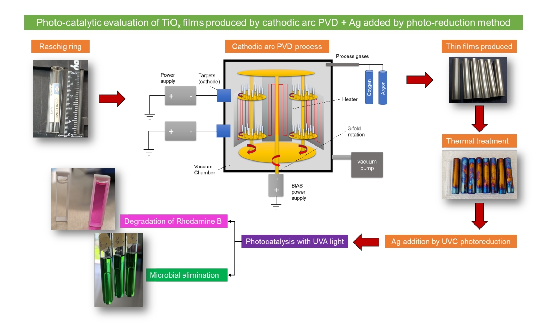
3.1. Synthesis of Thin Films by PVD on Borosilicate Raschig Rings

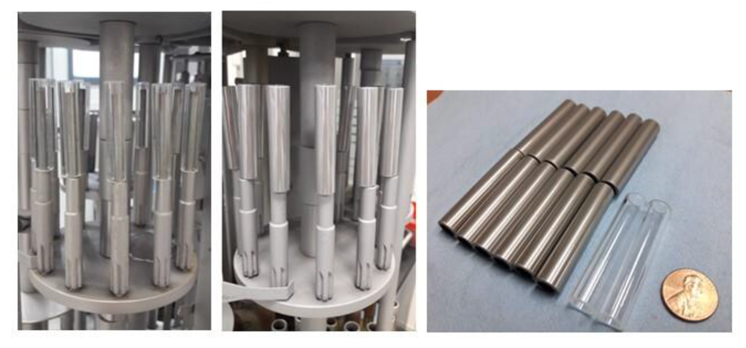
Commercial borosilicate Raschig rings with Ø = 10 mm, length = 50 mm, and wall thickness = 1.5 mm were used as substrates for TiOx deposition. First, the substrate rings were cleaned in an ultrasonic, using Elmasonic X-tra equipment (Singen, Germany) with an operating frequency of 45 KHz in ethanol for 15 min at room temperature. Then, they were dried and fixed in a 3-axis rotating planetary system for coating deposition, see left image in Figure 1. The rotating planetary system was introduced inside the coating chamber of the DOMINO mini semi-industrial equipment from OERLIKON (Bergisch Gladbach, Germany), where the TiOx coating was deposited applying the cathodic arc method, see the coated Rasching rings in the middle and right images in Figure 13.
Firstly, the rings are heated at ~450 °C using a 9 kW heating system and applying a continuous vacuum. The heating provides cleaning by evaporating residual moisture from the surface of the borosilicate rings. Before the coating step, the rings’ surface was cleaned with Ar+ ions produced through the arc enhanced glow discharge (AEGD) process. During the coating of the TiOx, a reaction takes place between the metallic Ti ions generated by erosion of the cathode material and the O ions. High purity gases are used for this process, oxygen (O2) with a purity of 99.998%, and Ar with a purity of 99.999%. The Ar and O2 flow rates were varied for each set of rings, and three sets of Ar/O2 ratios (440/60, 400/100, and 300/100) were selected and 11 Raschig rings were deposited during each coating run. The deposition time was maintained for 2 h by applying 80 V bias at a frequency of 20/80 KHz and a current of 150 A. Lastly, the coated samples were cooled inside the PVD chamber for ~90 min.
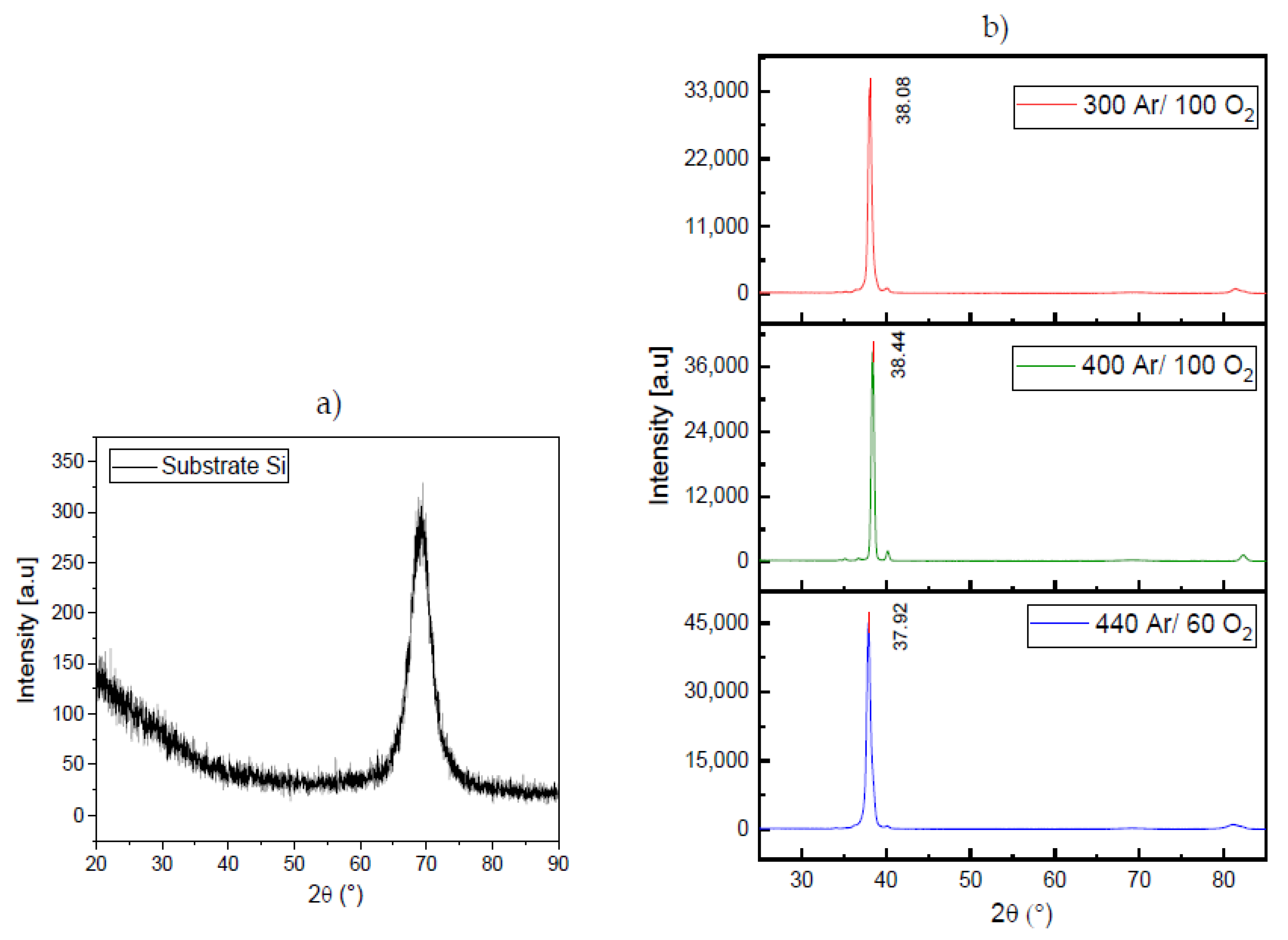
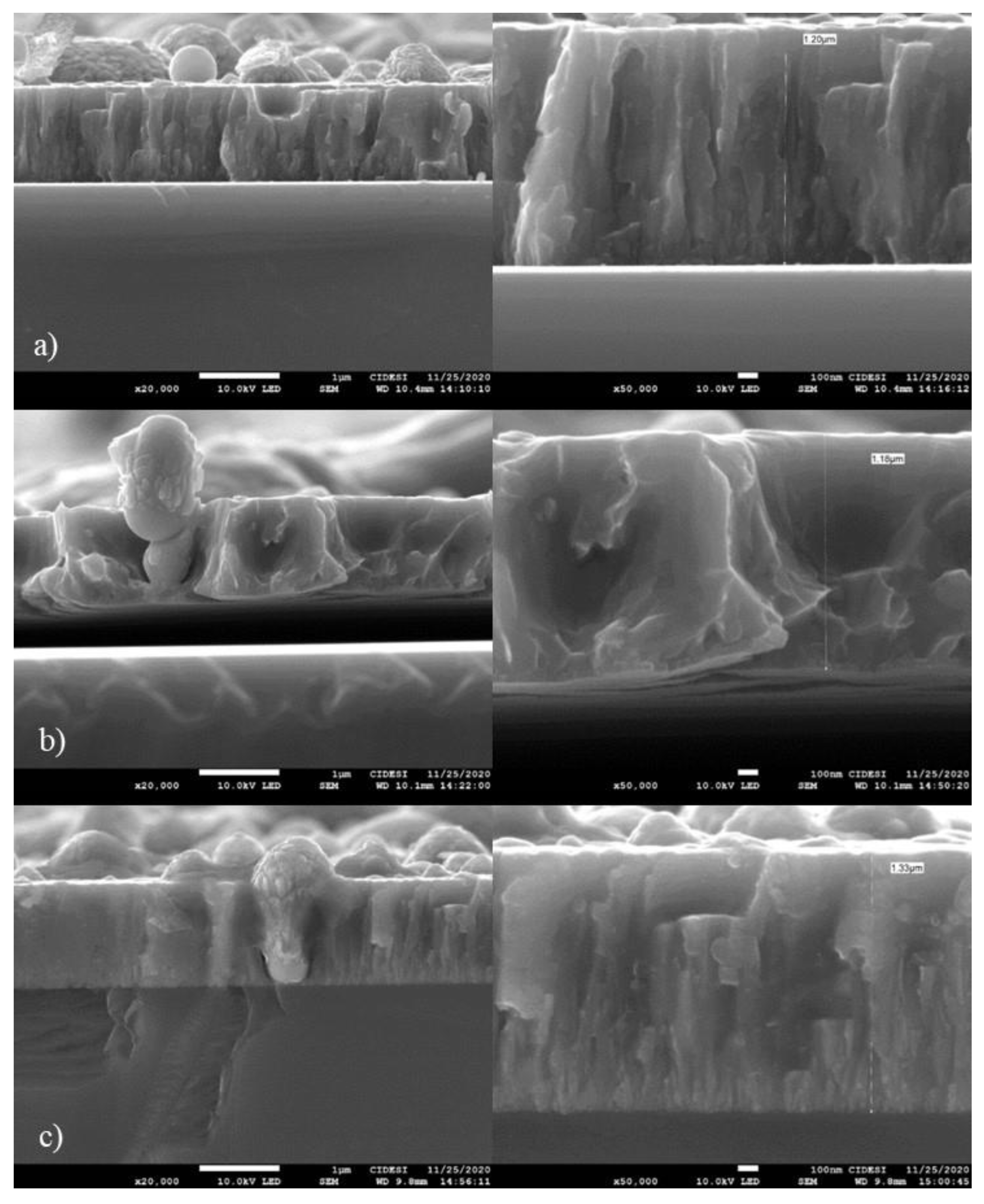
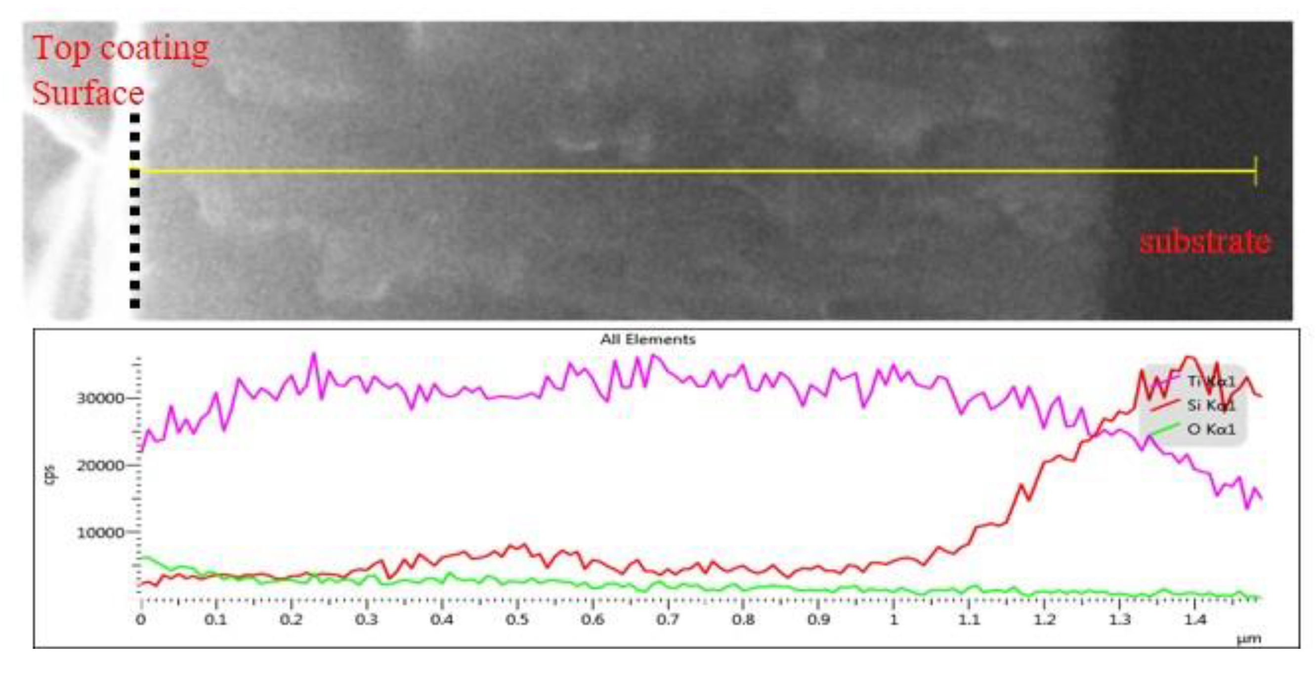
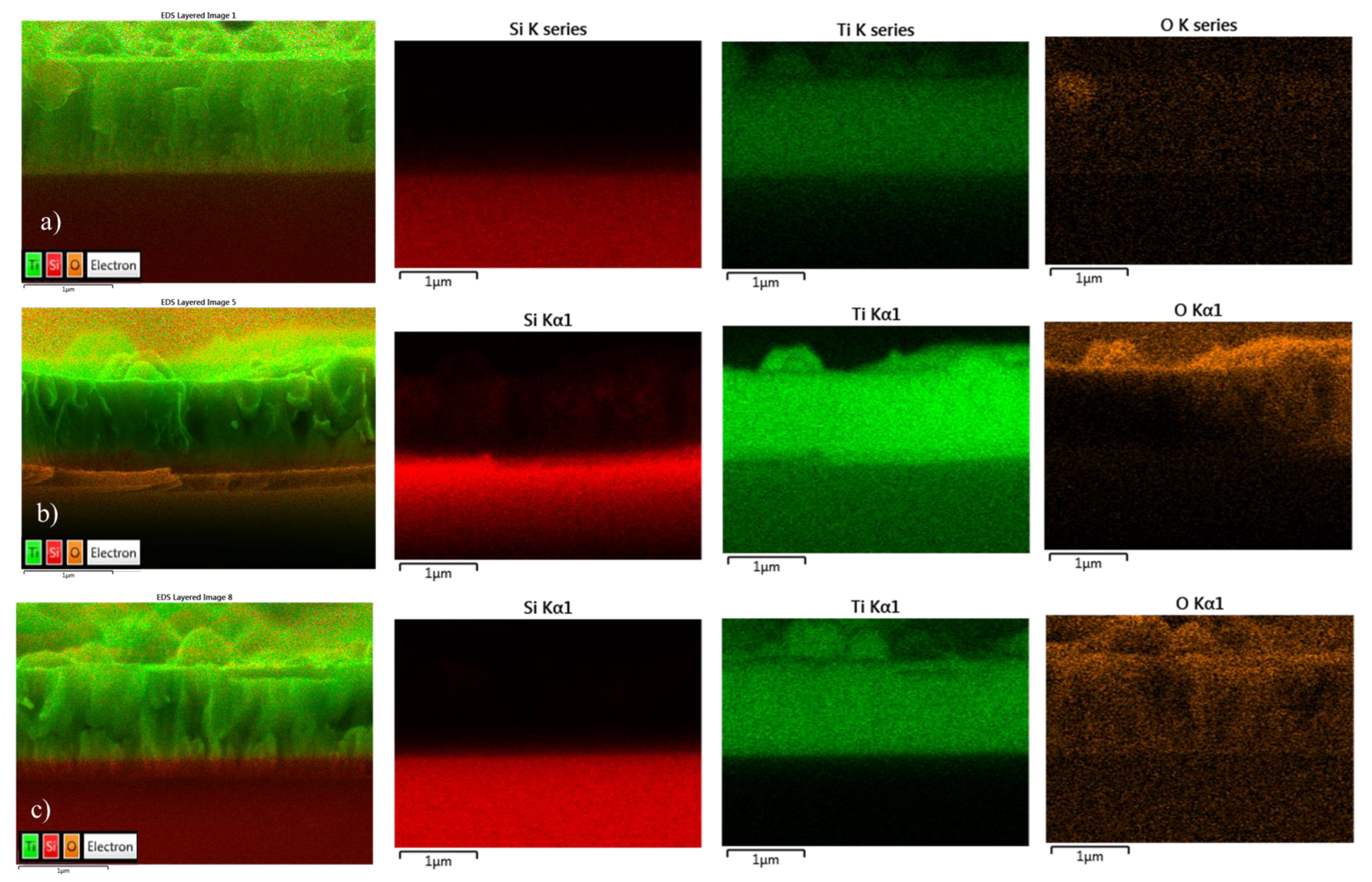
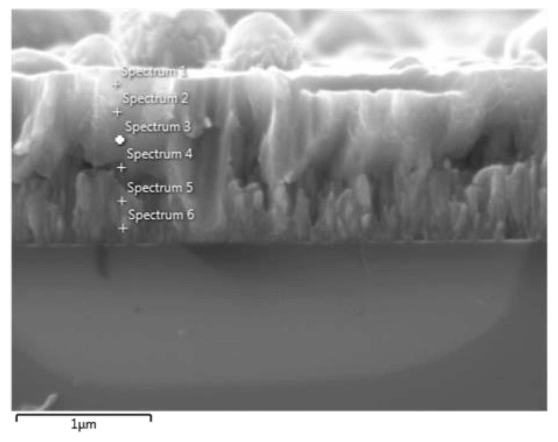
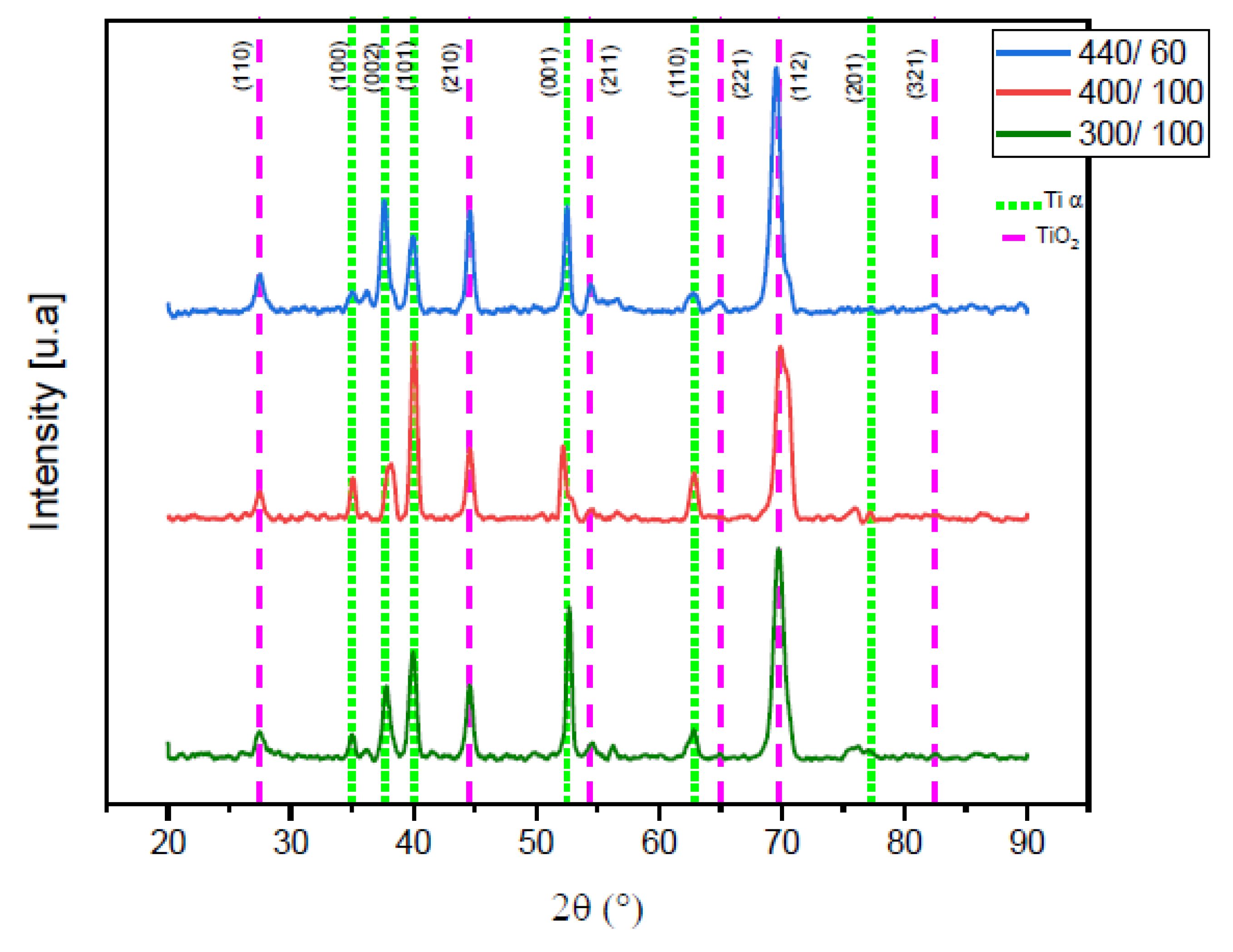
The ring sets were characterized by field emission scanning electron microscopy (FE-SEM) for surface analysis, thickness, and morphology of the TiOx films, X-ray energy dispersive spectrometry (EDS) for elemental analysis, and low angle X-ray diffraction (XRD) to identify crystalline phases in the coating. For this purpose, the XRD data were taken in a Rigaku diffractometer and by applying the parallel beam (PB) mode using Cu Kα radiation with a wavelength of 1.540593 Å, angle ω = 1°, an angular range from 20° to 90°, and a step size of 0.02°.

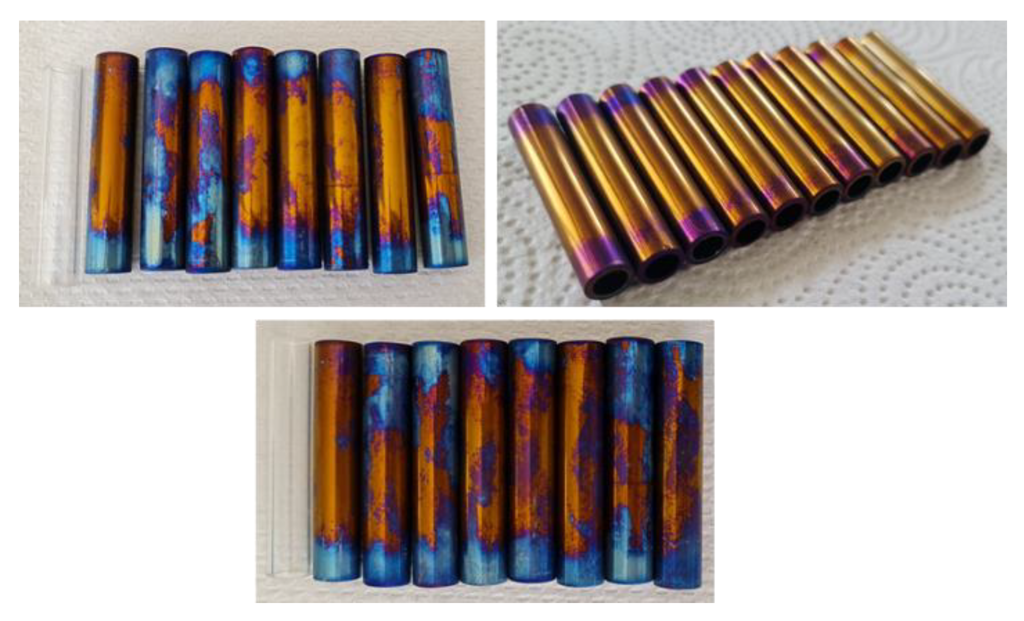
3.2. Heat Treatment after PVD Coating

The Raschig rings coated with the TiOx systems were heat-treated in a Thermo Scientific Lindberg Blue M furnace (Waltham, MA, USA) at 500 °C for 2 h. Using a heating ramp of 10 °C/min, this treatment was performed under standard conditions and air atmosphere. The heat treatment was performed in order to increase the O content on the surface of the coating according to [71,72]. Subsequently, the grazing incidence X-ray diffraction XRD method was applied to analyze the degree of oxidation and diffuse reflectance analysis was performed. Figure 14 shows the rings after heat treatment.

3.3. Doping of the TiOx with Silver

Silver (Ag) was added to the TiOx coating to activate it, and for this purpose, the photo-chemical reduction or photo-deposition methodology was applied, where the Ti/TiO2-coated rings were immersed twice for 5 min in a 1.7 wt. % AgNO3 aqueous solution with CAS number 7761-88-8. Subsequently, they were irradiated with a UVC source (λ = 254 nm) for 2 h to activate the impregnated Ag [24,73]. X-ray diffraction (XRD) and diffuse reflectance analysis were performed after this doping process.
Ag, is used, which is the most promising metal, serving as a conduction band electron attractor, increasing the hole-electron separation, and facilitating electron excitation by the creation of a local electric field [74,75]. In this way, e/h+ recombination is avoided. This property is due to the high reduction potential of the Ag+ ion (Ag+/ Ag0) producing metallic Ag on the TiO2 surface.

3.4. Dye Degradation Experiments

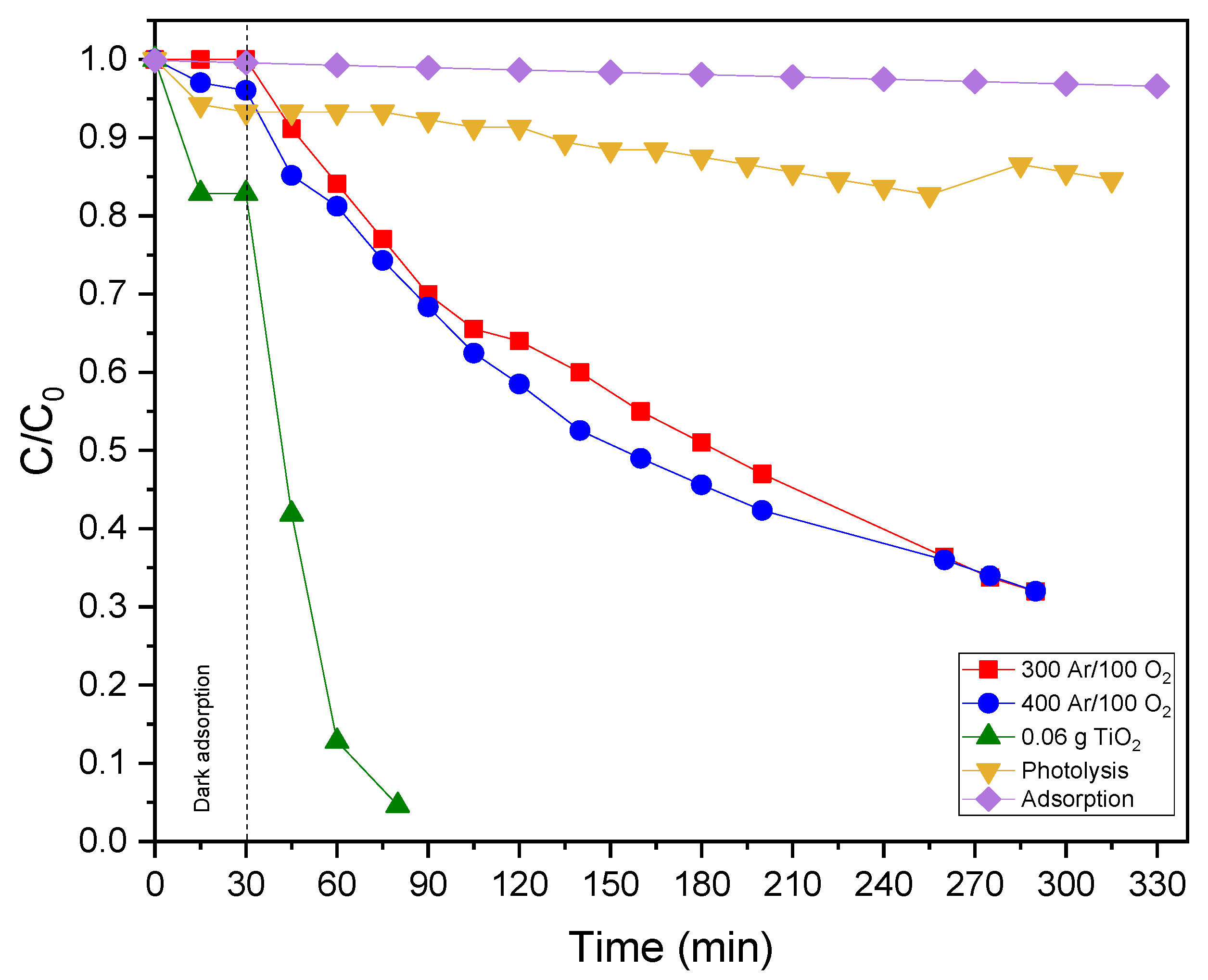
The photocatalytic activity of each set of rings coated with the Ag-doped TiO2 was evaluated in the degradation of rhodamine B (RhB) with CAS number 81-88-9. Firstly, the calibration curve of the rhodamine B dye was performed, taking as reference the wavelength of maximum absorption of the dye of 554 nm. The photocatalytic degradation reactions were carried out in the photocatalytic reactor where its geometry corresponds to a cylinder 90 cm long, with a diameter of 3 ½” made of polyvinyl chloride (PVC) and with a stainless steel reflecting surface. Inside the reactor there is a 20 W TecnoLite F20T8BLB UV-A lamp of short wavelength between 315 and 400 nm, which is inside a quartz tube of DExt = 3.2 cm, a wall thickness of 0.2 cm, and a length of 91.5 cm. The rings are attached to nylon threads from the top cover and are located in the free space between the quartz tube and the stainless steel surface, and the bottom is air inlet through an Elite 799 pump. The reactor operates in batch mode, with a volume of 2 l of a 0.5 ppm rhodamine B solution. In a typical photo-catalytic experiment, the aqueous solution was loaded into the reactor, left in the dark with stirring for 30 min to equilibrate, and then the UV-A lamp was turned on. Aliquots were taken at specific time intervals and analyzed using a BIOBASE BK-UV1800 spectrophotometer (Jinan, China). The obtained data were plotted to determine the reaction order. Subsequently, kinetic parameters, such as reaction rate, constant and half-life time, were calculated; this was done in order to evaluate the photo-catalytic efficiency of each set of TiO2-coated rings. In the reaction system, the following experimental arrangement was carried out in the degradation of rhodamine B: (a) use of immobilized TiO2 in the dark, (b) use of UV light lamp without immobilized TiO2 “photolysis”, (c) use of immobilized TiO2 with UV light lamp “photocatalysis”, and (d) degradation in the dark without catalyst.

3.5. Disinfection Experiments

The coated Ag-doped TiO2 rings were evaluated in the elimination of fecal coliforms. For these, two water matrices were used: one from the secondary treatment effluent (before chlorination process) of the wastewater treatment plant of GEA Ambiental, Morelia, Mexico, and synthetic water prepared in the laboratory.
For the preparation of synthetic water, a sample was taken from the secondary treatment effluent of the GEA Ambiental plant and bacterial growth was performed in MacConkey broth medium. After incubation for 24–48 h at 35 °C, bacterial cells were transferred to 50 mL conical polypropylene centrifuge tubes with flat caps and collected by centrifugation at 4000 rpm for 5 min at room temperature. The supernatant was discarded and the sediment was resuspended in sterile phosphate solution or 0.9% NaCl normal saline. This solution is called microbial stock solution [76]. To obtain the synthetic water to be used in photocatalytic disinfection, ~ 20 mL of the microbial stock solution is resuspended in 2 L of sterile distilled water and mixed with magnetic stirring.
In a typical experiment, the inoculated aqueous solution was loaded into the reactor, left in the dark with stirring for 30 min to equilibrate, and then exposed to UV-A radiation. An aliquot was extracted at the beginning and end of the process, which was after 4 h. In order to evaluate the percentage of elimination of fecal coliforms, both aliquots were analyzed by means of the microbiological test of the most probable number in multiple tubes (MPN) for fecal coliforms following the Mexican standard NMX-AA-042-SCFI-2015.
The following tests are performed in the reactor in the elimination of fecal coliforms: (a) use of immobilized TiO2 in darkness, (b) use of UV light lamp without immobilized TiO2 “photolysis”, (c) use of immobilized TiO2 with UV light lamp “photocatalysis” and (d) degradation in the darkness without catalyst.

4. Conclusions

Heterogeneous photocatalysis stands out among advanced oxidation processes (AOPs) as a promising and effective biocidal technique, with titanium dioxide (TiO2) being the most common catalyst employed for the purification of aqueous matrices. However, one of the main obstacles to putting it into practice is the separation of photocatalysts and the design of photocatalytic reactors, as most of the synthesized photocatalysts are in the form of powders. Alternatively, TiO2 can be fixed on a solid support, eliminating the need to add a catalyst separation process. In this case, it should be noted that the process offers lower efficiencies due to the smaller surface area of catalysts exposed to light and molecules to be removed.
The Ag-TiOx photocatalyst deposited with 400 Ar/100 O2 degraded ~68.18%, and the catalytic coating deposited with 300 Ar/100 O2 degraded ~68.02% of the rhodamine B dye at a concentration of 0.5 ppm in 2 L volume. In the disinfection, the TiOx 300 Ar/100 O2 coating had a fecal coliform removal of 83.69% and the TiOx 400 Ar/100 O2 coating removed 90.32%.
The results obtained cannot be directly compared with others because each of them used different reaction conditions, such as type of illumination, concentration of TiO2 and rhodamine B, reactor design, catalyst immobilization method, and coating substrate. All these parameters are factors that have an effect on the kinetics of rhodamine B degradation and bacterial killing. However, they are positive results on rhodamine B degradation for TiOx-Ag thin films if the results obtained for TiO2-Ag thin films synthesized with a sol-gel method deposited by dip coating on the Ag substrates and photo-chemical deposition shown in Table 1 are known.
  • Targeting the application of fixed photo-catalysts, TiOx coatings were deposited by cathodic arc physical vapor deposition on borosilicate Raschig rings. The deposit was designed for O grading using flow ratios of 440 Ar/60 O2, 400 Ar/100 O2, and 300 Ar/100 O2 in a semi-industrial coater unit. The arc-PVD plasma was stable in all Ar/O2 mixtures.
  • In the as-coated state, the TiOx catalysts are composed of pure Ti in the α phases. The EDS analysis showed that O and Ti varies in the depth of the coating, suggesting the formation of a system with a low amount of O producing a nonstoichiometric oxide (TiOx). A heat treatment was needed in order to increase the amount of O in the coating and build the crystalline TiO2 rutile phase.
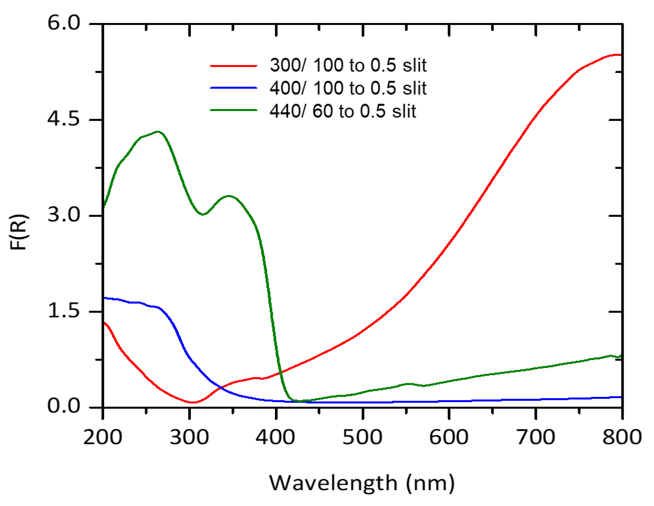
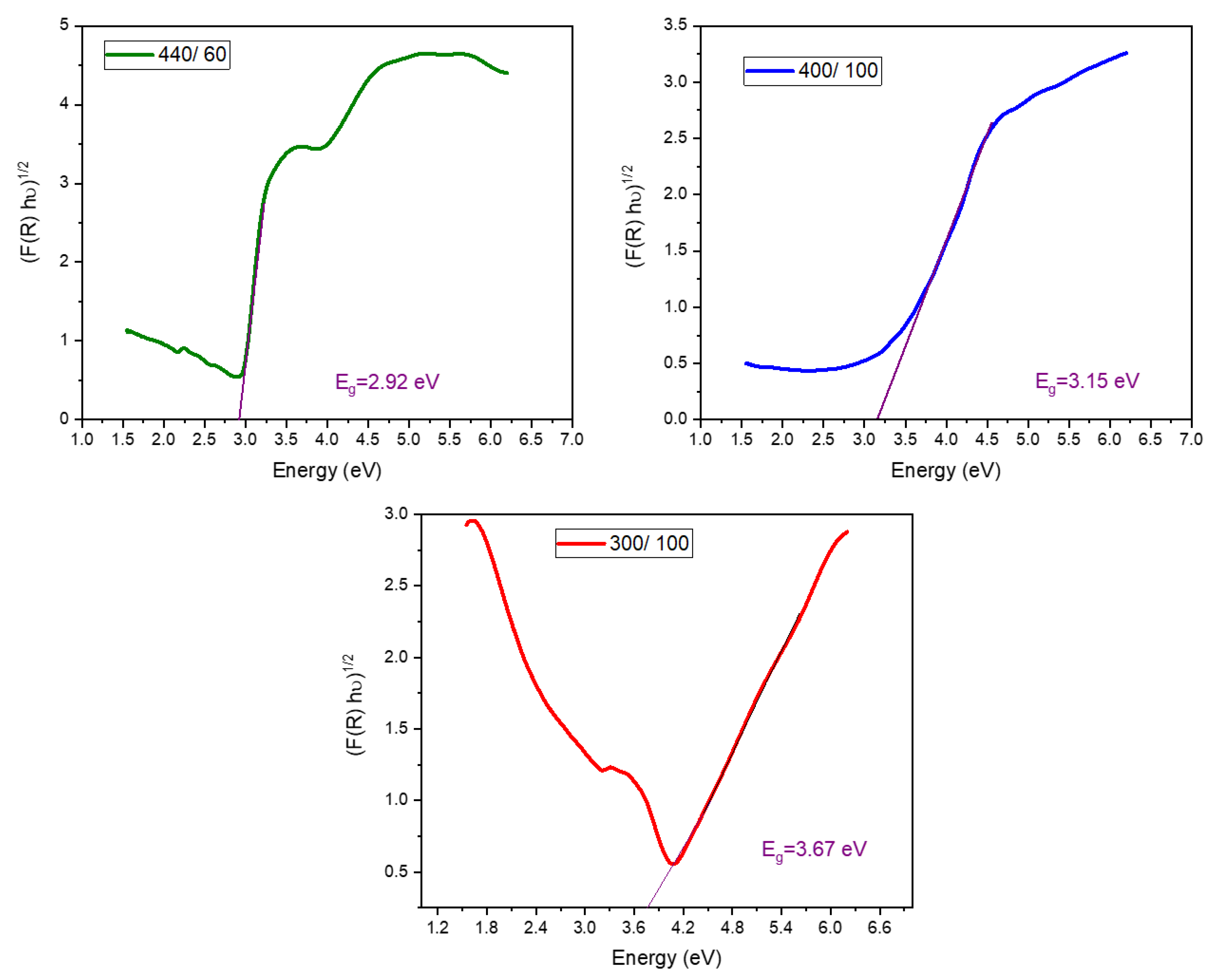
  • After the thermal treatment, the analysis by UV vis diffuse reflectance spectroscopy indicated that the TiOx catalytic coatings using 400 Ar/100 O2 and 300 Ar/100 O2 are activated under UVA radiation, while the coating 440 Ar/60 O2 absorbs in the visible spectrum.
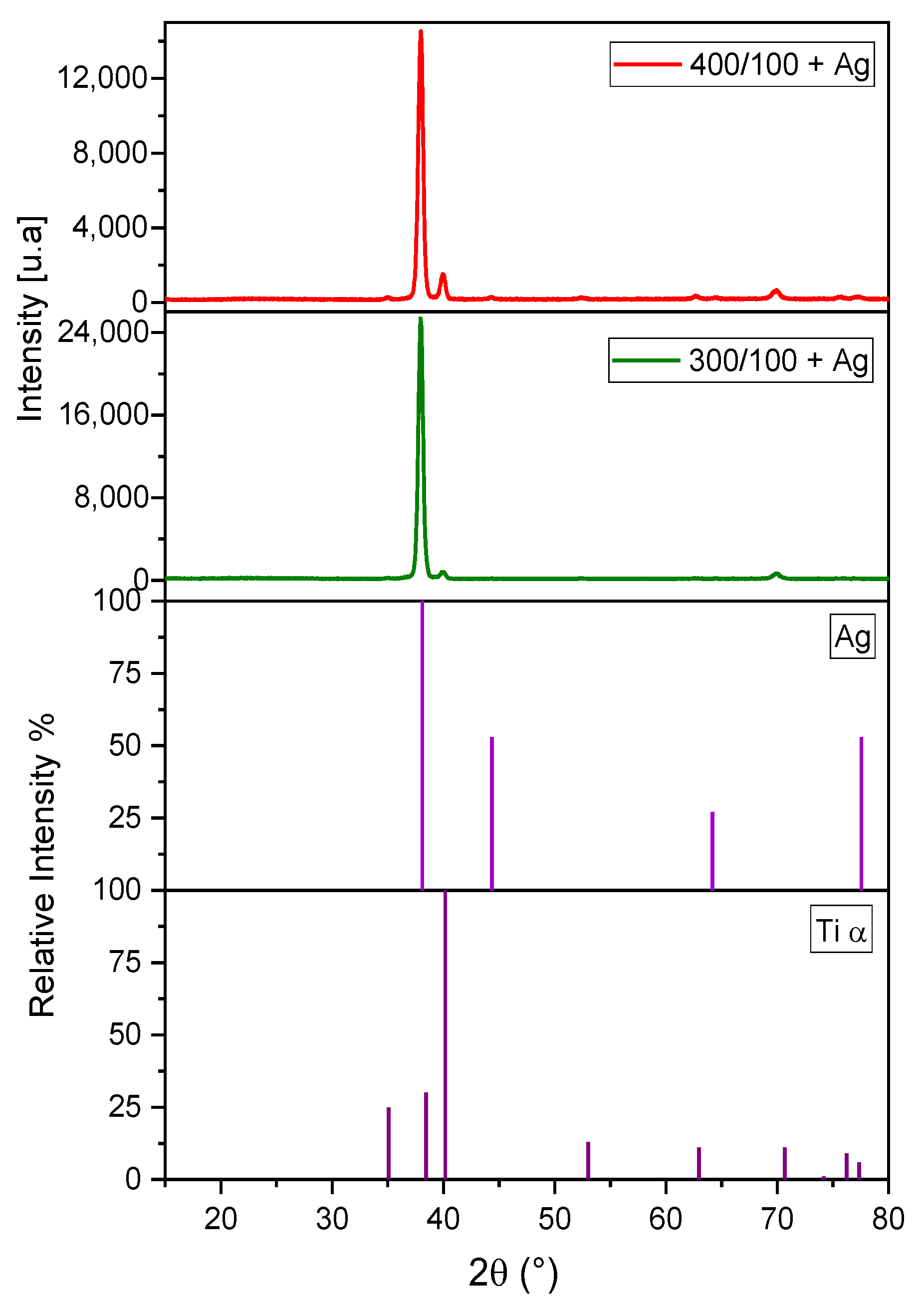
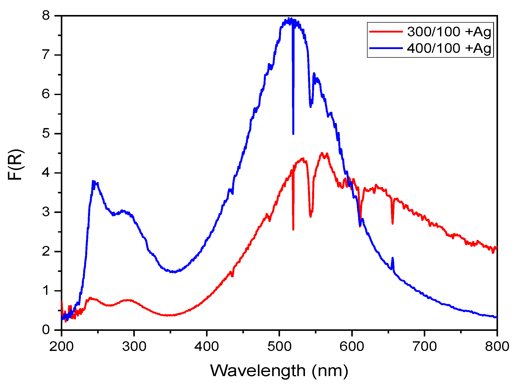
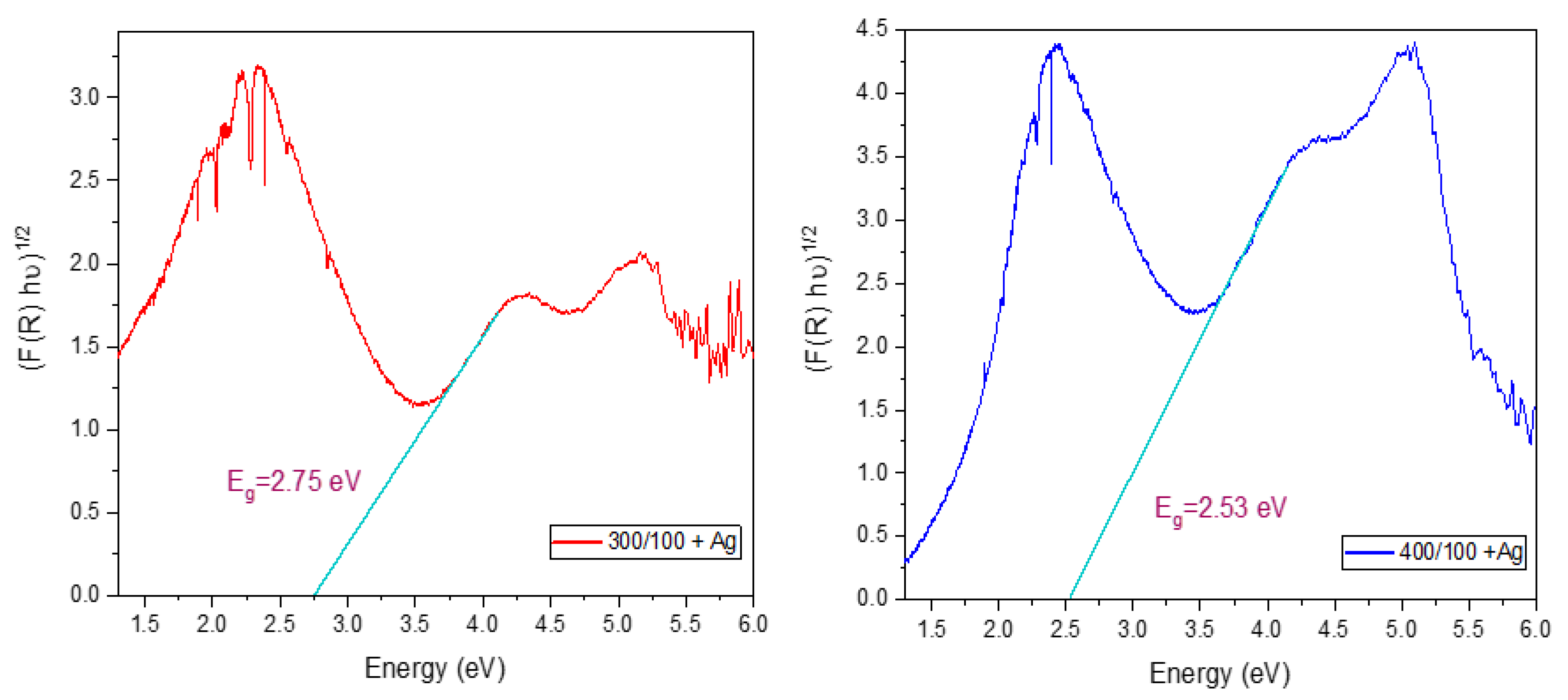
  • Silver, confirmed by XRD, was added by the UVC photo-reduction method in the TiOx photo-catalysts deposited using 400 Ar/100 O2 and 300 Ar/100 O2 sccm sets. In this Ag-TiOx photo-catalyst, a decrease in the bandgap energy was observed, thus showing moderate photo-catalytic activity in the degradation of rhodamine B at 0.5 ppm and the removal of fecal coliforms from a secondary treatment effluent and synthetic water.

Author Contributions

Conceptualization, F.U.-M., G.C.M.-R. and R.H.-A.; investigation, A.Y.R.-T.; resources, E.M.R.-M. and F.J.M.; writing—original draft preparation, A.Y.R.-T.; writing—review and editing, R.H.-A. and G.C.M.-R.; visualization, G.C.M.-R.; supervision, J.A.-C.; funding acquisition, R.H.-A. All authors have read and agreed to the published version of the manuscript.

Funding

Financial resources and materials were partially provided through the FORDECYT/247/2018 project 297265 (Fortalecimiento de las Capacidades de Investigación, Desarrollo e Innovación del CIDESI para Atender las Necesidades Científico-Tecnológicas en Manufactura Aditiva de la Industria en la Región Centro-Norte de México), FORDECYT 296384 (Operación de consorcio CONACyT en manufactura aditiva), and the 2015-02-1077 (Estudio y Entendimiento de la Degradación y Estabilidad de Sistemas de Recubrimientos Duros Lubricantes para Aplicaciones de Mediana y Alta Temperatura) Frontiers of Science program managed by CONACYT. Dr. R. Huirache-Acuña thanks to CIC 2022 UMSNH Project.

Acknowledgments

The authors thank the access provided to the PVD coater unit and characterization equipment of the National Lab.- CENAPROT in CIDESI-Querétaro. G.C. Mondragón-Rodríguez, thank the Investigadoras e Investigadores por México program (previously Cátedras-CONACYT) sponsored by CONACYT. Thanks to J. Martín Yañez Limón for facilitating the access to the UV-vis equipment at CINVESTAV in Querétaro, Mexico. R. Huirache-Acuña thanks to B. Millán-Malo and C. Peza-Ledesma from CFATA-UNAM for their XRD Technical support.

Conflicts of Interest

The authors declare no conflict of interest.

References

  1. Naffouti, W.; Nasr, T.B.; Mehdi, A.; Kamoun-Turki, N. Effect of sprayed solution flow rate on the physical properties of anatase TiO2 thin films. J. Electron. Mater. 2014, 43, 4033–4040. [Google Scholar] [CrossRef]
  2. Giordano, F.; Abate, A.; Correa, J.P.; Saliba, M.; Matsui, T.; Im, S.H.; Graetzel, M. Enhanced electronic properties in mesoporous TiO2 via lithium doping for high-efficiency perovskite solar cells. Nat. Commun. 2016, 7, 10379. [Google Scholar] [CrossRef] [PubMed]
  3. Park, J.; Jin, T.; Liu, C.; Li, G.; Yan, M. Three-Dimensional Graphene-TiO2 Nanocomposite Photocatalyst Synthesized by Covalent Attachment. ACS Omega 2016, 1, 351–356. [Google Scholar] [CrossRef]
  4. Rane, S.S.; Kajale, D.A.; Arbuj, S.S.; Rane, S.B.; Gosavi, S.W. Hydrogen, ethanol and ammonia gas sensing properties of nano-structured titanium dioxide thick films. J. Mater. Sci. Mater. Electron. 2017, 28, 9011–9016. [Google Scholar] [CrossRef]
  5. Lanin, S.N.; Vlasenko, E.V.; Kovaleva, N.V.; Zung, F.T. The adsorption properties of titanium dioxide. Russ. J. Phys. Chem. 2008, 82, 2152–2155. [Google Scholar] [CrossRef]
  6. Nunes, D.; Fortunato, E.; Martins, R. Flexible nanostructured TiO2-based gas and UV sensors: A review. Discov. Mater. 2022, 2, 2. [Google Scholar] [CrossRef]
  7. Ding, S.N.; Gao, B.H.; Shan, D.; Sun, Y.M.; Cosnier, S. TiO2 nanocrystals electrochemiluminescence quenching by biological enlarged nanogold particles and its application for biosensing. Biosens. Bioelectron. 2013, 39, 342–345. [Google Scholar] [CrossRef]
  8. Mun, K.S.; Alvarez, S.D.; Choi, W.Y.; Sailor, M.J. A stable, label-free optical interferometric biosensor based on TiO2 Nanotube Arrays. ACS Nano 2010, 4, 2070–2076. [Google Scholar] [CrossRef]
  9. Hou, Z.; Zhang, Y.; Deng, K.; Chen, Y.; Li, X.; Deng, X.; Cheng, Z.; Lian, H.; Li, C.; Lin, J. UV-emitting upconversion-based TiO2 photosensitizing nanoplatform: Near-infrared light mediated in vivo photodynamic therapy via mitochondria-involved apoptosis pathway. ACS Nano 2015, 9, 2584–2599. [Google Scholar] [CrossRef]
  10. Deepagan, V.G.; You, D.G.; Um, W.; Ko, H.; Kwon, S.; Choi, K.Y.; Yi, G.R.; Lee, J.Y.; Lee, D.S.; Kim, K.; et al. Long-Circulating Au-TiO2 Nanocomposite as a Sonosensitizer for ROS-Mediated Eradication of Cancer. Nano Lett. 2016, 16, 6257–6264. [Google Scholar] [CrossRef]
  11. Chinnamuthu, P.; Dhar, J.C.; Mondal, A.; Bhattacharyya, A.; Singh, N.K. Ultraviolet detection using TiO2 nanowire array with Ag Schottky contact. J. Phys. D Appl. Phys. 2012, 45, 135102–135105. [Google Scholar] [CrossRef]
  12. Zhang, H.; Ruan, S.; Li, H.; Zhang, M.; Lv, K.; Feng, C.; Chen, W. Schottky diode ultraviolet detector based on TiO2 nanowire array. IEEE Electron. Device Lett. 2012, 33, 83–85. [Google Scholar] [CrossRef]
  13. Yu, H.; Chen, J.; Wang, W.; Dai, Q. Optical waveguide lightmode spectroscopy (OWLS) as a sensor for thin film and quantum dot corrosion. Sensors 2012, 12, 17330–17342. [Google Scholar] [CrossRef]
  14. Wang, D.; Li, X.; Chen, J.; Tao, X. Enhanced photoelectrocatalytic activity of reduced graphene oxide/TiO2 composite films for dye degradation. Chem. Eng. J. 2012, 198, 547–554. [Google Scholar] [CrossRef]
  15. O’Regan, B.; Grätzel, M. A low-cost, high-efficiency solar cell based on dye-sensitized colloidal TiO2 films. Nature 1991, 353, 737–740. [Google Scholar] [CrossRef]
  16. Morozova, M.; Kluson, P.; Krysa, J.; Zlamal, M.; Solcova, O.; Kment, S.; Steck, T. Role of the template molecular structure on the photo-electrochemical functionality of the sol-gel titania thin films. J. Sol-Gel Sci. Technol. 2009, 52, 398–407. [Google Scholar] [CrossRef]
  17. Kment, S.; Hubicka, Z.; Kmentova, H.; Kluson, P.; Krysa, J.; Gregora, I.; Morozova, M.; Cada, M.; Petras, D.; Dytrych, P.; et al. Photoelectrochemical properties of hierarchical nanocomposite structure: Carbon nanofibers/TiO2/ZnO thin films. Catal. Today 2011, 161, 8–14. [Google Scholar] [CrossRef]
  18. Kment, S.; Kluson, P.; Hubicka, Z.; Krysa, J.; Cada, M.; Gregora, I.; Deyneka, A.; Remes, Z.; Zabova, H.; Jastrabik, L. Double hollow cathode plasma jet-low temperature method for the TiO2-xNx photoresponding films. Electrochim. Acta 2010, 55, 1548–1556. [Google Scholar] [CrossRef]
  19. Kment, S.; Kluson, P.; Bartkova, H.; Krysa, J.; Churpita, O.; Cada, M.; Virostko, P.; Kohout, M.; Hubicka, Z. Advanced methods for titanium (IV) oxide thin functional coatings. Surf. Coat. Technol. 2008, 202, 2379–2383. [Google Scholar] [CrossRef]
  20. Karuppuchamy, S.; Nonomura, K.; Yoshida, T.; Sugiura, T.; Minoura, H. Cathodic electrodeposition of oxide semiconductor thin films and their application to dye-sensitized solar cells. Solid State Ion. 2002, 151, 19–27. [Google Scholar] [CrossRef]
  21. Djošić, M.; Mišković-Stanković, V.; Janaćković, D.T.; Kačarević-Popović, Z.; Petrović, R. Electrophoretic deposition and characterization of boehmite coatings on titanium substrate. Colloids Surf. 2006, 274, 185–191. [Google Scholar] [CrossRef]
  22. Liu, S.; Chen, X. Preparation and characterization of a novel activated carbon-supported N-doped visible light response photocatalyst (TiO2 − xNy/AC). J. Chem. Technol. Biotechnol. 2007, 82, 453–459. [Google Scholar] [CrossRef]
  23. Okada, K.; Takamatsu, Y.; Tokudome, Y.; Nakahira, A.; Takahashi, M. Highly oriented growth of titanate nanotubes (TNTs) in micro and confinement spaces on sol-gel derived amorphous TiO2 thin films under moderate hydrothermal condition. J. Sol-Gel Sci. Technol. 2013, 65, 36–40. [Google Scholar] [CrossRef]
  24. Kment, S.; Kmentova, H.; Hubicka, Z.; Olejnicek, J.; Cada, M.; Krysa, J. Enhanced photocatalytic activity of silver-doped nanoparticulate TiO2 thin films with respect to the method of doping. Res. Chem. Intermed. 2015, 41, 9343–9355. [Google Scholar] [CrossRef]
  25. Muaz, A.K.M.; Hashim, U.; Ibrahim, F.; Thong, K.L.; Mohktar, M.S.; Liu, W.W. Effect of annealing temperatures on the morphology, optical and electrical properties of TiO2 thin films synthesized by the sol–gel method and deposited on Al/TiO2/SiO2/p-Si. Microsyst. Technol. 2016, 22, 871–881. [Google Scholar] [CrossRef]
  26. Chen, Y.; Wang, K.; Lou, L. Photodegradation of dye pollutants on silica gel supported TiO2 particles under visible light irradiation. J. Photochem. Photobiol. 2004, 163, 281–287. [Google Scholar] [CrossRef]
  27. Tonejc, A.M.; Turković, A.; Gotić, M.; Musić, S.; Vuković, M.; Trojko, R.; Tonejc, A. HREM, TEM and XRD observation of nanocrystalline phases in TiO2 obtained by the sol-gel method. Mater. Lett. 1997, 31, 127–131. [Google Scholar] [CrossRef]
  28. Kavaliunas, V.; Sestakauskaite, A.; Sriubas, M.; Laukaitis, G. Influence of Deposition Parameters on the Structure of TiO Thin Films Prepared by Reactive Magnetron Sputtering Technique. Lect. Notes Netw. Syst. 2019, 53, 49–57. [Google Scholar] [CrossRef]
  29. Sobrinho, V.S.S.; Neto, J.Q.M.; Lima, L.L.F.; Souza, I.A.; Libório, M.S.; Queiroz, J.C.A.; Sousa, R.R.M.; Almeida, E.O.; Feitor, M.C.; Costa, T.H.C. Optical-Electrical Properties and Thickness Analysis of TiO2 Thin Films Obtained by Magnetron Sputtering. Braz. J. Phys. 2020, 50, 771–779. [Google Scholar] [CrossRef]
  30. Horprathum, M.; Eiamchai, P.; Chindaudom, P.; Pokaipisit, A.; Limsuwan, P. Oxygen partial pressure dependence of the properties of TiO2 thin films deposited by DC reactive magnetron sputtering. Procedia Eng. 2012, 32, 676–682. [Google Scholar] [CrossRef] [Green Version]
  31. Guo, D.; Ito, A.; Goto, T.; Tu, R.; Wang, C.; Shen, Q.; Zhang, L. Preparation of rutile TiO2 thin films by laser chemical vapor deposition method. J. Adv. Ceram. 2013, 2, 162–166. [Google Scholar] [CrossRef]
  32. Wang, S.; Wang, K.; Jehng, J.; Liu, L. Preparation of TiO2/MCM-41 by plasma enhanced chemical vapor deposition method and its photocatalytic activity. Front. Environ. Sci. Eng. 2011, 6, 304–312. [Google Scholar] [CrossRef]
  33. Naffouti, W.; Jrad, A.; Nasr, T.B.; Ammar, S.; Turki-Kamoun, N. Structural, morphological and optical properties of TiO2: Mn thin films prepared by spray pyrolysis technique. J. Mater. Sci. Mater. Electron. 2016, 27, 4622–4630. [Google Scholar] [CrossRef]
  34. Doubi, Y.; Hartiti, B.; Labrim, H.; Fadili, S.; Tahri, M.; Belafhaili, A.; Siadat, M.; Thevenin, P. Experimental study of properties of TiO2 thin films deposited by spray pyrolysis for future sensory applications. Appl. Phys. A 2021, 127, 475. [Google Scholar] [CrossRef]
  35. Lahiri, R.; Ghosh, A.; Dwivedi, S.M.M.D.; Chakrabartty, S.; Chinnamuthu, P.; Mondal, A. Performance of Erbium-doped TiO2 thin film grown by physical vapor deposition technique. Appl. Phys. A Mater. Sci. Processing 2017, 123, 573. [Google Scholar] [CrossRef]
  36. Shubham, K.; Chakrabarti, P. Fabrication and characterization of metal/insulator/semiconductor structures based on TiO2 and TiO2/SiO2 thin films prepared by low-temperature arc vapor deposition. Electron. Mater. Lett. 2014, 10, 579–584. [Google Scholar] [CrossRef]
  37. Yap, Y.K.; Zhang, D. Physical Vapor Deposition. In Encyclopedia of Nanotechnology; Bhushan, B., Ed.; Springer: Dordrech, The Netherlands, 2015; pp. 1–8. [Google Scholar] [CrossRef]
  38. György, G.; Socol, E.; Axente, I.N.; Mihailescu, C.; Ducu, C.; Ciuca, S. Anatase phase TiO2 thin films obtained by pulsed laser deposition for gas sensing applications. Appl. Surf. Sci. 2005, 247, 429–433. [Google Scholar] [CrossRef]
  39. Brunella, M.; Diamanti, M.; Pedeferri, M.; Fonzo, F.; Casari, C.; Bassi, A. Photocatalytic behavior of different titanium dioxide layers. Thin Solid Film. 2007, 515, 6309–6313. [Google Scholar] [CrossRef]
  40. Sahm, H.; Zywitzki, O.; Glöß, D.; Modes, T.; Frach, P.; Goedicke, K. Structure and properties of crystalline titanium oxide layers deposited by reactive pulse magnetron sputtering. Surf. Coat. Technol. 2004, 180, 538–543. [Google Scholar]
  41. Heo, C.; Lee, S.; Boo, J. Deposition of TiO2 thin films using RF magnetron sputtering method and study of their surface characteristics. Thin Solid Film. 2005, 475, 183–188. [Google Scholar] [CrossRef]
  42. Zhang, F.; Wang, X.; Li, C.; Wang, H.; Chen, L.; Liu, X. Rutile-type titanium oxide films synthesized by filtered arc deposition. Surf. Coat. Technol. 1998, 110, 136–139. [Google Scholar] [CrossRef]
  43. Takikawa, H.; Matsui, T.; Sakakibara, A.; Bendavid, A.; Martin, P. Properties of titanium oxide film prepared by reactive cathodic vacuum arc deposition. Thin Solid Film. 1999, 348, 145–151. [Google Scholar] [CrossRef]
  44. Attenberger, W.; Thorwarth, G.; Manova, D.; Mändl, S.; Stritzker, B.; Rauschenbach, B. Interface properties of TiO2 on Si formed by simultaneous implantation and deposition of titanium and oxygen ions. Surf. Coat. Technol. 2001, 142, 412–417. [Google Scholar] [CrossRef]
  45. Leng, Y.X.; Chen, J.Y.; Sun, H.; Yang, P.; Wan, G.J.; Wang, J.; Huang, N. Properties of titanium oxide synthesized by pulsed metal vacuum arc deposition. Surf. Coat. Technol. 2004, 176, 141–147. [Google Scholar] [CrossRef]
  46. Zheng, P.; Wu, H.; Guo, J.; Dong, J.; Jia, S.; Zhu, Z. P–N co-doping induced structural recovery of TiO2 for overall water splitting under visible light irradiation. J. Alloy. Compd. 2014, 615, 79–83. [Google Scholar] [CrossRef]
  47. Noguez, C. Surface Plasmons on Metal Nanoparticles: The Influence of Shape and Physical Environment. J. Phys. Chem. C 2007, 111, 3606–3619. [Google Scholar] [CrossRef]
  48. Trabelsi, K.; Hajjaji, A.; Ka, I.; Gaidi, M.; Bessais, B.; el Khakani, M.A. Optoelectronic and photocatalytic properties of in situ platinum-doped TiO2 films deposited by means of pulsed laser ablation technique. J. Mater. Sci. Mater. Electron. 2017, 28, 3317–3324. [Google Scholar] [CrossRef]
  49. Hassan, A.; Kayani, Z.N.; Anwar, M. Effect of Au ions on structural, optical, magnetic, dielectric, and antibacterial properties of TiO2 dip-coated thin films. J. Mater. Sci. Mater. Electron. 2021, 32, 14398–14419. [Google Scholar] [CrossRef]
  50. Khan, S.; ul Haq, M.; Ma, Y.; Nisar, M.; Li, Y.; Khan, R.; Han, G.; Liu, Y. Structural and optical properties of macroporous Ag@TiO2 thin films prepared by a facile one-step sol–gel method. J. Sol-Gel Sci. Technol. 2019, 93, 273–280. [Google Scholar] [CrossRef]
  51. Heiba, Z.K.; Mohamed, M.B.; Badawi, A. Structural and Optical Characteristic of Cu-Doped TiO2 Thin Film. J. Inorg. Organomet. Polym. Mater. 2022, 32, 2853–2862. [Google Scholar] [CrossRef]
  52. Balakrishnan, A.; Appunni, S.; Chinthala, M.; Vo, D.V.N. Biopolymer-supported TiO2 as a sustainable photocatalyst for wastewater treatment: A review. Environ. Chem. Lett. 2022, 3, 1–28. [Google Scholar] [CrossRef]
  53. Obregón, S.; Rodríguez-González, V. Photocatalytic TiO2 thin films and coatings prepared by sol–gel processing: A brief review. J. Sol-Gel Sci. Technol. 2021, 102, 125–141. [Google Scholar] [CrossRef]
  54. ACLAC. Economic Commission for Latin America and the Caribbean. 2004. Available online: https://observatoriop10.cepal.org/es/tratados/convenio-estocolmo-contaminantes-organicos-persistentes (accessed on 3 March 2022).
  55. Anjaneyulu, R.B.; Mohan, B.S.; Naidu, G.P.; Muralikrishna, R. Visible light enhanced photocatalytic degradation of methylene blue by ternary nanocomposite, MoO3/Fe2O3/rGO. J. Asian Ceram. Soc. 2018, 6, 183–195. [Google Scholar] [CrossRef]
  56. Klut, M.E.; Bisalputra, T.; Antia, N.J. The use of fluorochromes in the cytochemical characterization of some phytoflagellates. Histochem. J. 1988, 20, 35–40. [Google Scholar] [CrossRef] [PubMed]
  57. Rani, S.A.; Pitts, B.; Stewart, P.S. Rapid Diffusion of Fluorescent Tracers into Staphylococcus epidermidis Biofilms Visualized by Time Lapse Microscopy. Antimicrob. Agents Chemother. 2005, 49, 728–732. [Google Scholar] [CrossRef] [PubMed]
  58. Rinne, W.E.; Deacon, J.E. Fluorescent Pigment and Immersion Stain Marking Techniques for Lepidomeda mollispinis and Cyprinodon nevadensis. Trans. Am. Fish. Soc. 1973, 102, 459462. [Google Scholar] [CrossRef]
  59. Sagoo, S.K.; Jockusch, R.A. The fluorescence properties of cationic rhodamine B in the gas phase. J. Photochem. Photobiol. A Chem. 2011, 220, 173–178. [Google Scholar] [CrossRef]
  60. Wilhelm, P.; Stephan, D. Photodegradation of rhodamine B in aqueous solution via SiO2@TiO2 nano-spheres. J. Photochem. Photobiol. A Chem. 2007, 185, 19–25. [Google Scholar] [CrossRef]
  61. Zhao, X.; Zhu, Y. Synergetic Degradation of Rhodamine B at a Porous ZnWO 4 Film Electrode by Combined Electro-Oxidation and Photocatalysis. Environ. Sci. Technol. 2006, 40, 3367–3372. [Google Scholar] [CrossRef]
  62. Mahmood, M.A.; Baruah, S.; Anal, A.K.; Dutta, J. Microbial Pathogen Inactivation Using Heterogeneous Photocatalysis. In Environmental Chemistry for a Sustainable World; Springer: Dordrech, The Netherlands, 2012; pp. 511–541. [Google Scholar]
  63. Hossain, M.A.; Elias, M.; Sarker, D.R.; Diba, Z.R.; Mithun, J.M.; Azad, M.A.K.; Siddiquey, I.A.; Rahman, M.M.; Uddin, J.; Uddin, M. Synthesis of Fe- or Ag-doped TiO2–MWCNT nanocomposite thin films and their visible-light-induced catalysis of dye degradation and antibacterial activity. Res. Chem. Intermed. 2018, 44, 2667–2683. [Google Scholar] [CrossRef]
  64. Hou, X.; Wu, X.; Liu, A. Studies on photocatalytic activity of Ag/TiO2 films. Front. Chem. China 2006, 1, 402–407. [Google Scholar] [CrossRef]
  65. Kader, S.; Al-Mamun, M.R.; Suhan, M.B.K.; Shuchi, S.B.; Islam, M.S. Enhanced photodegradation of methyl orange dye under UV irradiation using MoO3 and Ag doped TiO2 photocatalysts. Environ. Technol. Innov. 2022, 27, 102476. [Google Scholar] [CrossRef]
  66. Wang, X.; Hou, X.; Luan, W.; Li, D.; Yao, K. The antibacterial and hydrophilic properties of silver-doped TiO2 thin films using sol–gel method. Appl. Surf. Sci. 2012, 258, 8241–8246. [Google Scholar] [CrossRef]
  67. Molina-Reyes, J.; Romero-Morán, A.; Sánchez-Salas, J.L. Enhanced photocatalytic bacterial inactivation of atomic-layer deposited anatase-TiO2 thin films on rutile-TiO2 nanotubes. Photochem. Photobiol. Sci. 2020, 19, 399–405. [Google Scholar] [CrossRef]
  68. Araujo-Scharnber, A. Porous ceramic supported TiO2 nanoparticles: Enhanced photocatalytic activity for rhodamine B degradation. Bol. Soc. Esp. Cerám. Vidr. 2020, 59, 230–238. [Google Scholar] [CrossRef]
  69. Polo-Lopez, M.I.; Fernandez-Ibañez, P.; Garcia-Fernandez, I.; Oller, I.; Salgado-Trnsito, I.; Sichel, C. Resistance of Fusarium sp spores to tolar TiO2 photocatalysis: Influence of spore type and water. Scaling-Up Results 2010, 85, 1038–1048. [Google Scholar]
  70. Sichel, C.; Blanco, J.; Malato, S.; Fernández-Ibáñez, P. Effects of experimental conditions on E. coli survival during solar photocatalytic water disinfection. J. Photochem. Photobiol. A Chem. 2007, 189, 239–246. [Google Scholar] [CrossRef]
  71. Christensen, P.; Curtis, T.; Egerton, T.; Kosa, S.; Tinlin, J. Photoelectrocatalytic and photocatalytic disinfection of E. coli suspensions by titanium dioxide. Apllied Catal. B Environ. 2003, 41, 371–386. [Google Scholar] [CrossRef]
  72. Lopes, P.; Montagnoli, R.; Bidoia, E. Photocatalytic Degradation of phenol by thermal titanium dioxide thin layer electrodes. Water Air Soil Pollut. 2012, 223, 3673–3688. [Google Scholar] [CrossRef]
  73. Santos, A. Water Disinfection by Photocatalytic Process Using Titanium Dioxide Thermal Electrodes for Inactivation of Escherichia Coli and Staphylococcus aureus; Bioscience Institute; UNESP—Univ Estadual Paulista: Rio Claro, Brazil, 2008. [Google Scholar]
  74. Zhao, G.; Kozuka, H.; Yoko, T. Sol—gel preparation and photoelectrochemical properties of TiO2 films containing Au and Ag metal particles. Thin Solid Films 1996, 277, 147–154. [Google Scholar] [CrossRef]
  75. Herrmann, J.M.; Tahiri, H.; Ait-Ichou, Y.; Lassaletta, G.; Gonzalez-Elipe, A.R.; Fernandez, A. Characterization and photocatalytic activity in aqueous medium of TiO2 and Ag-TiO2 coatings on quartz. Appl. Catal. B Environ. 1997, 13, 219–228. [Google Scholar] [CrossRef]
  76. Zarei, M.; Jamnejad, A.; Khajehali, E. Antibacterial effect of silver nanoparticles against four foodborme pathogens. Jundishapur J. Microbiol. 2014, 7, e8720. [Google Scholar] [CrossRef] [PubMed] [Green Version]
Figure 1. X-ray diffraction patterns of (a) silicon substrate and (b) TiOx arc-PVD coatings deposited using different Ar/O2 flow ratios.
Inorganics 10 00148 g001
Figure 2. FE-SEM micrographs of the cross sections of the arc-PVD TiOx coating with Ar and O2 flow ratios of (a) 300/100, (b) 400/100, and (c) 440/60.
Inorganics 10 00148 g002
Figure 3. Linear elemental scan EDS analysis of the arc-PVD TiOx coating.
Inorganics 10 00148 g003
Figure 4. EDS mapping of the arc-PVD TiOx coatings deposited with (a) 440 Ar/60 O2, (b) 400 Ar/100 O2, and (c) 300 Ar/100 O2.
Inorganics 10 00148 g004
Figure 5. Micrograph showing the location of the EDS spectra from 1 to 6 in the TiOx coating deposited using a 300/100 Ar/O2 flow ratio.
Inorganics 10 00148 g005
Figure 6. XRD diffractograms of heat-treated arc-PVD TiOx coatings showing the characteristic peaks of hexagonal-Ti and rutile-TiO2.
Inorganics 10 00148 g006
Figure 7. Kubelka–Munk absorbance function F(R) vs. wavelength of the TiOx arc-PVD coatings.
Inorganics 10 00148 g007
Figure 8. Tauc diagrams to calculate the Eg of the rings with argon-oxygen flow sccm 440–60, 400–100 and 300–100.
Inorganics 10 00148 g008
Figure 9. XRD diffractogram of the arc-PVD 400 Ar/100 O2 and 300 Ar/100 O2 TiOx coating with heat treatment and photo-reduction of Ag with patterns in relative intensity of Ag and Tiα.
Inorganics 10 00148 g009
Figure 10. Kubelka–Munk absorbance function F(R) vs wavelength for heat-treated TiOx catalysts coated with Ag.
Inorganics 10 00148 g010
Figure 11. Tauc diagrams to calculate Eg of rings with TiOx catalytic coatings deposited using Ar-O2 flow ratio 300/100 and 400/100.
Inorganics 10 00148 g011
Figure 12. Change in rhodamine B concentration (2 L at 0.5 ppm RhB) with respect to time using different catalysts configurations. Arc-PVD Ag-TiOx coating (circle and square), anatase-phase TiO2 powder catalyst (triangle), photolysis (inverted triangle), and adsorption (rhombohedra).
Inorganics 10 00148 g012
Figure 13. Raching rings prepared for the PVD process (left), and substrates after arc-PVD TiOx deposition (middle and right).
Inorganics 10 00148 g013
Figure 14. Raschig rings after heat treatment.
Inorganics 10 00148 g014
Table 1. Research on dye degradation and disinfection by photocatalysis.
PhotocatalystPollutantPollutant ConcentrationReaction Time and Type of IrradiationResults
Ag-doped TiO2 thin films using a modified sol-gel method and photo-chemical deposition [24]Rhodamine B (RhB)10 mL of RhB solution with initial concentration of 10−4 mol−190 min, UV lamp (400 W)Degradation efficiency from 38% to 90%.
Fe- or Ag-doped TiO2 –MWCNT nanocomposite thin films by sol–gel drop coating method [63]Methylene blue (MB)200 mL of MB solution with initial concentration of 5 × 10⁻⁶ M240 min, visible-light (200 W)Degradation efficiency from 40.39% to 58.27%. kap = 0.002 and 0.003 min−1
Ag/TiO2 films by hybrid sol-gel method [64]Methyl orange (MO)100 mL of MO solution with initial concentration of 5 × 10⁻⁵ mol/L60 min, UV lamp (1000 W)Kap = 0.0014 min−1
Synthesis of Ag/TiO2 nanocomposites immobilized by Dip-coating on borosilicate glass [65]Methyl orange (MO)125 mL of MO solution with initial concentration of 10 ppm at a fixed pH (7.0)5.5 h, UV lamp (100 W)Degradation efficiency from 59.5% to 75.8%. kap = 0.003 min−1
Ag–TiO2 composite thin films deposited on glass slides by sol–gel spin coating technique [66]Escherichia coli10 mL E. coli solution with initial concentration of 4.46 × 10⁸ CFU/mL80 s, UV-C lamp (15 W)Bacterial elimination 94%
Atomic layer deposited anatase-TiO2 thin films on rutile-TiO2 nanotubes [67]Escherichia coli∼106 CFU mL−1120 min, UV-A lampBacterial elimination 40%
Table 2. Crystalline planes corresponding to Ti in α-phase.
Ti α
2ϴ Anglehkl PlaneIntensity [a.u.]
35.094(100)315.3
38.422(002)36,126.04
40.171(101)1690.58
53.005(102)20.89
77.37(201)20.24
82.292(004)1033.52
Table 3. Chemical composition estimated via the EDS spectra of TiOx arc-PVD coatings deposited using 300/100 Ar/O2 flow ratio.
Spectrum% Weight% Atomic
OTiOTi
112.1787.8329.3270.68
26.8693.1418.0781.93
34.5895.4212.5687.44
43.6696.3410.2189.79
54.8995.1113.3486.66
65.9994.0116.0183.99
79.2890.7223.4476.56
Table 4. Kinetic parameters for rhodamine B degradation reaction.
Reaction OrderReaction Speed (kap)Integration of kapLine Graph to Determine kapHalf Life Time (t1/2)
First
order
d [ C ] d t = k [ C ] [ C ] = [ C 0 ] e k t ln [ C ] v s   t ln ( 2 ) k
Table 5. Kinetic data of the photo-catalytic degradation of rhodamine B over Ag-TiOx catalyst deposited by arc-PVD.
PhotocatalystC/C0kap (min−1)Half Life Time (t1/2) (min)R2
TiOx coating 400 Ar /100 O20.3180.0041169.060.9937
TiOx coating 300 Ar/100 O20.3190.0038182.40.9951
Powder-TiO20.040.059511.640.9904
Table 6. Concentration of fecal coliforms in NMP/100 mL and Ulog in secondary effluent water from the GEA Ambiental plant using Ag-TiOx arc-PVD coatings on rings.
NMP/100 mL
Ag-TiOx Coatings
Time300 Ar/100 O2400 Ar/100 O2PhotolysisRings in the dark
046,0009300400400
4 h7500900400400
% removal83.6990.3200
logarithmic units
04.663.962.602.60
4 h3.872.952.602.60
Ulog reduction0.791.0100
Table 7. Concentration of fecal coliforms in NMP/100 mL and Ulog in synthetic water using Ag-TiOx arc-PVD coatings on rings.
NMP/100 mL
Ag-TiOx Coatings
Time300 Ar/100 O2400 Ar/100 O2PhotolysisRings in the dark
0150,00011,000,00024,000,00011,000,000
4 h21,0002,400,00024,000,00011,000,000
% removal8678.1800
logarithmic units
05.177.047.387.04
4 h4.326.387.387.04
Ulog Reduction0.850.6600
Publisher’s Note: MDPI stays neutral with regard to jurisdictional claims in published maps and institutional affiliations.

